Note: Descriptions are shown in the official language in which they were submitted.
CA 02529048 2005-12-09
WO 2005/016401 PCT/US2004/019121
IMPROVED INTRA-DERMAL DELIVERY OF BIOLOGICALLY ACTIVE
AGENTS
This application claims priority to U.S. Provisional Application Nos.
60/53,473 filed
on January 26, 2004; 60/502,225 filed on September 12, 2003; 60/477, 950 filed
on June 13,
2003; and 60/49,920 filed on July 25, 2003; each of which is incorporated
herein by
reference in its entireties.
1. FIELD OF THE INVENTION
[0001] The present invention relates to methods and devices for delivering one
or
more biologically active agents, particularly a diagnostic agent to the
intradermal
compartment of a subject's skin. The present invention provides an improved
method of
delivery of biologically active agents in that it provides among other
benefits, rapid uptake
into the local lymphatics, improved targeting to a particular tissue, improved
bioavailability,
improved tissue bioavailability, improved tissue specific kinetics, improved
deposition of a
pre-selected volume of the agent to be administered, and rapid biological and
pharmacodynamics and biological and pharmacokinetics. This invention provides
methods
for rapid transport of agents through lymphatic vasculature accessed by
intradermal delivery
of the agent. Methods of the invention are particularly useful for delivery of
diagnostic
agents.
2. BACKGROUND OF THE INVENTION
2.1 DELIVERY OF AGENTS TO THE SHIN
[0002] The importance of efficiently and safely administering pharmaceutical
agents such as diagnostic agents and drugs has long been recognized.
Difficulties associated
with ensuring adequate bioavailability and reproducible absorption of large
molecules, such
as proteins that have arisen out of the biotechnology industry, have been
recently highlighted
(Cleland et al., CuYr. Opif2. Biotechnol. 12: 212-219, 2001). The use of
conventional needles
has long provided one approach for delivering pharmaceutical agents to humans
and animals
by administration through the skin. In general, injection avoids harsh
conditions associated
with oral delivery that commonly mitigate the desired effects of most
biological therapies.
Injection may also provide faster therapeutic effect than oral administration.
Considerable
effort has been made to achieve reproducible and efficacious delivery needle-
based injection
while improving the ease of use and reducing patient apprehension and/or pain
associated
with conventional needles. Furthermore, certain transcutaneous delivery
systems eliminate
CA 02529048 2005-12-09
WO 2005/016401 PCT/US2004/019121
needles entirely, and rely upon simple hydrophobic adsorption, chemical
mediators or
external driving forces such as iontophoretic currents or electroporation or
thermal poration
or sonophoresis to breach the statum corneum (the outermost layer of the skin)
and deliver
agents through the surface of the skin. However, such delivery systems do not,
in general,
reproducibly traverse the skin barriers or deliver pharmaceutical agents to a
given depth
below the surface of the skin. Consequently, clinical results can be variable.
Thus,
mechanical breach of the stratum corneum, such as with needles, is believed to
provide the
most reproducible method of administration of agents through the surface of
the skin, and
provides control and reliability in the placement of the administered agents.
[0003] Approaches for delivering agents beneath the surface of the skin have
almost exclusively involved transdennal inj ections or infusions, i. e.
delivery of agents
through the skin to a site beneath the skin. Transdermal injections and
infusions include
subcutaneous, intramuscular or intravenous routes of administration of which,
intramuscular
(IM) and subcutaneous (SC) injections have been the most commonly used.
[0004] Anatomically, the outer surface of the body is made up of two major
tissue
layers, an outer epidermis and an underlying dermis, which together constitute
the skin (for
review, see Physiology, Bioclaefnistry, and Molecular Biology of the Skin,
Second Edition,
L.A. Goldsmith, Ed., Oxford University Press, New York, 1991). The epidermis
is
subdivided into five layers or strata of a total thickness of between 75 and
150 ~,m. Beneath
the epidermis lies the dermis, which contains two layers, an outermost portion
referred to as
the papillary dermis and a deeper layer referred to as the reticular dermis.
The papillary
dermis contains vast microcirculatory blood and lymphatic plexuses. In
contrast, the reticular
dennis is relatively acellular and avascular and made up of dense collagenous
and elastic
connective tissue. Beneath the epidermis and dennis is the subcutaneous
tissue, also referred
to as the hypodennis, which is composed of connective tissue and fatty tissue.
Muscle tissue
lies beneath the subcutaneous tissue.
[0005] As noted above, both the subcutaneous tissue and muscle tissue have
been
commonly used as sites for administration of pharmaceutical agents, including
diagnostic
agents. The dermis, however, has rarely been targeted as a site for
administration of agents,
and this may be due, at least in part, to the difficulty of precise needle
placement into the
intradermal compartment. Furthermore, even though the dermis, in particular,
the papillary
dermis has been known to have a high degree of vascularity, it has not
heretofore been
appreciated that one could take advantage of this high degree of vascularity
to obtain an
-2-
CA 02529048 2005-12-09
WO 2005/016401 PCT/US2004/019121
improved absorption profile for administered agents compared to subcutaneous
administration.
[0006] One approach to administration beneath the surface to the skin and into
the
region of the intradermal compartment has been routinely used in the Mantoux
tuberculin
test. In this procedure, a purified protein derivative is injected at a
shallow angle to the skin
surface using a 27 or 30 gauge needle (Flynn et al., Chest 106: 1463-5, 1994).
A degree of
uncertainty in placement of the injection can, however, result in some false
negative test
results. Moreover, the test has involved a localized inj ection to elicit a
response at the site of
injection and the Mantoux approach has not led to the use of intradermal
injection for
systemic administration of agents.
[0007] Some groups have reported on systemic administration by what has been
characterized as "intradennal" injection. In one such report, a comparison
study of
subcutaneous and what was described as "intradermal" injection was performed
(Autret et al.,
Thef~apie 46: 5-8, 1991). The pharmaceutical agent tested was calcitonin, a
protein of a
molecular weight of about 3600. Although it was stated that the drug was
injected
intradermally, the injections used a 4 mm needle pushed up to the base at an
angle of 60°.
This would have resulted in placement of the injectate at a depth of about 3.5
mm and into
the lower portion of the reticular dermis or into the subcutaneous tissue
rather than into the
vascularized papillary dermis. If, in fact, this group injected into the lower
portion of the
reticular dermis rather than into the subcutaneous tissue, it would be
expected that the agent
would either be slowly absorbed in the relatively less vascular reticular
dermis or diffuse into
the subcutaneous region to result in what would be functionally the same as
subcutaneous
administration and absorption. Such actual or functional subcutaneous
administration would
explain the reported lack of difference between subcutaneous and what was
characterized as
intradermal administration, in the times at which maximum plasma concentration
was
reached, the concentrations at each assay time and the areas under the curves.
[0008] Similarly, Bressolle et al., administered sodium ceftazidime in what
was
characterized as "intradermal" injection using a 4 mm needle (Bressolle et
al., J. PlZarm. Sci.
82:1175-1178, 1993). This would have resulted in injection to a depth of 4 mm
below the
skin surface to produce actual or functional subcutaneous injection, although
good
subcutaneous absorption would have been anticipated in this instance because
sodium
ceftazidime is hydrophilic and of relatively low molecular weight.
-3-
CA 02529048 2005-12-09
WO 2005/016401 PCT/US2004/019121
[0009] Another group reported on what was described as intradermal drug
delivery device (U.S. Patent No. 5,007,501). Injection was indicated to be at
a slow rate and
the injection site was intended to be in some region below the epidermis,
i.e., the interface
between the epidermis and the dermis or the interior of the dermis or
subcutaneous tissue.
This reference, however, provided no teachings that would suggest a selective
administration
into the dermis nor did the reference suggest any possible pharmacokinetic
advantage that
might result from such selective administration.
[0010] Thus, there remains a continuing need for efficient and safe methods
and
devices for administration of pharmaceutical agents, especially diagnostic
agents.
2.2 DELIVERY OF DIAGNOSTIC AGENTS FOR DIAGNOSIS OF
DISEASES
[0011] Cancer is one of the most significant chronic conditions of the 20th
century. The American Cancer Society's Caface~ Facts and Figures, 2003
indicates over 1.3
million Americans will receive a cancer diagnosis this yeax. In the US, cancer
is second only
to heart disease in mortality accounting for one of four deaths. In 2002, the
National
Institutes of Health estimated total costs of cancer totaled $171.6 billion
with $61 billion in
direct expenditures. Incidence of cancer is widely expected to increase as the
US population
ages further augmenting the impact of this condition. The current treatment
regimens of
chemotherapy and radiation essentially established in the 1970s and 1980s,
have not changed
dramatically. These treatments have limited utility since they are relatively
nonspecific
affecting processes in both normal and cancer cells. Another reason for the
continued slow
progress in treating cancer is that it arises primarily as a result of a
breakdown in regulation at
the molecular and cellular level. Although scientific understanding of cell
regulatory
processes is accelerating, the benefits of this knowledge are critically
dependent on early
detection and profiling of cancer at the cellular and molecular level in the
clinic.
[0012] Many efforts have been focused on improving the detection of cancer.
One recent advance in identifying cancer and its spread is the Sentinel Lymph
Node Biopsy
and Mapping procedure. Generally, this surgical procedure identifies the
lymphatic network
that drains the area in and around a tumor. Mapping this network allows the
surgeon to
visualize the patient's lymphatic system, aiding in the detection of cancerous
growths and
determining the lymphatic involvement in the disease. Diseased tissue and
involved lymph
nodes can be removed with greater efficiency and accuracy. The placement and
number of
-4-
CA 02529048 2005-12-09
WO 2005/016401 PCT/US2004/019121
involved lymph nodes affects subsequent treatment decisions. This is
especially important
for breast cancer patients. The sentinel mapping procedure employs intradermal
delivery of a
radioisotope-labeled tracer and a dye. The dye provides the visual enhancement
while the
tracer assists in identifying the sentinel lymph nodes that first drain from
the tumor tissue.
The tissue and nodes, once removed, are quickly evaluated by a waiting
pathologist who
examines the nodes and makes gross evaluations concerning cancer involvement.
For the
most part, macrometastasis can be identified, wlule micrometastasis requires a
more lengthy
examination post surgery. Together, the surgeon and pathologist decide how
much additional
tissue, as well as how many of the lymph nodes, are to be removed.
[0013] One problem with the current Sentinel Node Biopsy and Mapping
procedure is its lack of sensitivity and specificity. Identification of cancer
invasion into the
lymph node is done by gross observation. Micrometastasis cannot be detected
during the
procedure. The reagents used are non-specific and do not aid in identifying
rare cells.
Addition of specific reagents in this manner improves sensitivity by giving
the histologist and
surgeon a more specific and sensitive signal that will allow for
identification of rare cells in
the tissue. Intradennal delivery of these reagents has been developed and used
to substitute
subcutaneous delivery, because intradennal delivery eliminates background
signal from the
tissue surrounding the lymph nodes. The current manual intradennal delivery
works for
reducing the background signal due to dye in non-lymphatic tissues. Despite
obvious
advantages, the skill and experience required to reliably perform sentinel
node biopsies is a
significant barrier to widespread clinical use. Infectious diseases similarly
account for
significant morbidity and mortality. For example, the CDC estimates 42 million
people are
infected with HIV worldwide. Present diagnostic methods generally rely on in
vitro assay for
diagnostic profiling. However, information regarding disease loci is,
therefore lost. This
information is of potential import for staging and therapy selection.
[0014] The present invention describes a novel method for profiling a disease,
including infections using specific detection agents.
-5-
CA 02529048 2005-12-09
WO 2005/016401 PCT/US2004/019121
3. SUMMARY OF THE INVENTION
[0015] The present invention provides a method for administering one or more
biologically active agents, preferably a diagnostic agent, to a subject's
skin,.in which the
biologically active agent is delivered to the intradermal (m) compartment of
the subject's
skin. The present invention is based, in part, on the unexpected discovery by
the inventors
that when such agents are delivered to the m compartment, they are transported
to the local
lymphatic system rapidly as compared to conventional modes of delivery,
including
subcutaneous delivery and m Mantoux delivery, and thus provide the benefits
disclosed
herein. Although not intending to be bound by a particular mechanism of
action, agents
delivered in accordance with the methods of the invention are transported ih
vivo through the
local lymphatic system, excreted into the systemic blood circulation and into
deeper tissue
environments. The agent is then degraded or metabolized by, for example, the
liver, kidneys,
or spleen. Although not intending to be bound by a particular mechanism of
action, it is the
biomechanical manipulation of the extracellular matrix (ECM) through the
precise delivery
of agents in the intradermal compartment that enables rapid uptake into the
local lymphatics
and lymph nodes by the methods described herein.
[0016] The present invention provides an improved method of delivery of
biologically active agents, in that it provides among other benefits, rapid
uptake into the local
lymphatics, improved targeting to a particular tissue, i.e., improved
deposition of the
delivered agent into the particular tissue, i.e., group or layer of cells that
together perform a
specific function, improved systemic bioavailability, improved tissue
bioavailability,
improved deposition of a pre-selected volume of the agent to be administered,
improved
tissue-specific kinetics (i. e., includes improved or altered biological
pharmacodynamics and
biological pharmacokinetics ) rapid biological and pharmaco-dynamics (PD), and
rapid
biological and pharmacokinetics (PK). Such benefits of the invention are
improved over
other methods of delivering biologically active agents which deposit the agent
into deeper
tissue compartments than the intradermal compartment including for example
subcutaneous
compartment and intramuscular compartment. Such benefits of the methods of the
invention
are especially useful for the delivery of diagnostic agents. Intradermal
delivery of a
diagnostic agent in accordance with the methods of the invention deposits the
diagnostic
agent into the intradermal and lymphatic compartments, thus creating a rapid
and biologically
significant concentration of the diagnostic agent in these compartments. Rapid
diagnostics
-6-
CA 02529048 2005-12-09
WO 2005/016401 PCT/US2004/019121
can therefore be performed using less diagnostic agent with significant
advantages as outlined
herein.
[0017] Intradermally delivered biologically active agents have improved tissue
bioavailability in a particular tissue, including but not limited to, skin
tissue, lymphatic tissue
(e.g., lymph nodes), mucosal tissue, reproductive tissue, cervical tissue,
vaginal tissue and
any part of the body that consists of different types of tissue and that
performs a particular
function, i.e., an organ, including but not limited to lung, spleen, colon,
thymus. In some
embodiments, the tissue includes any tissue that interacts with or is
accessible to the
environment, e.g., skin, mucosal tissue. The invention encompasses any tissue
or organ with
a mucosal layer. Other tissues encompassed by the invention include without
limitation
Haemolymphoid System; Lymphoid Tissue (e.g., Epithelium-associated lymphoid
Tissue and
Mucosa-associated lymphoid Tissue or MALT (MALT can be further divided as
organized
mucosa-associated lymphoid Tissue (O-MALT) and diffused lymphoid tissue (D-
MALT));
primary Lymphoid Tissue (e.g., thymus and bone marrow); Secondary Lymphoid
Tissue
(e.g., lymph node, spleen, alimentary, respiratory and Urigenital). It will be
appreciated by
one skilled in the art that MALT secondary includes gut associated lymphoid
tissue (GALT);
Bronchial associated lymphoid tissue (BALT), and genitourinary system. MALT
specifically
comprises lymph nodes, spleen, tissue associated with epithelial surfaces such
as palentine
and nasopharyngeal tonsils, Peyer's Patches in the small intestine and various
nodules in the
respiratory and urinogenital systems, the skin and conjunctivia of the eye. O-
MALT includes
the peripharyngeal lymphoid ring of the tonsils (palentine, lingual,
nasopharyngeal and
tubal), Oesophageal nodules and similar lymphoid tissue scattered throughout
the alimentary
tract from the duuuodenum to the anal canal. The delivery of a biologically
active agent in
accordance with the methods of the invention results in improved tissue
bioavailability as
compared to when the same agent is delivered to the subcutaneous (SC)
compartment or
when the same agent is delivered by the intradermal (ID) Mantoux method.
Improved tissue
bioavailability of agents delivered in accordance with the methods of the
invention is
particularly useful when delivering diagnostic agents to the ID compartment,
as it reduces the
amount of the diagnostic agent required for each diagnostic procedure, which
may be difficult
and costly to obtain. The reduced amount of the diagnostic agent further
reduces the
likelihood of side effects associated with the diagnostic procedure, e.g.,
toxicity.
[0018] Intradermally delivered biologically active agents have improved tissue
bioavailability in a particular tissue compared to when the same agent is
delivered to a deeper
CA 02529048 2005-12-09
WO 2005/016401 PCT/US2004/019121
tissue compartment such as the SC compartment and the IM compartment. The
improved
tissue bioavailability of the agents delivered in accordance with the methods
of the invention
can be determined using methods and parameters known to those skilled in the
art, for
example, by measuring the total amount of the agent accumulated in a
particular tissue using,
for example, histological methods known to those skilled in the art and
disclosed herein.
Alternatively, improved tissue bioavailability of the agents can be assessed
as the amount of
the agent presented to the particular tissue, the amount of the agent
accumulated per mass or
volume of a particular tissue, amount of the agent accumulated per unit time
in a particular
mass or volume of a particular tissue.
[0019] Biologically active agents delivered in accordance with the methods of
the
invention are deposited in the intradermal compartment and first distributed
with high
bioavailability to the lymphatic tissue local to the administration site,
followed by a more
wide spread lymphatic delivery in to the general circulation. In some
embodiments, the
methods of the present invention are particularly effective for diagnosis of a
disease, disorder,
or infection in deeper tissues, e.g., iya vivo detection of an infection in an
organ or tissue such
as lung or inflammation of an organ or tissue such as appendix or joints.
[0020] Intradermally delivered biologically active agents, especially
diagnostic
agents, exhibit more rapid onset and clearance versus conventional delivery
including SC
delivery and m Mantoux delivery. The methods of the invention thus confer
several
advantages when delivering a diagnostic agent to the ID compartment of a
subject's skin.
First, the methods disclosed herein reduce potential side effects and
discomfort due to the
diagnostic procedures. Second, they enable the rapid and repeated trial of
sequential
procedures in a single diagnostic session. Third, they reduce the time
required in expensive
medical or imaging facilities. Fourth, they facilitate real time studies of
physiology. Fifth,
they reduce potential background signal generated by unbound and un-cleared
diagnostic
reagents. Sixth, patients experience reduced pain from the methods of the
invention in
comparison to pain perceived from IV administration, SC injection, Ma~itoux
injection, or
surgical biopsy.
[0021] Delivering biologically active agents, including diagnostic agents in
accordance with the methods of the invention is preferred over traditional
modes of delivery
including SC delivery and ID Mantoux delivery because the amount of the pre-
selected dose
of the agent deposited in the lymphatic tissue is increased, as measured, for
example, using
_g_
CA 02529048 2005-12-09
WO 2005/016401 PCT/US2004/019121
histopathological methods or other methods known to one skilled in the art,
such as
Fluorescence Activated Cell Sorting (FACS) and imaging methods disclosed
herein.
[0022] As used herein, delivery to the intradermal compartment or
intradermally
delivered is intended to mean administration of a biologically active agent
into the dermis in
such a manner that the agent readily reaches the richly vascularized papillary
dermis and is
rapidly absorbed into the blood capillaries and/or lymphatic vessels to become
systemically
bioavailable. Such can result from placement of the agent in the upper region
of the dermis,
i.e., the papillary dermis or in the upper portion of the relatively less
vascular reticular dermis
such that the agent readily diffuses into the papillary dermis. The controlled
delivery of a
biologically active agent in this dermal compartment below the papillary
dermis in the
reticular dermis, but sufficiently above the interface between the dennis and
the subcutaneous
tissue, should enable an efficient (outward) migration of the agent to the
(undisturbed)
vascular and lymphatic microcapillary bed (in the papillary dermis), where it
can be absorbed
into circulation via these microcapillaries without being sequestered in
transit by any other
cutaneous tissue compartment. In some embodiments, placement of a biologically
active
agent predominately at a depth of at least about 0.3 mm, more preferably, at
least about 0.4
mm and most preferably at least about 0.5 mm up to a depth of no more than
about 2.5 mm,
more preferably, no more than about 2.0 mm and most preferably no more than
about 1.7 mm
will result in rapid absorption of the agent. Although not intending to be
bound by a
particular mechanism of action, placement of the biologically active agent
predominately at
greater depths andlor into the lower portion of the reticular dermis may
result in less effective
uptake of the agent by the lymphatics, as the agent will be slowly absorbed in
the less
vascular reticular dermis or in the subcutaneous compartment.
[0023] Biologically active agents, including diagnostic agents delivered in
accordance with the methods of the invention will aclueve higher maximum
concentrations of
the agents and allow reduced overall dosing. Therefore, the dose can be
reduced, providing
an economic benefit, as well as a physiological benefit since lesser amounts
of the drug or
diagnostic agent has to be cleared by the body.
[0024] Another benefit of the invention is no change in systemic elimination
rates
or intrinsic clearance mechanisms of biologically active agents, including
diagnostic agents.
This indicates this dosing route has no change in the biological mechanism for
systemic
clearance. This is an advantage from a regulatory standpoint, since
degradation and clearance
pathways need not be reinvestigated prior to filing for FDA approval. This is
also beneficial
-9-
CA 02529048 2005-12-09
WO 2005/016401 PCT/US2004/019121
from a pharmacokinetics standpoint, since it allows predictability of dosing
regimes. Some
agents may be eliminated from the body more rapidly if their clearance
mechanism are
concentration dependent. Since ID delivery results in higher CmaXa clearance
rate may be
altered, although the intrinsic mechanism remains unchanged.
[0025] The improved benefits associated with m delivery of biologically active
agents in accordance with the methods of the invention can be achieved using
not only
microdevice-based injection systems, but other delivery systems such as needle-
less or
needle-free ballistic injection of fluids or powders into the m compartment,
enhanced
ionotophoresis through microdevices, and direct deposition of fluid, solids,
or other dosing
forms into the skin. In specific embodiments, the administration of the
biologically active
agent is accomplished through insertion of a needle or cannula into the
intradermal
compartment of the subject's skin.
[0026] The intradermal delivery of diagnostic agents in accordance with the
present invention are particularly beneficial in the diagnosis of diseases,
including chronic
and acute diseases, which include, but are not limited to, lymphoma, leukemia,
breast cancer,
melanoma, colorectal cancer, head and neck cancer, lung cancer, cancer
metastasis, including
micrometastasis, viral infections, e.g., HIV, RSV, immune disorders such as
rejection,
metabolic disorders, diseases or disorders of the lymphatic system, any
disease affecting the
lymph nodes, and infectious diseases. Chronic diseases according to the U.S.
National
Center for Health Statistics refers to a disease or disorder which lasts for
three months or
longer. Although not intending to be bound by a particular mechanism of
action, diagnostic
agents delivered in accordance with the methods of the invention are deposited
in the
intradermal compartment and taken up by the lymphatic system, where its
recognition and
binding of a particular cell in a particular tissue indicate the presence of a
cell or disease state.
The present invention is useful for diagnostic procedures including, but not
limited to,
surgical methods, biopsies, non-invasive screening and imaging and image-
guided biopsies.
[0027] The present invention provides improved methods for diagnosis andlor
detection of a disease, e.g., cancer, by improving sensitivity, the amount of
the agent
deposited, tissue bioavailability, faster onset and clearance of the delivered
diagnostic agent.
The invention provides a method for administration of at least one diagnostic
agent for the
detection of a disease, particularly cancer, comprising delivering the agent
into the ID
compartment of a subject's skin at a controlled rate, volume and pressure so
that the agent is
deposited into the ID compartment and taken up by the lymphatic vasculature.
-10-
CA 02529048 2005-12-09
WO 2005/016401 PCT/US2004/019121
[0028] The methods of the invention are particularly improved over
conventional
cancer detection procedures for the detection of a tumor seminal node, e.g.,
breast tumor
seminal node, or a lymph node that drains the tumor in a human subj ect,
because more than
75% of the pre-selected volume of the diagnostic agent is deposited into the
intradermal
compartment, relative to when the same pre-selected volume is delivered to the
intradermal
compartment by the traditional methods of delivery of such agents, e.g., ID
Mantoux method.
[0029] The present invention provides improved methods for current sentinel
node biopsy procedure and mapping surgical procedure by improving the uptake
and the
bioavailability of the diagnostic agents to the local lymphatic system. The
invention provides
a method for administration of at least one diagnostic agent for the detection
of a tumor
seminal node, e.g., breast tumor seminal node, or a lymph node that drains the
tumor in a
human subj ect, comprising delivering the agent into the intradermal
compartment of the
human subject's skin so that the agent is transported to the local lymphatic
system. In other
embodiments, the invention provides a method for administration of at least
one diagnostic
agent for the detection of a tumor seminal node, e.g., breast tumor seminal
node, or a lymph
node that drains the tumor in a human subject, comprising delivering the agent
into the
intradermal compartment of the human subject's skin so that the agent has a
higher tissue
bioavailability compared to when the same agent is delivered by the m Mantoux
method. In
yet other specific embodiments, the invention provides a method for
administration of at least
one diagnostic agent for the detection of a tumor sentinel node, e.g., breast
tumor sentinel
node, or a lymph node that drains the tumor in a human subject, comprising
delivering the
agent into the intradermal compartment of the human subj ect's skin so that
the agent has a
faster onset and clearance compared to when the same agent is delivered by the
m Mantoux
method.
[0030] The methods of the instant invention provide improved prognostic
methods using specific agents (versus non-specific agents) to assess
therapeutic efficacy of a
treatment regimen of a disease, for example, by monitoring cellular genetic
profiles in
assessing gene regulation and expression over time. Traditionally, ifa vitro
analysis of
cellular genetic profiles have been used to assess gene regulation and
expression over time as
a tool in assessing therapeutic efficacy. Such ifa vitro methods have numerous
shortcomings
including, but not limited to, inaccuracies, the removal of cells from the
body can cause the
destruction of RNA and DNA thereby altering the genetic profile in the
specimen,
information about the morphological locus of the genetic lesion is potentially
lost using ex-
-11-
CA 02529048 2005-12-09
WO 2005/016401 PCT/US2004/019121
vivo methods, and cell differentiation and regulation may be influenced by
removal from the
extracellular environment ifZ vivo. By using the methods of the present
invention, intradennal
administration of specific diagnostic agents capable of associating and/or
binding a specific
marker for a disease provides for assessment of disease as it exists in the
patient. Thus, the
methods taught by the present invention influence the choices of therapy
available to the
practitioner.
[0031] The methods of the invention are particularly useful for methods of
integrated diagnosis and therapy. Accurate diagnosis of a disease is largely
an unmet need
for example in oncology, where few diagnostic agents indicate which
therapeutic choices will
succeed with any reliability. The methods of the invention provide improved
methods for
integrated diagnosis and therapy by administration of formulations comprising
one or more
diagnostic agents in combination with one or more therapeutic agents. The
present invention
provides methods to target diagnostic agents and therapeutic agents to a
particular cell in a
particular tissue. In a specific embodiment, the invention encompasses
delivering
formulations comprising one or more diagnostic agents in combination with one
or more
therapeutic agents to the m compartment of a subject's skin such that a
specific action of the
diagnostic agent triggers an action, e.g., biological effect, of the
therapeutic agent. The
combination of targeted diagnostic delivery with targeted therapeutics
delivery in accordance
with the methods of the invention provides for enhanced patient care. This
embodiment
teaches the advantages of combining intradermal therapeutic delivery with
diagnostic agents.
The combination of delivering a diagnostic and a therapeutic agent to the m
compartment
provides a powerful tool for improving the treatment of a disease in a
subject.
3.1 DEFINITIONS
[0032] As used herein, "intradermal" refers to administration of a
biologically
active agent into the dermis in such a manner that the agent readily reaches
the richly
vascularized papillary dermis and is rapidly absorbed into the blood
capillaries and/or
lymphatic vessels to become systemically bioavailable. Such can result from
placement of the
agent in the upper region of the dennis, i.e., the papillary dermis or in the
upper portion of the
relatively less vascular reticular dermis such that the agent readily diffuses
into the papillary
dermis. The controlled delivery of a biologically active agent in this dermal
compartment
below the papillary dermis in the reticular dermis, but sufficiently above the
interface
between the dennis and the subcutaneous tissue, should enable an efficient
(outward)
-12-
CA 02529048 2005-12-09
WO 2005/016401 PCT/US2004/019121
migration of the agent to the (undisturbed) vascular and lymphatic
microcapillary bed (in the
papillary dermis), where it can be absorbed into systemic circulation via
these
microcapillaries without being sequestered in transit by any other cutaneous
tissue
compartment. In some embodiments, placement of a biologically active agent
predominately
at a depth of at least about 0.3 mm, snore preferably, at least about 0.4 mm
and most
preferably at least about 0.5 mm up to a depth of no more than about 2.5 mm,
more
preferably, no more than about 2.0 mm and most preferably no more than about
1.7 mm will
result in rapid absorption the agent. Although not intending to be bound by a
particular
mechanism of action, placement of the biologically active agent predominately
at greater
depths and/or into the lower portion of the reticular dermis or the SC
compartment which
results in less effective uptake by the lymphatics.
[0033] As used herein, "intradermal delivery" means the delivery of agents to
the
intradermal compartment as described by Pettis et al. in WO 02/02179 A1
(PCT/LTS01120782) and U.S. Application Serial No. 09/606,909; each of which is
incorporated herein by reference in their entireties.
[0034] As used herein, "ID Mantoux delivery" refers to the traditional ID
Mantoux tuberculin test where an agent is injected at a shallow angle to the
skin surface
using a 27 or 30 gauge needle and standard syringe (see, e.~., Flynn et al.,
Chest 106: 1463-5,
1994, which is incorporated herein by reference in its entirety). The Mantoux
technique
involves inserting the needle into the skin laterally, then "snaking" the
needle further into the
m tissue. The technique is known to be quite difficult to perform and requires
specialized
training. A degree of imprecision in placement of the delivery results in a
significant-number
of false negative test results. Moreover, the method involves a localized
injection to elicit a
response at the site of injection and the Mantoux approach has not led to the
use of
intradermal injection for systemic administration of agents. When delivering
the agent by m
Mantoux, the needle is substantially parallel to the surface at the skin,
preferably at an angle
of no more that 30° and best described as being between 10 ° and
15°. Mantoux deposition of
injectate, when performed correctly, results in an elliptical pattern with the
injectate in the SC
and ID tissues. ID deposition as described herein results in a rounded
deposition pattern of
the inj ectate contained in the ID tissue. When delivering an agent by the ID
Mantoux
method, the insertion angle of the needle is preferably at a 15° angle
parallel to the skin
surface. Histological examination of the injection site after an agent has
been administered
by m Mantoux results in an elliptical wheat deposition pattern, and a
substantial part of the
-13-
CA 02529048 2005-12-09
WO 2005/016401 PCT/US2004/019121
agent delivered gets deposited into the SC compartment of the skin rather than
the m
compartment. m Mantoux method is typically used clinically in diagnostic
procedures such
as seminal node biopsy procedures for detection of tumors, however the method
is quite
difficult to perform and requires specialized training and has numerous
limitations including,
sites of administration, complications of injection, and patient discomfort.
[0035] As used herein subcutaneous delivery refers to deposition of an agent
into
the subcutaneous layer of a subject's skin at a depth greater than 2.5 mm.
[0036] As used herein, "pharmacokinetics, pharmacodynamics and
bioavailability" are as described by Pettis et al. in WO 02/02179 Al
(PCT/LTSOl/20782
having a priority date of June 29, 2000).
[0037] As used herein, "improved pharmacokinetics" means increased
bioavailability, decreased lag time (Tag), decreased TmaX, more rapid
absorption rates, more
rapid onset and/or increased CmaX for a given amount of agent administered,
compared to
conventional administration methods.
[0038] As used herein, "bioavailability", means the total amount of a given
dosage of the administered agent that reaches the blood compartment. This is
generally
measured as the area under the curve in a plot of concentration vs. time.
[0039] As used herein, "lag time" means the delay between the administration
of
the agent and time to measurable or detectable blood or plasma levels. TmaX is
a value
representing the time to achieve maximal blood concentration of the agent, and
CmaX is the
maximum blood concentration reached with a given dose and administration
method. The
time for onset is a function of Tag, TmaX and CmaX, as all of these parameters
influence the time
necessary to achieve a blood (or target tissue) concentration necessary to
realize a biological
effect. Tmax and CmaX can be determined by visual inspection of graphical
results and can
often provide sufficient information to compaxe two methods of administration
of a agent.
However, numerical values can be determined more precisely by kinetic analysis
using
mathematical models and/or other means known to those of skill in the art.
[0040] As used herein, the term "particles" includes any formed element
comprising monomers, polymers, lipids, amphiphiles, fatty acids, steroids,
proteins, and other
materials known to aggregate, self assemble or which can be processed into
particles.
Particles also include unilamelar, multilamelar, random tortuous path and
solid morphologies.
Representative examples include liposomes, microcrystalline materials,
particulate MRI
-14-
CA 02529048 2005-12-09
WO 2005/016401 PCT/US2004/019121
contrast agents, polymeric beads (i.e. latex and HEMA), but most preferably
hollow particles,
such as microbubbles, useful for ultrasonic imaging.
[0041] As used herein "tissue" refers to a group or layer of cells that
together
perform a function including but not limited to, skin tissue, lymphatic tissue
(e.g., lymph
nodes), mucosal tissue, reproductive tissue, cervical tissue, vaginal tissue
and any part of the
body that consists of different types of tissue and that performs a particular
function, i.e., an
organ, including but not limited to lung, spleen, colon, thymus. As used
herein, tissue
includes any tissue that interacts with or is accessible to the environment,
e.g., skin, mucosal
tissue.
[0042] As used herein, "tissue-bioavailability" means the amount of an agent
that
is biologically available i~ vivo in a particular tissue. These amounts are
commonly
measured as activities that may relate to binding, labeling, detection,
transport, stability,
biological effect, or other measurable properties useful for diagnosis and/or
therapy. In
addition, it is understood that the definition of "tissue-bioavailability"
also includes the
amount of an agent available for use in a particular tissue. "Tissue-
bioavailability" includes
the total amount of the agent accumulated in a particular tissue, the amount
of the agent
presented to the particular tissue, the amount of the agent accumulated per
mass/volume of
particular tissue, and amount of the agent accumulated per unit time in a
particular mass/
volume of the particular tissue. Tissue bioavailability includes the amount of
an agent that is
available ih vivo in a particular tissue or a collection of tissues such as
those that make up the
vasculature and/or various organs of the body (i.e., a part of the body that
consists of different
types of tissue and that performs a particular function. Examples include the
spleen, thymus,
lung, lymph nodes, heart and brain).
[0043] As used herein, "conventional delivery" means any method for delivering
any material that has, or is thought to have, improved biological kinetics and
biological
dynamics similar to, or slower than, subcutaneous delivery. Conventional
delivery may
include subcutaneous, iontophoretic, and intradermal delivery methods such as
those
described in US 5,800,420 to Gross.
[0044] As used herein a "biological entity" includes but is not limited to a
cell,
group or collection of cells, a bacteria, a virus, a pathogen, a protein, a
plaque, and a parasitic
agent.
-15-
CA 02529048 2005-12-09
WO 2005/016401 PCT/US2004/019121
[0045] As used herein, "targeted delivery" means the use of intradermal
delivery
to particular specific tissues and/or organs and/or a biological entity not
otherwise accessed
or understood to be accessed by the conventional delivery methods.
[0046] Targeted tissues include, but are not limited to, the intradermal
compartment near the site of administration and the local lymphatic structures
including
initial lymphatics, lymphatic vessels, ducts and lymph nodes. Targeted tissues
also include
but are not limited to, skin tissue, lymphatic tissue (e.g., lymph nodes),
mucosal tissue,
reproductive tissue, cervical tissue, vaginal tissue and any part of the body
that consists of
different types of tissue and that performs a particular function, i.e., an
organ, including but
not limited to lung, spleen, colon, thymus. and any tissue that interacts with
or is accessible
to the environment, e.g., skin, mucosal tissue.
[0047] As used herein, a "specific agent" includes such compounds as proteins,
immunoglobulins (e.g., multi-specific Igs, single chafing Igs, Ig fragments,
polyclonal
antibodies and their fragments, monoclonal antibodies and their fragments),
peptides (e.g.,
peptide receptors, selectins), binding proteins (maltose binding protein,
glucose-galactose
binding protein)), Nucleotides, Nucleic Acids (e.g., PNAs, RNAs, modified
RNA/DNA,
aptamers), Receptors (e.g., Acetylcholine receptor), Enzymes (e.g., Glucose
Osicase, HIV
Protease and reverse transcriptase), Carbohydrates (e.g., NCAMs, Sialic
acids), Cells (e.g.,
Insulin & Glucose responsive cells), bacteriophage (e.g., filamentous phage),
viruses (e.g.,
HIV), chemospecific agents (e.g., cyptands, crown ethers, boronates).
[0048] As used herein, "chemospecific agent" means a chemically synthesized
molecule that binds specifically to a bio-molecule. Examples of chemospecific
agents
include, but are not limited to, PNAs such as GeneGRIPTM as commercialized by
Gene
Therapy Systems Inc., photoaptamers as commercialized by SomaLogic, sialic
acid binders
as described by Shinkai, S, et. al. J. A. Chem. Soc. 2001,123. 10239-10244,
Wang et al.,
Current Ofgahic Chemistry 2002, 6, 1285-1317, Striegler, S. Current Organic
ClaenZistfy
2003, 7, 81-102, Wang, et. al., Bioorgayaic & Medicifaal Chemistry Letters
2002, 2175-2177,
and boronic acids for detection of carbohydrates as described in US
2002/0143475
(Colorimetric and Fluorometric analysis of Carbohydrates). All of the above
mentioned
references are incorporated herein by reference in their entireties.
-16-
CA 02529048 2005-12-09
WO 2005/016401 PCT/US2004/019121
[0049] As used herein, a "non-specific agent" includes such compounds as dyes,
dye conjugates, radionuclides, or formed elements such as liposomes, colloids
or latex
particles.
[0050] As used herein, "marker" means any receptor, molecule or other chemical
or biological entity that is specifically found in tissue that it is desired
to identify, in
particular tissue affected by a disease or disorder (e.g. a metastases). Where
an antibody is
used as the tracer, the marker is an antigen. Examples of antigen markers
include CD4, CDB,
CD90 and other antigenic markers mentioned herein, as well as those that are
known in the
art. Non-limiting examples of such markers include: proteins or receptors such
as Her2/neu
or epidermal growth factor receptor (EGFR) for breast cancer, melastatin for
melanoma,
CD22 for lymphoma, and HIV protease for HIV infection. Markers may also be
carbohydrates such as sialic acids for metastases or NCAMs for neuroendocrine
disease or
cancer, cells that are glucose or insulin responsive for diabetes, viruses or
bacteriophage for
HIV or infectious diseases, nucleotides or nucleic acids such as aptamers for
genetic profiling
detection of disease or detection of disease molecular level. An example of
such a disease is
Diffuse Large B Cell lymphoma.
[0051] As used herein, the terms "disorder" and "disease" are used
interchangeably to refer to a condition in a subj ect. Diseases include to any
interruption,
cessation, or disorder of body functions, systems or organs. Diseases may
include any
disturbance of
[0052] As used herein, the term "cancer" refers to a neoplasm or tumor
resulting
from abnormal uncontrolled growth of cells. As used herein, cancer explicitly
includes,
leukemias and lymphomas. The term "cancer" refers to a disease involving cells
that have
the potential to metastasize to distal sites and exhibit phenotypic traits
that differ from those
of non-cancer cells, for example, formation of colonies in a three-dimensional
substrate such
as soft agar or the formation of tubular networks or weblike matrices in a
three-dimensional
basement membrane or extracellular matrix preparation. Non-cancer cells do not
form
colonies in soft agar and form distinct sphere-like structures in three-
dimensional basement
membrane or extracellular matrix preparations. Cancer cells acquire a
characteristic set of
functional capabilities during their development, albeit through various
mechanisms. Such
capabilities include evading apoptosis, self sufficiency in growth signals,
insensitivity to anti-
growth signals, tissue invasion/metastasis, limitless explicative potential,
and sustained
angiogenesis. The term "cancer cell" is meant to encompass both pre-malignant
and
17-
CA 02529048 2005-12-09
WO 2005/016401 PCT/US2004/019121
malignant cancer cells. In some embodiments, cancer refers to a benign tumor,
which has
remained localized. In other embodiments, cancer refers to a malignant tumor,
which has
invaded and destroyed neighboring body structures and spread to distant sites.
In yet other
embodiments, the cancer is associated with a specific cancer antigen.
[0053] As used herein, the terms "subject" and "patient" are used
interchangeably.
As used herein, a subject is preferably a mammal such as a non-primate (e.g.,
cows, pigs,
horses, cats, dogs, rats etc.) and a primate (e.g., monkey and human), most
preferably a
human.
4. DESCRIPTION OF THE FIGURES
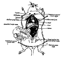
[0054] FIG. 1 MOUSE LYMPH NODES a diagram depicting the location
of draining lymph nodes in the mouse.
[0055] FIGS. 2A-E ID DELIVERY TO LYMPHATICS
[0056] A. shows highly stained superficial inguinal lymph nodes in the mouse 1
hour post intradermal delivery of 1% Evans Blue solution by the method of the
present
invention.
[0057] B: shows Intra-dermal (ID) vs. Subcutaneous (SC) Injection ofEvans
Blue Dye in Yorkshire Swine. Diagram of swine depicting location of injection
sites.
[0058] C. ID and SC injections. Arrow indicates location of SC injection.
[0059] D. ID and SC injections post mortem.
[0060] E. ID and SC injection site resection. Note the trafficking of the
Evans
Blue dye from the ID injection site to the inguinal node and depoting of the
dye at the SC
inj ection site.
[0061] FIGS. 3A and B. ID DELIVERY TO LYMPHATICS
[0062] A. shows the percentage of cells positive for CD90 and CD4 or CD8 or
CD 19 in the draining lymph node over time.
[0063] B. shows flow cytometry plots of labeled cell suspension from lymph
nodes of naive, 30 minutes, and 1-hour post anti-CD90-FITC antibody injection
mice (n = 2).
[0064] FIGS. 4A-C. ID DELIVERY TO LYMPHATICS
-18-
CA 02529048 2005-12-09
WO 2005/016401 PCT/US2004/019121
[0065] A. shows in vivo fluorescent staining of lymph tissue with injected
antibody sections at 1 hour post FITC-antibody injection under 40 times
magnification.
[0066] B shows H and E staining of the cells at 1 hour post FITC-antibody
injection under 40 times magnification.
[0067] C shows the overlay of 4a and 4b.
[0068] FIG. 5 ID DELIVERY PROFILE shows the path of the biologically
active agent after being intradermally delivered, by the method of the present
invention.
[0069] FIG. 6 IN VIVO TARGETED DIAGNOSTICS shows a diagram of
potential targets for delivery in the lymphatic system.
[0070] FIG. 7A AND B ID IN VIVO TARGETED DIAGNOSTICS. shows
comparative time profiles for ID and SC (SubQ) delivery of labeled antibody to
mouse lymph
nodes.
[0071] A. Delivery Method. Comparison of ID and SC delivery to Lymph
Nodes
[0072] B. Enhanced Detection of Lymphatic cells using ID Delivery Time
profile of antibody labeled cells in mouse lymph nodes
[0073] FIG. 8 IN VIVO TARGETED DIAGNOSTICS-APPLICATION
shows a diagram of how the method may be applied to a breast tumor, and a
demonstration of
T-cell labeling in mouse lymph node.
[0074] FIG. 9 shows results of injection of 50 ul EB through a 34G, l.Omm
needle at a rate of 45uL/min in a Yorkshire pig. The circled areas within the
reticular dermis,
separate from the main injection depot, show cross-sections of the draining
lymphatic vessels
(blue).
[0075] FIGS. 10 AND 11 show results of injection of 100uL of EB through a
34G, 1.0 mm needle at a rate of 45uL/min. in a Yorkshire pig.
[0076] FIGS.12 AND 13 show results of injection at two sites interdermally in
the flank of a Yorkshire pig with 100uL of EB through a 34G, 1.Omm needle at a
rate of
100uL/min.
[0077] FIG. 14 shows results of injection intradermally in the flank of a
Yorkshire pig with 100uL of EB through a 34G, l.Smm needle at a rate of
100uL/min.
-19-
CA 02529048 2005-12-09
WO 2005/016401 PCT/US2004/019121
[0078] FIG. 15 shows an example of lymphatic vessels (blue) from a 2mm
injection. Both a cross-section and a length-wise section can be seen in the
circled area.
[0079] FIG. 16 shows an example of lymphatic vessels (blue) trafficking the
intradermally injected Evans Blue dye from the site of injection to the
inguinal lymph nodes.
Insert shows close-up of resected inguinal lymph node.
[0080] FIGS. 17A-C shows the Number of Injected Fluorescent Beads present in
the Inguinal Lymph Node Over Time. Comparison of Intra-dermal and Subcutaneous
Inj ection.
[0081] A. SOnm sized beads
[0082] B. l,um sized beads
[0083] C. 10~.m sized beads.
[0084] FIGS.18 A-B PERCENT OF CD8 POSITIVE T CELLS, IN MOUSE
SPLEENS. Graphs depicting the Percent of CD8 Positive T Cells, in mouse
spleens, labeled
with CD90-FITC antibody over time. CD90-FITC a~itibody was either ID or SC
injected into
mice and spleens were monitored for cell-associated signal.
[0085] FIG. 19. IMAGING OF SWINE ABDOMINAL BLOOD
VASCULATURE AFTER 12.5 mg IV INJECTION OF ICG. Right Inguinal node
location depicted in box. Only blood vasculature is illuminated and not the
lymph nodes.
Imaging continued episodically for 30 minutes post injection without
illumination of lymph
nodes.
[0086] FIGS. 20A-C. DOSE SPARING-IV AND MICRONEEDLE ID
INJECTION. Imaging of Lymphatic vasculature and inguinal node of swine
immediately
following injection of ICG using a 34G, lmm depth, microneedle.
[0087] A. Three injections on swine abdomen (left side), top injection 200 uls
of
80ug/ml ICG, bottom 2 injections 75u1s of 80ug/ml ICG.
[0088] B. Imaging of lymphatic vasculature and left inguinal lymph node of
swine immediately after top inj ection from A.
[0089] C. Imaging of lymphatic vasculature and right inguinal nodes after 2
separate injections of 80ug/ml ICG on right hind leg. Note the individual
lymphatic
vasculature from each injection feeding separately into the nodes.
-20-
CA 02529048 2005-12-09
WO 2005/016401 PCT/US2004/019121
[0090] FIGS. 21A-D. DEMONSTRATION OF ICG DYE TRAFFICHING
SPEED. ICG injected H~ using 34G, lrilm depth, microneedle. Injection
performed above
left mammary chain of swine. ICG travel calculated to be 7cm/sec for this
injection.
[0091] FIG. 22 NEEDLE DEVICE. An exploded, perspective illustration of a
needle assembly designed according to this invention.
[0092] FIG. 23 NEEDLE DEVICE. A partial cross-sectional illustration of
the embodiment in FIG. 22.
[0093] FIG. 24 NEEDLE DEVICE. Embodiment of FIG. 22 attached to a
syringe body to form an injection device.
[0094] FIG. 25 ID INJECTION TECHNIQUE. A perspective view of one
technique for making an ID inj ection
[0095] FIG. 26 ID INJECTION TECHNIQUE. A perspective view of a
second technique for making an ID injection.
[0096] FIG. 27 ID INJECTION TECHNIQUE. A perspective view of a
third technique for making an ID injection.
[0097] FIG. 28 ID INJECTION TECHNIQUE. A perspective view of a
fourth technique for making an ID inj ection.
[0098] FIG. 29A AND B IDELIVERY OF CARDIO GREEN
IMAGING AGENT
[0099] A. INJECTIONS ON RIGHT HIND LEG AND LEFT SIDE
MAMMARY CHAIN. Left and right inguinal nodes illuminated.
[00100] B. INJECTIONS ON RIGHT HIND LEG AND LEFT SIDE
MAMMARY CHAIN. Inverted Image. Left and right inguinal nodes illuminated.
[00101] FIG. 30 COMPARISON OF MANTOUX AND ID DELIVERY.
The photo of the mantoux inj ection clearly shows the track of the needle
(blue line through
dennis leading to depot.) The majority of the EB was injected into the SC. The
photo of the
delivery with a 34G 1 mm needle shows that the injection was completely within
the dermis.
Drainage to the lymphatics can already be seen (circled).
[00102] FIGs 31 A and B. GRAPHS OF MAXIMUM AND AVERAGE
SUSTAINED PRESSURE AS A FUNCTION OF INSERTION DEPTH FOR BOTH
-21 -
CA 02529048 2005-12-09
WO 2005/016401 PCT/US2004/019121
VENTRAL AND DORSAL SWINE INJECTIONS. Maximum pressure was the single
highest pressure recorded during the first 100 seconds of infusion. Average
sustained
pressure was average for all pressure readings from 100 to 300 seconds. The
maximum and
average sustained pressures for each inj ection configuration were averaged
together and
plotted.
[00103] A. Average back pressure plotted as function of needle length. All
infusions in the ventral region of the animal.
[00104] B. Average back pressure plotted as function of needle length. All
infusions in the dorsal region of the animal.
[00105] FIG. 33. IN T~IVO STAINING. Graph depicting the percent of T and B
cells stained, in vivo, in the draining lymph nodes of mice.
5. DETAILED DESCRIPTION OF THE INVENTION
[00106] The present invention provides a method for administering one or more
biologically active agents, preferably a diagnostic agent, to a subject's
skin, in which the
biologically active agent is delivered to the intradermal (m) compartment of
the subject's
skin. The present invention is based, in part, on the unexpected discovery by
the inventors
that when such agents are delivered to the m compartment, they are transported
to the local
lymphatic system rapidly compared to conventional modes of delivery, including
subcutaneous delivery and m Mantoux delivery, and thus provide the benefits
disclosed
herein. Although not intending to be bound by a particular mechaiusm of
action, agents
delivered in accordance with the methods of the invention are transported in
vivo through the
local lymphatic system, excreted into the systemic blood circulation and into
deeper tissue
environments. The agent is then degraded or metabolized by, for example, the
liver, kidneys,
or spleen. Although not intending to be bound by a particular mechanism of
action, it is the
biomechanical manipulation of the extracellulax matrix (ECM) through the
precise delivery
of agents in the intradermal compartment that enables rapid uptake into the
local lymphatics
and lymph nodes by the method described herein.
[00107] The present invention provides an improved method of delivery of
biologically active agents in that it provides among other benefits, rapid
uptake into the local
lymphatics, improved targeting to a particular tissue, i.e., improved
deposition of the
-22-
CA 02529048 2005-12-09
WO 2005/016401 PCT/US2004/019121
delivered agent into the particular tissue, i.e., grbup or layer of cells that
together perform a
specific function, improved systemic bioavailability, improved tissue
bioavailability,
improved deposition of a pre-selected volume of the agent to be administered,
improved
tissue-specific kinetics rapid biological and pharmaco-dynamics (PD), and
rapid biological
and pharmacokinetics (PK). Such benefits of the methods of the invention are
especially
useful for the delivery of diagnostic agents. Intradermal delivery of a
diagnostic agent in
accordance with the methods of the invention deposits the diagnostic agent
into the
intradermal and lymphatic compartments thus creating a rapid and biologically
significant
concentration of the diagnostic agent in these compartments. Rapid diagnostics
can therefore
be performed using less diagnostic agent with significant advantages as
outlined herein.
[00108] Intradennally delivered biologically active agents have improved
tissue
bioavailability in a particular tissue, including but not limited to, skin
tissue, lymphatic tissue
(e.g., lymph nodes), mucosal tissue, reproductive tissue, cervical tissue,
vaginal tissue and
any part of the body that consists of different types of tissue and that
performs a particular
function, i.e., an organ, including but not limited to lung, spleen, colon,
thymus. In some
embodiments, the tissue includes any tissue that interacts with or is
accessible to the
environment, e.g., skin, mucosal tissue. Other tissue encompassed by the
invention include
without limitation Haemolymphoid System; Lymphoid Tissue (e.g., Epithelium-
associated
lymphoid Tissue and Mucosa-associated lymphoid Tissue or MALT (MALT can be
further
divided as organized mucosa-associated lymphoid Tissue (O-MALT) and diffused
lymphoid
tissue (D-MALT)); primary Lymphoid Tissue (e.g., thymus and bone marrow);
Secondary
Lymphoid Tissue (e.g., lymph node, spleen, alimentary, respiratory and
Urigenital). It will
be appreciated by one skilled in the art that MALT secondary includes gut
associated
lymphoid tissue (GALT); Bronchial associated lymphoid tissue (BALT), and
genitourinary
system. MALT specifically comprises lymph nodes, spleen, tissue associated
with epithelial
surfaces such as palentine and nasopharyngeal tonsils, Peyer's Patches in the
small intestine
and various nodules in the respiratory and urinogenital systems, the skin and
conjmlctivia of
the eye. O-MALT includes the peripharyngeal lymphoid ring of the tonsils
(palentine,
lingual, nasopharyngeal and tubal), Oesophageal nodules and similar lymphoid
tissue
scattered throughout the alimentary tract from the duuuodenum to the anal
canal.
Intradermally delivered biologically active agents have improved tissue
bioavailability in a
particular tissue compared to when the same agent is delivered to a deeper
tissue
compartment such as the SC compartment and the IM compartment.
- 23 -
CA 02529048 2005-12-09
WO 2005/016401 PCT/US2004/019121
[00109] The delivery of a biologically active agent in accordance with the
methods
of the invention results in improved tissue bioavailability as compared to
when the same
agent is delivered to the subcutaneous (SC) compartment or when the same agent
is delivered
by the intradermal (m) Mantoux method. The delivery of a biologically active
agent in
accordance with the methods of the invention results in improved tissue
bioavailability as
compared to when the same agent is delivered to a deeper tissue compartment,
e.g., SC, IM..
Improved tissue bioavailability of agents delivered in accordance with the
methods of the
invention is particularly useful when delivering diagnostic agents to the ID
compartment, as it
reduces the amount of the diagnostic agent required for each diagnostic
procedure, which
may be difficult and costly to obtain. The reduced amount of the diagnostic
agent further
reduces the likelihood of side effects associated with the diagnostic
procedure, e.g., toxicity.
[00110] The improved tissue bioavailability of the agents delivered in
accordance
with the methods of the invention can be determined using methods and
parameters known to
those skilled in the art, for example by measuring the total amount of the
agent accumulated
in a particular tissue using, for example, histological methods known to those
skilled in the
art and disclosed herein. Alternatively improved tissue bioavailability of the
agents can be
assessed as the amount of the agent presented to the particular tissue, the
amount of the agent
accumulated per mass or volume of particular tissue, amount of the agent
accumulated per
unit time in a particular mass or volume of the particular tissue.
[00111] Biologically active agents delivered in accordance with the methods of
the
invention are deposited in the intradermal compartment and first distributed
with high
bioavailability to the lymphatic tissue local to the administration site,
followed by a more
wide spread lymphatic delivery in to the general circulation. In some
embodiments, the
methods of the present invention are particularly effective for diagnosis of a
disease, disorder,
or infection in deeper tissues, e.g., in vivo detection of an infection in an
organ or tissue such
as lung or inflammation of an organ or tissue such as appendix or joints.
[00112] Biologically active agents delivered in accordance with the methods of
the
invention show immediate transport and uptake within at least 5 minutes, at
least 10 minutes,
at least 15 minutes, preferably no more than within 20 minutes after the inj
ection to the
lymphatic system, as monitored visually in real time using common methods in
the art (e.g.,
MRI, X-Ray, Ultrasound, CT, PET, SPELT, Optical (fluorescence,
bioluminescence,
chemiluminescence), photoacoustic, RAMAN and SERS imgaing) or in vitro using
common
methods in the art (e.g., histological examination, flow cytometry) and those
disclosed herein,
-24-
CA 02529048 2005-12-09
WO 2005/016401 PCT/US2004/019121
see, Section 6.2. Agents delivered in accordance with the methods of the
invention are
transported to the lymphatic system and deposited in a particular tissue with
velocities of at
least 10 cm per second, preferably at least 5-10 cm per second. It will be
appreciated by one
skilled in the art that the rate at which the agent is transported to the
lymphatic system and
deposited in a particular tissue depends on various parameters including but
not limited to
volume of injection, rate of injection, biochemical and physical
characteristic of the agent,
and site of injection.
[00113] In some embodiments, biologically active agents, including diagnostic
agents delivered in accordance with the methods of the invention specifically
recognize and
bind a cell in a particular tissue in which they are deposited. In other
embodiments,
biologically active agents delivered in accordance with the methods of the
invention are
delivered to the ID compartment so that the amount of the pre-selected dose of
the agent
deposited in the target tissue is increased by at least 0.1 % compared to when
the agent is
delivered outside of the intradermal space, e.g., subcutaneous compartment
(SC),
intramuscular compartment (IM). The invention contemplates that the amount of
the pre-
selected dose of the agent deposited in the target tissue is increased by at
least 100%, at least
150%, at least 200%, at least 200%, at least 250%, preferably by at least 350%
or 3.5x, up to
1750%, the amount achieved when the agent is administered by routes outside of
the
intradermal compartment, e.g., SC, IM and thus delivered to a deeper tissue
compartment.
[00114] The invention encompasses methods of delivering the biologically
active
agents to the ID compartment so that the amount of the pre-selected dose of
the agent
deposited in the target tissue is increased by the amounts specified herein
compared to when
the agent is delivered outside of the intradermal space, e.g., subcutaneous
compartment (SC),
intramuscular compartment (IM) such that the increase in amount is detected as
early as3
minutes post-injection, or as early as 3 hours post injection. Preferably the
increase in
deposition of the agent in the particular tissue may persist for at least 21
days, at least 27
days
[00115] In some embodiments, the concentration of the biologically active
agent
deposited in a particular tissue after ID delivery is about 5 nanograms of the
agent agent per
50 micrograms of the particular tissue; 10 picograms of the agent per 50
micrograms of the
particular tissue; 29 nanograms of the agent per SOmicrograms of the
particular tissue; 10
picograms of the agent per 50 micrograms of the particular tissue to about 29
nanograms of
the agent per 50 micrograms of the particular tissue; 10 picograms of the
agent per 50
- 25 -
CA 02529048 2005-12-09
WO 2005/016401 PCT/US2004/019121
micrograms of particular tissue to about 150 nanograms of the agent per 50
micrograms of
the particular tissue.
[00116] In other embodiments, the concentration of the biologically active
agent,
e.g., a diagnostic agent, deposited in a particular tissue after m delivery is
about 10 pg to
about 15 ug of the agent agent per 50 micrograms of the particular tissue, or
about 1 cg to
about 30 ng of the agent agent per 50 micrograms of the particular tissue.
(00117] Unlike subcutaneous delivery, intradermal methods, as described
herein,
enhance the biological kinetics, biological dynamics, and tissue
bioavailability of the
biologically active agents delivered, including diagnostic and therapeutic
agents. Intradermal
delivery of biologically active agents in accordance with the methods of the
invention are
taken up by the lymphatic system and deposited in a particular tissue without
the need of
"massaging" the injection site, which is unlike other conventional modes of
delivery,
including subcutaneous delivery. Biologically active agents delivered to the
subcutaneous
compartment do not achieve deposition in a target tissue and/or lymphatic
transport unless
the injection site is massaged to induce such transport of the delivered
agent. Although not
intending to be bound by a particular mechanism of action, delivery methods,
such as
intravenous injection, rely on dissemination of the agent of interest from the
general
circulation into the target tissue. Dissemination of the biologically active
agent into the tissue
is dependent on many variables and the bioavailability found in the general
circulation is not
always optimal for a given target tissue. The intravenous and subcutaneous
methods for
delivery of an agent are limiting especially when the target tissue is in the
lymphatic system.
In addition, intradermal delivery, as described by the present invention,
offers an alternate
transport mechanism in which a specific agent is presented to the intradermal
compartment
and flows to the general circulation via the lymphatic system and area
capillaries. Although
others have described intradermal delivery to lymphatic vasculature, none have
defined
specific conditions or devices for reliable access of these tissues. Although
not intending to
be bound by a particular mechanism of action, delivering biologically active
agents (as
liquids or suspensions) into the intradermal compartment in accordance with
the methods of
the invention results in increased interstitial pressure which, in turn, opens
the lymphatic
vasculature permitting high rates of sustained flow until fluid flow is
terminated. The
inventors have found that this lymphatic transport occurs surprisingly fast,
permitting
immediate access to the lymphatic vasculature and general circulation. Methods
of the
invention result in uptake of agents into the lymphatic system, rather than
capillary uptake,
-26-
CA 02529048 2005-12-09
WO 2005/016401 PCT/US2004/019121
thus resulting in the benefits disclosed herein including but not limited to
enhanced rate and
activity of targeting.
[00118] Biologically active agents delivered in accordance with the methods of
the
invention are deposited in the intradermal compartment and first distributed
with high
bioavailability to the lymphatic tissue local to the administration site,
followed by a more
wide spread lymphatic delivery in to the general circulation. In some
embodiments, the
methods of the present invention are particularly effective for diagnosis of a
disease, disorder
or infection in deeper tissues.
[00119] In some embodiments, the invention encompasses targeted intraderaml
delivery of a biologically active agent to a particular biological entity
including but not
limited to a cell, a group or collection of cells, a bacteria (e.g.,
EscheYichia coli, Klebsiella
praeumoniae, Staphylococcus au~eus, Ente~ococcus faecials, Candida albicans,
PYOteus
vulgaris, Staphylococcus viridans, and Pseudonaonas ae~uginosa), a pathogen
(e.g., B-
lymphotropic papovavirus (LPV); Bordatella pertussis; Borna Disease virus
(BDV); Bovine
coronavirus; Choriomeningitis virus; Dengue virus; a virus, E. coli; Ebola;
Echovirus 1;
Echovirus-11 (EV); Endotoxin (LPS); Enteric bacteria; Enteric Orphan virus;
Enteroviruses ;
Feline leukemia virus; Foot and mouth disease virus; Gibbon ape leukemia virus
(GALV);
Gram-negative bacteria ; Heliobacter pylori; Hepatitis B virus (HBV); Herpes
Simplex Virus;
HIV-1; Human cytomegalovirus; Human coronovirus; Influenza A, B & C ;
Legionella;
Leishmania mexicana; Listeria monocytogenes; Measles virus; Meningococcus;
Morbilliviruses; Mouse hepatitis virus; Murine leukemia virus; Murine gamma
herpes virus;
Murine retrovirus; Murine coronavirus mouse hepatitis virus; Mycobacterium
avium-M;
Neisseria gonorrhoeae; Newcastle disease virus; Parvovirus B19; Plasmodium
falciparum;
Pox Virus; Pseudomonas; Rotavirus; Samonella typhiurium; Shigella;
Streptococci; T-cell
lymphotropic virus 1; Vaccinia virus); a plaque, and a parasitic agent. Once
an agent is
delivered to a biological entity in accordance with the methods of the
invention, any of the
detection, imaging methods known to one skilled in the art and disclosed
herein can be used
to detect and image the entity. The methods of the invention encompasse
methods for
delivering a biologically active agent where the agent specifically binds a
biological entity.
[00120] Directly targeting the intradermal compartment as taught by the
invention
provides more rapid onset of effects of biologically active agents, including
diagnostic
agents, and higher bioavailability including, tissue bioavailability, relative
to other modes of
delivery of such agents, including subcutaneous and ID Mantoux delivery. The
inventors
-27-
CA 02529048 2005-12-09
WO 2005/016401 PCT/US2004/019121
have found that agents delivered in accordance with the methods of the
invention can be
rapidly absorbed and systemically distributed via controlled ID administration
that selectively
accesses the dermal vascular and lymphatic microcapillaries, thus the agents
may exert their
beneficial effects more rapidly than SC administration and ID Mantoux
delivery.
Additionally the inventors have found that delivering an agent into the ID
compartment takes
advantage of the dermal microcirculation and the interaction between
hydrostatic and osmotic
pressures, in the dermal extra-cellular matrix, and the lymphatic vessels. It
is in the dermal
interstitium that the blood and lymph systems interact in the skin. As blood
travels to the
smallest capillaries, plasma fluid and proteins are forced out into the
interstitial compartment.
Osmotic and biomechanical forces result in perfusion of the fluid through the
interstitium and
into the local initial lymphatics. The initial lymphatics are permeable to
macromolecules and
therefore act in maintaining osmotic and hydrostatic pressures within the
tissue compartment.
The typical flow rate of the lymphatics is 10-100 times less than the flow
rate of blood. The
lymphatic system consists of 5 major conduits. They include lymphatic
capillaries and
collecting vessels, found in the dermis, lymph nodes, trunks, and ducts. Lymph
forms when
interstitial fluid moves into the lymph capillaries. It then drains into the
collecting vessels.
The vessels pass through at least one but usually several lymph nodes
clusters. The vessels
leaving the nodes drain into larger trunks, which in turn lead into the ducts.
The ducts return
the lymph back to the bloodstream, completing the circuit (Swartz, M.A. 2001,
Adv. DYUg
Del. Rev. 50: 3-20). The lymphatic system flow is uni-directional with the
lymphatic
capillaries as the initial fluid collection conduit. In the interstitium the
lymphatic capillaries
primary role is to maintain hydrostatic and osmotic pressure in the tissue.
This is
accomplished through the interaction of the capillary anchoring filaments and
the extra-
cellular matrix (ECM). As fluid fills the interstitium, tissue pressure
increases and places
stress on the ECM, essentially stretching the tissue, and the anchoring
filaments holding the
lymphatics in place are pulled with the ECM. This movement pulls the lymphatic
capillary
open allowing the fluid to flow rapidly from the tissue in order to re-
establish appropriate
hydrostatic and osmotic pressures. Therefore, the mechanical integrity of the
ECM plays an
important role in the lymphatic function. Extensive or chronic degradation of
the ECM
eventually renders lymphatic vessels non-responsive to the changes in the
interstitium and
causes dysfunction (Swartz et al., 1999, JBionzech. 32(12):1297-307). Although
not
intending to be bound by a particular mechanism of action, it is the
biomechanical
manipulation of the ECM through the precise delivery of agents in the
interstitium that
- 28 -
CA 02529048 2005-12-09
WO 2005/016401 PCT/US2004/019121
enables rapid uptake into the local lyrnphatics and lymph nodes by the method
described
herein.
[00121] While tissue stress contributes greatly to the uptake of fluid
additional
factors contribute as well. These include the concentration, size, charge, and
molecular
weight of the pharmaceutical agent being delivered and the interplay between
these
characteristics and the surrounding intradermal tissue environment (Charman et
al., 1992
Lymphatic Transport of Drugs, Boca Raton: CRC Press Inc.). Manipulation of
these factors
is contingent upon exact and reproducible access to the interstitium. Even
though the dermis,
in particular, the papillary dermis has been known to have a high degree of
vascularity, it has
not heretofore been appreciated that one could take advantage of the high
degree of
vascularity as well as the interaction between the ECM and the lymphatic
vasculature to
obtain an improved absorption profile for administered agents compared to
alternative routes
of transdermal administration
[00122] Intradermally delivered biologically agents, especially diagnostic
agents,
exhibit more rapid onset and clearance versus conventional delivery including
SC delivery
and ll~ mantoux delivery. These properties confer several advantages when
delivering a
diagnostic agent to the ID compartment of a subject's skin. First, it reduces
potential side
effects and discomfort due to the diagnostic procedures. Second, it enables
the rapid and
repeated trial of sequential procedures in a single diagnostic session. Third,
it reduces the
time required in expensive medical or imaging facilities. Fourth, it
facilitates real time
studies of physiology. Fifth, it reduces potential background signal generated
by unbound
and un-cleared diagnostic reagents. Sixth, patients experience reduced pain
versus IV
administration, SC injection or surgical biopsy.
[00123] Delivering biologically active agents, including diagnostic agents in
accordance with the methods of the invention is preferred over traditional
modes of delivery
including SC delivery and ID Mantoux delivery because the amount of the pre-
selected dose
of the agent deposited in the lymphatic tissue is increased, as measured for
example using
histopathological methods or other methods known to one skilled in the art
such as flow
cytomety (FACS) or imaging.
[00124] As used herein, delivery to the intradermal compartment or
intradermally
delivered is intended to mean administration of a biologically active agent
into the dermis in
such a manner that the agent readily reaches the richly vascularized papillary
dermis and is
-29-
CA 02529048 2005-12-09
WO 2005/016401 PCT/US2004/019121
rapidly absorbed into the blood capillaries and/or lymphatic vessels to become
systemically
bioavailable. Such can result from placement of the agent in the upper region
of the dermis,
i.e., the papillary dermis or in the upper portion of the relatively less
vascular reticular dermis
such that the agent readily diffuses into the papillary dermis. The controlled
delivery of a
biologically active agent in this dermal compartment below the papillary
dermis in the
reticular dermis, but sufficiently above the interface between the dermis and
the subcutaneous
tissue, should enable an efficient (outward) migration of the agent to the
(undisturbed)
vascular and lymphatic microcapillary bed (in the papillary dermis), where it
can be absorbed
into systemic circulation via these microcapillaries without being sequestered
in transit by
any other cutaneous tissue compartment. In some embodiments, placement of a
biologically
active agent predominately at a depth of at least about 0.3 mm, more
preferably, at least about
0.4 mm and most preferably at least about 0.5 mm up to a depth of no more than
about 2.5
mm, more preferably, no more than about 2.0 mm and most preferably no more
than about
1.7 mm will result in rapid absorption of the agent. Although not intending to
be bound by a
particular mechanism of action, placement of the biologically active agent
predominately at
greater depths and/or into the lower portion of the reticular dermis may
result in less effective
uptake of the agent by the lymphatics as the agent will be slowly absorbed in
the less vascular
reticular dermis or in the subcutaneous region.
[00125] Biologically active agents, including diagnostic agents delivered in
accordance with the methods of the invention will achieve higher maximum
concentrations of
the agents. The inventors have found that agents administered to the ID
compartment are
absorbed more rapidly, with bolus administration resulting in higher initial
concentrations.
Therefore, the dose can be reduced, providing an economic benefit, as well as
a physiological
benefit since lesser amounts of the drug or diagnostic agent has to be cleared
by the body.
[00126] In accordance with the invention direct intradermal (ID)
administration
can be achieved using, for example, microneedle-based injection and infusion
systems or any
other means known to one skilled in the art to accurately target the
intradermal compartment.
Particular devices include those disclosed in WO 01/02178, published January
10, 2002; and
WO 02/02179, published January 10, 2002, U.S. Patent No. 6,494,865, issued
December 17,
2002 and U.S. Patent No. 6,569,143 issued May 27, 2003 all of which are
incorporated herein
by reference in their entirety, as well as those exemplified in FIGS. 22-24.
Micro-cannula-
and microneedle-based methodology and devices axe also described in U.S.
Application
Serial No. 09/606,909, filed June 29, 2000, which is incorporated herein by
reference in its
-30-
CA 02529048 2005-12-09
WO 2005/016401 PCT/US2004/019121
entirety. Standard steel cannula can also be used for infra-dermal delivery
using devices and
methods as described in IJ.S. Serial No. 417,671, filed October 14, 1999,
which is
incorporated herein by reference in its entirety. These methods and devices
include the
delivery of agents through narrow gauge (30G or narrower) "micro-cannula" with
a limited
depth of penetration (typically ranging from 10 ,um to 2 mm), as defined by
the total length of
the cannula or the total length of the cannula that is exposed beyond a depth-
limiting hub
feature.
[00127] The subject of intradermal delivery of the present invention is a
mammal,
preferably, a human. The biologically active agents delivered in accordance
with the
methods of the invention (with or without a tracer reagent) may be delivered
into the
intradermal compartment by a needle or cannula, usually from about 300 ~,m to
about 5 mm
long. Preferably, the needle or cannula is about 300 ~,m to about 1 mm long,
with the outlet
inserted into the skin of the subject to a depth of 1 mm to 3 mm. Preferably,
a small gauge
needle or cannula, between 30 and 36 gauge, preferably 31-34 gauge is used.
The outlet of
the needle or cannula is preferably inserted to a depth of 0.3mm (300um) to
l.5mm.
[00128] Improved pharmacokinetic parameters using methods of the invention can
be achieved using not only microdevice-based injection systems, but other
delivery systems
such as needle-less or needle-free ballistic injection of fluids or powders
into the ID
compartment, enhanced ionotophoresis through microdevices, and direct
deposition of fluid,
solids, or other dosing forms into the skin. In specific embodiments, the
administration of
the biologically active agent is accomplished through insertion of a needle or
cannula into the
intradermal compartment of the subject's skin.
[00129] The intradermal delivery of diagnostic agents in accordance with the
present invention are particularly beneficial in the diagnosis of the diseases
including chronic
and acute diseases which include, but are not limited to, lymphoma, melanoma,
leukemia,
breast cancer, colorectal cancer, cancer metastasis, diseases of the lymphatic
system, any
disease affecting the lymph nodes, e.g., axillary, politeal, lingual, viral
diseases, e.g., HIV,
immune disorders such as rej ection, metabolic disorders, and infectious
diseases. Although
not intending to be bound by a particular mechanism of action, diagnostic
agents delivered in
accordance with the methods of the invention are taken up by the intradennal
compartment
and delivered to the lymphatic system where its recognition and binding
indicate the presence
of a cell or disease state. The present invention is useful for diagnostic
procedures including,
-31-
CA 02529048 2005-12-09
WO 2005/016401 PCT/US2004/019121
but not limited to, surgical methods, biopsies, non-invasive screening and
image-guided
biopsies.
[00130] The present invention provides improved methods for cancer detection
and/or diagnosis by improving sensitivity, the amount of the agent deposited,
tissue
bioavailability, faster onset and clearance of the delivered diagnostic agent.
Additionally
methods of the invention are particularly improved over conventional cancer
detection
procedures for the detection of a tumor, e.g., breast tumor in a human
subject, because more
than 75% of the pre-selected volume of the diagnostic agent is deposited into
the intradermal
compartment, relative to when the same pre-selected volume is delivered to the
intradermal
compartment by the traditional methods of delivery of such agent, e.g., m
Mantoux method.
[00131] The present invention provides improved methods for current sentinel
node biopsy procedure and mapping surgical procedure by improving the uptake
and
bioavailability of the diagnostic agents to the local lymphatic system. The
invention provides
a method for administration of at least one diagnostic agent for the detection
of a tumor
seminal lymph node, e.g., breast tumor seminal lymph node, or a lymph node
that drains the
tumor in a human subject, comprising delivering the agent into the intradermal
compartment
of the human subject's skin so that the agent is transported to the local
lymphatic system. In
other embodiments, the invention provides a method for administration of at
least one
diagnostic agent for the detection of a tumor seminal lymph node or a lymph
node that drains
the tumor in a human subject, comprising delivering the agent into the
intradermal
compartment of the human subject's skin so that the agent has a higher tissue
bioavailability
compared to when the same agent is delivered by the m Mantoux method. In yet
other
specific embodiments, the invention provides a method for administration of at
least one
diagnostic agent for the detection of a tumor seminal lymph node, e.g., a
breast tumor
sentinal lymph node or a lymph node that drains the tumor in a human subject,
comprising
delivering the agent into the intradermal compartment of the human subject's
skin so that the
agent has a faster onset and clearance compared to when the same agent is
delivered by the
m Mantoux method.
[00132] The methods of the instant invention provide improved prognostic
methods using specific agents (versus non-specific agents) to assess
therapeutic efficacy of a
treatment regimen of a disease, for example by monitoring cellular genetic
profiles in
assessing gene regulation and expression over time. Traditionally ih vitro
analysis of cellular
genetic profiles have been used to assess gene regulation and expression over
time as a tool in
-32-
CA 02529048 2005-12-09
WO 2005/016401 PCT/US2004/019121
assessing therapeutic efficacy. Such in vitro methods have numerous
shortcomings including
but not limited to inaccuracies, the removal of cells from the body can cause
the destruction
of RNA and DNA thereby altering the genetic profile in the specimen;
information about the
morphological locus of the genetic lesion is potentially lost using ex-vivo
methods; and cell
differentiation and regulation may be influenced by removal from the
extracellular
environment ira vivo. By using the methods of the present invention,
intradennal
administration of specific diagnostic agents capable of associating and/or
binding a specific
marker for a disease provides for assessment of the disease as it exists in
the patient. Thus,
the methods taught by the present invention influence the choices of therapy
available to the
practitioner.
[00133] The methods of the invention are particularly useful in identification
of
impaired cellular metabolism of a disease or disorder, using for example
genomics and
proteomics technologies. In a specific embodiment, the methods of the
invention provide
improved methods for prognosis of cancer, particularly Diffuse Large B-cell
lymphoma
(DLBCL). Specific agents capable of distinguishing between DLBCL with and
without
active proliferative pathways can be delivered to the ID compartment and
allowed to traffic
throughout the lynphatic system. Binding of one agent would indicate a good or
poor
prognosis and thus enhanced effectiveness of therapy. In other specific
embodiments, the
methods of the invention provide improved methods for diagnosis of diseases
associated with
impaired signaling in the NFkb pathway by delivery of a specific agent to the
m
compartment, allowing the agent to traffic to a particular cells, where it
binds accordingly.
The signal from the binding can be visualized in vivo. Binding of the agent
indicates the
presence of an impairment in the pathway and will allow assessment of the
effectiveness of
therapy and the onset of potential drug resistance as therapy progresses
(00134] The methods of the invention are particularly useful for methods of
integrated diagnosis and therapy preferably including require complementary
and/or
concurrent diagnostics and monitoring. Accurate diagnosis of a disease is
largely an unmet
need for example in oncology, where few diagnostic agents indicate which
therapeutic
choices will succeed with any reliability. The methods of the invention
provide delivering
agents which specifically recognize a cell, e.g., a cancer cell, in a
particular tissue. Such
agents include without limitation antibodies, preferably therapeutic
monoclonal antibodies
disclosed herein. In a specific embodiment, the invention encompasses
delivering Herceptin,
a monoclonal antibody specific for Her2/neu positive breast cancer to the ID
compartment of
-33-
CA 02529048 2005-12-09
WO 2005/016401 PCT/US2004/019121
a subject's skin for improved diagnosis and therapy. The methods of the
invention provide
improved diagnosis of cancer subjects over traditional methods of diagnostic
of Her2/neu
positive cancer cells, which identifies the population that will most benefit
from this
therapeutic treatment while eliminating others that would not. Currently, such
ira vitro
diagnostic tests identifying the population that will most benefit from a
particular therapeutic
treatment produce "equivocal" or unclear results. By using the methods of the
present
invention, identification of the Her2/neu positive cells can be enhanced.
Thus, ih vivo
intradermal administration of Herceptin or a nucleic acid that identifies the
mRNA coding for
Her2/neu, provides for the ability to identify those individuals suitable for
integrated
diagnostics and monitoring. Using the methods of this invention the cells are
left intact
providing a greater chance for positive identification.
[00135] The methods of the instant invention provide improved methods for
tailoring therapies of a disease, disorder or infection using integrating
diagnostic methods of
the invention. The methods of the invention are applicable for current
tailored and non-
tailored treatment regimens. The methods of the invention allow a continuous
monitoring of
a treatment regimen in a subject. While tailored therapies of the future will
require integrated
diagnostics, current non-tailored treatment regimens could also benefit from
tailored
diagnostics of the instant invention. For example, those subjects diagnosed
with large diffuse
B cell lymphoma typically undergo CHOP therapy. Monitoring the effectiveness
of this
combined drug regimen is restricted to clinical changes and intermittent non-
specific imaging
and tissue biopsies. The ability to continually monitor treatment
effectiveness would allow
for earlier identification of drug resistance and metastasis. This could be
accomplished with
the administration of specific intradermal diagnostic reagents in the
therapeutic cocktail or in
combination with existing therapies.
[00136] The methods of the invention provide administration of formulations
comprising one or more diagnostic agents in combination with one or more
therapeutic
agents. The present invention provides methods to target diagnostic agents and
therapeutic
agents to cells of interest. In a specific embodiment, the invention
encompasses delivering a
diagnostic agent combined with a therapeutic agent to the m compartment of a
subject's skin
such that a specific action of the diagnostic agent triggers an action of the
therapeutic agent.
The combination of targeted diagnostic delivery with targeted therapeutics
delivery in
accordance with the methods of the invention provides for enhanced patient
care. This
embodiment teaches the advantages of combining intradermal therapeutic
delivery with
-34-
CA 02529048 2005-12-09
WO 2005/016401 PCT/US2004/019121
diagnostic agents. The combination of delivering a diagnostic and a
therapeutic agent to the
m compartment, provides a powerful tool for improving the treatment of a
disease in a
subj ect.
[00137] In yet other embodiments, the invention enables the use of specific
agents,
e.g., diagnostic agents, for binding andlor detecting a cellular event or
disease state in vivo.
As a result, the invention provides screening methods to identify a specific
agent needed to
bind to the cell of interest. In some embodiments, the invention provides
methods for in. vivo
screening of combinatorial libraries, both biological and chemical, to
identify suitable agents
(e.g., diagnostic target or moiety or therapeutic target or moiety) in the
library for the purpose
being tested. The ability to screen for agents in vivo using the methods of
the instant
invention enables identification of unique cellular and disease states.
[00138] In a specific embodiment, the invention provides using an animal model
of
interest, where libraries of agents can be injected intradermally and their
effects monitored
over time. Effects which can be monitored include for example relief of
symptoms or
binding to a tissue and/or cell of interest. In a preferred specific
embodiment, an animal
tumor model, e.g., a lymphoma mouse model could be used for screening
biologically active
agents, delivered intradermally that traffic to the lymph nodes. This would
enable the
detection of cancer cell states ifa vivo and possibly identify the active
triggers for metastases
and potential targets for therapeutic and diagnostic agents. These results
would then be
utilized to develop novel diagnostics for humans and other species.
5.1 COMPOSITIONS OF THE INVENTION
[00139] The invention encompasses compositions comprising one or more
biologically active agents in solution forms, particulate forms thereof and
mixtures thereof.
Compositions for use in the methods of the invention may be obtained from any
species or
generated by any recombinant DNA technology known to one skilled in the art.
Compositions comprising one or more biologically active agents may be from
different
animal species including, limited but not to, swine, bovine, ovine, equine,
etc. The chemical
state of such agents may be modified by standard recombinant DNA technology to
produce
agents of different chemical formulas in different association states.
[00140] The biologically active agent used in the methods of the invention
encompasses any molecule that either specifically or non-specifically binds a
molecule in
vivo and is capable of producing a biological effect in vivo. The biologically
active agents
-35-
CA 02529048 2005-12-09
WO 2005/016401 PCT/US2004/019121
may either be naturally occurring molecules or those derived using a synthetic
process or
recombinant process, using common methods known to one skilled in the art.
Biologically
active agents of the invention may recognize specifically or non-specifically
a recognition
moeity on a particular cell in a particular tissue. Often, these specific
agents contain
structural or functional properties in common with known biological entities.
These
biologically active agents may either be naturally occurring recognition
molecules or those
derived using a synthetic process or recombinant process, using common methods
known to
one skilled in the art.
[00141] In other embodiments, the biologically active agent is a biomimetic in
nature, comprising naturally occurnng structural motifs while incorporating
additional or
modified functional groups for transport, targeting, enhanced binding,
stability, or detection.
[00142] Examples of biologically active agents that can be used in the methods
of
the instant invention include without limitation, immunoglobulins (e.g., Multi-
specific Igs,
Single chain Igs, Ig fragments), Proteins, Peptides (e.g., Peptide receptors,
PNAs, Selectins,
binding proteins (maltose binding protein, glucose binding protein)),
Nucleotides, Nucleic
Acids (e.g., PNAS, RNAs, modified RNAIDNA, aptamers), Receptors (e.g.,
Acetylcholine
receptor), Enzymes (e.g., Glucose Oxidase, HIV Protease and reverse
transcriptase),
Carbohydrates (e.g, NCAMs, Sialic acids), Cells (e.g" Insulin & Glucose
responsive cells),
bacteriophags (e.g., filamentous phage), viruses (e.g., HIV), Chemospecific
agents (e.g.,
Cyptands, Crown ethers, Boronates).
[00143] Particularly preferred biologically active agents that may be used in
the
instant invention are therapeutic antibodies that can be used diagnostically
which include but
are not limited to HERCEPTIN~ (Trastuzumab) (Genentech, CA) which is a
humanized anti-
HER2 monoclonal antibody for the treatment of patients with metastatic breast
cancer;
REOPRO~ (abciximab) (Centocor) which is an anti-glycoprotein IIb/IIIa receptor
on the
platelets for the prevention of clot formation; ZENAPAXC~ (daclizumab) (Roche
Pharmaceuticals, Switzerland) which is an immunosuppressive, humanized anti-
CD25
monoclonal antibody for the prevention of acute renal allograft rejection;
PANOREXTM
which is a murine anti-17-IA cell surface antigen IgG2a antibody (Glaxo
Wellcome/Centocor); BEC2 which is a murine anti-idiotype (GD3 epitope) IgG
antibody
(ImClone System); IMC-0225 which is a chimeric anti-EGFR IgG antibody (ImClone
System); VITAXINTM which is a humanized anti-exV~33 integrin antibody (Applied
Molecular
Evolution/MedIrmnune); Campath 1H/LDP-03 which is a humanized anti CD52 IgG1
-36-
CA 02529048 2005-12-09
WO 2005/016401 PCT/US2004/019121
antibody (Leukosite); Smart M195 which is a humanized anti-CD33 IgG antibody
(Protein
Design Lab/Kanebo); RITUXANTM which is a chimeric anti-CD20 IgGl antibody
(IDEC
Pharm/Genentech, Roche/Zettyaku); LYMPHOCIDETM which is a humanized anti-CD22
IgG antibody (Immunomedics); ICM3 is a humanized anti-ICAM3 antibody (ICOS
Pharm);
IDEC-114 is a primatied anti-CD80 antibody (IDEC Phann/Mitsubishi); ZEVALINTM
is a
radiolabelled marine anti-CD20 antibody (IDEC/Schering AG); IDEC-131 is a
humanized
anti-CD40L antibody (IDEC/Eisai); IDEC-151 is a primatized anti-CD4 antibody
(IDEC);
IDEC-152 is a primatized anti-CD23 antibody (IDEC/Seikagaku); SMART anti-CD3
is a
humanized anti-CD3 IgG (Protein Design Lab); SGl.l is a humanized anti-
complement
factor 5 (CS) antibody (Alexion Pharm); D2E7 is a humanized anti-TNF-a
antibody
(CATIBASF); CDP870 is a humanized anti-TNF-a Fab fragment (Celltech); IDEC-151
is a
primatized anti-CD4 IgGl antibody (IDEC Pharm/SmithKline Beecham); MDX-CD4 is
a
human anti-CD4 IgG antibody (Medaxex/Eisai/Genmab); CDP571 is a humanized anti-
TNF-
a IgG4 antibody (Celltech); LDP-02 is a humanized anti-a4~37 antibody
(LeukoSite/Genentech); OrthoClone OKT4A is a humanized anti-CD4 IgG antibody
(Ortho
Biotech); ANTOVATM is a humanized anti-CD40L IgG antibody (Biogen); ANTEGRENTM
is a humanized anti-VLA-4 IgG antibody (Elan); and CAT-152 is a human anti-TGF-
X32
antibody (Cambridge Ab Tech).
[001] Other examples of antibodies that can be used in accordance with the
instant
invention are listed in Table 1 below.
Table 1: Monoclonal antibodies for Cancer Therapy that can be used in
accordance
with the invention.
Company Product Disease Target
Abgenix ABX-EGF Cancer EGF receptor
AltaRex OvaRex ovarian cancer tumor antigen CA125
BravaRex metastatic tumor antigen MUC1
cancers
Antisoma Theragyn ovarian cancer PEM antigen
(pemtumomabytrrium-
90)
_ Therex breast cancer PEM antigen
Boehringer blvatuzumab head & neck CD44
In~elheim cancer
Centocor/J&J Panorex Colorectal 17-lA
cancer
ReoPro PTCA gp IIIb/IIIa
ReoPro Acute MI gp IIIb/IIIa
ReoPro Ischemic stroke gp IIIb/IIIa
-37-
CA 02529048 2005-12-09
WO 2005/016401 PCT/US2004/019121
Company Product Disease Target
Corixa Bexocar NHL CD20
CRC MAb, idiotypic lO5AD7colorectal gp72
cancer
Technology vaccine
Crucell Anti-EpCAM cancer Ep-CAM
Cytoclonal MAb, lung cancer non-small NA
cell
lung cancer
Genentech Herceptin metastatic breast HER-2
cancer
Herceptin early stage HER-2
breast cancer
Rituxan Relapsed/refract CD20
ory low-grade
or
follicular NHL
Rituxan intermediate & CD20
high-grade NHL
MAb-VEGF NSCLC, VEGF
metastatic
MAb-VEGF Colorectal VEGF
cancer,
metastatic
AMD Fab age-related CD18
macular
E-26 (2"d gen. IgE) allergic asthmaIgE
& rhinitis
IDEC Zevalin (Rituxan + low grade of CD20
yttrium-90) follicular,
relapsed or
refractory,
CD20-positive,
B-cell NHL
and
Rituximab-
refractory
NHL
ImClone Cetuximab + innotecan refractory EGF receptor
colorectal
carcinoma
Cetuximab + cisplatin & newly diagnosedEGF receptor
radiation or recurrent
head
& neck cancer
Cetuximab + gemcitabine newly diagnosedEGF receptor
metastatic
pancreatic
carcinoma
Cetuximab + cisplatin + recurrent or EGF receptor
SFU or Taxol metastatic
head
& neck cancer
Cetuximab + carboplatin newly diagnosedEGF receptor
+ paclitaxel non-small cell
lung carcinoma
-38-
CA 02529048 2005-12-09
WO 2005/016401 PCT/US2004/019121
Company Product Disease ' ' Target
Cetuximab + cisplatin head & neck EGF receptor
cancer
(extensive
incurable local-
regional disease
& distant
metasteses)
Cetuximab + radiation locally advancedEGF receptor
head & neck
carcinoma
BEC2 + Bacillus small cell mimics ganglioside
lung
Calinette Guerin carcinoma GD3
BEC2 + Bacillus melanoma mimics ganglioside
Calinette Guerin GD3
IMC-1C11 colorectal VEGF-receptor
cancer
with liver
metasteses
ImmonoGen nuC242-DM1 Colorectal, nuC242
gastric, and
pancreatic
cancer
ImmunoMedics LymphoCide Non-Hodgkins CD22
lymphoma
LymphoCide Y-90 Non-Hodgkins CD22
lymphoma
CEA-Cide metastatic CEA
solid
tumors
CEA-Cide Y-90 metastatic CEA
solid
tumors
CEA-Scan (Tc-99m- colorectal CEA
cancer
labeled arcitumomab) (radioimaging)
CEA-Scan (Tc-99m- Breast cancer CEA
labeled arcitumomab) (radioimaging)
CEA-Scan (Tc-99m- lung cancer CEA
labeled arcitumomab) (radioimaging)
CEA-Scan (Tc-99m- intraoperativeCEA
labeled arcitumomab) tumors (radio
imaging)
LeukoScan (Tc-99m- soft tissue CEA
labeled sulesomab) infection
(radioimaging)
LymphoScan (Tc-99m- lymphomas CD22
labeled) (radioimaging)
AFP-Scan (Tc-99m- liver 7 gem-cellAFP
labeled) cancers
(radioimaging)
Intracel HumaRAD-HN (+ head & neck NA
yttrium-90) cancer
-39-
CA 02529048 2005-12-09
WO 2005/016401 PCT/US2004/019121
Company . Product Disease Target
HumaSPECT colorectal NA
imaging
Medarex MDX-101 (CTLA-4) Prostate and CTLA-4
other cancers
MDX-210 (her-2 Prostate cancerHER-2
overexpression)
MDX-210/MAK Cancer HER-2
Medlmmune Vitaxin Cancer av~33
Merck KGaA MAb 425 Various cancersEGF receptor
IS-IL-2 Various cancersEp-CAM
Millennium Campath (alemtuzumab)chronic CD52
lymphocytic
leukemia
NeoRx CD20-streptavidin Non-Hodgkins CD20
(+
biotin-yttrium 90) lymphoma
Avidicin (albumin metastatic NA
+
NRLU13) cancer
Peregrine Oncolym (+ iodine-131)Non-Hodgkins HLA-DR 10 beta
lymphoma
Cotara (+ iodine-131)unresectable DNA-associated
malignant proteins
Pharmacia C215 (+ staphylococcalpancreatic NA
Corporation enterotoxin) cancer
MAb, lung/kidney lung & kidneyNA
cancer
cancer
nacolomab tafenatoxcolon & NA
(C242 + staphylococcalpancreatic
enterotoxin) cancer
Protein DesignNuvion T cell CD3
Labs malignancies
SMART M195 AML CD33
SMART 1D10 NHL HLA-DR antigen
Titan CEAVac colorectal CEA
cancer,
advanced
TriGem metastatic GD2-ganglioside
melanoma &
small cell lung
cancer
TriAb metastatic breast MUC-1
cancer
Trilex CEAVac colorectal CEA
cancer,
advanced
TriGem metastatic GD2-ganglioside
melanoma &
small cell lung
cancer
-40-
CA 02529048 2005-12-09
WO 2005/016401 PCT/US2004/019121
Company Product Disease Target
TriAb metastatic breast MUC-1
cancer
Viventia NovoMAb-G2 Non-Hodgkins NA
Biotech radiolabeled lymphoma
Monophann C colorectal & SK-1 antigen
pancreatic
carcinoma
GlioMAb-H (+ gelonin gliorna, NA
toxin) melanoma &
neuroblastoma
Xoma Rituxan Relapsedlrefract CD20
ory low-grade or
follicular NHL
Rituxan intermediate & CD20
high-grade NHL
ING-1 adenomcarcino Ep-CAM
ma
[00144] In one specific embodiment, the invention encompasses compositions
comprising biologically active agents comprising one or more diagnostic
agents. In another
specific embodiment, the invention encompasses compositions comprising
biologically active
agents which comprise at least one diagnostic and at least one therapeutic
agent. In one
embodiment, the biologically active agent comprises a marker that identifies
the cell type of a
particular disease or disorder (e.g., a cancer), along with a therapeutic
agent, e.g., an agent
capable of killing diseased cells. For example, a marker identifying an
undesirable cell type
may be conjugated with a toxin capable of inactivating or killing the target
cells.
[00145] Therapeutic agents that may be used in the compositions of the
invention
include but are not limited to chemotherapeutic agents, radiation therapeutic
agents,
hormonal therapeutic agents, immunotherapeutic agents, immunomodulatory
agents, anti-
inflammatory agents, antibiotics, anti-viral agents, and cytotoxic agents.
[00146] Non-limiting examples of anti-inflammatory agents include non-
steroidal
anti-inflammatory drugs (NSAIDs), steroidal anti-inflammatory drugs, beta-
agonists,
anticholingeric agents, and methyl xanthines. Examples of NSAIDs include, but
are not
limited to, aspirin, ibuprofen, celecoxib (CELEBREXTM), diclofenac
(VOLTARENTM),
etodolac (LODINETM), fenoprofen (NALFONTM), indomethacin (INDOCINTM),
ketoralac
(TORADOLTM), oxaprozin (DAYPROTM), nabumentone (RELAFENTM), sulindac
(CLINORILTM), tolmentin (TOLECTINTM), rofecoxib (VIOXXTM), naproxen (ALEVETM,
-41-
CA 02529048 2005-12-09
WO 2005/016401 PCT/US2004/019121
NAPROSYNTM), ketoprofen (ACTRONTM) and nabumetone (RELAFENTM). Such NSAIDs
function by inhibiting a cyclooxgenase enzyme (e.g., COX-1 and/or COX-2).
Examples of
steroidal anti-inflammatory drugs include, but are not limited to,
glucocorticoids,
dexamethasone (DECADRONTM), cortisone, hydrocortisone, prednisone
(DELTASONETM),
prednisolone, triamcinolone, azulfidine, and eicosanoids such as
prostaglandins,
thromboxanes, and leukotrienes.
[00147] Examples of immunomodulatory agents include, but axe not limited to,
methothrexate, ENBREL, REMICADETM, leflunomide, cyclophosphamide, cyclosporine
A,
and macrolide antibiotics (e.g., FK506 (tacrolimus)), methylprednisolone (MP),
corticosteroids, steriods, mycophenolate mofetil, rapamycin (sirolimus),
mizoribine,
deoxyspergualin, brequinar, malononitriloamindes (e.g., leflunamide), T cell
receptor
modulators, and cytokine receptor modulators, corticosteroids, cytokine
agonists, cytokine
antagonists, and cytokine inhibitors.
[00148] Examples of antibiotics include, but are not limited to, macrolide
(e.g.,
tobramycin (Tobi~)), a cephalosporin (e.g., cephalexin (Keflex~), cephradine
(Velosef~),
cefuroxime (Ceftin~), cefprozil (Cefzil~), cefaclor (Ceclor~), cefixime
(Suprax~) or
cefadroxil (Duricef~)), a clarithromycin (e.g., clarithromycin (Biaxin~)), an
erythromycin
(e.g., erythromycin (EMycin~)), a penicillin (e.g., penicillin V (V-Cillin K~
or Pen Vee
K~)) or a quinolone (e.g., ofloxacin (Floxin~), ciprofloxacin (Cipro~) or
norfloxacin
(Noroxin~)),aminoglycoside antibiotics (e.g., apramycin, arbekacin,
bambermycins,
butirosin, dibekacin, neomycin, neomycin, undecylenate, netilinicin,
paxomomycin,
ribostamycin, sisomicin, and spectinomycin), amphenicol antibiotics (e.g.,
azidamfenicol,
chloramphenicol, florfenicol, and thiamphenicol), ansamycin antibiotics (e.g.,
rifamide and
rifampin), carbacephems (e.g., loracaxbef), carbapenems (e.g., biapenem and
imipenem),
cephalosporins (e.g., cefaclor, cefadroxil, cefamandole, cefatrizine,
cefazedone, cefozopran,
cefpimizole, cefpiramide, and cefpirome), cephamycins (e.g., cefbuperazone,
cefinetazole,
and cefininox), monobactams (e.g., aztreonam, carumonam, and tigemonam),
oxacephems
(e.g., flomoxef, and moxalactam), penicillins (e.g., amdinocillin,
amdinocillin pivoxil,
amoxicillin, bacampicillin, benzylpenicillinic acid, benzylpenicillin sodium,
epicillin,
fenbenicillin, floxacillin, penamccillin, penethamate hydriodide, penicillin o-
benethamine,
penicillin 0, penicillin V, penicillin V benzathine, penicillin V hydrabamine,
penimepicycline, and phencihicillin potassium), lincosamides (e.g.,
clindamycin, and
lincomycin), amphomycin, bacitracin, capreomycin, colistin, enduracidin,
enviomycin,
-42-
CA 02529048 2005-12-09
WO 2005/016401 PCT/US2004/019121
tetracyclines (e.g., apicycline, chlortetracycline, clomocycline, and
demeclocycline),
2,4-diaminopyrimidines (e.g., brodimoprim), nitrofurans (e.g., furaltadone,
and furazolium
chloride), quinolones and analogs thereof (e.g., cinoxacin" clinafloxacin,
flumequine, and
grepagloxacin), sulfonamides (e.g., acetyl sulfamethoxypyrazine,
benzylsulfamide,
noprylsulfamide, phthalylsulfacetamide, sulfachrysoidine, and sulfacytine),
sulfones (e.g.,
diathymosulfone, glucosulfone sodium, and solasulfone), cycloserine,
mupirocin,
chloramphenicols, erythromycin, penicillin, streptomycin, vancomycin,
trimethoprimsulfamethoxazols, and tuberin.
[00149] Examples of anti-viral agents include, but are not limited to,
protease
inhibitors, nucleoside reverse transcriptase inhibitors, non-nucleoside
reverse transcriptase
inhibitors and nucleoside analogs, zidovudine, acyclovir, gangcyclovir,
vidarabine,
idoxuridine, trifluridine, and ribavirin, as well as foscarnet, amantadine,
rimantadine,
saquinavir, indinavir, amprenavir, lopinavir, ritonavir, the alpha-
interferons; adefovir,
clevadine, entecavir, and pleconaril
[00150] Other therapeutic agents which can be used with the present invention
include but are not limited to Alpha-1 anti-trypsin, Anti-Angiogenesis agents,
Antisense,
butorphanol, Calcitonin and analogs, Ceredase, COX-II inhibitors,
dermatological agents,
dihydroergotamine, Dopamine agonists and antagonists, Enkephalins and other
opioid
peptides, Epidermal growth factors, Erythropoietin and analogs, Follicle
stimulating
hormone, G-CSF, Glucagon, GM-CSF, granisetron, Growth hormone and analogs
(including
growth hormone releasing hormone), Growth hormone antagonists, Hirudin and
Hirudin
analogs such as Hirulog, IgE suppressors, Insulin, insulinotropin and analogs,
Insulin-like
growth factors, Interferons, hiterleukins, Luteinizing hormone, Luteinizing
hormone
releasing hormone and analogs, Heparins, Low molecular weight heparins and
other natural,
modified, or synthetic glycoaminoglycans, M-CSF, metoclopramide, Midazolam,
Monoclonal antibodies, Pegylated antibodies, Pegylated proteins or any
proteins modified
with hydrophilic or hydrophobic polymers or additional functional groups,
Fusion proteins,
Single chain antibody fragments or the same with any combination of attached
proteins,
macromolecules, or additional functional groups thereof, Narcotic analgesics,
nicotine, Non-
steroid anti-inflammatory agents, Oligosaccharides, ondansetron, Parathyroid
hormone and
analogs, Parathyroid hormone antagonists, Prostaglandin antagonists,
Prostaglandins,
Recombinant soluble receptors, scopolamine, Serotonin agonists and
antagonists, Sildenafil,
Terbutaline, Thrombolytics, Tissue plasminogen activators, TNF, and TNF
antagonist, the
- 43 -
CA 02529048 2005-12-09
WO 2005/016401 PCT/US2004/019121
vaccines, with or without carriers/adjuvants, including prophylactics and
therapeutic antigens
(including but not limited to subunit protein, peptide and polysaccharide,
polysaccharide
conjugates, toxoids, genetic based vaccines, live attenuated, reassortant,
inactivated, whole
cells, viral and bacterial vectors) in connection with, addiction, arthritis,
cholera, cocaine
addiction, diphtheria, tetanus, HIB, Lyme disease, meningococcus, measles,
mumps, rubella,
varicella, yellow fever, Respiratory syncytial virus, tick borne Japanese
encephalitis,
pneumococcus, streptococcus, typhoid, influenza, hepatitis, including
hepatitis A, B, C and E,
otitis media, rabies, polio, HIV, parainfluenza, rotavirus, Epstein Barr
Virus, CMV,
chlamydia, non-typeable haemophilus, moraxella catarrhalis, human papilloma
virus,
tuberculosis including BCG, gonorrhoea, asthma, atheroschlerosis malaria, E-
coli,
Alzheimer's Disease, H. Pylori, salmonella, diabetes, cancer, herpes simplex,
human
papilloma and the like other substances including all of the major.
therapeutics such as agents
for the common cold, Anti-addiction, anti-allergy, anti-emetics, anti-obesity,
antiosteoporeteic, anti-infectives, analgesics, anesthetics, anorexics,
antiarthritics,
antiasthmatic agents, anticonvulsants, antidepressants, antidiabetic agents,
antilustamines,
anti-inflammatory agents, antimigraine preparations, antimotion sickness
preparations,
antinauseants, antineoplastics, antiparkinsonism drugs, antipruritics,
antipsychotics,
antipyretics, anticholinergics, benzodiazepine antagonists, vasodilators,
including general,
coronary, peripheral and cerebral, bone stimulating agents, central nervous
system stimulants,
hormones, hypnotics, immunosuppressives, muscle relaxants, parasympatholytics,
parasyrnpathomimetrics, prostaglandins, proteins, peptides, polypeptides and
other
macromolecules, psychostimulants, sedatives, and sexual hypofunction and
tranquilizers.
[00151] A biologically active agent, e.g., a diagnostic agent, may be
conjugated to
a therapeutic moiety such as a cytotoxin (e.g., a cytostatic or cytocidal
agent), a therapeutic
agent or a radioactive element (e.g., alpha-emitters, gamma-emitters, etc.).
Cytotoxins or
cytotoxic agents include any agent that is detrimental to cells. Examples
include paclitaxol,
cytochalasin B, gramicidin D, ethidium bromide, emetine, mitomycin, etoposide,
tenoposide,
vincristine, vinblastine, colchicin, doxorubicin, daunorubicin, dihydroxy
anthracin dione,
mitoxantrone, mithramycin, actinomycin D, 1-dehydrotestosterone,
glucocorticoids, procaine,
tetracaine, lidocaine, propranolol, and puromycin and analogs or homologs
thereof.
Therapeutic agents include, but are not limited to, antimetabolites (e.g.,
methotrexate, 6-
mercaptopurine, 6-thioguanine, cytarabine, 5-fluorouracil decarbazine),
alkylating agents
(e.g., mechlorethamine, thioepa chlorambucil, melphalan, carmustine (BSNLn and
lomustine
-44-
CA 02529048 2005-12-09
WO 2005/016401 PCT/US2004/019121
(CCNU), cyclothosphamide, busulfan, dibromomannitol, streptozotocin, mitomycin
C, and
cisdichlorodiamine platinum (II) (DDP) cisplatin), anthracyclines (e.g.,
daunorubicin
(formerly daunomycin) and doxorubicin), antibiotics (e.g., dactinomycin
(formerly
actinomycin), bleomycin, mithramycin, and anthramycin (AMC)), and anti-mitotic
agents
(e.g., vincristine and vinblastine), prednisone and adriomycin.
[00152] Moreover, a biologically active agent can be conjugated to therapeutic
moieties such as a radioactive materials or macrocyclic chelators useful for
conjugating
radiometal ions (see above for examples of radioactive materials). In certain
embodiments,
the macrocyclic chelator is 1,4,7,10-tetraazacyclododecane-N,N',N",N"-
tetraacetic acid
(DOTA) which can be attached to the antibody via a linker molecule. Such
linker molecules
are commonly known in the art and described in Denardo et al., 1998, Clin
Cancer Res.
4:2483-90; Peterson et al., 1999, Bioconjug. Chem. 10:553; and Zimmerman et
al., 1999,
Nucl. Med. Biol. 26:943-50 each incorporated by reference in their entireties.
[00153] Techniques for conjugating such therapeutic moieties to biologically
active
agents, e.g., antibodies, are well known; see, e.g., Arnon et al., "Monoclonal
Antibodies For
Immunotargeting Of Drugs In Cancer Therapy", in Monoclonal Antibodies And
Cancer
Therapy, Reisfeld et al. (eds.), 1985, pp. 243-56, Alan R. Liss, Inc.);
Hellstrom et al.,
"Antibodies For Drug Delivery", in Controlled Drug Delivery (2nd Ed.),
Robinson et al.
(eds.), 1987, pp. 623-53, Marcel Dekker, Inc. ); Thorpe, "Antibody Carriers Of
Cytotoxic
Agents In Cancer Therapy: A Review", in Monoclonal Antibodies '84: Biological
And
Clinical Applications, Pinchera et al. (eds.), 1985, pp. 475-506); "Analysis,
Results, And
Future Prospective Of The Therapeutic Use Of Radiolabeled Antibody In Cancer
Therapy",
in Monoclonal Antibodies For Cancer Detection And Therapy, Baldwin et al.
(eds.), 1985,
pp. 303-16, Academic Press; and Thorpe et al., Inamunol. Rev., 62:119-58,
1982.
[00154] In some embodiments, the compositions of the invention comprise an
effective amount of a biologically active agent and one or more other
additives. Additives
that may be used in the compositions of the invention include for example,
wetting agents,
emulsifying agents, or pH buffering agents. The compositions of the invention
may contain
one or more other excipients such as saccharides and polyols. Additional
examples of
pharmaceutically acceptable carriers, diluents, and other excipients are
provided in
Remin~ton's Pharmaceutical Sciences (Mack Pub. Co. N.J. current edition, all
of which is
incorporated herein by reference in its entirety.
- 45 -
CA 02529048 2005-12-09
WO 2005/016401 PCT/US2004/019121
[00155] The invention encompasses compositions in which the biologically
active
agent is in a particulate form, i.e., is not fully dissolved in solution. In
some embodiments, at
least 30%, at least 50%, at least 75% of the biologically active agent is in
particulate form.
Although not intending to be bound by a particular mode of action,
compositions of the
invention in which a biologically active agent is in particulate form have at
least one agent
which facilitates the precipitation of the agent. Precipitating agents that
may be employed in
the compositions of the invention may be proteinacious, e.g., protamine, a
cationic polymer,
or non-proteinacious, e.g., zinc or other metals or polymers.
[00156] In some embodiments, a tracer agent may be concurrently administered
with the biologically active agent to facilitate the tracing and examination
of the biologically
active agent. The tracer agent may include, but is not limited to, visible
dyes, fluorescent
dyes, radioisotopes, microbubbles, or magnetic spin labels. Such tracer agents
can be easily
observed by the conventional techniques. Detection of the labeled agents or
the tracer agents
may be accomplished using ex vivo or ifz vivo, invasive or non-invasive, using
methods
known in the art.
[00157] The form of the biologically active agent to be delivered or
administered
include solutions thereof in pharmaceutically acceptable diluents or solvents,
emulsions,
suspensions, gels, particulates such as micro- and nanoparticles either
suspended or
dispersed, as well as in-situ forming vehicles of the same. The compositions
of the invention
may be in any form suitable for intradermal delivery. In one embodiment, the
intradermal
composition of the invention is in the form of a flowable, inj ectible medium,
i, e., a low
viscosity composition that may be injected in a syringe or pen. The flowable
injectible
medium may be a liquid. Alternatively the flowable injectible medium is a
liquid in which
particulate material is suspended, such that the medium retains its fluidity
to be injectible and
syringable, e.g., can be administered in a syringe.
[00158] The invention encompasses formulations comprising at least one
biologically active agent, wherein the the concentration of the agent is
between about 20
ug/mL to 100 mglmL. In a specific embodiment, the concentration of the agent
is is aboutl0
mg/mL. In another specific embodiment, the concentration of the agent is about
100 mg/mL.
In some embodiments, the amount of the at agent delivered in accordance with
the methods
of the invention is between about 5 and 10 ug.
-46-
CA 02529048 2005-12-09
WO 2005/016401 PCT/US2004/019121
[00159] The invention also includes compositions comprising particle reagents
for
diagnostic and/or therapeutic use and methods of delivery thereof. In brief,
particles of
defined shape and surface characteristics may be suspended in liquid media and
delivered for
example through micro needles to the intradermal compartment, e.g., generally
less than 5
mm below the epidermis and preferably between 1 and 3 rnm below the epidermis.
These
particles are then transported through the lymphatic vasculature to lymph
nodes. Particle
migration rate may be contingent on size and surface charge.
[00160] As used herein, the term "particles" includes any formed element
comprising monomers, polymers, lipids, amphiphiles, fatty acids, steroids,
proteins, and other
materials known to aggregate, self assemble or which can be processed into
particles.
Particles also include unilamelar, multilamelar, random tortuous path and
solid morphologies
including but not limited to liposomes, microcrystalline materials,
particulate MRI contrast
agents, polymeric beads (i.e., latex and HEMA), but most preferably hollow
particles, such as
microbubbles, which are particularly useful for ultrasonic imaging.
[00161] In one preferred embodiment, the invention encompasses particles
comprising of one or more biologically active agents including therapeutic and
diagnostic
agents which may result in site selective non-invasive dissolution of said
particles to deliver
the agent. In a specific embodiment, the invention encompasses compositions
comprising an
ultrasound contrast agent (e.g., a microbubble) comprising a therapeutic
and/or diagnostic
agent, e.g., doxorubicin. Although not intending to be bound by a particular
mechanism of
action, once introduced the particles are actively or passively trafficked
into the area and
regional draining lymph nodes. As the particles move into these tissues an
ultrasound probe
detects their presence and, at the appropriate frequency, breaks the particle
open; its contents
then diffuse into nearby tissues allowing for high local agent concentration
only at the disease
locus without need for systemic delivery. Additionally, the particle may
further comprise a
diagnostic agent so that dispersion of the agent is limited to the immediate
tissue for
additional analysis.
[00162] The advantages of such particle delivery systems include but are not
limited to, (1) improved targeting of the lymphatic system tissue via targeted
m delivery.
Using such delivery systems, disease response occurs in the lymphatic tissue
and direct
access to this process may offer greater effectiveness of therapeutics and
improved diagnostic
capabilities; (2) Improved therapeutic and diagnostic outcomes. Local delivery
of
therapeutics to tissue of greatest interest offers the possibility of improved
clinical outcomes
-47-
CA 02529048 2005-12-09
WO 2005/016401 PCT/US2004/019121
due to altered PK and PD profiles. With local delivery in accordance with the
methods of the
invention less agent than traditional systemic delivery may be used in order
to achieve the
desired clinical or diagnostic outcome with the associated decrease in side
effects. The
methods of the invention result in an increased sensitivity and speed for
diagnostic
assessment due to local delivery of high concentration agent.
[00163] Particles as described herein are delivered intradermally and may be,
non-
specific non-tissue binding, or specific tissue and/or cell binding (that is,
the particle may
bind to a particular biological entity or may have a targeting molecule
attached to it), and
may be associated with therapeutic or diagnostic moieties via various methods.
The particles
themselves may be the therapeutic or diagnostic agent or they may encapsulate,
entrap, or
bind the therapeutic or diagnostic agent. The invention encompasses all drug
classes and
diagnostic agents. The therapeutic or diagnostic agents used in the methods
and
compositions of the invention may or may not be cell or tissue targeted.
[00164] In some specific embodiments, the particles comprise one or more
diagnostic agents. Although not intending to be bound by a particular
particles provide signal
amplification needed for diagnosis of rare events using imaging methods known
in the art and
disclosed herein.
[00165] In some embodiments, particle reagents may further comprise
therapeutic
agents which are carried with the particles into the lymphatic system and
delivered at rates
determined by particle composition. In some specific embodiments, the
particles comprise
therapeutic agents in combination with one or more diagnostic agents. Although
not
intending to be bound by a particular mechanism of action, particles provide
for extended
targeted release of agents to particular tissues and/or organs rather than
release to the general
circulation. Consequently, toxicity is reduced and therapeutic effect is
maximized.
[00166] In particularly preferred embodiments, the compositions used in the
methods of the invention comprise of nanoparticles.
[00167] One preferred embodiment of this aspect of the invention relates to a
composition comprising small non-specific microbubbles and a method for
delivering the
composition using intradermal methods to a particular tissue, e.g., lymphatic
tissue, or a
particular organ. Although not intending to be bound by a particular mechanism
of action
microbubbles are rapidly transported through the lymphatic circulation and may
be detected
using for example ultrasonic imaging. The invention thus provides improved
methods for
- 4~ -
CA 02529048 2005-12-09
WO 2005/016401 PCT/US2004/019121
detecting cancer metastases for example to sentinel lymph nodes, andlor
improved methods
for evaluating lymphedema, e.g., a common morbidity associated with extensive
axillary
lymph node dissection. The methods of the invention are improved over
conventional cancer
diagnostics such as those disclosed in, e.g., Creager, A. J.; Geisinger, K.
R.; Shiver, S. A.;
Perier, N. D.; Shen, P.; Shaw, J.; Young, P. R.; Levine, E. A. "Intraoperative
Evaluation of
Sentinel Lymph Nodes for Metastatic Breast Cat°cinoma by Imprint
Cytology" Mod Pathol
2002,15(11), 1140-1146.
[00168] In other specific embodiments the invention encompasses hypoxia
detection via intradermal delivery of oxygen responsive particles.
[00169] The intradermal compositions of the present invention can be prepared
as
unit dosage forms. A unit dosage per vial may contain 0.1 to 0.5 mL of the
composition. In
some embodiments, a unit dosage form of the intradermal compositions of the
invention may
contain 50 ~,L to 100 ~L, 50 p,L to 200 ~L, or 50 ~,L to 500 ~.L of the
composition. If
necessary, these preparations can be adjusted to a desired concentration by
adding a sterile
diluent to each vial. Compositions administered in accordance with the methods
of the
invention are not administered in volumes whereby the intradermal space might
become
overloaded leading to partitioning to one or more other compartments, such as
the SC
compartment.
5.2 DIAGNOSTIC USES
[00170] The present invention provides improved methods for diagnosis and/or
detection of a disease, disorder, or infection by improving sensitivity, the
amount of the agent
deposited, tissue bioavailability, faster onset and clearance of the delivered
biologically
active agent, e.g., diagnostic agent. The biologically active agents,
particularly diagnostic
agents disclosed herein can be used for diagnostic purposes to detect,
diagnose, or monitor
diseases, disorders or infections. The invention provides a method for
administration of at
least one diagnostic agent for the detection of a disease, particularly
cancer, comprising
delivering the agent into the ID compartment of a subject's skin at a
controlled rate, volume
and pressure so that the agent is deposited into the ID compartment and taken
up by the
lymphatic vasculature.
[00171] The methods of the invention encompass administering a diagnostically
effective, preferably non-toxic amount of an agent to a mammal, such that the
agent is
imageable and detectable with sufficient resolution through the methods
disclosed herein and
-49-
CA 02529048 2005-12-09
WO 2005/016401 PCT/US2004/019121
known to one skilled in the art, e.g., ultrasound or magnetic resonance
imaging to permit
visualization of intranodal architecture. Preferably the agents administered
in accordance
with the methods of the invention are deposited in a particular tissue, e.g.,
in the lymph
nodes; and the agent is imaged in the subject. The agent may be images within
about 2
weeks of said administration, within about 1 months of said administration,
within about 2
months of said administration, or within about 3 months of said
administration.
[00172] In some embodiments, the invention provides a method for the detection
or
diagnosis of a disease, disorder or infection, comprising: (a) delivering one
or more
diagnostic agents to the ID compartment of the subject's skin; (b) assaying
the expression of
a specific gene known to have aberrant expression or levels resulting in the
disease, disorder,
or infection in a subject using one or more agents that specifically bind to a
cell expressing
the specific gene; and (b) comparing the level of the expression of the gene
with a control
level, e.g., levels in normal tissue samples, whereby an increase in the
assayed level
compared to the control level is indicative of the disease, disorder or
infection.
[00173] One aspect of the invention is the detection and diagnosis of a
disease,
disorder, or infection in a human. In one embodiment, diagnosis comprises: (a)
administering
to a subject an effective amount of a labeled biologically active agent by
delivering the agent
to the m compartment of the subject's skin so that the agent specifically
binds a cell that
resides in the target tissue; (b) waiting for a time interval following the
administration of the
agent for permitting the labeled agent to preferentially concentrate at sites
in the subject
where specific binding to the target tissue occurs (and for unbound labeled
agent to be
cleared to background level); (c) determining background level; and (d)
detecting the labeled
agent in the subject, such that detection of labeled agent above the
background level indicates
that the subject has the disease, disorder, or infection. In accordance with
this embodiment,
the agent is labeled with an imaging moiety which is detectable using an
imaging system
known to one of skill in the art. Background level can be determined by
various methods
including, comparing the amount of labeled agent detected to a standard value
previously
determined for a particular system.
[00174] The present invention provides improved methods for current sentinel
node biopsy procedure and mapping surgical procedure by improving the uptake
and the
bioavaialability of the diagnostic agents to the local lymphatic system. The
invention
provides a method for administration of at least one diagnostic agent for the
detection of a
tumor, e.g., breast tumor, or a lymph node that drains the tumor in a human
subject,
-50-
CA 02529048 2005-12-09
WO 2005/016401 PCT/US2004/019121
comprising delivering the agent into the intradermal compartment of the human
subject's skin
so that the agent is transported to the local lymphatic system. In other
embodiments, the
invention provides a method for administration of at least one diagnostic
agent for the
detection of a tumor, or a lymph node that drains the tumor in a human
subject, comprising
delivering the agent into the intradermal compartment of the human subject's
skin so that the
agent has a higher tissue bioavailability compared to when the same agent is
delivered by the
m Mantoux method. In yet other specific embodiments, the invention provides a
method for
administration of at least one diagnostic agent for the detection of a tumor,
e.g., a breast
tumor, or a lymph node that drains the tumor in a human subject, comprising
delivering the
agent into the intradermal compartment of the human subj ect's skin so that
the agent has a
faster onset and clearance compared to when the same agent is delivered by the
m Mantoux
method.
[00175] The methods of the invention are particularly improved over
conventional
cancer detection procedures for the detection of a tumor, e.g., breast tumor
or a lymph node
that drains the tumor in a human subject, because more than 75% of the pre-
selected volume
of the diagnostic agent is deposited into the intradermal compartment,
relative to when the
same pre-selected volume is delivered to the intradermal compartment by the
traditional
methods of delivery of such agents, e.g., m Mantoux method.
[00176] In a specific embodiment, the invention encompasses a diagnostic
method
for cancer comprising the following: antibody specific for a particular cell
type, i.e., breast
cancer, labeled with a dye that is detectable upon exposure to a specific
light source is
injected intradermally into and around the tissue of interest. The surgeon
using a unique light
source (hand held or incorporated into another instrument (e.g., specially
designed
eyeglasses)) follows the path of the labeled antibody in the lymph nodes
looking for
metastases and cancer spread. In alternative embodiments, the label is
radioactive or
magnetic with an appropriate external source to track the label, and in some
cases, may be
one that is not capable of being detected until the specific agent binds to
its target. ). The
diagnostic agents of the invention are particularly useful for cancer
prognosis since oxygen
concentration proximal to tumors often indicates susceptibility to radiation
(see, e.g., Lo et
al., 1995, BioclZemistfy 20, 11,727-11730) and photodynamic therapies (see,
e.g., Mcllroy et
al., 1998, JPlaotochem Photobiol, 43, 47-55).
[00177] In one embodiment, the present invention provides a method
particularly
useful for diagnosis of cancer metastasis. In the diagnostic method, a
biologically active
-51-
CA 02529048 2005-12-09
WO 2005/016401 PCT/US2004/019121
agent is intradermally delivered to a location suspected of having a tumor,
and the
biologically active agent is transported to the local lymphatic system so that
the lymphatic
system, including the lymph nodes draining the location, are identified.
Microexamination is
then performed on the identified lymph nodes to determine whether cancer cells
have
migrated into the lymph nodes, i.e., that metastasis has occurred. Further
transport beyond the
lymphatic tissue provides a unique mode for rapid delivery of biologically
active agents and
enhanced tissue-bioavailability. For example, ProstaScintTM (Cytogen) is an
lilIn labeled
monoclonal antibody used for staging prostate cancer; 99TC labeled anti CD-15
monoclonal
antibodies have been used for highly sensitive and specific identification of
equivocal
appendicitis (Kipper, S. L. et. al. Jouf~raal of Nuclear MedicifZe 2000 41
(3), 449-455). The
invention encompasses administration of Cytogen to the ID compartment of a
subject's skin
to provide an improved diagnostic application of prostate cancer.
[00178] The invention encompasses a method for administration of at least one
diagnostic agent for the detection of a tumor or a lymph node that drains the
tumor to a
human subject, comprising delivering the agent into the intradermal
compartment of the
human subject's skin so that the agent is transported to the local lymphatic
system.
Preferably the diagnostic agents delivered in accordance with the methods of
the invention
have a higher tissue bioavailability, faster onset and clearance compared to
when the same
agent is delivered by the ID Mantoux method. Most preferably the amount of the
pre-
selected dose of the agent deposited in the lymphatic tissue is increased by
at least 100% , at
least 150%, at least 200%, at least 250%, at least 300%, at least 350%
compared to when the
same agent is delivered by the ID Mantoux method. In yet other preferred
embodiments, the
amount of the pre-selected dose of the agent deposited in the lymphatic tissue
is increased by
at least 100% , at least 150%, at least 200%, at least 250%, at least 300%, at
least 350%
compared to when the same agent is delivered to a deeper tissue compartment,
e.g., SC
compartment, IM compartment.
[00179] In one preferred embodiment, the invention provides an improved method
for the diagnosis of metastasis of tumor cells, comprising: delivering a
biologically active
agent that is transported in vivo to the lymphatic system, tracing the
biologically active agent
to determine the lymphatic system draining the location, and microexamining
the lymphatic
system for metastasis.
[00180] It is an object of the invention to provide a method for delivering a
biologically active agent, e.g., a diagnostic agent, to a subject comprising
administering a
-52-
CA 02529048 2005-12-09
WO 2005/016401 PCT/US2004/019121
biologically active agent into an intradermal compartment of the subject's
skin, wherein the
biologically active agent specifically associates with or binds to a marker of
a disease or
disorder. Preferably, the biologically active agent demonstrates improved
biological kinetics
or biological dynamics or tissue-bioavailability compared to conventional
methods of
delivery.
[00181] The present invention provides a method for diagnosing a disease,
disorder, or infection having a specific marker, by administering a
biologically active agent
for said disease or disorder using the methods disclosed herein, tracing the
biologically active
agent and determining whether any specific binding of said agent occurs, such
binding
indicating the probability of said disease or disorder. The biologically
active agents of the
invention can be used diagnostically to, monitor the development or
progression of a disease,
disorder or infection as part of a clinical testing procedure to, e.g.,
determine the efficacy of a
given treatment regimen.
(00182] The methods of the instant invention provide improved prognostic
methods using specific agents (versus non-specific agents) to assess
therapeutic efficacy of a
treatment regimen of a disease, for example, by monitoring cellular genetic
profiles in
assessing gene regulation and expression over time. Traditionally, ira vitro
analysis of
cellular genetic profiles have been used to assess gene regulation and
expression over time as
a tool in assessing therapeutic efficacy. Such ih vitro methods have numerous
shortcomings
including, but not limited to, inaccuracies, the removal of cells from the
body can cause the
destruction of RNA and DNA thereby altering the genetic profile in the
specimen,
information about the morphological locus of the genetic lesion is potentially
lost using ex-
vivo methods, and cell differentiation and regulation may be influenced by
removal from the
extracellular environment in vivo. By using the methods of the present
invention, intradermal
administration of specific diagnostic agents capable of associating and/or
binding a specific
marker for a disease provides for assessment of disease as it exists in the
patient. Thus, the
methods taught by the present invention influence the choices of therapy
available to the
practitioner.
[00183] The methods of the invention are particularly useful for methods of
integrated diagnosis and therapy. Accurate diagnosis of a disease is largely
an unmet need
for example in oncology, where few diagnostic agents indicate which
therapeutic choices will
succeed with any reliability. The methods of the invention provide improved
methods for
integrated diagnosis and therapy by administration of formulations comprising
one or more
-53-
CA 02529048 2005-12-09
WO 2005/016401 PCT/US2004/019121
diagnostic agents in combination with one or more therapeutic agents. The
present invention
provides methods to target diagnostic agents and therapeutic agents to a
particular cell in a
particular tissue. In a specific embodiment, the invention encompasses
delivering
formulations comprising one or more diagnostic agents in combination with one
or more
therapeutic agents to the m compartment of a subject's skin such that a
specific action of the
diagnostic agent triggers an action, e.g., biological effect, of the
therapeutic agent. The
combination of targeted diagnostic delivery with targeted therapeutics
delivery in accordance
with the methods of the invention provides for enhanced patient care. This
embodiment
teaches the advantages of combining intradermal therapeutic delivery with
diagnostic agents.
The combination of delivering a diagnostic and a therapeutic agent to the ID
compartment
provides a powerful tool for improving the treatment of a disease in a subj
ect.
[00184] In some embodiments, the invention encompasses repeated administration
of one or more labeled specific agents (e.g., an antibody) intradermally in
the area of interest,
prior to external screening process (i.e., mammography or other imaging
system). Each
specific agent is then monitored during the procedure. Specific agents may be
a part of a
diagnostic kit with pre-filled syringes) or delivery device(s). In one
embodiment,
monitoring of a disease, disorder or infection is carried out by repeating the
method for
diagnosing the disease, disorder or infection, for example, one month after
initial diagnosis,
six months after initial diagnosis, or one year after initial diagnosis.
[00185] The present invention also provides a method for delivering
biologically
active agent to a subject, in which the biologically active agent is
administered to the
intradermal compartment of the subject and is transported in vivo to the local
lymphatic
system. Thus, the biologically active agent reaches the local lymphatic system
before it is
excreted, degraded, or metabolized by, for example, the liver, kidneys, or
spleen. In some
embodiments, the biologically active agent comprises an immunoglobulin, a
protein or
peptide, a nucleotide, polynucleotide or nucleic acid, a ligand for a neuron
receptor, an
enzyme, a carbohydrate, cellular therapeutic agent, a chemospecific agent, or
a combination
thereof. Further, a tracer agent may be concurrently administered with the
biologically active
agent, or the biologically active agent itself may be labeled so that it can
be traced irz vivo.
The tracing and examination of the tracer agent or self labeled biologically
active agent may
be conducted by ex vivo flow cytometry, histological methods, or other ex vivo
techniques
known in the art, or in vivo using, SPECT, PET, MRI, fluorescence,
luminescence,
-54-
CA 02529048 2005-12-09
WO 2005/016401 PCT/US2004/019121
bioluminescence, optical imaging, photoacoustic imaging, RAMAN and SERS or
other ih
vivo imaging techniques known in the art.
[00186] For agents that are administered by injection, the limits of the
targeted
tissue depth are controlled inter alia by the depth to which the needle or
cannula outlet is
inserted, the exposed height (vertical rise) of the outlet, the volume
administered, and the rate
of administration. Suitable parameters can be determined by persons of skill
in the art
without undue experimentation.
[00187] The invention encompasses administering one or more diagnostic agents
employing surgical and non-surgical methods. For suitable agents, imaging via
an external
monitor (i. e., MRI, PET, CAT Scan, or mammography) outside of the surgical
site is used.
Non-surgical methods may be use for diseases which include, but are not
limited to, breast
cancer, lymphoma, colorectal and prostate cancer imaging and screening, early
detection of
rare cells indicative of a disease state, chronic diseases such as rheumatoid
arthritis, and
blood borne pathogens such as HIV.
(00188] Detection can be facilitated by coupling the biologically active agent
to a
detectable substance. Examples of detectable substances include various
enzymes, prosthetic
groups, fluorescent materials, luminescent materials, bioluminescent
materials, radioactive
materials, positron emitting metals, visisble dyes, fluorescent dyes,
radioisotopes, magnetic
spin labels, and non-radioactive paramagnetic metal ions. The detectable
substance may be
coupled or conjugated either directly to the biologically active agent or
indirectly, through an
intermediate (such as, for example, a linker known in the art) using
techniques knomn in the
art. See, for example, U.S. Patent No. 4,741,900 for metal ions which can be
conjugated to
antibodies for use as diagnostics according to the present invention. Such
diagnosis and
detection can be accomplished by coupling the biologically active agent to
detectable
substances including, but not limited to, various enzynes, enzymes including,
but not limited
to, horseradish peroxidase, alkaline phosphatase, beta-galactosidase, or
acetylcholinesterase;
prosthetic group complexes such as, but not limited to, streptavidin/biotin
and avidin/biotin;
fluorescent materials such as, but not limited to, umbelliferone, fluorescein,
fluorescein
isothiocyanate, rhodamine, dichlorotriazinylamine fluorescein, dansyl chloride
or
phycoerythrin; luminescent material such as, but not limited to, luminol;
bioluminescent
materials such as, but not limited to, luciferase, luciferin, and aequorin;
radioactive material
such as, but not limited to, bismuth (213$1), carbon (14C), chromium (slCr),
cobalt (s~Co),
fluorine (18F), gadolinium (ls3Gd, is9Gd), gallium (68Ga, 6~Ga), germanium
(68Ge), holmium
-55-
CA 02529048 2005-12-09
WO 2005/016401 PCT/US2004/019121
(166HQ)~ indium (llsIn, 113In, 112In, 111IT1), iodine (1311, l2sl' 123I' lays
lanthallllllll (140La),
lutetium (l~~Lu), manganese (s4Mn), molybdenum (99Mo), palladilJUm (lo3Pd),
phosphorous
(32P)~ praseodymium (laaPr), promethium (149Pm), rhenium (186Re, 188Re),
rhodium (los~)~
ruthemium (9~Ru), samarium (ls3Sm), scandium (4~Sc), selenium (~sSe),
strontium (gsSr),
sulfur (3sS), technetium (99Tc), thallium (2°1Ti), tin (113Sn, ll~Sn),
tritium (3H), xenon (ls3Xe),
ytterbium (169' l~s~)~ yttrium (9oY), zinc (6sZn); positron emitting metals
using various
positron emission tomographies, and non-radioactive paramagnetic metal ions.
[00189] The invention encompasses any detection method known in the art and
exemplified herein including but not limited to ex vivo or iya vivo, invasive
or non-invasive.
Detection of the labeled agents and biologically active agents in accordance
with the methods
of the invention may be done using optical methods (e.g., time resolved and
life time
fluorescence spectroscopy, luminescence, or bioluminescence ,
chemiluminescence); flow
cytometry, fluorescence in the infrared region, histological examination,
ultrasonography,
photoacoustics spectroscopy, Raman spectroscopy, and surface enhanced raman
spectroscopy. W preferred embodiments, the examination and tracing of the
location of the
biologically active agent is by way of ifa vivo imaging. Any suitable method
of irz vivo
imaging known in the art, including, for example, SPELT, optical imaging,
photoacoustic
imaging, RAMAN and SERS CAT, PET, may be used in the methods of the invention.
[00190] It will be understood in the art that the size of the subject and the
imaging
system used will determine the quantity of imaging moiety needed to produce
diagnostic
images. In the case of a radioisotope moiety, for a human subject, the
quantity of
radioactivity injected will normally range from about 5 to 20 millicuries of
99"'Tc. The
labeled antibody will then preferentially accumulate at the location of cells
which contain the
specific protein. Ira vivo tumor imaging is described in S.W. Burchiel et al.,
"hnmunopharmacokinetics of Radiolabeled Antibodies and Their Fragments."
(Chapter 13 in
Tumor Imaging: The Radiochemical Detection of Cancer, S.W. Burchiel and B. A.
Rhodes,
eds., Masson Publishing Inc. (1982); which is incorporated herein by reference
in its
entirety.) Depending on several variables, including the type of label used
and the mode of
administration, the time interval following the administration for permitting
the labeled
molecule to preferentially concentrate at sites in the subject and for unbound
labeled
molecule to be cleared to background level is 6 to 48 hours or 6 to 24 hours
or 6 to 12 hours.
In another embodiment the time interval following administration is 5 to 20
days or 5 to 10
days.
-56-
CA 02529048 2005-12-09
WO 2005/016401 PCT/US2004/019121
[00191] Presence of the labeled molecule can be detected in the subject using
methods known in the art for ih vivo scanning. These methods depend upon the
type of label
used. Skilled artisans will be able to determine the appropriate method for
detecting a
particular label. Methods and devices that may be used in the diagnostic
methods of the
invention include, but are not limited to, computed tomography (CT), whole
body scan such
as position emission tomography (PET), magnetic resonance imaging (MRI),
single photon
emission computer tomography (SPELT), X-Ray, Optical (spectrophotometric)
imaging and
sonography.
[00192] In a specific embodiment, the biologically active agent is labeled
with a
radioisotope and is detected in the patient using a radiation responsive
surgical instrument
(Thurston et al., U.S. Patent No. 5,441,050). In another embodiment, the
biologically active
agent is labeled with a fluorescent compound and is detected in the patient
using a
fluorescence responsive scanning instrument. In another embodiment, the
biologically active
agent is labeled with a positron emitting metal and is detected in the patient
using positron
emission-tomography. In yet another embodiment, the biologically active agent
is labeled
with a paramagnetic label and is detected in a patient using magnetic
resonance imaging
(MRI).
[00193] The invention encompasses in vivo imaging agents delivered in
accordance
to the methods of the invention. Such agents can be detected using the
appropriate imaging
modality. Imaging modalities include but are not limited to ultrasound, MRI,
CT, PET,
SPELT, Fluorescent, Chemiluminescent, Bioluminescence, X-Ray, and
Photoacoustic
imaging. The invention encompasses in vivo imaging of a disease, disorder, or
infection
using the biologically active agents and other agents disclosed herein, e.g.,
tracer agents,
imaging agents. Once a biologically active agent is delivered to a subject,
the subject may be
imaged appropriately which can be during the injection, immediately after
injection, and/or at
an appointed times post injection. The images obtained can be continuous (real
time) or
episodic in manner. The images can be used to locate structures, i.e., lymph
nodes, identify
architectural features including obstructions, flow rate of the agent, and
identify rare events.
[00194] The present invention encompasses delivering contrast agents suitable
for
imaging by one or more imaging techniques. Any contrast agent known in the art
is
contemplated within the methods and compositions of the invention. In some
embodiments,
the contrast agents are in particulate form and are adapted to be
preferentially taken up by the
lymphatic system upon administration. These contrast agents can be radiopaque
materials,
-57-
CA 02529048 2005-12-09
WO 2005/016401 PCT/US2004/019121
MRI imaging agents, ultrasound imaging agents, and any other contrast agent
suitable for a
device that images an animal body. Contrast agents for use in the methods of
the invention
are preferably nontoxic and/or non-radioactive. There are two major classes of
contrast
agents: paramagnetic and superparamagnetic; each of which is contemplated
witlun the
methods of the invention. Paramagnetic agents have unpaired electron spins
that facilitate
relaxation of nuclei, usually water protons, that can closely approach them
(within 1 nm).
These agents decrease both T1 and T2, are effective in uM concentrations, and
can be
incorporated in chelates with favorable biodistribution and toxicity profiles.
Schering's
patented product, GdDTPA (gadolinium diethylenetriaminepentaacetic acid), is
an
outstanding example of several commercially available such agents. In some
embodiments
the contrast agents are incorporated into macromolecules to avoid uptake by
the systemic
circulation. Combination with albumin, other biological molecules of
appropriate size, latex,
dextran, polystyrene or other nontoxic natural or synthetic polymer, or
encapsulation in
liposomes, can be accomplished using methods known to those skilled in the
art.
[00195] The invention further encompasses non-specific contrast agents
including
but not limited to: MRI contrast agents (e.g., gadolinium, paramagnetic
particles, super-
paramagnetic particles), ultrasound contrast agents (e.g., microbubbles), CT
contrast agents
(e.g., radiolabels), X-Ray contrast agents (e.g., Iodine), PET contrast agents
(e.g., any 2
photon emitter, F19, Fluoro-deoxy-glucose), Photoacoustic contrast agents
(e.g., dyes,
various light absorbing molecules), Optical contrast agents (e.g.,
Fluorescent: CYS,
squaraines, near infrared dyes, i.e. indocyanine green, lanthanide fluors
(e.g., Europium,
Turbium).
[00196] In a particular example, microbubble ultrasound contrast agent is
delivered
as described herein. An ultrasound probe is positioned either at the injection
site or at a
regional lymph node site. Although not intending to be bound by a particular
mechanism of
action the contrast agent is delivered to the intradermal compartments and
immediately
travels through the lymphatic vessels and to the lymph node. The ultrasound
probe detects
the contrast agent as it passes beneath the probe. Both diagnostic flow rate
and architecture
information, including obstructions, can be obtained. In this embodiment, the
images can be
obtained continuously (real time) or in an episodic manner.
[00197] In specific embodiments, the invention encompasses a method for
diagnosing a disease affecting the lymph nodes which is improved over
traditional
lymphography methods known in the art. The methods of the invention
encompasses using
-58-
CA 02529048 2005-12-09
WO 2005/016401 PCT/US2004/019121
ultrasound or magnetic resonance imaging. The methods of the invention
encompass
administering a diagnostically effective, non-toxic amount, non-radioactive
contrast agent to
a mammal, such that the agent is imageable with sufficient resolution through
ultrasound or
magnetic resonance imaging to permit visualization of intranodal architecture;
permitting the
contrast agent to localize in the lymph nodes; and imaging the lymph nodes of
the mammal
in which said contrast agent has localized with magnetic resonance imaging or
ultrasound
within about 2 weeks of said administration, within about 1 months of said
administration,
within about 2 months of said administration, or within about 3 months of said
administration.
[00198] In some embodiments, , magnetic resonance images further comprise an
additional step of making sure to pre-image the subject prior to injection of
the agent, e.g.,
contrast agent. In some embodiments, Multiple images post injection are
obtained over time
and compared to the pre-image. The invention encompasses methods for detection
and
location of lymph nodes, as well as information concerning other tissues,
organs and
biological entities using methods disclosed herein and known to those skilled
in the art, e.g.,
CT, PET, SPECT, Optical (e.g., Fluorescent, Chemiluminescent) and X-Ray
imaging.
5.2.1 DISEASES
[00199] The methods of the invention can be used for improved diagnosis of
cancers and related disorders including but not limited to, the following:
Leukemias
including, but not limited to, acute leukemia, acute lyrnphocytic leukemia,
acute myelocytic
leukemias such as myeloblastic, promyelocytic, myelomonocytic, monocytic,
erythroleukemia leukemias and myelodysplastic syndrome, chronic leukemias such
as but not
limited to, chronic myelocytic (granulocytic) leukemia, chronic lymphocytic
leukemia, hairy
cell leukemia; polycythemia vera; lymphomas such as but not limited to
Hodgkin's disease,
non-Hodgkin's disease; multiple myelomas such as but not limited to smoldering
multiple
myeloma, nonsecretory myeloma, osteosclerotic myeloma, plasma cell leukemia,
solitary
plasmacytoma and extramedullary plasmacytoma; Waldenstrom's macroglobulinemia;
monoclonal gammopathy of undetermined significance; benign monoclonal
gammopathy;
heavy chain disease; bone and connective tissue sarcomas such as but not
limited to bone
sarcoma, osteosarcoma, chondrosarcoma, Ewing's sarcoma, malignant giant cell
tumor,
fibrosaxcoma of bone, chordoma, periosteal sarcoma, soft-tissue sarcomas,
angiosarcoma
(hemangiosarcoma), fibrosarcoma, Kaposi's sarcoma, leiomyosarcoma,
liposarcoma,
lymphangiosarcoma, neurilemmoma, rhabdomyosarcoma, synovial sarcoma; brain
tumors
-59-
CA 02529048 2005-12-09
WO 2005/016401 PCT/US2004/019121
including but not limited to, glioma, astrocytoma, brain stem glioma,
ependymoma,
oligodendroglioma, nonglial tumor, acoustic neurinoma, craniopharyngioma,
medulloblastoma, meningioma, pineocytoma, pineoblastoma, primary brain
lymphoma;
breast cancer including, but not limited to, adenocarcinoma, lobular (small
cell) carcinoma,
intraductal carcinoma, medullary breast cancer, mucinous breast cancer,
tubular breast
cancer, papillary breast cancer, Paget's disease, and inflammatory breast
cancer; adrenal
cancer, including but not limited to, pheochromocytom and adrenocortical
carcinoma; thyroid
cancer such as but not limited to papillary or follicular thyroid cancer,
medullary thyroid
cancer and anaplastic thyroid cancer; pancreatic cancer, including but not
limited to,
insulinoma, gastrinoma, glucagonoma, vipoma, somatostatin-secreting tumor, and
carcinoid
or islet cell tumor; pituitary cancers including but not limited to, Cushing's
disease, prolactin-
secreting tumor, acromegaly, and diabetes insipius; eye cancers including but
not limited to,
ocular melanoma such as iris melanoma, choroidal melanoma, and cilliary body
melanoma,
and retinoblastoma; vaginal cancers, including but not limited to, squamous
cell carcinoma,
adenocarcinoma, and melanoma; vulvar cancer, including but not limited to,
squamous cell
carcinoma, melanoma, adenocarcinoma, basal cell carcinoma, sarcoma, and
Paget's disease;
cervical cancers including but not limited to, squamous cell carcinoma, and
adenocarcinoma;
uterine cancers including but not limited to, endometrial carcinoma and
uterine sarcoma;
ovarian cancers including but not limited to, ovarian epithelial carcinoma,
borderline tumor,
germ cell tumor, and stromal tumor; esophageal cancers including but not
limited to,
squamous cancer, adenocarcinoma, adenoid cyctic carcinoma, mucoepidermoid
carcinoma,
adenosquamous carcinoma, sarcoma, melanoma, plasmacytoma, verrucous carcinoma,
and
oat cell (small cell) carcinoma; stomach cancers including but not limited to,
adenocarcinoma, fungating (polypoid), ulcerating, superficial spreading,
diffusely spreading,
malignant lymphoma, liposarcoma, fibrosarcoma, and carcinosarcoma; colon
cancers; rectal
cancers; liver cancers including but not limited to hepatocellular carcinoma
and
hepatoblastoma, gallbladder cancers including but not limited to,
adenocarcinoma;
cholangiocarcinomas including but not limited to, pappillary, nodular, and
diffuse; lung
cancers including but not limited to, non-small cell lung cancer, squamous
cell carcinoma
(epidermoid carcinoma), adenocarcinoma, large-cell carcinoma and small-cell
lung cancer;
testicular cancers including but not limited to, germinal tumor, seminoma,
anaplastic, classic
(typical), spermatocytic, nonseminoma, embryonal carcinoma, teratoma
carcinoma,
choriocarcinoma (yolk-sac tumor), prostate cancers including but not limited
to,
adenocarcinoma, leiomyosarcoma, and rhabdomyosarcoma; penal cancers; oral
cancers
-60-
CA 02529048 2005-12-09
WO 2005/016401 PCT/US2004/019121
including but not limited to, squamous cell carcinoma; basal cancers; salivary
gland cancers
including but not limited to, adenocarcinoma, mucoepidermoid carcinoma, and
adenoidcystic
carcinoma; pharynx cancers including but not limited to, squamous cell cancer,
and
verrucous; skin cancers including but not limited to, basal cell carcinoma,
squamous cell
carcinoma and melanoma, superficial spreading melanoma, nodular melanoma,
lentigo
malignant melanoma, acral lentiginous melanoma; kidney cancers including but
not limited
to, renal cell cancer, adenocarcinoma, hypernephroma, fibrosarcoma,
transitional cell cancer
(renal pelvis and/ or uterer); Wilms' tumor; bladder cancers including but not
limited to,
transitional cell carcinoma, squamous cell cancer, adenocarcinoma,
caxcinosaxcoma. In
addition, cancers include myxosarcoma, osteogenic sarcoma, endotheliosarcoma,
lymphangioendotheliosarcoma, mesothelioma, synovioma, hemangioblastoma,
epithelial
carcinoma, cystadenocaxcinoma, bronchogenic carcinoma, sweat gland carcinoma,
sebaceous
gland carcinoma, papillary carcinoma and papillary adenocarcinomas (for a
review of such
disorders, see Fishman et al., 195, Medicine, Zd Ed., J.B. Lippincott Co.,
Philadelphia and
Murphy et al., 1997, Informed Decisions: The Complete Book of Cancers
Diagnosis,
Treatnzeyat, arad Recovery, Viking Penguin, Penguin Books U.S.A., Inc., United
States of
America). Accordingly, the methods and agents of the invention are also useful
in the
diagnosis of a variety of cancers or other abnormal proliferative diseases,
including (but not
limited to) the following: carcinoma, including that of the bladder, breast,
colon, kidney,
liver, lung, ovary, pancreas, stomach, cervix, thyroid and skin; including
squamous cell
carcinoma; hematopoietic tumors of lymphoid lineage, including leukemia, acute
lymphocytic leukemia, acute lymphoblastic leukemia, B-cell lymphoma, T-cell
lymphoma,
Berketts lymphoma; hematopoietic tumors of myeloid lineage, including acute
and chronic
rnyelogenous leukemias and promyelocytic leukemia; tumors of mesenchymal
origin,
including fibrosarcoma and rhabdomyoscarcoma; other tumors, including
melanoma,
seminoma, tetratocarcinoma, neuroblastoma and glioma; tumors of the central
and peripheral
nervous system, including astrocytoma, neuroblastoma, glioma, and schwannomas;
tumors of
mesenchymal origin, including fibrosafcoma, rhabdomyoscarama, and
osteosarcoma; and
other tumors, including melanoma, xenoderma pegmentosum, keratoactanthoma,
seminoma,
thyroid follicular cancer and teratocarcinoma. It is also contemplated that
cancers caused by
aberrations in apoptosis would also be treated by the methods and compositions
of the
invention. Such cancers may include but not be limited to follicular
lymphomas, carcinomas
with p53 mutations, hormone dependent tumors of the breast, prostate and
ovary, and
precancerous lesions such as familial adenomatous polyposis, and
myelodysplastic
-61-
CA 02529048 2005-12-09
WO 2005/016401 PCT/US2004/019121
syndromes. In specific embodiments, malignancy or dysproliferative changes
(such as
metaplasias and dysplasias), or hyperproliferative disorders, diagnosed more
effectively by
the methods and compositions of the invention in the ovary, bladder, breast,
colon, lung, skin,
pancreas, or uterus. In other specific embodiments, sarcoma, melanoma, or
leukemia is
diagnosed more effectively by the methods and compositions of the invention.
[00200] Cancers associated with the cancer antigens may diagnosed more
effectively by administration of the agents of the invention, For example, but
not by way of
limitation, cancers associated with the following cancer antigen may be
diagnosed more
effectively by the methods and compositions of the invention. KS 1/4 pan-
carcinoma antigen
(Perez and Walker, 1990, J. Immunol. 142:32-37; Bumal, 1988, HybYidoma
7(4):407-415),
ovarian carcinoma antigen (CA125) (Yu et al., 1991, Cancer Res. 51(2):48-475),
prostatic
acid phosphate (Tailor et al., 1990, Nucl. Acids Res. 18(1):4928), prostate
specific antigen
(Henttu and Vihko, 1989, BioclZem. BioplZys. Res. Comm. 10(2):903-910; Israeli
et al., 1993,
Cancer Res. 53:227-230), melanoma-associated antigen p97 (Estin et al., 1989,
J. Natl.
Cancer Instit. 81(6):445-44), melanoma antigen gp75 (Vijayasardahl et al.,
1990, J. Exp.
Med. 171(4):1375-1380), high molecular weight melanoma antigen (HMW-MAA)
(Natali et
al., 1987, Cancer 59:55-3; Mittelman et al., 1990, J. Clira. Iravest. 86:2136-
2144)), prostate
specific membrane antigen, carcinoembryonic antigen (CEA) (Foon et al., 1994,
P~oc. Am.
Soc. Clin. Oncol. 13:294), polymorphic epithelial mucin antigen, human milk
fat globule
antigen, Colorectal tumor-associated antigens such as: CEA, TAG-72 (Yokata et
al., 1992,
Cancey~ Res. 52:3402-3408), C017-lA (Ragnhammar et al., 1993, Int. J. Cancer
53:751-
758); GICA 19-9 (Herlyn et al., 1982, J. Clin. Immunol. 2:135), CTA-l and LEA,
Burkitt's
lymphoma antigen-38.13, CD19 (Ghetie et al., 1994, Blood 83:1329-1336), human
B-
lymphoma antigen-CD20 (Reff et al., 1994, Blood 83:435-445), CD33 (Sgouros et
al., 1993,
J. Nucl. Med. 34:422-430), melanoma specific antigens such as ganglioside GD2
(Saleh et
al., 1993, J.Timnunol., 151, 3390-3398), ganglioside GD3 (Shitara et al.,
1993, Cancer
Inamunol. Immunothe~. 36:373-380), ganglioside GM2 (Livingston et al., 1994,
J. Clin.
Oncol. 12:1036-1044), ganglioside GM3 (Noon et al., 1993, Cancer Res. 53:5244-
5250),
tumor-specific transplantation type of cell-surface antigen (TSTA) such as
virally-induced
tumor antigens including T-antigen DNA tumor viruses and envelope antigens of
RNA tumor
viruses, oncofetal antigen-alpha-fetoprotein such as CEA of colon, bladder
tumor oncofetal
antigen (Hellstrom et al., 1985, Cancer. Res. 45:2210-2188), differentiation
antigen such as
human lung carcinoma antigen L6, L20 (Hellstrom et al., 1986, Cancers Res.
46:3917-3923),
-62-
CA 02529048 2005-12-09
WO 2005/016401 PCT/US2004/019121
antigens of fibrosarcoma, human leukemia T cell antigen-Gp37 (Bhattacharya-
Chatterjee et
al., 1988, J. oflmmuh. 141:1398-1403), neoglycoprotein, sphingolipids, breast
cancer antigen
such as EGFR (Epidermal growth factor receptor), HER2 antigen (p185HE~),
polymorphic
epithelial mucin (PEM) (Hilkens et al., 1992, Trerads ifZ Bio. Chem. Sci.
17:359), malignant
human lymphocyte antigen-APO-1 (Bernhard et al., 1989, Science 245:301-304),
differentiation antigen (Feizi, 1985, Nature 314:53-57) such as I antigen
found in fetal
erthrocytes and primary endoderm, I (Ma) found in gastric adencarcinomas, M18
and M39
found in breast epithelium, SSEA-1 found in myeloid cells, VEPB, VEP9, Myl,
VIM-DS,and
D156-22 found in colorectal cancer, TR.A-1-85 (blood group H), C14 found in
colonic
adenocarcinoma, F3 found in lung adenocarcinoma, AH6 found in gastric cancer,
Y hapten,
Ley found in embryonal carcinoma cells, TLS (blood group A), EGF receptor
found in A431
cells , El series (blood group B) found in pancreatic cancer, FC10.2 found in
embryonal
carcinoma cells, gastric adenocaxcinoma, CO-514 (blood group Lea) found in
adenocarcinoma, NS-10 found in adenocarcinomas, CO-43 (blood group Leb), G49,
EGF
receptor, (blood group ALeb/Le'') found in colonic adenocarcinoma, 19.9 found
in colon
cancer, gastric cancer mucins, TSAR found in myeloid cells, R24 found in
melanoma, 4.2, Go3,
D1.1, OFA-1, GM2, OFA-2, GD2, M1:22:25:8 found in embryonal carcinoma cells
and SSEA-
3, SSEA-4 found in 4-8-cell stage embryos. In another embodiment, the antigen
is a T cell
receptor derived peptide from a cutaneous T cell lymphoma (see Edelson, 1998,
Tlae Cafacef~
.IouYtaal 4:62).
[00201] The biologically active agents, particularly diagnostic agents
disclosed
herein can be used for diagnostic purposes to detect, diagnose, or monitor
infections (e.g.,
lymphangitis, pneumonia, slymphadenitis, streptococcus, RSV). Infectious
diseases that can
be detected, diagnosed, or monitored by the agents of the invention are caused
by infectious
agents including but not limited to viruses, bacteria, fungi, protozae, and
viruses.
[00202] Viral diseases that can be detected, diagnosed, or monitored using the
agents of the invention in conjunction with the methods of the present
invention include, but
are not limited to, those caused by hepatitis type A, hepatitis type B,
hepatitis type C,
influenza, varicella, adenovirus, herpes simplex type I (HSV-I), herpes
simplex type II (HSV-
II), rinderpest, rhinovirus, echovirus, rotavirus, respiratory syncytial
virus, papilloma virus,
papova virus, cytomegalovirus, echinovirus, arbovirus, huntavirus, coxsackie
virus, mumps
virus, measles virus, rubella virus, polio virus, small pox, Epstein Barr
virus, human
- 63 -
CA 02529048 2005-12-09
WO 2005/016401 PCT/US2004/019121
immunodeficiency virus type I (HIV-I), human immunodeficiency virus type II
(HIV-I17, and
agents of viral diseases such as viral miningitis, encephalitis, dengue or
small pox.
[00203] Bacterial diseases that can be detected, diagnosed, or monitored using
the
agents of the invention in conjunction with the methods of the present
invention, that are
caused by bacteria include, but are not limited to, mycobacteria rickettsia,
mycoplasma,
neisseria , S. pneumonia, Borrelia burgdorferi (Lyme disease), Bacillus
antracis (anthrax),
tetanus, streptococcus, staphylococcus, mycobacterium, tetanus, pertissus,
cholera, plague,
diptheria, chlamydia, S. aureus and legionella.
[00204] Protozoal diseases that can be detected, diagnosed, or monitored using
the
agents of the invention in conjunction with the methods of the present
invention, that are
caused by protozoa include, but are not limited to, leishmania, kokzidioa,
trypanosoma or
malaria.
[00205] Parasitic diseases that can be detected, diagnosed, or monitored using
the
agents of the invention in conjunction with the methods of the present
invention, that are
caused by parasites include, but are not limited to, chlamydia and rickettsia.
5.3 INTRADERMAL ADMINISTRATION OF BIOLOGICALLY ACTIVE
AGENTS
[00206] The invention encompasses methods for intradermal delivery of
biologically active agents, particularly diagnostic agents, described and
exemplified herein to
the intradermal compartment of a subject's skin, preferably by directly and
selectively
targeting the intradennal compartment, particularly the dermal vasculature,
without entirely
passing through it. Once the biologically active agents, particularly
diagnostic agents for use
in the methods of the invention are prepared, the agent is typically
transferred to an injection
device for intradermal delivery, e.g., a syringe or pen. The biologically
active agents,
particularly diagnostic agents may be in a commercial preparation, such as a
vial or cartridge,
specifically designed for intradermal injection. The biologically active
agents, particularly
diagnostic agents of the invention are administered using any of the
intradermal devices and
methods known in the art or disclosed in WO 01/02178, published January 10,
2002; and WO
02/02179, published January 10, 2002, U.S. Patent No. 6,494,865, issued
December 17, 2002
and U.S. Patent No. 6,569,143 issued May 27, 2003 all of which are
incorporated herein by
reference in their entirety.
-64-
CA 02529048 2005-12-09
WO 2005/016401 PCT/US2004/019121
[00207] The actual method by which the intradermal administration of the
biologically active agents, particularly diagnostic agents is targeted to the
intradermal
compartment is not critical as long as it penetrates the skin of a subject to
the desired targeted
depth within the intradermal compartment without passing through it. In most
cases, the
device will penetrate the skin to a depth of about 0.5-2 rnm. The invention
encompasses
conventional injection needles, catheters or microneedles of all known types,
employed
singularly or in multiple needle arrays. The dermal access means may comprise
needle-less
devices including ballistic injection devices. The terms "needle" and
"needles" as used
herein are intended to encompass all such needle-like structures with any
bevel or even
without a point. The term microneedles as used herein are intended to
encompass structure
30 gauge and smaller, typically about 31-50 gauge when such structures are
cylindrical in
nature. Non-cylindrical structures encompass by the term microneedles would
therefore be
of comparable diameter and include pyramidal, rectangular, octagonal, wedged,
and other
geometrical shapes. They too may have any bevel, combination of bevels or may
lack a
point. The methods of the invention also include ballistic fluid injection
devices, powder jet
delivery devices, piezoelectric, electromotive, electromagnetic assisted
delivery devices, gas-
assisted delivery devices, of which directly penetrate the skin to provide
access for delivery
or directly deliver agents to the targeted location within the dermal
compartment.
[00208] Preferably however, the device has structural means for controlling
skin
penetration to the desired depth within the intradermal compartment. This is
most typically
accomplished by means of a widened area or hub associated with the shaft of
the dermal-
access means that may take the form of a backing structure or platform to
which the needles
are attached. The length of microneedles as dermal-access means are easily
varied during the
fabrication process and are routinely produced in less than 2 mm length.
Microneedles are
also a very sharp and of a very small gauge, to further reduce pain and other
sensation during
the injection or infusion. They may be used in the invention as individual
single-lumen
microneedles or multiple microneedles may be assembled or fabricated in linear
arrays or
two-dimensional arrays as to increase the rate of delivery or the amount of
agent delivered in
a given period of time. The needle may ej ect its agent from the end, the side
or both.
Microneedles may be incorporated into a variety of devices such as holders and
housings that
may also serve to limit the depth of penetration. The dermal-access means of
the invention
may also incorporate reservoirs to contain the agent prior to delivery or
pumps or other
-65-
CA 02529048 2005-12-09
WO 2005/016401 PCT/US2004/019121
means for delivering the drug or other agent under pressure. Alternatively,
the device
housing the dermal-access means may be linked externally to such additional
components.
[00209] The intradermal methods of administration comprise microneedle-based
inj ection and infusion systems or any other means to accurately target the
intradermal
compartment. The intradermal methods of administration encompass not only
microdevice-
based injection means, but other delivery methods such as needle-less or
needle-free ballistic
injection of fluids or powders into the intradermal compartment, enhanced
ionotophoresis
through microdevices, and direct deposition of fluid, solids, or other dosing
forms into the
skin.
[00210] The invention provides a method for an improved method of delivering
biologically active agents, particularly diagnostic agents into the
intradermal compartment of
a subject's skin comprising the steps of providing a delivery device, e.g.,
such as those
exemplified in FIGs. 22-24, including a needle cannula having a forward needle
tip and the
needle cannula being in fluid commuiucation with an agent contained in the
delivery device
and including a limner portion surrounding the needle cannula and the limiter
portion
including a skin engaging surface, with the needle tip of the needle cannula
extending from
the limiter portion beyond the skin engaging surface a distance equal to
approximately 0.5
mm to approximately 3.0 mm and the needle cannula having a fixed angle of
orientation
relative to a plane of the skin engaging surface of the limiter portion,
inserting the needle tip
into the skin of an animal and engaging the surface of the skin with the skin
engaging surface
of the limiter portion, such that the skin engaging surface of the limiter
portion limits
penetration of the needle cannula tip into the dermis layer of the skin of the
animal, and
expelling the agent from the delivery device through the needle cannula tip
into the skin of
the animal.
(00211] In other preferred embodiments, the invention encompass selecting an
injection site on the skin of the subject, cleaning the injection site on the
skin of the subject
prior to expelling the biologically active agents, particularly diagnostic
agents from the
delivery device into the skin of the subject. In addition, the method
comprises filling the
delivery device with the biologically active agents, particularly diagnostic
agents of the
invention. Further, the method comprises pressing the skin engaging surface of
the limiter
portion against the skin of the subject and applying pressure, thereby
stretching the skin of
the subject, and withdrawing the needle cannula from the skin after injecting
the agent. Still
further, the step of inserting the forward tip into the skin is further
defined by inserting the
-66-
CA 02529048 2005-12-09
WO 2005/016401 PCT/US2004/019121
forward tip into the skin to a depth of from approximately 1.0 mm to
approximately 2.0 mm,
and most preferably into the skin to a depth of 1.5 mm + 0.2 to 0.3 mm. FIGS.
25-2~
exemplify specific embodiments of the intradermal methods of the invention.
[00212] In a preferred embodiment, the step of inserting the forward tip into
the
skin of the animal is further defined by inserting the forward tip into the
skin at an angle
being generally perpendicular to the skin within about fifteen degrees, with
the angle most
preferably being generally ninety degrees to the skin, within about five
degrees, and the fixed
angle of orientation relative to the skin engaging surface is further defined
as being generally
perpendicular. In the preferred embodiment, the limiter surrounds the needle
cannula, having
a generally planar flat skin engaging surface. Also, the delivery device
comprises a syringe
having a barrel and a plunger received within the barrel and the plunger being
depressable to
expel the agent from the delivery device through the forward tip of the needle
cannula, e.g.,
see FIGS. 22-24.
[00213] In a preferred embodiment, expelling the biologically active agents,
particularly diagnostic agents, from the delivery device is further defined by
grasping the
hypodermic needle with a first hand and depressing the plunger with an index
finger of a
second hand and expelling the agent from the delivery device by grasping the
hypodermic
needle with a first hand and depressing the plunger on the hypodermic needle
with a thumb of
a second hand, with the step of inserting the forward tip into the skin of the
animal further
defined by pressing the skin of the animal with the limiter. In addition, the
method may
fixr ther comprise the step of attaching a needle assembly to a tip of the
barrel of the syringe
with the needle assembly including the needle cannula and the limiter, and may
comprise the
step of exposing the tip of the barrel before attaching the needle assembly
thereto by
removing a cap from the tip of the barrel. Alternatively, the step of
inserting the forward tip
of the needle into the skin of the subject may be further defined by
simultaneously grasping
the hypodermic needle with a first hand and pressing the limiter against the
skin of the animal
thereby stretching the skin of the animal, and expelling the agent by
depressing the plunger
with an index finger of the first hand or expelling the agent by depressing
the plunger with a
thumb of the first hand. The method further encompasses withdrawing the
forward tip of the
needle cannula from the skin of the subject after the agent has been injected
into the skin of
the subject. Still further, the method encompasses inserting the forward tip
into the skin
preferably to a depth of from approximately 1.0 mm to approximately 2.0 mm,
and most
preferably to a depth of 1.5 mm + 0.2 to 0.3 mm.
-67-
CA 02529048 2005-12-09
WO 2005/016401 PCT/US2004/019121
[00214] Preferably, prior to inserting the needle cannula 24 (see FIGs. 22-
24), an
injection site upon the skin of the subject is selected and cleaned.
Subsequent to selecting
and cleaning the site, the forward end 40 of the needle camula 24 is inserted
into the skin of
the subject at an angle of generally 90 degrees until the skin engaging
surface 42 contacts the
skin. The skin engaging surface 42 prevents the needle cannula 42 from passing
through the
dermis layer of the skin and inj ecting the agent into the subcutaneous layer.
While the needle
cannula 42 is inserted into the skin, the agent is intradermally injected. The
agent may be
prefilled into the syringe 60, either substantially before and stored therein
just prior to making
the injection. Several variations of the method of performing the injection
may be utilized
depending upon individual preferences and syringe type. In any event, the
penetration of the
needle cannula 42 is most preferably no more than about 1.5 mm because the
skin engaging
surface 42 prevents any further penetration.
[00215] Also, during the administration of an intradermal injection, the
forward
end 40 of the needle cannula 42 is embedded in the dermis layer of the skin
which results in a
reasonable amount of back pressure during the injection of the biologically
active agents,
particularly diagnostic agents of the invention. In order to reach this
pressure with a minimal
amount of force having to be applied by the user to the plunger rod 66 of the
syringe, a
syringe barrel 60 with a small inside diameter is preferred such as 0.183"
(4.65 mm) or less.
The method of this invention thus comprises selecting a syringe for injection
having an inside
diameter of sufficient width to generate a force sufficient to overcome the
back pressure of
the dermis layer when the biologically active agents, particularly diagnostic
agents is
expelled from the syringe to make the injection.
[00216] In addition, since intradermal inj ections are typically carned out
with
small volumes of the biologically active agents, particularly diagnostic
agents to be injected,
i.e., on the order of no more than 0.5 ml, and preferably around 0.1 ml, a
syringe barrel 60
with a small inside diameter is preferred to minimize dead compartment which
could result in
wasted agent captured between the stopper 70 and the shoulder of the syringe
after the
injection is completed. Also, because of the small volumes of agents, e.g., on
the order of 0.1
ml, a syringe barrel with a small inside diameter is preferred to minimize air
head
compartment between the level of the agent and the stopper 70 during process
of inserting the
stopper. Further, the small inside diameter enhances the ability to inspect
and visualize the
volume of the agents within the barrel of the syringe.
-68-
CA 02529048 2005-12-09
WO 2005/016401 PCT/US2004/019121
[00217] As shown in FIG.25, the syringe 60 may be grasped with a first hand
112
and the plunger 66 depressed with the forefinger 114 of a second hand 116.
Alternatively,
the plunger 66 may be depressed by the thumb 118 of the second hand 116 while
the syringe
60 is held by the first hand. In each of these variations, the skin of the
subject is depressed,
and stretched by the skin engaging surface 42 on the limiter 26. The skin is
contacted by
neither the first hand 112 nor the second hand 116.
[00218] An additional variation has proven effective for administering the
intradermal injection of the present invention. This variation includes
gripping the syringe 60
with the same hand that is used to depress the plunger 66. The syringe 60
being gripped with
the first hand 112 while the plunger is simultaneously depressed with the
thumb 120 of the
first hand 112. This variation includes stretching the skin with the second
hand 114 while the
injection is being made. Alternatively, as shown in FIG. 26, the grip is
reversed and the
plunger is depressed by the forefinger 122 of the first hand 112 while the
skin is being
stretched by the second hand 116.
[00219] The methods of the invention result in improved pharmacokinetics of
the
administered agents. By "improved pharmacokinetics" it is meant that an
enhancement of
pharmacokinetic profile is achieved as measured, for example, by standard
pharmacokinetic
parameters such as time to maximal plasma concentration (Tmax), the magnitude
of maximal
plasma concentration ~Cmax~ or the time to elicit a minimally detectable blood
or plasma
concentration (Tag). By enhanced absorption profile, it is meant that
absorption is improved
or greater as measured by such phannacokinetic parameters. The measurement of
pharmacokinetic parameters and determination of minimally effective
concentrations are
routinely performed in the art. Values obtained are deemed to be enhanced by
comparison
with a standard route of administration such as, for example, subcutaneous
administration or
intramuscular administration. In such comparisons, it is preferable, although
not necessarily
essential, that administration into the intradermal layer and administration
into the reference
site such as subcutaneous administration involve the same dose levels, i.e.,
the same amount
and concentration of agent as well as the same carrier vehicle and the same
rate of
administration in terms of amount and volume per unit time. Thus, for example,
administration of a given agnent into the dermis at a concentration such as
100 ~.g/mL and
rate of 100 ~,L per minute over a period of 5 minutes would, preferably, be
compared to
administration of the same agent into the subcutaneous compartment at the same
concentration of 100 ~.glmL and rate of 100 p.L per minute over a period of 5
minutes.
-69-
CA 02529048 2005-12-09
WO 2005/016401 PCT/US2004/019121
[00220] The above-mentioned PK and PD benefits are best realized by accurate
direct targeting of the dermal capillary beds. This is accomplished, for
example, by using
microneedle systems of less than about 250 micron outer diameter, and less
than 2mm
exposed length. Such systems can be constructed using known methods of various
materials
including steel, silicon, ceramic, and other metals, plastic, polymers,
sugars, biological and or
biodegradable materials, and/or combinations thereof.
[00221] It has been found that certain features of the intradermal
administration
methods provide clinically useful PK/PD and dose accuracy. For example, it has
been found
that placement of the needle outlet within the skin significantly affects
PK/PD parameters.
The outlet of a conventional or standard gauge needle with a bevel has a
relatively large
exposed height (the vertical rise of the outlet). Although the needle tip may
be placed at the
desired depth within the intradermal compartment, the large exposed height of
the needle
outlet causes the delivered agent to be deposited at a much shallower depth
nearer to the skin
surface. As a result, the agent tends to effuse out of the skin due to
backpressure exerted by
the skin itself and to pressure built up from accumulating fluid from the
injection or infusion
and to leak into the lower pressure regions of the skin, such as the
subcutaneous tissue. That
is, at a greater depth a needle outlet with a greater exposed height will
still seal efficiently
where as an outlet with the same exposed height will not seal efficiently when
placed in a
shallower depth within the intradermal compartment. Typically, the exposed
height of the
needle outlet will be from 0 to about 1 mm. A needle outlet with an exposed
height of 0 mm
has no bevel and is at the tip of the needle. In this case, the depth of the
outlet is the same as
the depth of penetration of the needle. A needle outlet that is either formed
by a bevel or by
an opening through the side of the needle has a measurable exposed height. It
is understood
that a single needle may have more than one opening or outlets suitable for
delivery of agents
to the dermal compartment.
[00222] It has also been found that by controlling the pressure of injection
or
infusion the high backpressure exerted during ID administration can be
overcome. By
placing a constant pressure directly on the liquid interface a more constant
delivery rate can
be achieved, which may optimize absorption and obtain the improved
pharmacokinetics.
Delivery rate and volume can also be controlled to prevent the formation of
wheals at the site
of delivery and to prevent backpressure from pushing the dermal-access means
out of the skin
and/or into the subcutaneous region. The appropriate delivery rates and
volumes to obtain
these effects may be determined experimentally using only ordinary skill.
Increased spacing
-70-
CA 02529048 2005-12-09
WO 2005/016401 PCT/US2004/019121
between multiple needles allows broader fluid distribution and increased rates
of delivery or
larger fluid volumes.
[00223] The administration methods useful for carrying out the invention
include
both bolus and infusion delivery of the biologically active agents to humans
or animals
subj ects. A bolus dose is a single dose delivered in a single volume unit
over a relatively
brief period of time, typically less than about 10 minutes. Infusion
administration comprises
administering a fluid at a selected rate that may be constant or variable,
over a relatively more
extended time period, typically greater than about 10 minutes. To deliver an
agent, the
dermal-access means is placed adjacent to the skin of a subject providing
directly targeted
access within the intradermal compartment and the agent or agents are
delivered or
administered into the intradermal compartment where they can act locally or be
absorbed by
the bloodstream and be distributed systematically. The dermal-access means may
be
connected to a reservoir containing the agent or agents to be delivered.
[00224] Delivery from the reservoir into the intradermal compartment may occur
either passively, without application of the external pressure or other
driving means to the
agent or agents to be delivered, and/or actively, with the application of
pressure or other
driving means. Examples of preferred pressure generating means include pumps,
syringes,
pens, elastomer membranes, gas pressure, piezoelectric, electromotive,
electromagnetic or
osmotic pumping, or Belleville springs or washers or combinations thereof. If
desired, the
rate of delivery of the agent may be variably controlled by the pressure-
generating means.
[00225) In some embodiments, the invention encompasses methods for controlling
the pharmacokinetics of administered biologically active agents by combining
the advantages
of delivery to two or more compartments or depths within skin. In particular,
the invention
provides a method for delivering a biologically active agent, particularly a
diagnostic agent as
described herein to the shallow SC and ID compartments to achieve a hybrid pK
profile that
has a portion similar to that achieved by ID delivery and another portion
similar to that
achieved by SC delivery. This provides rapid and high peak onset levels of the
biologically
active agent, particularly a diagnostic agent as well as a lower prolonged
circulating level of
the agent. Such methods are disclosed in U.S. Application Serial No. 10/429,
973, filed May
6, 2003 which is incorporated herein by reference in its entirety. In some
embodiments, the
biologically active agent, particularly a diagnostic agent is delivered to a
site or, sites that
include two or more compartments. In other embodiments, biologically active
agent,
-71 -
CA 02529048 2005-12-09
WO 2005/016401 PCT/US2004/019121
particularly a diagnostic agent is delivered to multiple sites that each
include one or more
compartments.
[00226] The methods of the invention encompass controlled delivery of the
biologically active agent, particularly a diagnostic agent using algorithms
having logic
components that include physiologic models, rules based models or moving
average methods,
therapy pharmacokinetic models, monitoring signal processing algorithms,
predictive control
models, or combinations thereof.
[00227] The methods of the invention encompass a method for combinations of
shallow SC and m delivery to achieve improved PK outcomes. These outcomes are
not
achievable using solely one delivery compartment or another. Multiple site
deposition via
proper device configuration and/or dosing method may obtain unique and
beneficial results.
The underlying technical principle is that the PK outcome of microneedle
delivery is specific
to the deposition depth and patterning of the administered fluid, that such
deposition can be
controlled mechanically via device design and engineering or by technique such
as fluid
overloading of the m compartment.
[00228] In addition, the invention includes needles (micro or otherwise) for
SC
inj ection having a length less than Smm length. Shallow SC delivery to a
depth of about
3mm yields almost identical PK to deep SC using traditional techniques. The
utility of
shallow SC delivery alone to yield more controlled profiles has never been
exploited. In fact,
previously depths of less than Smm have been considered to not be within the
SC
compartment.
[00229] Mixed delivery either by device design or technique results in
biphasic or
mixed kinetic profiling. Minor differences in device length (1 mm vs. 2 mm vs.
3 mm) 30
yield dramatic differences in PK outcomes. SC-like profiles can be obtained
with needle
lengths often assumed to locate the end of the needle within the m
compartment. Shallow
SC delivery is more consistent and uniform in PK outcomes than standard SC
delivery. The
limits of the targeted tissue depth are controlled inter' alia by the depth to
which the needle or
cannula outlet is inserted, the exposed height (vertical rise) of the outlet,
the volume
administered, and the rate of administration. Suitable parameters can be
determined by
persons of skill in the art without undue experimentation.
[00230] The invention encompasses administering the compositions of the
invention intradermally as disclosed herein in combination with other routes
of delivery
_72_
CA 02529048 2005-12-09
WO 2005/016401 PCT/US2004/019121
including for example, subcutaneous-intradermal interface, intransal (IN),
parenteral
administration (e.g., intramuscular, intraperitoneal, intravenous and
subcutaneous), epidural,
and mucosal (e.g., intranasal and oral routes). The compositions may be
administered by any
convenient route, for example, by infusion or bolus injection, by absorption
through epithelial
or mucocutaneous linings (e.g., oral mucosa, rectal and intestinal mucosa,
etc.) and may be
administered together with other biologically active agents. Administration
can be systemic
or local. In addition, pulmonary administration can also be employed, e.g., by
use of an
inhaler or nebulizer, and formulation with an aerosolizing agent. See, e.g.,
U.S. Patent Nos.
6,019,968; 5,985, 320; 5,985,309; 5,934,272; 5,874,064; 5,855,913; 5,290,540;
and
4,880,078; and PCT Publication Nos. WO 92119244; WO 97/32572; WO 97/44013; WO
98/31346; and WO 99/66903, each of which is incorporated herein by reference
in its
entirety.
5.3.1 DEVICES FOR INTRADERMAL ADMINISTRATION
[00231] The biologically active agents, including the diagnostic agents of the
invention are administered using any of the devices and methods known in the
art or
disclosed in WO 01/02178, published January 10, 2002; and WO 02/02179,
published
January 10, 2002, U.S. Patent No. 6,494,865, issued December 17, 2002 and U.S.
Patent No.
6,569,143 issued May 27, 2003 all of which are incorporated herein by
reference in their
entirety.
[00232] Preferably the devices for intradermal administration in accordance
with
the methods of the invention have structural means for controlling skin
penetration to the
desired depth within the intradermal space. This is most typically
accomplished by means of
a widened area or hub associated with the shaft of the dermal-access means
that may take the
form of a backing structure or platform to which the needles are attached. The
length of
microneedles as dermal-access means are easily varied during the fabrication
process and are
routinely produced in less than 2 mm length. Microneedles axe also a very
sharp and of a
very small gauge, to further reduce pain and other sensation during the
injection or infusion.
They may be used in the invention as individual single-lumen microneedles or
multiple
microneedles may be assembled or fabricated in linear arrays or two-
dimensional arrays as to
increase the rate of delivery or the amount of substance delivered in a given
period of time.
The needle may ej ect its substance from the end, the side or both.
Microneedles may be
incorporated into a variety of devices such as holders and housings that may
also serve to
limit the depth of penetration. The dermal-access means of the invention may
also
- 73 -
CA 02529048 2005-12-09
WO 2005/016401 PCT/US2004/019121
incorporate reservoirs to contain the substance prior to delivery or pumps or
other means for
delivering the drug or other substance under pressure. Alternatively, the
device housing the
dermal-access means may be linked externally to such additional components.
[00233] The intradermal methods of administration comprise microneedle-based
injection and infusion systems or any other means to accurately target the
intradermal space.
The intradermal methods of administration encompass not only microdevice-based
injection
means, but other delivery methods such as needle-less or needle-free ballistic
injection of
fluids or powders into the intradermal space, enhanced ionotophoresis through
microdevices,
and direct deposition of fluid, solids, or other dosing forms into the skin.
[00234] In some embodiments, the present invention provides to a delivery
device
including a needle assembly for use in making intradermal injections. The
needle assembly
has an adapter that is attachable to prefillable containers such as syringes
and the like. The
needle assembly is supported by the adapter and has a hollow body with a
forward end
extending away from the adapter. A limiter surrounds the needle and extends
away from the
adapter toward the forward end of the needle. The limner has a skin engaging
surface that is
adapted to be received against the skin of an animal such as a human. The
needle forward
end extends away from the skin engaging surface a selected distance such that
the limiter
limits the amount or depth that the needle is able to penetrate through the
skin of an animal
[00235] In a specific embodiment, the hypodermic needle assembly for use in
the
methods of the invention comprises the elements necessary to perform the
present invention
directed to an improved method delivering biologically active agents,
including the
diagnostic agents into the skin of a subject's skin, preferably a human
subject's skin,
comprising the steps of providing a delivery device including a needle cannula
having a
forward needle tip and the needle cannula being in fluid communication with an
agent
contained in the delivery device and including a limiter portion surrounding
the needle
cannula and the limiter portion including a skin engaging surface, with the
needle tip of the
needle cannula extending from the limiter portion beyond the skin engaging
surface a
distance equal to approximately 0.5 mm to approximately 3.0 mm and the needle
cannula
having a fixed angle of orientation relative to a plane of the skin engaging
surface of the
limiter portion, inserting the needle tip into the skin of an animal and
engaging the surface of
the skin with the skin engaging surface of the limiter portion, such that the
skin engaging
surface of the limiter portion limits penetration of the needle cannula tip
into the dermis layer of
-74-
CA 02529048 2005-12-09
WO 2005/016401 PCT/US2004/019121
the skin of the animal, and expelling the substance from the drug delivery
device through the
needle cannula tip into the skin of the animal.
[00236] In a specific embodiment, the invention encompasses a drug delivery
device as disclosed in FIG. 22 - FIG. 24 illustrate an example of a drug
delivery device which
can be used to practice the methods of the present invention for making
intradermal injections
illustrated in FIGS. 25-28. The device 10 illustrated in FIGS. 22-24. includes
a needle
assembly 20 which can be attached to a syringe barrel 60. Other forms of
delivery devices
may be used including pens of the types disclosed in U.S. Patent No.
5,279,586, U.S. Patent
Application Serial No. 09/027,607 and PCT Application No. WO 00/09135, the
disclosure of
which are hereby incorporated by reference in their entirety.
[00237] The needle assembly 20 includes a hub 22 that supports a needle
cannula
24. The limiter 26 receives at least a portion of the hub 22 so that the
limner 26 generally
surrounds the needle cannula 24 as best seen in FIG 22.
[00238] One end 30 of the hub 22 is able to be secured to a receiver 32 of a
syringe. A variety of syringe types for containing the substance to be
intradermally delivered
according to the present invention can be used with a needle assembly
designed, with several
examples being given below. The opposite end of the hub 22 preferably includes
extensions
34 that are nestingly received against abutment surfaces 36 within the limiter
26. A plurality
of ribs 38 preferably are provided on the limiter 26 to provide structural
integrity and to
facilitate handling the needle assembly 20.
[00239] By appropriately designing the size of the components, a distance "d"
between a forward end or tip 40 of the needle 24 and a skin engaging surface
42 on the
limiter 26 can be tightly controlled. The distance "d" preferably is in a
range from
approximately 0.5 mm to approximately 3.0 mm, and most preferably around 1.5
mm ~ 0.2
mm to 0.3 mm. When the forward end 40 of the needle cannula 24 extends beyond
the skin
engaging surface 42 a distance within that range, an intradermal injection is
ensured because
the needle is unable to penetrate any further than the typical dermis layer of
an animal.
Typically, the outer skin layer, epidermis, has a thickness between 50-200
microns, and the
dermis, the inner and thicker layer of the skin, has a thickness between 1.5-
3.5 mm. Below
the dermis layer is subcutaneous tissue (also sometimes referred to as the
hypodermis layer)
and muscle tissue, in that order.
-75-
CA 02529048 2005-12-09
WO 2005/016401 PCT/US2004/019121
[00240] As can be best seen in FIG 22, the limiter 26 includes an opening 44
through which the forward end 40 of the needle cannula 24 protrudes. The
dimensional
relationship between the opening 44 and the forward end 40 can be controlled
depending on
the requirements of a particular situation. In the illustrated embodiment, the
skin engaging
surface 42 is generally planar or flat and continuous to provide a stable
placement of the
needle assembly 20 against an animal's skin. Although not specifically
illustrated, it may be
advantageous to have the generally planar skin engaging surface 42 include
either raised
portions in the form of ribs or recessed portions in the form of grooves in
order to enhance
stability or facilitate attachment of a needle shield to the needle tip 40.
Additionally, the ribs
38 along the sides of the limiter 26 may be extended beyond the plane of the
skin engaging
surface 42.
[00241] Regardless of the shape or contour of the skin engaging surface 42,
the
preferred embodiment includes enough generally planar or flat surface area
that contacts the
skin to facilitate stabilizing the injector relative to the subject's skin. In
the most preferred
arrangement, the skin engaging surface 42 facilitates maintaining the inj
ector in a generally
perpendicular orientation relative to the skin surface and facilitates the
application of pressure
against the skin during inj ection. Thus, in the preferred embodiment, the
limiter has
dimension or outside diameter of at least 5 mm. The major dimension will
depend upon the
application and packaging limitations, but a convenient diameter is less than
15 mm or more
preferably 11-12 rnrn.
[00242] It is important to note that although FIG. 22 and 23 illustrate a two-
piece
assembly where the hub 22 is made sepaxate from the limiter 26, a device for
use in
connection with the invention is not limited to such an arrangement. Forming
the hub 22 and
limiter 26 integrally from a single piece of plastic material is an
alternative to the example
shown in FIGS 22 and 23. Additionally, it is possible to adhesively or
otherwise secure the
hub 22 to the limiter 26 in the position illustrated in FIG. 24 so that the
needle assembly 20
becomes a single piece unit upon assembly.
[00243] Having a hub 22 and limiter 26 provides the advantage of making an
intradermal needle practical to manufacture. The preferred needle size is a
small Gauge
hypodermic needle, commonly known as a 30 Gauge or 31 Gauge needle. Having
such a
small diameter needle presents a challenge to make a needle short enough to
prevent undue
penetration beyond the dermis layer of an animal. The limiter 26 and the hub
22 facilitate
utilizing a needle 24 that has an overall length that is much greater than the
effective length
-76-
CA 02529048 2005-12-09
WO 2005/016401 PCT/US2004/019121
of the needle, which penetrates the individual's tissue during an injection.
With a needle
assembly designed in accordance herewith, manufacturing is enhanced because
larger length
needles can be handled during the manufacturing and assembly processes while
still
obtaining the advantages of having a short needle for purposes of completing
an intradermal
inj ection.
[00244] FIG 24 illustrates the needle assembly 20 secured to a drug container
such
as a syringe 60 to form the device 10. A generally cylindrical syringe body 62
can be made
of plastic or glass as is known in the art. The syringe body 62 provides a
reservoir 64 for
containing the substance to be administered during an injection. A plunger rod
66 has a
manual activation flange 68 at one end with a stopper 70 at an opposite end as
known in the
art. Manual movement of the plunger rod 66 through the reservoir 64 forces the
substance
within the reservoir 64 to be expelled out of the end 40 of the needle as
desired.
[00245] The hub 22 can be secured to the syringe body 62 in a variety of known
manners. In one example, an interference fit is provided between the interior
of the hub 22
and the exterior of the outlet port portion 72 of the syringe body 62. In
another example, a
conventional Luer fit arrangement is provided to secure the hub 22 on the end
of the syringe
60. As can be appreciated from FIG 6, such needle assembly designed is readily
adaptable to
a wide variety of conventional syringe styles.
[00246] This invention provides an intradermal needle injector that is
adaptable to
be used with a variety of syringe types. Therefore, this invention provides
the significant
advantage of facilitating manufacture and assembly of intradermal needles on a
mass
production scale in an economical fashion.
[00247] Prior to inserting the needle cannula 24, an injection site upon the
skin of
the animal is selected and cleaned. Subsequent to selecting and cleaning the
site, the forward
end 40 of the needle cannula 24 is inserted into the skin of the animal at an
angle of generally
90 degrees until the skin engaging surface 42 contacts the skin. The skin
engaging surface 42
prevents the needle cannula 42 from passing through the dennis layer of the
skin and
injecting the substance into the subcutaneous layer.
[00248] While the needle cannula 42 is inserted into the skin, the substance
is
intradermally injected. The substance may be prefilled into the syringe 60,
either
substantially before and stored therein just prior to making the injection.
Several variations
of the method of performing the injection may be utilized depending upon
individual
77 -
CA 02529048 2005-12-09
WO 2005/016401 PCT/US2004/019121
preferences and syringe type. In any event, the penetration of the needle
cannula 42 is most
preferably no more than about 1.5 mm because the skin engaging surface 42
prevents any
fizrther penetration.
[00249] Also, during the administration of an intradermal inj ection, the
forward
end 40 of the needle cannula 42 is embedded in the dermis layer of the skin
which results in a
reasonable amount of back pressure during the inj ection of the substance.
This back pressure
could be on the order of 76 psi. In order to reach this pressure with a
minimal amount of
force having to be applied by the user to the plunger rod 66 of the syringe, a
syringe barrel 60
with a small inside diameter is preferred such as 0.183" (4.65 mm) or less.
The method of
this invention thus includes selecting a syringe for injection having an
inside diameter of
sufficient width to generate a force sufficient to overcome the back pressure
of the dermis
layer when the substance is expelled from the syringe to make the injection.
[00250] In addition, since intradermal injections are typically carried out
with
small volumes of the substance to be injected, i.e., on the order of no more
than 0.5 ml, and
preferably around 0.1 ml, a syringe barrel 60 with a small inside diameter is
preferred to
minimize dead space which could result in wasted substance captured between
the stopper 70
and the shoulder of the syringe after the injection is completed. Also,
because of the small
volumes of substance, on the order of 0.1 ml, a syringe barrel with a small
inside diameter is
preferred to minimize air head space between the level of the substance and
the stopper 70
during process of inserting the stopper. Further, the small inside diameter
enhances the
ability to inspect and visualize the volume of the substance within the barrel
of the syringe.
[00251] As shown in FIGS 22-24 the syringe 60 may be grasped with a first hand
112 and the plunger 66 depressed with the forefinger 114 of a second hand 116.
Alteniatively, as shown in FIGS. 8-10 the plunger 66 may be depressed by the
thumb 118 of
the second hand 116 while the syringe 60 is held by the first hand. In each of
these
variations, the skin of the animal is depressed, and stretched by the skin
engaging surface 42
on the limiter 26. The skin is contacted by neither the first hand 112 nor the
second hand
116.
[00252] An additional variation has proven effective for administering the
intradermal inj ection of the present invention. This variation includes
gripping the syringe 60
with the same hand that is used to depress the plunger 66. FIG. 22 shows the
syringe 60
being gripped with the first hand 112 while the plunger is simultaneously
depressed with the
_78_
CA 02529048 2005-12-09
WO 2005/016401 PCT/US2004/019121
thumb 120 of the first hand 112. This variation includes stretching the skin
with the second
hand 114 while the injection is being made. Alternatively, as shown in FIG.
22, the grip is
reversed and the plunger is depressed by the forefinger 122 of the first hand
112 while the
skin is being stretched by the second hand 116. However, it is believed that
this manual
stretching of the skin is unnecessary and merely represents a variation out of
habit from using
the standard technique.
[00253] In each of the variations described above, the needle cannula 24 is
inserted
only about 1.5 mm into the skin of the animal. Subsequent to administering the
injection, the
needle cannula 24 is withdrawn from the skin and the syringe 60 and needle
assembly 20 are
disposed of in an appropriate manner. Each of the variations were utilized in
clinical trials to
determine the effectiveness of both the needle assembly 20 and the present
method of
administering the intradermal injection.
-79-
CA 02529048 2005-12-09
WO 2005/016401 PCT/US2004/019121
6. EXAMPLES
[00254] The following examples are illustrative, and should not be viewed as
limiting the scope of the present invention. Reasonable variations, such as
those that occur to
reasonable artisan, can be made herein without departing from the scope of the
present
invention.
6.1 DYE STAINING AND TRACING IN 1~IY0.
[00255] MATERIALS AND METHODS. In Vivo cell staining and all the
following experiments were conducted under an approved IACUC protocol. Balblc
mice
(Charles River Laboratories, Raleigh, NC), 6-8 weeks old, were anesthetized
(IsoFlurane,
Abbott Laboratories, Chicago, IL) and injected intradermally with 1% Evans
Blue dye
solution using a standard syringe with 34 gauge needle. The mice were
dissected one hour
post injection and the location of the dye observed. The mouse, as the human,
has several
main groups of easily identified draining lymph nodes.
[00256] RESULTS. FIG. 1 illustrates the inguinal nodes that were targeted by
the
inj ection. FIG. 2 shows that the superficial inguinal lymph nodes were highly
stained with
the dye. The remaining dye at the injection site had not yet been trafficked
to the lymph
node. Therefore, one hour post injection, it was apparent that the dye had
been transported to
the lymph node as evidenced by the dark staining.
6.2 DYE STAINING AND TRACING IN hlhO : COMPARISON OF SC
AND ID DELIVERY. (EXAMPLE lA)
[00257] MATERIALS AND METHODS. In vivo cell staining and all the
following experiments were conducted under an approved IACUC protocol. A
Yorkshire
swine (Charles River Laboratories, Raleigh, NC), 20-25 kg, was anesthetized
(IsoFlurane,
Abbott Laboratories, Chicago, IL) and injected with 1% Evans Blue dye solution
(1)
intradermally (ID) and perpendicular to the skin using a standard syringe with
a 34 gauge
needle 1 mm in length or (2) subcutaneously, at approximately a 30 degree
angle, using a
standard syringe with a 25 gauge/half inch (14 mm) needle (approximate depth
of 7 mm).
Injections were placed on the left dorsal side of the swine below the
diaphragm and next to
one another, as indicated in FIG. 2B. Visual observation made during ID
injection noted
immediate transport of the dye from the site of injection moving toward the
regional draining
lymph node, the inguinal node. This transport was visible through the skin and
was
-80-
CA 02529048 2005-12-09
WO 2005/016401 PCT/US2004/019121
extremely rapid, traversing tissues at velocities of up to 10 cm per second.
Visual
observation of the subcutaneous injected dye indicated no apparent transport
of the dye. The
swine was euthanized and dissected 10 minutes post injection and the location
of the dye
observed.
[00258] RESULTS. As evidenced in FIG. 2C to 2D, the ID injected dye moved
rapidly through the lymphatic vasculature to the inguinal node while the
subcutaneous
injected dye remained at the site of injection and was not transported to the
inguinal node.
Therefore, it was apparent that ID injection was superior to subcutaneous for
rapid targeted
delivery of agents to the lymphatic system.
6.3 ANTIBODY STAINING AND FLOW CYTOMETRY (EXAMPLE 2)
[00259] MATERIALS AND METHODS. The model employed a fluorescein
(FITC) labeled rat anti-CD90 (T cell marker) antibody. CD90 was a marker
present on
mature T lymphocyte cells. This reagent offered the opportunity to
specifically label, in vivo,
cells resident in lymph nodes. The antibody was introduced via a single bolus
intradermal
injection using a 34G lmm needle/catheter apparatus in the dorsal axes.
Trafficking of the
antibody to the inguinal lymph nodes was monitored over time by flow cytometry
and
histological examination of relevant tissue sections (see Example 3).
[00260] Anesthetized Balb/c mice, 6-8 weeks old, as described above, were
injected with a rat anti-CD90 (T cell marker) monoclonal antibody (clone 30-
H12
Pharmingen, BD Biosciences, San Jose, CA, specific for thpnocytes, T
lymphocytes and
some dendritic cells) at lug/gram mouse as a single bolus intradermal
injection using a 34G
intradermal apparatus (needle/catheter configuration) in a total volume of 50
~.L (20-25
~,Ls/lower side of dorsum of shaved mouse). At the appropriate time post
injection the mice
were sacrificed, and the inguinal lymph nodes and other appropriate tissues
(spleen, thymus,
kidney) were removed and prepared for flow cytometry analysis or histological
examination
(Example 3). Antibody amount could be as low as Sug/mouse.
[00261] For flow cytometry analysis, the tissue was placed in petri dishes
containing lOml cold Rl'MI buffer (RPMI 1640, 5% FBS, 1% Pen/Strep, 0.5% (3-
mercaptoethanol, Invitrogen Life Technologies, Carlsbad, CA) for the lymph
nodes, thymus,
and kidneys. Spleens were placed in l Oml cold red blood cell lysis buffer
(0.16M NH4C1
(Sigma, St. Louis, MO), l OmM KHC03). Single cell suspensions were prepared by
mashing
the tissue through a 200, mesh screen (VWR Scientific Products, West Chester,
PA) under
-81-
CA 02529048 2005-12-09
WO 2005/016401 PCT/US2004/019121
sterile conditions. Cell counts were taken using a 1:20 dilution from the
resulting cell
solution. Cells were centrifuged at 1500 rpm for 15 minutes at 4°C.
Supernatant was
aspirated and the cells were washed once with Smls RPMI buffer and centrifuged
as earlier.
Supernatant was aspirated and the cells were resuspended in Pharmingen stain
buffer
(Pharmingen, BD Biosciences, San Jose, CA) at 2-4 x 10⁸ cells/ml for flow
staining.
Approximately 1 x 10' cells, 25 ,uL of the resuspended cells, was added to a
well of a 96 well
plate. Staining cocktail, 25 ,uL, was added to the cells in the well and mixed
by pipetting.
The cocktail consisted of the following labeled antibodies each at O.Olmg/ml
in Pharmingen
Stain buffer, CYSPE-MAC1 (Caltag Laboratories, Burlingame, CA), CYSPE-GRl, APC-
CD19, PE-CD4, APC-Cy7-CD8 (Pharmingen, BD Biosciences, San Jose, CA).
[00262] The cell/stain mix was incubated for 1 hour at 4°C in the dark.
The wells
were washed with 150u1s FacsFlow buffer (Pharmingen) and centrifuged at
1500rpm for 5
minutes at 4°C. The supernatant was aspirated and the wash was
repeated. The washed cells
were resuspended in lml of cold FacsFlow buffer and kept on ice in the dark
until analyzed
by flow cytometry using a FACS Vantage SE. Cell analysis was gated for
granulocytes and
macrophages.
[00263] The lymph nodes were removed and the cells stained ifa vitro for
analysis
using the T cell markers CD4 and CD8 along with CD19 for B cell
identification. The
injected antibody contained the Fc region and binding to the Fc receptor on B
cells was
anticipated.
[00264] RESULTS. The results showxn in FIGs. 3A and 3B were obtained via
flow cytometry and indicate the rapid transport, in as little as 15 minutes,
of the antibody
from the intradermal compartment into the lymph node with subsequent binding
to the CD90
molecule on the T cells and uptake by the B cells through the Fc receptor.
FITC+ cells were
observed at frequencies above 20% for up to 2 hours. The percentage of cells
that bind the
labeled antibody fluctuates over time as the circulating T cells flow into and
then out of the
lymph node. The antibody-labeled cells do not show up in the spleen until 6-10
hours post
injection. Attempts at subcutaneous delivery of the labeled antibody met with
confounded
results as the tissue surrounding the lymph nodes contained high background
signal from the
antibody and was indistinguishable from specific node signal. General
observations of the
data indicate greater uptake and signal with intradermal delivery as compared
to
subcutaneous delivery.
_g2_
CA 02529048 2005-12-09
WO 2005/016401 PCT/US2004/019121
6.4 HISTOLOGICAL EXAMINATION. (EXAMPLE 3)
[00265] MATERIALS AND METHODS. Tissues sections of the draining
inguinal lymph nodes as obtained in Example 2 at each time point were examined
by
histological examination. The collected tissue was prepared as frozen sections
in OCT media
(Triangle Biomedical Sciences, Durham, NC). Samples were flash frozen on dry
ice/2-
methylbutane and then stored at -80°C until sectioning. Tissues were
serially sectioned at a
depth of 12 microns and adhered to poly-L-lysine (Sigma) coated glass slides.
The adhered
tissue sections were hemolysin (Sigma) and eosin Y (Sigma) stained and mounted
in
VectaMount solution (Vector Laboratories, Burlingame, CA) and dried.
Microscopic
examination was conducted using a Nikon Eclipse TE300 confocal microscope.
[00266] RESULTS. The sections were stained with hematoxylin and eosin (H&E)
and then microscopically examined. FIGs. 4A-4C showed the tissue from the
lymph node of
a mouse one hour after injection of the fluorescently labeled anti-CD90
antibody. As
evidenced here, the inj ected fluorescent antibody did bind irz vivo (FIG. 4A)
to cells in the
tissue indicating that it had maintained biological activity and signal. The
mouse model
showed successful targeted delivery of diagnostic reagents to the lymphatic
system.
6.5 ADMINISTRATION OF DYE AT VARIOUS DEPTHS. VOLUMES,
AND RATES IN THE SKIN (EXAMPLES 4-9).
[00267] MATERIALS AND METHODS. The following experiments were
conducted under an approved IACUC protocol. Yorkshire Swine (Charles River
Laboratories, Raleigh, NC), approximately 20-25 kg, were anesthetized (Rompun
4 mg/kg,
Xylazine 2 mg/kg, and Ketamine 2 mg/kg and maintained on 2% isoflurane) and
injected
intradermally with 1% Evans Blue (EB) dye solution at various volumes and
needle
penetration depths using a standard syringe and a 34 gauge needle. Injection
volume and rate
was controlled manually or with a Harvard Apparatus PhD 2000 programmable
pump.
[00268] The skin at the injection site, including the injected material and
surrounding tissue, was immediately excised after the injection. The tissue
was flash frozen
on dry ice/2-methylbutane and then stored at -80°C until sectioning.
The frozen tissue was
cut longitudinally through the needle insertion point and immediately examined
microscopically and photographed. Microscopic examinations were conducted
using a Nikon
SMZ-U dissecting scope with a Nikon FX-35PX 35mm camera mount. Alternatively,
after
injection, the swine was euthanized and the tissue resected and photographed.
-83-
CA 02529048 2005-12-09
WO 2005/016401 PCT/US2004/019121
6.5.1 ID ADMINISTRATION OF 50uL WITH A 346,1.0 mm needle at
a rate of 45uL/min (EXAMPLE 4)
[00269] One anesthetized Yorkshire swine was injected intradermally in the
flank
with 50 pL of EB through a 346, 1.0 mm needle at a rate of 45 ~,L/min. A 2cm2
section
around the injection site was excised and processed as described above. The
results are
shown in FIG. 9. The circled areas within the reticular dermis, separate from
the main
injection depot, show cross-sections of the draining lymphatic vessels (blue)
6.5.2 ID ADMINISTRATION OF 100uL WITH A 346 1.0 mm needle
at a rate of 45uL/min (EXAMPLE 5)
[00270] One anesthetized Yorkshire swine was injected interdermally in the
flank
with 100 ~,L of EB through a 346, 1.0 mm needle at a rate of 45 ~L/min. The
skin sites were
excised, flash frozen in methyl butane and cross-sectioned through the needle
insertion point.
The results are shown in FIG.s 10 and 11. In FIG. 10, the circled blue area
within the
reticular dermis, to the right of the main injection depot, shows a length-
wise section of the
draining lymphatic vessel. In contrast, a subpapillary capillary is shown
within the same
circle (red spot). In FIG. 11, the lymphatic vessels (blue spots) can be seen
in at least five
distinct areas around the injection depot.
6.5.3 ID ADMINISTRATION OF 100uL WITH A 346, l.Omm needle
at a rate of 100uL/min (EXAMPLE 6)
(00271] One anesthetized Yorkshire swine was injected in two sites
interdermally
in the flank with 100 ~L of EB through a 346, 1.Omm needle at a rate of 100
~,L/min. The
depots were allowed to remain in the skin for 5 minutes before excision. The
results are
shown in FIG.s 12 and 13. In FIG. 12, the lymphatic vessels (blue) are clearly
visible
through the skin of the pig, leading away from the injection point (under the
white gauze),
towards the draining lymph node. In FIG. 13, surgical cut down confirms the
drainage path
seen previously in the excised tissue samples and in FIG.12.
6.6 ID ADMINISTRATION OF 100uL WITH A 346, l.5mm needle at a rate
of 100uL/min (EXAMPLE 7)
[00272] One anesthetized Yorkshire swine was injected intradermally in the
flank
with 100 ~.L of EB through a 346, l.Smm needle at a rate of 100 ~,L/min. A 2
cm2 section
around the injection site was excised immediately following injection, flash
frozen in methyl
-84-
CA 02529048 2005-12-09
WO 2005/016401 PCT/US2004/019121
butane and cross-sectioned through the needle insertion point. As can be seen
in FIG. 14, EB
passes through the cannula and begins to generate pressure in the intradermal
compartment.
When sufficient pressure develops, the lymphatic vasculature opens and rapid
transport of EB
is sustained until EB delivery ceases. FIG. 14 shows an example of lymphatic
vessels visible
from a l.Smm injection (circled).
6.7 ID ADMINISTRATION OF SOuL WITH A 34G, 2mm needle at a rate of
45uL/min (EXAMPLE 81
[00273] One anesthetized Yorkshire swine was injected intradermally in the
flank
with 50 uL of EB through a 34G, 2mm needle at a rate of 45 ~,L/min. A 2cmz
section around
the injection site was excised immediately following injection, flash frozen
in methyl butane
and cross-sectioned through the needle insertion point. The results are shown
in FIG. 15.
6.8 ID ADMINISTRATION OF 200 uL WITH A a 34G, lmm needle/catheter
(EXAMPLE 9)
[00274] One anesthetized Yorkshire swine was manually injected intradermally
above the right hoof with 200u1s of EB through a 34G, lmm needle/catheter.
Within
seconds, the dye traveled from the site of injection to the draining inguinal
lymph node; the
rate of travel could be visualized through the skin and approached 20 cml
second.. Twenty
minutes post injection the tissue was resected from the site of injection to
the draining
inguinal lymph nodes demonstrating long-range transport through lymphatic
vasculature to
deep tissues. The results are shown in FIG. 16.
6.9 DELIVERY OF BEADS TO THE SHIN (EXAMPLE 10): ID V. SC
[00275] The following example describes the advantages of infra-dermal
delivery
compared to subcutaneous delivery of agents for targeted delivery to the local
lymphatic
system.
[00276] MATERIALS AND METHODS. In Yivo particle injection and the
following experiments were conducted under an approved IACUC protocol. Balb/c
mice
(Charles River Laboratories, Raleigh, NC), 6-8 weeks old, 16-20g, were
anesthetized
(IsoFlurane, Abbott Laboratories, Chicago, IL) and injected (1) mantoux style
with
fluoresecently labeled beads (Spherotech Inc., Libertyville, IL) SOnm, 100mn,
l~,m, or 10~,m
in size as a single bolus dorsal infra-dermal injection using a 34G, lmm
length, infra-dermal
apparatus (needle/catheter configuration) or (2) with a dorsal bolus
subcutaneous injection
-85-
CA 02529048 2005-12-09
WO 2005/016401 PCT/US2004/019121
using a 30G needle, half inch/syringe apparatus in a total volume of 60~,1s
(30~,1s/lower side
of dorsum of shaved mouse). The number of beads delivered varied from size to
size,
however, all mice within each bead set received the same number of beads. At
the
appropriate time post injection the mice were sacrificed, and the inguinal
lymph nodes were
removed and prepared for flow cytometry analysis.
[00277] For flow cytometry analysis, the tissue was placed in petri dishes
containing lOml cold sterile H20, in order to facilitate cell lysis. Single
cell suspensions
were prepared by mashing the tissue through a 200, mesh screen (VWR Scientific
Products,
West Chester, PA) under sterile conditions creating a cell/bead suspension.
The cell/bead
suspension was centrifuged at 1500 rpm for 5 minutes at 4°C.
Supernatant was aspirated and
the pellet was resuspended in Pharmingen FacsFlow buffer (Pharmingen, BD
Biosciences,
San Jose, CA) and kept on ice in the dark until analyzed by flow cytometry
using a FACE
Vantage SE. Analysis was gated for fluorescent signal and the number of beads
present in
the sample counted.
[00278] RESULTS. Results as shown in FIG. 17 demonstrate improved bead
delivery to the lymph node via infra-dermal delivery over subcutaneous
injection for all bead
sizes tested.
6.10 COMPARISON OF ID VS SC DELIVERY OF SPECIFIC REAGENT
TO SPLEEN TISSUE
[00279] Enclosed herein is an additional example of the benefits of targeted
intradermal (ID) delivery. This example shows the improvement/enhancement of
117 delivery
of targeted reagents to the spleen compared to subcutaneous delivery.
[00280] MATERIALS AND METHODS.
[00281] Animal Care: The following experiment was conducted under an
approved IACUC protocol. Balb/c mice (Charles River Laboratories, Raleigh, NC)
6-8
weeks old, 16-20g, were anesthetized (acepromozine, xylazine, ketamine) and
injected
intradermally (ID) (modified mantoux) using a standard syringe and a 34gauge
(34G), lmm
needle/catheter or subcutaneously (SC) using a standard syringe and a 27gauge
needle.
[00282] Fluorescent antibody infections. Anesthetized Balb/c mice, 6-8 weeks
old, were injected, as described above, with 20ugs total, of a fluorescein
isothiocyanate
(FITC) labeled rat anti-CD90 (T cell marker) monoclonal antibody (clone 30-H12
-86-
CA 02529048 2005-12-09
WO 2005/016401 PCT/US2004/019121
Pharmingen, BD Biosciences, San Jose, CA, specific for thymocytes, T
lymphocytes and
some dendritic cells) as a single bolus injection in a total volume of SO~Is
(20-25~,1s/lower
side of dorsum of shaved mouse). At the appropriate time post injection the
mice were
sacrificed, and the spleen removed and prepared for flow cytometry analysis.
(00283] Flow Cytometry: For flow cytometry analysis, the tissue was placed in
petri dishes containing l Oml cold red blood cell lysis buffer (0.16M NH4C1
(Sigma, St. Louis,
MO), l OmM KHC03). Single cell suspensions were prepared by mashing the tissue
through
a 200, mesh screen (VWR Scientific Products, West Chester, PA) under sterile
conditions.
Cell counts were taken using a 1:20 dilution from the resulting cell solution.
Cells were
centrifuged at 1500 rpm for 15 minutes at 4°C. Supernatant was
aspirated and the cells were
washed once with Smls RPMI buffer and centrifuged as earlier. Supernatant was
aspirated
and the cells were resuspended in Pharmingen stain buffer (Pharmingen, BD
Biosciences,
San Jose, CA) at 2-4 x 10⁸ cells/ml for flow staining. Approximately 1 x 10'
cells, 25,u1s of
the resuspended cells, were added to a well of a 96 well plate. Staining
cocktail, 25~C1s, was
added to the cells in the well and mixed by pipetting. The cocktail consisted
of a
combination of the following labeled antibodies, as appropriate, each at
O.Olmg/ml in
Pharmingen Stain buffer, CYSPE-MAC 1 (Caltag Laboratories, Burlingame, CA),
CYSPE-
GRl, APC-CD19, PE-CD4, APC-Cy7-CD8 (Pharmingen, BD Biosciences, San Jose, CA).
The cell/stain mix was incubated for 1 hour at 4°C in the dark. The
wells were washed with
150u1s FacsFlow buffer (Pharmingen) and centrifuged at 1500rpm for 5 minutes
at 4°C. The
supernatant was aspirated and the wash was repeated. The washed cells were
resuspended in
lml of cold FacsFlow buffer and kept on ice in the dark until analyzed by flow
cytometry
using a FAGS Vantage SE. Cell analysis was gated for granulocytes and
macrophages.
[00284] RESULTS: FIG. 18 shows the binding of the injected CD90-FITC
antibody to T cells over time, post injection, in the spleen of mice. Initial
appearance of the
antibody in the spleen is lhour post injection. This delayed signal can be
attributed to the
heavy anesthesia used in the experiment. However, the percentage of cells
labeled with the
injected antibody was consistently higher in the ID injected mice than the SC
injected mice
indicating not only access to the spleen via ID injection but also greater
tissue bio-
availability.
6.11 DELIVERY OF CARDIO GREEN IMAGING AGENT(INDOCYANINE
GREEN; "ICG")
_87_
CA 02529048 2005-12-09
WO 2005/016401 PCT/US2004/019121
[00285] Enclosed herein is an additional example of the use of targeted
intradermal
(ID) delivery for the delivery of Cardiogreen (indocyanine green; "ICG"), an
approved in
vivo imaging agent for clinical use. This example shows the utility of
targeted delivery as
described in the patent mentioned above. This example complements the previous
examples
showing delivery of Evans Blue to swine and shows delivery of a near infrared
(NIR) dye,
lymphatic flow rate and dose sparing.
MATERIALS AND METHODS
[00286] Animal Care: All experiments were conducted under an approved
IACUC protocol. Yorkshire swine (Charles River Laboratories, Raleigh, NC), 20-
25kg, were
anesthetized (telazol/xylazine/ketamine mix (35, 17.5, 17.5 mg/kg
respectively, followed by
continued isoflurane inhalation) and injected intradermally (90° angle)
using a 34G, lmm
needle/catheter and a standard syringe. IV injections were performed using a
27G, half inch
needle and delivered through a venous catheter. All recovered swine were
intubated and
hydrated throughout the procedure.
[00287] Dye Infections: Yorkshire swine were injected ID as described above,
with 200u1s of 250ug/ml indocyanine green (ICG) in sterile water (Fluka
Chemical Corp.,
Milwaukee, WI). Injections sites included the right hind leg, and at the first
and second teat
of the left mammary chain. Additional injections of 200 ~ls and 75 p,ls were
performed at
80ug/ml indocyanine green in order to determine lymphatic flow rates.
Intravenous
injections were performed as described above with Smls of 2.5 mg/mL ICG.
[00288] Image Acguisition: Near infrared images were obtained using a tungsten
lamp (Dolan-Jenner, Lawrence, MA) fitted with a 750nm excitation filter (Omega
Optical,
Brattleboro, VT), a CCD camera (Kowa Co., Supercircuits CCTV camera model b/w
Hi-Res
ExVision) fitted with a 790 nm long pass emission filter (Omega Optical) and a
Canon ZR-20
mini-DV camcorder. Images were acquired from the beginning of the injection
until 40
minutes post injection. Images were processed using Adobe Premier v6.01
editing software.
Speed of infusion through the lymphatic vessels determined from film footage.
[00289] RESULTS: As evidenced here, the images show that accurate targeting
of lymphatic vasculature was achieved (FIGS. 19-21 and 29A and B). Lymphatic
vessels and
lymph nodes were easily visualized. Speed through the lymphatic vessels is
effected by the
volume injected, the rate of the infusion and the characteristics of the
material infused. At a
_8g_
CA 02529048 2005-12-09
WO 2005/016401 PCT/US2004/019121
concentration of SOug/ml ICG, the speed through the lymphatic vessels was
determined to be
5-10 cm/sec. In addition, dose-sparing effects were observed (FIG. 20 and 21).
An IV
injection of 12.5 mgs of ICG, while illuminating the circulatory vasculature,
did not
illuminate the lymphatic vessels or any lymph nodes. An ID injection of 1000
fold less ICG,
6 and l6ugs, did illuminate the lymphatic vasculature and draining inguinal
lymph node.
These results are indicative of improved sensitivity of ID delivery of imaging
agents and as
such indicate that further reduced amount of agent may be used to achieve the
desired result
when using advanced imaging techniques.
6.12 COMPARISON OF ID AND MANTOUX INJECTIONS
[00290] Enclosed herein is an additional example of the benefits of targeted
intradermal (ID) delivery. This example shows the improvement/enhancement of
targeted
delivery over the current Mantoux injection practice.
[00291] MATERIALS AND METHODS. The following experiment was
conducted under an approved IACUC protocol. Yorkshire Swine (Charles River
Laboratories, Raleigh, NC), approximately 20-25kg, were anesthetized (Rompun
4mg/kg,
Xylazine 2mg/kg, and Ketamine 2mg/kg and maintained on 2% isoflurane) and
injected
intradermally with 1 % Evans Blue (EB) dye solution using either a 34G, lmm
needle or a
standard mantoux injection using a 27G needle. Injection volume and rate was
controlled
manually or with a Harvard Apparatus PhD 2000 programmable pump.
[00292] The skin at the injection site, including the injected material and
surrounding tissue, was immediately excised after the injection. The tissue
was flash frozen
on dry ice/2-methylbutane and then stored at -~0°C until sectioning.
The frozen tissue was
cut longitudinally through the needle insertion point and immediately examined
microscopically and photographed. Microscopic examinations were conducted
using a Nikon
SMZ-U dissecting scope with a Nikon FX-35PX 35mm camera mount.
[00293] Three anesthetized Yorkshire swine was injected intradermally in the
flank
with 25uL of EB through either a 34G, l .Omm needle at a rate of 45uL/min, or
as a standard
mantoux inj ection with a 27G needle. A 2cm2 section around the inj ection
site was excised
and processed as described above. Each injection was performed a total of
three times.
Measurements were taken of depot width, height and overall depth within the
skin and a t-
Test (Two-Sample Assuming Unequal Variances) was run on the data.
-89-
CA 02529048 2005-12-09
WO 2005/016401 PCT/US2004/019121
[00294] RESULTS. The average injection with the ID 34G, lmm needle had a
significantly (p=0.05) smaller width than the Mantoux injection. The greater
width within
the SC compartment is expected due to the lower density of the tissue.
Injections into the
area tend to spread laterally just below the dennis following injection. There
was no
significant difference (p=0.45) between the overall heights of the injections.
The depth
within the tissue sample, however, did show a significant difference (p=0.03).
The average
lower depth of the lmm needle was l.2mm shallower than the Mantoux injection.
[00295] The result demonstrates the significant differences between Mantoux
and
34G intradermal injections (see FIG. 30). The 34G lmm needles delivered
compounds at
shallower depths and more repeatedly into the ID compartment than can be
accomplished
through standard Mantoux methods.
6.13 INFUSION PRESSURE DIFFERENCES AS A FUNCTION OF
NEEDLE INSERTION DEPTH AND TISSUE ENVIRONMENT
[00296] This example demonstrates the differences observed with ID delivery as
a
function of infusion pressure, needle insertion depth and tissue environment.
MATERIALS AND METHODS:
[00297] Animal Care: All experiments were conducted under an approved
IACUC protocol. Yorkshire swine (Charles River Laboratories, Raleigh, NC), 20-
25kg, were
anesthetized (telazol/xylazine/ketamine mix (35, 17.5, 17.5 mg/kg
respectively) and
maintained on 2% isoflurane) and injected intradermally (90° angle)
using a 34G depth
limited 1.0, 1.5, 2.0, or 3.0 mm needle/catheter and a 500 ~.L Hamilton
syringe. The catheter
contained a WPI pressure gauge to measure injection pressure. Injection volume
and rate
was controlled with a Harvard Apparatus PhD 2000 programmable pump. All
recovered
swine were intubated and hydrated throughout the procedure.
[001] An anesthetized swine was injected, as described above, with 100 uls of
saline/injection at a rate of 100 uls/hour. Injections were performed both
dorsally and
ventrally. Multiple inj ections (4) with each needle configuration were
conducted and
pressure measurements recorded continuously throughout the injection.
[00298] RESULTS: FIG. 31 A and B depicts the maximum and average
sustained pressures recorded as a function of needle depth . As shown in FIG.
31 delivery
-90-
CA 02529048 2005-12-09
WO 2005/016401 PCT/US2004/019121
pressure during intradermal infusion depends on depth of penetration as
controlled by needle
length. Infusions using 1 and 1.5 mm needles have the highest pressure while 2
and 3 mm
needles recorded lower pressures. It was observed that higher-pressure
injections were
accompanied by typical bleb formation (swelling and blanching of the skin)
while lower
pressure injections had reduced or absent blebs. A major contributing factor
to these pressure
differences is the deposition of fluid in the intradermal tissue versus the
subcutaneous tissue.
As the needle depth approaches and then reaches the subcutaneous tissue the
infusion
pressure decreases. The skin in the dorsal region was observed to provide more
resistance to
infusion than the ventral region; however, the trend in both regions was the
same with
decreasing resistance with increasing needle depth. Table 1 shows a summary of
the Back
pressure during in vivo intradermal infusion using various length 34ga
needles.
Table 1. Back pressure during in vivo intradermal infusion using various
length 34ga
needles.
pressure
(fig)
depth ventral sustainventral dorsal sustaindorsal
(mm) max max
1 367 1014 1997 2783
1.5 321 552
2 202 440 1372 1575
3 103 329 315 336
6.14 DELIVERY OF A COCKTAIL OF ANTIBODIES TO THE
LYMPHATIC SYSTEM
[00299] This example shows ID delivery of a cocktail of monoclonal antibodies
and their binding to the target cells in the draining lymph nodes. Also, the
methods section is
written to explain either the single monoclonal antibody injection of CD90-
FITC, already in
the patent, or the cocktail as delivered here.
MATERIALS AND METHODS:
[00300] Animal Care: All experiments were conducted under an approved
IACUC protocol. Balb/c mice (Charles River Laboratories, Raleigh, NC) 6-8
weeks old, 16-
20g, were anesthetized (Isoflurane, Abbott Laboratories, Chicago, IL) and
injected
intradermally (modified mantoux) using a standard syringe and a 34gauge (34G),
lmm
needle/catheter.
-91 -
CA 02529048 2005-12-09
WO 2005/016401 PCT/US2004/019121
[00301] Fluorescent antibody injections. Anesthetized Balb/c mice, 6-8 weeks
old, were injected, as described above, with 20ugs total, of a fluorescein
isothiocyanate
(FITC) labeled rat anti-CD90 (T cell marker) monoclonal antibody (clone 30-H12
Pharmingen, BD Biosciences, San Jose, CA, specific for thymocytes, T
lymphocytes and
some dendritic cells) or a combination of FITC-rat anti-CD90 and Phycoerythrin
(PE) labeled
rat anti-CD19 (B cell marker, clone 1D3 Pharmingen, BD Biosciences, San Jose,
CA specific
for B lymphocytes at all stages of development), monoclonal antibody cocktail
(10:8ug total
respectively) as a single bolus intradermal injection using a 34G intradermal
apparatus
(needle/catheter configuration) in a total volume of SO~,Is (20-25,u1s/lower
side of dorsum of
shaved mouse). At the appropriate time post injection the mice were
sacrificed, and the
superficial inguinal lymph nodes and other appropriate tissues (spleen,
thymus, kidney) were
removed and prepared for flow cytometry analysis or histological examination.
(00302] Flow Cytometry: For flow cytometry analysis, the tissue was placed in
petri dishes containing l Oml cold RPMI buffer (RPMI 1640, 5% FBS, 1 %
Pen/Strep, 0.5% (3-
mercaptoethanol, Invitrogen Life Technologies, Carlsbad, CA) for the lymph
nodes, thymus,
and kidneys. Spleens were placed in l Oml cold red blood cell lysis buffer
(0.16M NH4C1
(Sigma, St. Louis, MO), l OmM I~HC03). Single cell suspensions were prepared
by mashing
the tissue through a 200p, mesh screen (VWR Scientific Products, West Chester,
PA) under
sterile conditions. Cell counts were taken using a 1:20 dilution from the
resulting cell
solution. Cells were centrifuged at 1500 rpm for 15 minutes at 4°C.
Supernatant was
aspirated and the cells were washed once with Smls RPMI buffer and centrifuged
as earlier.
Supernatant was aspirated and the cells were resuspended in Pharmingen stain
buffer
(Pharmingen, BD Biosciences, San Jose, CA) at 2-4 x 10⁸ cells/ml for flow
staining.
Approximately 1 x 10' cells, 25,u1s of the resuspended cells, were added to a
well of a 96 well
plate. Staining cocktail, 25p,1s, was added to the cells in the well and mixed
by pipetting.
The cocktail consisted of a combination of the following labeled antibodies,
as appropriate,
each at O.Olmg/ml in Pharmingen Stain buffer, CYSPE-MAC1 (Caltag Laboratories,
Burlingame, CA), CYSPE-GRl, APC-CD19 (for CD90 only injected mice and
controls), PE-
CD4, APC-Cy7-CD8 (Pharmingen, BD Biosciences, San Jose, CA). Naive mice were
stained with the above labeled antibodies as well as FITC-CD90.
[00303] The cell/stain mix was incubated for 1 hour at 4°C in the dark.
The wells
were washed with 150u1s FacsFlow buffer (Pharmingen) and centrifuged at
1500rpm for 5
minutes at 4°C. The supernatant was aspirated and the wash was
repeated. The washed cells
-92-
CA 02529048 2005-12-09
WO 2005/016401 PCT/US2004/019121
were resuspended in lml of cold FacsFlow buffer and kept on ice in the dark
until analyzed
by flow cytometry using a FACS Vantage SE. Cell analysis was gated for
granulocytes and
macrophages.
[00304] RESULTS: FIG. 32 demonstrates the in vivo labeling of both T and B
cells in the draining lymph nodes of mice. In vitro staining controls
indicated the available T
and B cell population were 80 and 9 percent respectively. The conditions
tested here did not
stain all of the available cells in the lymph node, antibody concentrations
were not optimized,
but specific in vivo staining with a cocktail of monoclonal antibodies was
observed.
-93-