Note: Descriptions are shown in the official language in which they were submitted.
CA 02529651 2014-09-12
1
TEST STRIP WITH FLARED SAMPLE RECEIVING CHAMBER
FIELD OF THE INVENTION
The present invention relates generally to the testing of body fluids for
concentration of analytes and more particularly to a test strip or biosensor
for such
testing.
BACKGROUND
Test strips are often used to measure the presence and/or concentrations of
selected analytes in test samples. For example, a variety of test strips are
used to
measure glucose concentrations in blood to monitor the blood sugar level of
people
with diabetes. These test strips include a reaction chamber into which a
reagent
composition has been deposited. Current trends in test strips require smaller
test
samples and faster analysis times. This provides a significant benefit to the
patient,
allowing the use of smaller blood samples that can be obtained from less
sensitive
areas of the body. Additionally, faster test times and more accurate results
enable
patients to better control their blood sugar level.
In connection with smaller sample volumes, it is known to provide test strips
having a sufficiently small reaction chamber such that sample fluid is drawn
therein
by capillary action, which is a phenomenon resulting from the surface tension
of the
sample fluid and the thermodynamic tendency of a liquid to minimize its
surface area.
For example, U. S. Patent No. 5,141, 868 discloses a test strip having a
cavity sized
sufficiently small to draw sample liquid therein by capillary action. The
cavity is
defined by two parallel plates spaced about 1 mm apart by two epoxy strips
extending
lengthwise along lateral sides of the plates. The cavity is open at both ends,
one of
which receives the sample, and the other of which allows air to escape. The
cavity
includes an electrode structure and carries a coating of a material
appropriate to the
test to be performed by the test strip.
Various other test strip designs include capillary cavities that draw sample
fluid therein and include vent openings to allow air to escape. As one should
appreciate, capillary channels in current test strip designs are typically
very small and
CA 02529651 2014-09-12
2
are continually being designed smaller to reduce the amount of sample needed
for
testing. However, the smaller the capillary entrance width, the more difficult
it
becomes to accurately apply (or "target") a small sample volume to the
capillary of
the test strip. Targeting is even more important in segments of the
demographic with
impaired vision and/or reduced dexterity because it is more difficult for this
segment
to accurately align their fingers with the dosing edge of a test strip.
Furthermore, the
sample fluid sometimes undesirably hesitates before being drawn into the
capillary, a
phenomenon referred to as "dose hesitation". It would be desirable to overcome
the
difficulties associated with small capillaries in test strip design.
SUMMARY OF THE INVENTION
In one aspect of the invention, there is provided a test strip for analyzing a
fluid, comprising: a test strip body defining a sample receiving chamber
having a
flared portion terminating in a fluid receiving opening and an elongated
portion
extending inwardly from the flared portion, wherein the elongated portion is
defined
by a pair of parallel opposing sidewalls and an end wall connecting the
sidewalls; and
the flared portion is defined by a pair of walls that narrow in a direction
toward the
elongated portion; the test strip body defining a vent in communication with
the
sample receiving chamber, whereby air can escape from the vent as fluid is
drawn
into the sample receiving chamber; the test strip further comprising: a base
substrate;
a covering layer overlying the base substrate; and a spacing layer sandwiched
between
the base substrate and the covering layer, the spacing layer defining a void
that
defines the perimeter of the sample receiving chamber between the base
substrate and
the covering layer; the test strip further comprising a testing area
positioned in the
sample receiving chamber for analyzing the fluid; wherein the fluid receiving
opening
is disposed at a tapered fluid receiving end of the test strip.
In another aspect of the invention, there is provided a test strip for
analyzing a
fluid, comprising: a test strip body including a base substrate, a spacing
layer, and a
covering layer, the spacing layer defining a sample receiving chamber having
an entry
portion extending inwardly from a fluid receiving opening disposed at a
tapered fluid
receiving end of the test strip, and a test portion extending inwardly from
the entry
CA 02529651 2014-09-12
3
portion, the entry portion being defined by a first pair of walls that narrow
in a
direction from the fluid receiving opening to the test portion, the test
portion being
defined by a second pair of walls that are substantially parallel, the test
strip body
further defining a vent in communication with the sample receiving chamber,
whereby air can escape therefrom as fluid is drawn into the sample receiving
chamber, and a testing area positioned in the test portion of the sample
receiving
chamber for analyzing the fluid.
In one aspect of the invention there is provided a method of making a test
strip, comprising; (a) providing a base substrate, a spacing layer and a
covering layer;
(b) forming a void defining a sample receiving chamber in the spacing layer,
the void
having an elongated portion and a bulbous portion, wherein the void extends
through
the spacing layer, and the sample receiving chamber is bounded by the covering
layer
and the base substrate; (c) laminating the spacing layer to the base substrate
and
laminating the covering layer to the spacing layer, thereby forming a test
strip
precursor; and (d) cutting through the precursor to make the test strip, the
cut crossing
the bulbous portion of the void and forming a sample receiving end edge of the
test
strip, wherein the sample receiving chamber has a flared portion, terminating
in a
sample receiving opening at the sample receiving end edge of the test strip
and the
sample receiving end edge is positioned at a dosing end of the test strip and
the dosing
end is tapered. In particular the flared portion is an entry portion extending
inwardly
from a fluid receiving opening, and a test portion extends inwardly from the
entry
portion, the entry portion being defined by a first pair of walls that narrow
in a
direction from the fluid receiving opening to the test portion, and the test
portion
being defined by a second pair of walls that are substantially parallel, said
base
substrate, spacing layer and covering layer defining a test strip body having
a vent in
communication with the sample receiving chamber; and said test portion having
a
testing area.
In one embodiment, the present invention provides a test strip with a sample
receiving chamber having a novel flared portion that terminates in a sample
receiving
opening. The flared portion provides a reservoir from which sample fluid can
be
CA 02529651 2014-09-12
4
drawn into the capillary or sample receiving chamber, aids the user in
introducing the
sample to the test device, and reduces dose hesitation. In preferred
embodiments, the
hydrophilic reagent layer extends to the dosing end or side of the test strip
and further
promotes wicking of the sample into the sample receiving chamber and thus
further
reduces dose hesitation.
In one form thereof, the present invention provides a test strip comprising a
base substrate and a covering layer overlying the base substrate, the covering
layer
further comprising a vent. A spacing layer is disposed between the covering
layer and
the base substrate, and the spacing layer has a void that defines a sample
receiving
chamber disposed between the base substrate and the covering layer.
The sample receiving chamber defines a flared portion that terminates in a
fluid receiving opening. The vent is in communication with the sample
receiving
chamber, whereby air can escape from the vent as fluid is drawn into the
sample
receiving chamber.
In a preferred form thereof, the covering layer, the base substrate and the
spacing layer are substantially flat, such that the sample receiving chamber
comprises
a substantially constant height and a width that varies at the flared portion.
More
preferably, the sample receiving chamber includes an elongated portion having
a
substantially constant width extending inwardly from the flared portion. The
covering
layer and the base substrate include substantially aligned edges that comprise
a fluid
receiving end or side of the test strip at which the fluid receiving opening
is located.
In a preferred form, the aligned edges comprise a notch which further defines
the
opening. The notch is smaller than the flared portion and is disposed
centrally with
respect to the flared portion.
In another preferred form, at least one electrode and a reagent layer are
disposed in the sample receiving chamber, and the reagent layer covers at
least one
electrode. Most preferably, the reagent layer extends to the sample receiving
opening.
In another preferred form, the present invention provides a test strip
comprising a base substrate having a reagent layer disposed thereon. A
covering layer
overlies the base substrate and a sample receiving chamber is disposed between
the
CA 02529651 2014-09-12
base substrate and the covering layer. The sample receiving chamber comprises
a
flared portion defining a sample receiving opening, and the reagent layer
extends to
the sample receiving opening.
In a preferred form thereof, the test strip includes a slot in communication
with
5 the sample receiving chamber, the slot defining a vent opening in the
covering layer
that allows air to escape as fluid enters the sample receiving chamber. The
sample
receiving chamber comprises an elongated portion extending inwardly from the
flared
portion. The flared portion is defined by a pair of opening walls that narrow
in a
direction toward the elongated portion, whereas the elongated portion is
defined by
substantially parallel walls that are connected by an end wall.
In another form thereof, the present invention provides a method of making a
test strip. A base substrate, a spacing layer and a covering layer are
provided. A void
is formed in the spacing layer, the void having an elongated portion and a
bulbous
portion. The spacing layer is laminated to the base substrate and the covering
layer is
laminated to the spacing layer, thereby forming a test strip precursor. A cut
is made
through the precursor to make the test strip, the cut crossing the bulbous
portion of the
void and forming a sample receiving edge of the test strip, wherein the void
defines a
sample receiving chamber having a flared portion terminating in a sample
receiving
opening at the sample receiving edge of the test strip.
In a preferred form of this inventive method, the sample receiving edge
comprises an
end of the test strip, the sample receiving opening is flared outwardly, and
the dosing
end of the test strip is tapered.
The present invention provides a very easy-to-dose test strip and provides a
robust but flexible manufacturing process. The various other features that
characterize
the invention are pointed out with particularity herein. For a better
understanding of
the invention, its advantages, and objectives obtained therefrom, reference
should be
made to the drawings and to the accompanying description, in which there is
illustrated and described preferred embodiments of the invention.
CA 02529651 2005-12-15
WO 2004/113900
PCT/US2004/019684
6
BRIEF DESCRIPTION OF THE DRAWINGS
Referring now to the drawings, wherein like reference numerals and letters
indicate corresponding structure throughout the several views:
FIG.1 is a perspective view of a test strip or biosensor in accordance with
the present invention.
FIG. 1A is an enlarged fragmentary perspective view of the test strip shown
in FIG. 1, illustrating one embodiment of the novel vent opening or slot.
FIG. 1B is an enlarged fragmentary perspective view illustrating an
alternate embodiment of the vent opening or slot in accordance with the
present
invention.
FIG. 1C is an enlarged fragmentary perspective view illustrating another
alternate embodiment of the vent opening or slot and also illustrating an
alternate
configuration of the opening to the sample receiving chamber of the biosensor
in
accordance with the present invention.
FIG. 2 is an exploded, perspective view of the biosensor of FIG. 1.
FIG. 3 is a cross-sectional view of a portion of the biosensor of FIG. 1,
additionally illustrating adhesive layers that have been omitted from FIGS. 1-
2.
FIG. 4 is a top, plan view of a portion of the biosensor of FIG. 1, with
portions broken away to show underlying details.
FIGS. 5 and 5A show a process flow diagram for a method for producing a
biosensor in accordance with the present invention.
FIG. 6 is a perspective view showing the reel-to-reel processing and cutting
of a web material useful in forming the bottom substrate of the biosensor of
the
present invention.
FIG. 7 is a perspective view of a portion of a webbing, showing an
exemplary pattern of electrical components on the base substrate.
FIG. 8 is a perspective view of a portion of the webbing of FIG. 7 and
including a reagent composition coated thereon.
FIG. 9 is an exploded, perspective view showing a spacing layer and the
associated adhesive layers and release liners.
CA 02529651 2005-12-15
WO 2004/113900
PCT/US2004/019684
7
FIG. 10 is an exploded perspective view of a portion of the spacing layer
with pre-capillary chambers cut out and the spacing layer being aligned for
lamination to a base substrate having electrode patterns thereon.
FIG. 11 is a perspective view of an assembly of the base substrate with the
spacing layer.
FIG. 12 is an exploded, perspective view showing the combination of the
body and chamber covers for assembly onto the base substrate and spacing
layer.
FIG. 13 is a perspective view of a portion of an assembly including the
several layers comprising the biosensor.
FIG. 14 is a perspective view of a portion of webbing including several
detachable biosensors.
FIG. 15 is a perspective view of a single biosensor separated from the
assembled webbing.
FIG. 16A is a top view of a single biosensor separated from assembled
webbing.
FIG. 16B is a fragmentary top view of a web or test strip precursor
illustrating a void with a flared end that will ultimately form the sample
receiving
chamber for a biosensor such as that shown in FIG. 16A.
FIG. 16C is a fragmentary top view of a web or test strip precursor
illustrating a light bulb shaped void that will ultimately form the sample
receiving
chamber for a biosensor such as that shown in FIG. 16A.
FIG. 17A is a top view of a single biosensor separated from assembled
webbing.
FIG. 17B is a fragmentary top view of a web or test strip precursor
illustrating a chalice shaped void that will ultimately form the sample
receiving
chamber for a biosensor such as that shown in FIG. 17A.
FIG. 17C is a fragmentary top view of a web or test strip precursor
illustrating a keyhole shaped void that will ultimately form the sample
receiving
chamber for a biosensor such as that shown in FIG. 17A.
FIG. 18A is a top view of a single biosensor separated from assembled
webbing.
CA 02529651 2005-12-15
WO 2004/113900
PCT/US2004/019684
8
FIG. 18B is a fragmentary top view of a web or test strip precursor
illustrating a T-shaped void that will ultimately form the sample receiving
chamber
for a biosensor such as that shown in FIG. 18A.
FIG. 18C is a fragmentary top view of a web or test strip precursor
illustrating a T-shaped void that will ultimately form the sample receiving
chamber
for a biosensor such as that shown in FIG. 18A.
FIG. 19A is a fragmentary top view of a single test strip illustrating a Y-
shaped flared portion leading inwardly from a straight dosing edge of the test
strip
to an elongated portion.
FIG. 19B is a fragmentary top view of a single test strip illustrating a
curved profile for a dosing edge and a curved shaped flared portion leading
inwardly on the test strip to an elongated portion.
FIG. 19C is a fragmentary top view of a single test strip illustrating a
curved concave profile for a dosing edge and a Y-shaped flared portion leading
inwardly on the test strip to an elongated portion.
CA 02529651 2005-12-15
WO 2004/113900
PCT/US2004/019684
9
DESCRIPTION OF THE PREFERRED EMBODIMENTS
For the purposes of promoting an understanding of the principles of the
invention, reference will now be made to the specific embodiments illustrated
herein and specific language will be used to describe the same. It will
nevertheless
be understood that no limitation of the scope of the invention is thereby
intended.
Any alterations and further modifications in the described processes or
devices,
and any further applications of the principles of the invention as described
herein,
are contemplated as would normally occur to one skilled in the art to which
the
invention relates.
System
The present invention relates to a system that is useful for assessing an
analyte in a sample fluid. The system includes devices and methods for
evaluating
the sample fluid for the target analyte. As more fully described hereafter,
the
evaluation may range from detecting the presence of the analyte to determining
the
concentration of the analyte. The analyte and the sample fluid may be any for
which the test system is appropriate. For purposes of explanation only, a
preferred
embodiment is described in which the analyte is glucose and the sample fluid
is
blood or interstitial fluid. However, the present invention clearly is not so
limited
in scope.
Sensor
One component of the system is an electrochemical sensor including a
sample-receiving chamber for the sample fluid, and a suitable reagent for
producing an electrochemical signal in the presence of the test analyte. The
sensor
preferably comprises a disposable test strip, particularly one having a
laminar
construction providing an edge opening which communicates with the sample-
receiving chamber. The reagent is disposed within the sample-receiving chamber
in position to provide the electrochemical signal to a working electrode also
positioned within the chamber. In appropriate circumstances, such as for
glucose
detection, the reagent may contain an enzyme and optionally a mediator.
Meter
The sensor is used in combination with a meter for determination of the
analyte in the sample fluid. The meter conventionally includes a connection
with
CA 02529651 2005-12-15
WO 2004/113900
PCT/US2004/019684
the electrodes of the sensor and circuitry to evaluate the electrochemical
signal
corresponding to the concentration of the analyte. The meter may also include
means for determining that the sample fluid has been received by the sensor,
and
that the amount of sample fluid is sufficient for testing. The meter typically
will
5 store and display the results of the analysis, or may alternatively
provide the data to
a separate device.
Analyte ¨ Characteristic
The system can provide either a qualitative or quantitative indication for the
analyte. In one embodiment, the system indicates simply the presence of the
10 analyte in the sample fluid. The system may also provide a reading of
the quantity
or concentration of the analyte in the sample fluid. In a preferred
embodiment, it is
a feature of the present invention that a highly accurate and precise reading
of the
analyte concentration is quickly obtained from a small volume of sample fluid.
Analyte ¨ Type
The system is useful for the detennination of a wide variety of analytes.
The test strip, for example, is readily adapted for use with any suitable
chemistry
that can be used to assess the presence of the analyte. Most preferably, the
system
is configured and used for the testing of an analyte in a biological fluid.
Such
analytes may include, for example, glucose, cholesterol, HDL cholesterol,
triglycerides, lactates, lactate dehydrogenase, alcohol, uric acid, and 3-
hydroxybutric acid (ketone bodies). Commensurate modifications to the system
will be apparent to those skilled in the art. For purposes of explanation, and
in a
particularly preferred embodiment, the system is described with respect to the
detection of glucose in a biological fluid.
Interferants
Test methodologies may be variously affected by the presence of
interferants in the sample fluid. For example, the testing for glucose in a
blood
sample may be impacted by such factors as oxygen, bilirubin, hematocrit, uric
acid, ascorbate, acetaminophen, galactose, maltose, and lipids. The present
system
is adaptable to minimize or eliminate the adverse effects of interferants that
may
also be present in the sample fluid. These effects may be addressed by
appropriate
selection of test materials and parameters, such as by the selection of
chemistries
CA 02529651 2005-12-15
WO 2004/113900
PCT/US2004/019684
11
that are known to be impacted less, or not at all, by possible interferants.
As is
known in the art, other steps may also be taken to address possible
interferant
effects, such as the use of coatings or films that prevent the interferant
from
entering the test zone. In addition, modifications to the electrode
configurations or
interrogation methods can be used to minimize the effect of interferants.
Fluid Type
The system is useful with a wide variety of sample fluids, and is preferably
used for the detection of analytes in a biological fluid. In this context, the
teiin
"biological fluid" includes any bodily fluid in which the analyte can be
measured,
for example, interstitial fluid, dermal fluid, sweat, tears, urine, amniotic
fluid,
spinal fluid and blood. The temi "blood" in the context of the invention
includes
whole blood and its cell-free components, namely plasma and serum. In
addition,
the system is useful in connection with control fluids that are used in
conventional
fashion to verify the integrity of the system for testing.
In a preferred embodiment, the system is employed for the testing of
glucose. The sample fluid in this instance may specifically include, for
example,
fresh capillary blood obtained from the finger tip or approved alternate sites
(e.g.,
forearm, palm, ear lobe, upper arm, calf and thigh), fresh venous blood, and
control
solutions supplied with or for the system.
The fluid may be acquired and delivered to the test strip in any fashion.
For example, a blood sample may be obtained in conventional fashion by
incising
the skin, such as with a lancet, and then contacting the test strip with fluid
that
appears at the skin surface. It is an aspect of the present invention that the
test strip
is useful with very small fluid samples. It is therefore a desirable feature
of the
invention that only a slight incising of the skin is necessary to produce the
volume
of fluid required for the test, and the pain and other concerns with such
method can
be minimized or eliminated.
It is also well known that different locations on the skin will produce more
or less amounts of blood upon lancing. The finger tip, for example, is a
commonly
used site for obtaining a blood sample because it produces a relatively large
amount of blood upon lancing. However, it is also known that areas that
produce
larger volumes of blood are generally associated with greater degrees of pain
for
CA 02529651 2005-12-15
WO 2004/113900
PCT/US2004/019684
12
the user. It is therefore an additional advantage of the present system that
the
required volume of sample fluid is sufficiently small that the test strip is
useful
with the amount of blood typically obtained upon lancing less productive, but
also
less painful, areas of the skin, such as the palm and upper arm. The use of
these
locations to obtain sample fluids for testing is sometimes referred to as
"alternate
site testing". The present invention is particularly well suited to use with
sample
fluids, e.g., blood or interstitial fluid, obtained at these alternate sites.
Test Strip ¨ General
Introduction.
The test strip includes several basic components. The strip comprises a
small body defining a chamber in which the sample fluid is received for
testing.
This "sample-receiving chamber" is filled with the sample fluid by suitable
means,
preferably by capillary action, but also optionally assisted by pressure or
vacuum.
The sample-receiving chamber includes electrodes and chemistry suitable for
producing an electrochemical signal indicative of the analyte in the sample
fluid.
Basic Description.
Referring in particular to the drawings, there is shown a preferred
embodiment of a test strip useful in accordance with the present invention.
The
test strip 10 includes a base substrate 12, a spacing layer 14 and a covering
layer 16
comprising body cover 18 and chamber cover 20. The spacing layer 14 includes a
void portion 22 to provide a sample-receiving chamber 24 extending between the
base substrate 12 and the covering layer 16.
The base substrate 12 carries an electrode system 26 including a plurality of
electrodes 28 and electrode traces 30 terminating in contact pads 32. The
electrodes are defined as those portions of electrode traces 30 that are
positioned
within the sample-receiving chamber 24. Various configurations of the
electrode
system 26 may be used, as set forth hereafter. A suitable reagent system 33
overlies at least a portion of the electrodes or electrode pairs 28 within the
sample-
receiving chamber.
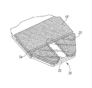
The body cover 18 and the chamber cover 20 overlying the spacing layer
16 define a slot 34 therebetween, the slot defining a vent opening
communicating
with the sample-receiving chamber to allow air to escape the chamber as a
sample
CA 02529651 2005-12-15
WO 2004/113900
PCT/US2004/019684
13
fluid enters the chamber from the edge opening or fluid receiving opening 35.
The
test strip therefore includes a dosing end 36 and a meter insertion end 38.
The
shape of the dosing end is typically distinguishable from the meter end so as
to aid
users. As shown in FIG. 1, preferably, dosing end 36 is tapered or narrowed to
form a trapezoidal shape to aid users in aligning the sample-receiving chamber
24
of test strip 10 with a fluid sample. The tapered shape of the dosing end 36
minimizes the available contact surface of the dosing end to the user's skin,
thereby aiding the alignment of the sample-receiving chamber 24 with the fluid
sample, as described in more detail below.
In addition, strip graphics and contrasting colors at the dosing end are
preferably used to further improve the intuitiveness of the strip design.
Similarly,
at the meter insertion end, chevron 31 indicates the direction of insertion of
the
strip into the meter. Further, chevron 31 is sized and positioned on the test
strip 10
such that the chevron 31 is inside the meter, and therefore hidden from view,
when
the test strip 10 is properly inserted into the meter. The size and position
of
chevron 31 (as opposed to an arrow) lessens the likelihood that users will jam
a test
strip marked with the chevron 31 into the meter and damage or destroy the test
strip 10.
General Dimensions.
The test strip is a relatively small device that is dimensioned for
compactness and ease of storage and use. In a typical embodiment, the strip
length
is on the order of 20 to 50 mm, preferably about 33 to about 38 mm, in length,
and
5 to 15 mm, preferably about 7 to about 9 mm, in width. The distance from the
slot or vent opening 34 to the edge of the meter is sized to provide a "grab
area"
where there is no blood present, and to guard against blood contamination of
the
meter contact area, and therefore may be in the range of 5 to 35 preferably 13
mm. The length of the test strip portion (from the meter insertion end 38)
that is
inserted into the meter is preferably 6.0 mm along the long axis of the test
strip.
The preferred laminar construction of the test strip also provides a device
that is relatively thin. The minimal thickness of the strip allows ready
packaging
of the strip in appropriate containers that are convenient for the user. For
example,
CA 02529651 2005-12-15
WO 2004/113900
PCT/US2004/019684
14
the overall thickness of the test strip may be about 500 to 525 Am. The
thickness
of the test strip portion that is inserted into the meter contact may be about
250 pcm.
Substrate
The test strip includes a base substrate 12 which comprises an insulating
material supporting the electrode system and other components. Typically,
plastics
such as vinyl polymers, polyimides, polyesters, and styrenes provide the
electrical
and structural properties which are required. Further, because the test strip
is
preferably mass producible from rolls of material, it is desirable that the
material
properties be appropriate to have sufficient flexibility for roll processing,
while
also giving a useful stiffness to the finished strip. The base substrate can
be
selected as a flexible polymeric material such as polyester, especially high
temperature polyester materials; polyethylene naphthalate (PEN); and
polyimide,
or mixtures of two or more of these. Polyimides are available commercially,
for
example under the trade name Kapton , from E.I. duPont de Nemours and
Company of Wilmington, DE (duPont). A particularly preferred base substrate
material is 1VIELlNEX 329 available from duPont.
Electrodes
Type.
The invention relates to an "electrochemical sensor", which is a device
configured to detect the presence of, and/or measure the concentration of, an
analyte by way of electrochemical oxidation and reduction reactions within the
sensor. These reactions are transduced to an electrical signal that can be
correlated
to an amount or concentration of the analyte. The test strip therefore
includes an
electrode system 26 comprising a set of measuring electrodes, e.g., at least a
working electrode and a counter electrode, within the sample-receiving
chamber.
The sample-receiving chamber is configured such that sample fluid entering the
chamber is placed in electrolytic contact with both the working electrode and
the
counter electrode. This allows electrical current to flow between the
measuring
electrodes to effect the electrooxidation or electroreduction of the analyte.
In the context of the present invention, a "working electrode" is an
electrode at which analyte is electrooxidized or electroreduced with or
without the
agency of a redox mediator. The term "counter electrode" refers herein to an
CA 02529651 2005-12-15
WO 2004/113900
PCT/US2004/019684
electrode that is paired with the working electrode and through which passes
an
electrochemical current equal in magnitude and opposite in sign to the current
passed through the working electrode. The term "counter electrode" is meant to
include counter electrodes which also function as reference electrodes (i.e.,
5 counter/reference electrodes).
Electrode material.
The working and counter electrodes, and the remaining portions of the
electrode system, may be formed from a variety of materials, as known in the
art.
The electrodes should have a relatively low electrical resistance and should
be
10 electrochemically inert over the operating range of the test strip.
Suitable
conductors for the working electrode include gold, palladium, platinum,
carbon,
titanium, ruthenium dioxide, and indium tin oxide, and iridium, as well as
others.
The counter electrode may be made of the same or different materials, e.g.,
silver/silver chloride. In a preferred embodiment, the working and counter
15 electrodes are both gold electrodes.
Electrode application.
The electrodes may be applied to the base substrate in any fashion that
yields electrodes of adequate conductivity and integrity. Exemplary processes
are
well known in the art, and include, for example, sputtering, printing, etc. In
a
preferred embodiment, gold electrodes are provided by coating the base
substrate
and then removing selected portions of the coating to yield the electrode
system. A
preferred removal method is laser ablation, and more preferably broad field
laser
ablation.
Laser ablative techniques typically include ablating a single metallic layer
or a multi-layer composition that includes an insulating material and a
conductive
material, e.g., a metallic-laminate of a metal layer coated on or laminated to
an
insulating material (discussed below). The metallic layer may contain pure
metals,
alloys, oxides, or other materials, which are metallic conductors. Examples of
metals or metallic-like conductors include: aluminum, carbon (such as
graphite),
cobalt, copper, gallium, gold, indium, iridium, iron, lead, magnesium, mercury
(as
an amalgam), nickel, niobium, osmium, palladium, platinum, rhenium, rhodium,
CA 02529651 2010-11-17
16
selenium, silicon (such as highly doped polycrystalline silicon), silver,
tantalum,
tin, titanium, tungsten, uranium, vanadium, zinc, zirconium, mixtures thereof,
and
alloys or solid solutions of these materials. Preferably, the materials are
selected to
be essentially unreactive to biological systems; such materials include: gold,
platinum, palladium, iridium, silver, or alloys of these metals or indium tin
oxide.
The metallic layer may be any desired thickness. In a preferred embodiment,
the
thickness is about 50 nm.
Configuration.
The electrode system may have a variety of configurations suited to the
operation of the test strip and meter. For any embodiment, the working and
counter electrodes preferably are positioned and dimensioned to minimize the
volume of sample fluid required to cover them. It is also preferable that the
electrodes be configured to maintain a current flux of sufficient magnitude as
to be
measurable using a relatively inexpensive hand-held meter.
By way of further example, a preferred embodiment includes a counter
electrode which extends around both sides of the working electrode. The
counter
electrode therefore has two elements, one in front of the working electrode
and the
other behind the working electrode, as the sample fluid enters the sample-
receiving
chamber. More specifically, the counter electrode includes elements 40 and 42
which extend across the sample-receiving chamber. Each of these elements is
about 250 gm wide. The working electrode element 44 has a width of about 250
gm, and is spaced from each of the two counter electrode elements by about 255
gra. It will be appreciated that this is only one of a number of
configurations for
the measuring electrodes.
The traces 30 and the contact pads 32 may be provided in a variety of
fashions consistent with their intended function relative to the test strip.
These
components of the electrode system are preferably composed of the same
material
as the electrodes, and are preferably applied to the base substrate in the
same
manner and simultaneously with the application of the electrodes. In a
preferred
embodiment, the traces and contact pads are gold, and are formed by laser
ablation,
particularly as described in United States Patent 7,073,246
entitled Method of Making a Biosensor.
CA 02529651 2010-11-17
=
=
17
However, alternate materials and methods of application may be employed.
Chemistry
Reagent Composition.
The test strip includes a chemical reagent within the sample-receiving
chamber for reacting with the test analyte to produce the electrochemical
signal
that represents the presence of the analyte in the sample fluid. The reagent
layer
can include a variety of active components selected to determine the presence
and/or concentration of various analytes. The test chemistry is therefore
selected
in respect to the analyte to be assessed. As is well known in the art, there
are
numerous chemistries available for use with each of various analytes. For
example, in one preferred embodiment, the test strip of the present invention
can
include one or more enzymes, co-enzymes, and co-factors, which can be selected
to determine the presence of glucose in blood. The selection of an appropriate
chemistry is therefore well within the skill in the art, and further
description herein
is not required in order to enable one to make and use the test strips with
various
analytes.
Adjuvants.
In conventional fashion, the reagent chemistry may include a variety of
= 20 adjuvants to enhance the reagent properties or characteristics. For
example, the
chemistry may include materials to facilitate the placement of the reagent
composition onto the test strip and to improve its adherence to the strip, or
for
increasing the rate of hydration of the reagent composition by the sample
fluid.
Additionally, the reagent layer can include components selected to enhance the
physical properties of the resulting dried reagent layer, and the uptake of a
liquid
test sample for analysis. Examples of adjuvant materials to be used with the
reagent composition include thickeners, viscosity modulators, film formers,
stabilizers, buffers, detergents, gelling agents, fillers, film openers,
coloring agents,
and agents endowing thixotropy.
In a preferred embodiment of the test sample, the majority of the chamber
is hollow before use. In the very small sample chamber of the test strips
according
to the present invention, it is preferable that the reagent layer be thin and
uniform.
CA 02529651 2005-12-15
WO 2004/113900
PCT/US2004/019684
18
Since the sample-receiving chamber is very small, less than about 1 1, the
depth
or vertical height of the chamber is small. Consequently, the reagent layer
should
not occupy the majority of the internal cavity of the chamber. The reagent
layer
should be sufficiently thin to leave ample space for the test sample in the
chamber.
Further, the liquid test sample will hydrate or dissolve the thin reagent
layer more
quickly. As discussed in the above reaction scheme, the mediator and mediator
redox products diffuse through and within the reagent layer/gradient to the
electrodes. The reactive components and intermediates will have a short
distance
to diffuse through a thin reagent, therefore, diffusion to the electrodes will
occur in
less time. Additionally, the capture efficiency of mediator redox products at
an
electrode will be greater for a thin layer of enzyme than a thick layer.
Conversely, a thick reagent layer will take more time for the liquid test
sample to hydrate or dissolve, and a thick reagent layer will increase the
time that
it takes for the mediator/mediator redox products to approach the electrodes.
This
can delay the time to determine the analyte concentration and introduce errors
into
the determination.
It is preferred that the reagent layer have a uniform thickness. Thickness
inhomogeneity can lead to variability in determining the analyte
concentration. In
a preferred embodiment, the reagent layer has a uniform thickness throughout
the
entire sample receiving chamber. In this preferred embodiment, the reagent
layer
is not thicker around the perimeter of the sample receiving chamber adjacent
the
vertical side walls that define the chamber than in the central portion of the
chamber. Consequently, the reagent layer does not exhibit a meniscus profile.
The reagent composition is formulated as a viscous solution that can be
deposited in a thin, uniform layer on the base layer. The reagent composition
includes thickeners and thixotropic agents to enhance the physical properties
of the
reagent layer. The thickeners are selected to provide a thick, liquid matrix
having
the remaining components homogeneously dispersed therein. The thickening and
thixotropic agents also inhibit the liquid or semi-paste material from running
or
spreading over the surface of the base layer after it has been deposited and
before it
dries. After the reagent composition is deposited, it quickly dries to a
readily
hydratable matrix.
CA 02529651 2005-12-15
WO 2004/113900
PCT/US2004/019684
19
The reagent composition is provided to dry rapidly either with air drying or
heat drying. After drying, the deposited reagent layer exhibits a thickness of
between about 1 micron and about 20 microns. More preferably, the dried
reagent
layer exhibits a thickness of between about 2 microns and about 6 microns.
The reagent composition can be deposited on the test strip surface using a
variety of coating methods including slot-die coating, curtain coating, hot
melt
coating, rotary screen coating, doctor blade or air knife coating, Meyer bar
coating,
and reverse roll coating techniques. These techniques are known to those
skilled in
the art. Preferably, the reagent layer is deposited on the flexible web as a
wet
composition at a thickness of between about 40 lana and about 100 pm. More
preferably, the reagent composition is deposited as a wet composition at a
thickness of between about 60 Inn and about 8011M. The composition may be
applied as a uniformly thin layer of a reagent directly on top of the
measuring
electrodes and along the length of a web of multiple test strips, as a
continuous
narrow band. In preferred embodiments, the narrow band has a width of between
about 7 mm and 8 mm and a dry thickness of between about 3 um and about 20
um. The composition may also be applied onto other electrodes that may reside
in
the sample-receiving chamber, depending on the desired functionality of such
extraneous electrodes.
Spacing Layer
Configuration.
The test strip includes a spacing layer 14 which overlies the base substrate
and which in part defines the sample-receiving chamber. In particular, the
spacing
layer 14 includes a void portion 22 substantially defining the height and the
perimeter of the sample-receiving chamber 24. The void portion 22 is
conveniently placed to have an edge opening whereby the sample fluid is
contacted
with the edge opening to enter into the sample-receiving chamber. The edge
opening preferably is located at the end of the test strip, although it will
be
appreciated that placement on a side edge is also useful.
CA 02529651 2005-12-15
WO 2004/113900
PCT/US2004/019684
Materials.
The spacing layer 14 may be made of any material useful for fabrication
with the test strip. Because the spacing layer partially defines the height of
the
5 sample-receiving chamber, the material should have sufficient strength at
thicknesses appropriate to the desired height of the chamber. Another function
of
the spacing layer is to protect the electrode traces that extend along the
upper
surface of base substrate 12. The material should also be readily attached to
the
base substrate and the cover materials, either by heat-sensitive or pressure-
sensitive
10 adhesives, or other means, such as heat or laser welding. Examples of
suitable
materials include a 100 Am PET, or PEN foil coated or combined with adhesives
such as ARCare 90132 from Adhesives Research Inc.
Covering Layer
Configuration.
15 A covering layer 16 is received over and attached to the spacing layer
14.
One function of the covering layer is to form the top surface of the sample-
receiving chamber. Another function is the provision of a hydrophilic surface
to
aid in acquisition of the test sample. In addition, the covering layer 16
preferably
defines a vent opening 34 that allows air to escape from the interior of the
chamber
20 as the sample fluid enters and moves into the sample-receiving chamber.
The covering layer can be formed as a unitary piece with slot 34' formed as
a recess on the underside thereof, as shown in FIG. 1B. For mass production
purposes, slot 34' would be substantially straight as shown and extend across
the
entire width of the test strip, such that air would vent from the sample
receiving
chamber 24 to the vent openings formed on opposite lateral sides of the test
strip.
However, the slot could comprise a groove or recess that only extends from the
chamber 24 to one side of the test strip, although such configuration is not
preferred for mass production purposes.
Another alternate embodiment is shown in FIG. 1C, in which chamber
cover 20 "overlaps" body cover 18. In this arrangement, a small end portion 37
of
cover layer 20 is bent upwardly and extends across the edge of body cover 18.
A
slot 34" is thereby formed having roughly a triangular shaped cross section as
can
CA 02529651 2005-12-15
WO 2004/113900
PCT/US2004/019684
21
be seen at the edges of the strip, at which there are triangular shaped
openings that
allow air to escape. In this "overlap" arrangement, the precise placement of
the
chamber cover 20 with respect to body cover 18 along the lengthwise direction
of
the strip is not critically important. That is, the amount of chamber covering
material overlapping body cover 18 can vary without affecting the dimensions
or
placement of the slot. This has advantages in manufacturing, as will become
apparent with reference to the discussion below.
Preferably, body cover 18 and chamber cover 20 comprise two separate
members for ease in fabrication and in forming the vent opening. Body cover 18
and chamber cover 20 are both disposed in substantially the same horizontal
plane. The chamber cover 20 substantially covers the void portion 22 of the
spacing layer, and forms the top of the sample-receiving chamber. The chamber
cover preferably includes a hydrophilic coating or treatment 21 on its
underside, as
described in more detail below. The body cover and the chamber cover are
positioned end to end in the lengthwise direction along the test strip and
include
slot 34 therebetween as shown in FIG. 1A. The slot is located adjacent the
interior
end of the void portion 22 of the spacing layer, and in the preferred
embodiment in
FIG. 1A, forms a small gap that spaces chamber cover 20 from body cover 18.
The gap constitutes the vent opening 34 in communication with the sample-
receiving chamber. Slot 34 is substantially straight and extends across the
width of
test strip 10. Slot 34 is oriented substantially perpendicular to the
longitudinal or
lengthwise axis of test strip 10. Sample fluid entering the sample-receiving
chamber will expel air through the vent opening defined by slot 34. If the
slot be
formed as a gap, some or most of the air expelled will exit from the top of
the test
strip.
The slot is located at a position relative to the sample-receiving chamber
that is interior of the location of the electrode system 26. Sample fluid
entering the
sample-receiving chamber will progress as far as the vent opening, but no
further.
When viewed from the top, the slot provides a visual indication of a "fill-
line," as
described herein. The placement of the vent opening therefore assures that
sufficient sample fluid can be received to fully cover the electrode system.
At the
CA 02529651 2005-12-15
WO 2004/113900
PCT/US2004/019684
22
same time, the placement of the vent opening will inhibit continued wicking of
the
sample fluid beyond the region of the electrode system.
The formation of the slot and vent opening by the spacing of the body
cover and the chamber cover is further advantageous because it avoids the need
to
otherwise form an aperture in the covering layer or base layer. In the prior
art, it
has been an approach to form the vent opening by punching a hole in either the
top
or bottom film foiming the sample-receiving chamber, which presents
fabrication
issues because of the need to precisely locate the hole relative to the sample-
receiving chamber. While this approach is also suitable for a test strip, the
preferred design described herein avoids the need to align the vent opening
laterally relative to the test strip. Further, the present design is well
suited to mass
production of the test strips by roll processing techniques, as described
hereafter.
At the same time, the vent construction may be made in a manner to inhibit
the wicking of sample fluid laterally along the slot beyond the middle area
that
overlies the sample receiving chamber 24. For example, the body cover is
preferably secured to the spacing layer by means of an adhesive 46, as shown
in
FIG. 3. The use of a hydrophobic adhesive will inhibit blood, interstitial
fluid, and
other aqueous liquids from moving along the laterally-extending slot by
capillary
action. The entire body cover, or portions adjacent to the vent opening, may
also
be hydrophobic to inhibit wicking. Materials and methods for providing
hydrophobic properties for a surface of a material are well known in the art.
The
chamber cover may be secured to the spacing layer by the same or different
adhesive than adhesive 46, as explained below.
Adhesive 49 secures the spacing layer to the base substrate 12. Adhesive
46, as well as adhesive 49 and the material for spacing layer 14, are all
formed of
substantially hydrophobic material in the illustrated embodiment. As such, the
vertical walls of the capillary chamber formed in strip 10 are hydrophobic. By
contrast, the floor of the chamber is covered with a hydrophilic reagent and
the
underside of layer 20 is coated with a hydrophilic coating 21 (FIG. 2). In
other
words, the horizontal surfaces in the capillary are hydrophilic while the
vertical
surfaces are hydrophobic. This has been found to promote good wicking of the
CA 02529651 2010-11-17
=
=
23
sample into the capillary chamber, yet prevents unwanted migration of the
sample
laterally from the chamber, e.g., between the spacer layer and the base
substrate.
Materials.
5 The body cover and chamber cover may be made of any materials useful
for fabrication with the test strip. The materials for the body cover and the
chamber cover may be the same or different. The materials should be readily
attached to the spacing layer, either by heat-sensitive or pressure-sensitive
adhesives, or other means such as heat or laser welding. Examples of suitable
10 materials for both the chamber cover and body cover include
approximately 127
Am thick foil of PET. The chamber cover preferably includes a hydrophilic
layer
21 as disclosed in WO 02/085185, ARFlow 90191 from Adhesives Research Inc.
The covering layer 16 may also be used to facilitate viewing of the sample
fluid as it enters the sample-receiving chamber. This is accomplished by
providing
15 a contrast in color or shading between the chamber and the surrounding
area. For
example, in one approach the portion of the spacing layer 14 that surrounds
void.
22 is provided with a color that contrasts with the color of the bottom of the
= sample-receiving chamber, e.g., the color of the chemical reagent layer
positioned
at the bottom of the chamber. This contrasting color may be provided, for
20 example, by the application of an ink or other coloring agent to the
portions of the
spacing layer adjacent the sample-receiving chamber. A colored section 23 of
layer 14 is pictured in FIG. 2. The chamber cover 20 is then provided as a
transparent or translucent material that allows the user to view the chamber
and the
adjacent spacing layer. As sample fluid enters from the edge of the test
strip, the
25 user is able to observe its progress as it moves by capillary action
toward the vent
opening. This type of feature is further described in US Patent No. 5,997,817,
issued to Crismore et al. on December 7, 1999.
Capillary
30 The sample-receiving chamber formed by the base substrate, spacing
layer
and chamber cover essentially comprises several sections into which the sample
fluid will travel. A first, entry section 48 extends from the edge opening to
the
CA 02529651 2005-12-15
WO 2004/113900
PCT/US2004/019684
24
area of the measuring electrode system. A second, test section 50 extends
through
the area of the electrode system. A third section 52 extends from the
measuring
electrode system to the vent opening. It will be appreciated that the testing
of the
sample fluid occurs in the area of the electrode system in the test section.
However, the sample fluid will also fill the other sections of the chamber in
the
course of filling the test strip.
Dimensions.
The height and width of the sample-receiving chamber are selected based
upon a variety of considerations, including the fluid being tested and the
analyte at
issue. For example, the chamber dimensions are preferably sized to promote
capillary flow of the test fluid into the chamber. Preferred chamber heights
for use
with blood, for example, are from about 50 Am to about 200 m, and most
preferably from 120 to 180 AM. In a preferred embodiment, the chamber height
is
about 150 Am. The width of the chamber may similarly be selected to match a
desired sample fluid and analyte. For example, the chamber should be
sufficiently
wide to expose a desired amount of the working and counter electrodes, and
should
be narrow enough to avoid the requirement of an undue amount of sample fluid
for
testing. The width of the sample-receiving chamber and the width of the
working
electrode define the area of the working electrode. The area represents a
further
design consideration as it relates to signal amplitude and instrumentation
design.
Volume.
The sample-receiving chamber is preferably provided as having a minimal
volume, in order to reduce the amount of sample fluid needed for conducting a
test.
The overall sample-receiving chamber, including all of the three sections
extending
from the edge opening to the vent opening, has a total volume that can be
considered to be a factor of the area of the chamber from the edge to the
vent, and
the height of the chamber from the base substrate to the chamber cover 20.
However, the "net chamber volume" comprises the volume of sample fluid
required to fill this space. The net chamber volume of the sample-receiving
chamber will be the equivalent of the total chamber volume less the volume
occupied by the electrodes, the reagent, and perhaps other items such as a
sorbent
material, if included.
CA 02529651 2005-12-15
WO 2004/113900
PCT/US2004/019684
As previously indicated, the volume of the overall sample-receiving
chamber is comprised of the volumes attributable to the three sections of the
chamber. Each of the sections is generally sized to be as small as practical
for the
operation of the test strip. However, there are considerations, and possibly
other
5 functions, that will impact on the size of each section.
The chamber volumes are a factor of both height and area. The height is a
result of the thickness of the spacing layer and the thickness of the
adhesives used
to secure the spacing layer to the other layers. For example, the base
substrate and
the chamber cover are attached to opposite sides of the spacing layer. One
method
10 of attachment is the heat or laser sealing of the materials. It is
preferred, however,
to attach these layers by the use of suitable adhesives, such as heat-
sensitive or
pressure-sensitive adhesives. In this approach, the height of the sample-
receiving
chamber, i.e., the distance between the facing surfaces of the bottom
substrate and
the chamber cover, will be impacted by the thickness of the adhesive layers.
As
15. shown in FIG. 3, chamber 24 is bounded on its bottom side by reagent
layer 33 and
its top side by coating 21 of chamber cover 20. However, adhesive layers 46
and
49 as well as spacing layer 14 define the total height of chamber 24.
Further, in a preferred embodiment, the reagent layer 33 extends between
base substrate 12 and spacing layer 14 and indeed extends the entire width of
the
20 test strip, as described below. The height of the chamber may therefore
also be
increased due to the presence of the reagent layer underlying the spacing
layer. In
this embodiment, and if adhesive is employed, it has been found that the
adhesive
may combine with the test reagent, at least to an extent that causes the
adhesive to
fill somewhat into and around the reagent. The heights of the reagent and
adhesive
25 layers therefore are not necessarily additive in the final test strip.
Rather, the
height of the resulting space between the base substrate and the spacing layer
is
somewhat less than the combination of the heights of the separate reagent and
adhesive layers prior to lamination.
It has also been found that the combination of the adhesive and the reagent
advantageously helps to create a seal along the edge of the sample-receiving
chamber. This inhibits sample fluid from wicking into the reagent material
present
CA 02529651 2005-12-15
WO 2004/113900
PCT/US2004/019684
26
in the space between the base substrate and the spacing layer in the time
frame
necessary for performing a test.
The first entry section is available to receive the sample fluid and direct it
to the measuring electrodes. This section can be quite small in size, and may
comprise only a short segment of the chamber. The length of this section is
preferably less than 1200 pm.
The second testing section includes the test or measuring electrodes, and is
also sized to require a minimal volume of sample fluid. A primary factor
controlling the size of this second section will be the type, number, size,
signal
strength, and configuration of the measuring electrodes. The length of this
section
is preferably about 1260 AM. A preferred volume is about 0.265 pcL, based on a
capillary height of 0.15mm, and a capillary width of 1.4 mm.
The sample fluid moves past the measuring electrodes and into the third
section. This provides assurance, and preferably allows for specific
confirmation,
that the measuring electrodes are properly wetted. This confirmation may be by
visual observation by the user, or by automatic detecting means. For example,
dose sufficiency electrodes may be placed in this section to detect when the
sample
fluid has progressed into this section to a point that the wetting of the
measuring
electrodes is assured. This can be used as a trigger for initiating the
application of
the potential to the electrodes. The length of this section is preferably 50
to 500
ium, and more preferably 255 to 400 inn. The volume is preferably 0.01 to 0.1
AL,
and more preferably 0.05 to 0.08 pt.
In a preferred embodiment, the overall net chamber volume of the sample-
receiving chamber is less than about 1 AL, and is more preferably less than
about
0.5 pl. Desirable ranges for the net chamber volume of the sample-receiving
chamber include volumes from about 0.15 to about 1.4 [IL, more preferably from
about 0.4 to about 0.7
Sorbent.
The sample chamber may be otherwise empty, which is preferred, or may
alternatively include a sorbent material. Suitable sorbent materials include
polyester, nylon, cellulose, and cellulose derivatives such as nitrocellulose.
A
sorbent material could be included to facilitate the uptake of the sample
fluid by
CA 02529651 2005-12-15
WO 2004/113900
PCT/US2004/019684
27
assisting in wicking the fluid into the chamber. The use of a sorbent material
would also serve to further reduce the void volume of the sample-receiving
chamber for reception of the sample fluid.
Fill Method.
The preferred method of filling the sample chamber is by capillary action.
In addition, the filling of the test strip can be augmented by other means,
such as
applying a pressure on the sample fluid to push it into the sample chamber,
and/or
creating a vacuum on the sample chamber to pull the sample fluid into the
chamber.
Hydrophilic coating.
For purposes of capillary filling of the sample-receiving chamber, various
approaches are available to facilitate the movement of the sample fluid into
the
chamber. For example, any or all of the surfaces defining the chamber may be
selected or treated to improve hydrophilicity. Such treatment may comprise the
use of known hydrophilic materials, application of a hydrophilic material onto
the
surface, or treatment of the surfaces to increase hydrophilicity, as described
below.
In addition, the reagent composition may be formulated to be readily hydrated
and
to encourage filling of the sample-receiving chamber. As previously indicated,
a
sorbent may also be used.
Testing for Analyte
The electrochemical sensor is operated by applying a suitable potential or
series of potentials across the working and counter electrodes, and across the
dose
sufficiency electrodes. When a mediator is used, the magnitude of the required
potential across the working and counter electrodes will be dependent on the
redox
mediator. Moreover, the potential at the electrode where the analyte is
electrolyzed is typically large enough to drive the electrochemical reaction
to or
near completion, but the magnitude of the potential is preferably not large
enough
to induce significant electrochemical reaction of interferants. For glucose,
for
example, an applied potential difference typically is between about +100 mV
and
about +550 mV when using a DC potential. When using AC potentials these can
be typically be 5 to 100 mV RMS.
CA 02529651 2005-12-15
WO 2004/113900
PCT/US2004/019684
28
A potential may be applied before or after the sample begins to enter the
sample-receiving chamber. However, a potential is preferably applied after the
sample has entered the chamber, and more preferably after it has been
determined
that there is a sufficient amount of sample in the sample-receiving chamber
for
conducting a test. The timing of the application of a potential may be
triggered in
a variety of fashions, including visual observation by the user, a time delay
following sampling of the fluid to the test strip, or upon electrical or other
automated detection of a sufficient amount of sample fluid in the chamber. The
visual and electrical alternatives also may act as redundant failsafes to
assure
proper operation of the device. Preferably, the test strip and system utilize
separate
detecting means, such as dose sufficiency electrodes, for determining when the
fluid sample has sufficiently filled the chamber.
When a potential is applied and the sample fluid is in the sample-receiving
chamber, an electrical current will flow between the working electrode and the
counter electrode. The current can be a result of the electrolysis of the
analyte in
the sample fluid when a potential of sufficient magnitude is applied. In this
case
electrochemical reaction occurs via the redox mediator, generally as
previously
described. In the case where small amplitude potential is applied,
particularly in
the case of AC potentials, the current is produced not necessarily by
electrolysis,
but by ionic motion and response of the dielectric in the sample chamber.
Those
skilled in the art will recognize that there are many different reaction
mechanisms
that will achieve the same result.
Control solution
A test may be applied to the test strip after dosing to confirm that a control
solution, and even that the correct control solution, has been administered.
The
control solutions aid the user in confirming that the entire system is
functioning
within design specifications, and that the test strips have not been stored
improperly or otherwise mistreated. Acceptable strips will recover values
within
specified tolerance ranges for the particular strip lot being tested. The
tolerance
ranges in question will be published for each strip lot on the container
label.
CA 02529651 2005-12-15
WO 2004/113900
PCT/US2004/019684
29
Method of Making Strip
In a preferred embodiment, the sensor comprises a multi-layered, laminate
test strip 10. As previously described, the laminate includes a base substrate
12, a
spacing layer 14, and a covering layer 16. These components may be assembled
in
various ways. For example, the components may be assembled by use of
adhesives, heat sealing, laser welding, and a variety of other suitable
techniques
appropriate for securing the adjacent materials. The test strips are
preferably
assembled in a large number on a single sheet or web, and the strips are
thereafter
separated for storage and use.
The laminate test strip may be assembled sequentially by successively
laying down one layer at a time. Alternatively, the test strip can be prepared
by
assembling and processing individual components or layers, which are then
laminated together to provide the functional test strip. In one preferred
form, two
or more basic components of the test strip are prepared simultaneously. Then
in
one or a series of assembly or laminating steps, the basic components are
combined to produce the test strip, which may or may not require further
processing. In a preferred embodiment, the test strip is assembled from three
basic
components: a metallized substrate preferably with a reagent layer coated on
metallic electrodes defined on the substrate, a spacing layer having a cavity
preformed therein, and one or more top or cover layers.
With such small dimensions for the sample-receiving chamber, the
characteristics of the reagent layer can have a significant impact on the
operation
of the test strip, particularly in view of hydration and mixing
characteristics. The
reproducibility of the quantity, location, thickness and other properties of
the
reagent layer is therefore important. It is therefore desirable for the
composition to
include materials which specifically enhance the physical characteristics,
such as
the uniformity and flatness, of the applied layer.
In one particular aspect, the test strip includes a unique manner of
incorporating the reagent. The reagent is placed in the sample-receiving
chamber
at least on the working electrode, and preferably also on the counter
electrode. The
reagent may be applied to the test strip in a variety of fashions as is well
CA 02529651 2005-12-15
WO 2004/113900
PCT/US2004/019684
understood in the art. In a preferred embodiment, the reagent composition is
applied as a thin coating over the electrodes supported on the base substrate.
More particularly, the reagent is placed onto the base substrate in a manner
that positions the reagent composition between the base substrate and the
spacing
5 layer. This manner of application helps to make the reagent layer more
flat and
uniform in thickness. In contrast, a procedure of the prior art has been to
first
prepare the reaction well or cavity, and to then fill the reagent into the
well.
However, this can result in a more uneven reagent layer due to phenomena such
as
the formation of a meniscus at the perimeter of the well. This in turn can
cause the
10 reagent to have a different thickness adjacent to the side walls of the
reaction well
than in the interior portion, which can cause inconsistency in the filling of
the
chamber, prolonged dissolution intervals, and inconsistent mixing of the
reagent
with the sample fluid, and the ultimate test results. By placing the reagent
onto the
base substrate before the spacing layer is added, there is no meniscus effect
to
15 disrupt the even layering of the reagent as it dries on the base
substrate. In
addition, this method of application facilitates the mass production of the
test
strips.
Referring to the drawings, the test strip 10 is shown as including a reagent
layer 33 that extends between the bottom substrate 12 and the spacing layer
14.
20 More particularly, the reagent forms a layer 33 which covers both the
top surface
of the bottom substrate 12 and the electrodes 28. The reagent covers at least
the
working electrode, and preferably also the counter electrode. In the most
preferred
embodiment, the reagent layer extends the full width of the test strip. The
reagent
layer also preferably extends from the end edge to the dose sufficiency
electrodes,
25 and most preferably to the vent opening. The reagent layer thus extends
under the
spacing layer and is sandwiched between the spacing layer and the base
substrate.
The reagent composition is applied to the bottom or base substrate in any
suitable fashion that provides a desired and uniform layer which will
ultimately
extend under the spacing layer. The reagent is preferably applied in a
continuous
30 coating directly onto the bottom substrate, and onto the electrodes
received
thereon. As described hereafter, the reagent composition is most preferably
applied in the course of producing a large quantity of test strips on a
webbing of
CA 02529651 2005-12-15
WO 2004/113900
PCT/US2004/019684
31
material. In this manner, the reagent may be applied in the way of a
continuous
stripe of material that extends over a substrate roll that is later separated
into
individual test strips. The reagent composition is allowed to dry or otherwise
set
up and the spacing layer is applied thereover.
In a related aspect, a preferred manner of securing the spacing layer to the
bottom substrate is the use of an adhesive. In addition to securing the layers
together, it has been found that the adhesive will sufficiently engage with
the
reagent composition as to help to seal the space between the bottom substrate
and
the spacing layer. The adhesives preferably placed on the spacing layer, which
is
laminated onto the base substrate. The adhesive thereby contacts the portion
of the
reagent which extends under the spacing layer.
Although the spacing layer of the illustrated embodiment is fowled from
Melinex material with adhesives on both sides thereof, it is also possible to
form
spacing layer 14 as a continuous adhesive material, such as a double-sided
tape.
For example, a 5 to 6 millimeter thick ARCare Adhesive could be used in lieu
of
spacing layer 14.
In a further aspect, a preferred embodiment is described in which the
analyte is glucose. In the case of glucose, the active components of the
reagent
composition will typically include an oxidoreductase, such as an enzyme for
glucose; optionally a co-enzyme or co-factor; and a redox mediator. These
components are typically dissolved or suspended in a matrix. The liquid test
sample hydrates or dissolves the matrix, and the analyte diffuses through the
matrix to react with one or more of the active components. Typically, the
enzyme
oxidizes the glucose in the test sample to gluconolactone and/or gluconic
acid.
The mediator, in turn, reacts with or oxidizes the reduced enzyme, and
consequently the mediator is reduced in the process. The reduced mediator can
be
detected at one of the electrodes on the test strip.
In a specific example of an oxidation/reduction reaction scheme useful for
detecting glucose in human blood, a test sample containing glucose reacts with
an
enzyme such as Glucose-Di-Oxidoreductase (Gluc-Dor), and optionally a co-
enzyme or cofactor such as pyrrolo-quinoline-quinone (PQQ), in the presence of
a
redox mediator. The mediator may include, for example, benzoquinone,
transition
CA 02529651 2005-12-15
WO 2004/113900
PCT/US2004/019684
32
metal complexes, e.g., potassium ferricyanide, osmium derivatives (e.g.,
osmium
bipyridyl complexes such as described in WO 98/35225) and nitrosoanaline
derivatives (see U.S. Patent No. 5,286,362). This produces the oxidized form
of
the analyte, gluconolactone, and the reduced form of the redox mediator. The
mediator thereafter shuttles the redox equivalent of mediator product, the
reduced
mediator, to the electrode surface by diffusion. There the mediator is
oxidized
quantitatively at a defined anodic potential, and the resulting current is
related to
the apparent glucose concentration.
A representation of the reaction sequences for this reaction system using a
nitrosoaniline derivative is provided below in Equation 1.
CA 02529651 2005-12-15
WO 2004/113900 PCT/US2004/019684
33
H3c. H3c
IcH2cHzoH o/CH2CH2OH
fkk NCI
cf cH2cH20H 90 CH2CH2OH
Nitrosoanaline Derivative (31.1144)
(1st Enzymatic Reduction) Glucose
GI uc-DO
2H* Gluconolactone
Very H3C =
Fast
Kinetics ICH2CH2OH
CH2CH2OH
( Reduced Form)
C OH -
r
/CH2CH2OH
(Counter Electrode] H/ \hiH2CH2OH [ Oxidation at the
Anode]
2e - C
(2nd Enzymatic Reduction
Glucose
QD Quinonediimine(0x) H.. 2e -
GI uc-DOR (QD)
Gluconolactone
H+ H3C =
H
CH2CH2OH
ik/ = /
CH2CH2OH
IThenylenediamine (Red>
(PD)
File Mediator 311144 RXN Rey 09092002-Text.CDX
(1)
As shown, the nitrosoaniline derivative, o-methoxy-[N,N-bis-(2-
hydroxyethyl)]-p-nitrosoaniline, initially exists as a mixture of two isomers,
or
tautomers, in equilibrium with each other. Reaction of Gluc-Dor with glucose
in
the test sample yields gluconolactone and the reduced form of Gluc-Dor (Glue-
Dor2H+). The reduced form of Gluc-Dor (Gluc-Dor2H+) reacts rapidly with the
nitrosoaniline derivative, which is reduced and which regenerates Gluc-Dor.
The
reduced nitrosoaniline derivative then undergoes hydrolysis to form
quinonediimine (QD). In a second enzymatic, redox reaction, Gluc-Dor reacts
with glucose to yield another molecule of Gluc-Dor2H+ and gluconolactone. The
CA 02529651 2005-12-15
WO 2004/113900
PCT/US2004/019684
34
Gluc-Dor2H+ reacts with (is oxidized by) quinonediimine to regenerate Gluc-
Dor,
and produces a phenylene diamine derivative (PD). PD is then oxidized at the
working electrode to produce a current related to glucose concentration.
Additionally, at the counter electrode QD can be reduced to PD.
Adjuvants.
With such small dimensions for the sample-receiving chamber, the
characteristics of the reagent layer can have a significant impact on the
operation
of the test strip, particularly in view of hydration and mixing
characteristics. The
control and reproducibility of the quantity, location, width, thickness, and
other
properties of the reagent layer become more important as the chamber volume
decreases and test time diminishes. It is therefore desirable for the
composition to
include materials that specifically enhance the physical characteristics, such
as the
uniformity and flatness, of the applied layer. Additionally, the method of
application can impact the physical characteristics, control, and
reproducibility of
the reagent layer.
The reagent composition can therefore also include a variety of adjuvants to
enhance the reagent properties or characteristics. For example, the
composition
may include adjunct materials to facilitate the placement of the reagent
composition onto the test strip and to improve its adherence to the strip. The
composition can also include materials to increase its rate of hydration
and/or
increase its influence on the capillary action to fill the chamber with the
test
sample. Examples of addjunct materials to be used with the reagent composition
include thickeners, viscosity modulators, film formers, stabilizers, buffers,
detergents, gelling agents, fillers, film opening agents, coloring agents, and
agents
endowing thixotropy.
The adjuvant materials or components can impact the application,
reproducibility and physical properties of the reagent layer. The adjunct
materials
can include one or more of the following:
Thickeners may include, for example, (1) starches, gums (e.g., pectin, guar
gum, locust bean (carob seed) gum, konjac gum, xanthan gum, alginates, and
agar), casein, gelatin, and phycocolloids; (2) cellulose and semi-synthetic
cellulose
derivatives (carboxymethyl-cellulose, methyl cellulose,
hydroxymethylcellulose,
CA 02529651 2005-12-15
WO 2004/113900
PCT/US2004/019684
hydroxyethylcellulose, methylhydroxyethylcellulose); (3) polyvinyl alcohol and
carboxy-vinylates; and (4) bentonite, silicates, and colloidal silica.
Preferred
thickeners include a combination of a xanthan gum sold under the trade name
Keltrol F by CP Kelco US, Inc., and carboxylmethyl cellulose sold under the
trade
5 name AQUALON CMC 7F PH by Hercules Inc., Aqualon Division.
Film forming and thixotropic agents useful in the reagent composition
include polymers and silica. Preferred thixotropic agents include silica sold
under
the trade name Kieselsaure Sipernate FK 320 DS by Degussa AG. Preferred film
forming agents include polyvinylpyrrolidone, sold under the trademark
10 polyvinylpyrrolidon Kollidon 25, by BASF, and polyvinyl propionate
dispersion.
Stabilizers for the enzyme in the reagent can be selected from sacchhrides
and mono-or di-fatty acid salts. Preferred stabilizers include trehalose sold
under
the trade name D-(+)-Trehalose dihydrate by Sigma Chemical Co. and sodium
succinate.
15 Detergents can be selected from water-soluble soaps, as well as water-
soluble synthetic surface-active compounds such as alkali, earth alkali or
optionally substituted ammonium salts of higher fatty acids, e.g., oleic or
stearic
acid, mixtures of natural fatty acids, for example, from coconut or tallow
oil, fatty
sulphates, esters of sulphonic acids, salts of alkyl sulphonic acids taurine
salts of
20 fatty acids, fatty acid amides, and ester amides. Preferred detergents
for the
present invention include an ester amide, n-octanoyl-N-methylglucamide, sold
under the trade name Mega-8 by Dojindo Molecular Technologies, Inc., and a
fatty
acid salt, N-methyl ley' taurate sodium salt, sold under the trade name
Geropon
T77 by Rhodia ETCH (Home, Personal Care and Industrial Ingredients).
25 It should be understood that one or more of the specific additives above
described can exhibit additional properties and consequently could be
categorized
in one or more of the classes above noted.
Mediator.
A mediator for use in the reagent composition can be selected as any
30 chemical species (generally electroactive) which can participate in a
reaction
scheme involving an enzyme, an analyte, and optionally a cofactor, and
reaction
products thereof, to produce a detectable electroactive reaction product.
Typically,
CA 02529651 2005-12-15
WO 2004/113900
PCT/US2004/019684
36
participation of the mediator in the reaction involves a change in its
oxidation state
(e.g., a reduction), upon interaction with any one of the analyte, the enzyme,
or a
cofactor, or a species that is a reaction product of one of these (e.g., a
cofactor
reacted to a different oxidation state). A variety of mediators exhibit
suitable
electrochemical behavior. A mediator can preferably also be stable in its
oxidized
folin, may optionally exhibit reversible redox electrochemistry, can
preferably
exhibit good solubility in aqueous solutions, and preferably reacts rapidly to
produce an electroactive reaction product. Examples of suitable mediators
include
benzoquinone, meldola blue, other transition metal complexes, potassium
ferricyanide, osmium derivatives (see WO 98/35225) and nitrosoanaline- based
mediators (see US Patent 5,286,362). In a preferred embodiment, the reagent
composition utilizes a nitrosoaniline-based chemistry.
Preferred mediators include N-(2-hydroxyethyl)-1V-p-nitrosophenyl-
piperazine, N,N-bis-(2-hydroxyethyl)-p-nitrosoaniline, o-methoxy-[N,N-bis-(2-
hydroxyethy1)]-p-nitrosoaniline, p-hydroxynitrosobenzene, N-methyl-N'-(4-
nitrosopheny1)-piperazine, p-quinone dioxime, N,N-dimethyl-p-nitrosoaniline,
N,N-
diethyl-p-nitrosoaniline, N-(4-nitrosopheny1)-morpholine, N-benzyl-N-(5'-
carboxypenty1)-p-nitrosoaniline, N,N-dimethy1-4-nitroso-1-naphthylamine, N,N,3-
trimethy1-4-nitrosoaniline, N-(2-hydroxyethyl)-5-nitrosoindoline, N,N-bis-(2-
hydroxyethyl)-3-chloro-4-nitrosoaniline, 2,4-dimethoxy-nitrosobenzene, N,N-bis-
(2-methoxyethyl)-4-nitrosoaniline, 3-methoxy-4-nitrosophenol, N-(2-
hydroxyethyl)-6-nitroso-1,2,3,4-tetrahydroquinoline, N,N-dimethy1-3-chloro-4-
nitrosoaniline, N,N-bis-(2-hydroxyethyl)-3-fluoro-4-nitrosoaniline, N,N-bis-(2-
hydroxyethyl)-3-methylthio-4-nitrosoaniline, N-(2-hydroxyethyl)-N-(2-(2-
methoxyethoxy)-ethyl)-4-nitrosoaniline, N-(2-hydroxyethyl)-N-(3-methoxy-2-
hydroxy-1-propy1)-4-nitrosoaniline, N-(2-hydroxyethyl)-N-(3-(2-hydroxyethoxy)-
2-
hydroxy-1-propy1)-4-nitrosoani line, N-(2-hydroxyethyl)-N-(2-(2-hydroxyethoxy)-
ethyl)-4-nitrosoaniline. Particularly preferred mediators according to the
present
invention include N,N-bis-(2-hydroxyethyl)-p-nitrosoaniline, o-methoxy-[N,N-
bis-
(2-hydroxyethyl)j-p-nitrosoaniline, and N-(2-hydroxyethyl)-N-(2-(2-
hydroxyethoxy)-ethyl)-4-nitrosoaniline.
An exemplary reagent composition is listed below in Table I.
CA 02529651 2005-12-15
WO 2004/113900 PCT/US2004/019684
37
Table I
Components Function Amount Abs. %solids w/w. Note.
Keltrol F Thickener 11.60 g 0.24%
Carboxy methylcellulose Thickener 27.24 g 0.57%
Kieselsaure Sipemat 320 DS Film Opener 97.01 g 2.01%
Polyvinylpyrrolidine PVP K25 Film Former 89.33 g 1.85%
Propiofan Film Former 257.09 5.34%
GlucDOR Apo-Enzyme 19.127 g 0.40% 0.673 MU/g
pyrrolo-quinoline quinine
Co-Factor 0.5329g 0.01%
(PQQ)
Na-Succinate Stabilizer 23.23 g 0.48%
Trehalose Stabilizer 23.6 g 40.49%
KH2PO4 Buffer 12.02g 0.39%
K2HPO4 x 3 H20 Buffer 43.43 g 0.90%
Nitrosoaniline Mediator 41.26 g 0.86%
Mega 8 Detergent 13.23 g 0.27%
Geropon T77 Detergent 1.405 g 0.03%
KOH 5N Adjust Buffer 36.47 g 0.76%
Water total 4114.52 g
Sum 4817.80g
Solids 14.6%
CA 02529651 2005-12-15
WO 2004/113900
PCT/US2004/019684
38
Mixing.
The components of the reagent composition are admixed with water to
provide a homogeneous, viscous suspension. The order of addition is not
critical
for the invention. A sufficient amount of the buffer solution is added to
maintain
the reagent composition at a pH of about 7. Typically the selected components
are
pre-mixed with water to provide a variety of stock solutions that can be
combined
to yield the final reagent composition. For example, a buffer solution can be
prepared by combining the phosphate salts and, optionally, the sodium
succinate.
Other stock solutions include: the thickening agents, i.e., Keltrol F and the
carboxymethyl cellulose; the surfactants, i.e., Geropon T77 and Mega 8; the
enzyme and co-enzyme or cofactor; and the mediator.
The following provides an example of the preparation of a reagent
composition. The reagent composition can be prepared by first preparing the
following stock solutions:
Buffer Solution pH 6.9 to 7.1
Amount (gm)
1120 1214.62
KH2PO4 18.27
K2BP04 43.43
Na succinate 23.23
Keltrol F Solution
Amount (gm)
H2O 287.06
Buffer Solution 101.35
Keltrol F 11.60
Carboxymethylcellulose (CMC) Solution
Amount (gm)
H20 1334.76
Na CMC1 27.24
1. Na CMC is a sodium salt of carboxymethyl cellulose sold by Hercules Inc.,
Aqualon Division
CA 02529651 2005-12-15
WO 2004/113900
PCT/US2004/019684
39
Silica Suspension
Amount(gm)
H20 722.99
Sipernat 3201
1 Kieselsaure Sipemat 320 DS (Silica) sold by Degussa AG.
Polyvinylpyrrolidone (PVP) Solution
Amount (gm)
Buffer Solution 226.03
Mega 81 13.23
Geropon T772 1.405
PV135 89.33
1. Mega 8 is n-octanoyl-N-methylglucamide sold by Dojindo Molecular
Technologies Inc.
2. Geropon T77 is N-methyl oleyl taurate sodium salt sold by Rhodia ETCH.
3. PVP is Polyvinylpyrrolidone K25 sold by BASF.
Trehalose Solution'
Amount (gm)
H20 36.4
Trehalose 23.6
1 This trehalose solution is used only in preparing the "Enzyme Solution"
listed
below.
PQQ Solution
Amount (gm) _
Buffer Solution 1st 101.59
addition
PQQ 0.533
Buffer Solution 2nd 30.0
addition
Enzyme Solution
Amount (gm)
PQQ Solution 132.12
Gluc-Dor 19.13
(673 U/mg Ly)
Trehalose Solution 58.75
Mediator Solution
CA 02529651 2005-12-15
WO 2004/113900
PCT/US2004/019684
Amount (gm)
Buffer Solution 782.27
Mediator 41.26
5 N KOH 36.47
The buffer solution, Keltrol F solution, CMC solution, and the Silica
suspension were prepared a day before. These solutions can then be combined as
listed below to prepare the reagent composition.
5 Final Reagent Composition
Thickener I 331.51 g
(Keltrol F solution)
Thickener II (CMC 1262.9 g
Solution)
PVP Solution 315.05 g
Silica suspension 762.3 g
Propiofan solution 257.09 g
Mediator Solution 855.84 g
Enzyme Solution 196.65 g
5N KOH as required to
achieve final pH of
6.9 to 7.1
Water (bidistilled) 518.69 g
For this reagent prior to coating, the final pH was 6.96 and did not need
adjustment
with 5N KOH solution. The measured viscosity was 111 mPas, which is in the
correct range for coating of 105 to 115 mPas.
10 FIGS. 5 and 5A present a flow chart illustrating a preferred process 100
for
preparing a test strip useful in accordance with the present invention.
Process 100
begins in the central process line 101 at stage 102 with selection of a film
material
for the base layer or base substrate. In a preferred embodiment, the film is
provided as a continuous roll having a width and length suitable for
fabricating a
15 large number of test strips. In subsequent finishing steps, the
processed film can
be subdivided to provide a single strip or web having a width that
approximates the
length of the test strip and includes a series of test strips, or can be die
cut to
provide individual test sensors.
From stage 102, the film proceeds to stage 104 where it is pretreated to
20 receive a metal coating and coated with the metal in one continuous
process. The
CA 02529651 2005-12-15
WO 2004/113900
PCT/US2004/019684
41
pretreatment 164 (discussed below) can be used to clean or modify the surface
to
provide a uniform coating thickness and better adhesion of the subsequent
metallized layer 166 (discussed below). The pretreatment can include
subjecting
the film to corona discharge or Argon plasma. Immediately after this pre-
treatment, a uniform conductive coating is applied to the film as shown at
106.
Alternatively, suitable substrates with metal coatings can be obtained
commercially.
The metallic layer may contain pure metals, oxides, alloys, or other
materials, which are metallic conductors. Examples of metals or metallic-like
conductors include: aluminum, carbon (such as graphite), cobalt, copper,
gallium,
gold, indium, iridium, iron, lead, magnesium, mercury (as an amalgam), nickel,
niobium, osmium, palladium, platinum, rhenium, rhodium, selenium, silicon
(such
as highly doped polycrystalline silicon), silver, tantalum, tin, titanium,
tungsten,
uranium, vanadium, zinc, zirconium, mixtures thereof, and alloys or solid
solutions
of these materials. Preferably, the materials are selected to be essentially
unreactive to biological systems; such materials include: gold, platinum,
palladium, iridium, or alloys of these metals. The metallic layer may be any
desired thickness.
The conductive coating is preferably a metal layer that is applied by a
variety of methods, including but not limited to sputtering, physical vapor
deposition (PVD), plasma assisted vapor deposition (PAVD), chemical vapor
deposition (CVD), electron beam physical vapor deposition (EBPVD), and/or
metal-organic chemical vapor deposition (MOCVD). Vapor deposition is typically
performed under vacuum. These techniques are well known in the art and can be
used to selectively provide a uniformly thin coating of metal onto a
substrate. The
resulting metallized film can be inspected to ensure that the metal coating is
uniform and free of material defects.
The roll of metallized film next encounters stage 108 where it is subdivided
and/or sized to provide webs having a width that approximates the final length
of
an individual test strip. The slicing can be accomplished using fixed-knife
slitting
equipment well-known in the art.
CA 02529651 2005-12-15
WO 2004/113900
PCT/US2004/019684
42
A single web proceeds to stage 110 for patterning the electrodes, traces,
and contacts or pads. At this stage, the electrodes, traces, and contact pads
are
formed by removing metal from the surface of the web strip. The excess metal
can
be removed using a variety of techniques well known in the art. At this stage,
one
or more indexing or registration marks can be formed either on a first edge
proximate to the electrodes, the opposite second edge proximate to the
electrode
pad, on both edges or anywhere in between. The indexing marks, particularly
those at an edge, can be used in subsequent operations to align layered
components
prefabricated in separate operations.
In a preferred method, the metal is laser ablated to eliminate undesired
portions of the metal and leave the desired electrical components. In
accordance
with this method, selected areas are laser etched simultaneously, in a "broad
field",
as opposed to using linear movement of a focused laser beam. This broad field
laser ablation method provides a precise metal pattern rapidly and at reduced
cost
as compared to other approaches. Corona treatment of the patterned substrate
is
then conducted at stage 111.
The patterned web continues on to stage 112, where a reagent layer is
deposited onto the electrodes. In a preferred embodiment, the reagent layer is
deposited as a continuous elongate stripe extending adjacent or close to the
first
edge, and overlying the measuring electrodes formed on the patterned web. As
previously noted, the reagent is consequently located across the full width of
the
test strip, including the area laterally outside of the sample-receiving
chamber and
between the base substrate and the spacing layer. Also as noted, this will
facilitate
the drying of the reagent without discontinuities, edge effects, or other
variances
that would detract from providing a thin, flat, uniform reagent layer within
the
sample-receiving chamber. The reagent includes a combination of components,
and is formulated to dry rapidly with minimal or no running after deposition,
typically by manipulating the thixotropy of the coated reagent film.
This stripe may be applied in any suitable fashion which provides the
desired extent and uniformity of thickness, precision of the stripe edge,
homogeneity, and the like. Preferred methods are capable of applying the
desired
CA 02529651 2005-12-15
WO 2004/113900
PCT/US2004/019684
43
coating at relatively high speed and high batch size. Suitable methods of
application are well known in the art and therefore are not detailed herein.
Preparing the Spacing Layer
Referring now to process line 114, a process flow for preparing the spacing
layer is illustrated. Beginning at stage 116, a material is selected to
prepare a
spacing layer for laminating on top of the reagent coated web prepared at
stage
112. The base film for the substrate can be selected from a variety of
materials.
The spacing layer material, similar to the base layer, can be provided as an
elongate roll which can be conveniently processed rapidly and with high
efficiency. Preferred materials include a polyester film sold under the trade
name
MELINEX by DuPont. Other materials suitable for use in the present invention
could include PEN. The spacing layer material has a thickness specifically
selected to provide a desired chamber depth (or height) in each of the test
strips
when combined with the thicknesses of any joining layers that are used to
laminate
the spacer to the other strip components. In preferred embodiments, the
spacing
layer is selected to have a thickness between about 75 lam and about 150 pm,
more
preferably from about 100 pm to about 125 pa. As noted above, the spacing
layer
can be formed of a double-sided adhesive.
The spacing layer is preferably formed as a continuous film having a series
of gaps that will align with the electrodes on the bottom substrate webbing.
The
manner of joining the spacing layer and bottom substrate will impact the
method
for preparing the spacing layer. For example, if the spacing layer is to be
heat
welded to the bottom substrate, then the spacing layer may simply be die cut
to
provide the appropriately spaced chamber gaps. However, a preferred method is
the use of thin, non-interfering adhesives that join the adjacent layers. In
accordance with this preferred method, a spacing layer is prepared for
combination
with the previously described substrate webbing as set forth hereafter.
The spacing layer film is prepared having the desired width for
combination with the remainder of the test strip components. The spacing layer
film may include an opaque portion, e.g., a section 23 of it is printed blue
or
another color for use in visualizing the sample-receiving chamber, as
described
elsewhere. The spacing layer film is laminated on the bottom side with a
CA 02529651 2005-12-15
WO 2004/113900
PCT/US2004/019684
44
combination adhesive and release liner, and on the top side with a similar
combination adhesive and liner.
At stage 118, two transfer adhesives are laminated to the spacing layer
material: the first transfer adhesive is laminated to the top surface of the
spacing
layer, and the second transfer adhesive is laminated to the bottom surface of
the
spacing layer. Preferably, the transfer adhesives are the same adhesive;
however,
in alternative embodiments, the first and second transfer adhesives can be
different
from each other. In preferred embodiments, the transfer adhesives are selected
from commonly used and known adhesives, including pressure sensitive
adhesives.
Preferred adhesives exhibit sufficient hydrophobicity to prevent or inhibit
the test
sample in the chamber from wicking out between the spacing layer and the
reagent
layer or base substrate. An example of a suitable sensitive adhesive is ARCare
90132 from Adhesives Research Inc. The adhesives are provided with a release
liner to prevent premature adhesion of the spacing layer during processing.
The
release liners are disposed on the exterior surface of the first and second
transfer
adhesives, facing outward from the spacing layer material.
The spacing layer with the adhesive release liners on the top and bottom
surfaces progresses to stage 120. At stage 120, the cavity which will fowi the
sample-receiving chamber is punched in the spacing layer. In one embodiment,
the cavity is punched using a "kiss cut" method. The kiss cut method cuts
through
the upper release liner, the upper adhesive, the spacing layer, and the lower
adhesive, but not through the lower release liner. In subsequent operations,
simply
removing the lower release liner will then remove the punched out portions of
the
lower adhesive, the spacing layer, the upper adhesive, and the upper release
liner
from the punched spacing layer. In other embodiments, the cavity can be
punched
through with a hollow die. The hollow die completely punches or cuts through
the
spacing layer, the two adhesives, and the two release liners, with the punched
out
portion subsequently removed in the hollow die. The spacing or pitch between
each cavity is determined and accurately controlled to allow accurate mating
of the
punched spacing layer over the electrodes using one or both of the indexing
marks
patterned in the reagent-coated web.
CA 02529651 2005-12-15
WO 2004/113900
PCT/US2004/019684
At stage 122, the lower release liner on the spacing layer is removed, taking
with it the kiss cut portions and exposing the adhesive on the underside
surface of
the spacing layer. Proceeding on to stage 124 in process line 101, the spacing
layer
is laminated over the reagent-coated web using one or more of the indexing
marks
5 previously patterned on the web to correctly align each cavity formed in
the
punched spacing layer directly on top of an electrode set to provide a web-
spacing
layer laminate. At stage 126 in the central process line 101, the upper
release liner
covering the upper adhesive on the web-spacing layer laminate is removed in
preparation for attaching the cover layer.
10 Laminating on the Cover Portions
At stage 128, a material for a body cover is introduced into the process. In
preferred examples, the material is a flexible polymeric material and may be
selected, for example, MELINEX 454 or MELINEX 339 from du Pont. The
material for the body cover is sized to have a width sufficient to overlay at
least a
15 portion of the electrode traces in the sample-receiving chamber of the
test strip.
Referring now to process line 130, beginning at stage 131, a film material is
selected to provide a chamber cover over the cavity, reagent, and measuring
electrodes on the web-spacing layer laminate. In preferred embodiments, the
body
cover material is provided as a clear poly(ethylene-terephthalate) (PET) or
20 poly(ethylene-naphthalate) (PEN) film having a thickness between about
100 pm
and about 200 lam. The coating may preferably include a release liner, which
can
be removed immediately prior to laminating over the web-spacing layer. The
chamber cover is preferably made from a hydrophilic material or the bottom
surface of the chamber cover may be treated or coated to make it hydrophilic
as
25 indicated at 134.
At stage 138, the film material can be sized to a desired width sufficient to
form the chamber cover to overlay the cavity and electrodes.
Proceeding to stage 140, the body cover from stage 128 and the chamber
cover from stage 138 are laminated to the web-spacing layer laminate. In
preferred
30 embodiments, the body cover and chamber cover are simultaneously
laminated
with the web-spacing layer laminate. The body cover is positioned over a
portion
of the electrode traces proximate to the electrodes formed on the base
substrate.
CA 02529651 2005-12-15
WO 2004/113900
PCT/US2004/019684
46
The chamber cover is positioned over the cavity, reagent, and measuring
electrodes
on the web-spacing layer laminate. The body cover and chamber cover are
separated by a gap to form a vent 34 at the interior end of the cavity formed
in the
test strip.
As described, the chamber cover is placed near the edge of the strip to
overlie the cut out portion of the spacing layer, leaving the innermost
portion of the
cut out uncovered. As just described, this chamber cover preferably includes a
hydrophilic underside to promote the wicking of fluid into the reagent
chamber.
The chamber cover is spaced slightly from the body cover to form a gap which
thereby communicates with the sample-receiving chamber and serves as a vent
opening for air to escape as fluid enters the chamber, as described above.
The opacity of the spacing layer and the transparency of the chamber cover
cooperate to allow a user of the final test strip to better view the progress
of a test.
As constructed, the bottom substrate or reagent layer coated thereon is
visible
through the cut out in the spacing layer and through the transparent chamber
cover.
The bottom substrate and/or reagent has a light color, e.g., bright yellow,
which
contrasts with the opaque coloring of the spacing layer. Therefore, the
progress of
a fluid through the capillary channel can be easily monitored by the person
using
the test. Further, since the slot 34 is configured to be hydrophobic on the
body
cover side and hydrophilic on the chamber cover side, fluid will abruptly stop
when it reaches the slot, thus presenting a sharply defined fill-line which in
turn
provides a clear indication to the user that sufficient fluid sample has been
received
into the chamber.
Separating the Test Strips
From stage 140, the finish processing steps for the fabrication of test strips
are performed. At stage 141, a profile cut is made across the end of the web
of test
strips 10. Typically, the profile cut is made in two steps. First, the web is
sheared
by a cutting blade moving across the web, thereby forming a straight edge;
then the
web is die-cut to produce the desired profiled shape (a taper for the
illustrated
embodiment) of the dosing end 36 of the test strips.
At stage 142, any graphics or logos are printed on the test strips 10.
CA 02529651 2005-12-15
WO 2004/113900
PCT/US2004/019684
47
At stage 143, a decision is made whether to manufacture a single, die-cut
test strip similar to the single test strip 10 above discussed. If so, then
the multi-
layered laminate from stage 143 proceeds to stage 144 to be die cut into
single test
strips.
Alternatively, the multi-layered laminate from stage 143 proceeds to stage
148, where it is kiss cut to define individual test strips and to perforate or
weaken
the boundaries between adjacent test strips on the ribbon. Additionally, at
stage
149 the ends of the test strips are die cut along the laminated ribbon. One
end of
the web is cut to form the fluid receiving end of the test sensor with a Y-
shaped
opening leading into the cavity. The test strips may be divided into cards
comprising a number, e.g., 25, of strips which are only fence cut and then
folded to
be stacked in a vial or dispenser.
Proceeding out of either stage 144 or 149, the processed strips or ribbon of
strips are inspected and ultimately packaged for use by the consumer at stage
146
or 150, respectively.
FIGS. 6-16 illustrate in greater detail some of the components and/or
process stages previously described with respect to FIGS. 5 and 5A. FIG. 6
illustrates a perspective view of one embodiment of a base film for use in
forming
the test strip. Base film 160 is preferably provided as a flexible film or web
material that is rolled onto one or more rollers 162 with processes 164, 166
proceeding on the material between the rollers.
The pretreated upper surface of the film is metallized using a sputtering,
PVD, CVD, EBPVD, MOCVD or another suitable process, illustrated by reference
number 166 and described more fully above, to deposit a uniform coating of a
metal or metal alloy. The processes can use a single or multiple target source
for
the metallic layer. The metallized film 168 can then be sectioned or
subdivided
into a plurality of metallized films, e.g., 170a, 170b, and 170c, by cutting
or slicing
the film as illustrated by reference number 172. Each separated roll of the
conductive, metallized film 170 can then be rolled upon a single core or upon
a
plurality of different cores as preferably desired.
The electrical components are formed from the conductive film, as shown
in one embodiment in FIG. 7. The metallic surface of film 170 is treated to
CA 02529651 2005-12-15
WO 2004/113900
PCT/US2004/019684
48
remove any metallic component that is not desired to form the electrodes,
traces,
contact pads, or other intended features. This process can be precisely
controlled
using laser ablation or other technology. The process provides a plurality of
sets of
electrodes 182, traces 184, and contact pads 186. The process can also provide
a
plurality of indexing or registration marks 176 along a first edge 178 and/or
similar
registration marks 177 along the opposite edge 180. As shown in FIG. 7,
repeating
features of the electrode pattern faitu registration markings 177. Preferably,
each
set of electrodes and/or contacts is associated with at least one index or
registration
mark, 176 and 177, respectively.
FIG. 8 illustrates a portion of a reagent-coated web 188. The reagent
composition is deposited on the surface of the flexible web material. The
reagent
layer 190 is deposited using a variety of coating methods including curtain
coating,
hot melt coating, rotary screen coating, doctor blade or air knife coating,
Meyer bar
coating, and reverse roll coating techniques. Preferably, the reagent layer is
deposited on the flexible web as a wet composition at a thickness of between
about
50 lam and about 100 p.m, more preferably, between about 60 p.m and about 90
gm. Web 188 can be provided by coating a uniformly thin layer of reagent 190
directly on top of electrode sets 182 and along the length of web 188 as a
continuous narrow band 192. In preferred embodiments, the narrow band 192 has
a width of between about 5 mm and 9 mm and a dry thickness of between about 2
Rin and about 10 11111. As depicted in FIG. 8, the reagent layer 190 is
translucent.
FIG. 9 is an exploded view of a spacing layer assembly 194, which can be
assembled in accordance with the present invention. Spacing layer assembly 194
comprises a spacing layer 196 preferably formed of a polymeric material.
Spacing
layer 196 includes a band or section 197 that is colored (corresponding to
section
23, FIG. 2). In the manufacturing process, spacing layer 196 is provided in a
roll
198 and is then overcoated with adhesives on top and bottom.
The top adhesive is provided in a roll 200 which further comprises a top or
"tight" release liner 202, which is adapted to withstand further processing,
an
adhesive 204, and a lower or "easy" release liner 206. Preferred adhesives 204
for
use in the present invention include a pressure-sensitive adhesive sold under
the
trade name ARCare 90132 by Adhesives Research Inc. During assembly, bottom
CA 02529651 2005-12-15
WO 2004/113900
PCT/US2004/019684
49
release liner 206 is removed and the resulting adhesive 208 having the top
release
liner 202 still present is adhered to spacing layer 196 as indicated in the
top of FIG.
9.
Similarly, the bottom adhesive is provided in a roll 210 which further
comprises a top or "tight" release liner 212, which is adapted to withstand
further
processing, an adhesive 214, and a lower or "easy" release liner 216.
Preferred
adhesives 214 for use in the present invention include a pressure-sensitive
adhesive
sold under the trade name ARCare 90132 by Adhesives Research Inc. During
assembly, bottom release liner 216 is removed and the resulting adhesive 218
having its top release liner 212 facing away from spacing layer 196 is adhered
to
spacing layer 196 as indicated in FIG. 9. It should be understood that
adhesive 204
can be the same or different from adhesive 214.
FIG. 10 illustrates spacing layer 196 that has been die cut to form pre-
capillaries 220a, 220b, 220c, etc., and is ready to be laminated to a web of
base
substrate material 188 as described with reference to FIG. 8. Pre-capillaries
220
can be formed using a "kiss-cut" technique in which a die cuts through the top
release liner 202, adhesive 204, spacing layer 196, and adhesive 214, but not
release liner 212, which, as noted above, faces away from spacing layer 196.
Release liner 212 is then removed along with portions of the top release liner
202,
adhesive 204, spacing layer 196, and adhesive 214 that had been cut through.
These portions that are cut through comprise "capillary trim," i.e., a
sandwich of
layers shaped like pre-capillaries 220. This "trim" is removed along with
release
liner 212, leaving the cavities 220 devoid of any material. As release liner
212 is
removed, it can be inspected to ensure that it always contains the capillary
trim just
described. The resulting series of cavities 220 are spaced from each other a
desired
distance selected to position each one of the channels of the series of
channels 220
directly over a measuring electrode set in the test strip. The spacing layer
196
having its lower adhesive exposed can then be aligned with web 188 by means of
indexing marks 176 and laminated thereto. Each capillary channel of the series
of
channels 220 overlays one set of measuring electrodes 182.
FIG. 11 illustrates an assembly 230 formed by the lamination of spacing
layer 196 to web 188. In FIG. 11, the upper release liner 202 has been removed
CA 02529651 2005-12-15
WO 2004/113900
PCT/US2004/019684
from the top adhesive 208, which makes assembly 230 ready for assembly of
additional material thereto. As shown in FIG. 12, a web 240 of chamber
covering
layer material and a web 234 of body covering material are aligned over the
exposed upper adhesive 208 of assembly 230 and are ready to be adhered
thereto.
5 As depicted in FIG. 12, chamber covering layer 240 is clear and includes
a
hydrophilic coating (see coating 21, FIG. 2) on at least the side that faces
cavities
220. This facilitates wicking or transport of the liquid sample into the
sample-
receiving chamber and over the electrodes and reagent layer. Body cover 234 is
opaque, is colored as shown, and is preferably hydrophobic. Covering layer 240
10 and body cover 234 can be provided on reels like those described above
with
reference to FIG. 9.
Preferably, chamber covering material 240 is slightly thinner than body
covering material 234. After the chamber covering material 240 and body
covering material 234 are laminated to the other layers (described below), the
15 assembly is rewound to await the final processing steps. If body
covering material
234 is thicker than chamber covering material 240, then body covering material
234 will absorb more of the pressure or force imparted to the web as it is
rewound
and stored. Thus, if any adhesive squeezes out of the web as it is rewound,
the
adhesive will squeeze out around the body covering material 234 and not the
20 chamber covering material 240. Advantageously, the thinner chamber cover
thus
reduces the possibility of the adhesive squeezing out from under it during
roll
processing and entering the capillary zone where it could degrade or destroy
the
test strips ultimately produced. Additionally when assembly 260 (discussed
below)
is re-rolled onto a core, the pressure exerted on the chamber covering
material 240
25 is less than that exerted on body covering material 234 which minimizes
the
possibility of damage during processing to the capillary chamber.
Assembly 260 shown in FIG. 13 is produced by laminating webs 234 and
240 to the assembly 230 shown in FIG. 12 and then trimming the end of the web
to
form dosing edge 250. Dosing edge 250 is preferably formed by a shear cut in
30 which the cutting blade moves across the end of the web as indicated by
arrow
252. By contrast, it is more difficult to use a die punching technique without
damaging the capillaries. The shear cut along dosing edge 250 also cuts away a
CA 02529651 2005-12-15
WO 2004/113900
PCT/US2004/019684
51
portion of the pre-capillaries 220 and defines the final volume of capillaries
222.
Capillaries 222 preferably include a flared or Y-shaped opening as shown.
Preferably, a gap 262 is formed between the chamber covering web and the body
covering web and this gap will ultimately provide a vent opening in the
individual
test strips. In preferred embodiments, the gap has a width of between 1.0 mm
and
about 1.6 mm. As noted above, however, the gap could be replaced by using a
unitary covering layer having a notch foinied on its underside (FIG. 1B) or by
having the chamber cover overlap the body cover or vice versa. (FIG. 1C).
With further reference to FIG. 13, assembly 260 is ready for further
processing as indicated by dashed lines 262 and 264. In FIG. 14 there is shown
the
kiss-cut strip 276 having a plurality of individual test strips, e.g., 278a,
278b, and
278c, detachably connected together. It can be observed that kiss-cut strip
276 has
been trimmed or cut at its upper end along lines 262 in FIG. 13 to have a
profile
and/or configuration suitable to facilitate capturing a very small fluid
sample in
each of the series of capillary channels 222. In the illustrated embodiment,
kiss-cut
strip 276 has a flat working end 280 exposing the end of the sets of Y-cut
capillary
channels 222. The resulting configuration of second edge 282 can be provided
to
facilitate insertion of a single strip into a meter (not shown). For example,
the
second edge 282 can have a registration mark and/or tabs, cut slots, or other
configurations designed to allow insertion of a single strip into the meter in
only
one direction. For example, with reference to FIG. 13, the edges 177 of
contact
pads 288 are spaced by a constant pitch, "P" as shown, and edges 177 can
therefore
be used as registration marks. As in other processing steps, the indexing or
registration marks 176 and 177 on either the first edge and/or the second edge
can
be used to accurately "kiss cut" and trim the individual test strips from the
laminated structure 260.
FIG. 15 is a perspective view of one embodiment of a punched cut test strip
290 formed by cutting through the dashed lines 264 shown in FIGS. 13 and 14.
Strip 290 illustrated in FIG. 15 has been substantially described above as
test strip
10. Strip 290 is provided as an individual test strip separate from any other
test
strip.
CA 02529651 2005-12-15
WO 2004/113900
PCT/US2004/019684
52
Flared Portion of Sample Receiving Chamber
As described above and in more detail in this section, embodiments
incorporating the invention recited in the appended claims include a sample
receiving chamber having a flared portion that terminates in a sample
receiving
opening. As one should appreciate, the capillary action that draws sample
fluid
into the capillary-sized sample receiving chamber is the result of adhesion of
the
fluid to the walls of the capillary channel as well as the surface tension of
the fluid
to be sampled. Adhesion of the fluid to the walls of the capillary channel
results in
a force acting on the fluid at its edges and results in a meniscus. The
surface
tension, or cohesion of the liquid molecules of the fluid, acts to hold the
surface
intact, so instead of the edges moving inward in the capillary, the entire
liquid
sample is pulled into the capillary. Capillary action occurs when the adhesion
to
the walls of the capillary channel is stronger than the cohesive forces
between the
liquid molecules in the bodily fluid sampled.
In a uniform capillary tube, the height to which capillary action will be able
to lift liquid depends on the surface tension of the liquid and the weight of
the
liquid. By reducing the size of the capillary, the ratio of adhesion of the
fluid to
the surface of the capillary to the weight or mass of liquid to be drawn into
the
capillary is increased, thereby increasing the net force which pulls the fluid
into the
capillary. Consequently, capillaries that are smaller in size are able to draw
liquid
more quickly and to a greater extent as compared to larger capillaries. Thus,
capillary channels are typically very small and are continually being designed
smaller to lessen the amount of sample needed for testing.
However, the smaller the capillary entrance width, the more difficult it
becomes to accurately apply (or "target") a 0.05 1 or 1 1 sample volume to
the
capillary of the test strip. In the embodiments described below, the capillary
channels are flared or "Y-shaped"; i.e., they narrow in a direction inwardly
of the
sample receiving opening. Advantageously, because the capillary channel
narrows beyond the opening or entrance, the increased forces provided by the
narrow and elongated part of the channel will draw fluid from the flared
portion,
which can also be referred to and thought of as a "pre-chamber." Furthermore,
this
CA 02529651 2005-12-15
WO 2004/113900
PCT/US2004/019684
53
pre-chamber acts as a virtual "reservoir," which minimizes underdosing and
minimizes the amount of sample that must be supplied to the strip.
A phenomena in which a sample is applied to the opening of a test strip but
hesitates to be drawn into the capillary is known as dose hesitation. As
should be
appreciated, this dose hesitation increases the time required in order to
collect an
adequate sample. Extending the hydrophilic reagent layer to the dosing end of
the
test strip and coating the underside of the chamber cover with a hydrophilic
reagent
reduces dose hesitation and promotes good wicking of the fluid sample into the
capillary. Reducing dose hesitation in turn reduces fluid collection times and
makes fluid collection easier.
The capillary channels in the embodiments illustrated in FIGS. 16-19 are
flared or "Y-shaped." The flared portion provides a wider target area for
depositing a fluid sample onto the test strip. Further, the dosing end 36 of a
test
strip or biosensor is tapered to form a trapezoidal shaped profile in which a
dosing
edge 39 (FIG. 1) of the test strip is narrower than the width of the remainder
of the
test strip. The tapered end reduces the length of the edge that is wasted,
i.e., the
portion of the edge that sample fluid can contact but not enter the sample-
receiving
chamber. Additionally, the Y-shaped opening increases the target area for the
fluid
sample. The combination of this tapered end and flared portion produces a
synergistic effect, in that it provides a test strip that will draw sample
fluid into the
sample receiving chamber no matter where along the dosing edge 39 the sample
makes contact. This greatly reduces the chance of user error in dosing the
strip.
Further, it is not necessary to cover the entire opening with sample fluid.
This
feature greatly facilitates the use of small liquid volumes.
Turning now to FIG. 16A, a test strip or biosensor 300 is shown, which is
substantially identical to biosensor 10 illustrated hereinabove. FIG. 16B
illustrates
a portion of a precursor or web 302, which corresponds to the structure shown
in
FIG. 12 after the chamber cover 240 and body cover 234 have been laminated. As
shown in FIG. 16B, chamber cover 304 is clear and is spaced from body cover
306. Chamber cover 304 overlies the spacer layer, which has been formed with a
series of voids 308. The voids include an elongated portion 310 and a bulbous
portion 312. The bulbous portion can take a variety of shapes and comprises a
CA 02529651 2005-12-15
WO 2004/113900
PCT/US2004/019684
54
shape which ultimately determines the shape of the flared opening, as
described in
more detail below. The spacing or spacer layer from which voids 308 are formed
in FIG. 16B is similar to that shown in FIGS. 10-12, described above. That is,
the
void extends all of the way to the edge of the spacing layer and therefore
defines a
discontinuous periphery. In other words, the void extends to the edge of the
spacing layer.
By contrast, as shown in FIG. 16C, an alternate set of voids 314 having
continuous peripheries is fondled in the spacing layer of precursor or web
315.
Voids 314 comprise a light bulb shape, having bulbous portions 316 and
elongated
portions 318. In either case, the dosing edge of the test strip is formed by
cutting
through the precursor along dashed line 320 (FIG. 16B) or dashed line 322
(FIG.
16C), thereby forming the structure 260 shown in FIG. 13. As the cut is made,
preferably by shearing across the web in the direction indicated in FIG. 13,
the cut
extends or spans across the bulbous portion of the void. Placement of the cut
(lines
320 and 322) should be precise with respect to the longitudinal or lengthwise
direction of the test strips ultimately formed from the web, such that the
volume of
the capillaries of the completed biosensors is consistent and within a tight
tolerance. Of course, misplacement of the dosing edge affects capillary volume
more when the capillary or sample receiving chamber has a flared portion than
when it has parallel walls. Cutting along lines 320 and 322 not only forms the
dosing end of the biosensors, but also forms the sample receiving openings of
the
test strips, which openings are aligned with the dosing edges. It should also
be
appreciated that while the dosing edge is shown in the illustrated embodiments
as
formed on the end of the test strips, it could also be disposed on a side, for
example.
After the dosing edge of the web is formed, the web may be cut along lines
324 to form individual biosensors or test strips 300. As shown in FIG. 16A,
biosensor 300 has a capillary or sample receiving chamber that includes a
flared
portion 328 that terminates in a sample receiving opening 330. An elongated
portion 332 extends inwardly from the flared portion. As shown in FIGS. 16A-
16C, the elongated portion is formed of substantially parallel walls that are
defined
by the void in the spacing layer, while the walls of the flared portion angle
CA 02529651 2005-12-15
WO 2004/113900
PCT/US2004/019684
outwardly or laterally as the flared portion extends away from the elongated
portion. The capillary or sample receiving chamber 326 communicates with vent
opening 334 that is formed as a gap between chamber cover 304 and body cover
306, as also described elsewhere.
5 It should also be appreciated that, in preferred embodiments, the cut
along
lines 320 and 322 of FIGS. 16B-C also cuts through the reagent layer (see
FIGS.
10-14), such that the reagent layer extends to and is coextensive with the
dosing
edge of the biosensors, as can be appreciated with respect to FIG. 2 discussed
elsewhere herein. Since the reagent is hydrophilic, this advantageously
promotes
10 wicking of the sample into the biosensor and thus further discourages
dose
hesitation. Additionally, the cut forming the dosing edge also advantageously
removes the most uneven portion of the reagent layer, thereby leaving behind
an
extremely flat and uniform reagent layer in the sample receiving chamber.
By way of non-limiting example only, the flared portion 328 can have a
15 length of about 0.80 +1- 0.2 mm; a width at the sample receiving opening
of about
2.9 mm; and the flared walls form an angle of about 1100. Other suitable
dimensions for the sample receiving chamber are listed hereinabove. One of
skill
in the art should readily appreciate that the dimensions just noted are given
merely
as an example, and the dimensions of a biosensor falling within the limits of
the
20 appended claims could vary widely from those just given.
Indeed, many other shapes for the flared portion are possible. For example,
turning now to FIGS. 17A-17C, biosensor 350 is similar to biosensor 300 shown
in
FIG. 16A, except that the flared portion 352 of sample receiving chamber 354
has
curved walls rather than straight ones. In FIG. 17B, the voids 356 are formed
as
25 chalice shaped, whereas the voids 358 in FIG. 17C are shaped like
keyholes. In
either event, the cuts along dashed lines 362 form the dosing edge of the
strips that
will in turn be formed by cutting along lines 364.
Similarly, turning to FIGS. 18A-18C, biosensor 400 is similar to biosensor
300 shown in FIG. 16A, except that the flared portion 402 of sample receiving
30 chamber 404 has a T-shape. In FIG. 18B, the voids 406 are formed as T-
shaped,
and the voids 408 in FIG. 18C are also T-shaped, having thinner cross-members
of
the "T." In either event, the cuts along dashed lines 410 (FIG. 18B) and 412
(FIG.
CA 02529651 2005-12-15
WO 2004/113900
PCT/US2004/019684
56
18C) form the dosing edge of the strips that will in turn be formed by cutting
along
lines 364.
Referring to FIG. 1C, an optional notch 41 can be formed at the dosing end
of test strip 10. Notch 41 may help reduce dose hesitation and provides a
tactile
sensation to the user that a finger or other alternate site is located
correctly with
respect to biosensor 10 when depositing a blood sample. The notch is disposed
centrally with respect to the fluid receiving opening and is formed by cutting
the
same size and shape cut-out portions 43 and 45 in the aligned edges of
covering
layer 16 and base substrate 12, respectively. Cut-out portions 43 and 45 align
with
one another as shown.
FIGS. 19A-19C illustrate various alternate embodiments of test strips or
biosensors having flared portions and/or notches that can be formed with the
present invention. In FIG. 19A, an end of a biosensor 500 is shown in which
the
dosing end 502 comprises a flat or straight edge. Sample receiving chamber 504
includes a Y-shaped flared portion 506 leading inwardly on the strip to an
elongated portion 508. An optional V-shaped notch 510 can be cut through the
covering layer and base substrate. In FIG. 19B, an end of a biosensor 512 is
shown
in which the dosing end 514 comprises a curved profile. Sample receiving
chamber
516 includes a curved shaped flared portion 518 leading inwardly on the strip
to an
elongated portion 520. An optional curved notch 522 can be cut through the
covering layer and base substrate. Finally, in FIG. 19C, an end of a biosensor
524
is shown in which the dosing end 526 comprises a curved concave profile.
Sample
receiving chamber 528 includes a Y-shaped flared portion 530 leading inwardly
on
the strip to an elongated portion 532. An optional V-shaped notch 534 can be
cut
through the covering layer and base substrate.
Example
By way of specific example, a test strip is formed based on the described
method and using materials as follows. The bottom substrate is surface coated
with a 50 nm layer of gold, and is slit to widths of 43-45 mm. Laser ablation
(308
nm) is performed using a field size of approximately 40 mm x 10 mm. The
spacing layer assembly includes a spacing layer film of white Melinex TM 339,
and
CA 02529651 2005-12-15
WO 2004/113900
PCT/US2004/019684
57
a thickness of 0.1016 or 0.127 mm (4 or 5 mil). The bottom and top adhesives
are
an Adhesive Research Arcare 90132 adhesive at 0.0254 or 0.0127 mm (1 or 1/2
mil), sandwiched between release liners having a thickness of 0.0508mm (2
mil).
The capillary channels are formed with a width of 1.500 mm, +/- 0.050 mm, and
a
pitch (spacing) of 9 mm, +/- 0.150 mm.
The body cover 18 comprises a strip of Melinex 454, 453 or 339 material,
0.127 mm (5 mil) thick. The chamber cover 20 comprises a polyester or
polyethylene naphthate material fowled, for example, from Melinex 454 or 453,
0.1016 mm (4 mil) thick. As indicated, the chamber cover may be preferably
treated or coated to have a hydrophilic underside adjacent to the capillary
channel
to promote wicking of the blood specimen into the channel. In a preferred
embodiment, a Melinex 453 foil (4 mil) for chamber cover 20 is coated on its
underside with a hydrophilic material 21, ARCare 90037 from Adhesives Research
Inc. Preferably the chamber cover material is initially formed as a wider
material,
and is slit to the desired width after preparation.
CA 02529651 2005-12-15
WO 2004/113900
PCT/US2004/019684
58
Test Strip Examples
The following materials will be used in the strip:
Base substrate layer 12 Melinex 329-9 mil or 329 ¨ 10 mil
Conductive Layer 26 Sputtered gold ¨50 nm
Lower Adhesive Layer 49 AR ARCare 90132 PSA ¨ 1 to 0.5 mil
Spacing layer 14 Melinex 329 or 339 ¨ 4 to 5 mil
Adhesive Layer 46 AR ARCare 90132 PSA ¨ 1 to 0.5 mil
Body Cover 18 Melinex 339 or 329 or 454 ¨ 5 mil
Chamber Cover 20 Melinex 339 or 329 or 454 ¨ 4 mil
Hydrophilic foil 21 ARCare 90037
Storage of Strips
Strips may be packaged in a variety of ways. For example, strips may be
packaged into flip-top plastic vials (e.g., 10, 25 or 50 count). All
containers
include desiccant materials necessary to ensure acceptable shelf-life. Test
strips
preferably display a minimum shelf life of 18 months when stored between 4 -
32 C in tightly closed containers as provided.
While preferred embodiments incorporating the principles of the present
invention have been disclosed hereinabove, the present invention is not
limited to
the disclosed embodiments. Instead, this application is intended to cover such
departures from the present disclosure as come within known or customary
practice in the art to which this invention pertains and which fall within the
limits
of the appended claims.