Note: Descriptions are shown in the official language in which they were submitted.
CA 02530203 2005-12-19
WO 2004/114218 PCT/US2004/019715
SYSTEM AND METHOD FOR ADAPTIVE MEDICAL IMAGE REGISTRATION
TECHNICAL FIELD
The present disclosure relates generally to medical imaging techniques. More
particularly, the present disclosure describes systems and methods for
adaptively
registering medical images in accordance with relationships between particular
parameters, for example, patient movement and spatial resolution.
BACKGROUND
Medical imaging technologies can provide detailed information useful for
differentiating, diagnosing, or monitoring the condition, structure, and/or
extent of
various types of tissue within a patient's body. In general, medical imaging
technologies detect and record manners in which tissues respond in the
presence of
applied signals and/or injected or ingested substances, and generate visual
representations indicative of such responses.
A variety of medical imaging technologies exist, including Computed
Tomography (CT), Positron Emission Tomography (PET), Single Photon Emission
Computed Tomography (SPELT), and Magnetic Resonance Imaging (MRI). Any given
medical imaging technology may be particularly well suited for differentiating
between
specific types of tissues. A contrast agent administered to the patient may
selectively
enhance or affect the imaging properties of particular tissue types to
facilitate improved
tissue differentiation. For example, MRI may excel at distinguishing between
various
types of soft tissue, such as malignant and/or benign breast tumors or lesions
that are
contrast enhanced relative to healthy breast tissue in the presence of
Gadolinium DPTA
or another contrast agent.
Particular imaging techniques, for example, certain MRI techniques, may scan a
volume of tissue within an anatomical region of interest. Scan data
corresponding to an
anatomical volume under consideration may be transformed into or reconstructed
as a
series of planar images or image "slices." For example, data generated during
a breast
MRI scan may be reconstructed as a set of 40 or more individual image slices.
Any
given image slice comprises an array of volume elements or voxels, where each
voxel
corresponds to an imaging signal intensity within an incremental volume that
may be
defined in accordance with x, y, and z axes or dimensions. The z axis commonly
corresponds to a distance increment between image slices, that is, image slice
-1-
CA 02530203 2005-12-19
WO 2004/114218 PCT/US2004/019715
thickness.
Medical imaging techniques may generate or obtain imaging data corresponding
to a given anatomical region at different times or sequentially through time
to facilitate
detection of changes within the anatomical region from one scan series to
another.
Temporally varying, t issue d ependent c ontrast a gent uptake p roperties m
ay facilitate
accurate identification of particular tissue types. For example, in breast
tissue, healthy
or normal tissue exhibits different contrast agent uptake behavior over time
than
tumorous tissue. Moreover, malignant lesions exhibit different contrast agent
uptake
behavior than benign lesions ("Measurement and visualization of physiological
parameters in contrast-enhanced breast magnetic resonance imaging," Paul A.
Armitage et al., Medical Imaging Understanding and Analysis, July 2001,
University of
Birmingham).
In view of the foregoing, comparisons between 1 ) an image obtained prior to
contrast agent administration (i.e., a "pre-contrast image") and one or more
corresponding images obtained following contrast agent administration (i.e.,
"post-
contrast images"); and/or 2) a temporal sequence of post-contrast images
relative to
each other may serve to highlight differences between and/or within tissues,
thereby
aiding medical diagnostic procedures.
Medical images can be characterized by their spatial resolution. As previously
indicated, an MRI slice comprises a set of volume elements or voxels, where
each
voxel corresponds to a signal intensity or value for a quantized tissue
volume. An
exemplary MRI slice may have a resolution of 256 x 256 voxels with respect to
x and y
reference directions or axes, where each voxel represents imaging data for a
1.0 x 1.0
x 2.5 mm3 tissue volume relative to x, y, and z axes, respectively.
Successful detection, characterization, and/or identification of tissue
boundaries
and/or s mall t issue s tructures s uch a s n ewly o r recently d eveloped I
esions o r t issue
abnormalities requires the ability to identify tissue boundaries and/or
indicate temporal
tissue changes at the level of fractional voxels, individual voxels, and/or
very small
voxel groups. If a patient moves even slightly during or between image
acquisition
procedures, the imaged shape, size, and/or relative location of a given tissue
boundary
or structure may be distorted or shifted relative to its actual shape, size,
and/or location.
Unfortunately, some patient movement will essentially always exist. Patient
movement
may arise from several sources, including changes in patient relaxation or
tension
. levels over time, for example, prior to, during, and following injection of
a contrast
-2-
CA 02530203 2005-12-19
WO 2004/114218 PCT/US2004/019715
agent; minor positional adjustments; and respiration. Patient movement can be
particularly problematic when imaging nonrigid or readily deformable
anatomical
structures such as breasts.
To reduce the effects of patient motion upon imaging accuracy, medical imaging
techniques may include registration correction procedures. Current
registration
correction procedures involve selection of a reference image from within an
image
series; generation or determination of motion estimation parameters; and
motion
correction of acquired images with respect to the reference image. The motion
correction involves image resampling with subvoxel accuracy. Such resampling
may
occur, for example, through an interpolation procedure. Unfortunately, image
resampling itself can degrade or deteriorate the spatial resolution of imaging
information. S uch degradation can be d ependent upon one or m ore a spects o
f the
registration correction procedure itself. A need exists for a system and
method that
situationally c onsider t he p otential i mpact t hat r egistration c
orrection m ay h ave a pon
imaging accuracy.
BRIEF DESCRIPTION OF THE DRA1NINGS
Figure 1 is a side view schematic illustration of an exemplary precontrast
image
slice in which a lesion has been imaged within spatial boundaries
corresponding to a
first voxel.
Figure 2 is a side view schematic illustration of a first and a second
exemplary
postcontrast image slice in which a lesion has been imaged across a voxel
belonging to
the first postcontrast slice and a voxel belonging to the second postcontrast
slice as a
result of patient or tissue motion.
Figure 3 is a graph relating a f ractional normalized tissue displacement to a
n
uncorrected and a corrected postcontrast signal enhancement percentage when
precontrast imaging signals are less than or generally less than background
imaging
signals.
Figure 4 is a graph relating a f ractional normalized tissue displacement t o
a n
uncorrected and a corrected postcontrast signal enhancement percentage when
precontrast and background imaging signals are equal or essentially equal.
Figure 5 is a graph relating a f ractional normalized tissue displacement to a
n
uncorrected and a corrected postcontrast signal enhancement percentage when
precontrast imaging signals are greater than or generally greater than
background
-3-
CA 02530203 2005-12-19
WO 2004/114218 PCT/US2004/019715
imaging signals.
Figure 6 is a flowchart of a procedure for adaptive registration of medical
images
according to an embodiment of the invention.
Figure 7 is a flowchart of exemplary evaluation and selective correction
procedures according to an embodiment of the invention.
Figure 8 is a flowchart of exemplary evaluation and selective correction
procedures according to another embodiment of the invention.
Figure 9 is a block diagram of a system for adaptive registration of medical
images according to an embodiment of the invention.
DETAILED DESCRIPTION
The present disclosure describes systems and/or methods for adaptive
registration of medical images. Depending upon embodiment details, adaptive
medical
image registration may be based upon relationships between various imaging
parameters and/or results obtained from image analysis. Such parameters and/or
results may include image resolution in one or more dimensions; an amount of
patient
or tissue movement in one or more dimensions; and/or relative imaging signal
intensity
levels at one or more times for particular categories of tissue. Portions of
the following
description detail manners in which various embodiments of the present
invention may
be applied in an MRI context, particularly MRI imaging of breast tissue.
Notwithstanding, various embodiments of systems and/or methods in accordance
with
the present invention may be applicable to essentially any type of medical
imaging
technology and/or technique that utilizes a contrast agent.
In general, at any particular time, the intensity of an imaging signal
associated
with any given voxel d epends upon the types of tissues within an anatomical
region
corresponding to the voxel; the presence or absence of a contrast agent in
such
tissues; and the temporal manners in which such tissues respond following
contrast
agent administration. In several types of breast MRI situations, normal or
healthy tissue
exhibits a background signal intensity in the absence of a contrast agent,
while
abnormal or tumorous tissue exhibits a low or reduced signal intensity
relative to the
background intensity. Prior to contrast agent administration, abnormal tissue
therefore
typically appears darker than normal tissue. In the presence of a contrast
agent,
lesions or certain types of abnormal tissue typically exhibit an enhanced or
increased
signal intensity relative to the background intensity. In certain breast MRI
situations,
-4-
CA 02530203 2005-12-19
WO 2004/114218 PCT/US2004/019715
MRI situations involving other anatomical regions, and/or imaging applications
involving
other imaging technologies, relationships between background, precontrast,
and/or
postcontrast signal intensity may differ, in manners understood by those
skilled in the
art.
On an individual voxel basis, the relative degree to which an imaging signal
corresponding to a lesion or abnormal or undesirable tissue is enhanced at any
given
time following contrast agent administration may be defined as a signal
enhancement
percentage that is normalized to a lowest signal intensity within a voxel.
Commonly,
this lowest signal intensity is either the background signal intensity or the
abnormal
tissue's precontrast signal intensity.
Patient or tissue movement or motion may cause an imaging signal
corresponding to a lesion within any particular voxel to be displaced and/or
distorted
into a set of adjacent, adjoining, and/or proximate voxels in any given
direction or
dimension. When tissue movement occurs during acquisition of a given single
slice,
imaging signal distortion affects voxels within the plane of that slice. When
tissue
movement continues or occurs between one slice acquisition and another,
imaging
signal displacement and/or distortion can affect' voxels in different slices.
Following
tissue motion, the extent to which a lesion is imaged in an adjacent,
adjoining, or
proximate voxel relative to voxel resolution may affect signal enhancement
percentages
for the voxels involved, as further detailed hereafter.
Figure 1 is a schematic illustration of an exemplary precontrast image slice
100
in which a lesion 150 has been imaged within spatial boundaries corresponding
to a
first precontrast voxel 110. In the precontrast slice 100, the lesion 150 may
be imaged
as having a precontrast signal intensity (shown in dark gray) that is less or
lower than a
background signal intensity (shown in light gray). Following data or signal
corresponding to acquisition of a set or series of precontrast image slices
that includes
the exemplary precontrast slice 100, a contrast agent may be administered.
After
contrast agent administration, image acquisition corresponding to a set or
series of
postcontrast slices may occur. Relative to breast MRI, contrast agent uptake
within a
lesion may provide a peak postcontrast imaging signal intensity approximately
60 to 90
seconds after contrast agent administration. Patient movement during or after
precontrast imaging may affect how the lesion 150 is imaged in one or more
postcontrast slices.
Figure 2 is a schematic illustration of a first 200 and a second 202 exemplary
-5-
CA 02530203 2005-12-19
WO 2004/114218 PCT/US2004/019715
postcontrast image slice, in which the lesion 150 of Figure 1 has been imaged
as
spanning a portion of a first postcontrast voxel 210 within the first
postcontrast slice 200
and a portion of a second postcontrast voxel 212 within the second
postcontrast slice
202 as a r esult o f patient o r t issue motion. I n t he p ostcontrast s
lices 2 00, 2 02, t he
lesion 150 may be imaged as having a postcontrast intensity (shown in white)
that is
greater or higher than its precontrast intensity.
As a result of patient or tissue motion, the lesion 150 has been imaged within
spatial locations corresponding to two voxels 210, 212 across separate slices
200, 202
rather than within a spatial extent corresponding to a single voxel 110 within
a single
slice 100. Such motion has therefore caused a partial volume artifact or
imaging error.
In response to this or a similar type of volume artifact or imaging error, the
present
invention in one embodiment may initiate or perform a registration correction
procedure
in a selective or adaptive manner.
In the absence of any type of registration correction, an uncorrected signal
enhancement percentage corresponding to the first postcontrast voxel 210 may
be
given by
%Eaio a = ((1- a) * POST + a, * BG) - PRE) / PRE [1]
where BG corresponds to a background signal intensity; PRE corresponds to a
precontrast signal intensity associated with the lesion 150; POST corresponds
to a
postcontrast signal intensity associated with the lesion 150; and a may be
defined as a
distance that the tissue of interest (i.e., the contrast enhanced lesion) or
an imaging
signal corresponding thereto has shifted along a particular axis or direction
relative to a
voxel resolution along that axis or direction. In other words, a may represent
a
resolution normalized fractional shift of contrast enhanced tissue, which
corresponds to
a resolution normalized amount of patient motion. The value of a may be a
measured,
estimated, approximated, and/or derived quantity based upon imaging
information
and/or implementation details.
In a manner analogous to that for Equation [1], an uncorrected signal
enhancement percentage for the second postcontrast voxel 212 may be given by
%Eal2" _ ((a, * POST + (1 - a) * BG) - BG) / BG [2]
During or in association with registration correction, image resampling may be
-6-
CA 02530203 2005-12-19
WO 2004/114218 PCT/US2004/019715
performed in a variety of manners depending upon implementation details. For
example, image resampling may be performed in accordance with a linear, a
polynomial, a spline, or a sine based procedure, or in accordance with
essentially any
type of resampling technique capable of providing subvoxel accuracy. These
registration processes are well know in the art and need not be described in
greater
detail herein.
In accordance with an exemplary linear interpolation based registration
correction, a registration corrected signal enhancement percentage for the
first
postcontrast voxel 210 may be given by
((1- a) * POST + a * BG) - ((Z - a) * PRE + a * BG)
%E2lo o = [3]
((1-a)*PRE+a*BG)
In like manner, a registration corrected signal enhancement percentage for the
second postcontrast voxel 212 may be given by
(a*POST+(1-a)*BG)-(a*PRE+(1-a)*BG)
%E212 c = L4']
(a*PRE+(1-a)*BG)
Valuation of Equations [1] through [4] yields different results depending upon
the
value of a. Thus, the degree to which tissue is contrast enhanced depends upon
patient motion relative to voxel or image resolution. Furthermore, the
numerical
behavior of Equations [1] through [4] depends upon relative relationships
between
background, precontrast, and postcontrast imaging signal intensities. A
variety of
useful imaging signal intensity reference relationships may be defined,
including (a)
precontrast signal intensity less than background signal intensity; (b) equal
or
essentially equal background and precontrast signal intensities; and (c)
precontrast
signal intensity greater than background signal intensity. In many or most
types of
breast MR imaging situations, precontrast signal intensity is typically less
than
background signal intensity and thus reference relationship (a) generally
holds. The
applicability of a particular signal intensity reference relationship to a
given medical
imaging situation may depend upon imaging technology and/or techniques
employed;
tissue types under consideration; contrast agent type; and/or other factors.
Manners in
CA 02530203 2005-12-19
WO 2004/114218 PCT/US2004/019715
which various imaging signal intensity reference relationships m ay affect an
imaging
signal enhancement percentage are considered in detail hereafter.
In imaging situations in which an imaging signal intensity or value associated
with a postcontrast lesion is expected to be higher or greater than an
intensity
associated with a precontrast lesion, accurate lesion identification may be
aided when a
signal enhancement percentage is increased or maximized. Such imaging
situations
typically include breast MRI. In certain embodiments, the present invention
may
adaptively select, initiate, and/or perform a registration correction
procedure in a
manner that maximizes a likelihood of lesion enhancement.
Figure 3 is a graph 300 relating a fractional normalized tissue displacement a
to
an uncorrected postcontrast signal enhancement percentage curve or line 310
and a
corrected postcontrast signal enhancement percentage curve or line 320 when
precontrast imaging signals corresponding to a lesion are less than or
generally less
than background imaging signals. The curve 320 comprises two curve portions
showing the percent enhancement from voxel 1 and voxel 2, respectively, with
the
curve 320 showing only the maximum value of the percent enhancement. The
percent
enhancement from voxel 1 is shown on the left portion of the curve 320 for
values of a
less than approximately 0.5. For values of a greater than 0.5, the portion of
the curve
320 is due to the percent enhancement. The uncorrected curve 310 is generated
based upon Equation [1], while the corrected curve 320 is based upon Equations
[3]
and [4]. In Figure 3, the values of BG, PRE, and POST are respectively defined
as
150, 100, and 200.
In Figure 3, if a is approximately equal to 0.3, for example, a postcontrast
enhancement percentage corresponding to the uncorrected curve 310 is higher or
larger than that corresponding to the corrected curve 320. In such an imaging
situation,
one embodiment of the present invention may avoid or omit performing a
correction or
image resampling in order to enhance or maximize imaging accuracy, such that
an
imaging result more closely represents or indicates actual lesion boundaries
and/or
processes occurring therein. In the event that a is approximately equal to
0.8, for
example, a corrected curve 320 provides a higher or larger postcontrast
enhancement
percentage than an uncorrected curve 310, and thus in one embodiment the
present
invention may perform a correction or image resampling in order to increase,
enhance,
or maximize imaging accuracy in such a situation.
More generally, below a transition value or a transition range of a, an
_g_
CA 02530203 2005-12-19
WO 2004/114218 PCT/US2004/019715
uncorrected curve 310 may provide a higher or larger enhancement percentage
than a
corrected curve 320, while the corrected curve 320 may provide a higher
enhancement
percentage than the uncorrected curve 310 above the transition value or
transition
range of a. As shown in Figure 3, a transition value or transition range of a
may be
approximately between 0.6 and 0.8. The transition value or transition range of
a may
vary depending upon imaging technology, clinical conditions, and/or various
embodiment details (possibly including a manner of estimating or determining
a). In
imaging situations in which maximization of sensitivity to postcontrast signal
enhancement percentage is desired and PRE is expected to be less than BG,
particular
embodiments of the present invention may initiate or perform a first type of
correction,
for example, a 2D correction, when a measured, estimated, approximated, or
derived
value of a is below a certain transition value or falls within a first range;
and initiate or
perform a second type of correction, for example, a 3D correction, when a
value of a is
above such a transition value or falls within a second range.
Figure 4 is a graph 400 relating a fractional normalized tissue displacement a
to
an uncorrected postcontrast signal enhancement percentage curve or line 410
and a
corrected postcontrast signal enhancement percentage curve or line 420 when
precontrast and background imaging signals are equal or essentially equal. The
curve
420 comprises two curve portions showing the percent enhancement from voxel 1
and
voxel 2, respectively, with the curve 420 showing only the maximum value of
the
percent enhancement. In Figure 4, the values of BG and PRE are defined as 100,
and
the value of POST is defined as 200. In a manner similar to that described
above with
reference to Figure 3, a transition value for a may approximately equal 0.5,
and/or a
transition range for a may approximately be between 0.45 and 0.55, under
conditions
corresponding or generally corresponding to Figure 4. Thus, imaging accuracy
may be
enhanced or maximized in certain embodiments by performing a first type of
correction
or avoiding a correction when a is less than approximately 0.5; and performing
a
second type of correction when a is greater than approximately 0.5. As
indicated in
Figure 4, a correction may be unnecessary, avoided, or omitted when a is less
than
approximately 0.5 because imaging accuracy is unaffected or generally
unaffected in
such a situation. That is, the equations defining the uncorrected postcontrast
curve 410
and the corrected postcontrast curve 420 generate identical or essentially
identical
results w hen a i s I ess t han a pproximately 0.5, a nd t hus c orrection m
ay b a a voided.
-9-
CA 02530203 2005-12-19
WO 2004/114218 PCT/US2004/019715
Avoidance of a correction when a is less than approximately 0.5 may eliminate
unnecessary computation and save time.
Figure 5 is a graph 500 relating a fractional normalized tissue displacement a
to
an uncorrected postcontrast signal enhancement percentage curve or line 510
and a
corrected postcontrast signal enhancement percentage curve or line 520 when
precontrast imaging signals are greater than or generally greater than
background
imaging signals. As discussed above with respect to Figures 3-4, the curve 520
comprises two curve portions showing the percent enhancement from voxel 1 and
voxel
2, respectively, with the curve 520 showing only the maximum percent
enhancement.
In Figure 5, the values of BG, PRE, and POST are respectively defined as 100,
150,
and 200. As shown in Figure 5, the corrected curve 520 enhances, increases, or
maximizes imaging accuracy relative to the uncorrected curve 510 under such
circumstances. Thus, in one embodiment, the present invention may perform a
correction when PRE is greater than BG independent of a value of a; or
possibly
determine a different type of resampling procedure that may give rise to a
transition
value or transition region for a when PRE is greater than BG, and selectively
initiate or
perform a correction in accordance therewith.
The foregoing examples considered an effect of patient motion relative to
resolution along a single axis or dimension. Certain embodiments may consider
patient
or tissue motion along an axis that corresponds to a lowest image resolution.
In MRI
situations, an axis of lowest resolution typically corresponds to image slice
thickness,
and is commonly defined as a z axis. In general, various embodiments of
systems
and/or methods in accordance with the present invention may adaptively
consider
resolution normalized fractional shifts (i.e., a) and/or mathematical
equivalents thereto
and/or analogs thereof along or in multiple dimensions, including a dimension
of lowest
resolution. Depending upon embodiment details, systems and methods in
accordance
with the present invention that consider an ax, an ay, and/or an a~ and/or one
or more
mathematical equivalents thereto and/or analogs thereof may adaptively select
between performing no correction, a two dimensional (2D) correction, and/or a
three
dimensional (3D) correction.
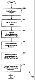
Figure 6 is a flowchart of a procedure 600 for adaptive registration of
medical
images according to an embodiment of the invention. In one embodiment, the
adaptive
registration procedure 600 includes an acquisition procedure 602 that involves
acquiring, generating, retrieving, receiving, and/or obtaining imaging data
- 10
CA 02530203 2005-12-19
WO 2004/114218 PCT/US2004/019715
corresponding to a set or series of medical images. In one embodiment, the
acquisition
procedure 602 involves or is directed toward precontrast and/or postcontrast
image
slices, which may correspond to breast images or other types of MR images.
The adaptive registration procedure 600 may further include a tiling procedure
604 involving determination of whether registration should consider a local
subset of
imaging data or global imaging data; and identification or specification of
one or more
local subset parameters if applicable. Relative to breast MRI, a tiling
procedure 604
may determine whether to perform registration in accordance with a window or
subset
of imaging information associated with a plurality of image slices. For
example, a tiling
procedure 604 may determine that registration corresponding to precontrast
and/or
postcontrast imaging data for a left breast is appropriate or required, and
ignore
imaging data for a right breast. Depending upon embodiment details, one or
more
portions of a tiling procedure 604 may involve manual input and/or an
automated
procedure.
The adaptive registration procedure 600 may additionally include a motion
estimation procedure 606, which involves estimating, approximating, or
determining
patient or tissue motion based upon the imaging data under consideration.
Motion
estimation may involve generating one or more motion vectors by determining
and/or
optimizing a set of spatial transform parameters defined in accordance with a
motion
model. The motion model may be capable of accounting for various types of 2D
or 3D
motion or deformation of rigid and/or nonrigid tissues.
In general, a number of motion estimation techniques suitable for medical
imaging and/or image processing may be applicable to various embodiments of
the
present invention. Descriptions of such motion estimation techniques may be
found in
references such as (a) "Comparison and Evaluation of Retrospective
Intermodality
Brain Image Registration Techniques," West et al., JCAT 1997; and (b) "A
Survey of
Medical Image Registration," Maintz and Viergever, Medical Image Analysis,
1998.
In one embodiment, the motion estimation procedure 606 may involve selection
or identification of a motion model and/or a motion estimation technique;
selection or
identification of a reference image; and/o'r determination of one or more
motion vectors
or parameters for a set of images relative to the reference image along an
axis or
direction of lowest resolution (typically the axis corresponding to slice
thickness in MRI
situations) as well as one or more other axes. Selection of a motion model
and/or a
motion estimation technique may involve manual input and/or one or more
automated
-11-
CA 02530203 2005-12-19
WO 2004/114218 PCT/US2004/019715
procedures, possibly based upon clinical conditions such as imaging technology
type
and configuration, tissue types under consideration, image resolution, and/or
other
factors. In general, motion estimation in accordance with a nonrigid motion
model is
more computationally intensive than motion estimation in accordance with a
rigid
motion model, and 3D motion estimation is more computationally intensive than
2D
motion estimation. Thus, selection or identification of a motion model and/or
a motion
estimation technique may additionally or alternatively be based upon available
computational resources or capabilities. An alternate embodiment may rely upon
a
single type of motion model and/or motion estimation technique.
In accordance with the motion estimation procedure 606, determination of one
or
more motion vectors and/or motion parameters may involve operations in a
spatial
(voxel) domain or a spectral (frequency) domain. In one embodiment, a motion
estimation procedure 606 may i nvolve a m inimization of g ray I evel o r
voxel p roperty
based similarity measures in accordance with an optimization procedure (for
example,
a simplex minimization, a direction set, a conjugate gradient, or a simulated
annealing
procedure). Gray level similarity measures may include a sum of squared or
absolute
differences; a cross-correlation measure; an intensity ratio variance; mutual
information;
andlor a deterministic or stochastic sign change. A sum of squared differences
technique may be computationally efficient at the possible expense of some
accuracy,
while a mutual information technique may be highly accurate at the possible
expense of
some computational speed. In an embodiment that performs motion estimation in
accordance with an affine transform, a least squares technique rather than an
optimization search may provide a direct solution.
During the motion estimation procedure 606, images or imaging data under
consideration may be scaled or reduced in size to generate an initial motion
estimate;
and then scaled or increased to an original size to generate a final motion
estimate,
which may provide increased robustness. For example, a motion estimation
procedure
604 directed toward images characterized by a 512x512 resolution may first-
estimate
motion at a 64x64 resolution, then estimate motion at a 128x128 resolution,
subsequently estimate motion at a 256x256 resolution, and finally estimate
motion at a
512x512 resolution. Images or imaging data under consideration may
additionally or
alternatively be subdivided into smaller blocks for local motion estimation,
which may
be useful for estimating nonrigid motion.
The adaptive registration procedure 600 may further include an evaluation
-12-
CA 02530203 2005-12-19
WO 2004/114218 PCT/US2004/019715
procedure 608 that involves comparing a result generated or obtained by the
motion
estimation procedure 606 to a correction threshold. In one embodiment, a
motion
estimation result may comprise a motion vector that may specify or indicate an
estimated m otion value along one or more a xes, including an axis
corresponding to
lowest image resolution. As indicated above, for MRI image data, the axis of
lowest
resolution is typically an axis corresponding to image slice thickness, and is
commonly
defined as z. In one embodiment, a correction threshold may comprise a
resolution
value corresponding to one or more axes, including a lowest resolution axis,
where
each such resolution value is multiplied by a corresponding fraction. Thus, a
correction
threshold may specify or correspond to a fractional resolution along one or
more axes.
The evaluation procedure 608 may determine whether one or more motion
estimation results are greater or less than corresponding correction
thresholds.
Alternatively or additionally, the evaluation procedure 608 may determine
whether one
or more motion estimation results fall within particular corresponding
correction ranges.
Evaluation or comparison of a motion estimation result relative to a
fractional resolution
value may be mathematically equivalent or analogous to determining an a value
of a
type described above. The value of a correction threshold may be influenced by
imaging technology; actual and/or expected background, precontrast, and/or
postcontrast signal intensities; and/or other factors. One or more correction
thresholds
may be stored in and/or retrieved from a memory such as a lookup table based
upon
applicability to particular clinical situations.
The adaptive registration procedure 600 may further include a selective
correction procedure 610 that involves selectively initiating or performing an
image
resampling or correction in accordance with a 2D or 3D rigid or nonrigid
correction
based upon a result obtained by or in conjunction with an evaluation procedure
608.
Depending upon implementation details, the selective correction procedure 610
may
additionally involve selectively avoiding image resampling or correction.
Exemplary
evaluation 608 and adaptive registration 600 procedures are further described
below.
Following the selective correction procedure 610, the adaptive registration
procedure
600 may a Iso i nclude a n adjustment p rocedure 612 t hat i nvolves p
erforming filtering
and/or other image processing operations. These adjustment procedures may
include,
by way of example, noise reduction, contrast enhancement, and window and level
procedures. Such adjustment procedures are well known in the art and need not
be
described herein.
-13-
CA 02530203 2005-12-19
WO 2004/114218 PCT/US2004/019715
Figure 7 is a flowchart of exemplary evaluation 608 and selective correction
610
procedures according to an embodiment of the invention. Relative to Figure 6,
like
reference numbers indicate like procedures, and the z axis corresponds to a
lowest
resolution dimension. In one embodiment, the evaluation procedure 608
determines
whether estimated motion along the z axis equals, approximately equals, or
exceeds a
correction threshold, for example, 0.5 times z axis resolution. Thus, if
resolution along
z equals 1.5 mm, the evaluation procedure 608 may determine whether estimated
motion along z exceeds 0.75 mm (corresponding to a situation in which a equals
0.5).
If so, the selective correction procedure 610 may initiate and/or perform a 3D
rigid or
nonrigid correction; otherwise, the selective correction procedure 610 may
initiate
and/or perform a 2D rigid or nonrigid correction. A 3D correction may provide
greater
accuracy than a 2D correction, but will generally require significantly more
computational time than a 2D correction. Thus, unless patient or tissue
movement is
significant relative to resolution, a 3D correction may be unnecessary, and
particular
embodiments of the invention may save time by performing a 2D correction.
Performance of a rigid or nonrigid correction may be dependent upon a type of
,motion
model and/or motion estimation technique employed during the motion estimation
procedure 606, clinical conditions, tissue types under consideration,
available
computational resources, and/or computation time goals or constraints.
Figure 8 is a flowchart of exemplary evaluation 608 and selective correction
610
procedures according to another embodiment of the invention. Relative to
Figure~.6, like
reference numbers indicate like procedures, and the z axis corresponds to a
lowest
resolution dimension. In one embodiment, the evaluation procedure 608 may
determine whether estimated motion along the z axis is less than a first z
axis
correction threshold, for example, 0.1 times z axis resolution. If so, image
resampling
or correction is avoided. Otherwise, the evaluation procedure 608 may
determine
whether estimated motion' along the z axis equals, approximately equals, or
exceeds a
second z axis correction threshold, for example, 0.5 times z axis resolution.
If so, the
selective correction procedure 610 may initiate and/or perform a 3D rigid or
nonrigid
correction. Otherwise, the evaluation procedure 608 may determine whether
estimated
motion along an x or y axis equals, approximately equals, or exceeds a
corresponding x
axis or y axis correction threshold. In one embodiment, an x or y axis
correction
threshold may be, for example, 0.8 times x or y resolution, respectively. If
estimated
motion along an x or y axis meets the aforementioned conditions, the selective
- 14-
CA 02530203 2005-12-19
WO 2004/114218 PCT/US2004/019715
correction procedure 610 may initiate and/or perform a 3D rigid or nonrigid
correction.
Otherwise, the selective correction procedure 610 may initiate or perform a 2D
rigid or
nonrigid correction. Performance of a rigid or nonrigid correction may be
dependent
upon a type of motion model and/or motion estimation technique employed,
and/or
computational resources.
Figure 9 is a block diagram of a system 900 for adaptive medical image
registration according to an embodiment of the invention. The system 900 may
comprise a medical imaging system 910, at least one data storage unit 920, and
an
adaptive registration computer 940. In one embodiment, each element 910, 920,
940 is
coupled to a computer network 990. The medical imaging system 910 may comprise
an MRI or other type of imaging system. The data storage unit 920 may comprise
one
or more hard disk drives, and may possibly comprise a Network Attached Storage
(NAS) device. The data storage unit 920 may receive, store, and/or transfer
imaging
data as well as other information.
The adaptive registration computer 940 may comprise one or more portions of a
medical image analysis platform. The adaptive registration computer 940 may
include
a processing unit and a memory, and may further include one or more of a disk
drive
and/or other data storage devices (e.g., optical and/or magnetooptical data
storage
devices, tape drives, flash memory based drives, etc.), an input device, and
an output
device. The memory, the disk drive, and/or other data storage devices may
comprise
one or more portions of computer readable media that store program
instructions and
possibly data for performing one or more adaptive medical image registration
procedures and/or operations associated therewith in accordance with
particular
embodiments o f t he i nvention. D epending a pon i mplementation details, t
he n etwork
990 may comprise one or more local or private networks such as a Local Area
Network
(LAN) and/or one or more public networks such as the Internet. In an alternate
embodiment, the medical imaging system 910 and the adaptive registration
computer
940 may each have a separate data storage unit 920, and imaging data and/or
other
information stored upon removable media may be manually transferred between
such
data storage units 920.
From the foregoing, it will be appreciated that specific embodiments of the
invention have been described herein for purposes of illustration, but that
various
modifications may be made without deviating from the spirit and scope of the
invention.
Accordingly, the invention is not limited except as by the appended claims.
-15-