Note: Descriptions are shown in the official language in which they were submitted.
CA 02532713 2006-01-12
VESSEL SEALER AND DIVIDER WITH
ROTATING SEALER AND CUTTER
BACKGROUND
The present disclosure relates to an electrosurgical instrument and
method for performing endoscopic surgical procedures. More particularly, the
present disclosure relates to an endoscopic bipolar electrosurgical forceps
and
method of using same which includes an end effector having a movable jaw and a
.
fixed jaw, the fixed jaw including a rotatable electrode having a sealing
surface and a
cutting edge. Further, a non-conductive stop member is associated with one or
both
of the opposing jaw members. The non-conductive stop member is designed to
control the gap distance between opposing jaw members and enhance the
manipulation and gripping of tissue during the sealing and dividing process.
Technical Field
Endoscopic forceps utilize mechanical action to constrict, grasp,
dissect and/or clamp tissue.
Endoscopic electrosurgical forceps utilize both
mechanical clamping action and electrical energy to effect hemostasis by
heating
the tissue and blood vessels to coagulate, cauterize and/or seal tissue.
1
CA 02532713 2006-01-12
Endoscopic instruments are inserted into the patient through a
cannula, or port, that has been made with a trocar or similar such device.
Typical
sizes for cannulas range from three millimeters to twelve millimeters. Smaller
cannulas are usually preferred, and this presents a design challenge to
instrument
manufacturers who must find ways to make surgical instruments that fit through
the
cannulas.
Certain endoscopic surgical procedures require cutting blood vessels
or vascular tissue. However, due to space limitations surgeons can have
difficulty
suturing vessels or performing other traditional methods of controlling
bleeding, e.g.,
clamping and/or tying-off transected blood vessels. Blood vessels, in the
range
below two millimeters in diameter, can often be closed using standard
electrosurgical techniques. However, if a larger vessel is severed, it may be
necessary for the surgeon to convert the endoscopic procedure into an open-
surgical procedure and thereby abandon the benefits of laparoscopy.
Several journal articles have disclosed methods for sealing small blood
vessels using electrosurgery. An article entitled Studies on Coaoulation and
the
Development of an Automatic Computerized Bipolar Coagulator, J. Neurosurg.,
Volume 75, July 1991, describes a bipolar coagulator which is used to seal
small
blood vessels. The article states that it is not possible to safely coagulate
arteries
with a diameter larger than 2 to 25 mat A second article is entitled
Automatically
Controlled Bipolar Electrocoagulation ¨ "COA-COMP", Neurosurg. Rev. (1984),
pp.187-190, describes a method for terminating electrosurgical power to the
vessel
so that charring of the vessel walls can be avoided.
2
CA 02532713 2006-01-12
As mentioned above, by utilizing an electrosurgical forceps, a surgeon
can either cauterize, coagulate/desiccate and/or simply reduce or slow
bleeding, by
controlling the intensity, frequency and duration of the electrosurgical
energy applied
through jaw members to the tissue. The electrode of each jaw member is charged
to a different electric potential such that when the jaw members grasp tissue,
electrical energy can be selectively transferred through the tissue.
In order to effect a proper seal with larger vessels, two predominant
mechanical parameters must be accurately controlled - the pressure applied to
the
vessel and the gap distance between the electrodes - both of which are
affected by
the thickness of the sealed vessel. More
particularly, accurate application of
pressure is important to oppose the walls of the vessel; to reduce the tissue
impedance to a low enough value that allows enough electrosurgical energy
through
the tissue; to overcome the forces of expansion during tissue heating; and to
contribute to the end tissue thickness which is an indication of a good seal.
It has
been determined that a typical fused vessel wall is optimum between 0.001 and
0.006 inches. Below this range, the seal may shred or tear and above this
range the
lumens may not be properly or effectively sealed.
Electrosurgical methods may be able to seal larger vessels using an
appropriate electrosurgical power curve, coupled with an instrument capable of
applying a large closure force to the vessel walls. It is thought that the
process of
coagulating small vessels is fundamentally different than electrosurgical
vessel
sealing. For the purposes herein, "coagulation" is defined as a process of
desiccating tissue wherein the tissue cells are ruptured and dried. Vessel
searing is
defined as the process of liquefying the collagen in the tissue so that it
reforms into a
g
3
CA 02532713 2006-01-12
fused mass. Thus, coagulation of small vessels is sufficient to permanently
close
them. Larger vessels need to be sealed to assure permanent closure.
U.S. Patent No. 2,176,479 to Willis, U.S. Patent Nos. 4,005,714 and
4,031,898 to Hiltebrandt, U.S. Patent Nos. 5,827,274, 5,290,287 and 5,312,433
to
Boebel et as., U.S. Patent Nos. 4,370,980, 4,552,143, 5,026,370 and 5,116,332
to
Lottick, U.S. Patent No. 5,443,463 to Stern et al., U.S. Patent No. 5,484,436
to
Eggers et al. and U.S. Patent No. 5,951,549 to Richardson et al., all relate
to
electrosurgical instruments for coagulating, cutting and/or sealing vessels or
tissue.
However, some of these designs may not provide uniformly reproducible pressure
to
the blood vessel and may result in an ineffective or non-uniform seal.
For the most part, these instruments rely on clamping pressure alone
to procure proper sealing thickness and are not designed to take into account
gap
tolerances and/or parallelism and flatness requirements which are parameters
which, if properly controlled, can assure a consistent and effective tissue
seal. For
example, it is known that it is difficult to adequately control thickness of
the resulting
sealed tissue by controlling clamping pressure alone for either of two
reasons: 1) if
too much force is applied, there is a possibility that the two poles will
touch and
energy will not be transferred through the tissue resulting in an ineffective
seal; or 2)
if too low a force is applied, the tissue may pre-maturely move prior to
activation and
sealing and/or a thicker, less reliable seal may be created.
Typically and particularly with respect to endoscopic electrosurgical
procedures, once a vessel is sealed, the surgeon has to remove the sealing
instrument from the operative site, substitute a new instrument through the
cannula
and accurately sever the vessel along the newly formed tissue seal. As can be
4
CA 02532713 2006-01-12
appreciated, this additional step may be both time consuming (particularly
when
sealing a significant number of vessels) and may contribute to imprecise
separation
of the tissue along the sealing line due to the misalignment or misplacement
of the
severing instrument along the center of the tissue sealing line.
Several attempts have been made to design an instrument which
incorporates a knife or blade member which effectively severs the tissue after
forming a tissue seal. For example, U.S. Patent No. 5,674,220 to Fox et al.
discloses a transparent vessel sealing instrument which includes a
longitudinally
reciprocating knife which severs the tissue once sealed. The instrument
includes a
plurality of openings which enable direct visualization of the tissue during
the sealing
and severing process. This direct visualization allows a user to visually and
manually regulate the closure force and gap distance between jaw members to
reduce and/or limit certain undesirable effects known to occur when sealing
vessels,
thermal spread, charring, etc. As can be appreciated, the overall success
of
creating a tissue seal with this instrument is greatly reliant upon the user's
expertise,
vision, dexterity, and experience in judging the appropriate closure force,
gap
distance and length of reciprocation of the knife to uniformly, consistently
and
effectively seal the vessel and separate the tissue at the seal.
U.S. Patent Nos. 5,702,390 and 5,944,718 to Austin et al. disclose a
vessel sealing instrument which includes a pivoting, triangularly-shaped
electrode
which is rotatable from a first position to coagulate tissue to a second
position to cut
tissue. As described above, the user must rely on direct visualization and
expertise
5
CA 02532713 2006-01-12
to control the various effects of sealing and culling tissue. Additionally,
since there
is no means to control the gap distance, there is a risk of the electrodes of
the
instrument to come into contact with each other, regardless of the position of
the
triangularly-shaped electrode, and cause a short between electrodes resulting
in
damage to the instrument and/or connected energy source, e.g. electrosurgical
generator. Further, to change operation of the instrument from coagulating to
cutting, the instrument must be removed from the operative site and the
electrode
rotated by loosing a set screw which further adds time and complexity to the
procedure.
Thus, a need exists to develop an endoscopic electrosurgical
instrument which effectively and consistently seals and separates vascular
tissue
and solves the aforementioned problems. This
instrument regulates the gap
distances between opposing jaws members, reduces the chances of short
circuiting
the opposing jaws during activation and assists in manipulating, gripping and
holding
the tissue prior to and during activation and separation of the tissue.
SUMMARY
According to an aspect of the present disclosure, an electrosurgical
instrument for sealing and dividing tissue includes a housing having a shaft
attached
thereto which defines a longitudinal axis. " First and second opposing jaw
members
are coupled to the shaft; the first jaw member having a conductive surface and
being
movable relative to the second jaw member and the second jaw member being
fixed
relative to the shaft and having a conductive electrode rotatable along the
6
CA 02532713 2006-01-12
longitudinal axis. The rotatable electrode includes a sealing surface on one
side
thereof and a cutting edge on a second side thereof. A source of
electrosurgical
energy is connected to each jaw member such that the jaw members are capable
of
conducting energy through tissue held therebetween. The electrosurgical
instrument
also includes at least one non-conductive stop member operatively associated
with
at least one of the first and second jaw members which controls the distance,
e.g., a
gap distance, between the jaw members when tissue is held therebetween. In
another aspect, the gap distance between the jaw members is fixed. The gap
distance is typically in the range of about 0.001 inches to about 0.006
inches.
The electrosurgical instrument further includes a rotating assembly for
rotating the electrode of the second jaw member and/or for rotating the second
jaw
member. The rotating assembly includes a dial disposed within the housing for
setting a desired position of the electrode and an elongated tube disposed
within the
shaft coupling the dial to the electrode. The dial selectively orients the
electrode of
the second jaw member from a first operable position wherein the sealing
surface of
the electrode is generally parallel to the conductive surface of the first jaw
member
for sealing tissue to a second operable position wherein the cutting edge of
the
electrode is generally perpendicular to the conductive surface of the first
jaw
member for dividing tissue.
According to another aspect of the present disclosure, the forceps
include a housing having a shaft attached thereto, the shaft defining a
longitudinal
axis. First and second opposing jaw members are coupled to the shaft; the
first jaw
7
CA 02532713 2006-01-12
=
member includes a conductive surface and is movable relative to the second jaw
member and the second jaw member is fixed relative to the shaft and includes
an
electrode rotatable along the longitudinal axis. The rotatable electrode
includes a
sealing surface and a cutting edge. At least one non-conductive stop member is
disposed on at least one of the first and second jaw members which controls
the
distance between the jaw members when tissue is held therebetween. A rotating
assembly is included which rotates the electrode of the second jaw member from
a
first operable position wherein the sealing surface of the electrode is
generally
parallel to the conductive surface of the first jaw member for sealing tissue
to a
second operable position wherein the cutting edge of the electrode is
generally
perpendicular to the conductive surface of the first jaw member for dividing
tissue.
According to a further aspect of the present disclosure, a method for
searing and dividing tissue is provided. The method includes the steps of:
providing an electrosurgical instrument comprising a housing having:
a shaft attached thereto which defines a longitudinal axis;
first and second opposing jaw members coupled to the shaft,
the first jaw member having a conductive surface and being movable relative to
the
second jaw member and the second jaw member being fixed relative to the shaft
and having an electrode rotatable along the longitudinal axis, the rotatable
electrode
having a sealing surface and a cutting edge; and
at least one non-conductive stop member disposed on at least
=
one of the first and second jaw members which controls the distance between
the
jaw members when tissue is held therebetween;
8
CA 02532713 2006-01-12
positioning the sealing surface of the rotatable electrode to be
generally parallel to the conductive surface of the first jaw member;
approximating tissue by closing the first and second jaw members;
applying electrosurgical energy to the first and second jaw members to
seal the tissue;
opening the first and second jaw members and repositioning the
electrode so the cutting edge is generally perpendicular to the conductive
surface of
the first jaw member; and
closing the first and second jaw members on the tissue seal and
applying electrosurgical energy to divide the tissue at the seal.
BRIEF DESCRIPTION OF THE DRAWINGS
Various embodiments of the subject instrument are described herein
with reference to the drawings wherein:
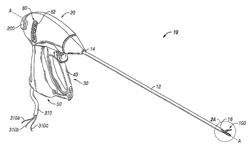
Fig. 1 is a perspective view of an endoscopic forceps showing a handle
and an end effector according to the present disclosure;
Fig. 2A is an enlarged, left perspective view of the end effector
assembly with jaw members shown in an open configuration for sealing vessels;
Fig. 2B is an enlarged, left perspective view of the end effector'
assembly with the jaw members shown in an open configuration for cutting
vessels;
Fig. 3A is an end view of the end effector assembly of FIG. 2A showing
the conducting surfaces in a configuration for sealing vessels;
9
CA 02532713 2006-01-12
Fig. 3B is an end view of the end effector assembly of FIG. 2B showing
the conducting surfaces in a configuration for cutting vessels;
Fig. 4 is an enlarged, side view of the end effector assembly;
Fig. 5 is an enlarged perspective view of the rotating assembly;
Fig. 6A is an enlarged perspective view of a sealing site of a tubular
vessel;
Fig. 6B is a longitudinal cross-section of the sealing site taken along
line 6B-6B of Fig. 6A; and
Fig. 6C is a longitudinal cross-section of the searing site of Fig. 6A after
separation of the tubular vessel.
DETAILED DESCRIPTION
Turning now to the several Figures, one embodiment of an endoscopic
bipolar forceps 10 is shown for use with various surgical procedures and
generally
includes a housing 20, a handle assembly 30, a rotating assembly 80 and an end
effector assembly 100 which mutually cooperate to grasp, seal and divide
tubular
vessels and vascular tissue 150 (Fig. 6A). Although the majority of the figure
drawings depict a bipolar forceps 10 for use in connection with endoscopic
surgical
procedures, the present disclosure may be used for more traditional open
surgical
procedures. For the purposes herein, the forceps 10 is described in terms of
an
CA 02532713 2013-06-12
endoscopic instrument, however, it is contemplated that an open version of the
forceps may also include the same or similar operating components and features
as
described below.
Forceps 10 includes a shaft 12 which has a distal end 16 dimensioned
to mechanically engage the end effector assembly 100 and a proximal end 14
which
mechanically engages the housing 20. .In the drawings and in the descriptions
which
follow, the term "proximal", as is traditional, will refer to the end of the
forceps 10
which is closer to the user, while the term "distal" will refer to the end
which is further
from the user. Further, the shaft 12 defines a longitudinal axis 'A-A" through
the
forceps 10.
As best seen in Fig. 1, forceps 10 also includes an electrosurgical
cable 310 which connects the forceps 10 to a source of electrosurgical energy,
e.g.,
a generator (not shown). Generators such as those sold by Valleylab - a
division of
Tyco Healthcare LP, located in Boulder, Colorado are used as a source of
electrosurgical energy, e.g., FORCE EZT14 Electrosurgical Generator, FORCE FX-
rm
Electrosurgical Generator, FORCE ICTMI FORCE 2114 Generator, SurgiStatTm 11.
One such system is described in commonly-owned U.S, Patent No. 6,033,399
entitled "ELECTROSURGICAL GENERATOR WITH ADAPTIVE POWER
CONTROL". Other systems have been described in commonly-owned U.S. Patent
No. 6,187,003 entitled "BIPOLAR ELECTROSURGICAL INSTRUMENT FOR
SEALING VESSELS".
11
CA 02532713 2006-01-12
The generator includes various safety and performance features
including isolated output, independent activation of accessories. The
electrosurgical
generator includes Valleylab's Instant ResponseTm technology features which
provides an advanced feedback system to sense changes in tissue 200 times per
second and adjust voltage and current to maintain appropriate power. The
Instant
Responsirm technology is believed to provide one or more of the following
benefits
to surgical procedure:
= Consistent clinical effect through all tissue types;
= Reduced thermal spread and risk of collateral tissue damage;
= Less need to "turn up the generator"; and
= Designed for the minimally invasive environment.
Cable 310 is internally divided into a plurality of cable leads 310a,
310b, 310c which each transmit electrosurgicai energy through their respective
feed
paths through the forceps 10 to the end effector assembly 100 as explained in
more
detail below.
Handle assembly 30 includes a fixed handle 50 and a movable handle
40. Fixed handle 50 is integrally associated with housing 20 and handle 40 is
movable relative to fixed handle 50 as explained in more detail below with
respect to
the operation of the forceps 10. Rotating assembly 80 may be integrally
associated
with the housing 20 and is rotatable approximately 180 degrees in either
direction
12
CA 02532713 2013-06-12
about the longitudinal axis "A-A". Details of the rotating assembly 80 are
described in
more detail with respect to Figs. 2A, 2B, 3A, 3B and 5.
As mentioned above, end effector assembly 100 is attached at the
distal end 16 of shaft 12 and includes a pair of opposing jaw members 110 and
120
as shown in Figs. 2A and 2B. Movable handle 40 of handle assembly 30 is
ultimately
connected to a drive assembly (not shown) which, together, mechanically
cooperate
to impart movement of the jaw members 110 and 120 from an open position
wherein
the jaw members 110 and 120 are disposed in spaced relation relative to one
another, to a clamping or closed position wherein the jaw members 110 and 120
cooperate to grasp tissue 150 (Fig. 6B) therebetween or to cut tissue (Fig.
6C). The
specific functions and operative relationships of these elements and the
various
internal-working components of forceps 10 are described in more detail in
commonly
assigned, co-pending application U.S. Patent Publication No. US2004/0254573
entitled "VESSEL SEALER AND DIVIDER FOR USE WITH SMALL TROCARS AND
CANNULAS" by Dycus et al.
It is envisioned that the forceps 10 may be designed such that it is fully
or partially disposable depending upon a particular purpose or to achieve a
particular
result. For example, end effector assembly 100 may be selectively and
releasably
engageable with the distal end 16 of the shaft 12 and/or the proximal end 14
of shaft
12 may be selectively and releasably engageable with the housing 20 and the
handle assembly 30. In either of these two instances, the forceps 10 would be
considered "partially disposable or "reposable", i.e., a new or different end
effector
13
CA 02532713 2013-06-12
assembly 100 (or end effector assembly 100 and shaft 12) selectively replaces
the
old end effector assembly 100 as needed. As can be appreciated, the presently
disclosed electrical connections would have to be altered to modify the
instrument to
a reposable forceps.
As shown best in Figs. 2A and 2B, the end effector assembly 100
includes opposing jaw members 110 and 120 which cooperate to effectively grasp
tissue 150 for sealing purposes and to divide the tissue 150 once sealed. The
end
effector assembly 100 is designed as a unilateral assembly, Le., jaw member
120 is
fixed relative to the shaft 12 and jaw member 110 pivots about a pivot pin 103
to
grasp tissue 150.
More particularly, the unilateral end effector assembly 100 includes
one stationary or fixed jaw member 120 mounted in fixed relation to the shaft
12 and
pivoting jaw member 110 mounted about a pivot pin 103 attached to the
stationary
jaw member 120. A reciprocating sleeve 60 is slidingly disposed within the
shaft 12
and is remotely operable by the drive assembly (not shown). The above
mentioned
U.S. Patent Publication No. US2004/0254573 describes one example of a drive
assembly which may be utilized for this purpose. The pivoting jaw member 110
includes a detent or protrusion 117 which extends from jaw member 110 through
an
aperture 62 disposed within the reciprocating sleeve 60. The pivoting jaw
member
110 is actuated by sliding the sleeve 60 axially within the shaft 12 such that
a distal
end 63 of the aperture 62 abuts against the detent 117 on the pivoting jaw
member
110 (see Fig. 4). Pulling the sleeve 60 proximally closes the jaw members 110
and
14
CA 02532713 2006-01-12
120 about tissue 150 grasped therebetween and pushing the sleeve 60 distally
opens the jaw members 110 and 120 for grasping purposes.
As best shown in Fig. 2A, jaw member 110 also includes a jaw housing
116 which has an insulative substrate or insulator 114 and an electrically
conducive
surface 112.
Insulator 114 is preferably dimensioned to securely engage the
electrically conductive sealing surface 112. This may be accomplished by
stamping,
by overmolding, by overmolding a stamped electrically conductive sealing plate
and/or by overmolding a metal injection molded seal plate.
All of these manufacturing techniques produce jaw member 110 having
an electrically conductive surface 112 which is substantially surrounded by an
insulating substrate 114. The insulator 114, electrically conductive sealing
surface
112 and the outer, non-conductive jaw housing 116 may be dimensioned to limit
and/or reduce many of the known undesirable effects related to tissue sealing,
e.g.,
flashover, thermal spread and stray current dissipation.
Alternatively, it is also
envisioned that the jaw member 110 may be manufactured from a ceramic-like
material and the electrically conductive surface 112 is coated onto the
ceramic-like
jaw members 110.
= 20
It is envisioned that the electrically conductive sealing surface 112 may
also include an outer peripheral edge which has a pre-defined radius and the
insulator 114 meets the electrically conductive sealing surface 112 along an
adjoining edge of the sealing surface 112 in a generally tangential position.
CA 02532713 2013-06-12
Preferably, at the interface, the electrically conductive surface 112 is
raised relative to
the insulator 114. These and other envisioned embodiments are discussed in co-
pending, commonly assigned W002/080786 entitled "ELECTROSURGICAL
INSTRUMENT WHICH REDUCES COLLATERAL DAMAGE TO ADJACENT TISSUE"
by Johnson et al. and co-pending, commonly assigned W002/080785 entitled
"ELECTROSURGICAL INSTRUMENT WHICH IS DESIGNED TO REDUCE THE
INCIDENCE OF FLASHOVER" by Johnson et al.
Jaw member 120 includes similar elements to jaw member 110 such
as jaw housing 126 having an insulator 124. Unlike jaw member 110, jaw member
.120 includes a rotatable electrode 122. The rotatable electrode 122 has at
least two
operable positions. A first position is employed during vessel sealing and a
second
position is employed during vessel dividing or cutting. As best seen in Figs.
3A and
3B, the rotating electrode '122 includes three surfaces, namely, a first
surface 134, a
second surface 136 and a third surface 138.
Referring to Fig. 3A, when in a first operable position, the first surface
134 of electrode 122 is generally and substantially parallel to the conductive
sealing
surface 112 of first jaw member 110. In this position, first surface 134 and
conductive sealing surface 112 will facilitate grasping of tissue. Upon
activation of
electrosurgical energy and upon application of pressure within the predefined
range
of about 3 kg/cm2 to about 16 kg/cm2 and upon grasping the tissue within a
16
CA 02532713 2013-06-12
predefined gap range of about .001 inches to about .006 inches, arid
preferably from
about .002 inches to about .004 inches, the tissue dispersed between the jaw
members will seal into a single fused mass with limited demarcation between
tissue
layers. As explained in more detail below, a series of stop members are
operatively
associated with at least one of the jaw members to maintain a gap distance "G"
(FIG. 6A) between opposing tissue containing surfaces 112 and 134. As
explained in
the above-identified U.S. Patent Publication No. US2004/0254573, handle 40 and
fixed handle 50 include a camnning mechanism which, upon activation thereof,
maintains pressure between opposing sealing surfaces between about 3 kg/cm2 to
about 16 kg/cm2. U.S. Patent Publication No. US2005/0203504 and U.S. Patent
Publication No. US2004/0015163 include exemplitive details regarding the
various
electrical parameters which need to be closely monitored and controlled to
optimize
the vessel sealing process for various tissue thicknesses and tissue types.
Referring to Fig. 38, second surface 136 and third surface 138 of
electrode 122 meet to form culling edge 130. When the forceps is selectively
rotated
to the second operable position, the cutting edge 130 is generally
perpendicular to
sealing surface '112. When the jaw members 110, 120 are moved to a closed
position, cutting edge 130 comes into close proximity with sealing surface 112
to
electromechanically sever or cut sealed tissue as will be described below in
relation
to Fig. 6C.
17
CA 02532713 2013-06-12
As mentioned above, rotatable electrode 122 (and/or jaw member 110
of sealing surface 112) includes at least one and preferably a plurality of
stop
members 140 operatively associated with the first surface 134 of the electrode
122.
Stop members 140 are configured to define a gap 'G" (Fig. 6A) between opposing
sealing surfaces 112 and 134 of jaw members 110 and 120 during tissue sealing.
It
is envisioned that a series of stop members 140 may be employed on one or both
jaw members 110 and 120 (and/or sealing surfaces 112 and 134) depending upon a
particular purpose or to achieve a desired result. A detailed discussion of
these and
other envisioned stop members 140 as well as various manufacturing and
assembling processes for attaching and/or affixing the stop members 140 to the
jaw
members 110, 120 are described in commonly-assigned, co-pending W002/080796
entitled "VESSEL SEALER AND DIVIDER WITH NON-CONDUCTIVE STOP
MEMBERS" by Dycus et al.
Stop members 140 are affixed/attached to the jaw member(s) by
stamping, thermal spraying, overmolding and/or by an adhesive. The stop
members
project from about 0.001 inches to about 0.006 inches and, preferably, from
about
0.002 inches to about 0.004 inches from the inner-facing surface of at least
one of
the jaw members. It is envisioned that the stop members may be made'from an
insulative material such as parylene, nylon and/or ceramic. Other materials
are also
contemplated, e.g., syndiotactic polystryrenes such as QUESTRA manufactured
by
DOW Chemical, Syndiotactio-polystryrene (S PS), Polybutylene Terephthalate
(PBT),
Polycarbonate (PC), Acrylonitrile Butadiene Styrene (ABS), Polyphthalamide
(PPA),
Polymide, Polyethylene Terephthalate (PET), Polyamide-imicle (PAI), Acrylic
18
CA 02532713 2006-01-12
(PMMA), Polystyrene (PS and HIPS), Polyether Sulfone (PES), Aliphatic
Polyketone,
Acetal (DOM) Copolymer, Polyurethane (PU and TPU), Nylon with Polyphenylene-
oxide dispersion and Acrylonitrile Styrene Acrylate.
As explained in detail below and as best seen in FIG. 5, rotatable
electrode 122 is designed to be fixed to the end of a rotating tube 162 which
is part
of the rotating assembly 80 such that rotation of the tube 162 via dial 82
will impart
rotation to the electrode 122. In contrast to U.S. Patent Application Serial
No.
10/460,926, the rotating assembly is designed to rotate electrode 122 and not
the
end effector assembly 100. More particularly, rotating tube 162 includes an
elongated guide slot 160 disposed in an upper portion thereof which is
dimensioned
to carry lead 310a therealong. Lead 310a carries a first electrical potential
to
movable jaw 110. As explained in more detail below with respect to the
internal
electrical connections of the forceps, a second electrical connection from
lead 310c
is conducted through the tube 160 to the electrode 134 of fixed jaw member
120.
The electrical leads 310a, 310b, 310c and 311 are fed through the
housing 20 by electrosurgical cable 310. More particularly, the
electrosurgical cable
310 is fed into the bottom of the housing 20 through fixed handle 50. Lead
310c
extends directly from cable 310 into the rotating assembly 80 and connects to
electrode 122 to conduct the second electrical potential to fixed jaw member
120.
Leads 310a and 310b extend from cable 310 and connect to the hand switch or
joy-
stick-Eke toggle switch 200. The specific functions and operative
relationships of
these elements and the various internal-working components of forceps 10 are
19
CA 02532713 2013-06-12
described in more detail in commonly assigned, co-pending U.S. Publication No.
US2004/0254573 entitled "VESSEL SEALER AND DIVIDER FOR USE WITH SMALL
TROCARS AND CANNULAS" by Dycus et al.
When the switch 200 is depressed, electrosurgical energy is
transferred through leads 310a and 310c to jaw members 110 and 120,
respectively.
It is envisioned that a safety switch or circuit (not shown) may be employed
such that
the switch cannot fire unless the jaw members 110 and 120 are closed and/or
unless the jaw members 110 and 120 have tissue 150 held therebetween. in the
latter instance, a sensor (not shown) may be employed to determine if tissue
150 is
held therebetween. In addition, other sensor mechanisms may be employed which
determine pre-surgical, concurrent surgical (i.e., during surgery) and/or post
surgical
conditions. Still other sensor mechanisms, e.g., a toggle switch or the like,
may be
positioned on the tube 162 to determine the relative position of electrode
122, i.e.,
seal activation or cut activation.
The sensor mechanisms may also be utilized with a closed-loop
feedback system coupled to the electrosurgical generator to regulate the
electrosurgical energy based upon one or more pre-surgical, concurrent
surgical or
post surgical conditions. Various sensor mechanisms and feedback systems are
described in commonly-owned, co-pending U.S. Patent Publication No.
US2004/0015163 entitled "METHOD AND SYSTEM FOR CONTROLLING OUTPUT OF
CA 02532713 2013-06-12
RF MEDICAL GENERATOR" filed on May 1, 2003.
It is envisioned that cable leads 310a and 310c are fed through
respective halves 82a and 82b of the rotating assembly 80 in such a manner to
allow
rotation of the shaft 162 (via rotation of the rotating assembly 80) in the
clockwise or
counter-clockwise direction without unduly tangling or twisting the cable
leads 310a
and 310c, More particularly, each cable lead 310a and 310c is fed through a
series
of conjoining slots, e.g., 84, located in the two halves 82a and 82b of the
rotating
assembly 80. Each conjoining pair of slots are large enough to permit rotation
of the
rotating assembly 80 without unduly straining or tangling the cable leads 310a
and
310c. The presently disclosed cable lead feed path is envisioned to allow
rotation of
the rotation assembly approximately 180 degrees in either direction, which, in
turn,
rotates electrode 122 from a first position for sealing tissue to a second
position for
cutting tissue.
Figs. 6A through 6C illustrate the sealing and cutting of tissue
employing the forceps 10 according to the present disclosure. Before
approximating
tissue, a user will select an operable position of rotatable electrode 122 via
rotating
assembly 80. Here, the electrode 122 is placed in the first operable position
to
perform vessel sealing where the first surface 134 is generally parallel to
sealing
surface 112 (see FIG. 3A). As the handle 40 is squeezed, the reciprocating
sleeve
60 is pulled proximally which, in turn, causes aperture 62 of sleeve 60 to
proximally
cam detent 117 and close the jaw member 110 relative to jaw member 120. The
21
CA 02532713 2013-06-12
reciprocating sleeve's 60 load is converted to a torque about the jaw pivot
103. As a
result, a specific closure force can be transmitted to the opposing jaw
members 110
and 120 between about 3 kg/cm2 to about 16 kg/cm2.
As can be appreciated and as discussed in U.S. Patent Publication No.
US2004/0254573 the unique combination of the mechanical advantage of the
over-the-center pivot along with the compressive force associated with the
drive
assembly facilitate and assure consistent, uniform and accurate closure
pressure
about the tissue 150 within the desired working pressure range of about 3
kgfcm2 to
about 16 kgfcm2 and, preferably, about 7 kg/cm2 to about 13 kg/cm2. By
controlling
the intensity, frequency and duration of the electrosurgical energy applied to
the
tissue 150, the user can seal the tissue. As mentioned above, two mechanical
factors play an important role in determining the resulting thickness of the
sealed
tissue and effectiveness of the seal 150, i.e., the pressure applied between
opposing
jaw members 110 and 120 and the gap distance '1G" between the opposing sealing
surfaces 112, 134 of the jaw members 110 and 120 during the sealing process.
However, thickness of the resulting tissue seal 152 cannot be adequately
controlled
by force alone. In other words, too much force and the two jaw members 110 and
120 would touch and possibly short resulting in little energy traveling
through the
tissue 150 thus resulting in a bad tissue seal 152. Too little force and the
seal 152
would be too thick.
Applying the correct force is also important for other reasons: to
oppose the walls of the vessel; to reduce the tissue impedance to a low enough
22
CA 02532713 2006-01-12
value that allows enough current through the tissue 150; and to overcome the
forces
of expansion during tissue heating in addition to contributing towards
creating the
required end tissue thickness which is an indication of a good seal 150.
As mentioned above, at least one jaw member, e.g., 120, may include
a stop member 140 operatively associated therewith which limits the movement
of
the two opposing jaw members 110 and 120 relative to one another. For example,
the stop member 140 may extend from the sealing surface 134 a predetermined
distance according to the specific material properties (e.g., compressive
strength,
thermal expansion, etc.) to yield a consistent and accurate gap distance "G"
during
sealing (Fig. 6A). The gap distance between opposing sealing surfaces 112 and
134 during sealing ranges from about 0.001 inches to about 0.006 inches and,
more
preferably, between about 0.002 and about 0.004 inches.
Alternatively, the non-conductive stop members 140 can be molded
onto the jaw members 110 and 120 (e.g., overmolding, injection molding, et.),
stamped onto the jaw members 110 and 120 or deposited (e.g., deposition) onto
the
jaw members 110 and 120. For example, one technique involves thermally
spraying
a ceramic or porcelain material onto the surface of the jaw member 110 and 120
to
form the stop members 140. Several thermal spraying techniques are
contemplated
which involve depositing a broad range of heat resistant and insulative
materials on
various surfaces to create stop members 140 for controlling the gap distance
between electrically conductive surfaces 112 and 134.
23
CA 02532713 2006-01-12
As energy is being selectively transferred to the end effector assembly
100, across the jaw members 110 and 120 and through the tissue 150, a tissue
seal
152 forms isolating two tissue halves 150a and 150b. At this point and with
other
known vessel sealing instruments, the user must remove and replace the forceps
10
with a cutting instrument (not shown) to divide the tissue halves 150a and
150b
along an approximate center line B-B of the tissue seal 152. As can be
appreciated,
this is both time consuming and tedious and may result in inaccurate tissue
division
across the tissue seal 152 due to misalignment or misplacement of the cutting
instrument along the ideal tissue cutting plane, e.g., center line B-B.
Once the tissue seal 152 forms, the jaw members 110 and 120 may be
opened by re-grasping the handle 40. Once the jaw members are opened, the
rotatable electrode 122 is moved into its second operable position via
rotating
assembly 80, where cutting edge 130 is generally perpendicular to sealing
surface
112. Once the electrode 122 is set, the handle 40 is re-grasped closing jaw
members 110 and 120 bringing cutting edge 130 into close proximity of sealing
surface 112 to divide tissue 150 along at point 154. The tissue may be cut
utilizing
mechanical cutting action, electro-mechanical cutting action or simply
electrical
cutting action depending upon a particular purpose and depending upon the
particular configuration of cutting edge 130.
it can be appreciated since forceps 10 can seal and divide tissue
without removing the forceps 10 from the operative site the intended procedure
can
be performed more quickly. Additionally, the seal 152 will be divided
uniformly since
24 =
CA 02532713 2013-06-12
the user will not have to locate the center of the seal after inserting a
different
instrument, e.g., a cutting instrument.
From the foregoing and with reference to the various figure drawings,
those skilled in the art will appreciate that certain modifications can also
be made to
the present disclosure without departing from the scope of the same. For
example,
the forceps 10 (and/or the electrosurgical generator used in connection with
the
forceps 10) may include a sensor or feedback mechanism (not shown) which
automatically selects the appropriate amount of electrosurgical energy to
effectively
seal the particularly-sized tissue grasped between the jaw members 110 and 120
and subsequently select the appropriate energy to selectively cut the tissue.
The
sensor or feedback mechanism may also measure the impedance across the tissue
during sealing and provide an indicator (visual and/or audible) that an
effective seal
has been created between the jaw members 110 and 120. Examples of such sensor
systems are described in commonly-owned U.S. Patent Publication No.
US2004/0015163 entitled "METHOD AND SYSTEM FOR CONTROLLING OUTPUT
OF RF MEDICAL GENERATOR" filed on May 1, 2003.
It is envisioned that the outer surface of the end effector assembly 100
may include a nickel-based material, coating, stamping, metal injection
molding
which is designed to reduce adhesion between the jaw members 110 and 120 with
the surrounding tissue during activation and sealing. Moreover, it is also
contemplated that the conductive surfaces 112 and 134 of the jaw members 110
CA 02532713 2006-01-12
and 120 may be manufactured from one (or a combination of one or more) of the
following materials: nickel-chrome, chromium nitride, MedCoat 2000
manufactured
by The Electrolizing Corporation of OHIO, inconel 600 and tin-nickel. The
tissue
conductive surfaces 112 and 134 may also be coated with one or more of the
above
materials to achieve the same result, i.e., a anon-stick surface". As can be
appreciated, reducing the amount that the tissue 'sticks" during sealing/and
cutting
improves the overall efficacy of the instrument.
One particular class of materials disclosed herein has demonstrated
superior non-stick properties and, in some instances, superior seal quality.
For
example, nitride coatings which include, but not are not limited to: TiN, ZrN,
TiAlN,
and CrN are preferred materials used for non-stick purposes. CrN has been
found
to be particularly useful for non-stick purposes due to its overall surface
properties
and optimal performance. Other classes of materials have also been found to
reducing overall sticking. For example, high nickel/chrome alloys with a Ni/Cr
ratio
of approximately 5:1 have been found to significantly reduce sticking in
bipolar
instrumentation. One particularly useful non-stick material in this class is
Inconel
600. Bipolar instrumentation having sealing surfaces 112 and 134 made from or
coated with N1200, N1201 (-100% Ni) also showed improved non-stick performance
over typical bipolar stainless steel electrodes.
As can be appreciated, locating the switch 200 on the forceps 10 has
many advantages. For example, the switch 200 reduces the amount of electrical
cable in the operating room and eliminates the possibility of activating the
wrong
26
CA 02532713 2006-01-12
instrument during a surgical procedure due to "line-of-sight" activation. It
is also
envisioned that the switch 200 may be disposed on another part of the forceps
10,
e.g., the fixed handle 50, rotating assembly 80, housing 20, etc.
It is also envisioned that the forceps may be dimensioned to include a
fixed gap within the range of about 0.001 inches to about 0.006 inches by
providing
a stop member on another part of the end effector assembly, e.g., proximal
andfor
distal to the conductive surfaces, on the insulative housing 116 and/or 126,
and/or
as part of the pivot 103. In addition, it is envisioned that the detent 117
and aperture
62 arrangement may be dimensioned to limit the distance between conductive
surfaces 112 and 122.
While several embodiments of the disclosure have been shown in the
drawings, it is not intended that the disclosure be limited thereto, as it is
intended
that the disclosure be as broad in scope as the art will allow and that the
specification be read likewise. Therefore, the above description should not be
construed as limiting, but merely as exemplifications of preferred
embodiments.
Those skilled in the art will envision other modifications within the scope
and spirit of
the claims appended hereto.
27