Note: Descriptions are shown in the official language in which they were submitted.
CA 02532790 2014-02-14
SYSTEM AND METHOD FOR MULTI-ANALYTE DETECTION
2. BACKGROUND OF THE INVENTION
2.1 Field of the Invention
100021 Generally, the present invention relates to a system and method for
multiple
analyte detection. More particularly, multiple analytes are contained within a
single sample
and analyzed simultaneously using high-speed digital signal processing.
2.2 Description of Related Art
[0003] Disease analysis, research, and drug development depend heavily on
laboratory assay analysis. An example of an assay that has become commonplace
in
today's laboratory is the immunoassay. Many other types of laboratory analyses
are also
conducted in today's laboratories, such as analysis on blood, urine, serum,
blood plasma,
and other body fluids for proteins, viruses, bacteria, and other conditions.
[0004] Traditionally, laboratory assay analyses were very time consuming
processes, requiring lab personnel to perform precise measurements of reagents
and
samples, mixtures, centrifuging, etc. Each step of these analyses typically
needs to be
repeated multiple times to acquire statistically significant data.
Furthermore, the processes
are often wasteful of costly solvents, solutions, reagents, require numerous
man-hours, and
are generally slow.
10005] Accordingly, automated devices were envisioned and developed to
quicken
the process, generate more accurate results and make the process more
economical and
efficient. However, a drawback of these devices is that a limited number of
analyses can be
run on any given sample at any time. Therefore, much time is still required to
analyze a
sample for multiple analytes.
[0006] Accordingly, a system and method for testing multiple analytes, with
little or
no human intervention, from a single test sample would be highly desirable.
- t -
CA 02532790 2016-11-23
3. BRIEF SUMMARY OF THE INVENTION
100071 According to one embodiment, a system and method is provided fOr the
simultaneous detection of multiple analytes. The system comprises a housing
and a reagent
carousel rotatably coupled to the housing. Also rotatably coupled with the
housing is an
incubator carousel. Magnetic material is associated W ith the incubation
carousel fin-
assisting in washing samples held by the incubator carousel. Furthermore, at
least one robot
is coupled to the housing. The robot is configured to manipulate the reagent
carousel and/or
the incubator carousel. Reaction vessel handlers are responsible for moving
reaction vessels
between locations, such as a pre-testing location, testing location, and post-
testing location.
There is also at least one laser based detector for analyzing samples
following mixing the
sample with reagents.
100081 In a preferred embodiment, there is a flow cytometer for conducting
the
analysis of the samples. It is also preferred that washers are included for
washing the robots
and robot probes.
100091 According to another embodiment the incubator carousel further
includes a
plurality of rings. Each ring is rotatable with respect to the other rings
such that high
throughput is achieved from the system.
100101 According to yet another embodiment, the reagent carousel is
configured to
contain reagent kits. The reagent kits are accessible by at least one robot.
The kits and
reagent carousel are designed to be utilized during use of the system and
without
interruption or overall system processes.
100 l Oa] In a further embodiment of the present invention there is
provided a multiple
analyte detection system, comprising: a chassis; a reagent storage assembly
having a rotatable
reagent carousel coupled to said chassis; a rotatable incubator carousel
assembly coupled to
said chassis and configured to receive a plurality of reaction vessels,
wherein said reagent
storage assembly comprises a reagent cooler, wherein said reagent carousel is
rotatably
coupled within said reagent cooler, and wherein said reagent carousel is
configured to hold a
reagent pack within said reagent carousel and said reagent cooler is
configured to maintain the
reagent pack at a predetermined temperature; at least one automated reaction
vessel handler
coupled to said chassis and configured to deliver a reaction vessel to said
rotatable incubator
carousel assembly; and a detector module attached to said chassis, wherein
said detector
module comprises a flow cytometer.
1001%] In another embodiment of the present invention there is provided an
automated
method for multiple analvte detection using a multiple analyte detection
system having a
chassis, comprising: loading a reaction vessel into a rotatable incubator
carousel assembly;
- 2 -
CA 02532790 2016-11-23
adding a sample to said reaction vessel; transferring a reagent from a
rotatable reagent carousel
into the reaction vessel, wherein said rotatable reagent carousel is rotatably
coupled within a
= reagent cooler, and wherein said rotatable reagent carousel is configured
to hold a reagent pack
within said rotatable reagent carousel and said reagent cooler is configured
to maintain the
reagent pack at a predetermined temperature; incubating the sample at a
predetermined
temperature range; transferring the sample to a detector module Ibr analysis;
analyzing the
Sat11 pie with the detector module to detect an analyte within the sample.
wherein said detector
module comprises a flow cytometer, and wherein the rotatable incubator
carousel assembly,
the rotatable reagent carousel, the reagent cooler and the detector module are
attached to the
chassis of the multiple analyte detection system.
[0010cl In yet a further embodiment of the present
invention there is provided an
apparatus, comprising: a chassis; an automated sample processing module for
processing
samples; a detector module attached to said chassis, said detector module
including an
analyzer for analyzing processed samples, wherein the detector module
comprises a flow
cytometer; and a host computer communicating with said sample processing
module and said
detector module, said host computer including a processor and memory including
instructions
for controlling said sample processing module and said detector module, \
herein said sample
processing module comprises: a rack handling assembly configured to manipulate
a plurality
of racks, each rack holding a plurality of sample containers; an incubation
and separation
carousel assembly rotatably coupled to said chassis; a reaction vessel supply
assembly for
storing and supplying a plurality of reaction vessels; a reaction vessel
handler assembly for
transferring reaction vessels from said reaction vessel supply assembly to
said incubation and
separation carousel assembly; a reagent storage assembly including a rotatable
reagent
carousel adapted to hold a plurality of reagent packs. wherein said reagent
storage assembly
comprises a reagent cooler, wherein said rotatable reagent carousel is
rotatably coupled within
said reagent cooler, and wherein said rotatable reagent carousel is configured
to hold a reagent
pack within said rotatable reagent carousel and said reagent cooler is
configured to maintain
the reagent pack at a predetermined temperature: at least one reaction vessel
handler
configured to transfer reaction vessels from said reaction vessel supply and
to manipulate the
reaction vessels within said incubation and separation carousel assembly; a
sample handler
having a sample aspiration probe and a sample robot for transferring samples
from the
plurality of sample containers to the reaction vessels on said incubation and
separation
carousel; a reagent handler having a reagent probe and a reagent robot for
transferring reagents
from the reagent packs; and a detector transfer robot tar transferring
processed samples to the
detector module.
- 2a -
I
CA 02532790 2016-11-23
4. BRIEF DESCRIPTION OF THE DRAWINGS
100111 For a better understanding of the nature and objects of the
invention,
reference should be made to the following detailed description, taken in
conjunction with
the accompanying drawings, in which:
[0012] FIG. 1 is a block diagram showing general features of a multi-
analyte
detection ("MAD") system according to an embodiment of the present invention:
10013] FIG. 2 is an perspective view of a MAD system according to an
embodiment
of the present invention, partially disassembled to show component modules and
subsystems;
[0014] FIG. 3 is a top schematic view of the MAD system of FIG. 2:
100151 FIG. 4 shows typical flow crometer fluidics as incorporated in an
embodiment of the present invention:
100161 FIG. 5 shows a Bow cuvette design and wavelengths of two laser beams
- 2b -
CA 02532790 2006-01-17
WO 2005/008219 PCT/US2004/023204
according to an embodiment of the present invention;
[0017] FIG. 6 shows an optics design according to an embodiment of the
present
invention;
[0018] FIG. 7 shows a bead "map" showing relative fluorescence signals
from two
classifier dyes for a set of 25 beads according to an embodiment of the
present invention;
and
[0019] FIG. 8 shows washing of magnetic beads using electromagnets
according to
an embodiment of the present invention;
[0020] FIG. 9 is a perspective view of a sample rack handler assembly
according to
an embodiment of the present invention;
[0021] FIG. 10 is a block diagram of the specimen rack handler assembly
of FIG. 9
is a flow chart depicting a method of using the specimen rack handler of FIGS.
2 and 3;
[0022] FIG. 11 is a perspective view of a reaction vessel ("RV") handler
and supply
assembly according to an embodiment of the present invention;
[0023] FIG. 12 is a perspective view of a reaction vessel supply sub-
assembly of
FIG. 11;
[0024] FIGS. 13A-C depict a RV detection mechanism for use in the RV
supply
assembly of FIG. 12;
[0025] FIG. 14 is an illustration of operation of RV the supply assembly
of FIG. 12;
[0026] FIG. 15 is a block diagram of an RV supply assembly according to
the
present invention;
[0027] FIG. 16 is an illustration of a reaction vessel handler assembly
according to
an embodiment of the present invention;
[0028] FIGS. 17-19 are detailed illustrations of a reaction vessel
handler and related
components;
[0029] FIG. 20 is a block diagram depicting an RV handler assembly;
[0030] FIG. 21 shows a reaction vessel according to an embodiment of the
present
invention;
[0031] FIG. 22 is a perspective view of a sample handler assembly
according to an
embodiment of the present invention;
[0032] FIG. 23 is a cross-sectional view of a sample aspiration probe
and clean
station according to an embodiment of the present invention;
[0033] FIG. 24 is a block diagram depicting a sample handler robot;
[0034] FIGS. 25 and 26 are perspective view of a reagent storage
assembly
according to an embodiment of the present invention.
-3 -
CAJD: 503832.1
CA 02532790 2006-01-17
WO 2005/008219 PCT/US2004/023204
[0035] FIGS. 27A and B are cross-sectional perspective views of the
reagent storage
assembly of FIG. 25;
[0036] FIGS. 28A-C show a reagent pack and bottles according to an
embodiment
of the present invention;
[0037] FIGS. 29A-C show a reagent pack piercer according to an
embodiment of the
present invention;
[0038] FIG. 30 shows a reagent storage assembly clutch mechanism
according to an
embodiment of the present invention;
[0039] FIGS 31A-C are cross-sectional side views showing operation of
the pack
piercer of FIGS. 29A-C;
[0040] FIG 32. is a perspective view of a reagent pack lid opener
according to an
embodiment of the present invention;
[0041] FIG. 33 is a perspective view of a reagent robot assembly
according to an
embodiment of the present invention;
[0042] FIG. 34 is a top view of the reagent robot of FIG. 33, showing a
range of
movement;
[0043] FIG. 35 is a block diagram of the reagent robot assembly of FIG.
33;
[0044] FIG. 36 is an exploded perspective view of an incubator and
separation
carousel assembly according to an embodiment of the present invention;
[0045] FIG. 37 is a block diagram of the incubator and separation
carousel assembly
of FIG. 36;
[0046] FIG. 38 is an illustration depicting a separation mechanism
employed in an
embodiment of the incubator and separation carousel assembly of FIG. 36;
[0047] FIG. 39 is a perspective view of a partially assembled incubator
and
separation carousel assembly of FIG. 36;
[0048] FIG. 40 shows a wash station according to an embodiment of the
present
invention;
[0049] FIG. 41 is a block diagram of a wash station according to an
embodiment of
the present invention;
[0050] FIG, 42 is a perspective view of a detector transfer robot
assembly according
to an embodiment of the present invention;
[0051] FIG. 42 is a block diagram depicting the detector transfer robot;
[0052] FIGS. 44A and 44B depict a reaction vessel waste assembly
according to an
embodiment of the present invention;
[0053] FIGS. 45-48B show a fluidics system according to an embodiment of
the
- 4 -
CAJD: 503832.1
CA 02532790 2006-01-17
WO 2005/008219 PCT/US2004/023204
present invention;
[0054] FIGS 49-51 are flow charts depicting an example of a method of use
of a
MAD system for performing a serology IgG assay panel; and
[0055] FIG. 52 is a detailed functional block diagram of a MAD system
according
to the present invention.
[0056] Like reference numerals refer to corresponding parts throughout
the several
views of the drawings.
5. DETAILED DESCRIPTION OF THE INVENTION
5.1 Overview of the Multi-Analvte Detection ("MAD") System
[0057] A general overview of the technology employed in the system
follows. It
should be appreciated by one of ordinary skill in the art that the following
description of the
technology employed in the system is intended as exemplary and educational and
is not
intended to limit the invention. Furthermore, any numerical values, ranges,
materials,
temperatures, times, or the like, given below are preferred values, not
intended to limit the
present invention. Following the general description is a more detailed
description of each
of the modules and various sub-modules and other components.
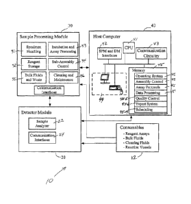
[0058] Referring to FIG. 1, the multi-analyte detection system 10 (also
referred to
herein as MAD system 10) includes a detector module ("DM") 20, a sample
processing
module ("SPM") 30, and a host computer 40. Each of these modules 20, 30, 40
includes
assemblies, sub-assemblies, and/or components that perform various tasks
within the overall
system and method of using the MAD. (Note that the terms "assembly" and "sub-
assembly" are used throughout to help identify levels of component systems
within overall
system 10, however such term are not meant to limit the invention and may be
used
interchangeably.)
[0059] For example, sample processing module 30 includes assemblies for
specimen
handling 31, reagent storage 32, incubation and assay processing 33, control
of on-board
assemblies and processes 34, storage and delivery of fluids and storage of
wastes 35,
cleaning and maintenance functions 36, and for communication 36 between and
among the
various assemblies and modules. Detector module 20 includes an analyzer 22 for
analyzing
samples processed by SPM 30 and interfaces for communicating with other
modules and
assemblies and for sending assay data to host 40.
[0060] Host computer system 40 is generally responsible for providing
oversight of
necessary operations from instrument control to results evaluation, data
storage, quality
- 5 -
CAJD: 503832.1
CA 02532790 2006-01-17
WO 2005/008219 PCT/US2004/023204
control, mainframe bidirectional interfacing, and operator assistance.
Specific instrument
control activities include movement of samples, pipetting samples, pipetting
reagents,
flushing samples, analyzing samples, reporting data, and troubleshooting the
device.
[0061] Host computer 40 preferably includes a processor 41, or central
processing
unit (CPU) 41, memory 45, interfaces 42 for communicating with modules 20, 30,
other
communication circuitry 45, and a user interface 44 that can include, e.g., a
monitor, a
keyboard, a trackball, or mouse, and/or a touch screen for data entry. Host
computer system
40 also preferably, although not necessarily, includes a printer or other
peripheral output
devices. Memory 45 includes software and data for performing various
operations and
control of system 10. For example, memory 45 typically includes software
modules such
as: an operating system 45-1; one or assembly control modules 45-2 including
instructions
for operation and control of modules 20, 30 and other subsystems and
components of
system 10; assay protocols 45-3; analyzing, processing and storing data 45-4;
quality
control 45-5; an expert system 45-6 and a instructions and data related to
scheduling 45-7 of
assays and procedures.
[0062] Referring to FIG. 2, a preferred embodiment of MAD system 10 is a
self
contained, fully automated random assay system incorporating a sample
processing module
30 having a number of automated assemblies and components for processing
samples and a
detector module 20 for analyzing the samples.
[0063] MAD system 10 is capable of multiplexing a number of assays in
the same
reaction tube and preferably employs a multiplexed bead-based chemistry system
for
performing numerous assays. For example, in one embodiment, system 10 up to 25
or more
individual assays, in another embodiment up to 100 individual assays, and in
yet another
embodiment more than 100 individual assays simultaneously in a single reaction
vessel.
[0064] Generally, detector module 20 utilizes an advanced analyzer to
detect the
presence of analytes, such as antigens, antibodies receptors, peptides,
oligonucleotides,
DNA, RNA, small molecules, viruses, viroids, cells, and the like, in patient
samples by
integrating the technologies of fluoroimmunoassay and flow cytometry. This
combination
of advanced technologies allows system 10 to perform, e.g., at least 200
measurements per
analyte per specimen, and about 100,000 measurements per minute, yielding up
to about
2,200 results per hour. Detector module 20 incorporates a flow cytometer that
detects
labeled microspheres or beads in a sample, and communicates with hardware and
software
in host computer 40 for assay control and data analysis. Optionally, detector
module 20
incorporates its own computer processor and memory for providing some level of
control
and analysis and communicating with host 40.
- 6 -
CAJD: 503832.1
CA 02532790 2006-01-17
WO 2005/008219 PCT/US2004/023204
[0065] In a preferred embodiment a flow cytometer of detector module 20
analyzes
individual microspheres by size and fluorescence, distinguishing preferably
three
fluorescent colors, green (550 - 610 nm emission), orange (585 - 650 nm
emission), and red
(>650 nm emission), simultaneously. Microsphere size, determined by 90-degree
light
scatter, is used to eliminate microsphere aggregates from the analysis. Orange
and red
fluorescence are used for microsphere classification, and green fluorescence
is used for
quantification of analyte. Additional details and examples of detector module
20 are
described in section 5.2 below.
[0066] Major assemblies and components of sample processing module 30
include a
specimen rack handler assembly 50, a reaction vessel handler assembly 52, a
reaction vessel
supply system 54, an incubator and separation carousel 56, a reagent storage
assembly 58, a
reagent robot 60, a wash robot 62, a solid waste system 64, a sample handler
66, and a
fluidics system 68. Each of these components are shown in FIG. 3 and described
in more
detail in section 5.3 below.
5.2 Detector Module
5.2.1 Overview of Detector Module ("DM")
[0067] According to a preferred embodiment, detector module 20 is a dual-
laser
flow cytometer system including fluidic, electronic and optical subassemblies.
The main
optical components of the detector module are: red laser for bead
classification, green laser
for label excitation, a PMT (photomultiplier tube) for detecting label
emissions and
photodiodes to detect signal coming as a result of excitation of the
classification dyes in
microparticles.
[0068] More particularly, in one embodiment detector module 20 is an
advanced
immunoassay analyzer incorporating SUSPENSION ARRAY (LUMINEX
CORPORATION). Briefly, SUSPENSION ARRAY analysis involves the process of
analyzing populations of microspheres (or "beads") having unique intensities
of red and
near infrared dyes, allowing each bead population to be identified and
analyzed separately.
Bead populations are distinguished with unique binding molecules making each
population
sensitive to a particular analyte. A fluorescent indicator dye is then used to
quantify the
amount of bound analyte on each bead. Calibrators convert average population
intensity
into analyte concentration. Performing the chemistry on microspheres, e.g., on
the surface
of microspheres, leads to significant reduction in reagents, yielding
significantly lower costs
for consumables. Additional details may be found in U.S. Patent Numbers
6,592,822;
6,528,165; 6,524,793; 6,514,295; 6,449,562; 6,411,904; 6,366,354; 6,268,222;
6,139,800;
- 7 -
CAJD: 503832.1
CA 02532790 2014-02-14
6,057,107; 6,046,807; 5,981,180; 5,802,327; and 5,736,330; as well as in
published U.S.
Application Numbers 20030132538, and 20020132609.
[0069] Detector module 20, in one embodiment, includes a flow cytometer
having a
one or more lasers, optics, photodiodes, a photomultiplier tube, and digital
signal processing
to perform simultaneous, discrete measurements of fluorescent microspheres. In
one
embodiment, three avalanche photodiodes and a high sensitivity photomultiplier
tube
(PMT) receive photon signals from the microspheres. Detector module 20 in this
example
digitizes the waveforms and delivers the signals to a digital signal processor
(DSP). The
detector module works with the SPM and host computer to perform multiplexed
analysis
simultaneously by using the flow cytometer and digital signal processor to
perform real-
time analysis of multiple microsphere-based assays. Because a flow cytometer
has the
ability to discriminate different particles on the basis of size and/or
fluorescence emission
color, multiplexed analysis with different microsphere populations is
possible. Differential
dyeing microspheres, emitting light at two different wavelengths, allows
aggregates to be
distinguished and permits discrimination of, in one embodiment, up to about 25
different
sets of microspheres, in another embodiment up to about 100 different sets of
microspheres,
and in yet another embodiment more than 100 different sets of microspheres.
Several
control beads are used in every analysis to ensure quality control of the
results. The system
can analyze small-molecular weight (e.g., T4) and large molecular weight
analytes
including, for example, IgG, IgA, and IgM antibodies, and glycoprotein
hormones.
[0070] In one embodiment, fluorescence excitation a preferred embodiment of
a
MAD system involves two solid state lasers. These lasers illuminate the
microspheres as
they flow single file through the cuvette. The fluorescent signals are
discriminated with
selective emission filters and are converted into intensity units by using the
DSP. There are
two different fluorophores present within the microbeads which emit with two
different
emission profiles that are separately measured in order to define the address
(test analysis)
of the bead.
[0071] Immunochemical reactants, such as antigens, antibodies receptors,
peptides,
oligonucleotides, DNA, RNA, small molecules, viruses, viroids, cells, and the
like of these
assays become bound to the surfaces of uniquely addressed fluorescent
microscopic beads.
The fluorescent spectral address of each bead identifies each of the assays
performed
simultaneously on a single sample. Based on its fluorescent signature, every
microsphere is
classified to its own unique region. In addition, each bead is scanned for the
presence of a
reporter fluorescence that quantifies the bead-assigned assay at the bead
surface. The MAD
- 8 -
CA 02532790 2006-01-17
WO 2005/008219 PCT/US2004/023204
microspheres are highly uniform, polystyrene-based particles that have been
crosslinked
during polymerization to provide physical strength and stability. They also
contain a
magnetic core so that they can be attracted to an electromagnet to facilitate
facile washing.
The beads may be magnetic, paramagnetic, superparamagnetic or the like to
facilitate
processing and washing of samples. Varying ratios of different fluorochromes
embedded
within each microsphere give each bead a unique spectral address. Each
microsphere is
dyed to emit light in a certain classification channel. All microspheres of
given emissions
represent a distinct assay within a multiplex of assays. A reporter channel is
used to detect
fluorescence bound to the surface of each microsphere, and each reporter
emission
quantitates each of the distinct assays. Only one reporter is needed for a
multiplex of
assays. To ensure the stability of this address, the microspheres should be
protected from
light and high temperatures.
[0072] One technology employed in the MAD system is flow cytometry. Flow
cytometry is a technique that simultaneously measures and then analyzes
multiple physical
characteristics of single particles, usually cells, as they flow in a fluid
stream through a
beam of light, most commonly from a laser. As a technique, flow cytometry is
somewhat
analogous to fluorescent microscopy; one major difference is that flow
cytometry provides a
digital result. In flow cytometry, measurements are performed on particles
(e.g., cells or
microbeads) in liquid suspension, which flow one at a time through a focused
light (e.g.,
laser) beam at rates up to several thousand particles per sec.
[0073] The properties measured by flow cytometry may include a
particle's relative
size, relative granularity, internal complexity, and relative fluorescence
intensity. These
characteristics are determined using an optical-to-electronic coupling system
that records
how the cells or beads scatter incident light and emit fluorescence.
[0074] The detector module flow cytometer includes three main systems -
detector
fluidics, optics, and electronics. The detector fluidics system transports
particles in a stream
to a laser beam for interrogation. A diagram of a typical flow cytometer
fluidics system is
shown in FIG. 4. Details of the MAD flow cuvette design and the wavelengths of
the two
laser beams used in MAD system are provided in FIG. 5. The optics system
consists of
lasers to illuminate the particles in the sample stream and optical filters to
direct the
resulting light signals to the appropriate detectors. Details of the MAD
detector optics
design are provided in FIG. 6. The electronics system converts the detected
light signals
into electronic signals that can be processed by the computer 40.
[0075] As shown in FIG. 4, in the MAD flow cytometer 100, particles 102
are
carried to the laser intercept 104 in a fluid stream; this liquid is referred
to as sheath fluid
- 9-
CAJD: 503832.1
CA 02532790 2006-01-17
WO 2005/008219 PCT/US2004/023204
106. Particles 102 are carried by a microscopic jet of buffer and are
hydrodynamically
focused in the center of the fast moving stream 106 of the sheath fluid. The
particles 102
pass one by one (some particles in the flow stream may not be individual
entities; particles
may be bound to each other or in such close proximity that they are detected
as a single
bead) through an intense beam of excitation light in the measuring region of
the flow
cytometer. Each particle thereby produces short flashes of fluorescence, the
intensities of
which are proportional to the content of the fluorescently labeled
constituent. FIG. 4 shows
a flow cytometry fluidics system according to an embodiment of the present
invention
showing the flow direction, injector tip 108, flow cell 110, laser beam 104,
and sheath fluid
106.
[0076] The fluorochromes emit fluorescent light many times during the
transit time
of a bead through the flow cuvette.
[0077] Suspended particles or cells preferably from about 0.2 to about
150
micrometers in size are suitable for flow cytometric analysis. The portion of
the fluid
stream where particles are located is called the sample core. When particles
pass through
the laser intercept, they scatter laser light without loss or gain of energy.
Any light excited
molecules present on the particle fluoresce. The scattered and fluorescent
light are collected
by appropriately positioned lenses. A combination of beam splitters and
filters directs the
scattered and fluorescent light to the appropriate detectors. The detectors
produce electronic
signals proportional to the optical signals striking them.
[0078] List mode data (photon recordings are recorded in a list) are
collected for
each particle or event. The characteristics or parameters of each event are
based on light
scattering and fluorescent properties. The data are collected and stored in a
computer.
These data can later be analyzed to provide information about subpopulations
within the
sample.
[0079] FIG. 5 details a MAD flow cytometry cuvette flow cell 110
according to an
embodiment of the present invention showing the cell dimensions and the laser
beam
wavelengths 114, 116. The dimensions shown, e.g., approximately 200 inn x 200
Inn, are
approximate inner dimensions of a suitable flow cuvette. According to one
embodiment,
the outer dimensions are preferably about 2.2 mm by 2.2 mm. One skilled in the
art will
appreciate that cuvettes of other dimensions or characteristics are known and
may be used
without departing from the scope of the invention.
[0080] FIG. 6 details a MAD optics design according to an embodiment of
the
present invention showing flow cell 110, reflectors 124,126, lasers 116,114,
lenses 120,
122, and detectors 128,130.
- 10 -
CAJD: 503832,1
CA 02532790 2006-01-17
WO 2005/008219 PCT/US2004/023204
[0081] In a preferred embodiment, detector module 20 is equipped with
two lasers
114 and 116. Particular characteristics of lasers 114 and 116 are described
below, however
one skilled in the art will appreciate that numerous other laser systems and
analyzers are
known in the art and may be used depending upon the types of assays one
desires to
perform; any such lasers or alternative analyzers or detector modules may be
employed in
detector module as part of the overall MAD system without departing from the
scope of the
present invention.
[0082] In this example, diode laser 116 produces about 7 mWatts of about
635 nm
(red) light. This laser is also referred to herein as red laser 116 or the
classifier laser 116
since it is used to excite the classifier dyes within a bead leading to the
identification of the
bead region to which the bead belongs.
[0083] The second laser, laser 114, is preferably a diode-pumped, solid
state,
continuous wave (CW), doubled Nd:YAG laser. In this example embodiment, laser
114
produces about 15 mWatts of power and about 532 nm (green) light. Laser 114 is
also
referred to herein as green laser 114 or reporter laser 114 since it functions
to excite the
reporter (label) groups at the microsphere or bead surface. Green laser 114
preferably has a
power stability of less than +/- 2% over 8 hours, and a beam diameter of 0.32
mm +/- 10%.
Laser 114 uses yttrium aluminum garnet (YAG) as the matrix material, doped
with
neodymium (Nd:YAG). A 15 mm lens is used as the primary focusing lens for the
532 nm
laser 114.
[0084] Further, in this embodiment, there are two 4 mm lens assemblies
120, 122
located approximately 900 mm from the laser beam path. They are precision
aligned to the
cuvette and collect the fluorescent signals, both reporter and classifier. A
550 nm-610 rim
reflector is used to deflect the reporter signal to the single photomultiplier
in the MAD
system. A 630 nm-760 nm reflector is used to deflect the classifier signal to
two
classification channel avalanche photodiodes. One photodiode detects the "red'
classifier
dye emission and the other the "orange" classifier dye emission. Orthogonal
scattered light
(from the beads) is also measured on the system using a third avalanche
photodiode. A
block, referred to as the "U" block, houses the classifier and doublet
discriminator lenses,
the diodes, and circuitry. In one embodiment, the flow cuvette is made of
quartz and has a
width and depth of 200 p.m. The light gathered by the photodetectors initially
exits through
one of the cuvefte's walls, is reflected by a mirror, and finally passes
through a filter before
reaching the detector. The numerical aperture of the detector is 0.62. The
mirror causes
about 2% loss in light intensity and the filter cuts the intensity by about
50%. Fluorescence
emission from the bead surface takes place at all angles and only a few
percent of the total
- 11 -
CAJD: 503832.1
CA 02532790 2006-01-17
WO 2005/008219 PCT/US2004/023204
emitted photons are collected by the first mirror due to the physical
limitations of the optics
design. Therefore, only a very small percentage (e.g., probably < 1%) of
emitted photons
are actually recorded by the detector.
[0085] Alternatively, other more powerful 532 nm lasers 114 may be used
to
improve the analytical sensitivity of immunoassays. In particular, an
approximately 50
mWatt laser photobleaches the primary detector molecule, B-phycoerythrin, used
as a
reporter for most assays. The optimal wattage for 532 laser 114 is about 10-20
mWatt in
this example, however other wattages or lasers may be used.
[0086] There are two fluidic paths in this MAD system detector module.
The first
path involves a syringe driven mechanism that controls the small volume sample
uptake.
This syringe driven system transports a user specified volume of sample from a
sample
container to the reaction vessel (RV). After reaction incubation(s), the
sample is injected
into the flow cuvette at a steady rate for analysis. Following analysis, the
sample path is
automatically purged with sheath buffer by the second fluidics path. This
process
effectively removes residual sample within the tubing, valves, and probe. The
second
fluidics path is driven under positive air pressure and supplies the sheath
fluid to the
cuvette.
[0087] As will be described in more detail below, in this embodiment,
following the
last aspiration of wash, MAD system 10 dispenses wash buffer, e.g.,
approximately 20-70
1.1.1, more preferably about 50 ttl, into the reaction vessel (RV) in
preparation for
fluorescence reading. Approximately all of this volume is aspirated to the
detector. The
first approximately 5 pl is ignored by the detector before reading commences.
The length
of time to read the beads is generally dependent on the bead concentration
since in this
example there is a defined number of beads read for all regions, e.g., in this
example about
200 beads read for all bead regions. This typically requires from about 5 to
25 sec.
Counting of beads on the MAD system 10 terminates when either the defined
number of
each regional bead in the assay panel are counted, or when the allocated time
for
fluorescence analysis is completed. Since not all beads are at exactly the
same
concentration, most bead sets will acquire more than 200 counts. Analysis
terminates with
the last bead region to reach the defined number, e.g., 200, counts. The
allocated time is
determined by the sample flow rate and by its volume. The assay cycle time for
the
preferred timing sequence used by the IgG and IgM serology panels, and the
systemic
autoimmune panel, is, e.g., 30-40 sec, more preferably about 36 sec. The
percent of the
cycle time that the MAD system is reading fluorescent beads with the preferred
timing
sequence is therefore in this particular example about 28% to 53%. Of course,
other time
- 12 -
CAJD: 503832.1
CA 02532790 2006-01-17
WO 2005/008219 PCT/US2004/023204
cycles may apply depending upon system set-up and types of assays performed.
[0088] The transit time for an individual bead to travel from the flow
cuvette 110
location in which it is interrogated by red laser 116 to the location where
the bead is
illuminated by green laser 114 is about 35 sec. The instrument's firmware
measures the
exact transit time during the detection calibration step. In an actual assay,
the detector
measures this time from bead's coincident CL1 and CL2 readings (see FIG. 7),
and then
measures the conjugate derived fluorescence (RP-1) (see FIG. 7). Since the
flow rate is
approximately constant, MAD system 10 "knows" that a given green fluorescent
signal is
associated with the red and orange emissions registered 35 sec earlier.
Because the bead
concentration in the flow stream is low, there is only a very low statistical
chance that the
RP-1 signal would be misassigned to the wrong bead, Le., a close proximity
second bead in
the flow cell. Since the flow rate for the MAD system is nominally 2.3 m/sec,
a bead would
flow 81 microns in 35 sec. The distance between the focal points for the two
lasers 114,
116 is therefore about 81 microns.
[0089] Because the fluorescence readings of the red 116 and green 116
emissions
must be temporally coordinated, it is desirable that the flow rate remain
constant within a
narrow range. Some change in flow rate does occur and the MAD system can
accommodate minor fluctuations. Larger changes in the flow rate of beads
through the
detector would cause significant problems. When detector module 20 of
instrument 10 is
calibrated, not only are the voltage settings adjusted to attain the three pre-
designated RFI
readings, but the instrument also adjusts for the time required for bead
transit from the red
116 to the green 114 laser illuminations. As the flow rate changes due to
pressure changes
in the system, the transit time changes in a near linear relationship. Once
MAD system 10
is calibrated and the transit time established, subsequent change in pressure
(and thus flow
rate) will affect the results, leading to deleterious effects including lower
RFI values and
decreased precision.
[0090] In one embodiment, detector module 20 takes somewhat less time to
read the
beads than the LUMINEX LX100, all else being equal. This is because the MAD
system's
analyzer preferably employs efficient magnets for wash and separation of
analytes. Fewer
beads are therefore lost with the MAD system 10, the concentration of beads in
the flow cell
is higher, and the time to count 200 beads per region is less.
[0091] Fluorescent measurements of beads on detector module 20 are
gated, where a
gate is a boundary that defines a subset or sub-population of events. Gates
are set by
electronically drawing boundaries around the data subsets. Gates can be used
either for data
acquisition or analysis. Inclusive gates select only the events that fall
within (and on) the
- 13 -
CAJD: 503832.1
CA 02532790 2006-01-17
WO 2005/008219 PCT/US2004/023204
boundary. Exclusive gates select only the events that fall outside of the
boundary. (The
gates described in this paragraph are data acquisition and exclusive gates).
One gate is
fixed by the firmware and two gates are optionally set by the operator. The
first gate is
automatically established and determines whether the CL1 and CL2 signals for
an
individual bead fit within one of the established bead map regions. If it
does, the bead
passes that gate. If not, the data for the bead is filtered out and
subsequently ignored. The
second gate, which is operator established through the user interface, is the
doublet
discriminator gate. This gate is based on the light scatter measurement of the
bead. The
purpose of this gate is to exclude bead aggregate events (larger than an
individual bead) or
bead debris events (smaller than an individual bead). The third filter is the
RP-1 gate. This
gate excludes two types of events - zero RFI beads and very bright beads (high
statistical
outliers) from further data analysis. The last gate is also user defined.
[0092] Fluorescent spillover occurs when two or more emission spectra
overlap so
that selective filtering cannot occur. In some flow analyzers, emission
spillover is corrected
using a technique called compensation. Compensation involves subtraction of
some
emission percentage from another emission signal. One embodiment of system 10
does not
use compensation; however, the reporter signal does not significantly spill
over into the
classification emission.
[0093] The reporter fluorochrome is bound to the surface of the
microsphere and
provides raw analytical data. Because a microsphere suspension provides near
liquid phase
reaction kinetics, each microsphere of a particular spectral address
theoretically binds an
equal number of reporter molecules. Equal binding results in a statistically
even
distribution of reporter on each microsphere in a set. This means numerous
replicates for
each microsphere population are measured from a single well. The confidence in
a given
measurement strengthens with increased replicate measurements. For adequate
confidence,
200 events per microsphere set in each well is usually sufficient.
5.2.1 Characteristics and Uses of Microspheres
[0094] In a preferred embodiment of system 10, instead of employing
commonly
used microtiter wells to host the immunochemistry-based assays, the
immunoassay
reactions occur on the surface of microscopic, magnetic, polystyrene-core
beads known as
microspheres. Suitable microspheres are disclosed, for example in the LUMINEX
patents
listed above. Although such microspheres are not necessarily part of detector
module 20,
they are described herein as they are integral to the principals of operation
of an exemplary
embodiment of the detector module 10 as described.
- 14 -
CAJD: 503832.1
CA 02532790 2006-01-17
WO 2005/008219 PCT/US2004/023204
[0095] Prior to use, microspheres are maintained in suspension in a bead
reagent
solution. Individually dyed with combinations of two different fluorescent
dyes (red and
orange), a microsphere may have one of many possible levels of classifier dye
fluorescent
intensities. The various combinations of dyes create a sets of, in one
embodiment, up to 25
uniquely color-coded microsphere sets, in another embodiment up to 100
uniquely color-
coded microsphere sets, and in yet another embodiment more than 100 uniquely
color-
coded microsphere sets. In one embodiment, antigens or antibodies indicative
of a specific
bacterial or viral antigen, protein, or other molecule are coated onto the
surfaces of each
uniquely color-coded bead set, making each different microsphere set
representative of a
different assay.
[0096] Because in this example each microsphere is coated with antigens
or
antibodies specific for a given condition, each microsphere is equivalent to
an individual
microtiter well used in many enzyme-linked immunosorbent immunoassays
(ELISAs).
Alternatively, beads may be coated with proteins, antibodies, ligands or the
like in order to
run a wide variety of assays and assay formats. MAD sysiem 10 can
simultaneously run
(multiplex), according to one embodiment, up to 25 assays in a single reaction
vessel, in
another embodiment up to 100 assays in a single reaction vessel, and in yet
another
embodiment, more than 100 assays in a single reaction vessel, using as little
as 5 111 of
sample. Beads may include flourescent or other labels, or may be secondarily
labeled
during processing with labeled antibodies that bind to a target molecule after
it is bound to a
bead.
[0097] FIG. 7 shows microspheres serving as the vehicle for molecular
reactions.
The microspheres are approximately 8.0 im polystyrene microspheres that bear
carboxylate
functional groups on the surface. The microspheres are available in 25
distinct sets 134 that
are classified by the flow cytometer by virtue of the unique orange/red
emission profile of
each set, as shown in FIG. 7. Micro spheres of this size provide sufficient
surface area for
covalent coupling of 1-2 x 10⁶ target molecules per microsphere. In use,
fluorescence
classification of dual-labeled fluorescent microspheres is used. Two-
dimensional dot plots
report the classification of a 25 microsphere set based on simultaneous
analysis of
logarithmic orange fluorescence (FL2) and logarithmic red fluorescence (FL3).
FIG. 7 also
shows the positioning of each numbered bead set with regions 6 (labeled "134-
6")and 100
(labeled 134-100) containing the least and most amount, respectively, of
combined orange
and red dyes.
[0098] Fluorescent reactants, e.g., fluorescent antibodies, antigens, or
nucleic acid
probes provide specific signals for each reaction in a multiplexed assay.
Because each
- 15-
CAJD: 503832.1
CA 02532790 2006-01-17
WO 2005/008219 PCT/US2004/023204
fluorescent reactant binds specifically to a target that is present on only
one bead set in a
multiplexed assay, the soluble reactants do not need to be differentially
labeled. All
fluorescent molecules are labeled with a fluorophore such as the organic green-
emitting
dyes BODIPY and fluorescein isothiocyanate, or a more commonly used biological
fluorophore such as phycoerythrin. Any fluorochrome can be used as a reporter;
however,
each fluorochrome has a characteristic emission spectrum which affects the
amount of
spillover into the orange fluorescence channel. In one example green-emitting
fluorophores
are used.
[0099] To prepare a multiplexed assay, individual sets of microspheres
are
conjugated with the target molecules required for each reaction. Target
molecules may be
antigens, antibodies, oligonucleotides, receptors, peptides, etc. Fluorescent
reactants may
be complementary oligonucleotides, antigens, antibodies, receptors, etc.,
i.e., any molecule
that will specifically bind to the target molecule. After optimizing the
parameters of each
assay separately in a nonmultiplexed format, the assays can be multiplexed by
simply
mixing the different sets of microspheres. The fluorescent reactants also are
mixed to form
a cocktail for the multiplexed reactions. The microspheres are then reacted
with a mixture
of analytes, for example in a biological sample, followed by the cocktail of
fluorescent
reactants. After a short incubation period, the mixture of microspheres, now
containing
various amounts of fluorescence on their surfaces, are analyzed with the flow
cytometer.
Data acquisition, analysis, and reporting are performed in real time on all
microsphere sets
included in the multiplex. As each microsphere is analyzed by the flow
cytometer, the
microsphere is classified into its distinct set on the basis of orange and red
fluorescence, and
the green fluorescence value is recorded. Two hundred individual microspheres
of each set
are analyzed and the median value of the green fluorescence is reported.
[00100] With respect to protein antigens, methods for coupling proteins
to bead
surfaces are will known. For example, covalent coupling of protein antigens to
bead surface
carboxyl groups by amide bond formation requires protonated carboxylic acids
for the
initial activation and esterification steps of the conjugation reactions.
Since the pKa of
carboxylic acids is higher in ethanol than in water, conducting the initial
steps in buffered
ethanol is advantageous compared to conducting them in aqueous buffer. For
example,
ethanol raises the pKa of methacrylic acid from pH 4.9 to approximately pH
6.0, which
means that approximately ten-fold more carboxyl groups will be available for
coupling
when the beads are activated and esterified at pH 5.0 in buffered ethanol
compared to pH
5.0 in aqueous buffer. However, because the classification dyes used to define
the spectral
addresses of the beads are soluble in organic solvents and are only infused
into the bead
- 16-
CAJD: 503832.1
CA 02532790 2006-01-17
WO 2005/008219 PCT/US2004/023204
surface, it seemed possible that exposure of the beads to ethanol would result
in leeching of
these dyes. Depending on the extent of extraction, leeching of classification
dyes could
reduce classification efficiency, and if extreme, could even cause
misclassification. Other
potential effects of ethanol include increasing bead autofluorescence and
decreasing bead
dispersion in aqueous solution. The objective of a conducted experiment
described below
was therefore to determine whether exposure of dyed beads to ethanol
compromised
classification efficiency, increased autofluorescence, and/or decreased
dispersion in aqueous
solution. To address these issues, dyed beads were exposed to absolute ethanol
for four
hours and sampled at 30 minute intervals. At the end of the exposure period,
the beads were
evaluated sequentially in an LX-100 flow fluorometer and classification,
autofluorescence,
and bead dispersion data were acquired. The data showed no change in
classification
efficiency and bead dispersion for the duration of the study, however, there
was a nominal
increase in autofluorescence for some regions after 120 minutes and a
substantial increase
for these regions after four hours. Overall, the data from these experiments
indicated that
activation and esterification of bead surface carboxyl groups for 60 minutes
in 90% ethanol
would not have adverse effects on the classification efficiency,
autofluorescence, and
dispersion of dyed beads. A consequence of this could be rearrangement of the
bead
surface ultrastructure through redistribution of hydrophilic and hydrophobic
polymers, and a
change in the ability of the beads to remain dispersed in aqueous solution.
This concern
could be dismissed however, because doublet discriminator data did not reveal
any impact
of ethanol on bead dispersion.
[00101] The data from this study indicated that exposure of dyed beads to
ethanol for
60 minutes during carboxyl activation and esterification would not have any
consequence
on classification efficiency, autofluorescence, or dispersion of the beads in
aqueous
solution. Thus, exploiting the power of ethanol to raise the pKa of bead
surface carboxylic
acids offers a practical opportunity to increase the coupling efficiency of
bead ligation
procedures.
[00102] An embodiment of detector 20 uses a 14-bit analog to digital
converter
(ADC). The resolution in terms of histogram bin width is about 1/32,000. This
is fairly
limited resolution. A current version of the DSP in the LUMINEX obtains eight
fluorescent
readings from each bead during its lifetime in the focused region of the
reporter laser. The
RFI values reported by the DSP of the detector are essentially an integration
of the eight
fluorescent readings per bead. The height (direction of flow) of the laser-
illuminated region
in the detector flow cell is about 30 [tm and the velocity of the sample
stream is roughly 2.3
m/sec. Thus, the residence time of the bead in the illuminated region is about
13 sec.
- 17 -
CAJD: 503832.1
CA 02532790 2006-01-17
WO 2005/008219 PCT/US2004/023204
Assuming a fluorescence lifetime of 5 nsec for the reporter label, the beads
could be excited
and then fluoresce 2,600 times (13 sec/5 nsec) during the transit time
(assuming no
photobleaching). Therefore, reporter emission is recorded for less than 1% of
the time the
bead-bound label is in the laser light path.
[00103] In one embodiment, detector module 20 is a flow cytometer that
uses a two-
step calibration initiation. One calibrator (CAL-1) adjusts the correct gain
for the bead
classifier photodiode detectors (CL1 and CL2) and for the doublet
discriminator detector
(DD). The second calibrator (CAL-2) adjusts the gain for the reporter PMT
(RP1). A
current practice is to calibrate with CAL-1 to target values and calibrate CAL-
2 to a fixed
value, for example a value of approximately 17,000 irrespective of the target
value. In one
embodiment, the CAL-2 target value is around 3,800 10%.
[00104] In flow cytometry, electronic gating typically is used to isolate
classes of
cells or particles. Often, gating is used to differentiate one population of
cells or particles
from other populations. Doublet discriminator (DD) gating is also used to
eliminate debris
and aggregates from the counting statistics. The within-RV CV% values are
lower when
DD gates are used. In the case of the detector, differentiation is
accomplished via region
gating (CL1 and CL2) and there is a partial gate on the reporter channels that
allows
elimination of very low RFI events (usually between 0-2). DD gating is almost
exclusively
used by flow cytometers to eliminate debris and aggregates.
[00105] In addition to recording three different fluorescence
measurements, MAD
system 10 also detects light scatter. The collected scattered light is
orthogonal scatter, also
referred to as right-angle, side-angle, or wide-angle scatter. Orthogonally
scattered light is a
good reflection of the size of the particle from which the light impinged and
was scattered.
Side-angle scatter is easily capable of distinguishing a bead from a bead
doublet (two beads
stuck to each other or in very close vicinity). Detection of bead doublets is
called doublet
discrimination. In flow cytometry it is the usual practice to eliminate
recorded doublet
events from the data analysis since their physicochemical behavior is often
aberrant when
compared to single beads. Elimination of doublets can improve the precision
and accuracy
of an assay. When light impinges on a particle it is scattered without loss of
energy in many
directions. The magnitude and angles of scatter depend on such parameters as
particle size,
density, and shape.
[00106] Right-angle light scatter is detected on the MAD instrument with
an
avalanche photodiode, the same type of detector used to classify the beads
into regions.
[00107] FIG. 8 schematically represents a process of washing microspheres
140, or
beads, using magnets 142. Microspheres can be magnetic or have metallic
properties or
-18-
CAJD: 503832.1
CA 02532790 2006-01-17
WO 2005/008219 PCT/US2004/023204
other properties that allow them to be attracted to magnets 142 during
washing. When
washing is required, reaction vessels 144 containing beads 140 in a solution
146 (e.g. a
buffer solution or an assay reagent) are placed near two strong electromagnets
142 (e.g., in
separation carousel 55 of FIG. 3). Magnets 142 attract and hold beads 140 to
the sides of
the reaction vessel 144. Liquid 146 is then aspirated from the reaction vessel
leaving the
beads on the vessel 144 sides attracted to magnets 142. After removal of the
magnets 142
as shown in FIG. 8C, and beads are resuspended in another volume of liquid 146
as shown
in FIG. 8D.
5.3 Sample Processing Module ("SPM")
5.3.1 General Features of SPM
[00108] Referring again to FIG. 3, one embodiment of sample processing
module 30
can include a number of subsystems and components for automating sample
handling and
assay procedures of system 10. Such subsystems can include a specimen rack
handler
assembly 50, a reaction vessel handler assembly 52, a reaction vessel supply
system 54, an
incubator and separation carousel 56, a reagent storage assembly 58, a reagent
robot 60, a
wash robot 62, a solid waste system 64, a sample handler 66, and a fluidics
system 68.
[00109] Each of the various subsystems and their interactions are
described in more
detail in sections below. Each of these subsystems and their physical
relationship to other
subsystems and components within SPM 30 are described in the context of an
exemplary
embodiment of system 10, and such examples are not intended to limit the
invention. One
skilled in the art will appreciate that variations in each subsystem and/or
their interactions
may be made without departing from the scope of the invention.
[00110] For example, in the embodiment of system 10 detailed below, SPM
30
includes twenty seven discrete stepper motors for actuation of various
instrument robotic
systems. These include multiple high speed high power carousel drives capable
of better
than, e.g., 0.5 sec positioning times, for moving incubation and separation
carousel 56 and
reagent carousel 70. In addition, multiple X, Y robots having, e.g., at least
0.5 mm
placement accuracy, preferably about 0.2mm or better placement accuracy, are
employed
for driving specimen rack handler 50, reaction vessel handlers 52, reagent
robot 60, sample
handler 66, wash robot 62, and detector transfer robot 74. Additionally, Z-
theta robots may
be used to minimize probe movement times even further for time critical
actions. One
skilled in the art will appreciate that different number and types of
subsystems and drive
motors may be employed without departing from the overall spirit of the
present invention.
- 19-
CAJD: 503832.1
CA 02532790 2006-01-17
WO 2005/008219 PCT/US2004/023204
[00111] Motor drives and sensor feedback are provided by a number (e.g.,
four or
more) of custom designed stepper and sensor control printed wire assemblies
(PWA's; also
termed herein printed circuit boards, or PCB's), each capable of
simultaneously driving
multiple motors. Each has the capacity for multiple, e.g., up to 24, sensor
inputs used for
positional feedback and motor step loss detection.
[00112] As will be described in more detail below with respect to some
subsystems,
e.g., specimen rack handler 50, RV handlers 52, reagent robot 60 and incubator
carousel
assembly 56, integrated circuit boards related to each subsystem of SPM 30
preferably
communicate over an integrated compact PCI bus main system processor board of
host
computer 40. The main system processor board of host 40 can be, e.g., a
Pentium III or the
like single board computer running at 850 MHz, with 128 Mbytes on board RAM
and 48
Mbytes flash disk permanent storage. Alternatively host 40 can also be a
remote server
which the device communicates with over a network. The logging of operational
data to a
controlling host via USB minimizes the requirement for data storage on system
10.
[00113] SPM 30 as described in the example below also incorporates four
or more
pipetting probes for manipulating samples and reagents, e.g., a sample handing
probe
associated with sample hander assembly 66, a reagent probe associated with
reagent robot
assembly 60, a wash dispense probe associated with wash robot 62, and a
detector probe
associated with detector robot 74. Sample handler 66 probe is used to aspirate
and dispense
samples from within tubes 167 on specimen rack handling assembly 50 into
reaction vessels
on incubation and carousel assembly 56. Reagent robot 60 probe is used to
aspirate and
dispense reagents from reagent packages 80 in reagent carousel 70 into
reaction vessels on
incubation and separation carousel. In one embodiment, reagent robot 60 and
sample
handler 66 probes share a common tapered-tip design and are interchangeable.
Wash robot
62 probe and is used to dispense wash solution into reaction vessels on
incubation and
separation carousel 56.. Detector robot 74 probe is used to aspirate the
completed assay
bead solution into detector 20 for analysis.
[00114] Each of the probes described herein are preferably made of
stainless steel
with an internal polished surface to reduce nonspecific binding. In one
embodiment,
internal diameter of sample handler 66 and reagent 60 probes at the tapered
tip is 475 25
tun (e.g., approximately 60-fold wider than the diameter of the magnetic
beads). All probes
except the detector probe are preferably tapered and beveled. Sample handler
66 and
reagent robot 60 probes preferably incorporate sensors such as capacitive
liquid level
sensors for accurate and repeatable sensing of the liquid surface height, as
well as pressure
sensing for detection of blockage and/or when a probe is in contact with the
bottom of an
- 20 -
CAJD: 503832.1
CA 02532790 2014-02-14
empty vessel.
1001151 Additional details of each of the subsystems and major components
of
sample processing module 30 follow.
5.3.2 Specimen Rack Handler Assembly
[00116] FIGS. 9 and 10 depict a specimen rack handler assembly 50 (also
referred to
herein as "specimen handler" or "rack handler") according to an embodiment of
the present
invention. Specimen handler 50 moves samples from an input area 150 through an
instrument work area 160 to an output area 164. In doing so, the specimen
handler moves
the samples into an aspiration position. The specimen handler identifies each
rack 166 and
sample tube 167 by reading a barcode contained on each. There is a STAT (Short
Turn
Around Time) drawer 156 that provides the user with a mechanism for inserting
a rack 166
to be sampled and tested out of sequence. The specimen rack handler assembly
50 also
allows for continued uninterrupted operation while a user adds or removes
samples.
[00117] Specimen handler assembly 50 generally includes an input area 150,
151, a
main work area or horizontal platform 160, a robotic finger 152, a look-ahead
offline
platform 154, a STAT drawer 156, an aspiration offline platform 158, a look-
ahead barcode
reader 162, an aspiration offline barcode reader 164, and sample trays and
racks 166.
1001181 Robotic Finger 152 is designed to move sample racks 166 along
horizontal
platform 160 without interfering with any of the other specimen handler 50
components. In
one embodiment, finger 152 employs a 2-axis mechanism (horizontal in the x-
axis and
rotational). The two-axes work in conjunction to allow the finger to either
push a rack (e.g.,
to the right or left) or bypass a rack. The horizontal axis provides the
horizontal motion
while rotational axis provides the option for either engaging or disengaging
from pushing
the racks.
[00119] Referring to FIG. 10, horizontal movement of finger 152 is belt-
driven by a
stepper motor 222. This motor/belt drive assembly 222 is preferably located
underneath the
horizontal platform 160 to minimize its interference with other specimen
handler 50
components and accessibility to users.
1001201 The rotational axis movement is also belt-driven by a stepper motor
226.
Motor 226 preferably rotates a square shaft (on which the horizontal motion
occurs) of
finger 152 to move finger 152 down to a disengaged position (as shown) or up
into an
engaged position. In the engaged position, finger 152 can slide racks 166 from
right to left
along horizontal platform as shown in FIG. 9 under the power of horizontal
stepper motor
CA 02532790 2006-01-17
WO 2005/008219 PCT/US2004/023204
222.
[00121] The number of sensors monitor the position and status of the
robotic finger
52. A finger horizontal home optical sensor 223 located on farthest right side
of the
specimen handler 50 determines the horizontal home position of finger 52. A
Finger
horizontal step optical sensor 224 located on the farthest right side of the
specimen handler
50 determines the horizontal position of finger 152. A finger rotational home
optical
sensor 228 located on farthest right side of specimen handler 50 determines
the rotational
home position of the finger 152. A finger rotational step optical sensor 230
located on the
farthest right side of the specimen handler 50 determines the rotational
position of the finger
152. Each of the motors 222, 226 and sensors 223, 224, 228, 230 is
electrically connected
to system 10 via backplane printed wire assembly 200.
[00122] The look-ahead offline platform 154 is designed to move the racks
(and
associated specimen tubes) off the horizontal platform 160 and identify them
for the host
control 40 software. Look-ahead barcode reader 162 is used to read and
identify barcodes
of both samples 167 and rack 166 when the look-ahead offline platform pulls a
rack 166 off
horizontal platform 160.
[00123] Scheduling software in host computer 40 optimizes the throughput
of the
instrument and uses the information provided by barcodes on each of the racks
166 and
sample tubes 167.
[00124] Movement of the offline look-ahead platform 154 is driven by an
offline
look-ahead motor 202. Motor 202 moves platform 154 (and a rack 166 located
thereon) in a
y-axis direction (e.g., in a direction perpendicular to the long axis of
horizontal platform
160) to present racks 166 and sample tubes 167 to bar code reader 162. A
number of optical
sensors are present on the look-ahead offline platform 154. For example, a
look-ahead
offline rack optical sensor 210 determines the presence of a rack 166 on the
platform. This
sensor 210 is located on the top rear of the look-ahead offline platform
154204. A look-
ahead offline platform home optical sensor determines the horizontal (y-axis)
home position
for the look-ahead offline platform. Sensor 204 is located behind the
horizontal platform
160 near barcode reader 162. A look-ahead offline platform step optical sensor
206
determines the horizontal (y-axis) position for the look-ahead offline
platform. This sensor
206 is located behind the horizontal platform 160 on the rotational drive
gear. Rack sensor
210 communicates with circuit board 208 and each sensor 202, 204, 206, 210
electrically
communicates with system 10 through backplane 200.
[00125] The STAT drawer 156 is designed to provide the user the ability
to place a
rack 166 (with samples 167) at the head of the queue for immediate processing
by
-22-
CAJD: 503832.1
CA 02532790 2006-01-17
WO 2005/008219 PCT/US2004/023204
instrument 10. Rack 166 is placed just prior to aspiration offline platform
158 so it will be
moved onto platform 158 as soon as the current rack is finished. No other
action is required
by the user, aspiration offline platform 158 will automatically transfer the
rack and sample
information to the scheduling software in host 40.
[00126] A sensor 234, e.g., such as a mechanical plunger type sensor,
located on the
underside of STAT drawer 156 determines the status of the STAT drawer 156.
[00127] The aspiration offline platform 158 is designed to move the racks
(and
associated specimen tubes) off the specimen handler 50 horizontal platform
160, identify
them for the software and provide an aspiration location for the sample
handler assembly 66
(which includes a robotically-controlled sample aspiration probe). As with
barcode reader
162, aspiration barcode reader 164 associated with aspiration offline platform
is used to
identify both the rack 166 and individual tube 167 when aspiration offline
platform 158pulls
them off of the horizontal platform 160. Typically, this is just confirming
the information
obtained by barcode reader 162 of the look-ahead offline platform 154, but
occasionally,
e.g., when STAT drawer 156 is used, barcode reader 162 will be providing new
information
to the scheduling software.
[00128] Similar to look-ahead platform 147, aspiration or sample offline
platform
158 is driven by a motor 212. Motor 212 moves platform 158 (and a rack 166
located
thereon) in a y-axis direction (e.g., in a direction perpendicular to the long
axis of horizontal
platform 160) to present racks 166 and sample tubes 167 to aspiration bar code
reader 164.
[00129] The number of optical sensors are present on the aspiration
offline platform
158. For example, an aspiration offline rack optical sensor 220located on the
top rear of
aspiration offline platform 158determines the presence of rack 166 on the
platform.
Aspiration offline home optical sensor 214, e.g., located behind horizontal
platform 160
near the barcode reader 164, determines the horizontal (y-axis) home position
for aspiration
offline platform 158. An aspiration offline step optical sensor 216, e.g.,
located behind the
horizontal platform on the rotational drive gear, determines the horizontal (y-
axis) position
for the aspiration offline platform 158.
[00130] Sensor 220 communicates with rack sense circuit board 218, and
all of
sensors 212, 214, 216, 164 and 218 communicate with backplane 200.
[00131] Horizontal platform 160 is designed to provide a stable
horizontal surface for
rack movement, input and output areas 151, 162 for sample trays, and manual
rack input
150 and output 164 areas 150, 164.
[00132] Manual input area 150 provides the ability to add single racks
166 to the
queue. Manual input area 150 is located at the far left of specimen handler
assembly 50.
-23 -
CAJD: 503832.1
CA 02532790 2006-01-17
WO 2005/008219 PCT/US2004/023204
When a rack 166 is placed here, sensor 220 is tripped notifying software in
host 40. Finger
152 moves the rack 166 to the end of the queue. An optional manual input
optical sensor
146, e.g., located behind the back wall of the specimen handler 50, includes a
mirror
mounted on the specimen handler 50 front. Placement of a rack 166 in manual
input area
150 breaks the reflected beam of sensor 246.
[00133] Input tray area 151 provides a location for the placement of a
sample tray
165. Once a sample tray 165 is placed, it essentially becomes part of
horizontal platform
160 over which racks 166 are moved.
[00134] Input tray area sensor 252, e.g., located underneath specimen
rack handler
assembly 50, is preferably a magnetic reed type sensor. Sample trays 165 are
equipped with
magnets in the base to trip sensor 252 when the sample tray 165 is properly
placed.
[00135] Look-ahead area 155 provides a storage area for racks 165
awaiting
aspiration. Output holding area 157 provides an output area for racks (after
sample
aspiration). Typically, output holding area 157 will only have racks when the
output tray
162 is missing or full.
[00136] Output tray area 162 provides a location for the placement of a
sample tray
165. Output tray area sensor 232, e.g., located underneath specimen rack
handler 50, is
preferably a magnetic reed type sensor similar to sensor 252. As described
above, sample
trays 165 are equipped with magnets in the base that trip sensor 252 when such
sample tray
165 is properly placed. An optional output tray capacity sensor, e.g., located
behind the
specimen handler 50, monitors how many racks 165 are on the output tray 162.
After a
specified number of racks, e.g., ten, have moved onto output tray area 162,
such sensor trips
software in host 40 to request the customer to remove and empty the output
tray.
[00137] Manual output area 164 provides a location for the removal of
single racks
166 from the output area 162.
[00138] In one embodiment, sample trays 165 are designed to carry ten
racks 166.
Sample trays 165 allow large numbers of racks 166 to be added to or removed
from the
instrument 10 with ease. When not in the instrument (being carried) a self-
locking
mechanism is used to ensure the racks 166 cannot inadvertently slide off from
the tray 165.
The self-locking mechanism automatically unlocks when the rack is placed on
the
instrument 10 or on a flat surface.
[00139] During typical operation of specimen handler assembly 50, sample
tubes 167
are loaded into racks 166 that contain up to five bar-coded sample tubes in
each rack.
Optionally, racks 166 are loaded into a tray 165 that hold up to ten racks in
a queue awaiting
tube barcode reading and sample processing. Controls and calibrators are also
loaded into
-24-
CAJD: 503832.1
CA 02532790 2006-01-17
WO 2005/008219 PCT/US2004/023204
the same racks and are appropriately barcoded for identification. After tray
165 is placed in
the input tray area 151 on horizontal platform 16, input tray sensor 252
detects tray 165.
Finger 152 retracts and moves all the way to the left of manual input area
150. Finger 152
then rotates to the horizontal position, and moves racks 166 to the right
until the look-ahead
rack sensor 210 detects a rack on the look-ahead offline platform 154. Look-
ahead offline
platform 154 retracts, allowing the look-ahead barcode reader 162 to read the
barcodes.
[001401 After reading the barcodes, the look-ahead offline platform 154
returns to the
original position. Finger 152 moves the rack off the look-ahead offline
platform 154 and
onto the look-ahead area 155. The rack is here until aspiration offline
platform 158 is
available. Once the aspiration offline platform is available, finger 152 moves
the rack onto
aspiration offline platform 158 until the aspiration rack sensor 220 detects
the rack on
aspiration offline platform 158. Aspiration offline platform 158 then retracts
into SPM 30
allowing the aspiration barcode reader 164to read the barcodes.
[001411 Next, specimen robot 66 (see FIG. 3) obtains samples from the
sample tubes
167 in the rack 166. Aspiration offline platform 158 then returns to the
original position.
Finger 152 moves the rack off the aspiration offline platform 158 to the
output area 164.
The rack is held in the output holding area 164 until the output tray area 162
is available.
Finger 152 moves the rack onto a tray in output tray area 162. Finally, the
output tray is
removed from instrument 10.
5.3.3 Reaction Vessel Handling and Supply Assembly
[001421 Referring to FIG. 11, reaction vessel handing and supply assembly
260
generally includes a reaction vessel supply 262 ("RV supply 262") and two
reaction vessel
handlers 52a and 52b. RV handlers 52a, 52b and their associated motors and
components
are also collectively referred to herein as RV handler sub-assembly 52 or
simply RV
handlers 52.
5.3.3.1 Reaction Vessel Supply
[00143] FIGS. 11-16 depict a reaction vessel supply system 262 according
to an
embodiment of the present invention. Reaction vessel supply system 262
generally includes
a reaction vessel supply bin 264, or hopper, a reaction vessel supply motor
266, belt 268,
fans 270, and belt guards 272. Reaction vessel supply system 268 stores enough
reaction
vessels to supply device 10 for a long period, e.g., six or eight hours or
more, of continuous
operation. Reaction vessel supply system 262 also presents reaction vessels to
a position
where the reaction vessel handlers 52a, 52b can retrieve them. The maximum
time interval
between presentations of reaction vessels to pick up position is preferably 45
seconds or
-25 -
CAJD: 503832.1
CA 02532790 2006-01-17
WO 2005/008219 PCT/US2004/023204
less, more preferably 33 seconds or less. Reaction vessel supply system 262
stores
reactions vessels in a clean environment such that the reaction vessels do not
become
contaminated and interrupt the integrity of results of device 10.
[00144] Referring to FIGS. 12-14, RV storage sub-system 262 includes a
lugged belt
that loops through bulk storage bin 264, or hopper 264, which has a funnel
shaped section
274 at its bottom. A geared down stepper motor 266 is used to drive belt 268
through
storage bin 264, mixing RVs and collecting RVs with pick up lugs 269 attached
to belt 268.
RVs that are picked up and reach the top of the belt's 268 travel are detected
by an optical
through beam detector 280. Two static wedge shaped belt guards 272 and a fan
style
blower 270 with duct 271 remove RVs that are incorrectly presented by lugs 269
before
they reach optical beam detector 280. Lugged belt 268 is stopped whenever a RV
is
detected. RV handler 52a is then used to transfer the detected RV into
incubator carousel
57 for use in the assay process.
[00145] The RV supply bin 264 includes a front-loading style door 265
that acts as a
chute, when opened, to gather any errant RVs. The operator can add RVs to the
RV Supply
(without interrupting instrument operation) at any time by simply opening door
265 and
adding RVs. RV supply belt 268, also known as hopper belt 268, is located at
the rear of
the RV supply bin 264.
[00146] RV supply bin 264 has two sensors 276, 278 to determine the RV
level in
supply hopper 264. Sensors 276, 278 are preferably infrared through beam
sensors. In one
embodiment, sensors provide feedback when the RV level drops below an amount
sufficient
to provide a specified amount of run time, e.g., over an hour of instrument
run time at 100
RVs per hour, and to detect when the RV supply bin 264 has been refilled. For
example, the
"high" sensor 278 might detect an approximate RV full level as approximately
800 RVs and
"low" sensor 276 might be set for approximately 200 RVs.
[00147] Referring to FIGS. 13A-13C, the presence of an RV 290 in a belt
lug 269 is
determined by RV presentation optical sensors 280a and 280b. These sensors
determine
when RV 290 has been picked up by RV Supply belt 268 and has arrived at the
presentation
position 282 (location where RV Handler 52a picks up supply RVs). These
sensors 280a,
280b are located at the top of the supply belt 268.
[00148] In one embodiment, sensors 280a and 280b are dual infrared
through beam
sensors that permit differentiation between a lug 269 containing a RV 290 (as
shown in
FIG. 13A) and an empty lug. Beam sensors 280a, 280b are positioned so that RV
290, if
present, breaks the beam of sensor 280a and beam of sensor 280b is broken by
belt lug 269.
[00149] As an empty lug 269 passes through the detection point 282
sensors 280a
- 26 -
CAJD: 503832.1
CA 02532790 2006-01-17
WO 2005/008219 PCT/US2004/023204
and 280b see the logic pattern in FIG. 13B. As a lug 269 containing a RV 290
passes
through the detection point sensors 280a and 280b see the logic pattern in
FIG. 31C. The
dotted line indicates when the hopper control software would stop the belt 268
and wait for
the RV handler 52a to remove RV 290. Both through beam sensors 280a, 280b,
preferably
employ a modulated signal to reduce sensitivity to ambient light conditions.
[00150] Referring now to FIG. 14, RVs are moved from the supply bin 264 to
the
presentation point 282 by a motor 266 and belt 268 drive system. Motor 266 is
preferably,
although not necessarily, located below supply bin 264 as shown. Motor 266 and
its
associated gears, e.g., lower pulley 288 and drive belt 271, move lug belt 268
around lower
and upper pulleys 288 and 291, respectively. As belt 268 turns, lugs 269 move
through RV
supply bin 264 and pick up RVs 290. Lugs 269 are preferably specially designed
to pickup
one RV 290 at a time regardless of the RV's orientation within the supply bin
264.
[00151] Fan 270 and belt guards 272 are designed to ensure that only one
RV is
presented to RV Handler 52a. Fan 270, here mounted on the side of the RV
Supply Bin
264, essentially blows any incorrectly positioned or additional RVs off belt
lugs 269. Belt
guards 272, located in the vertical corridor between the RV supply bin and
presentation
point 282, provide a direct physical barrier to incorrectly positioned or
additional RVs on
belt lugs 269. Additionally, a stainless steel rooftop may be mounted to the
Supply Bin
body to physically remove any improperly positioned RVs.
[00152] Referring to the block diagram of FIG. 15, optical sensors 280a,
280b and
hopper sensors 276 and 278 are associated with one or more printed wire
assemblies, e.g.,
PWA 284, which communicates with backplane 200. In addition, RV supply motor
266
and belt 268 are monitored by RV supply step optical sensor 286 to determine
if step-loss is
occurring during movement of belt 268. In this embodiment, sensor 286 is
preferably
located on rotational gear 288 used to move the belt. When a step-loss (error
or collision) is
detected, the drive current to motor 266 is removed in an effort to minimize
the possibility
of damaging carousel 56 or the object it has collided with. During the
movement of motor
266, the control software calculates the exact time when the encoder will be
in the middle of
either a slot or the adjoining rib. The software then checks at each
calculated time to
compare the sensor 286 state to the predicted state. If a discrepancy is
detected, a position
error is reported.
[00153] In one embodiment, reaction vessels are presented within the
following
approximate tolerances: Z-axis, +/- 1.0mm; Y-axis, +/- 1.0mm, X-axis, +/-
0.5mm. In
some cases, such tolerances may be preferable to help avoid reaction vessel
jamming in the
pickup lugs or not being correctly placed into incubation carousel 57.
- 27 -
CAJD: 503832.1
CA 02532790 2006-01-17
WO 2005/008219 PCT/US2004/023204
[00154] The drive torque applied to the belt may be increased by the use
of a
reduction drive, e.g., a15:80 or similar reduction drive, on motor 266. Also,
belt pulleys
288, 290 preferably have ridged edges to maintain belt alignment and are
preferably
mounted with their axes approximately square to belt 268 centerline. A user
interface, e.g.,
interface 44 on computer 40, notifies a user when the RV supply hopper 264
needs to be
refilled.
[00155] Optionally, the RV supply assembly 260 includes a timeout
feature. For
example, if the RV belt 268 is unable to pickup and present a RV within a
defined period of
time, e.g., 30 or more seconds from the last presentation, the RV supply
assembly 260 will
cease operation and the instrument 10 will notify the user via the user
interface 44.
[00156] In use, host computer 40 or a control subsystem determines that
an RV is
required and causes stepper motor 266 to drive belt 268 through supply bin
264, mixing
RVs and collecting RVs with pick-up lugs 269. RVs that are improperly picked
up or
positioned, are then removed by belt guards 272 and a fan 270 before they
reach
presentation point 282. At presentation point 282, optical beam detectors
280a, 280b detect
the presence of an RV 290 and a lug 269. The lugged belt is stopped when a RV
290 is
detected within a lug 269. RV handler 52a then transfers the presented RV 290
into
incubator carousel 57 for use in the assay process.
5.3.3.2 Reaction Vessel Handlers
[00157] FIGS. 16 to 21 show a reaction vessel handler sub-assembly 52
according to
an embodiment of the present invention. Reaction vessel handler assembly 52
generally
includes horizontal drives 302a, 302b and vertical drives 304a, 304b, gripper
assembly
306a, 306b, and horizontal tracks 300 on common gantry 72. Reaction vessel
handler
assembly 52 moves reaction vessels 290 from various locations to other
locations,
automatically aligns the reaction vessels 290 at pick-up, detects the presence
of reaction
vessels held within the gripper assembly 306a, 306b, and provides feedback of
RV handler
52a, 52b location and status to the computer 40. In a preferred embodiment,
reaction vessel
handler assembly 52 includes two reaction vessel handlers 52a, 52b, operating
simultaneously to increase the efficiency and speed of system 10.
[00158] Referring back to FIG. 3, RV handler assembly 52 cooperates with
specimen
rack handler 50, incubation and separation carousel assembly 56, RV storage
supply 262
and RV waste 64 assemblies. Each RV handler 52a, 52b interfaces with different
components of the instrument 10. In some cases, they may interact with the
same
component but they do so at different locations. TABLE 1 provides examples of
RV
- 28 -
CAJD: 503832.1
CA 02532790 2006-01-17
WO 2005/008219 PCT/US2004/023204
handler 52a, 52b interaction with other components. Additional details of the
interaction
between the various component modules and subassemblies will be described
herein in
more detail in later sections.
TABLE 1. Reaction Vessel Handler Interactions
RV Handler Interacts with
RV Handler 52a = RV Supply 262
= RV Waste 64
= Detector Transfer Robot 74
= Separation Carousel 55
= Incubator Carousel 57(all three rings)
RV Handler 52b = Separation Carousel 55
= Incubator Carousel 57 (two outer rings only)
[00159] Referring to FIG. 17, each of the RV handler mechanisms 52a and
52b
includes a common horizontal linear travel axis 300 (y-axis) upon a common
gantry 72 and
separate but preferably, although not necessarily, similar gripper assemblies
306a and 306b,
which include a vertical linear axis (z-axis) and gripping axis (theta axis).
[00160] Each RV Handler 52a, 52b is associated with a horizontal drive
302, a
vertical drive 304, and a gripper assembly 306, respectively. Note that
because RV handler
52a, 52b include essentially the same components and sub-assemblies, the terms
RV
handler assembly 52, horizontal drive 302, vertical drive 304, gripper
assembly 306, etc.,
are occasionally used herein without using "a" or "b" to designate a
particular RV handler.
[00161] Horizontal Drives 302 move the RV Handlers 52 horizontally (e.g.,
along y-
axis)along gantry 72. Motor 266 and associated gearing are mounted on the
stationary
gantry 72. RV Handlers 52 are attached by slide 309 to rail 300 and attached
to motor 302
by belt 312 such that motor 302 and associated gearing drive belt 312 and move
RV handler
along rail 300.
[00162] The horizontal range of motion for RV Handler 52a is constrained
to travel
over the RV supply 262, incubator carousel 57 (e.g., forward half only),
separation carousel
55 and the detector robot 74. The horizontal range of motion for RV Handler
52b is
constrained to travel over incubator carousel 57 (rear half only) and
separation carousel 55.
[00163] Horizontal drive 302 preferably has two sensors to determine its
position. A
horizontal home optical sensor preferably determines the horizontal home
position of the
RV handlers 52. Horizontal step optical sensor preferably determines the
horizontal
position of the RV Handlers 52. Both sensors are preferably located on gantry
72.
[00164] Vertical Drive 304 moves grippers 307 vertically (z-axis) on the
RV
Handlers 52a, 52b. Motor and gearing of vertical drive 304 are mounted in the
moving RV
Handler 52a, 52b. Gripper assembly 306 rides on a rail bearing 316 (located
vertically
-29 -
CAJD: 503832.1
CA 02532790 2006-01-17
WO 2005/008219 PCT/US2004/023204
within the RV Handler 52) and is attached to the motor and gearing 304 by a
belt 318 and
pulleys 317, 319. In this embodiment, although not necessarily, vertical range
of motion for
both RV handlers 52a, 52b is approximately the same and constrained by hard
stops 315
located at the end of the rail bearing.
[00165] To prevent RV handlers 52 from dropping a RV when the power is
interrupted (either power loss or e-stopped), the vertical drive control
circuitry incorporates
a dynamic braking feature preferably. During dynamic braking, gripper head 306
is
prevented from free falling by the back EMF created in the motor winding. The
dynamic
braking circuitry works by shorting the windings of the vertical axis stepper
motors with a
relay connected to the 24V power supply. Since the drive current to the
stepper motor is
provided by the same power supply it ensures that dynamic braking is enabled
as soon as
the drive current is interrupted.
[00166] Vertical drive 304 has two sensors to determine its position. A
vertical home
optical sensor, preferably located on the RV handler 52 determines the
vertical home
position of the RV Handler. A vertical step optical sensor, also preferably
located on RV
handler 52, determines the vertical position of the RV handler 52.
[00167] FIG. 18A-C shows additional detail of gripper head assembly 306
with
FIGS. 18A and 18C having gripper drive motor 305 and support bracket 332
removed. In
this embodiment, gripper assemblies 306 employ a cam driven scissor 330
mechanism
actuated by a stepper motor 305. The motor 305 and cam are mounted on the
gripper
assembly 306. Gripper assembly 306 uses coil springs 320 and specially
designed gripper
jaws 307to provide a consistent gripping force and compensation for
misalignment of RVs.
[00168] Sealed ball bearings can be are used in both jaws to prevent wear
and
increase alignment accuracy. Due to the light axial spring load on the gripper
jaws 307 no
spacer has been used in this embodiment to separate the bearings in each jaw.
Both jaws
are lightly spring 320 loaded against the alignment/pivot pin 336. FIG. 18B
shows the
alignment pin 336, gripper jaws 307 and spring 320. A nyloc nutsert 338
prevents pivot pin
336 from moving. The thread on the alignment pin is preferably cut undersize
to permit
alignment to the gripper motor bracket 332.
[00169] Referring to FIG. 18C, gripper assemblies 306 use one or more,
e.g. two,
optical sensors 332, 334 to determine if an RV 310 is present in the gripper
jaws 307.
Sensors 332, 334 are located on each RV Handler 52a, 52b. Each sensor 332, 334
monitors
one side of gripper jaws 307.
[00170] In one embodiment, flexible wire cables are used to prevent
fatigue failures
for the moving components on the vertical and horizontal axes. The thin nature
of the cable
- 30 - CAM:
503832.1
CA 02532790 2006-01-17
WO 2005/008219 PCT/US2004/023204
helps minimize stresses within the wires thus maximizing fatigue life. The
cables are
capable of, e.g., greater than 5,000,000 moving cycles with a minimum bend
diameter of
approximately 40mm or more. The cables are preferably constrained in both the
vertical
and horizontal directions to limit bending. The cables are also routed to
avoid rubbing on
potentially damaging surfaces. Strain relief is provided by mechanically
clamping the
cables to a connection point.
[00171] Gripper jaws 307 are aligned in the x-axis (e.g., left to right
in FIG. 18B) by
adjusting alignment pin 336 that runs through the gripper jaws.
[00172] Referring to FIGS. 19A and 19B, in one embodiment gripper jaws
307
include two jaw pieces 307a and 307b. Each jaw piece 307a and 307b includes
grip
features, e.g., 340, 342, 344, 346, 348 and 350, to position and hold RVs.
Examples of grip
features for a primary 307a and secondary 307b jaw piece are shown, however
other grip
patterns or features may be used. In this embodiment, primary jaw 307a of FIG.
19a
includes two contoured grip features 340 and 342 configured to position and
hold RVs in a
vertical orientation. A rib 344 helps prevent upward sliding of an RV.
Secondary jaw piece
307b includes a single point of contact 348 that contacts the side of an RV
and helps hold it
against features 340 and 342 of primary jaw. A second, smaller, feature 346
only contacts
the upper flange of an RV (see fig. 21) if the RV is pulled vertically out of
jaws 307. An
upper rib 350 prevents upward shifting of an RV in the event of a collision.
[00173] Referring to the block diagram of FIG. 20, motors 302, 304 and
305 of RV
handlers 52a and 52b communicate with backplane circuit board 200 through RV
handler
gantry circuit board 356. RV handler vertical motor 304 communicates through
RV handler
head circuit board 354. Gripper motor 305 communicates with gripper motor
board 352,
which communicates with RV handler head circuit board 354. One skilled in the
art will
appreciate that a different arrangement of circuit boards or other control
features may be
employed.
5.3.3.3 Reaction Vessel (RV) Design
[00174] Referring to FIG. 21, a typical reaction vessel 290 according to
the present
invention employs a non-nesting design compatible with the hopper style RV
supply system
262 described herein. RV 290 generally is dimensioned as a tube or vessel
having a body
368 and an open end 360. External ribs 366 at end of RV opposite opening 360
help
prevent nesting with other RVs. In addition, a flange 362 provides an upper
stop surface for
grippers 307 and extends internally to form a lip on the inside of RV opening
to further
reduce possibility of RV nesting. A locating shoulder 364 provides a lower
stop surface for
- 31 -
CAJD: 503832.1
CA 02532790 2006-01-17
WO 2005/008219 PCT/US2004/023204
grippers 307 and may be used to support RV 290 when placed in a nest or
carousel such as
incubation and separation carousel 56. RV 290 preferably includes similar
internal
geometry as the existing B-R RVs (BIO-RAD part no. 223-9391) over the bottom
half of
the RV.
5.3.3.4 Operation of Reaction Vessel Handling and Supply Assembly
[00175] During use, RV Handler 52a removes an RV 290 from RV Supply 260
as
described previously and places RV 290 in incubator carousel 57 (see FIG. 3).
Then,
sample and reagent are added to RV 290 using sample handler assembly 66 and
reagent
robot assembly 60, respectively as described below. RV 290 is incubated for a
specified
amount of time (chemistry dependent). RV Handler 52b then moves RV 290 from
the
incubator carousel 57 to the separation carousel 55, where the sample
undergoes a wash and
separation process within RV 290.
[00176] After the wash and separation process, RV Handler 52b moves RV
290 from
separation carousel 55 back to incubator carousel 57. Additional reagents or
conjugates are
added if need. Incubation and washing are repeated as required by the
particular assay.
After assay preparation is complete, RV handler 52a then moves the RV 290 from
the
incubator carousel 57, or in some cases from wash and separation carousel 55
or some other
location, to the detector transfer robot 74. Typically, interaction of RV 290
with the RV
handlers 52a, 52b is finished at this point.
[00177] Each RV Handler 52a and 52b is capable of adjustment and
alignment with
other components of system 10. For example:
= Alignment of RV handler 52a to the incubator carousel 57 (all three
rings) in
the x-y-z directions.
= Alignment of RV handler 52a to the separation carousel 55 in the x-y-z
directions.
= Alignment of RV handler 52a to the detector robot 74 in the y-z
directions.
= Alignment of RV handler 52a to RV supply 260 in the y-z directions.
= Alignment of RV handler 52a to RV waste 700 in the y-z directions.
= Alignment of RV handler 52b to the separation carousel 55 in the x-y-z
directions.
= Alignment of RV handler 52b to the incubator carousel 57 (two outer rings
only) in the x-y-z directions.
5.3.4 Specimen Handler Assembly
[00178] FIGS. 22-24 show a specimen handler assembly 66, also termed
herein
"sample handler assembly 66", "sample handler 66" or "specimen handler 66",
according to
- 32 -
CAJD: 503832.1
CA 02532790 2006-01-17
WO 2005/008219 PCT/US2004/023204
an embodiment of the present invention. Sample Handler assembly 66 includes a
number of
subsystems and components, including a specimen probe 370 (also termed "sample
probe"),
a sample robot 380, a sample liquid level sensor 390, a blockage detection
sensor 392, and a
clean station 90.
[00179] Specimen probe 370 is designed to aspirate and dispense specimens
and be
easily cleaned in clean station 90. Sample robot 380 preferably includes a
probe mount 376
for holding sample probe 370 and moves probe 370 in the y and z directions,
e.g.,
horizontally along gantry 72 and rail bearing 382 and vertically to engage
sample tubes 167
and RVs 290. Similar to RV Handlers 52, which are located on opposite side of
gantry 72,
sample robot 380 is driven by a horizontal and vertical motors such as stepper
motors 384,
386 (see FIG. 23). One or more optical sensors 388 provide accurate monitoring
of probe
370 position.
[00180] Generally, sample handler 66 aspirates a sample from the specimen
rack
handler 50 and dispenses the sample into an reaction vessel 290 held on
incubator carousel
assembly 56. Sample handler 66 monitors specimen probe 370 for any loss of
function or
blockage, minimizes dead volume required using liquid level sensing, and
minimizes the
amount of sample used. Sample handler assembly 66 also cleans the specimen
probe 370 to
minimize any carryover between samples and/or reagents and provides the
ability to sample
various volumes.
5.3.4.1 Sample Probe
[00181] Sample, or specimen, probe 370 is designed to aspirate and
dispense samples
and undergo easily cleaned. Referring to FIG. 23, sample probe 370 includes a
head 372 for
engaging with probe mount 376 of robot 380 (FIG. 22) and a tapered tip 374.
Internal
diameter of the lumen 376 of probe 370 is preferably reduced at tip 374 to
enhance dispense
accuracy. Sample probe preferably uses a dual cavro pump system (discussed
below with
respect to system fluidics) to aspirate, dispense and clean. A smaller, e.g.,
2501AL, syringe
performs the aspiration and dispense functions while a larger, e.g. 2.5 mL,
syringe performs
the cleaning function. Probe lumen 376 is fluidly connected with system 10
fluidics
through tube 378 (see FIG. 22) attached to probe mount 376. In a preferred
embodiment,
sample probe 370 is replaceable and shares a common design with reagent probe
61.
Additional features of the sample/reagent probes are described in section
5.3.5 below.
[00182] Sample probe 370 is preferably mounted on an electrically
insulated
material, which facilitates the operation of the sample handlers liquid level
sensing (LLS)
system 390 described below.
-33-
CAJD: 503832.1
CA 02532790 2006-01-17
WO 2005/008219 PCT/US2004/023204
5.3.4.2 Sample Robot
[00183] Sample robot 380 is designed to mount the sample probe 370 and
move the
probe in vertical and horizontal directions along gantry 72. Referring to FIG.
24, stepper
motors 384 and 386 provide the motion in each axis using a pre-tensioned belt
(e.g., belt
383 of FIG. 23 connects to horizontal motor 384 on opposite end of gantry 72).
Optical
sensors provide accurate monitoring of the probe position. For example, a
horizontal step
optical sensor 388 determines the horizontal position of sample robot 380.
This sensor is
preferably located on the gantry. Optionally, a horizontal home optical sensor
also located
on gantry 72 determines the horizontal home position of the sample robot 380.
Similar to
RV handlers 52, vertical home optical sensors and vertical step optical
sensors can be
located on sample robot 380 may be used to determine the vertical home
position and
instantaneous position, respectively, of robot 380.
5.3.4.3 Sample Liquid Level Sensing (LLS) and Blockage Detection
[00184] The LLS system 390 is designed to detect the entrance and exit of
the sample
probe 370 from liquid. The LLS uses the change in capacitance that occurs when
a probe
enters or exits liquid. Due to the small change in capacitance that occurs,
stray capacitance
is minimized by electrically isolating the sample probe 370 using a relatively
non-
conducting probe head 372 and mount 376 compared with conducting body of probe
370.
[00185] Sample blockage detection 392 includes a pressure sensor that
monitors the
pressure variations that occur during aspirating or dispensing samples. The
pressure ranges
that occur during normal (good) aspiration and dispense are well known. When a
probe
becomes blocked the pressure variations change and fall outside of the known
values. When
this occurs, an auto-recovery is initiated by host 40. Probe 370 is cleaned
using pre-defined
protocols. If this does not correct the problem, the user is notified to take
corrective action.
5.3.4.4 Clean Station
[00186] Referring again to FIG. 23, clean station 90, mounted on SPM 30
chassis
near the incubation and separation carousel 56, is designed to clean probe
370. Probe 370 is
cleaned before and after each sample aspiration and dispense. To achieve the
carryover
specifications, a two stage clean station 90 is used. In the first clean
stage, probe 370
discharges directly into drain 400 (which carries contaminated liquid away).
Then, in the
second clean stage, the probe moves into a well 402 where liquid 404 (e.g.,
wash buffer) is
pumped through the probe and fills well 404. In this stage, the inside and
outside of probe
- 34 -
CAJD: 503832.1
CA 02532790 2006-01-17
WO 2005/008219 PCT/US2004/023204
tip 374 are washed. Vacuum extraction through port 402 is used to remove used
wash fluid
374.
5.3.4.5 Example of Operation of Sample Handler Assembly
[00187] In use, after sample probe 370 is cleaned in the clean station
90, probe moves
vertically to the home position over a specimen rack located on aspiration
platform 158 of
rack handler 50. Robot 380 moves probe 370 vertically down until specimen
liquid is
detected by the LLS system 390. After liquid detection, the sample robot 380
moves a fixed
distance further into the liquid and the specimen is aspirated. During
aspiration, blockage
detection system 392 monitors the aspiration pressure to determine if the
sample probe is
blocked (either partially or completely). Additionally, during aspiration the
sample robot
380 moves the probe down to ensure the probe stays in the liquid. After
aspiration, sample
robot 380 moves to vertical home position. During this movement, the LLS
system
monitors the liquid level to ensure the probe exit from the liquid is at an
expected position.
[00188] Specimen handler assembly 66 then moves horizontally along gantry
72 to
incubator carousel 57 and down to set position for specimen dispense. The
specimen is
dispensed into an RV on the incubator carousel 57. Sample Robot 380 then moves
sample
probe 370 back up to the vertical home position. Specimen handler assembly 66
moves
horizontally back to clean station 90 stage one clean position 400. Sample
robot 380 then
moves sample probe 370 down to the clean station 90 and discharges the over-
aspirate
volume and a set amount of buffer into clean station 90. Sample robot 380
moves sample
probe 370 to the stage two clean position 374. Wash Buffer is pumped through
the sample
probe 370 into the clean station 90 to clean both the inside and outside of
the probe. Finally
sample robot 380 returns to the vertical home position.
- 35 -
CAJD: 503832.1
CA 02532790 2006-01-17
WO 2005/008219 PCT/US2004/023204
5.3.5 Reagent Storage Assembly
5.3.5.1 Overview of Reagent Storage Assembly
FIGS. 25-32 depict the components and relative functions of a reagent storage
assembly 58
according to an embodiment of the present invention. In particular, FIGS. 25
and 26 depict
reagent storage assembly with storage lid 412 open (FIG. 25) and closed (FIG.
26).
Reagent storage 58 generally includes a reagent cooler 410, rotational and
agitation drives
440, 441, a barcode reader 442, reagent carousel 70, pack piercer 420, and a
pack lid opener
430. The reagent storage generally stores reagents under favorable conditions.
Preferably,
the temperature of the reagent storage is at least about 8 C and at most
about 10 C.
Reagent storage 58 also maintains bead homogeneity, preferably within about I
5 percent,
both horizontally and vertically, within the reagents. Reagent storage 58 also
preferably can
store up to 20 reagent packs within a removable carousel. The reagent storage
preferably
maintains performance characteristics in an ambient temperature of at least
about 15 C and
at most about 30 C. Reagent storage minimizes dead volume within reagent
bottles,
minimizes reagent loss through evaporation, identifies reagent packs with
barcodes, allows
a user to change individual reagent packs and/or change or remove the entire
reagent
carousel 70. Furthermore, reagent storage automatically opens reagent pack
covers to
aspirate reagents and automatically pierces the reagent bottle caps prior to
reagent
aspiration.
[00189] In one embodiment, the reagent carousel 70 preferably includes
twenty or
more variable reagent pack or kit positions 456. Two or more of the positions
preferably
contain a detector clean kit (70% isopropyl alcohol) and a detector
calibration kit (two bead
sets stored in separate bottles). The assay panel reagent kits each preferably
contain up to
four liquid reagents. Also, the reagent motor movement is bi-directional. The
reagent
carousel is chilled to about 2-8 C while the assay incubator is kept at about
37 C ( 0.5
C). The temperature of the refrigerated reagent compartment is maintained by
compressed
liquid refrigeration mounted beneath the incubator.
[00190] Additional details regarding components and sub-assemblies of
reagent
storage assembly 58 are described below.
5.3.5.2 Reagent Cooler
[00191] Reagent cooler 410 maintains a relatively constant ambient
temperature, e.g.,
between approximately 8 C and 10 C, within the reagent storage 58. Reagent
cooer 410
accomplishes this using an insulated housing 411 and a vapor compressor 445 or
other
refrigeration mechanism to cool the reagents.
-36 -
CAJD: 503832.1
CA 02532790 2006-01-17
WO 2005/008219 PCT/US2004/023204
[00192] In one embodiment, cooler 410 includes a 12/24V vapor compressor
445 unit
with a charge of, e.g., 132A refrigerant, an air-cooled condenser to dissipate
heat, a fan to
force air through the condenser, a drier to remove excess moisture from the
refrigerant, a
capillary tube expansion valve, a stainless steel tub evaporator 411 or
housing which
surrounds the reagents and provides cooling, insulation 413 around the
evaporator tub to
provide insulation and prevent external condensation; and an insulating lid
412 to prevent
heat flow into the system.
[00193] Evaporator tub 411 is preferably stainless steel or the like and
surrounded by
structural foam insulation 413. The stainless steel provides high durability
and consistency
of manufacture, while the structural foam minimizes the heat conducted into
the
refrigeration system through the evaporator walls and reduces condensation on
the exterior
surfaces of the reagent storage 58. A structural foam thickness of
approximately 17
millimeters is desirable to helps prevent potential condensation under ambient
conditions of
40 C and 90%RH, however other types and thicknesses of insulation may be used.
A
capillary tube can be used as a refrigeration expansion valve.
[00194] Reagent cooler lid 412 provides insulation for the top of reagent
cooler 410,
and access to the reagent carousel 70 for the reagent probe 60, reagent pack
piercer 420, and
reagent pack lid opener 430. In a preferred embodiment, reagent cooler lid 412
is moved by
the same motor and drive assembly as the pack piercer 420 discussed below. A
lid home
optical sensor,e.g., similar to other optical sensor described herein,
determines the home
position of lid 412. This sensor preferably is near pack piercer/lid motor and
gearing 446.
5.3.5.3 Rotational and Agitation Drives
[00195] Referring to FIG. 26, rotational drive 440 is a belt-driven
turntable powered
drive motor. In one embodiment, rotational drive employs a 48-volt motor,
although other
motors may be used. Rotational drive 440 moves rotates carousel the reagent
packs
(rotationally) to the various positions required for pack piercing, reagent
aspiration and pack
presentation (for replacement).
[00196] Agitation drive 441 performs the two functions of agitating the
reagent
bottles and lifting the reagent carousel 70 up to the installation/removal
position.
Preferably, the same drive motor is used in both functions. A simple clutch of
two pins
(moving in slots) on a lead screw/sun gear assembly 450 (e.g., see FIG. 27B)
is used to
accommodate each function. The agitation drive 441 agitates the reagent
bottles by rotating
lead screw/sun gear assembly 450, 451, e.g., a the 75-millimeter lead
screw/sun gear
assembly 450, 451, back and forth. The sun gear 451 then rotates two rows of
planetary
-37 -
CAJD: 503832.1
CA 02532790 2006-01-17
WO 2005/008219 PCT/US2004/023204
gears 452, which include heads 453 upon which the reagent bead bottles sit.
[00197] Planetary gears 452 include a plastic bushing, which sits on a
stainless steel
bearing, to eliminate potential corrosion. Planetary gears 452 are supported
on 3mm
stainless steel pins with spherical tops. In a preferred embodiment, sizing
and finish of the
pin was chosen to minimize wear between the pin and the hub of the planetary
gear. The
pin length was chosen to elevate the planetary gear hub above any fluid that
may be present
in the drive mechanism.
[00198] The motor 441 lifts or lowers the reagent carousel 70 by rotating
pins (in
either direction) and engaging the lead screw. Once the pins have engaged the
lead
screw/sun gear 450, 451, the reagent carousel 70 is raised or lowered. To
balance the
friction forces while raising and lowering the lifting platen 455, vertical
guide pins 457 are
positioned symmetrically on the turntable. In this embodiment, a 75mm lead
screw 450 was
chosen to provide stability for the lifting platen 455 in the raised position.
The lead
screw/sun gear 450, 451 and platen gear materials were selected for their
noise and wear
reduction properties. Example gears and suggested suitable materials are
listed below in
TABLE 2.
[00199] An agitation drive home optical sensor located on the rotational
gear driven
by the agitation drive motor 440 can be used to determine the rotational home
position of
the Agitation Drive.
[00200] An agitation drive proximity sensor can be used to determine a
vertical home
position of the agitation drive 441 and to determine if a reagent carousel 70
is loaded. This
sensor is preferably located on a post that penetrates the tub floor.
[00201] The vertical position of the lifting platen 455 can be determined
by rotating
the turntable to align a pin, e.g., steel pin 457 on the bottom of the lifting
platen and the
proximity sensor. The lifting platen 455 can then be homed by lowering the
platen until the
pin 457 actuates the proximity sensor. The proximity sensor detects the
presence of a
reagent carousel 70 by detecting one or more pins, for example four steel
pins, that are
pushed to the same level as the home steel pin when a reagent carousel is
loaded. In one
embodiment, proximity sensor is a reed switch.
[00202] A rotational drive home optical sensor can be used to deterimine
the
rotational home position of rotational drive 440. This sensor is preferably
located on the
rotation gear driven by the rotational drive motor.
[00203] The addition of a reagent pack to carousel 70 may cause the
agitation drive
to agitate the reagent packs continuously for four minutes to ensure the
complete suspension
of the bead.
-38 -
CAJD: 503832,1
CA 02532790 2006-01-17
WO 2005/008219 PCT/US2004/023204
5.3.5.4 Barcode Reader
[00204] Referring to FIG. 27B, reagent pack barcode reader 442 views
barcodes on
reagent packs 80 (See FIG. 28) through a heated window 443 on the reagent
cooler 410. In
one embodiment, barcode reader 442 type is a MICROSCAN 710 reader. Other
barcode
readers are known and may be suitable.
[00205] The barcode reader window 443 is heated to prevent external and
internal
condensation. The electrical resistance of the heating element is, e.g.,
between 30 ¨100
Ohms. The barcode reader heated 443 window operates whenever reagent storage
58 is
refrigerated. The control algorithm is a simple open loop that modulates the
drive current to
window 443 to match the resistance of the window and the ambient temperature.
5.3.5.5 Reagent Carousel and Kits
[00206] Referring to the embodiment depicted in FIGS. 28A-C, prior to
use, both the
bead and conjugate reagents are stored in packs 80 contained within reagent
carousel 70.
Reagent Carousel 70 is removable and has slots 465 for holding up to twenty or
more
reagent packs 80. Each pack 80 has capacity for up to four bottles 82, e.g., 2
bead bottles
and 2 conjugate bottles. In some cases, diluent or other reagent fluids may be
provided.
Whether each bottle gets used in a particular pack is chemistry dependent. All
reagent
packs are identifiable, e.g., barcoded 472, preferably during the
manufacturing process.
Barcode 472 includes information such as reagent type, bead lot, expiration
date, etc..
Barcode 472 is preferably located on the outer end of reagent pack 80 enabling
identification after it is inserted into one of slots 456 of carousel 70.
[00207] To limit reagent and conjugate evaporation as well as preventing
dust and
other particles entering the bottles, the reagent pack 80 (also termed reagent
kit or reagent
cartridge) design incorporates two levels of seals. A primary foil seal
covering each bottle
is used to prevent spillage and evaporation during shipping and prior to use.
A secondary
seal, or flip top lid 470 covers all reagent bottles 82 in pack. The lid can
be connected to a
pushrod mechanism inside the individual reagent pack 80 and spring loaded in
the closed
position as shown in FIG. 28A. As the pack lid opener pushes down on the
pushrod as
described below, lid 470 is forced open on this individual pack.
[00208] Examples of a bead reagent bottles are shown in FIGS. 28B and
28C.
Reagent bottles 82 include an access hole at top of bottle 82 to allow reagent
transfer robot
60 access. A lip 461 on top edge serves as an attachment point for foil
sealing which covers
hole 460 until reagent is ready for use, at with time foil seal is pierced by
pack piercer 420.
-39-
CAJD: 503832.1
CA 02532790 2006-01-17
WO 2005/008219 PCT/US2004/023204
A vertical rib 462 on the side of bottle 82 promotes vertical mixing of the
bead suspension.
Without such rib 462 or other mechanism, significant vertical gradients in
bead
concentration may exist even with high levels of agitation. Radial ribs 464
across bottom of
bottle 82 engage planetary gear heads 453 to facilitate agitation when the
beads have settled
out of suspension. A circumferential rib 469 on bottom helps centralize bottle
82 on
planetary gear heads 453. A relatively conical or tapered bottom 466 helps
minimize dead
volume. During storage outside of the instrument 10 or transportation the
beads may settle
out of suspension and typically concentrate in the lowest point in the bead
bottle, which in
this case is an approximately flat bottom, e.g., approximately 6mm in
diameter, to distribute
settled beads. Other versions of the bead bottle have a conical center,
however, when the
beads settled they may form a tight clump in the center of the bottle and may
be difficult to
re-suspend. With a flat portion 468 on the bottom, settled beads form a thin
film across the
entire flat surface instead of clumping and may require less agitation during
re-suspension.
[00209] Reagent pack 80, also termed reagent kit or cartridge, can be
stored on the
instrument once opened, and includes the appropriate reagents to carry out
sample testing,
wash solution is typically, although not necessarily supplied separately by
the system
fluidics. In one embodiment, each reagent kit includes one bottle of sample
diluent, one
bottle of coated magnetic beads, and one bottle of conjugate as shown in FIG.
28A.
Preferably, pack 80 includes enough reagent material to carry out a large
number of tests,
e.g.,100 tests.
[00210] Reagent kits 80 may contain reagents including beads that allow
the end user
to control and monitor the quality of results for each individual sample. For
example, two
specific bead "regions" may be used. First, in order to ensure an appropriate
sample has
been added to the reaction, the M.A.D. system chemistry can employ a bead
called "Serum
Verification Bead." This bead ensures that either serum or plasma was used in
the assay,
and that the appropriate volume of sample has been added to the reaction. If
samples other
than serum or plasma (i.e., urine, cerebral spinal fluid, nasal aspirates
etc.) are used in the
assay, or if the incorrect volume of sample is added to the reaction vessel,
the Serum
Verification Bead will identify a possible issue, and flag the results.
Second, in order to
ensure consistency of reading by the lasers within detector 20, the system
chemistry can
employ a bead called "Internal Standard Bead." This bead adjusts for variable
laser detector
response, standardizing the laser for each read.
[00211] Reagent carousel 70 also provides an interface between the
reagent bottles
and the agitation gears 453. Agitation of each reagent bottle 82 about its
vertical axis assists
to maintain bead homogeneity within the reagents. As described above, reagent
storage 58
- 40 -
CAJD: 503832.1
CA 02532790 2006-01-17
WO 2005/008219 PCT/US2004/023204
design rotates individual reagent bottles with an oscillatory motion to
achieve consistent
homogeneity. The bottle design includes a vertical rib to promote mixing both
radially and
vertically within the bead suspension. As shown in FIG. 26, each Reagent Pack
contains 2
bead bottles, which are agitated by rotating each container about its vertical
axis.
[00212] Reagent temperature sensors may be used to monitor the
temperature of the
reagent storage 58. Reagent storage 58 utilizes thermistors temperature
sensors, for
example two sets of two sensors. One set of thermistors, referred to as air
sensors, measure
the air temperature within the reagent cooler 410. These sensors provide the
primary
feedback for the temperature control algorithm. The second set, referred to as
the wall
sensors, measure the temperature of the evaporator wall. The feedback from
these sensors
is used to prevent excessive refrigeration of the evaporator and potential
icing problems.
Redundant temperature sensors are used to assist in troubleshooting during
service and
maintenance.
5.3.5.6 Pack Piercer
[00213] Referring to FIGS. 29A-29C, reagent pack piercer 420 is a sub-
system
within the reagent storage assembly 58 of the MAD instrument 10. Reagent pack
piercer
420 is used to open the primary seal on each reagent bottles 82 in a pack 80
just prior to the
first reagent aspiration from that reagent pack 80. Once opened, the primary
seal cannot be
closed. The secondary seal, or reagent pack lid 470, is used to prevent
evaporation after the
primary seal is opened.
[00214] Reagent pack piercer 446, located on reagent cooler lid 412,
includes a drive
mechanism 446 attached to the rear of the reagent storage and a piercer arm
421 which
extends over the reagent cooler 410 to actuate a number of piercer pins 480,
in this case four
as reagent packs 80 in this example are designed to hold up to four reagent
bottles 82.
Piercer arm 421 also supports the reagent storage insulation lid 412 and
reagent pack lid
opener mechanism 430. The reagent pack piercer 446 is used to open the primary
seals on
reagent bottles 82 prior to the first reagent aspiration from the reagent pack
80. The primary
pack seals are opened by driving the piercer pins 480 down through the sealing
foil and to
create a set of holes that match the diameter of the piercer pin tips 481 (see
FIG. 31A-C).
The piercer pins 480 are then retracted so reagent can be aspirated from the
reagent pack.
[00215] In this embodiment, pack piercer 446 and the reagent cooler lid
412 use the
same motor 446. Pack piercer 446 uses the motor only when lid 412is in the
closed
position. Once the lid is closed, the motor drives the lever arm 421, with
piercers 480, 481
at the end, down into the reagent packs 80.
- 41 -
CAJD: 503832.1
CA 02532790 2006-01-17
WO 2005/008219 PCT/US2004/023204
[00216] Drive mechanism 446 preferably, although not necessarily includes
the
following features. A ball screw drive is used to prevent high friction forces
variations due
the high working loads. A belt drive is used due to the space constraints and
to provide
easier service access to the motor. Lead nut is free to float within the
retainer so that side
loads do not cause excessive wear or degradation to the bearings within the
nut. All side
loads are carried by two sealed bearings on the lower ends of the drive links.
A spherical
bearing connects the drive links to the piercer arm 421 to prevent possible
binding caused
by over constraining the mechanism.
[00217] A number of sensors or detectors are association with pack
piercer 420. For
example, a pack piercer home optical sensor located on piercer arm 421
determines the
home position of pack piercer 420 from which the piercers 480 can lower into
the reagent
packs 82.
[00218] A pack piercer/lid step optical sensor determines if step-loss is
occurring
during either pack piercing or lid movement (raising or lowering). This sensor
is located on
the rotational gearing used to pierce and move the lid.
[00219] The pack piercer also uses an optical sensor to detect the
position where the
insulation lid seals against the evaporator top edge. The design was chosen
because it
eliminates any problems due to backlash in the reagent lid system.
[00220] An optional lid closed sensor 482 using a flag 485 that breaks
optical sensor
482 (e.g., see FIG. 31A) serves as a collision detection sensor during the lid
lower action. If
the lid is obstructed during the lower move the lid will move relative to the
piercer arm, the
closed sensor is used to flag the collision notifying the software to halt the
axis and prompt
the user to remove the obstruction.
[00221] A pack detector mechanism detects the top of the reagent pack
during the
pierce stroke. This design uses a spring-loaded pin with a flag 485 that
breaks an optical
sensor 484. This particular design helps avoid problems due to variation in
pack height or
backlash in the pack piercer mechanism.
[00222] A pierce detector flag 492 and sensor 494 mechanism checks the
height of
each of the piercer pins at the completion of the pierce stroke, e.g., an
elevated pin 480
indicates that the pierce has been incomplete. Because each of the piercer
pins is spring
loaded, the height of the highest pin is measure via the pierce detector's
spring-loaded plate
492 and an optical sensor 494.
[00223] A piercer interlock 486 located on the main arm 421 helps prevent
piercer
probe tips from being exposed while the reagent storage lid 412is raised.
[00224] A mechanical clutch 488, shown in FIG. 30, is optionally included
as the
- 42 -
CAJD: 503832.1
CA 02532790 2006-01-17
WO 2005/008219 PCT/US2004/023204
drive mechanism may be sufficiently highly geared that a user is unable to
back drive the
mechanism even if power is not applied to the stepper motor. To prevent
operators
damaging themselves or the instrument 10, clutch 488 is monitored by a sensor
489.
Disengaging the clutch will cause the instrument 10 to Estop.
[00225] To ensure alignment to reagent carousel 70, pack piercer 420
optionally
utilizes a floating guide block on the piercer tips 481. During initial part
of the pierce stroke
the floating guide block locates onto the carousel top and centers the piercer
pins over the
reagent bottles. In one embodiment, guide block can tolerate a +1- 2.0mm or
more of
misalignment between the piercer mechanism and the reagent carousel.
[00226] FIGS. 31A - 31C depict a typical operation of pack piercer 420
after piercer
is initialized during instrument startup, leaving the reagent lid 412 in the
closed position.
Prior to the first reagent aspiration from a pack 80 (e.g., 3 to 10 seconds,
preferably
approximately 6 seconds), reagent carousel 70 is rotated to position the
reagent pack 80
directly under the piercer pins 480. Once in position, the pierce probes 481
are driven
down. The spring loaded lid actuation pin opens the secondary seal 470 on the
reagent pack
80.
[00227] The piercer pin guide block 490 engages the reagent carousel and
centers the
piercer pins 480 over the reagent bottles 82. The pack detection pin touches
the top of the
reagent pack, trips the optical sensor 484 and sets the pierce depth to which
to drive. As the
probe tip 481 drives down, the piercer probe tip shoulders 483 bottom out on
the bottle tops
as shown in FIG. 31C.
[00228] Spring loading on each pin 80 takes up the difference in bottle
heights while
the piercer 481 tip travels to the full pierce depth. At the pierce depth the
pierce detection
optical sensor494 senses whether all piercer pins have successfully pierced
the pack. After
successfully piercing the bottles 82, the piercer pins are retracted and the
reagent lid 470 is
returned to the closed position.
[00229] As mentioned above, pack piercer 420 is also used to open and
close the
reagent storage lid 412. During a typical open and close procedure, reagent
storage
insulation lid 412 is closed and held down by gravity. To open the lid,
piercer 420 lifts the
reagent storage insulation lid to the point where it engages on the underside
of the external
cover. The piercer continues to lift, raising the external cover to a point
where a pulley on
the front of the piercer engages in a slot on the external cover. As the
piercer continues to
lift, the external cover reaches a point where the gas strut overcomes the
weight of the
cover. The piercer is now required to hold the external cover down. When
piercer reaches
the top of it's travel, lid is fully open and reagent carousel can be raised.
To lower the lid
- 43 -
CAJD: 503832.1
CA 02532790 2006-01-17
WO 2005/008219 PCT/US2004/023204
the same steps are repeated in reverse.
5.3.5.7 Reagent Pack Lid Opener
[00230] FIG. 32 depicts a reagent pack lid opener assembly 430. Lid
opener 430 is a
sub-assembly within the reagent storage assembly 58 of the MAD system 10.
Reagent pack
lid opener 430 opens and closes the secondary seal (e.g., reagent pack lid
470) on a reagent
pack 80 just prior to aspiration of a reagent. To minimize reagent evaporation
reagent pack
lid opener 430 does not open the secondary seal on other reagent packs 80,
which are not
required for that particular reagent aspiration process.
[00231] Reagent pack lid opener 430 preferably performs up to 3 or more
lid open
and close operations per test. In one example, based on 800 tests per day, 365
days per for
7 years, this equates to over 6,000,000 cycles.
[00232] Reagent pack lid opener 430 is designed to minimize the amount it
encroaches on the reagent storage lid 412 insulation. In one example, a lid
insulation
thickness of approximately 15 mm assists in minimizing or preventing
condensation
forming on the outside of the lid when the instrument is operating at high end
of the
temperature and humidity range.
[00233] Reagent pack lid opener 430 optionally has a low profile to
prevent clashing
with the reagent transfer robot 60 during aspiration (e.g., see FIG. 25). The
underside of the
reagent transfer robot, at the bottom of its vertical travel in each of the
four aspiration
positions, defines the top of the operating space envelope.
[00234] Reagent pack lid opener 430 is aligned to reagent pack piercer
mechanism
using a hole and slot with 2 dowel pins. As the pack piercer 420 is aligned to
the Reagent
Carousel using a jig, the lid opener 430 mechanism inherits the same
alignment.
[00235] To provide the maximum alignment tolerance between the reagent
carousel
70 and lid opener mechanism 430, the design uses large diameter buttons 512 in
the reagent
carousel 70 and smaller actuation pins 500 in the lid opener. The lid opening
buttons in the
reagent carousel provide accurate alignment with the lid 470 hinge on each
reagent pack 80
and a large target area for the actuation pins 500.
[00236] During the "open pack lid" action the reagent pack opener gearing
508,
including a smaller gear 514 attached to stepper motor 510 and a larger gear
that drives pin
actuation arm 501, overdrives the vertical axis to ensure packs lids are
always opened fully.
Larger gear 512 in the vertical drive is spring 504 loaded to prevent
steploss. This permits
the stepper motor 510 to drive through the full range of movement regardless
of where the
carousel 70 is positioned, within the 2.0mm vertical tolerance zone.
-.44-
CAJD: 503832.1
CA 02532790 2006-01-17
WO 2005/008219
PCT/US2004/023204
[00237] In one embodiment, no direct steploss detection is used on the
reagent pack
lid opener 430. In the case of steploss on either the horizontal or vertical
axis during the lid
opening process will, at worst, cause the reagent transfer robot 60 to
steploss on its descent
into the reagent pack 80. While the initial steploss is not detected,
respective home optical
sensors, e.g., vertical (rotational) home sensor 506 and horizontal home
sensor 502, will
sense that steploss has taken place on the vertical axis when actuation finger
501is raised to
close the pack lid; or on the horizontal axis when finger 501 prepares to open
well 500a.
[00238] As shown in FIG. 32 reagent pack lid opener includes an actuation
mechanism 499 attached to the top of the reagent storage lid 412 and actuation
pins 500a-
500d which are used to transfer the actuation force through lid 412 to the top
of the reagent
carousel 70. Actuation pins 500a-500d (generally termed actuation pin 500) are
located to
align with the reagent pack 80 lid above the aspiration points for each of, in
this case, four
reagent bottles. Lid opener 430 uses a finger 501 to depress the actuation
pins 500a-d and
utilizes a spring, e.g., 505d or similar mechanism for each pin 500 to return
to the raised
position. Finger 501 has a horizontal axis 503, which permits finger 501 to
travel between
the four actuation pins 500.
[00239] In one example of typical operation, the pack lid opener 430 is
initialized
during instrument startup. Approximately 3 seconds prior to a reagent
aspiration, finger 501
is moved to position it above the required actuation pin, e.g., 500a. This
action is called
"Prepare to open pack lid". Just prior to a reagent aspiration, finger 501 is
rotated down, the
actuation pin 500a engages the reagent carousel 70 and the reagent pack lid
470 is opened.
This action is called "Open pack lid". The reagent robot then lowers reagent
probe 61 into
the reagent pack 80, reagent is aspirated and reagent probe 61 is extracted.
Finger 501 is
rotated to a raised position and spring 505 force returns the actuation pin
500 to the raised
position. This action is called "Close pack lid".
5.3.5.8 Typical Operation of Reagent Storage Assembly
[00240] FIG. 27 depicts a typical operation of reagent storage assembly 58.
Briefly,
the reagent storage 58 lifts the lid and raises the lifting platen 455 to the
reagent carousel 70
installation position. Lifting platen 455 then rotates to present the desired
reagent pack
location to the user. The user installs either a new pack or replaces the
entire reagent
carousel 70. Reagent storage 58 lowers lifting platen 455 and closes the lid
412. Barcodes
472 are read to identify any new reagent packs 80.
[00241] Reagent storage
58 agitates the reagent pack for a period of time, e.g., four
- 45 -
CAJD: 503832.1
CA 02532790 2006-01-17
WO 2005/008219 PCT/US2004/023204
minutes, and reagent packs are ready for use. The reagent packs are
periodically agitated to
ensure uniform bead density throughout each bottle 82.
[00242] If the instrument 10 requires a specific reagent (in this case,
from a new
pack), the new reagent pack is rotated to a pack piercing position below pack
piercer 420.
Pack piercer 420 then moves down, opens the reagent pack lid, and breaks the
primary seal
on all of the reagent and conjugate bottles within the pack. Pack piercer 420
returns to the
ready position allowing reagent pack lid 470 to close. Reagent pack 80 is
rotated to the
proper aspiration position (based on which reagent or conjugate bottle is to
aspirated from).
Pack lid opener 430 opens the reagent pack lid 470. Reagent probe robot 60
aspirates the
desired amount of reagent or conjugate. Pack lid opener 430 then closes the
reagent pack
lid 470.
5.3.5.9 Example
Specifications of Reagent Storage Assembly components
[00243] The following TABLES 2 - 12 provide specifications for various
reagent
storage assembly 58 components described above. The specifications and values
provided
in the table are intended only as examples according to one embodiment, and
are in no way
limiting of the scope of the invention.
TABLE 2 - Sample materials each of the reagent storage assembly gears
Gear Material
Sun gear Acetyl
Planetary gear hub Lubriloy D
Inner planetary gear Polyurethane (95 shoreA)
Outer planetary gear Nylon (6,6)
TABLE 3 - details the drive ratios for the agitation and turntable axes.
Secondary
Stepper Pulley Degrees, Steps per
Axis Pulley
(No. of teeth) fNo. of teeth) per step degree
Agitation 24 192 0.225 4.4444
Turntable 24 192 0.225 4.4444
TABLE 4 - details the drive ratios for the carousel lifter and agitation
drive.
Axis Drive ratio
Lead screw pitch 25mm/rev
Agitation gear ratio 212:28
- 46 -
CAJD: 503832.1
CA 02532790 2006-01-17
WO 2005/008219 PCT/US2004/023204
TABLE 5 - Suggested agitation parameters
Agitation Amplitude Frequency
Period
New pack 450 degrees of rotation
1.5 Hz 4 minutes on
introduction at the bead bottle
450 degrees of rotation 3 seconds on
Steady state running 1.5 Hz
at the bead bottle 12 seconds off
TABLE 6- Suggested nominal distances between hardware elements.
Angular displacement Default Angle
Carousel checking pin 1 - from lifting platen vertical home pin 32 deg CW
Carousel checking pin 2 - from lifting platen vertical home pin 122 deg CW
Carousel checking pin 3 - from lifting platen vertical home pin 212 deg CW
Carousel checking pin 4 - from lifting platen vertical home pin 302 deg CW
Agitation drive slot length 144 deg
Lifting platen lowered (drive pins at end of drive slots) ¨ Dog
300 deg ACW
clutches disengaged
Agitation drive pin centred in slot ¨ Agitation drive pin at end of
72 deg
drive slot
Maximum lifter travel (rotational) 1656 deg
Maximum lifter travel (distance) mm
Stopping distance on proximity sensor ¨ equivalent to 0.35 mm
deg
vertical
Back off distance for proximity sensor 72 deg
Stopping distance on back off from proximity sensor 10 deg
TABLE 7 - Suggested nominal drive currents for drive motors.
Motor duty Current
High hold current
¨ used by turntable motor during agitation and carousel raise and 1 amp
lower
Low hold current
0.5 amps
¨ used by turntable and agitation motor while stationary
Agitation current
6 amps
¨ used by agitation motor during agitation
Move current
4 amps
¨ used by turntable and agitation motor during carousel rotation
Acceleration and deceleration current
6 amps
¨ used by turntable and agitation motor during carousel rotation
- 47 - CAJD: 503832.1
CA 02532790 2006-01-17
WO 2005/008219 PCT/US2004/023204
Motor duty Current
Lower carousel move current
¨ used by agitation motor during carousel lower (the low current 1 amp
eliminates the danger of crushing users fingers)
Lower carousel acceleration and deceleration current
¨ used by agitation motor during carousel lower (the low current 1.5 amps
eliminates the danger of crushing users fingers)
TABLE 8 - Suggested acceleration and velocities for the hardware elements.
Parameter Default Value
Disengage and engage dog clutch velocity 45 deg/sec
Raise and lower lifting platen acceleration 1000 deg/sec2
Raise and lower lifting platen velocity 450 deg/sec
Vertical home velocity 100 deg/sec
TABLE 9 - Suggested default soft set-ups for the turntable and agitation
drive.
Position ¨Relative to home Default Distance
Home = 154.1 clockwise from the front centre
Lifting Platen vertical home pin 30.4 CW
Lifting platen lowered (drive pins at end of drive slots) ¨ Lifting
1440 deg CW
platen raised
Pack 1 ¨ Outer Bead Bottle Aspirate Position 56.5 CCW
Pack 1 ¨ Inner Bead Bottle Aspirate Position 51.4 CCW
Pack 1 ¨ Outer Conjugate Bottle Aspirate Position 43.2 CCW
Pack 1 ¨ Inner Conjugate Bottle Aspirate Position 30.8 CCW
Pack 1 ¨ Barcode 1 Read Position 80.9 CW
Pack 1 ¨ Barcode 2 Read Position 80.9 CW
Pack 1 ¨ Pierce Position 115.9 CW
Pack 1 ¨ User Access Position 154.1 CCW
TABLE 10 - Suggested reagent bottle nominal positions
Position Distance from rotational axis
Well 1 (Outer bead bottle) 148.45 mm
Well 2 (Inner bead bottle) 120.15 mm
Well 3 (Outer conjugate bottle) 100.45 mm
Well 4 (Inner conjugate bottle) 87.55 mm
-48 -
CAJD: 503832.1
CA 02532790 2006-01-17
WO 2005/008219 PCT/US2004/023204
TABLE 11 - Suggested nominal vertical heights of the reagent storage elements
Interfacing element Distance above Base Support
Top of evaporator wall 171 mm
Top of lid under reagent probe 188 mm
Base of reagent bottles 90.55 mm
Top of reagent bottles
TABLE 12 - Suggested drive ratios for sample stepper motors.
Axis ¨ position Drive pulley Steps / mm mm / Step
Lead screw Lead screw 2 mm/rev 100 0.01
¨ 7 times the lever ratio of the
Piercer tips 14 0.0714
ball screw
5.3.6 Reagent Robot and Probe Assembly
[00244] FIGS. 33-35 show a reagent robot and probe assembly 60, also
referred to
simply as reagent robot 60 or reagent transfer assembly 60, according to an
embodiment of
the present invention. Reagent robot 60 generally includes a probe arm and
head assembly
520 for mounting a reagent probe 61 similar to aspiration probe 370 described
above, a
rotational motor assembly 522, a vertical column and motor assembly 524, a
base 526, a
power in and rotational circuit board (PCB) 528, a vertical movement PCB 534,
and a liquid
level sense PCB 536. Reagent robot 60 accurately aspirates reagent and
conjugate from
reagent storage 58 and dispenses the reagent and/or conjugate into reaction
vessels in
incubator carousel 57. The reagent robot further provides liquid level sensing
for use in
tracking reagent usage. During use, the reagent robot moves to "stow" position
when
reagent storage assembly 58 is opened and pauses operation. Furthermore,
reagent robot
60cleans the reagent probe 61 in a wash station similar to aspiration wash
station 90 to
prevent any material carryover and/or contamination.
[00245] Probe 61 (shown with reagent robot 60 in FIG. 25) is threaded
into probe
head 521 (also termed probe mount 521). Reagent probe 61 transfers reagents
from the
reagent storage 58 to RVs on the incubator carousel 57. In the present
embodiment, reagent
probe includes the same features as, and is interchangeable with, specimen
probe 370
described above with respect to FIGS. 22 and 23. For example, the tip of probe
61 has a
reduced internal diameter to increase fluid dispense velocity and aid dispense
accuracy.
Top of probe 61 is preferably connected near probe mount 521 to a reagent
syringe pump
- 49 -
CAJD: 503832.1
CA 02532790 2006-01-17
WO 2005/008219 PCT/US2004/023204
(e.g., a Cavro XP3000 syringe pump or the like) by a length of rigid walled
PTFE or similar
tubing filled with buffer fluid.
[00246] Probe arm and head assembly 520 includes probe mount 521, the
connection
to the reagent syringe pump and a probe blockage sensor, or pressure
transducer. Arm 520
(and therefore, the probe) is adjusted horizontally by using the slotted
screws 523 attaching
arm 520 to the vertical column 525. As shown in FIG. 34, this adjustment
provides the
radial alignment required to align the probe for accurate movement between
clean station
527, reagent storage 58 (e.g., four positions such as access ports 530a-d),
and incubator
carousel assembly 56 (e.g., two positions such as access ports 532a and 532b).
[00247] Head 521 provides a connection for the fluidics tubing and the
liquid level
sensing 536 to the reagent probe 61. Probe 61 is preferably threaded into the
underside of
head 521 with a small o-ring providing a seal. Head 521 provides adjustment of
the probe
in the z- angular direction relative to the probe head. The adjustment is in
the x-y directions
of the probe head only. Essentially, this moves the probe tip in either the x-
y directions
while the probe base (threaded into the probe head) is stationary.
[00248] A pressure transducer can be used to detect partial or complete
blockages of
the probe. The instrument monitors the pressure transducer for pressures
outside the range
of normal operation. Pressures outside of the normal operating range can be
due to a
number of factors. Error codes for each out of range pressure exist and assist
with
troubleshooting. The liquid level sensor 536, probe mount 521, reagent probe
61 and tubing
are optionally interchangeable with those used on specimen handler assembly
66.
[00249] Rotational motor assembly 522 drives a belt and gear 527 to
rotate vertical
column 525 and thus reagent probe arm 520 through a fixed arc as shown in FIG.
34. Hard
stops are located at the end of the travel arc. During normal operation, the
travel arc is from
incubator carousel assembly 56 middle ring, corresponding to port 532b, to the
reagent
carousel inner hole 530d. Clean station 527 is also included (approximately at
the center) in
the arc of motion.
[00250] The rotational home position of the probe preferably does not
correspond to
clean station 527 center. This design allows the reagent robot 60 clean
position to be
adjusted rotationally using motor assembly 522 to turn vertical column 525.
[00251] Vertical column and motor assembly 524 moves the reagent arm 520
and
probe 61 in the vertical direction. The amount of travel is governed by
different parameters
for each specific location (e.g., incubator carousel 57, clean station 527,
and reagent storage
58). The vertical travel at the incubator carousel 57 and clean station 527 is
set to a fixed
position determined during instrument alignment procedures. Liquid level
sensor 536
- 50 -
CAJD: 503832.1
CA 02532790 2006-01-17
WO 2005/008219 PCT/US2004/023204
determines the vertical travel at the reagent storage 58. Each parameter can
be modified
using a service program interface within the MAD host 40 software. The
vertical movement
is accomplished using motor 524 which employs a belt and gear system to move
the top
portion 538 of the vertical column 525 up and down. Additionally, a spring 529
is used to
help hold column 538 in the up position when holding current is removed from
the motor.
Essentially, this prevents the probe from becoming damaged by collision with
other parts
when the instrument power is turned off.
[00252] Base 526 is mounted to the instrument SPM 30 chassis by two or
more bolts,
and provides a stable platform on which all other reagent robot 60 components
are mounted.
Base 526 also incorporates hard range of motion stops that physically prevent
reagent robot
60 from moving outside the defined range of motion.
[00253] Example characteristics of vertical axis 524 and theta axis 522
stepper
motors that may be employed in reagent robot 60 are shown in tables 13 and 14,
respectively. In both examples below, stepper motors for the vertical524 and
theta 522 axis
are attached to a pre-tensioned belt, which is attached to the vertical arm
525 of reagent
robot 60.
TABLE 13 - Example characteristics of a vertical axis stepper motor
Feature Number Comment
Resolution of stepper 200 steps per turn Full stepping
PCD of pulley 11.2 mm 14 tooth pinion
Resolution on the Axis 0.17584mm/step
TABLE 14 - Example characteristics of a theta axis stepper motor
Feature Number Comment
Drive ratio 5.714285714 14 tooth pinion and 80 tooth gear
Resolution of stepper 1600 micro steps per 200 step per turn with 8
micro steps per
rev full step
Radius of point on Axis 168 mm Probe arm radius
Resolution on the Axis 0.115395 mm
Resolution on the Axis 0.039375 degrees 0.039375
5.3.6.1 Reagent Robot Circuit Boards and Sensors
[00254] Referring to FIG. 35, power in and rotational printed circuit
board (PCB) 528
provides a single electrical connection to the instrument via backplane 200,
operational
power and sensing to horizontal, or rotational, motor 522, and operational
voltages (drive &
sensing voltages) to all other reagent robot 60 components. The input signals
from the
- 51 -
CAJD: 503832.1
CA 02532790 2006-01-17
WO 2005/008219 PCT/US2004/023204
instrument are received via a large ribbon cable attached to the PCB. The
operational power
and sensing for the rotational motor 522 is distributed to the motor and
optical sensor 540
by two wire connections to the PCB. The remaining operational voltages (for
the rest of
reagent robot 60 components) are transferred via a single cable to the
vertical movement
PCB 534. Rotational step optical sensor 540 determines the rotational position
of the
Reagent Robot and provides step loss monitoring. The sensor is located on the
rotational
drive motor assembly 522. A rotational home optical sensor (not separately
shown)
preferably determines the rotational home position of reagent robot 60. This
sensor is
located on the power in and rotational PCB 528. If replacement is required,
the entire PCB
is replaced. PCB 528 in this embodiment is located on a bracket that is
directly mounted to
base 526 as shown in FIG. 33.
[00255] Vertical movement PCB 534 provides power and sensing to the
vertical
motor 524 and power to the liquid level sense PCB 536. The operational power
and sensing
for vertical motor 524 is distributed to motor 524 and optical sensor 542 by
two wire
connections to PCB 534. Liquid level sense PCB 536 is connected via a ribbon
cable.
Vertical movement PCB 534 is mounted on the non-moving portion of vertical
column 525.
Vertical step optical sensor 542 determines the vertical position of reagent
robot 60 and
provides step loss monitoring. This sensor 542 is located on vertical drive
motor assembly
524 and is connected to the vertical movement PCB 534. Vertical home optical
sensor (not
separately shown) is located on PCB 534 and determines the vertical home
position of the
robot 60. If replacement of this sensor is required, the entire PCB is
replaced.
[00256] Liquid level sense PCB 536 uses capacitance sensing to determine
when the
probe comes into contact with a liquid. The instrument uses this information
to determine
the probe insertion depth into the reagent pack and reagent volume remaining
within the
bottle. Liquid level sense PCB 536 is mounted on the moving portion 538 of the
vertical
column 525. Additionally, a ground lug from the reagent storage assembly 58 is
attached to
the reagent robot to ensure proper operation of liquid level sensor 536.
5.3.6.2 Operation of Reagent Robot
[00257] In an example of typical operation of reagent robot 60, probe 61
is cleaned at
the clean station 527. Reagent robot 60 returns to the ready position over
clean station 527.
Reagent robot 60 then rotates probe 61 over a specific well, e.g., port 530a,
in reagent
storage 58. Reagent robot 60 then lowers the probe into the reagent pack and
aspirates a
specific volume of reagent. After aspiration, reagent robot 60 lifts the probe
above the
reagent storage assembly 58 and rotates over the incubator carousel 57. Once
over the
- 52 -
CAJD: 503832.1
CA 02532790 2006-01-17
WO 2005/008219 PCT/US2004/023204
proper location of incubator carousel 57, robot 60 lowers probe 61 into a
hole, e.g., 532a, in
the incubator carousel 57 lid. Reagent robot 60 then dispenses the reagent
into an RV on
the incubator carousel 57. After dispensing reagent, the reagent robot 60
raises the probe
from incubator carousel 57 and rotates probe 61 to the center of the clean
station 527.
Finally, reagent robot lowers probe 61 into the clean station 527 for
cleaning.
5.3.7 Incubator and Separation Carousel Assembly
5.3.7.1 Overview of
Incubator and Separation Carousel Assembly
[00258] FIGS. 36-42 depict the structure and function of an incubator and
separation
carousel assembly 56 (also generally referred to herein as "incubator carousel
assembly"
56) according to an embodiment of the present invention. Referring to FIG. 36,
the
incubator and separation carousel assembly 56 is designed to incubate the
reaction vessels at
predetermined temperatures. Preferably, assembly 56 incubates specimens at not
less than
about 34 C and not more than about 39 C with a stability of about 0.5 C
over an
interval of about 45 minutes. The assembly 56 also agitates the reaction
vessel during
incubation at defined intervals for approximately about forty percent of the
incubation
period. Preferably, the agitation frequency is about 22 Hertz with an
amplitude (peak to
peak) of about six millimeters for about three seconds. Of course, other
incubation and/or
agitation parameters may appropriate for a given assay and be used without
departing from
the scope of this invention. The outer separation carousel 55 portion of
incubator carousel
assembly 56 also magnetically holds the beads (described below) in reaction
vessels when
liquid contained in the reaction vessel is aspirated. Preferably, each
reaction vessel located
on separation carousel 55 is associated with two magnets, positioned to
maximize bead
retention as described in more detail below. Incubator and separation carousel
56 also
moves the reaction vessels to defined positions for aspiration, dispense, and
movement. It is
preferred that the time required to move any reaction vessel to another
location is less than
about 0.5 sec.
[00259] Incubator and separation carousel assembly 56 interfaces with
other sub-
modules and components of system 10, e.g., as shown below in TABLE 15.
TABLE 15 - Incubator And Separation Carousel Assembly Interactions*
Incubator Carousel interfaces with; Separation Carousel interfaces with
RV Handler 52a RV Handler 52a
RV Handler 52b RV Handler 52b
Reagent Transfer Robot Wash Aspiration Probes
Specimen Transfer Robot Wash Dispense Probe
- 53 -
CASD: 503832.1
CA 02532790 2006-01-17
WO 2005/008219 PCT/US2004/023204
* Both Carousels interface with RV Handler I and RV Handler 2, however they
typically do so in different
locations.
[00260] Referring to FIG. 36 the incubator and separation carousel
assembly 56 has a
number of components, including, e.g., an incubator lid 550, an inner
incubator carousel 57,
an outer separation carousel 55, a separation carousel drive 552, an incubator
carousel drive
554, a tub-incubator chamber 556, 558, and a fan and heater assembly 560, 561.
Typically,
all of the components are mounted on or attached, directly or indirectly, to a
main support
casting 562, that is attached to the main instrument 10 chassis.
[00261] Incubator lid 550 is insulated to maximize thermal performance
and to
minimize heat loss. A single large knurled nut 551, located in the lid center,
is used for
removal and replacement of lid 550. Holes in lid 551 allow the RV handlers
(e.g. holes
554), reagent transfer robot 60, wash robot 62, and specimen aspirate robot 66
to access
RVs located on either incubator carousel 57 or separation carousel 55.
5.3.7.2 Incubator Carousel
[00262] Incubator carousel 57 holds a large number, e.g., 100, RVs within
two outer
rings 57a, 57b of forty RV receptacles each and one inner ring 57c of twenty
RVs. Outer
two rings 57a,b are used for incubating samples while the inner ring 57c is
used as RV
storage only when "temporary" situations occur. For example, a temporary
storage occurs
when instrument 10 is unable to process the RVs to either the detector 20 or
the waste 64.
[00263] Incubator carousel 57 is agitated at a frequency of, e.g., 22
Hertz with an 6
millimeter amplitude (peak to peak) for approximately 3 seconds. Samples are
agitated for
approximately 29% of the time spent on incubator carousel 57. Other incubation
parameters may be used.
[00264] TABLE 3 provides and example of which incubator carousel rings
interface
with the various other components of the instrument.
TABLE 16 - Incubator Carousel Ring Interfaces
Interfacine device Outer ring 57a Middle ring 57b Inner ring
57c
RV Handler 52a Yes Yes Yes
RV Handler 52b Yes Yes No
Specimen transfer robot Yes No No
Reagent transfer robot Yes Yes No
[00265] Referring to the block diagram of FIG. 37, temperature sensors
570 and 572
are utilized to monitor the temperature of the incubator carousel 57. The
incubator chamber
- 54 -
CAJD: 503832.1
CA 02532790 2006-01-17
WO 2005/008219 PCT/US2004/023204
interior temperature and the ambient incubator chamber 556 housing temperature
are
monitored using dual thermistor assemblies 570, 572 respectively, to provide
redundancy.
The thermistor assemblies 570, 572 are connected to the temperature sensor
PCB, mounted
inside the incubation chamber 556, which is covered to prevent fluid spills
from damaging
the circuit. The temperature sensor PCB 574 is mounted to provide cable
connection access
from the outside of the incubator tub.
5.3.7.3 Separation Carousel
[00266] Separation carousel 55 holds approximately 40 RVs in a single
outer ring on
an independently driven carousel (e.g., driven by motor assembly 552).
Separation carousel
55 runs concentrically around the outside of incubator carousel 57. Separation
carousel 55
has several characteristics differentiating it from the incubator carousel 57.
For example,
one differentiating characteristic is a number of magnets 580 (with backing
plates 582)
installed in the inner portion of the separation carousel 55 as shown in FIG.
38. Magnets
580 hold the microspheres in the RV (which is held in RV receptacle 584) while
wash
buffer is added and removed as described above with respect to FIG. 8. Backing
plates 582
provide a secure base for the magnets as well as containing the magnetic field
to separation
carousel 55.
[00267] Additionally, due to the high magnetic strength used, separation
carousel 55
optionally has a steel magnetic shield 586 installed between the separation
carousel and the
incubator carousel. Magnetic shield 586 prevents stray magnetic field from
interfering with
the chemistries occurring on the incubator carousel 57.
[00268] The magnetic separation process is used to allow removal of spent
sample
and reagent following incubation as well as washing of the beads to remove non-
specifically
bound sample and conjugate reagent. When a RV is placed into separation
carousel 55
magnets 582 attract the beads into a patch on the inner wall of the RV. After
a separation
period of 30-90 seconds, more preferably 55-65 seconds (dependent on scheduler
and
timing) the carousel moves the RV under the wash aspirate probes. The probes
are driven
to the bottom of the RV where they aspirate all the remaining fluid out of the
RV without
aspirating any of the beads. The attraction of the beads to separation magnets
282 forces the
bead patch to remain held to the RV wall. Once all the excess fluid has been
removed the
aspiration probes are retracted from the RV. Separation carousel 55 positions
the RV under
the wash dispense probe where wash buffer is dispensed into the RV. The wash
buffer
flows over the bead patch and removes non-specifically bound analyte and
conjugate
reagent. The separation and wash process is repeated a number of times as
specified by the
- 55 -
CAJD: 503832.1
CA 02532790 2006-01-17
WO 2005/008219 PCT/US2004/023204
chemistry protocol.
[00269] TABLE 17 suggests which instrument components typically interface
with
separation carousel 55.
TABLE 17 - Separation Carousel Interfaces
Separation
Interfacing device
Carousel
RV Handler 52a Yes
RV Handler 52b Yes
Wash Aspirate Probes Yes
Wash Dispense Probe Yes
Specimen transfer robot No
Reagent transfer robot No
5.3.7.4 Separation Carousel Drive
[00270] Referring to FIG. 39, separation carousel drive 552 contains a
servo-motor
552a, a pulley 590a, an encoder ring 592, tensioning spring 596 and a timing
belt 594. All
these components work together to move separation carousel 55 to locations
required by the
host 40 software to complete various tasks (e.g., RV movement, wash buffer
dispense or
aspiration).
[00271] Servomotor 552a, which is commonly used in reagent storage
drives, is well
suited to its role of slewing separation carousel 55 back and forth. Pulley
590a is sized to
maintain proper meshing with timing belt 594. The pulleys 590a and 590b on the
separation carousel 55 and incubator carousel 57 are preferably similar.
[00272] Encoder ring 592a, which is mounted on pulley 59a, allows the
sensor to
determine the position of the separation carousel 55.
[00273] Tensioning spring 596 maintains a specific tension on timing belt
594
throughout its operational lifetime. This reduces vibration, noise and
eliminates problems
associated with belt wear. Tensioning spring 596 is mounted to the motor
mounting 598 and
the instrument chassis. Timing belts 594a, 594b, which are preferably the same
or similar
for both separation and incubator carousel, is preferably although not
necessarily a
KEVLAR reinforced polyeruthane continuous timing belt.
[00274] A separation carousel rotational step optical sensor determines
the rotational
position of the outer carousel 55. The sensor is located in the incubator
carousel housing
556 on the rotational gear, while the optical flags are located on the bottom
of the rotational
gear. The optical sensors use quadrature encoders to determine and monitor the
position of
the carousels. The following describes the operation of the sensors with
quadrature
- 56 -
CAJD: 503832.1
CA 02532790 2006-01-17
WO 2005/008219 PCT/US2004/023204
encoders.
[00275] Quadrature encoders associated with quadrature optical sensor
circuit board
576 of FIG. 37 provide feedback to the control software for the carousel
drives 552, 554.
The quadrature encoders sense the encoder ring 592a, 592b on each of the
carousel pulleys,
providing feedback from the carousel side of the drive system and detecting
belt breakage
or slippage.
[00276] Each carousel 55, 57 has its home position identified by one slot
on the step
loss detection castellated ring 592a, 592b, which is 150% larger than the
other slots. The
home position is found by rotating the carousel until both sides of the slot
have been
detected. The carousel is then driven back to the center of the slot and is
considered homed.
[00277] Both of the carousel motor drives 552, 554 in the assembly 56 use
a
quadrature encoder for position feedback. Two techniques are used to monitor
the drive
positions during rotations and while static.
[00278] The basic feedback is provided by an optical quadrature encoder
positioned
under a slotted ring 592a, 592b that is incorporated into the web of the large
drive pulleys
590a, 590b on each of the carousel drives. The encoder produces two slightly
out of phase
signals. The rotational direction of a slot passing between the sensor's
detector and emitter
is determined by the order in which the sensor signals change state.
[00279] The first technique used for tracking the carousel positions uses
a high-speed
counter to track the total number of slots passed. Slots detected in clockwise
direction are
added to the total count while slots detected in counter-clockwise direction
are subtracted
from the total. Due to the speed of the counter the cumulative slot count is
capable of
tracking slots during fast moves and agitation even where mechanical vibration
causes
effects similar to switch bounce. The counter output is predominantly used
after a carousel
move or after agitation to check that the carousels are in the correct
position prior to
interfacing with another device.
[00280] The second technique used for position tracking is used to detect
loss of
position control during moves and agitation. When an error or collision is
detected the
drive current to the motor is instantly removed in an effort to minimise the
possibility of
damaging the carousel or the object it has collided with. During a move or
agitation the
control software calculates the exact time when the encoder will be in the
middle of either a
slot or the adjoining rib. The software then checks at each calculated time to
compare the
sensor state to the predicted state. If a discrepancy is detected a position
error is reported.
- 57 -
CAJD: 503832.1
CA 02532790 2006-01-17
WO 2005/008219 PCT/US2004/023204
5.3.7.5 Incubator Carousel Drive
[00281] Incubator carousel drive 554 is essentially the same or similar
to the
separation carousel drive 552 except for the motor and bearing. While the
separation
carousel drive does not use a bearing, the constant agitation of the incubator
carousel 57
benefits from the use of a bearing to provide impact and vibration cushioning.
The
demands on the incubator carousel motor, slew and agitation, suggest that this
motor
preferably is more durable and reliable than the motor used in the separation
carousel.
Additionally, the bearing chosen provides both impact and vibration cushioning
and
prevention of bearing failure due to localized welding and consequent pitting.
[00282] Incubator carousel rotational step optical sensor determines the
rotational
position of the Incubator. The sensor is located in the incubator carousel
housing
underneath the rotational gear.
5.3.7.6 Tub ¨ Incubator Chamber
[00283] Incubator chamber 556, 558 is preferably fabricated from
structural
polyurethane foam to provide good thermal insulation. The thermal insulation
is beneficial
to prevent heat flow into the rest of the instrument and to provide a constant
air temperature
throughout the incubator. The incubator chamber minimizes the heat loss by
insulating the
casting, minimizing air leakage through the timing belt slots and providing a
cover for the
wash aspirate and dispense probes. Incubator chamber 556, 558 also provides an
overflow
drain to prevent any liquid from reaching heater 561.
[00284] Additionally, mounts for the reagent transfer robot and specimen
transfer
robot clean stations are preferably located on the incubator chamber.
5.3.7.7 Fan and Heater
[00285] The incubator assembly 56 is heated to provide an optimised
stable
environment for the assay chemistry. A predefined warm-up time for the
incubator is
preferable because the incubator is generally only constantly heated during
assay chemistry.
[00286] To avoid adding mass to incubator carousel 57 an air heating
element 561
and fan 561 are beneficial to control temperature inside the incubator. Heater
561 and fan
560 are preferably automatically configured to permit either 110/240 volt
compatibility
when the instrument is configured to the source voltage.
[00287] A single use thermal fuse protects the heater element from any
over-
temperature conditions that may occur. The nominal trip temperature for the
thermal fuses
is 70 C (-184 F). Software will typically flag any thermal fuse trips or
defective heater as a
-58 -
CAJD: 503832.1
CA 02532790 2006-01-17
WO 2005/008219 PCT/US2004/023204
temperature out of range condition.
[00288] Fan 560 is preferably equipped with a tacho output to enable
detection of fan
failure or error. Fan and heater assembly 560, 561 is removed from the bottom
of the
incubator and separation carousel assembly 56 without requiring the accessing
of either
carousel.
[00289] Fan 560 circulates the heated air around below the incubation
carousel. Dual
thermistors monitor the air temperature 570 and provide feedback to the heater
control
circuitry 561a, 561b. The air thermistors are mounted on the metal housing
surrounding the
drive mechanisms.
5.3.7.8 Operation of Incubator and Separation Carousel Assembly
[00290] According to one embodiment, typical operation of incubator and
separation
carousel assembly 56 is as follows. An RV is removed from the RV supply 262
and
delivered to incubator carousel 57 outer ring 57a by RV Handler 52a. Incubator
carousel 57
rotates the RV to the reagent dispense position where reagent is added.
Incubator carousel
then rotates the RV to the sample dispense position where sample is added.
Incubator
Carousel 57 then agitates and incubates the RV.
[00291] After agitation and incubation of the sample in the RV, incubator
carousel 57
rotates the RV to a RV pickup position where the RV is removed from the
incubator
carousel placed on separation carousel 55 by RV Handler 52b. The separation
carousel
rotates the RV to the wash aspirate probe position where the wash probe robot
removes the
liquid. Separation carousel 55 rotates the RV to the wash dispense position
where liquid is
added to the RV.
[00292] After washing, separation carousel 55 rotates the RV to a RV
pickup position
where the RV is removed from the separation carousel and placed in the
incubator carousel
57 middle ring by RV Handler 52b. If required, the incubator carousel will
also rotate the
RV to accommodate any the addition of any other reagents. Incubator carousel
57 holds
and agitates the RV until the incubation time is completed. Incubator carousel
57 then
rotates the RV to a RV pickup position where the RV is removed by RV Handler
52a and
delivered to detector robot 74.
5.3.8 Wash Station Robot
[00293] Referring to FIGS. 40-41, according to one embodiment, a wash
robot 62
includes a wash probe head and column assembly 610 a clean station, and a wash
dispense
probe 620. Wash robot 600 aspirates waste fluids from reaction vessels while
the separation
- 59-
CAJD: 503832.1
CA 02532790 2006-01-17
WO 2005/008219 PCT/US2004/023204
carousel 55 holds the beads, by magnetic force, within the reaction vessels.
The wash robot
provides a uniform aspiration of liquid when aspirating from between one to
four reaction
vessels. The wash robot cleans all wash aspiration probes after use. Angled
tip probes 612
are preferred to minimize probe blockage, however, blockage detection sensors
are used to
detect blockage of any wash aspiration probe. The wash system also provides
the wash
buffer for washing and re-suspending the beads.
[00294] Wash probe head and column assembly 610 move the wash probes
vertically
to the following positions: an RV aspirate position, a probe cleaning position
and a ready
position. Wash probe head 614, to which one or more probes 612 are installed
(e.g., two,
four or more probes 612), is mounted on the top of the column 616. Column 616
is driven
vertically using a stepper motor 618 and belt drive.
[00295] Wash robot 62 includes sensors home position and steploss. For
example, a
wash probe vertical home optical sensor 622 (see FIG. 41) determines the
vertical home
position of the wash probe head and column. This sensor is located on top of
the wash robot
housing. A wash probe rotational steploss optical sensor, optionally
determines if steploss
during the vertical movement of the wash probes 612 has occurred. An encoder
wheel can
be is mounted on the stepper motor 618. Horizontal wash motor 630 and wash
well are also
optionally used. Motors 618, 630 and sensors, with as sensor 622 and 632
preferably
communicate with system 10 through circuit board 634 and backplane 200.
[00296] The clean station 62 is mounted on a tilting axis, which moves
between a
cleaning position and a retracted position. In the cleaning position, the
clean station is
rotated into the path of probes 612. In the retracted position, the clean
station is rotated out
of the probe path.
[00297] A clean station home optical sensor optionally determines the
tilt home
position of the clean station. In one embodiment, a clean station home optical
sensor is
located on the rear of the wash probe housing and is tripped by a flag
rotating on the stepper
motor.
[00298] A wash probe blockage sensor optionally determines if a wash
probe 612 is
blocked. Such a wash probe blockage sensor is preferably a conductive type
sensor with one
end mounted in the clean station and the other on the wash probes 612. If
liquid remains in
the clean station after cleaning, then the wash probes 612 did not perform the
clean
aspiration properly.
[00299] Wash dispense probe 620 is mounted on the right side of wash
robot 600.
The Wash dispense probe 620 dispenses a measured amount of wash fluid into the
RVs
located on the separation carousel. A FMI micropump, located in the fluidics
subsystem as
- 60 -
CAJD: 503832.1
CA 02532790 2006-01-17
WO 2005/008219 PCT/US2004/023204
described below, provides the wash fluid through a fluid line 636. Wash
dispense probe
620 is mounted on a retractable lever arm 624, which allows the probe to be
rotated out of
the way for service intervention.
[00300] During typical operation of the wash robot 600 RV handler 52b
removes an
RV from incubator carousel 57 and places it on the separation carousel 55. RV
contains a
mixture of sample, reagent and conjugate depending on how far the assay has
progressed.
Separation carousel 55 moves the RV under one of the four wash probes 612
(after a
predetermined separation time to allow the magnets to collect the beads
against the RV
wall). Wash robot (in the ready state with its clean station retracted) then
lowers the wash
probes into the RV to remove the liquid. Liquid is aspirated from the RV
through the wash
probe, e.g., by a nominal 15 kPA vacuum. After aspirating fluid from the RV,
wash robot
600 then raises the wash probes and clean station tilts forward under the wash
probes. The
Wash robot lowers the wash probes into the clean station and DI water Ms the
clean
station. The liquid is aspirated from the clean station by a nominal 301cPA
vacuum. Wash
robot then raises the wash probes and the clean station retracts. Next, the
separation
carousel moves the RV under the wash dispense probe, where wash dispense probe
dispenses a measured amount of wash buffer into the RV. The amount of wash
buffer
depends on the type of assay being performed and the progress of the RV in the
assay.
[00301] After a predetermined separation time to allow the magnets to
collect the
beads against the RV wall, the separation carousel then moves the RV under one
of the four
wash aspirate probes. The RV is again presented to the wash probes and the
wash probe
liquid aspiration is repeated. Depending on the type of chemistry being
performed the
washing of the beads can occur a number of times.
[00302] After the last wash, the RV is presented to the wash dispense
probe and a
resuspend volume of liquid is dispensed into the RV. Separation Carousel 55
moves the RV
to the RV Handler 52b pickup position. RV Handler 53b then removes the RV from
the
separation carousel 55 and places it on the incubator carousel 57.
5.3.9 Detector Transfer Robot Assembly
[00303] FIG. 42 shows a detector transfer robot 74, or detector robot 74,
according to
an embodiment of the present invention. Detector robot 74 generally includes a
reaction
vessel rotational arm and drive 650, a waste disposal lever 652, a clean
station and drive
654, and a detector module probe mount 656. Detector robot 74 presents
reaction vessels to
a detector module probe (also termed "detector probe", not shown) on mount 656
and
- 61 -
CAJD: 503832.1
CA 02532790 2006-01-17
WO 2005/008219 PCT/US2004/023204
disposes of the reaction vessels after sample aspiration. Detector robot 74
also cleans the
detector probe between each use and provides a stable mounting platform for
mounting the
detector probe.
[00304] RV rotation arm and drive 650 moves an RV from the "putdown"
position
(e.g., by RV Handler 52a on incubator carousel 57) and presents it to the
detector module
probe for aspiration. A drive motor 658 attached to a lead screw 660 rotates
and moves the
arm vertically. The vertical movement is accomplished by rotating the arm
until it hits a
hard stop (in this case, the vertical structure, or vertical stainless steel
rod 662), of detector
robot 74, then the lead screw 660 continues to turn, driving arm 664
vertically (either up or
down).
[00305] Referring to FIG. 43, the following sensors monitor the position
and
movement of the RV rotational arm and drive 650. A detector robot rotational
home optical
sensor 666 located on detector robot 74 housing is designed to determine the
rotational and
vertical home position of the detector robot 74. A detector robot rotation
step optical sensor
668 determines the rotational position of detector robot 74. This sensor is
located on the
rotational gearing above motor 658. Vertical positioning of the arm 664 is
controlled by
parameters set within the software. Vertical alignment is performed during
instrument
installation. After the initial alignment, modification of the alignment is
only required when
the distance between the detector module probe tip and the RV bottom exceeds
1.0 mm. An
optimum distance is between 0.5 ¨ 1 mm.
[00306] Referring again to FIG. 42, downward arm 664 motion at the pickup
position
triggers the action of the waste disposal lever 652. When the arm moves down
waste
disposal lever pushes the RV out of the rotational arm 664 and into the waste
chute 670.
Waste disposal lever 562 includes a prong and lever. The prong is used to push
the RV up
and out of the rotational arm 664 and the lever pushes the RV over into the
waste chute 670.
[00307] Clean station and drive 654 raises and lowers the clean station
672 to the
detector module probe. Clean station and drive 654 also uses a drive motor 674
and lead
screw 676 to vertically move clean station 672. All the fluidics connections
to the clean
station are located on the bottom of the clean station. This allows the clean
station to move
freely up and down without tangling the fluidics tubing. Clean station 672 has
multiple fluid
lines, e.g., one input fluid line (for cleaning the outside of the probe) and
two output fluid
lines (one for draining the clean station and one as an overflow).
[00308] Referring back to FIG. 43, the following sensors monitor the
status, position
and movement of the clean station and drive 654. Clean station home optical
sensor 678,
located on detector robot 74 housing, determines the vertical home position of
clean station
- 62 -
CAJD: 503832.1
CA 02532790 2006-01-17
WO 2005/008219 PCT/US2004/023204
672. Clean station vertical optical sensor 680, located on the rotational
gearing associated
with motor 674 used to move clean station 672, determines the vertical
position of the clean
station. Additionally, a clean station overflow sensor is a conductive sensor
mounted on or
within clean station 672 to monitor clean station for any blockage that would
cause clean
station 672 to overfill.
[00309] The detector module probe mount 656 secures the detector probe to
the top
of detector robot 74. Detector Module probe is placed through the top of the
mount 656 and
secured using the threaded nut. The mounting position is adjustable by a slot
and screw
combination. The probe has holes, for example five holes of approximately
0.005inch, or
about 0:0127mm, running through it and contains an internal screen.
[00310] During typical operation of detector robot 74, rotational arm 664
moves to an
RV acceptance position and RV Handler 52a places a RV in the RV holder 665 of
rotational
arm 664. Rotational arm 664 rotates clockwise to below the detector module
probe and
raises the RV to the aspiration position for the Detector Module probe in
probe mount 656
as shown in FIG. 42. Detector module probe aspirates the sample from the RV.
After
aspiration, rotational arm 664 lowers and rotates back to the pickup position.
Rotational
arm 664 then lowers causing the waste disposal prong to push the RV out of the
rotational
arm. Lowering the rotational arm activates spring loaded waste disposal lever
452 and the
lever moves forward and pushes the RV into the waste chute 670. Next, clean
station 672
rises to clean the detector module probe and rotational arm 664 returns to the
RV
acceptance position. Clean station 672 lowers after cleaning the probe.
[00311] While detector transfer robot assembly 74 is described above as
being an
assembly within sample processing module 30, robot assembly 74 may be part of
or
incorporated within detector module 20.
5.3.10 RV Waste Assembly
[00312] FIGS. 44A and 44B show a reaction vessel waste assembly 700
according to
an embodiment of the present invention. Reaction vessel waste assembly 700
generally
includes a reaction waste chute 670 (as described with respect to FIG. 42), a
reaction vessel
buffer area 702, a reaction vessel waste bin, and a reaction vessel waste
level tracker.
Reaction vessel waste system 700 preferably has quantity to accept waste from
up to eight
continuous hours of operation. A user of the device can remove and empty the
waste bin
706 without interrupting the operation of the device. Reaction vessel waste
bin 704, e.g.,
located in solid waste compartment 64 of device 10 (see FIG. 2) is easily
accessible and
fitted with biohazard bags for the collection of waste. The components of the
reaction
- 63 -
CAJD: 503832.1
CA 02532790 2006-01-17
WO 2005/008219 PCT/US2004/023204
vessel waste system are accessible for easy cleaning. Furthermore, there is a
monitoring
system that monitors the waste system and determines the presence of a
biohazard waste
bag and monitors the level of waste, alerting the user when the waste requires
emptying.
[00313] RV waste chute 670 is designed to catch the RVs dumped by the
detector
robot 74 as described above. A wide, sloping design (e.g., 30 degrees or more
on all
surfaces) ensures all RVs and liquid (contained within the RVs) are directed
into the RV
waste bin 704. RV and liquid are not allowed to block or pool in chute 670.
Since the RV
waste chute 670 will be interacting with waste (both RVs and their liquid),
periodic cleaning
of the chute is recommended. Chute is easily removed and cleaned by detaching
it from its
interface 708 with RV buffer area 702. In this embodiment, lower end of chute
760 fits
within an opening 710 on upper end of RV buffer 702.
[00314] RV buffer 702 is designed to catch and hold RVs and their liquid
waste
whenever the RV waste bin 704 is extended or pulled out of the instrument
chassis for
waste removal. RV buffer 702 includes a main body 703 that interfaces with RV
waste
chute 670, and a trap door 712. Trap door 712 is located towards the bottom
end of RV
waste chute 760 and is hingeably attached at two connection points 714 to body
703 such
that door 712 can move from a closed (i.e., blocking the passage of RVs and
fluid to waste
bin 704) to an open position as shown. One skilled in the art will recognize
that other door
or door attachment mechanisms may be used without departing from the scope of
this
invention.
[00315] In this embodiment, RV buffer 702 has the capacity to store
approximately 1
hour worth of RV and liquid waste (e.g., approximately 100 RVs and
approximately 2 ml of
fluid). RV waste bin 704 holds trap door 712 in the open position as shown
under normal
operation. However, when RV waste bin 704 is extended (opened) for waste
removal, trap
door 714 moves to the closed position blocking passage of disposed RVs. RV
buffer 702
preferably does not use any motors to move into position, rather door 712
moves between
open and closed positions by either gravity or contact with waste bin 704.
Optionally, a
buffer door latch or stop 716 limits forward travel of door 712 when in closed
position. In
other embodiments, buffer 702 employs spring mechanisms or the like to control
door
position.
[00316] RV buffer 702 preferably includes an optical sensor located in
the waste
chute or RV buffer area 702 to determine if trap door 712 is in an open or
closed state, and
to communicate the state to the user through the host PC 40 or some other
indicator such as
a visual (e.g., LED indicator) or audio indicator.
[00317] RV waste bin 704 is a pullout drawer, incorporating a handle 706
and slides
- 64 -
CAJD: 503832.1
CA 02532790 2006-01-17
WO 2005/008219 PCT/US2004/023204
718, capable of extending beyond the instrument footprint and is preferably is
configured to
accept standard biohazard bags, e.g., bags measuring approximately 14" X 19".
A
biohazard bag is secured within the RV waste bin 704, for example by spring
clips, to
prevent the bag from tearing when the bin is opened or closed. A bag detection
sensor
monitors the presence of the bag. In one embodiment, bag detection sensors are
a
conductive spring loaded contact switch system that is opened by the presence
of any plastic
biohazard bag. Removal of the bag trips the conductive sensor, causing
software in host PC
to determine no bag is present. In such embodiment, bag detection sensors are
two-wire
connections to a moving cable attached to the RV waste drawer.
[00318] Computer software, e.g., in host 40, also monitors RV waste bin
704 level by
incrementing the number of RVs in the waste each time a RV is transferred to
waste 700 by
detector robot 74. The level is reset to zero every time a bag change is
detected. To provide
accurate tracking of waste level, the bag detection sensors are activated as a
background
task during power up and continue until the instrument is powered down. The
sensors are
required to remain active when RV waste bin 704 is extended out on the drawer
slides 718,
as this is the time when the user will remove the waste bag. The sensor cable,
to allow the
drawer, is coiled to prevent tangling when the drawer is open or closed.
5.3.11 Cleaning Station Design
[00319] Three types of clean stations are used. All cleaning stations are
designed to
effectively clean the probe (inside and out) and minimize the consumables 12
used.
[00320] The Specimen and Reagent Probe Clean Stations are two-stage type
clean
stations. First, all contaminated waste is disposed of directly down the waste
tubing cleans
the probe. Then, the probe moves over and down into a well where liquid from
the probe
cleans interior and exterior of the probe.
[00321] The Detector transfer probe clean station is similar to the
Specimen Probe
and Reagent Probe Clean station except the Clean Station is a single stage
type clean. The
probe interior and exterior is cleaned by wash liquid supplied by the probe,
however the
probe is not moved from a dirty waste disposal position to a clean probe
position. The
detector probe clean station is aligned physically in the x-y axis using slots
in the clean
station. The probe insertion depth is a software controlled variable.
[00322] The Wash Probe Clean Stations clean the probes by filling the
clean station
with wash liquid, then using the probes to drain the stations. In these
stations, only a wash
liquid supply is required. The liquid is drained only via the probes. The
initial wash liquid
supply cleans the outside of the probe and the draining via the probes cleans
the interior.
-65 -
CAJD: 503832.1
CA 02532790 2006-01-17
WO 2005/008219 PCT/US2004/023204
The probes are aligned physically in the horizontal axis (combination of x-y
axis') using
slots in the clean stations for positioning. The rotational alignment and
probe insertion depth
are software-controlled variables.
5.3.12 System Fluidics
5.3.12.1 Overview of System Fluidics
[00323] FIGS. 45-48 show a fluidics system according to an embodiment of
the
present invention. The fluidics system generally supplies buffers, including
sheath, wash,
and deionized water for use in detection, cleaning, and sample movement. For
example, the
system provides for the addition of liquids and disposal of waste without
interruption of
operation. Furthermore, the system also accurately aspirates and dispenses
samples and
reagents of various quantities. Additionally, the system accurately dispenses
and aspirates
wash buffer from reaction vessels, cleans the outside of all probes, and
cleans the inside of
all reagent and specimen probes.
[00324] Internal bulk reagent and waste reservoirs contain enough capacity
on board
the device to allow all specimens currently in a process to be completed,
should external
supplies be exhausted.
[00325] Fluidics control system provides a wash buffer, sheath fluid for
the detector
and deionized water for cleaning probes. The fluidics control system
incorporates at least
three precision syringe pumps for specimen and reagent aspiration and
dispense, and an
FMI positive displacement dispensing pump for bead washing.
[00326] In one embodiment, the fluidics system includes a number of sub-
subsystems, each of which are discussed in more detail below.
5.3.12.2 Details of Subsystems of System Fluidics
DI Water Subsystem #1
[00327] The DI Water Subsystem stores and delivers DI water to the other
sub-
systems. DI water is used to clean various probes, flush valves and components
and perform
periodic maintenance. Additionally, as an added option DI Water Subsystem can
be directly
plumbed into the instrument.
[00328] The DI Water Subsystem includes a Bulk DI Water Supply (e.g., 5
Liter
Capacity), a customer accessible container with liquid level monitoring. The
Bulk DI Water
Supply has the capacity to supply the instrument with DI for an eight-hour
continuous run.
The liquid level sensing is used to notify the customer, via a software
interface, when
- 66 -
CAJD: 503832.1
CA 02532790 2006-01-17
WO 2005/008219 PCT/US2004/023204
additional DI is required. This container is not pressurized or under a
vacuum.
[00329] A DI Water Intake Pump pumps DI from the Bulk DI Water Supply to
the
Internal DI Water Supply. The pump is activated when the DI level in the
Internal DI Water
Supply falls below a specific level. A self-priming diaphragm pump is used in
this capacity.
[00330] A DI Water Intake Check Valve prevents DI water from traveling
from the
Internal DI Water Supply to the Bulk DI Water Supply. The check valve (1.5 psi
cracking
pressure) is required because the Internal DI Water Supply bottle is
pressurized.
[00331] An internal DI Water Supply such as a 1 Liter Capacity
pressurized internal
DI reservoir with liquid level monitoring. The low pressure provides the
motive force to
drive the DI water to the various pumps and clean station. The Internal DI
Water Supply
capacity is maintained to provide for the completion of all in-process assays
should the Bulk
DI Water Supply be exhausted or removed. The current minimum is 550 ml.
[00332] DI Selector Valve ¨ Provides either DI water or Wash Buffer to
the Fluid
Supply Manifold depending on the valve position.
[00333] Fluid Supply Manifold ¨ Provides either DI water or Wash Buffer
to the
various pumps and clean stations contained within the other sub-systems. The
DI Selector
Valve controls the supply of fluids (either DI or Wash Buffer) to the Fluid
Supply Manifold.
[00334] Tubing and Fittings ¨ Various small diameter tubing and fittings
used to
connect the components.
[00335] The DI Water Supply preferably has a number of sensors, for
example six
sensors. A thermistor type sensor is used in all DI Water sensing situations
due to the non-
conductive nature of DI Water. Thermistors are semiconductors that will change
resistance
with a change in temperature. As the DI Water covers or uncovers the
thermistor, the
resistance of the thermistor changes. In both the Bulk and Internal DI Water,
three
thermistors are required to correctly sense DI Water liquid levels.
[00336] Bulk DI Water Supply Cap Sensor ¨ Monitors the ambient
temperature and
provides a reference point for the software to identify changes in liquid
levels at the low and
high liquid level sensors.
[00337] Bulk DI Water Supply Low Liquid Level Sensor ¨ Monitors the low
liquid
level within the Bulk DI Water Supply. When tripped, this sensor causes the
software
interface to notify the customer that additional Bulk DI Water is required.
[00338] Bulk DI Water Supply High Liquid Level Sensor ¨ Monitors the high
liquid
level within the Bulk DI Water Supply. When tripped, this sensor causes the
software
interface to notify the customer that Bulk DI Water is full.
[00339] Internal DI Water Supply Cap Sensor ¨ Monitors the ambient
temperature
- 67 -
CAJD: 503832.1
CA 02532790 2006-01-17
WO 2005/008219 PCT/US2004/023204
and provides a reference point for the software to identify changes in liquid
levels at the low
and high liquid level sensors.
[00340] Internal DI Water Reservoir Low Liquid Level Sensor ¨ Monitors
the low
liquid level within the Internal DI Water Reservoir. When tripped, this sensor
causes the
Internal DI Reservoir to be filled from the Bulk DI Water Supply via the DI
Intake pump
(provided bulk DI water is present).
[00341] Internal DI Water Reservoir High Liquid Level Sensor ¨ Monitors
the high
liquid level within the Internal DI Water Reservoir. When tripped, this sensor
stops the
filling of the Internal DI Reservoir.
Wash Buffer Subsystem #2
[00342] The Wash Buffer Subsystem stores and delivers Wash Buffer to the
other
sub-systems. Wash Buffer is used as a hydraulic fluid, bead washing and re-
suspension,
probe cleaning fluid and specimen diluent. The Wash Buffer Subsystem includes
the
following components.
[00343] A Bulk Wash Buffer Supply¨ 10 Liter Capacity - A customer
accessible
container with liquid level monitoring. The Bulk Wash Buffer Supply has the
capacity to
supply the instrument with Wash Buffer for an eight-hour continuous run. The
liquid level
sensing is used to notify the customer, via a software interface, when
additional Wash
Buffer is required. This container is not pressurized or under a vacuum.
[00344] A Wash Buffer Intake Pump - Pumps Wash from the Bulk Wash Buffer
Supply to the Internal Wash Buffer Supply. The pump is activated when the Wash
Buffer in
the Internal Wash Buffer Supply falls below a specific level. A self-priming
diaphragm
pump is used in this capacity.
[00345] A Wash Buffer Intake Check Valve ¨ Prevents Wash Buffer from
traveling
from the Internal Wash Buffer Supply to the Bulk Wash Buffer Supply. The check
valve
(1.5 psi cracking pressure) is required because the Internal Wash Buffer
Supply bottle is
pressurized.
[00346] An Internal Wash Buffer Supply ¨ 1 Liter Capacity - A pressurized
internal
Wash Buffer reservoir with liquid level monitoring. The pressure provides the
motive force
to drive the Wash Buffer to the various pumps and clean station. The Internal
Wash Buffer
Supply capacity is always maintained to provide for the completion of all in-
process assays
should the Bulk Wash Buffer Supply be exhausted or removed. The current
minimum is 550
ml.
[00347] A DI Selector Valve ¨ Provides either DI water or Wash Buffer to
the Fluid
- 68 -
CAJD: 503832.1
CA 02532790 2006-01-17
WO 2005/008219 PCT/US2004/023204
Supply Manifold depending on the valve position.
[00348] A Fluid Supply Manifold ¨ Provides either DI water or Wash Buffer
to the
various pumps and clean stations contained within the other sub-systems. The
DI Selector
Valve controls the supply of fluids (either DI or Wash Buffer) to the Fluid
Supply Manifold.
1003491 Tubing and Fittings ¨ Various small diameter tubing and fittings
used to
connect the components.
[003501 In a preferred embodiment, Wash Buffer Supply has four sensors.
Conductive sensors are used in Wash Buffer. The sensors are simple stainless
steel probes
with a voltage applied to them. Wash Buffer completes a circuit between the
probe and a
common probe to trip the sensor. The common probe is also a stainless steel
probe.
[00351] o Bulk Wash Buffer Supply Low Liquid Level Sensor ¨ Monitors the
low
liquid level within the Bulk Wash Buffer Supply. When tripped, this sensor
causes the
software interface to notify the customer that additional Wash Buffer is
required.
[00352] o Bulk Wash Buffer Supply High Liquid Level Sensor ¨ Monitors the
high
liquid level within the Bulk Wash Buffer Supply.
[003531 o Internal Wash Buffer Reservoir Low Liquid Level Sensor ¨
Monitors the
low liquid level within the Internal Wash Buffer Reservoir. When tripped, this
sensor causes
the Internal Wash Buffer Reservoir to be filled from the Bulk Wash Buffer
Supply
(provided Wash Buffer is present). The sensor is currently set to trip when
the internal
capacity falls below 550 ml.
[00354] o Internal Wash Buffer Supply High Liquid Level Sensor ¨ Monitors
the
high liquid level within the Internal Wash Buffer Reservoir. When tripped,
this sensor stops
the filling of the Internal Wash Buffer Reservoir.
Sheath Subsystem #3
[00355] The Sheath Subsystem stores and delivers sheath to the Detector
Module.
Sheath is supplied to the Detector Module in both a precision metered flow and
a non-
pressurized flow.
[00356] The Sheath Subsystem includes of the following components;
[00357] o Bulk Sheath Supply ¨ 5 Liter Capacity - A customer accessible
container
with liquid level monitoring. The Bulk Sheath Supply has the capacity to
supply the
instrument with Sheath for an eight-hour continuous run. The liquid level
sensing is used to
notify the customer, via a software interface, when additional Sheath is
required. This
container is not pressurized or under a vacuum.
[00358] o Sheath Intake Pump ¨ Pumps Sheath from the Bulk Sheath Supply
to the
-69-
CAJD: 503832.1
CA 02532790 2006-01-17
WO 2005/008219 PCT/US2004/023204
Internal Sheath Supply. The pump is activated when the Sheath level in the
Internal Sheath
Supply falls below a specific level. A self-priming diaphragm pump is used in
this capacity.
[00359] o Sheath Intake Check Valve ¨ Prevents Sheath from traveling from
the
Internal Sheath Supply to the Bulk Sheath Supply. The check valve (1.5 psi
cracking
pressure) is required because the Internal Sheath Supply is pressurized.
[00360] o Internal Sheath Supply ¨ 1 Liter Capacity - An internal Sheath
reservoir
with liquid level monitoring. Either the Sheath Fluid Gear Pump or the
Detector Module
Syringe Pump draws the Sheath from the Internal Sheath Supply. The Internal
Sheath
Supply capacity is always maintained to provide for the completion of all in-
process assays
should the Bulk Sheath Supply be exhausted or removed. The current minimum is
550 ml.
[00361] o Detector Module Sheath Fluid Gear Pump ¨ Provides Sheath from
the
Internal Sheath Supply at a precise flow rate (90 uL/sec 5%) to the Detector
Module
Cuvette. The importance of the flow rate to the success of this technology
requires the use
of an extremely high quality pump.
[00362] o Syringe Pump Filter (1 lam) ¨ Filters the Sheath prior to use
by the
Detector Module Syringe Pump.
[00363] o Tubing and Fittings ¨ Various small diameter tubing and
fittings used to
connect the components.
[00364] The Sheath Supply has five sensors. Conductive sensors are used
in Sheath.
The sensors are simple stainless steel probes with a voltage applied to them.
Sheath
completes a circuit between the probe and a common probe to trip the sensor.
The common
probe is also a stainless steel probe.
[00365] o Bulk Sheath Supply Low Liquid Level Sensor ¨ Monitors the low
liquid
level within the Bulk Sheath Supply. When tripped, this sensor causes the
software interface
to notify the customer that additional Sheath is required.
[00366] o Bulk Sheath Supply High Liquid Level Sensor ¨ Monitors the high
liquid
level within the Bulk Sheath Supply. o Internal Sheath Reservoir Low Liquid
Level Sensor
¨ Monitors the low liquid level within the Internal Sheath Reservoir. When
tripped, this
sensor causes the Internal Sheath Reservoir to be filled from the Bulk Sheath
Supply
(provided Sheath is present).
[00367] o Internal Sheath Supply Mid Liquid Level Sensor ¨ Monitors the
mid liquid
level within the Internal Sheath Reservoir. This sensor ensures enough Sheath
fluid is
present within the reservoir to complete processing of all aspirated samples
in the
instrument. When tripped, this sensor stops the filling of the Internal Wash
Buffer
Reservoir.
-70 -
CAJD: 503832.1
CA 02532790 2006-01-17
WO 2005/008219 PCT/US2004/023204
[00368] o Internal Sheath Supply High Liquid Level Sensor ¨ Monitors the
high
liquid level within the Internal Sheath Reservoir. When tripped, this sensor
stops the filling
of the Internal Sheath Reservoir.
Wash, Separate and Re-Suspend Subsystem #4
[00369] The Wash, Separate and Re-Suspend System, aspirates liquid from
RVs,
disposes of aspirated waste, cleans Wash Probes, dispenses Wash Buffer to RVs.
The Wash,
Separate & Re-Suspend Subsystem consists of the following components;
[00370] o Wash Dispense Pump (FMI type) ¨Pumps accurately metered volumes
of
Wash Buffer to the Wash Dispense Probe. The amount of liquid pumped is
dependent on
the washing and re-suspension requirements for each individual assay.
[00371] o Wash Dispense Probe ¨ Dispenses the Wash Buffer provided by the
Wash
Dispense Pump into the RV.
[00372] o Wash Probes ¨ Aspirates liquid from RVs in the Separation
Carousel. The
Wash Probe Robot lowers the four Wash Probes into the RVs. The Wash Probes are
positioned (aligned) to provide a consistent liquid aspiration in any RV
position. The depth
of probe insertion is optimized to remove liquid and retain bead (in the bead
patch).
[00373] o Wash & Separate Waste Container ¨ Collects all liquid waste
aspirated
through the Wash Probes. This container serves as a temporary storage for
liquid waste
being moved from the Wash Probes to the Internal Waste Containers.
[00374] o Wash Waste Valve ¨ Allows liquid waste to be drawn from
temporary
storage (Wash & Separate Waste Container) to Waste Management System.
[00375] o Wash & Separate Clean Valve ¨ Allows DI Water to flow into the
Wash
Probe Clean Station.
[00376] o Wash Probe Clean Station ¨ Presents four individual clean
stations (as a
group) to the Wash Probes. The clean station is fitted with liquid level
sensing to identify
blockages in the Wash Probes. If liquid remains in one of the clean stations
after aspiration,
then the probe must be blocked.
[00377] o Tubing and Fittings ¨ Various small diameter tubing and fittings
used to
connect the components. The Wash, Separate & Re-Suspend System has two
sensors. Both
of these sensors are conductive sensors that use liquid to trip the sensor.
[00378] o Wash Probe Clean Station Liquid Level Sensor ¨ Monitors the
liquid
within the Wash Probe clean station. Since each probe has it's own clean
station (well), the
sensor monitor the liquid within the well. If liquid remains in any of the
wells after
cleaning, the sensor is tripped. This presence of liquid indicates the
blockage of one or more
of the Wash Probe. The customer is notified of this and directed to take
corrective action.
-71 -
CAJD: 503832.1
CA 02532790 2006-01-17
WO 2005/008219 PCT/US2004/023204
[00379] o Wash Separate Waste Reservoir Sensor ¨ Monitors the liquid
waste level
within the Wash Separate Waste Container. The sensor is mounted in the top of
the
container and is tripped when liquid fills the container. Under normal
operation, liquid does
not remain in the container.
Reagent Transfer Subsystem #5
[00380] The Reagent Transfer Subsystem aspirates and dispenses reagents
as well as
cleaning the Reagent Probe. The Reagent Robot physically transfers reagents
from the
Reagent Carousel to RVs on the Incubator Carousel. The Reagent Transfer
Subsystem
includes the following components:
[00381] o Reagent Syringe Pump ¨ A highly accurate 2.5 ml cavro syringe
pump
used to aspirate and dispense reagent and push DI Water through the Reagent
Probe for
cleaning.
[00382] o Reagent Probe ¨ Aspirates and dispenses reagents.
[00383] o Reagent Clean Station ¨ A fixed mounted clean station for the
Reagent
Probe. DI Water is pumped into the clean station when the Reagent Clean Valve
is opened.
[00384] o Reagent Clean Valve ¨ Allows DI Water to flow into Reagent
Clean
Station. o Reagent Waste Valve ¨ Allows liquid waste to flow from the clean
station to the
Waste Management System.
[00385] o Tubing and Fittings ¨ Various small diameter tubing and
fittings used to
connect the components. The Reagent Transfer System has two sensors.
[00386] o Reagent Probe Blockage Detection Unit ¨ Monitors the vacuum and
pressure used to aspirate and dispense reagent. If the Reagent Probe becomes
blocked the
vacuum or pressure will increase, tripping the sensor. The customer is
notified of this and
directed to take corrective action, such as cleaning or replacing the probe.
[00387] o Reagent Probe Liquid Level Sensor ¨ Detects when the Reagent
Probe
comes in contact with reagents. The change in capacitance is determines liquid
contact. The
software ensures the probe is inserted far enough for reagent aspiration, and
calculates of
reagent supplies remaining within the reagent pack uses this liquid level
sensing.
Specimen Transfer Subsystem #6
[00388] The Specimen Transfer Subsystem aspirates, moves and dispenses
specimen
samples into RVs on the Incubator Carousel. Additionally, it cleans the
Specimen Probe and
disposes of the waste. The Specimen Transfer includes the following
components.
[00389] o Specimen Syringe Pumps ¨ Two highly accurate cavro syringe type
pumps
- 72 -
CAJD: 503832.1
CA 02532790 2006-01-17
WO 2005/008219 PCT/US2004/023204
used to aspirate sample from sample tubes and dispense into RVs. The 2.5 ml
pump is used
exclusively for cleaning of the probe while the 250 IA pump is used for sample
aspiration
and dispense.
[00390] o Specimen Clean Syringe Pump o Blockage Detection Fitting ¨ A
sensor
designed to determine if the Specimen Probe is blocked. It is located on the
tubing between
the Specimen Probe and Specimen Aspirate Syringe Pump.
[00391] o Specimen Probe ¨ Aspirates and dispenses sample. o Specimen
Probe
Clean Station ¨ A fixed mounted clean station for the Specimen Probe. DI Water
is pumped
into the clean station when the Specimen Probe Clean Valve is opened.
[00392] o Specimen Probe Clean Valve ¨ Allows DI Water to flow into the
Specimen
Probe Clean Station.
[00393] o Specimen Waste Valve ¨ Allows liquid waste to flow from the
clean
station to the Waste Management System.
[00394] o Tubing and Fittings ¨ Various small diameter tubing and
fittings used to
connect the components.
[00395] The Specimen Transfer System preferably has two sensors,
including a
Specimen Probe Blockage Detection Unit that monitors the vacuum and pressure
used to
aspirate and dispense specimen. If the Specimen Probe becomes blocked the
vacuum or
pressure will increase, tripping the sensor. The customer is notified of this
and directed to
take corrective action, such as cleaning or replacing the probe. A Specimen
Probe Liquid
Level Sensor can be used to detect when the Specimen Probe comes in contact
with
specimen. The change in capacitance is determines liquid contact. The software
ensures the
probe is inserted far enough for reagent aspiration.
Detector Module Fluid Transfer Subsystem #7
[00396] The Detector Module Fluid Transfer Subsystem removes Detector
Module
waste and cleans the probe. The Detector Module Fluid Transfer Subsystem
includes the
following components:
[00397] o Detector Module Clean Station ¨ A vertical moving clean station
for
cleaning the Detector Module Probe.
[00398] o Detector Module Clean Valve ¨ Allows DI Water to flow into the
Detector
Module Clean Station. DI Water is pumped into the clean station when the
Detector Module
Clean Valve is opened.
[00399] o Detector Module Probe Waste Valve ¨ Allows liquid waste to flow
from
the clean station to the Waste Management System.
- 73 -
CAJD: 503832.1
CA 02532790 2006-01-17
WO 2005/008219 PCT/US2004/023204
[00400] o Detector Module Probe ¨ A fixed mounted probe through which
processed
samples are drawn into the Detector Module.
[00401] o Tubing and Fittings ¨ Various small diameter tubing and fittings
used to
connect the components.
[00402]
[00403] The Detector Module Fluid Transfer System contains a single sensor
relating
to the Fluidics.
[00404] o Detector Clean Station Overflow Sensor ¨ A conductive sensor,
located in
the top of the Clean Station, that monitors the clean station overfill for
liquid. This sensor is
designed to monitor the clean station or it's tubing and trigger an error when
they become
blocked.
Waste Management Subsystem #8
[00405] The Waste Management System removes and stores waste from the
other
sub-systems. The customer disposes of stored waste. Additionally, as an option
the Waste
Management System can be directly plumbed into an existing waste disposal
system. The
Waste Management System includes of the following components;
[00406] o Two General Purpose Waste Container ¨ A general temporary waste
collection containers (each 1 liter capacity). Typically, liquid waste from
the clean stations,
drains and clean station overflow is collected here, and then transferred to
the Internal
Waste Containers.
[00407] o General Purpose Waste Valve ¨ Allows liquid waste to flow from
the
General Purpose Waste Container to the General Purpose Manifold.
[00408] o General Purpose Manifold ¨ Acts as a five-function manifold to
processes
a wide variety of liquids (waste, Wash Buffer, and DI Water) to various
components of the
Fluidics System. Each function is independent of all other functions (within
the manifold)
and are described in the following:
[00409] Function 1; Collects waste from the General Purpose Waste
Container and Wash & Separate Waste Container and delivers it to the Waste
Delivery Manifold.
[00410] Function 2; Processes Wash Buffer from the Internal Wash
Buffer
Reservoir to the Fluid Supply Manifold. The DI Selector Valve controls the
flow of Wash Buffer.
[00411] Function 3; Provides a path for regulated low vacuum to be
monitored on the Fluidics Control Board and supply vacuum to the Wash &
- 74 -
CAJD: 503832.1
CA 02532790 2006-01-17
WO 2005/008219 PCT/US2004/023204
Separate Waste Container.
[00412] Function 4; Processes DI Water from the Internal DI Water
Reservoir
to the Fluid Supply Manifold (controlled by the DI Selector Valve) and
Wash Probe Clean Station (controlled by the Wash & Separate Clean Valve).
[00413] Function 5; Processes liquid waste from Detector Module
Clean
Station, Reagent Probe Clean Station, and Specimen Probe Clean Station to
the Waste Delivery Manifold.
[00414] o Waste Delivery Manifold ¨ Acts as a four-function manifold to
process
liquid waste and deliver vacuum and pressure to components requiring them.
Each function
is independent of the other functions (within the manifold) and are described
below.
[00415] Function 1; Collects liquid waste from General Purpose
Manifold
and delivers is to the Internal Waste Containers.
[00416] Function 2; Provides a path for unregulated pressure and
vacuum to
enter the Waste Management System. The Pressure/Vacuum Selector Valve
determines if pressure or vacuum is supplied. Upon exit of the manifold the
=
Waste Changeover Valve determines the destination of the pressure or
vacuum.
[00417] Function 3; Provides a path for a regulated high vacuum to
be
monitored on the Fluidics Control Board and supply vacuum to the Internal
Waste Reservoirs.
[00418] Function 4; Provides a path for a regulated pressure to be
monitored
on the Fluidics Control Board, pressurize the Internal Wash Buffer and DI
Water Reservoirs and pressurize the Internal Waste Container.
[00419] o Waste Changeover Valve ¨ Selects which output (waste and vacuum
or
pressure) from the Waste Delivery Manifold is delivered to the Bottle Selector
Waste
Manifold. Selecting waste and vacuum output, draws waste to the Internal Waste
Containers
(from the various sub-assemblies). While selecting pressure output, pushes
waste from the
Internal Waste Container to the Bulk Waste Container.
[00420] o Bottle Selector Waste Manifold ¨ Delivers the selected output
(waste and
vacuum or pressure) from the Waste Changeover Valve to the Internal Waste
Containers.
The Waste Changeover Valve determines the output delivery and the manifold
simply
delivers it to the desired location.
[00421] o Internal Waste Containers (Qty - 2) ¨ Provide liquid waste
storage with a
holding capacity of eight hours of instrument operation. During normal
operation, only one
container is filled at a time. Each container has three liquid level sensors.
A low level sensor
- 75 -
CAJD: 503832.1
CA 02532790 2006-01-17
WO 2005/008219 PCT/US2004/023204
indicates the container is present (the container will never empty below this
level, so if the
sensor registers liquid, a container must be present). A mid level sensor to
ensure that all the
samples in-process on the instrument can be completed. This is used to ensure
all in-process
samples can be completed without the removal of any waste. A high level sensor
to
determine when a container is full.
[00422] o Purge Selector Valve ¨ Determines which Internal Waste
Container will be
purged to the Bulk Waste Containers.
[00423] o Purge On/Off Valve ¨ Allows the Internal Waste Containers to be
purged
to the Bulk Waste Containers.
[00424] o Bulk Waste Selector Valve ¨ Determines which Bulk Waste
Container
waste is purged to from the Internal Waste Containers.
[00425] o Bulk Waste Containers ¨ 10 liter capacity (Qty 2) ¨ Provides
bulk liquid
waste storage. The customer is required to remove and empty the waste in these
containers.
Each container has a high liquid level sensor to determine when the container
is full.
[00426] o Tubing and Fittings ¨ Various small diameter tubing and
fittings used to
connect the components. The Waste Management System has ten sensors. All
sensors
within the Waste Management System are conductive type.
[00427] o GP Waste Container High Liquid Level Sensor ¨ Monitors the high
liquid
level within the GP Waste Container. When tripped this sensor, initiates the
removal of
waste from the GP Waste Container.
[00428] o GP Waste Container Low Liquid Level Sensor ¨ Monitors the low
liquid
level within the GP Waste Container. When tripped, this sensor stops the
removal of waste
from the GP Waste Container.
[00429] o Internal Waste Container 1 High Liquid Level Sensor ¨ Monitors
the high
liquid level waste within the Internal Waste Container 1. When tripped, this
sensor prevents
additional waste from being added to the container (assumes the container is
full).
[00430] o Internal Waste Container 1 Mid Liquid Level Sensor ¨ Monitors
the mid
liquid level waste within the Internal Waste Container 1. This sensor ensures
the waste
container contains adequate capacity to process all waste from samples
currently being
processed.
[00431] o Internal Waste Container 1 Low Liquid Level Sensor ¨ Monitors
the low
liquid level waste within the Internal Waste Container 1. Since a certain
amount of liquid
should always be present in the container, the tripping of this sensor
indicates the bottle has
been removed. When tripped, this sensor prevents additional waste from being
added to the
container.
- 76 -
CAJD: 503832.1
CA 02532790 2006-01-17
WO 2005/008219 PCT/US2004/023204
[00432] o Internal Waste Container 2 High Liquid Level Sensor ¨ Monitors
the high
liquid level waste within the Internal Waste Container 2. When tripped, this
sensor prevents
additional waste from being added to the container (assumes the container is
full).
[00433] o Internal Waste Container 2 Mid Liquid Level Sensor ¨ Monitors
the mid
liquid level waste within the Internal Waste Container 2. This sensor ensures
the waste
container contains adequate capacity to process all waste from samples
currently being
processed. o Internal Waste Container 2 Low Liquid Level Sensor ¨ Monitors the
low liquid
level waste within the Internal Waste Container 2. Since a certain amount of
liquid should
always be present in the container, the tripping of this sensor indicates the
bottle has been
removed. When tripped, this sensor prevents additional waste from being added
to the
container.
[00434] o Bulk Waste Container 1 High Liquid Level Sensor ¨ Monitor the
high
liquid level waste within the Bulk Waste Container 1. When tripped, this
sensor prevents
additional waste from being added to the container (assumes container is
full).
[00435] o Bulk Waste Container 2 High Liquid Level Sensor ¨ Monitor the
high
liquid level waste within the Bulk Waste Container 2. When tripped, this
sensor prevents
additional waste from being added to the container (assumes container is
full).
Pressure Subsystem #9
[00436] The Pressure Subsystem creates, stores and delivers pressure to
the other
sub-systems. Pressure is used to transfer fluids by other sub-systems. The
Pressure
Subsystem includes the following components.
[00437] o Pressure Pump ¨ Supplies pressure to the Pressure Accumulator.
The
pressure Accumulator holds pressure until needed by the instrument. The
Accumulator
Pressure Sensor on the Fluidics Control Board monitors the pressure.
[00438] o Pressure Regulator ¨ Regulates the pressure supplied to the
Waste Delivery
Manifold.
[00439] o Regulated Pressure Sensor ¨ Monitors the regulated pressure on
the Waste
Delivery Manifold.
[00440] o Accumulator Pressure Sensor ¨ Monitors the pressure in the
Pressure
Accumulator.
[00441] o Pressure/Vacuum Selector Valve ¨ Determines if pressure or
vacuum are
delivered to the Waste Delivery Manifold
[00442] o Tubing and Fittings ¨ Various small diameter tubing and
fittings used to
connect the components. The Pressure System has two sensors.
- 77 -
CAJD: 503832.1
CA 02532790 2006-01-17
WO 2005/008219 PCT/US2004/023204
[00443] o Accumulator Pressure Sensor ¨ Monitors the pressure within the
Pressure
Accumulator. The pressure is measured at the accumulator. The sensor is
located on the
Fluidics Board.
[00444] o Regulated Pressure Sensor ¨ Monitors the regulated pressure used
by the
DI Water and Wash Buffer Supplies. The regulated pressure is measured at the
Waste
Delivery Manifold and only during instrument warm up to ensure the regulator
is
functioning properly. The sensor is located on the Fluidics Board.
Vacuum Subsystem #I0
[00445] The Vacuum Subsystem creates, stores and delivers vacuum to the
other sub-
systems. Vacuum is used to transfer fluids by the other sub-systems. The
Vacuum
Subsystem includes of the following components;
[00446] o Vacuum Pump ¨ Supplies vacuum to the Vacuum Accumulator
[00447] o Vacuum Accumulator ¨ Holds vacuum until needed by the
instrument. A
liquid level sensor monitors the Vacuum Accumulator to prevent liquid from
being draw in.
[00448] o High Vacuum Regulator ¨ Regulates the high vacuum supplied to
the
Waste Delivery Manifold.
[00449] o Regulated High Vacuum Sensor ¨ Monitors the high vacuum at the
Waste
Delivery Manifold.
[00450] o Regulated Low Vacuum Sensor ¨ Monitors the low vacuum at the
General
Purpose Manifold.
[00451] o Accumulator Vacuum Sensor ¨ Monitors the vacuum levels within
the
Vacuum Accumulator.
[00452] o Tubing and Fittings ¨ Various small diameter tubing and fittings
used to
connect the components. The Vacuum System has four sensors.
[00453] o Accumulator Vacuum Sensor ¨ Monitors the vacuum within the
Vacuum
Accumulator. The vacuum is measured at the accumulator. The sensor is located
on the
Fluidics Board.
[00454] o Accumulator Liquid Level Sensor ¨ Monitors the amount of liquid
within
the Vacuum Accumulator. The addition of liquid to the Vacuum Accumulator is
the
indication of a problem.
[00455] o Regulated Low Vacuum Sensor ¨ Monitors the low regulated vacuum
used
by the Wash Buffer Supply and the Wash, Separate & Re-Suspend System and only
between washes during sample processing. The low regulated vacuum is measured
at the
General Purpose Manifold. The sensor is located on the Fluidics Board.
- 78 -
CAJD: 503832.1
CA 02532790 2006-01-17
WO 2005/008219 PCT/US2004/023204
[00456] o Regulated High Vacuum Sensor ¨ Monitors the high-regulated
vacuum
used by the Waste Management System. The high-regulated vacuum is measured at
the
Waste Delivery Manifold and only during instrument warm up to ensure the
regulator is
functioning properly. The sensor is located on the Fluidics Board.
Fluidics PCB #11
[00457] Although the Fluidics PCB is technically part of the Electronics
section, it is
important to discuss some of its function here to understand the operation of
the Fluidics
system. The Fluidics PCB provides a central location for all control and
sensor connections,
in the Fluidics system. This design minimizes the length of wiring required
and increases
serviceability of the Fluidics system. The Fluidics PCB has the five pressure
and vacuum (3
pressure, 2 vacuum) sensors located on it. They are connected to the various
other
components by tubing. The placement of the sensors on the Fluidics Board
allows the
sensor size to be minimized while maximizing reliability.
[00458] The Fluidics System performs a large number of tasks in both
serial and
parallel. To understand the Fluidics System, it is helpful that the workings
of the pressure
and vacuum are understood. The pressure side and vacuum side work in tandem to
perform
the various tasks required of the fluidics system by the instrument. The
fluidics system is
controlled by the electronics and computer system of the instrument. It should
be noted that
during any task performed by the fluidics system, the electronics and computer
system must
control both the pressure and vacuum sides. Failure to do so will result in
the fluidics
system being unable to successfully complete it's assigned task.
5.3.12.3 Internal Reservoir Sensing
[00459] The five internal reservoirs within the instrument allow
replacement of the
external bulk bottles without interruption of instrument operation. The
electrical connection
and liquid level sensing probes are the same or similar on four of them (Wash
Buffer,
Sheath, and two Waste).
[00460] The four reservoirs optionally are configured as shown in FIG.
48A and
using the following configuration of liquid level sensing probes
- 79-
CAJD: 503832.1
CA 02532790 2006-01-17
WO 2005/008219 PCT/US2004/023204
TABLE 18: Examples of liquid level sensing probes.
Reservoir Probe #1 Probe #2 Probe #3 Probe #4 Fluid Pick Up Line
Sheath OV Reference (55m1 550 ml 900 ml 55 ml Probe Yes.
At bottom of
Volume) Probe Probe (Blanking Probe) bottle (55m1)
Buffer OV Reference (55m1 550 ml 900 ml 55 ml Probe Yes.
At bottom of
Volume) Probe Probe (Blanking Probe) bottle (55m1)
Waste (2) OV Reference (55ml 550 ml 900 ml 55 ml Probe Yes.
At bottom of
Volume) Probe Probe bottle (55m1)
[00461] The DI water reservoir liquid level sensing is configured as
shown in FIG.
48A.
5.4 Host Computer System
[00462] Referring back to FIG. 1, host computer system 40 of the MAD
system 10
preferably includes a processor, a user in, keyboard, trackball for data
entry, touch screen,
VGA high resolution monitor, printer, and software. The computer system
preferably has a
dual microprocessor, one gigabyte of RAM memory, two 40 gigabyte hard drives,
and a
dual asynchronous serial interface. The computer system is responsible for
carrying out all
of the necessary operations from instrument control to results evaluation,
data storage,
quality control, mainframe bidirectional interfacing, and operator assistance.
The computer
system is connected to the analyzer through two USB ports, while the printer
is operated
through a parallel port. The computer system has a LAN and a modem line
connector.
[00463] The MAD system hardware and software provide complete control of
the
flow cytometer and performs real-time classification of the microspheres and
analysis of the
microsphere-based reactions simultaneously. The hardware includes a personal
computer
interface card that provides communication between the computer and the flow
cytometer.
The interface card has an on-board, high-speed digital signal processor that
is capable of
performing > 30 million mathematical functions per second. The software is a
WINDOWS2000/XPS-based 32-bit application that provides a "multiplexed mode"
for
automated multiplexed analysis, as well as a "data acquisition mode" for
nonautomated
gating and data acquisition. In addition, statistical analysis generated by
the software is
recorded to comma-separated-value (CSV) files that can be read by third-party
spreadsheet
programs.
[00464] The instrument control software is in excess of 100,000 lines of
C-H- code,
running under the QNX multi-threaded real time operating system, and performs
all
instrument control, specimen process scheduling, and error handling
operations.
Furthermore, instrument code can be upgraded or changed from the host via the
USB
interface or over a network from a remote server.
- 80 -
CAJD: 503832.1
CA 02532790 2006-01-17
WO 2005/008219 PCT/US2004/023204
[00465] Turning now to the software component of the MAD system, the host
software includes instructions for operating and controlling the multiple
robots incorporated
in the device. Furthermore, the host software controls movement of samples,
pipetting
samples, pipetting reagents, flushing samples, analyzing samples, reporting
data, and
troubleshooting the device.
[00466] In one embodiment, the MAD system software is divided into the
following
blocks:
[00467] (A) RUN ¨ execution and evaluation of testing;
[00468] (B) Quality Control ¨ long term quality control;
[00469] (C) System Parameter ¨ operational programming, e.g., test
applications,
mainframe interface configurations, and calculations mode; and
[00470] (D) System Functions ¨ system tests and troubleshooting
System operators spend the majority of their time in the RUN block. The RUN
block is
discussed below in some detail.
[00471] To begin a run the operator first enters the patient requisition
information
which includes the patient name/number and the test(s) or panel selections.
Patient
demographics may also be entered now or, for better time efficiency, after the
run has
begun. Several operator defined functions exist to allow for rapid requisition
entries. After
the sample requests are ordered, it is possible to segregate the samples or
specific assays.
[00472] In addition to the flexible sample handling, options are also
provided for the
run's calibration curve and controls. For each run the operator may choose
from: (1) a full
calibration curve, (2) one or two point adjustment, or (3) no calibrator,
using the stored
curve. Calibrators are run in duplicate and samples are run singularly. As
many as three
controls may be run per assay panel.
[00473] Other important functions of the RUN block include loading
status, run
optimization, run status, documentation, data conclusion/archive, and system
cleaning.
What follows is a brief overview of each of these modules. Loading status
provides a
printed summary of the requested run. This load list includes all bulk
solutions, reagents,
samples, cleaning solution, and RVs; it indicates their volumes required for
the run, and
provides the appropriate numbered position for each constituent on the
reagent, sample, and
incubator rotors.
[00474] One of the functions of the software is to calculate the
appropriate pipetting
sequences. This feature allows the assays of a specific run to be performed
during the
shortest possible time period and thus optimize the workload management.
[00475] The "Run" status allows the operator to determine at any time the
system's
-81 -
CAJD: 503832.1
CA 02532790 2006-01-17
WO 2005/008219 PCT/US2004/023204
progress in the run. It shows the individual procedural steps of pipetting,
incubation,
washing, and flow cytometric measurements for each assay in the run, including
their start
and end times.
5.5 Examples
5.5.1 General Method of Use of a MAD Instrument and System
[00476] In a preferred embodiment, a MAD instrument and system 10 allows
for
simultaneous detection of multiple analytes a single sample. Hence, the
instrument makes
possible a "multi-analyte detection system." The system is designed to
simultaneously
detect the presence of multiple different (up to approximately 25) antibodies
in a test sample
(e.g., a blood sample). This system utilizes the following basic steps:
[00477] 1) A test sample to be analyzed for the presence of one or more
"analytes" of
interest is contacted with a population of magnetic beads. A bar code is used
to identify the
test sample. In such a system, the analytes are particular antibodies that may
be present in
the test sample. Different beads within the population exhibit different
particular
combinations of fluorescent dyes and "analyte detectors." In a current
embodiment, the
analyte detectors are particular antigens. Each analyte detector binds a
particular analyte to
be detected. Specifically, each magnetic bead is uniquely dyed with two
distinct fluorescent
dyes. Thus, in the current BioPlex 2200 system, each analyte detector antigen
binds a
particular analyte antibody. The dyes are preferably oil-soluble or
hydrophobic and the
dyes are incorporated into the bead rather than being attached to the bead's
surface. Each
dye can have any of ten (10) or more possible levels of fluorescent intensity,
thus creating a
family of up to one hundred (100) or more spectrally addressed (color coded)
beads. Any
particular magnetic bead is identified via a specific combination of
fluorescent dyes (the
combination is referred to collectively as a "classification dye") the bead
exhibits. Each
magnetic bead exhibiting a particular classification dye also exhibits a
particular analyte
detector (antigen). Thus, by detecting a particular classification dye of a
magnetic bead, one
also identifies the specific analyte detector (antigen) the magnetic bead
exhibits. Generally,
the test sample and magnetic beads are incubated for approximately 15 minutes
at 37 C
inside the instrument reaction vessel.
[00478] 2) After incubation, the magnetic beads are washed (generally
three times) to
remove unbound test sample material. Specifically, when the reaction vessel is
ready to be
washed, a magnetic field is applied that draws the magnetic beads to the wall
of the reaction
vessel, where they are maintained during washing. After each washing, the
magnetic field
- 82 -
CAJD: 503832.1
CA 02532790 2006-01-17
WO 2005/008219 PCT/US2004/023204
is removed and the beads are resuspended. One skilled in the art will
appreciate that the
beads and/or magnets may incorporate various types of materials that either
produce or
respond to magnetic fields such that the beads are attracted to the side of a
reaction vessel
during washing.
[00479] 3) After washing, the only antibodies remaining in the reaction
vessel are
those that had bound to antigens present on the now washed magnetic beads. The
washed
magnetic beads, some or all of which may have bound analytes (antibodies of
interest)
present in the test sample, are then contacted with a labeled reporter
molecule that is a
fluorescently labeled secondary antibody designed to bind to any antibody
(e.g., any human
antibody or any particular class of human antibody) left remaining in the
reaction vessel.
Thus, those magnetic beads that bound an antibody from the test sample will,
in this step,
also bind a labeled reporter molecule. Generally, the beads and the labeled
reporter
molecule are incubated in the reaction vessel for approximately 15 minutes at
37 C.
[00480] 4) The magnetic beads are then washed again to remove any unbound
labeled reporter molecules, as described in 2), above.
[00481] 5) At this point, those antibodies from the test sample that had
bound to the
magnetic beads have been bound by a labeled reporter molecule. The reaction
vessel
containing the magnetic beads is introduced to the detector module 20. In this
example, a
suitable detector module includes flow cytometry mechanisms and at least two
lasers. The
two lasers are a red 635 nm classification laser and a green 532 nm reporter
laser. The
magnetic beads are then analyzed via flow cytometry involving a two laser
system, in which
one laser can detect and identify a classification dye and the second can
detect the labeled
reporter molecule. The identification of a particular classification dye
passing through a
particular flow cell indicates what analyte detector (antigen) the magnetic
bead carries. If
that bead is also determined to have bound a labeled reporter molecule, then
the test sample
contains the analyte (antibody) the particular magnetic bead is designed to
detect. As all the
magnetic beads are evaluated together via this two laser system, the presence
of each of the
multiple analytes (antibodies) is evaluated simultaneously. The results are
read, collated
and displayed and/or stored by a computer.
[00482] As pointed out above, the detector module in this example
utilizes flow
cytometry technology employing two lasers of different frequencies. One laser
excites the
fluorescent dye in the bead to identify the bead that just has passed through
the flow cell by
detecting the bead classification dye. The other laser excites the fluorescent
dye bound to
the labeled reporter molecule to indicate that an antibody from the test
sample is bound to
the magnetic particle.
- 83 -
CAJD: 503832.1
CA 02532790 2006-01-17
WO 2005/008219 PCT/US2004/023204
[00483] In particular, the detector module in this example involves two
solid-state
lasers. First, a red diode classification laser excites fluorochromes embedded
through the
dyes of the bead. When the red diode classification laser illuminates a dyed
bead, the
bead's fluorescent signature identifies it as a particular member of one of
the species of
classification dyes (a qualitative analysis). Software correlates the bead
species to the
particular analyte detector (antigen) present on the magnetic bead. Second, a
green reporter
laser simultaneously excites a fluorescent dye bound to the labeled reporter
molecule in the
assay. The amount of green fluorescence is directly proportional to the amount
of analyte
(antibody) captured in the immunoassay (a quantitative analysis).
[00484] Digital signal processing algorithms, for example in the host
computer or in
the detector module, provide real-time data acquisition from thousands of
beads per second.
Using high speed digital signal processing, about 100,000 beads can be
screened per minute
and up to 25 different analytes (antibodies) can be assessed in seconds.
Extrapolating the
standard curve allows the quantitation of each analyte in the sample.
[00485] According to one embodiment, a MAD system essentially comprises
two
sub-systems, namely an assay processing sub-system (e.g., a sample processing
module)
and an analysis sub-system (e.g., a detector module). The assay processing sub-
system
includes, inter alia, the following hardware components: reaction vessels
(RVs); RV
handlers; an RV hopper; reagent kits; specimen and reagent handlers and
probes; at least
one bar-code reader; a specimen input area including a specimen rack
positioned on a
specimen rack tray; a look-ahead platform or pre-view area; a specimen robot;
other
robotics for transferring the samples and/or reagents; a reagent carousel;
other carousels
including sample racks; a work surface; an incubation wheel or carousel; at
least one
specimen aspiration probe; a washing mechanism, including a wash carousel and
a wash
aspiration robot; and a specimen cleaning station. Furthermore, the assay
processing sub-
system includes, inter alia, the following software components: software used
to control
and monitor processing, such as the maintenance sentry, instrument sentry, and
sample
sentry; a laboratory automation track system (LATS); a laboratory information
system
(US); quality control (QC) tools; remote connectivity management software; and
user
interfaces.
5.5.2 Serology IgG
[00486] In one example, serology infectious disease testing covers all
areas of
infectious disease, including sexually transmitted diseases, pediatric
diseases, viral
infections, parasitic infections and others. As an example, a system serology
IgG reagent
- 84 -
CAJD: 503832.1
CA 02532790 2006-01-17
WO 2005/008219 PCT/US2004/023204
kit includes antigens for up to twelve or more specific infectious diseases.
In this example,
sixteen specific antigens are used for this purpose. Multiple antigens are
used for specific
disease association in order to increase assay specific reactivity, and to aid
in the clinician
diagnosis for staging of the specific disease. TABLE 19 provides of examples
of the
markers that may be included in a Serology IgG Reagent Kit.
TABLE 19: Serology IgG Reagent Kit Markers
DISEASE ANTIGEN 1 ANTIGEN 2 ANTIGEN 3
COATING
Toxoplasmosis T. gondii None None Single
Rubella Rubella None None Single
Cytomegalovirus CMV None None Single
Herpes Simplex 1 HSV-1 None None Single
Herpes Simplex 2 HSV-2 None None Single
Epstein Barr Virus VCA EBNA - 1 EA-D
Individual
Measles Measles None None Single
Mumps Mumps None None Single
Varicella Zoster Virus VZV None None Single
Syphilis T. pallidum r 15 T. pallidum r 47
None Individual
Lyme B. burgaorferi B. garinii B. afzelli
Individual
31
Helicobacter Pylori H. pylori 1 H. pylori 2
None Co-Coat
[00487] The first column identifies disease. The second, third, and
fourth indicate
what antigens are used to detect the disease. The last column identifies how
the antigens are
coated. Single indicates only one antigen. Individual indicates multiple
antigens for same
disease, however the listed antigens are coated onto separate beads for
identification. Co-
Coat indicates multiple antigens coated on the same bead.
[00488] Diseases with more than one antigen can be a powerful tool to
acquire
clinical diagnosis of a sample. For example, EBV infection uses three IgG
antigens to
identify the disease stage and progression. Offering all three antigens in one
tube will give
more consistent results, and will offer "possible" disease stage progression,
based on an
internal look-up table. Syphilis uses two specific recombinant proteins to
give higher
sensitivity and specificity for a syphilis infection, and may offer disease
staging "ideas" to
= the clinician based on pattern. Lyme offers three distinct strains of
Borrelia in order to
increase sensitivity, increase market usability, and other potential diseases
progression
"ideas" based on patterns. H. pylori uses two specific proteins
[00489] Referring to FIGS. 49-51, the mechanical processes of the
analyzer can be
illustrated by utilizing the serology IgG assay panel as an example. It
should, however, be
- 85 -
CAJD: 503832.1
CA 02532790 2006-01-17
WO 2005/008219 PCT/US2004/023204
appreciated that the sequence of steps or the combination of movements may be
altered.
The IgG assay panel uses three reagents in the reagent packs - magnetic beads
solution,
sample diluent, and conjugate solution. Once a test run is completed and the
instrument is
in the idle phase, the MAD system automatically flushes at regular time
intervals with wash
buffer so as to prevent drying of the probes. The MAD system performs all of
the assay
steps without operator intervention. A single assay panel or multiple random-
access assays
can be performed with minimal operator time.
5.5.3 Serology IgM
[00490] Detection of IgM antibodies is often used to identify an early or
acute
infection and is helpful to fully understand the progression of an infection.
[00491] Serology IgM in some cases offers a few more challenges to
antibody
detection than IgG. Two main problems that can occur with IgM detection
include IgG
replacement and Rheumatoid Factor replacement. First, if a sample has both IgG
and IgM
antibodies to one antigen, the IgG antibody will always bind to the antigen
first. The IgG
and IgM antibodies compete for the same antigen, and IgG will almost always
win, due to
steric hindrances and higher affinity. This can lead to false negative results
on a standard
IgM assay. Second, Rheumatoid factor, a protein found in a large percent of
the population,
"mimics" IgM antibodies. Rheumatoid factor will bind to antigens non-
specifically, leading
to false positive results in the assay. A "capture method" immunoassay helps
overcome
these problems.
[00492] In this example, a capture method for IgM detection uses an IgM
antibody
coated to the solid phase. This IgM antibody will bind to a IgM in a sample. A
conjugate,
made up of the specific antigen for testing, is then added to the sample, and
will bind to the
complex. This then generates a signal that can be detected.
[00493] For a standard indirect IgM assay, an extra step is generally
performed in
order to remove all the IgG antibodies and all the Rheumatoid Factor from the
sample. On
manual or semi-automated assays, this can cause major delays and problems for
programming.
[00494] The system 10 serology IgM reagent kit uses an indirect
technique, with the
absorption step included in the first incubation. The chemistry allowed within
the multiplex
format does not allow for "capture-like" tests to be developed. This serology
IgM reagent
kit has proven equivalence versus the capture methods where appropriate.
[00495] The serology IgM reagent kit offers essentially the same testing
procedure
and analyte package as Serology IgG described above, except that:
- 86-
CAJD: 503832.1
CA 02532790 2006-01-17
WO 2005/008219 PCT/US2004/023204
[00496] (a) EBV only offers VCA detection to IgM. Other markers are no
value to
determining disease stages and are not reimbursable.
[00497] (b) Syphilis still uses two distinct recombinant proteins,
however the
proteins used are T. pallidum 17 and T. pallidum r 47. Advantages are still
present using
two distinct antigens.
[00498] (c) H. pylori-only one antigen is used for detection. However,
more may be
used.
5.5.4 Autoimmune Systemic Reagent Kit
[00499] A MAD system 10 Autoimmune Systemic Reagent Kit offers antibody
detection against antigens normally identified as systems autoimmune antigens.
This
indicates a response that is system-wide, throughout the whole body, and
cannot be
identified to one specific organ or organs. Disease specific panels affecting
multiple organ
systems will be used in future product launches. TABLE 20 includes a list of
all the
antigens present in a MAD System Autoimmune Reagent Kit according to one
example.
TABLE 20: Antigens in Autoimmune Reagent Kit
SSA
SSA 52
SSB 48
Sm BB
Sm
SM RNP
RNP 68
RNP A
Ribosomal P
Nucleosome (DNP)
dsDNA (quantitative)
Centromere B
Sci-70
Jo-1
[00500] Diseases associated with the above markers, including systemic
lupus
erythematosus, scleroderma, sjogrens synaroma polymyositis, mixed connective
tissue
disease (MCTD), and CREST, will not be discussed in detail herein, however
they are know
in the field.
[00501] Software on the M.A.D. system host computer 40 preferably allows
the
Autoimmune Systemic Reagent Kit to report results in many different methods.
One
standout feature with this embodiment is the incorporation of a medical
decision device
system. This system incorporates database in system memory 45 including
results
generated from known clinical disease samples. As new samples are tested on
the system,
the antibody response results for that sample are "compared" to the internal
database. If the
pattern of antibody response is similar to a known clinical disease, the
system has the ability
- 87 -
CAJD: 503832.1
CA 02532790 2006-01-17
WO 2005/008219 PCT/US2004/023204
to report this correlation.
5.6 Functional Description of a MAD System
Figure 52 is a block diagram providing a functional description of a MAD
system
10, depicting interrelationships of the various functions as described herein
which may be
performed by the system.
[00502] As used throughout this specification MAD and BioPlex2200 are or
will be
used as trademarks covering this device, processes of this device, and/or any
part thereof.
The use of MAD and BioPlex2000 is intended to be used in a distinctive sense,
not a
generic description of the system or method being accomplished.
[00503] The foregoing descriptions of specific embodiments of the present
invention
are presented for purposes of illustration and description. For example, any
methods
described herein are merely examples intended to illustrate one way of
performing the
invention. They are not intended to be exhaustive or to limit the invention to
the precise
forms disclosed. Obviously many modifications and variations are possible in
view of the
above teachings. Also, any graphs and FIGS. described herein are not drawn to
scale. The
embodiments were chosen and described in order to best explain the principles
of the
invention and its practical applications, to thereby enable others skilled in
the art to best
utilize the invention and various embodiments with various modifications as
are suited to
the particular use contemplated. Furthermore, the order of steps in the method
are not
necessarily intended to occur in the sequence laid out. It is intended that
the scope of the
invention be defined by the following claims and their equivalents.
- 88 -
CAJD: 503832.1