Note: Descriptions are shown in the official language in which they were submitted.
CA 02532829 2005-12-16
WO 2004/112854 PCT/US2004/019805
SPECIFICATION
TECHNICAL FIELD
[0001] The invention generally relates to medical devices and procedures. The
invention more
particularly concerns a polymeric construct having interlaced and interlocked
fibers and products
formed from the fibrous polymer.
BACKGROUND ART
[0002] To better treat our aging population, physicians are looking for new
and better products
and methods to enhance the body's own mechanism to produce rapid healing of
musculoskeletal
injuries and degenerative diseases. Treatment of these defects has
traditionally relied upon the
natural ability of these types of tissue to repair themselves. In many
instances the body is unable
to repair such defects in a reasonable time, if at all. Advances in
biomaterials has allowed for
the creation of devices to facilitate wound healing in both bone and soft
tissues defects and
injuries. Such devices are used in tissue regeneration as tissue (e.g., bone)
graft scaffolds, for
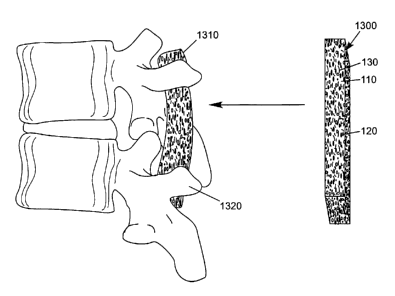
use in trauma and spinal applications, and for the delivery of drugs and
growth factors.
[0003] Bone and soft tissue repair is necessary to treat a variety of medical
(e.g., orthopedic)
conditions. For example, when hard tissue such as bone is damaged as a result
of disease or
injury, it is often necessary to provide an implant or graft to augment the
damaged bone during
the healing process to prevent further damage and stimulate repair. Such
implants may take
many forms (e.g., plugs, putties, rods, dowels, wedges, screws, plates, etc.),
which are placed
into the tissue. Typically, such implants can be rigid, flexible, deformable,
or flowable and can
be prepared in a variety of shapes and sizes. For non-rigid structural repair
materials (e.g.,
putties and pastes) to be conveniently used, they must be capable of being
formed into a variety
of complex shapes to fit the contours of the repair site. An accurately
configured implant that
substantially fills the defect site will enhance the integration of natural
bone and tissue to
provide better healing over time. The prior art discloses medical implants
that comprise, at least
partly, collagen (to be discussed later).
[0004] Collagen is the most abundant protein found in the body. The unique
chemistry of
2
CA 02532829 2005-12-16
WO 2004/112854 PCT/US2004/019805
collagen makes it an ideal polymer for structural and hemostatic applications
in both clinical and
diagnostic settings. Collagen, like all proteins, is comprised of amino acids
linked covalently
through peptide or amide linkages. The sequence of the amino acids, or the
primary structure,
outlines the three-dimensional structure of the protein, which in turn
dictates the function, and
properties of the molecule. Collagen is composed of three peptide chains
associated in a triple
helical orientation. These triple helices associate to form fibrils, which
ultimately make up
connective tissue and other structural members.
[0005] Collagen has been used in a number of applications in the art. For
example, one
application is for use in hemostatic devices for the stoppage of bleeding,
such as is described in
U.S. Pat. Nos. 5,310,407 (Casale) and 4,890,612 (Kensey). However, neither
teaches the use of
native insoluble fibrous collagen. In U.S. Pat. No. 5,425,769, Snyders, Jr.
discloses a
biocompatible and bioresorbable bone substitute with physical and chemical
properties similar to
bone, consisting of reconstituted fibrillar collagen within a calcium sulfate
di-hydrate matrix.
The ratios of calcium sulfate and collagen are adjusted for each application
and the bone
substitute is molded in situ to form a solid phase. Snyders Jr. discloses an
implant that remains
malleable only for a brief period, as the combination of fibrillar collagen
and calcium sulfate di-
hydrate matrix forms a hard composition. Furthermore, the collagen as
described in the '769
patent is neither interlocked, nor interlaced, relying on the calcium sulfate
to lend structural
integrity.
[0006] The polymer utilized for the implant may be combined in application
with a biologically
active agent to enhance the tissue healing response or enhance the mechanical
properties of the
implant (e.g., U.S. Patent No. 4,776,890 (Chu). Chu discloses a process for
creating matrix of
collagen containing mineral particles, such that when wetted, the matrix is
malleable and retains
its integrity. The matrix as claimed by Chu incorporates up to 10% of the mass
as collagen, and
relies on the physical characteristic of the particles comprising the bulk of
the matrix to lend the
integrity, and upon exposure to fluids, would lead to dissociation of the
material unless a cross-
linking step is performed. However, this cross-linking process is disfavored
by Chu, as it would
discourage bone tissue ingrowth.
[0007] Huc et al. (U.S. Patent No. 5,331,092) describes a process for
preparing medical pads by
CA 02532829 2005-12-16
WO 2004/112854 PCT/US2004/019805
grinding collagen, acidifying with acetic acid, homogenizing, molding and
freeze-drying. The
pad formed would readily fall apart upon exposure to aqueous fluids and thus
requires cross-
linking. The cross-linked pads hold together but have limited mechanical
strength limiting their
usefulness to hemostatic pads.
[0008] Nigam (U.S. Patent No. 4,948,540) described a process for preparing a
collagen dressing
material by creating slurry comprised of an acid solubilized collagen and a
non-solubilized
natively cross-linked collagen. The resultant slurry was molded, and freeze-
dried into a pad.
The pad did not have sufficient mechanical properties due to its excessive
porosity and thus was
compressed at a pressure of 15,000-30,000 psi and optionally cross-linked. To
improve strength
due to lack of fiber-to-fiber interaction, the device is compressed without
interlacing of the
individual fibers. The compression serves to compress in only one dimension,
placing the fibers
in close proximity in one orientation, rather than interlacing the fibers.
[0009] Li (U.S. Patent No. 5,206,028) described a process for preparing a
dense collagen
membrane by first freeze-drying a collagen dispersion of random fibers to form
a sponge. This
sponge was then humidified, compressed and subjected to chemical cross-
linking. The resultant
sponge was strong, having randomly entangled masses of fibers going in all
directions. This
device as described by Li lacks interlacing of the insoluble collagen as the
aqueous dispersion is
lyophilized without first interlacing the insoluble components.
[0010] Li (U.S. Patent No. 6,391,333) described a process wherein sheets of
oriented
biopolymeric fibers are formed into sheets by capturing them on a spinning
mandrill that was
rotated in a fibrous collagen slurry. The fibers were then compressed to force
them closer
together so they could be dried, preferably while in contact with a gluing
agent. The sheet was
then cut from the mandrill, inverted and cross-linked to form a sheet.
Additional sheets could be
individually stacked on top of each other to create thicker devices with
greater mechanical
strength. The device as constructed has fibers substantially aligned in
parallel planes, and lacks
equiaxial interlacing.
[0011] In PCT application WO 98/35653, Damien describes a process for
preparing an
implantable collagen putty material by acidifying a collagen solution to a pH
of between 3.0 to
4
CA 02532829 2005-12-16
WO 2004/112854 PCT/US2004/019805
6Ø This produces a non-fibrous dough like material that can be used to
suspend graft material.
At higher pH, the collagen precipitates out, becoming crumbly with a
consistency of wet sand.
[0012] It is well known to utilize a centrifuge or filtration press as a part
of a rinsing procedure,
or a 'wash step' to remove insoluble components contained within the solution.
Nishihara (U.S.
Patent No. 3,034,852) describes a process to solubilize previously insoluble
collagen fibers
without denaturation of the protein structure by using hydrolytic enzymes. In
the examples, the
author describes separation of the fibers from the wash solution by
centrifugation or filtration
press methods, the fibers are then brought back into solution. Additionally,
the fibers, which are
separated using this method are reconstituted fibers which tend to be small in
size.
[0013] Highberger, et. al. (U.S. Patent No. 2,934,446 and U.S. Patent No.
2,934,447) describe a
method, as well as, the physical preparation of collagen fiber masses to form
leather-like sheets
from hide scraps unusable in the traditional leather making process. This
psuedo-leather may
support small colonies of cells but would be unsuitable for tissue ingrowth.
The method of
concentration used is a precipitation technique, which creates a fiber
dispersion. This
slurry/dispersion as described included random clumps of undispersed or
entangled fibers.
Highberger combines a unique fiber that coacts with a high dissolved solids
content collagen
solution to form well knit,'or leather like sheet. In the '447 patent,
Highberger further refines the
process of the '446 patent by incorporating a kneading step, which works the
dough material to
make the product free from lumps, the kneading necessarily disrupts any
interlacing or
interlocking fibers prior to precipitating the solubilized collagen.
DISCLOSURE OF THE INVENTION
[0014] This invention includes malleable, biodegradable, fibrous compositions
for application
to a tissue site in order to promote or facilitate new tissue growth. One
aspect of this invention
is a fibrous component (e.g., collagen, chitosan, alginate, hyaluronic acid,
poly-lactic acid, poly-
caprolactone, and polyurethane) which provides unique mechanical and physical
properties, as
will be discussed.
[0015] Fibers may be suspended within a suspension fluid (preferably aqueous)
forming a
relatively homogenous slurry/dispersion. This dispersion preferably has a low
solid content
CA 02532829 2005-12-16
WO 2004/112854 PCT/US2004/019805
whereby the orgamzmg process, e.g., cenmtuganon, causes the material to have
preferable
mechanical and physical properties.
[0016] The physical properties may include, but not limited to, injectable,
flexible, compression
resistant, or elastic properties. Biologic properties may include, but not
limited to, conductive or
inductive properties for hard and soft tissues. Additionally, in a preferred
embodiment, additives
(e.g., fibers, particulate, or gels) may be used to further tailor the
material properties.
[0017] In a preferred embodiment, the degree of centrifugation is specified to
dictate the
physical properties of the resulting material; alternatively, or in
combination, a rehydration step
may be tailored to affect the physical properties of the material, as will be
discussed. As an
example, the properties of this material may be tailored such that exposure to
rehydration liquids
or bodily fluids (e.g., blood) will render the material to be self supporting.
That is, the material
will not readily slump under its own weight, even though it is readily
moldable by hand pressure.
This can be particularly useful during a procedure where the entire wound site
is not
immediately secured or enclosed by hard tissue or other constraint.
Additionally, the implantable
embodiments may contain biologically active agents, which may aid
osteoconductivity or
osteoinductivity.
[0018] In a preferred method, the material may be created by providing' a
vessel containing a
slurry, said slurry comprising a plurality of natural or synthetic polymer
fibers and at least one
suspension fluid, wherein the polymer fibers are substantially evenly
dispersed and randomly
oriented throughout the volume of the suspension fluid, or at least exhibiting
no significant
organization or preferred orientation; applying a force, e.g., centrifugal, to
said vessel containing
said slurry, whereupon said force serves to cause said polymer fibers to
migrate through the
suspension fluid and amass at a furthest extent of the vessel, generally at a
wall or similar
surface of the vessel, forming a polymer material, with said polymer material
comprising
6
CA 02532829 2005-12-16
WO 2004/112854 PCT/US2004/019805
polymer fibers of sufficient length and sufficiently viscous, interlaced, or
interlocked to retard
dissociation of said polymer fibers.
BRIEF DESCRIPTION OF THE DRAWINGS
[0019] FIG. 1 A, B, and C are enlarged, representative diagrams depicting the
arrangement of
interlacing and interlocking fibers of the present invention, wherein a force
(e.g., centrifugal) is
applied in orientation represented by the arrow.
[0020] FIG. 2 depicts a hydrated malleable mass 400 of interlaced fibers 110.
[0021] FIG. 3 depicts a human extremity 600 that has been surgically opened to
reveal bone 610. A
hydrated malleable mass 500 is being inserted into an exposed osseous defect
620.
[0022] FIG. 4 depicts an insertion of an interlaced fibrous putty 720 into a
confined tissue defect
700. The applied force 710 of the insertion is conducted by the interlaced
fibers of the putty, forcing
it into intimate contact with the walls defining the tissue defect.
[0023] FIG. 5A, B, and C depict a cylinder 1100 of interlaced fibers 110. The
fibers are represented
by open space defined by the dimensions of the cylinder. The interlocking of
the interlaced fibers
110 supports, confines, and locks the particulate material 120 and
biologically active agent 130
within a spatial conformation. The Cylinder 1100 is inserted inside of a
preformed structure or cage
1110 creating a spinal implant 1120.
[0024] FIG. SD depicts spinal implant 1120 being inserted into a defined space
1210 within two
vertebral bodies 1200.
[0025] FIG. 6 depicts dry sheet 1300 of interlaced fibers 110. The fibers are
represented by open
space defined by the dimensions of the sheet. The interlocking of the
interlaced fibers 110 supports,
confines, and locks the particulate material 120 and biologically active agent
130 within a spatial
conformation. As the dry sheet 1300 becomes hydrated, it becomes a conformable
mass 1310 that
can be approximated to the irregular topography of the transverse processes
1320 of the vertebrae.
7
CA 02532829 2005-12-16
WO 2004/112854 PCT/US2004/019805
[0026] FIG. 7 depicts the injection of a hydrated malleable mass 920 of
interlaced fibers from a
syringe 800 into a tissue defect 1000.
[0027] FIG. 8A depicts hollow intermedullary nail 1 S00 having openings 1510
containing graft
material 1520 composed of interlaced, interlocked fibers and particulate.
[0028] FIG 8B depicts the femoral portion of artificial hip prosthesis 1550
having a hollow stem
portion 1560 having openings 1 S 10 containing graft material 1520 composed of
interlaced,
interlocked fibers and particulate.
MODES FOR CARRYING OUT THE INVENTION
[0029] This invention is a malleable, biodegradable, fibrous composition for
application to a
tissue site in order to promote new tissue growth. One aspect of this
invention is a fibrous
component (e.g., collagen, chitosan, alginate, hyaluronic acid, polylactic
acid, poly-caprolactone,
and poly-urethane, etc.) (see table 1) which provides unique mechanical and
physical properties,
as will be discussed.
[0030] Fibers (e.g., collagen, chitosan, alginate, and hyaluronic acid), may
be obtained, for the
example of type I collagen, from bovine hide, which have been processed in a
manner known by
those skilled in the art. As an example, the hides are stripped from the
animal carcass and the
corium (i.e., the intermediate layer of an animal hide between the grain and
the flesh sides) is
subsequently split from the rest of the hide. The corium is limed using a
calcium hydroxide
solution to remove extraneous organic material. The limed corium is then
neutralized using an
acid solution and the excess salt produced is serially rinsed (e.g., with
water). The neutralized
corium is then ground using a mill type apparatus to tear apart the corium
into fibers. This
process maintains the native cross-links within the collagen. (During the
aging process of the
live animal, intermolecular and interfibrillar bonds are naturally formed
between collagen
fragments. These naturally occurring bonds are what distinguish the fibers of
collagen as
natively cross-linked fibers as opposed to reconstituted fibers of collagen).
The fibers are
suspended within a suspension fluid (preferably aqueous) forming a relatively
homogenous
slurry/dispersion. This dispersion preferably has a solid content ranging from
0.25 to 10%, but
8
CA 02532829 2005-12-16
WO 2004/112854 PCT/US2004/019805
most preferably m the range of 3 to 5% by weight.
[0031] The resultant slurry is concentrated by centrifugation; preferably, at
temperatures below
60 degrees Celsius to avoid degradation of the collagen and subsequent gelatin
formation. Speed
and therefore force, as well as time can be varied to create the desired
extent of fiber interlacing,
as will be discussed later. The slurry can be spun under forces of about 500
Xg (times gravity) to
forces as high as about 30,000 Xg and for times ranging from 10 seconds to 96
hours.
Preferably, the suspension is spun at forces of 500 Xg to 10,000 Xg and times
of 1 minute to 24
hours. Most preferably the suspension is spun at 3000Xg for about 5 minutes.
This creates, in a
preferred embodiment, a paste or putty-like structure containing interlocked
or interlaced
collagen fibers, as will be discussed, that may be molded and dried by either
evaporation to
create a high density non-porous unit, or by lyophilization to maintain the
three dimensional
structure and porosity. An additional preferred embodiment comprises a
material which is a
flowable, yet dense, material, which may be injected or otherwise molded. This
moldable
embodiment may be cast into a mold, or in situ, as will be discussed.
[0032] Referring to Figure 1A, the interlacing phenomenon occurs as the fibers
27 and 28
migrate down through the suspension fluid, and the individual fibers interlace
among other
fibers, herein represented by interlacing fiber 28 becoming interlaced with
other fibers 27,
entering a region 29 bordered by other fibers 27. In Figure 1 A, it is
desirable to start with a low
solids content (e.g., below 10%) slurry so that the fibers are uniformly
distributed and freely
moving prior to centrifugation. Entangled clumps of fibers (not shown) may
interfere with
proper interlacing and should be minimized by starting with the low
concentrations. Interlacing
is a non-directional interlocking of fibers 27 randomly in three dimensions
throughout the
material, as opposed to layer-like or directional entanglement of the fibers,
as will be discussed.
Figure 1B depicts interlacing 33 (represented by the zone within the dashed
circle), as the low
concentration fiber mix migrates and gradually intertwines itself during
centrifugation, but does
so in a three dimensional type of formation. Without wishing to be bound to
any particular
mechanism or explanation, it is believed that this phenomenon occurs because
there is a nearly
uniform load on all of the fibers 27, with the exaggerated gravitational load
(i.e., from the
centrifuge) tending to move the fiber 28, without necessarily rotating it.
This, in turn, causes the
fibers 27 and 28 to eventually coalesce and at least partially thread
themselves together, to some
9
CA 02532829 2005-12-16
WO 2004/112854 PCT/US2004/019805
degree as the interlacing fiber 28 enters the region 29 bordered by the other
fibers 27.
[0033] As the interlacing 33 continues, the fibers come into contact with the
surface of the
container, in the centrifuge, or other fibers already compressed with the
surface. As the fibers
continue the migration, and eventually "pile up" or amass, they become further
interlaced and
eventually the fibers 28 may be deformed or bent as the pressure of other
migrating fibers builds.
[0034] Referring to Figure 1 C, as the pressure builds, and the fibers 27 and
28 compress, the
interlacing step is completed, and the deformation of the fibers causes them
to interlock 34
(depicted by the dashed circle). It is this dual interaction of interlacing
and interlocking that
results in the unique properties of certain embodiments of the present
invention.
[0035] In these various embodiments, the fibers are not externally pressed
together (not shown),
which causes the higher end of the fibers to be pressed downward (causing
alignment) by the
externally applied force (e.g., a platen). The non-uniform force ultimately
causes a directional or
anisotropic fiber bundle; similarly, the evaporation of the fluid from a
preferential direction
causes fiber bundling and alignment at the evaporating surface/interface.
[0036] Additionally, interlacing restricts the motion of fibers within a unit
during rehydration,
since all external surfaces are comprised of fibers which intrude into the
material itself (not
shown), thereby retarding disassociation of fibers from the unit upon contact
with a fluid. Upon
rehydration the dried structure forms a paste or putty-like material similar
in characteristic to that
of the pre-dried material, as will be discussed.
[0037] Likewise, interlacing of the fibers also provides greater mufti-
directional consistency
(i.e., isotropy) in the mechanical properties as opposed to directional
mingling of fibers. That is,
the interlacing is three dimensional, and therefore provides uniform
properties in the three
dimensions; whereas the directional materials previously discussed yield
materials with
properties along the fiber axis (e.g., the plane of flattening or evaporation)
that are very different
from the properties perpendicular to the fiber axis.
[0038] Interlacing also provides biologic advantages over directional
entanglement by providing
CA 02532829 2005-12-16
WO 2004/112854 PCT/US2004/019805
an equiaxial structure (or a structural that more resembles an equiaxed
structure's lack of
directionality) for cellular infiltration as well as an advantageous platform
for tissue formation.
A structure that allows cells to infiltrate uniformly in all directions may
improve overall tissue
organization, and may also avoid, in soft tissue regeneration, the directional
bundling common in
fibrous scar tissue.
[0039] Centrifuging materials such as fibrous collagen will also create
chemical linkages aside
from physical interlacing that serve to reinforce the resulting matrix. In the
particular case for
collagen, the centrifugal force brings individual fibers and fibrils into
close molecular proximity,
and re-establishes non-covalent forces such as hydrogen bonding,
hydrophobic/hydrophillic
interactions, and electrostatic interactions, that these individual fibers and
fibrils previously
embodied in the native, pre-extracted tissue. These additional chemical
linkages may act to
create a pseudo-molecular weight increase to the matrix. Thus, the
combinatorial effects of
physical interlacing and chemical bonds impart unique cohesive properties and
viscosity to these
fibrous putties.
[0040] Although centrifugation is a preferred process, a similar interlacing
may be
accomplished by placing a low concentration slurry (e.g., less than 15%
polymer) into a closed
container and exposing it to a global force such as multi-axis gyroscopic
mixing. This multi-
axis mixing process causes flocculation of the fibers as they migrate through
the suspension fluid
towards the center of the closed container wherein they become interlaced with
one another. The
clumps of interlaced fibers that amass may then be exposed to a force that
extracts a portion of
the suspension fluid thereby allowing the fibers within the interlaced clumps
to become
interlocked with each other.
[0041] To improve the migration of fibers and prevent clumping during the
interlacing process,
as described above and expanded upon below, it is preferred to incorporate a
percentage (e.g.,
0%-50% by mass of fibers) of one or more lubricants (e.g., biocompatible oils,
hydrogels, liquid
polymers, low-molecular weight polymers, glycosaminoglycans, surfactants,
waxes, fatty acids,
fatty acid amines and metallic stearates such as zinc, calcium, magnesium,
lead and lithium
stearate, etc.) into the suspension fluid. A lubricant is defined as a
substance, which is capable of
making surfaces smooth or slippery. These characteristics are due to a
reduction in friction
11
CA 02532829 2005-12-16
WO 2004/112854 PCT/US2004/019805
between the polymer fibers to improve flow characteristics and enhance the
knitting and wetting
properties of the fibers. The lubricant may be liquid or solid and may be
suspended or dissolved
in a Garner solvent (e.g., water, alcohol, acetone, etc.). Additionally the
lubricant may only
become lubricious under shearing force or change in temperature. The lubricant
may remain in
its entirety in the final invention; may be partially removed in the
dehydration/desolvation
process; or, may be washed out or removed by methods known in the art during
further
processing. Lubricants that remain in the final invention may be biologically
active agents or
may form microstructures. Preferred lubricants include Tween-80, hyaluronic
acid, alginate,
glycerin or soluble collagen with the most preferred being acid soluble
collagen such as Semed S
produced by Kensey Nash Corporation (Exton, PA).
[0042] Additional ways in which to add lubricity include physically or
chemically altering the
surface of the fibers making up the composition. Such alterations can be
achieved through
chemical or physical attachment of a lubricious substance to the fibers,
temperature induced
phase changes to the surfaces of the fibers or partial solubilization of the
fibers through
alteration of the pH and/or conductivity of the free fluid or use of a
percentage of solvent for the
fibers within the free fluid. Other methods of creating lubricity are known to
those skilled in the
art, and are embraced by this disclosure.
[0043] During rehydration, the material absorbs liquid (e.g., water, bodily
fluids, blood, etc.) to
the limits of the void (i.e., pore) volume. Since a large portion of the
surface fibers are intruding
in the material, the inward ends are locked by mechanical and/or frictional
means. This aspect
of the interlacing phenomenon causes the material to remain intact while the
fluid ingresses,
with minimal fiber liberation to the non-absorbed liquid. In contrast,
directionally pressed
materials lose surface fiber during rehydration, since they are not anchored
or interlocked (i.e.,
they lay parallel to the surface of the material).
[0044] In these various embodiments, the material will freely absorb liquid
until the
approximate pre-dried volume is attained; at which point the material is in a
state of pseudo-
equilibrium. This state is achieved because the interlocked fibers are re-
hydrated, returning to
their natural state (i.e., centrifuged position). The continued absorption of
liquid will, over an
extended period of time, cause the fibers to de-interlace (i.e., the working
free of interlaced
12
CA 02532829 2005-12-16
WO 2004/112854 PCT/US2004/019805
other mechanical tocxmg mthm a region wnnout total fiber separation, allows
shifting or
movement of zones that remain interlaced. Resistance to fiber movement within
the interlaced
zones, combined with the frictional forces of the de-interlaced regions gives
the material the
paste or putty-like consistency. In contrast, traditional collagen materials
would rapidly
dissociate into the re-hydrating fluid.
[0045] The continued exposure to liquids will cause the material to swell
beyond the
aforementioned pseudo-equilibrium, and impart some de-interlacing, however,
this absorption
occurs at a rate significantly less than the rate prior to achieving pseudo-
equilibrium. This
attribute in itself, may be useful, because certain embodiments of this
invention may be sized for
particular types of defects, and the near equilibrium state will be more
easily achieved without
careful monitoring of the rehydration level of the material.
[0046] Whether the optimum absorption level is achieved may be moot, because
the size can be
altered or molded by applying pressure (e.g., squeezing) to the material to
cause the expulsion of
some of the absorbed liquid. This feature is attractive, since it renders the
material tailorable
with regard to size, shape and consistency. Additionally, in a preferred
embodiment, some or all
of the liquid may be absorbed in vivo, thereby causing intimate contact along
the entire defect
cavity.
[0047] As shown in Figure 2, the consistency of the various embodiments of the
invention
allows them to form a malleable putty/paste 400 that can conform to unique
shapes and contours
encountered in tissue-engineering applications. The interlocking of the
interlaced fibers 110
retards dissolution of the device, allowing it to be used in unconfined wounds
(e.g., segmental
defects as shown in Figure 3). Figure 3 depicts a human extremity 600 that has
been surgically
opened to reveal bone 610, a hydrated malleable mass 500 is being inserted
into an exposed
osseous segmental defect 620.
[0048] Similarly, the interlocking of the interlaced fibers 110 retards
dissolution of the device,
allowing it to be used in confined wound spaces (e.g., a tissue void as shown
in Figure 4).
Figure 4 depicts an insertion of an interlaced fibrous putty 720 into a
confined tissue defect 700.
13
CA 02532829 2005-12-16
WO 2004/112854 PCT/US2004/019805
The apples force ~ 1 U of the msernon is conducted by the interlaced fibers of
the putty, forcing
it into intimate contact with the walls defining the tissue defect.
[0049] The presence of interlaced fibers 110 in the devices makes it
particularly suited to those
tissue defects exposed to irngation, especially at volumes and/or flow rates
where current state
of the art putties fail (e.g., disintegrate, disassociate, break apart). The
embodiment of Figure 2
is suitable for, but not limited to: cell culture support and transfer,
cartilage defect filling, bone
void filling, soft tissue augmentation (e.g., removal of dermal creases) and
periurethral bulking
to combat urinary incontinences arising from such conditions as intrinsic
sphincter deficiency.
Additional possibilities include but are not limited to use as a spinal cage
filler (as shown in
Figure 5), depicting a cylinder of interlaced fibers. The fibers are
represented by open space
defined by the dimensions of the cylinder. The interlocking of the interlaced
fibers may support,
confine, and lock the particulate material and biologically active agent
within a spatial
conformation (as will be discussed later). The cylinder 1100 of Figure SA, is
inserted inside of a
preformed structure 1110 of Figure SB, or cage creating a spinal implant 1120
of Figure SC,
which may then be implanted substantially as shown in Figure SD.
[0050] An alternate use of the putty is depicted in Figures 8A and 8B, wherein
the putty
material is inserted inside of an intermedullary nail 1500 (Fig. 8A) and the
femoral shaft portion
1560 of a hip prosthesis 1550 (Fig. 8B). At least a portion of the rigid
implant, that which
extends into the bone, would be porous, or hollow and containing holes 1510
through the wall of
the implant capable of receiving the putty 1520 and allowing ingrowth of host
tissue. This
would allow bone to grow through and ultimately in and around the rigid
implant, thereby
effectively anchoring it to the rest of the surrounding bone. The putty
utilized as a graft material
may include particulates and/or biologically active agents. It is recognized
that the particulate
material itself may be functional as a biologically active agent (e.g.
Demineralized Bone Matrix,
etc.) The novel concept of providing a prosthesis containing a hollow zones)
capable of
receiving graft material is useful in the creation of dental implants or any
other implant, which
requires anchoring to host tissue such as an ocular prosthesis or mechanical
heart valve.
[0051] Alternatively, Figure 6 is another possible use for the putty, depicted
as a graft overlay to
retain osteoconductive/osteoinductive grafting material (e.g., harvested bone
chip, ceramics, etc.)
14
CA 02532829 2005-12-16
WO 2004/112854 PCT/US2004/019805
during a transverse process spinal fusion (as shown in Figure 6). FIG. 6
depicts dry sheet 1300
of interlaced fibers 110. The fibers are represented by open space defined by
the dimensions of
the sheet. The interlocking of the interlaced fibers 110 may support, confine,
and lock the
particulate material 120 and biologically active agent 130 within a spatial
conformation (as will
be discussed later). As the dry sheet 1300 becomes hydrated, it becomes a
conformable/malleable mass 1310 that can be approximated to the irregular
topography of the
transverse processes 1320 of the vertebrae. In these additional applications
the malleable
characteristics of the hydrated device will allow it to conform to the unique
shaped chambers of
the spinal cage or the irregular topography of the transverse process surgical
site. In a transverse
process surgical procedure the implant covers and secures the graft material
(not shown) in
place.
[0052] In another embodiment (not shown) the putty is preformed into a cup and
utilized to
retain graft material placed in and around the acetabulum during a hip
reconstruction. The putty
is then sandwiched between the host bone and the artificial cup that will
receive the prosthesis.
If additional toughness is required, the cup can be cross-linked to provide a
flexible article that ,
may further feature shape-memory. Additionally, the putty can be packed around
the stem of the
prosthesis prior to its insertion into the long bone cannel. If desired, a
novel prosthesis (as
described above for Figs. 8A and 8B) with a stem having a hollow cage-like
appearance can be
utilized. The putty is inserted into the cage wherein it is then dried and
sterilized. Optionally, the
dried putty can be cross-linked to prevent egress from the prosthesis. This
same novel cage-like
structure can be utilized for nails placed in the shaft of long-bones.
[0053] In another embodiment, the putty material contains reinforcing
materials such as long
threads, meshes or other fibers (not shown). The interlocking of the
interlaced fibers supports,
confines, and locks the reinforcing material within a spatial conformation.
This retards the
reinforcing material from migrating within or dissection from the putty or
paste. This can be
used to alter mechanical properties (e.g., compressive strength) as well as
enhance resistance to
disassociation of fibers form the construct. Additionally, the putty may
improve the
biocompatibility of the reinforcing material (e.g., improved cellular
migration within or adhesion
to a mesh). The reinforcing material may be centered within the construct,
located on or just
below one or more surfaces or interspersed throughout the entire construct.
CA 02532829 2005-12-16
WO 2004/112854 PCT/US2004/019805
[0054] In another embodiment the interlocking of the interlaced fibers is used
to control the
location and delivery of biologically active agents (e.g., growth factors,
hormones, BMP, drugs,
cells, viruses, etc.) (see table 2). The unique equiaxial formation of the
device controls flow of
fluid (e.g., blood, interstitial, etc.) within the device allowing for
tailored release properties. The
biologically active agents could be located within or supported between the
fibers making up the
device. Additionally, the biologically active agents could be mechanically or
chemically
attached or bonded to the fibers or suspended within a hydration fluid. This
hydration fluid may
contain a soluble polymer that suspends or binds the biologically active
agent. Additionally, the
hydration fluid containing the soluble polymer may be removed leaving the
soluble polymer as a
solid sheet or coating adhered to the fibers, an open laced network of strands
adhered to the
polymer fibers; a velour, felting or loosely woven sheet adhered to the
polymer fibers; or as a
porous foam or microstructure suspended between the fibers. In addition to
supporting a
biologically active agent, the hydrated soluble polymer can function as a
lubricant to aid in
partial de-interlacing of the polymer fibers during molding or implantation.
[0055] It is also conceived that in one embodiment of this invention the
material can contain an
additive that can be used to help deliver or retain the previously described
biologically active
agents. As an example, the interstices of the gross fibrous structure could be
invested with a
soluble polymer as defined above, e.g., a chemotactic ground substance, such
as the velour of
hyaluronic acid. A velour of chemotactic ground substance could accomplish
several
biochemical and biomechanical functions essential for wound repair. For
example, since
hyaluronic acid is extremely hydrophilic, it may be valuable for drawing body
fluid (e.g., blood,
bone marrow) or other fluid-based biologically active agents into the fibrous
device. Upon
hydration, the hyaluronic acid can become an ideal Garner for pharmacological
or biologically
active agents (e.g., osteoinductive or osteogenic agents such as the bone
morphogenetic protein
(BMP) and other bone-derived growth factors (BDGF)) by providing for chemical
binding sites,
as well as by providing for mechanical entrapment of the agent as the velour
forms a hydrogel.
[0056] It is also conceived that a source of growth factors (e.g., platelet-
rich plasma, bone
marrow cells, etc.), whether synthetic, autologous or allograft in
origination, can be delivered
with the device of this invention (e.g., incorporated into the implant during
manufacturing or
16
CA 02532829 2005-12-16
WO 2004/112854 PCT/US2004/019805
integrated into the device prior to implantation). For example, it is known
that one of the first
growth factors to initiate the cascade leading to bone regeneration are
platelet-derived growth
factor (PDGF) and transforming growth factor-beta (TGF-13). Each of these
growth factors is
derived from the degranulation of platelets at the wound, defect or trauma
site. It is believed that
increasing the presence of such platelets at the wound or trauma site can
increase the rate of
healing and proliferation needed to regenerate tissue (e.g., bone).
[0057] The application of platelet-rich plasma (PRP) is one way to deliver a
highly concentrated
dose of autologous platelets. PRP is easily prepared by extracting a small
amount of the
patient's blood and processing it, for example, using gradient density
centrifugation, to sequester
and concentrate the patient's platelet derived growth factors. Other
preparation methods remove
water from the huffy coat (i.e., coagulated blood coating) and utilize
filtering systems to
concentrate platelets and fibrinogen. It is believed that applying PRP or
other autologous growth
factors to the wound site in conjunction with the subject invention will
increase the amount of
PDGF and TGF-f3 available for jump-starting the healing process. PRP can be
prepared for
procedures with small volumes of blood, drawn by the doctor or nurse
presurgically. Typically,
40-100 ml of blood are drawn preoperatively and placed in a PRP preparation
unit. SmartPREP
(Harvest Technologies Corp., Norwell, MA) and UltraConcentrator (Interpore
Cross, Irvine, CA)
are device that have been shown to effectively produce PRP for OR, office
implant, and
periodontal uses.
[0058] Once the PRP is prepared, other additives (e.g., activator, growth
factor, drug, chemical,
bone, etc.) can be added to the plasma. For example, to infuse the implant
material of this
invention with a PRP gel preparation, the ratio of ingredients would include a
higher proportion
of PRP to allow the PRP to more effectively flow through and permeate through
the putty
material. It is also conceived that the de-hydrated putty can be inserted into
the PRP preparation
unit (e.g., centrifuge, concentration unit). In this fashion, the platelets
can be concentrated right
into or onto at least a portion of the implant directly. For example, some PRP
devices include a
centrifuge for separation of the blood components. The biomaterial implant
could be positioned
within the centrifuge such that the desired blood constituent is directed into
the implant material
during processing.
17
CA 02532829 2005-12-16
WO 2004/112854 PCT/US2004/019805
[0059] Other autologous materials can also be incorporated into and used in
conjunction with
the subject invention (e.g., autologous bone marrow cells (BMC)). Bone marrow
contains
osteogenic progenitor cells that have the ability to form and repair bone. The
marrow can be
harvested and dispersed into single cell suspensions. The cells can then be
concentrated (e.g.,
through filtering, centrifucation) or used as is. The resulting mixture can be
diluted and
implanted into the wound site, incorporated into the implant material, or
delivered by the
delivery system (e.g., syringe) with the materials of the subject invention.
[0060] In another embodiment, the interlocking of the interlaced fibers is
used to control the
location and orientation of particulate components compounded into the fibrous
material (e.g.,
ceramic, glass, glass-ceramic, metals, tricalcium phosphate, Hydroxylapatite,
calcium sulfate,
autologous bone graft, allograft bone matrix, polymers, microspheres,
microcapsules, hyaluronic
acid, collagen, chitosan, alginate, hyaluronic acid, poly-lactic acid, poly-
glycolic acid, poly-
caprolactone, polyurethane, etc). The particulate components may additionally
carry or serve to
deliver biologically active agents. The interlacing supports, confines, and
locks the particulate
components within a spatial conformation. This retards the particulate from
migrating within or
disassociating from the putty or paste. (When the fibrous material is combined
with a fine
powdered ceramic, the consistency is more chalk-like than that of putty or
paste formed with
larger ceramic particulate.) A fibrous slurry containing particulate may be
concentrated using
centrifugation as mentioned above, or particulate may be added as a solute to
a rehydrating
solvent. Alternatively, the particulate may be mechanically incorporated
(e.g., kneaded) into the
interlaced fibrous putty. The resulting material may be implanted or dried and
rehydrated with a
volume of liquid to yield a desired density or consistency to the paste or
putty. It should be noted
that previously dried putty is suitable for implantation dry wherein it is
rehydrated by body fluids
(e.g., blood).
[0061] When adding particulate, the addition of a soluble polymer to increase
the viscosity of
the aqueous solution prevents premature separation or stratification of the
particulate from the
collagen fibers in the final product. Additionally, the fluid containing the
soluble polymer can be
removed, leaving the particulate entrapped within the soluble polymer as a
coating on the fibers
or suspended between the fibers.
18
CA 02532829 2005-12-16
WO 2004/112854 PCT/US2004/019805
[0062] When porous particulates are used, the fibers randomly penetrate the
pores and become
interlaced along with the fibers, creating a single continuous network of
fibers and particulate.
The particulate may create a type of hub with fibers radiating out to other
hubs. This creates a
unique structure wherein particulate loss is reduced. Additionally, cells
migrating into the
structure along the fibers may be guided to the incorporated particulate.
[0063] The concentrations of the putty or paste, as well as the extent of
interlacing between
fibers that result from centrifizging, produce characteristics that range from
smooth injectable
gels to highly compact masses with elastic type qualities. For instance, the
centrifuge may be
used to spin down fibrous slurry into blocks or uniquely shaped molds (e.g.,
tubes, ears, nose,
cones, brick, plate, disk, ellipse, sheet, membrane, wedge, pin, rod,
cylinder, roles, cup, sphere,
semi-sphere, pyramid and a frustum of a cone, wedge or pyramid, etc).
Additionally, material
properties provided by the action of partial de-interlacing (as described
previously) of
centrifuged fibrous slurry allow the material to be: 1) injected through a
syringe; 2) pressed
between plates; 3) injection molded; 4) rolled out into flat sheets; 5) carved
and/or formed like
clay; or 6) aerated. This partial de-interlacing allows for an additional way
in which to form
shapes listed above.
[0064] In an embodiment, there may be a benefit to the creation of an
interlaced and interlocked
fibrous composition as described above, further featuring a plurality of
pores. The pores may be
created by techniques known in the art (e.g., a gas expansion process, a
freeze drying process,
etc.). The pores may be of a closed cell morphology, open cell and
intercommunicating
morphology, or a combination of both. The pores may be of a regular, ordered
size or shape, or
alternatively may vary in size or shape. The shape and size of the pore may be
manipulated
through various pore formation techniques known in the art.
[0065] For example, the flow caused by the movement of the fibers during
centrifuging may
create equiaxial and/or elongated pores (dependant of fiber length) within the
final product post-
drying (e.g., lyophilization, air-dried, etc.) Additionally, freeze rate,
freeze direction,
temperature gradients and insolubles (fibers and particulate) can be used to
control crystal
formation, that in turn controls pore size, shape and orientation.
Furthermore, as the liquid
freezes to solid crystals, the fibers and particulates have an impact on pore
formation as they
19
CA 02532829 2005-12-16
WO 2004/112854 PCT/US2004/019805
potentially interfere with crystal growth. As the temperature of a mixture is
lowered, crystals
form within the fluid surrounding the fibers and particulate. As the crystals
grow they force the
fiber and particulate material aside, thereby effectively increasing the
concentration of the
material between the crystals. The growth of the crystals may be disrupted as
they come in
contact with the fibers and particulate. This interruption of crystal growth,
either stops the
growth of the crystal or forces them to grow around the particles in an
irregular fashion. After
solidification the crystals of the frozen liquid are removed by methods know
in the art (e.g.
vacuum drying or leaching) leaving irregular pores.
[0066] Orientation of fibers, along with the interlacing (to keep the fibers
as one mass), that
allows putty-like dispersions to be injected through a syringe. This is
depicted in Figure 7,
wherein the syringe 800 injects a hydrated malleable mass 920 of interlaced
fibers into a tissue
defect 1000. The capability of the fibrous implant material in the form of the
hydrated malleable
mass 920 to be delivered via syringe 800 makes it suitable for use in
laparoscopic, arthroscopic
and endoscopic procedures. These minimally invasive surgical procedures
utilize cannulas and
trocars to remotely treat or repair a variety of injuries or maladies. It
should be understood that
the fibrous implant's unique viscosity allow for the delivery of the material
to remote sites
within the body. Once delivered, the material can remain intact and stable at
the delivery
location for a period of time post implantation.
[0067] It should also be noted that use of reinforcing materials (polymer
mesh, tricalcium
phosphate, etc.) or addition of biologically active agents (growth factors,
DBM, cells, drugs, etc.)
may be employed as a particulate or other addition. Additions may be made in
an effort to
increase the viscosity of the pre-centrifuge process liquid, but the addition
may also be used to
coat the fibers. This fiber coating may be employed to tailor the inner
environment of the
material, and may improve, e.g., the osteoconductivity or osteoinductivity.
These coatings or
other additions may be uniformly dispersed throughout the fibrous structure,
or more sporadic.
In a preferred embodiment, the coating or additive will create a
microstructure, adherent to the
fibrous macrostructure. In certain embodiments with microstructural additions
the
microstructure may be more prominent at junction points, or regions where
several fibers come
in contact with each other. In a preferred embodiment these microstructurally
coated junctions
serve to attract and nourish the inbound cells.
CA 02532829 2005-12-16
WO 2004/112854 PCT/US2004/019805
[0068] In another embodiment, the centrifuge process yields a material with a
viscous high-
density structure that, in itself, is useful for surgical procedures. For
example, this unique fiber
arrangement, regardless of the degree of interlacing or interlocking, if any,
renders the material
suitable for hand molding or injecting via syringe. The unique three-
dimensional nature of this
structure of this material exhibits properties not seen in the art.
[0069] In another embodiment, the materials made by these various processes
may be cross-
linked to impart improved characteristics such as: mechanical strength (e.g.,
suturablity,
compression, tension, etc.), and biodurability (e.g., resistant to enzymatic
and hydrolytic
degradation). This may be accomplished using several different cross-linking
agents, or
techniques (e.g., thermal dehydration, EDC, aldehydes (e.g., formaldehyde,
gluteraldehyde, etc.),
natural cross-linking agents such as genipin or proanthocyanidin, and
combinations thereof).
Each type of cross-linking agent/technique or combinations thereof imparts
diverse mechanical
and biological properties on the material. These properties are created
through the formation of
unique chemical bonds that stabilize the construct. This stabilization greatly
increases the
construct's ability to hold a shape and conformation, thereby, preserving the
interlaced
relationship between the fibers.
[0070] As an example of cross-linking, the construct may be placed in 100mM
EDC solution
contained in pH 5.4 MES buffer for 1 minute to 24 hours, preferably 4 hours.
This creates a
chemical bond between amino and carboxyl acid groups to form amide linkages.
The device is
then rinsed and dried by either lyophilization or simple evaporation.
[0071] This newly stabilized device, containing interlaced fibers, has
superior mechanical and
biological properties as compared to prior art constructs. The interlaced
fibrous structure guides
cellular ingrowth creating newly regenerated tissue that more closely
approximates natural tissue
than can be achieved via a random structure. Additionally, the three
dimensional interlaced
structure allows for the occurrence of directional, biomechanical stimulus
useful in the
regeneration of tissue which is exposed to mechanical motion. This can be seen
in tissues such
as cartilage, intervertebral discs, joint meniscus, blood vessel, heart
valves, or the like.
21
CA 02532829 2005-12-16
WO 2004/112854 PCT/US2004/019805
[UU72] In various embodiments of the invention the collagen putty, that has
been formed or
shaped by any methods known to those skilled in the art, can be cross-linked
to create uniquely
shaped biodurable medical devices (not shown). The devices may take on forms
such as sheets,
tubes, roles, blocks, cylinders brick, plate, disk, ellipse, membrane, wedge,
pin, rod cup, sphere,
semi-sphere, cone, pyramid, frustum of a cone, wedge or pyramid or pads useful
for tissue
augmentation, replacement, or repair. Additionally the devices can be shaped
into unique
anatomically specific shapes (e.g. nose, ear, chin, etc).
[0073] In one embodiment the interlocking of the interlaced fibers allows a
highly concentrated
putty to be rolled flat and stressed in three dimensions simultaneously,
producing an intact sheet
that can be cross-linked; whereas directionally oriented fibers would tear
apart or experience
separation during the flattening process (not shown). Therefore, this material
would be useful in
such applications as, but not limited to dura repair, skin grafting
procedures, hernia repair,
rotator cuff repair, ligament repair or bladder support or repair.
[0074] In another embodiment (not shown) the sheet produced in the previous
embodiment is
rolled prior to cross-linking to create a unique spiral configuration having a
plane separating
each successive revolution of the sheet. The plane provides unique compressive
qualities, that
when combined with the compressive qualities of the cross-linked interlaced
fibers, is ideal for
applications receiving directional compressive loads. These applications
include but are not
limited to joint meniscus, intervertebral disk and articular cartilage. In
another embodiment the
plane formed by the spiral configuration can be filled with materials to
enhance its mechanical or
biologic characteristics (e.g., reinforcing materials, particulates,
biologically active agents,
natural and synthetic polymers).
[0075] Various of these shaped embodiments may also be manufactured in
composite laminate
form. That is, flat sheet or shaped embodiments, may be affixed to other
materials, by pressing,
gluing, or means known to those skilled in the art. These macro-composites may
combine the
materials of these embodiments with material, i.e., with higher strength,
osteo conductivity,
resorbability, etc.
[0076] In another embodiment (not shown) a fibrous collagen slurry can be spun
down into a
22
CA 02532829 2005-12-16
WO 2004/112854 PCT/US2004/019805
mold that approximates the gross anatomy of a tissue or organ (e.g., blood
vessel, heart valve,
ear, nose, breast, finger-bones, long bone, acetabular cup, etc.) prior to
cross-linking. The
interlocking of the interlaced fibers, formed during this process, provides
superior shape holding
characteristics due to the unique resistance to fiber disassociation, as
previously described.
Constructs made using oriented fibers defined in prior art do not hold crisp
margins. Therefore,
material in this embodiment would be useful as, but not limited to, devices
for cosmetic and
reconstructive surgery, intervertebral disks, joint meniscus and hollow
tissues and organs (e.g.,
intestine, esophagus, ureter, etc.).
[0077] In another embodiment (not shown) a fibrous collagen slurry can be spun
down into a
mold containing a structure or component (e.g., ring, mesh, particulate,
screw, rod, screen, etc.)
to which the interlaced fibers migrate around, thereby creating a mechanical
lock, after which
cross-linking may occur. The interlocking of the interlaced fibers supports,
confines, and locks
the structure or component within a spatial conformation.
[0078] In another embodiment (not shown) the partial de-interlacing of zones
within a putty or
paste facilitates compression or injection of the material into or around
structures such as, but
not limited to: molds, screws, rings, rods, cavities, meshes or screens.
Injected into a tube mold
the material would be suitable as a vascular graft or nerve conduit. Injected
into more massive
and possibly complex shapes, the material would be suitable for applications
such as:
intervertebral disks, soft tissue augmentation, ocular prosthesis, joint
meniscus, bone void or soft
tissue filler, and applications in plastic and reconstructive surgery.
[0079] Additionally, material may contain reinforcing materials such as long
polymer threads or
meshes or may include particulates or biologically active agents. (e.g.,
growth factors, hormones,
bmp, drugs, cells, viruses, etc.) Additionally, the biologically active agents
could be located
within fibers making up the putty, mechanically or chemically attached to the
fibers making up
the putty, between the fibers, or suspended within a hydration fluid or second
soluble polymer
intermixed with the fibers of the putty material. The biologically active
agents and/or soluble
polymer intermixed with the fiber may be added prior to or after cross-
linking.
[0080] It is conceived the interlaced polymer material may be manufactured by
the
23
CA 02532829 2005-12-16
WO 2004/112854 PCT/US2004/019805
centrifugation process heretofore described, and may be sterilized and
packaged, or alternatively
dried (e.g., by lyophilization or evaporative processes) then sterilized and
packaged for later use.
It is also recognized that either the wet product or the dry product may be
terminally sterilized.
[0081] The following examples are given for purposes of illustration to aid in
understanding the
invention and it is to be understood that the invention is not restricted to
the particular
conditions, proportions and reagents set forth therein.
Example 1:
[0082] Fibrous Collagen, 4% solids in water by weight, pH 5.3-5.9, was placed
mixed with
powdered (6 micrometers) (3-tricalcium phosphate until a homogeneous mixture
was achieved.
This dispersion was centrifuged at 3200 Xg for 2 minutes to reduce the mixture
40% by volume.
The supernatant was poured off and discarded. The "pellet" was removed from
the centrifuge
tube, placed in a mold, and freeze-dried. This same processed was followed
with a larger
particle size (500-1000 micrometers) (3-tricalcium phosphate. When centrifuged
under the same
conditions the resulting dispersion was reduced 60% by volume.
Example 2:
[0083] Fibrous Collagen, 4% solids in water by weight, pH 5.3-5.9, was placed
in a centrifuge
tube. The dispersion was centrifuged at 8000 Xg for 24 hours. The supernatant
was poured off.
The solution was reduced by ~90% volume loss. This dough-like mass was then
shaped into a
mold and freeze-dried. The resultant sponge was then cross-linked using a
thermal dehydration
to lock in the molded shape. Upon rehydration, the resultant sponge held its
shape and showed
high resistance to compression. It was also noted that the sponge contained
some elastic
properties. These elastic properties allowed the sponge to be warped, twisted,
and manipulated
after which it returned to its original confirmation, thereby displaying shape
memory. This may
be useful when inserting through a narrowing.
Example 3:
[0084] Fibrous Collagen, 4% solids in water by weight, pH 5.3-5.9, was placed
in a centrifuge
tube. The dispersion was centrifuged at 8000 Xg for 4-5 hours. The supernatant
was poured off.
The solution was reduced by ~80% volume loss. This dough-like mass was then
rolled flat
24
CA 02532829 2005-12-16
WO 2004/112854 PCT/US2004/019805
using a rolling pin or a two roller system to create a high fiber density
sheet. The sheet was
freeze-dried and cross-linked using a 100 mM EDC solution (pH 5.4) in water.
The sheet was
allowed to soak in the cross-linking solution overnight and then serially
rinsed 3X for 2 hours
with agitation in water. This sheet exhibited high resistance to tearing and
ripping.
[0085] Table 1: Examples of Biodegradable Polymers for Construction of the
Device
Aliphatic polyesters
Bioglass
Cellulose
Chitin
Collagen
Copolymers of glycolide
Copolymers of lactide
Elastin
Fibrin
Glycolide/1-lactide copolymers (PGA/PLLA)
Glycolide/trimethylene carbonate copolymers (PGA/TMC)
Hydrogel
Lactide/tetramethylglycolide copolymers
Lactide/trimethylene carbonate copolymers
Lactide/s-caprolactone copolymers
Lactide/a-valerolactone copolymers
L-lactide/dl-lactide copolymers
Methyl methacrylate-N-vinyl pyrrolidone copolymers
Modified proteins
Nylon-2
PHBA/y-hydroxyvalerate copolymers (PHBA/HVA)
PLA/polyethylene oxide copolymers
PLA-polyethylene oxide (PELA)
Poly (amino acids)
Poly (trimethylene carbonates)
CA 02532829 2005-12-16
WO 2004/112854 PCT/US2004/019805
Poly hydroxyalkanoate polymers (PHA)
Poly(alklyene oxalates)
Poly(butylene diglycolate)
Poly(hydroxy butyrate) (PHB)
Poly(n-vinyl pyrrolidone)
Poly(ortho esters)
Polyalkyl-2-cyanoacrylates
Polyanhydrides
Polycyanoacrylates
Polydepsipeptides
Polydihydropyrans
Poly-dl-lactide (PDLLA)
Polyesteramides
Polyesters of oxalic acid
Polyglycolide (PGA)
Polyiminocarbonates
Polylactides (PLA)
Poly-1-lactide (PLLA)
Polyorthoesters
Poly-p-dioxanone (PDO)
Polypeptides
Polyphosphazenes
Polysaccharides
Polyurethanes (PU)
Polyvinyl alcohol (PVA)
Poly-(3- hydroxypropionate (PHPA)
Poly-(3-hydroxybutyrate (PBA)
Poly-a-valerolactone
Poly-(3-alkanoic acids
Poly-(3-malic acid (PMLA)
Poly-E-caprolactone (PCL)
Pseudo-Poly(Amino Acids)
26
CA 02532829 2005-12-16
WO 2004/112854 PCT/US2004/019805
Starch
Trimethylene carbonate (TMC)
Tyrosine based polymers
[0086] Table 2: Examples of Biological, Pharmaceutical, and other Therapies or
Agents
Deliverable via the Present Invention
Adenovirus with or without genetic material
Alcohol
Amino Acids
L-Arginine
Angiogenic agents
Angiotensin Converting Enzyme Inhibitors (ACE inhibitors)
Angiotensin II antagonists
Anti-angiogenic agents
Antiarrhythmics
Anti-bacterial agents
Antibiotics
Erythromycin
Penicillin
Anti-coagulants
Heparin
Anti-growth factors
Anti-inflammatory agents
Dexamethasone
Aspirin
Hydrocortisone
Antioxidants
Anti-platelet agents
Forskolin
27
CA 02532829 2005-12-16
WO 2004/112854 PCT/US2004/019805
GP IIb-IIIa inhibitors
eptifibatide
Anti-proliferation agents
Rho Kinase Inhibitors
(+)-traps-4-(1-aminoethyl)-1-(4 pyridylcarbamoyl)
cyclohexane
Anti-rejection agents
Rapamycin
Anti-restenosis agents
Adenosine AlA receptor agonists
Antisense
Antispasm agents
Lidocaine
Nitroglycerin
Nicarpidine
Anti-thrombogenic agents
Argatroban
Hirudin
GP IlblIIIa inhibitors
Anti-viral drugs
Arteriogenesis agents
acidic fibroblast growth factor (aFGF)
angiogenin
angiotropin
basic fibroblast growth factor (bFGF)
Bone morphogenic proteins (BMP)
epidermal growth factor (EGF)
fibrin
granulocyte-macrophage colony stimulating factor (GM CSF)
hepatocyte growth factor (HGF)
HIF 1
insulin growth factor-1 (IGF-1)
28
CA 02532829 2005-12-16
WO 2004/112854 PCT/US2004/019805
interleukin-8 (IL-8)
MAC-1
nicotinamide
platelet-derived endothelial cell growth factor (PD-ECGF)
platelet-derived growth factor (PDGF)
transforming growth factors alpha & beta (TGF .alpha., TGP beta.)
tumor necrosis factor alpha (TNF-.alpha.)
vascular endothelial growth factor (VEGF)
vascular permeability factor (VPF)
Bacteria
Beta blocker
Blood clotting factor
Bone morphogenic proteins (BMP)
Calcium channel blockers
Carcinogens
Cells
Chemotherapeutic agents
Ceramide
Taxol
Cisplatin
Cholesterol reducers
Chondroitin
Collagen Inhibitors
Colony stimulating factors
Coumadin
Cytokines prostaglandins
Dentin
Etretinate
Genetic material
Glucosamine
Glycosaminoglycans
GP IIb/)IIa inhibitors
29
CA 02532829 2005-12-16
WO 2004/112854 PCT/US2004/019805
L-703,081
Granulocyte-macrophage colony stimulating factor (GM-CSF)
Growth factor antagonists or inhibitors
Growth factors
Bone morphogenic proteins (BMPs)
Core binding factor A
Endothelial Cell Growth Factor (ECGF)
Epidermal growth factor (EGF)
Fibroblast Growth Factors (FGF)
Hepatocyte growth factor (HGF)
Insulin-like Growth Factors (e.g. IGF I)
Nerve growth factor (NGF)
Platelet Derived Growth Factor (PDGF)
Recombinant NGF (rhNGF)
Tissue necrosis factor (TNF)
Transforming growth factors alpha (TGP alpha)
Transforming growth factors beta (TGF beta)
Yascular Endothelial Growth Factor (VEGF)
Yascular permeability factor (UPF)
Acidic fibroblast growth factor (aFGF)
Basic fibroblast growth factor (bFGF)
Epidermal growth factor (EGF)
Hepatocyte growth factor (HGF)
Insulin growth factor-1 (IGF 1)
Platelet-derived endothelial cell growth factor (PD-ECGF)
Tumor necrosis factor alpha (TNF .alpha.)
Growth hormones
Heparin sulfate proteoglycan
HMC-CoA reductase inhibitors (statins)
Hormones
Erythropoietin
Immoxidal
CA 02532829 2005-12-16
WO 2004/112854 PCT/US2004/019805
Immunosuppressant agents
inflammatory mediator
Insulin
Interleukins
Interlukin-8 (IL-8)
Interlukins
Lipid lowering agents
Lipo-proteins
Low-molecular weight heparin
Lymphocites
Lysine
MAC-1
Methylation inhibitors
Morphogens
Nitric oxide (NO)
Nucleotides
Peptides
Polyphenol
PR39
Proteins
Prostaglandins
Proteoglycans
Perlecan
Radioactive materials
Iodine - 125
Iodine - 131
Iridium -192
Palladium 103
Radio-pharmaceuticals
Secondary Messengers
Ceram ide
Somatomedins
31
CA 02532829 2005-12-16
WO 2004/112854 PCT/US2004/019805
Statins
Stem Cells
Steroids
Surfactants
tween
triton
polysorbate
witconate
sulfonic tea
sodium oleate
Thrombin
Thrombin inhibitor
Thrombolytics
Ticlid
Tyrosine kinase Inhibitors
ST638
AG-17
Vasodilators
Histamine
Forskolin
Nitroglycerin
Vitamins
E
C
Yeast
Ziyphi fructus
[0087] The inclusion of groups and subgroups in Table 2 is exemplary and for
convenience only.
The grouping does not indicate a preferred use or limitation on use of any
drug therein. That is, the
groupings are for reference only and not meant to be limiting in any way
(e.g., it is recognized that
the Taxol formulations are used for chemotherapeutic applications as well as
for anti-restenotic
coatings). Additionally, the table is not exhaustive, as many other drugs and
drug groups are
32
CA 02532829 2005-12-16
WO 2004/112854 PCT/US2004/019805
contemplated for use in the current embodiments. There are naturally occurring
and synthesized
forms of many therapies, both existing and under development, and the table is
meant to include
both forms.
33