Note: Descriptions are shown in the official language in which they were submitted.
CA 02532837 2006-O1-16
WO 2005/035088 PCT/US2004/023182
METHODS FOR FABRICATION, USES AND COMPOSITIONS
OF SMALL SPHERICAL PARTICLES PREPARED
BY CONTROLLED PHASE SEPARATION
CROSS-REFERENCE TO RELATED APPLICATION:
This application claims priority to U.S. Provisional Application Serial No.
60/488,712
filed July 18, 2003, which is incorporated herein in its entirety by reference
and made a part
hereof.
FEDERALLY SPONSORED RESEARCH OR DEVELOPMENT:
Not Applicable.
BACKGROUND OF THE INVENTION:
Technical Field
The present invention relates to methods of production, methods of use, and
compositions of small spherical particles of an active agent. In accordance
with the method
of production, the active agent is dissolved in an aqueous or aqueous-miscible
solvent
containing a dissolved phase-separation enhancing agent (PSEA) to form a
solution in a
single liquid phase. The solution is then subjected to a liquid-solid phase
separation having
the active agent comprising the solid phase and the PSEA and solvent
comprising the liquid
phase. The liquid-solid phase separation can be induced in numerous ways, such
as changing
the temperature of the solution to below the phase transition temperature of
the system. The
method is most suitable for forming small spherical particles of therapeutic
agents which can
be delivered to a subject in need of the therapeutic agent. The method is also
most suitable
for forming solid, small spherical particles of macromolecules, particularly
macromolecules
which are heat labile, such as proteins.
Background Art
Several techniques have been used in the past for the manufacture of
biopolymer
nano- and microparticles. Conventional techniques include spray drying and
milling for
particle formation and can be used to produce particles of 5 ~,m or less in
size.
CA 02532837 2006-O1-16
WO 2005/035088 PCT/US2004/023182
-2-
U.S. Pat. No. 5,654,010 and U.S. Pat. No. 5,667,808 describe the production of
a solid
form of recombinant human growth hormone, hGH, through complexation with zinc
in order
to create an amorphous complex, which is then micronized through an ultrasound
nozzle and
sprayed down in liquid nitrogen in order to freeze the droplets. The liquid
nitrogen is then
allowed to evaporate at a temperature of -80°C and the resultant
material is freeze-dried.
Microparticles, microspheres, and microcapsules are solid or semi-solid
particles
having a diameter of less than one millimeter, more preferably less than 100
microns and
most preferably less than 10 microns, which can be formed of a variety of
materials,
including proteins, synthetic polymers, polysaccharides and combinations
thereof.
Microspheres have been used in many different applications, primarily
separations,
diagnostics, and drug delivery.
The most well known examples of microspheres used in separations techniques
are
those which are formed of polymers of either synthetic or natural origin, such
as
polyacrylamide, hydroxyapatite or agarose. In the controlled drug delivery
area, molecules
are often incorporated into or encapsulated within small spherical particles
or incorporated
into a monolithic matrix for subsequent release. A number of different
techniques are
routinely used to make these microspheres from synthetic polymers, natural
polymers,
proteins and polysaccharides, including phase separation, solvent evaporation,
coascervation,
emulsification, and spray drying. Generally the polymers form the supporting
structure of
these microspheres, and the drug of interest is incorporated into the polymer
structure.
Particles prepared using lipids to encapsulate target drugs are currently
available.
Liposomes are spherical particles composed of a single or multiple
phospholipid and/or
t
cholesterol bilayers. Liposomes are 100 nanometer or greater in size and may
carry a variety
of water-soluble or lipid-soluble drugs. For example, lipids arranged in
bilayer membranes
surrounding multiple aqueous compartments to form particles may be used to
encapsulate
water soluble drugs for subsequent delivery as described in U.S. Pat. No.
5,422,120 to Sinil
Kim.
Spherical beads have been commercially available as a tool for biochemists for
many
years. For example, antibodies conjugated to beads create relatively large
particles that have
binding specificity for particular ligands. Antibodies are routinely used to
bind to receptors
on the surface of a cell for cellular activation, are bound to a solid phase
to form antibody-
coated particles for immunoaffmity purification, and may be used to deliver a
therapeutic
CA 02532837 2006-O1-16
WO 2005/035088 PCT/US2004/023182
-3-
agent that is slowly released over time, using tissue or tumor-specific
antibodies conjugated
to the particles to target the agent to the desired site.
There is an on-going need for development of new methods for making particles,
particularly those that can be adapted for use in the drug delivery,
separations and diagnostic
areas. The most desirable particles from a utility standpoint would be small
spherical
particles that have the following characteristics: narrow size distribution,
substantially
spherical, substantially consisting of only the active agent, retention of the
biochemical
integrity and of the biological activity of the active agent. The particles
should provide a
suitable solid that would allow additional stabilization of the particles by
coating or by
microencapsulation. Further, the method of fabrication of the small spherical
particles would
have the following desirable characteristics: simple fabrication, an
essentially aqueous
process, high yield, and requiring no subsequent sieving.
SUMMARY OF THE INVENTION:
The present invention relates to methods of production and methods of use of
small
spherical particles of an active agent. In accordance with the method, the
active agent is
dissolved in a solvent containing a dissolved phase-separation enhancing agent
to form a
solution that is a single liquid phase. The solvent is preferably an aqueous
or aqueous
miscible solvent. The solution is then subjected to a liquid-solid phase
separation having the
active agent comprising the solid phase and the PSEA and solvent comprising
the liquid
phase. The liquid-solid phase separation can be induced in numerous ways, such
as changing
the temperature of the solution to below the phase transition temperature of
the solution.
In a preferred embodiment of the present invention, the method of subjecting
the
solution to a liquid-solid phase separation is by cooling the solution to
below the phase
transition temperature of the active agent in the solution. That temperature
may be above or
below the freezing point of the solution. For solutions in which the freezing
point is above
the phase transition temperature, the solution can include a freezing point
depressing agent,
such as polyethylene glycol or propylene glycol, to lower the freezing point
of the solution to
allow the phase separation in the solution to occur without freezing the
solution.
The phase-separation enhancing agent of the present invention enhances or
induces
the liquid-solid phase separation of the active agent in the solution when the
solution is
subjected to the step of phase change in which the active agent solidifies to
form a suspension
of small spherical particles as a discontinuous phase while the phase-
separation enhancing
CA 02532837 2006-O1-16
WO 2005/035088 PCT/US2004/023182
-4-
agent remains dissolved in the continuous phase. That is, the phase separating
enhancing
agent does not go through a change of phase, but the active agent does go
through a phase
change.
The method of producing the particles in the present invention may also
include an
additional step of controlling the liquid-solid phase separation of the
particles to control the
size and shape of the particles formed. Methods of controlling the phase-
separation include
control of the ionic strength, the pH, the concentration of the phase-
separation enhancing
agent, the concentration of the active agent in the solution, or controlling
the rate of change in
temperature of the solution, the control of these being either before the
phase-separation or a
change of any or several of these in order to induce the phase-separation.
In a preferred embodiment of the present invention, the small spherical
particles are
separated from the PSEA in the continuous phase after particle formation. In
yet another
preferred embodiment, the method of separation is by washing the solution
containing the
particles with a liquid medium in which the active agent is not soluble in the
liquid medium
while the phase-separation enhancing agent is soluble in the liquid medium.
The liquid
washing medium may contain an agent which reduces the solubility of the active
agent in the
liquid medium. The liquid washing medium may also contain one or more
excipients. The
excipient may act as a stabilizer for the small spherical particles or for the
active agent or the
carrier agent. The excipient may also imbue the active agent or the particle
with additional
characteristics such as controlled release of the active agent from the
particles or modified
permeation of the active agent through biological tissues.
In another preferred embodiment, while the small particles do not include the
PSEA,
they may be harvested in the presence of the PSEA phase for subsequent
processing steps
prior to separation from the PSEA phase.
In another preferred embodiment,. the solution is an aqueous solution
comprising an
aqueous or aqueous-miscible solvent.
The active agent of the present invention is preferably a pharmaceutically
active
agent, which can be a therapeutic agent, a diagnostic agent, a cosmetic, a
nutritional
supplement, or a pesticide. In a preferred embodiment of the present
invention, the active
agent is a macromolecule, such as a protein, a polypeptide, a carbohydrate, a
polynucleotide,
or a nucleic acid. In yet another preferred embodiment, the particles
containing the active
agent are suitable for in vivo delivery to a subject in need of the agent by a
suitable route,
CA 02532837 2006-O1-16
WO 2005/035088 PCT/US2004/023182
-5-
such as parenteral injection, topical, oral, rectal, nasal, pulmonary,
vaginal, buccal,
sublingual, transdermal, transmucosal, ocular, intraocular or otic.
BRIEF DESCRIPTION OF THE DRAWINGS:
FIG. 1 is a two-dimensional phase diagram plotting active agent concentration
against
temperature.
FIG. 2 is a cooling temperature profile.
FIG. 3a is a scanning electron micrograph (SEM) of the starting insulin
material.
FIG. 3b is an SEM of a small spherical particle of insulin (Example 4).
FIG. 4 is an HPLC analysis showing overall maintenance of chemical stability
of
insulin when prepared into small spherical particles.
FIGS. 5a and Sb are schematics demonstrating batch-to-batch reproducibility.
FIG. 6 is a schematic demonstrating batch-to-batch reproducibility.
FIG. 7 is a schematic diagram of the continuous flow through process for
making
insulin small spherical particles in Example 3.
FIG. 8 is a scanning electron micrograph (at 10 I~v and 6260X magnification)
of the
insulin small spherical particles produced by the continuous flow through
process in Example
3.
FIG. 9 is an HPLC chromatograph of dissolved insulin small spherical particles
prepared by the continuous flow through process in Example 3.
FIGS. l0a-lOd demonstrate the effect of sodium chloride on insulin solubility.
FIGS. 10e-1 Oh demonstrate the effect of different salts on insulin
solubility.
FIG. 10i is a Raman spectra of raw material insulin, insulin released from
small
spherical particles and insulin in small spherical particles.
FIG. 11 is an Andersen Cascade Impactor results for radiolabeled insulin of
Example
10.
FIG. 12 is a bar graph of P/I ratios for Example 8.
FIG. 13 is a scintigraphic image of a lung from Example 8.
FIG. 14a is a circular dichroism (CD) plot for alpha-1-antitrypsin (AAT).
FIG. 14b is a plot of activity against storage time at room temperature in
Example 17.
FIG. 14c is a plot of activity against storage time at 4°C in
Example 17.
FIGS 15-25b are DSC plots.
FIG. 26 is a plot of TSI Corporation Aerosizer particle size data.
CA 02532837 2006-O1-16
WO 2005/035088 PCT/US2004/023182
-6-
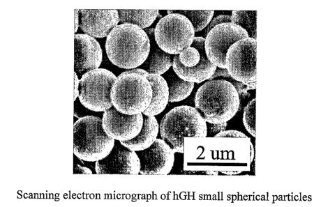
FIG. 27 is a SEM of human growth hormone (hGH) small spherical particles.
FIG. 28 is a chart showing insulin stability data in HFA-134a.
FIG. 29 is a chart comparing aerodynamic performance of Insulin using three
inhalation devices.
FIG. 30 is a chart of stability data of Insulin small spherical particles
compared to
Insulin starting material stored at 25°C.
FIG. 31 is a chart of stability data of Insulin small spherical particles
compared to
Insulin starting material stored at 37°C.
FIG. 32 is a chart of stability data of Insulin small spherical particles
compared to
Insulin starting material stored at 25°C.
FIG. 33 is a chart of stability data of Insulin small spherical particles
compared to
Insulin starting material stored at 37°C.
FIG. 34 is a chart of stability data of Insulin small spherical particles
compared to
Insulin starting material stored at 25°C.
FIG. 35 is a chart of stability data of Insulin small spherical particles
compared to
Insulin starting material stored at 37°C.
FIG. 36 is a bar graph of insulin aerodynamic stability using a Cyclohaler
DPI.
FIG. 37 is a light micrograph of DNase small spherical particles.
FIG. 38 is a chart of enzymatic activity of DNase.
FIG. 39 is a light micrograph of SOD small spherical particles.
FIG. 40 is a chart of enzymatic data for SOD small spherical particles.
FIGS. 41A-B are schematic illustrations of the continuous emulsification
reactor,
where FIG. 41A is a schematic illustration of the continuous emulsification
reactor when
surface active compound added to the continuous phase or the dispersed phase
before
emulsification, and FIG. 41B is a schematic illustration of the continuous
emulsification
reactor when the surface active compound is added after emulsification.
FIG. 42 illustrates the effect of PEG on the IVR profile of PLLA-encapsulated
HSA
particles (Example 32).
FIG. 43 illustrates the IVR profile of PLGA encapsulated LDS small spherical
particles (Example 33).
FIG. 44 illustrates the effect of pH of continuous phase on IVR profile of
PLGA
encapsulated insulin small spherical particles (Example 31).
CA 02532837 2006-O1-16
WO 2005/035088 PCT/US2004/023182
-7_
FIG. 45 illustrates the IVR profile of PLGA encapsulated hGH small spherical
particles (Example 34).
FIG. 46 illustrates the effect of the microencapsulation variables (pH of
continuous
phase and matrix material) on formation of INS dimers in encapsulated INSms
(Example 35).
FIG. 47 illustrates the effect of the microencapsulation variables (pH of
continuous
phase and matrix material) on formation of HMW species in encapsulated INSms
(Example
35).
FIG. 48 illustrates in-vivo release of recombinant human insulin from
unencapsulated
and encapsulated pre-fabricated insulin small spherical particles in rats
(Example 36).
FIG. 49 is an SEM of the particles of Example 27.
DETAILED DESCRIPTION OF THE INVENTION:
The present invention is susceptible to embodiments in many different forms.
Preferred embodiments of the invention are disclosed with the understanding
that the present
disclosure is to be considered as exemplifications of the principles of the
invention and are
not intended to limit the broad aspects of the invention to the embodiments
illustrated.
The present invention is related to methods of production and methods of use
and
composition of small spherical particles of an active agent. In accordance
with the method of
production, the active agent is dissolved in a solvent containing a dissolved
phase-separation
enhancing agent to form a solution that is a single liquid continuous phase.
The solvent is
preferably an aqueous or aqueous-miscible solvent. The solution is then
subjected to a phase
change, for example, by lowering the temperature of the solution to below the
phase
transition temperature of the active agent, whereby the active agent goes
through a liquid
solid phase separation to form a suspension of small spherical particles
constituting a
discontinuous phase while the phase-separation enhancing agent remains in the
continuous
phase.
Phases:
The Continuous Phase
The method of the present invention of preparing small spherical particles of
an active
agent begins with providing a solution having the active agent and a phase-
separation
enhancing agent dissolved in a first solvent in a single liquid phase. The
solution can be an
organic system comprising an organic solvent or a mixture of miscible organic
solvents. The
CA 02532837 2006-O1-16
WO 2005/035088 PCT/US2004/023182
_g_
solution can also be an aqueous-based solution comprising an aqueous medium or
an
aqueous-miscible organic solvent or a mixture of aqueous-miscible organic
solvents or
combinations thereof. The aqueous medium can be water, normal saline, buffered
solutions,
buffered saline, and the like. Suitable aqueous-miscible organic solvents
include, but are not
limited to, N-methyl-2-pyrrolidinone (N-methyl-2-pyrrolidone), 2-pyrrolidinone
(2-
pyrrolidone), 1,3-dimethyl-2-imidazolidinone (DMI), dimethylsulfoxide,
dimethylacetamide,
acetic acid, lactic acid, acetone, methyl ethyl ketone, acetonitrile,
methanol, ethanol,
isopropanol, 3-pentanol, n-propanol, benzyl alcohol, glycerol, tetrahydrofuran
(THF),
polyethylene glycol (PEG), PEG-4, PEG-8, PEG-9, PEG-12, PEG-14, PEG-16, PEG-
120,
PEG-75, PEG-150, polyethylene glycol esters, PEG-4 dilaurate, PEG-20
dilaurate, PEG-6
isostearate, PEG-8 palmitostearate, PEG-150 palmitostearate, polyethylene
glycol sorbitans,
PEG-20 sorbitan isostearate, polyethylene glycol monoalkyl ethers, PEG-3
dimethyl ether,
PEG-4 dimethyl ether, polypropylene glycol (PPG), polypropylene alginate, PPG-
10
butanediol, PPG-10 methyl glucose ether, PPG-20 methyl glucose ether, PPG-15
stearyl
ether, propylene glycol dicaprylate/dicaprate, propylene glycol laurate, and
glycofurol
(tetrahydrofurfuryl alcohol polyethylene glycol ether), alkanes including
propane, butane,
pentane, hexane, heptane, octane, nonane, decane, or a combination thereof.
The single continuous phase can be prepared by first providing a solution of
the
phase-separation enhancing agent, which is either soluble in or miscible with
the first solvent.
This is followed by adding the active agent to the solution. The active agent
may be added
directly to the solution, or the active agent may first be dissolved in a
second solvent and then
together added to the solution. The second solvent can be the same solvent as
the first
solvent, or it can be another solvent selected from the list above and which
is miscible with
the solution. It is preferred that the agent is added to the solution at an
ambient temperature
or lower, which is important particularly for heat labile molecules, such as
certain proteins.
What is meant by "ambient temperature" is a temperature of around room
temperature of
about 20°C to about 40°C. However, the system can also be heated
to increase the solubility
of the active agent in the system as long as heating does not cause
significant reduction in the
activity of the agent.
CA 02532837 2006-O1-16
WO 2005/035088 PCT/US2004/023182
-9-
The Phase-Separation EnhancingAgent
The phase-separation enhancing agent (PSEA) of the present invention enhances
or
induces the liquid-solid phase separation of the active agent from the
solution when the
solution is subjected to the step of phase separation in which the active
agent becomes solid
or semi-solid to form a suspension of small spherical particles as a
discontinuous phase while
the phase-separation enhancing agent remains dissolved in the continuous
phase. The phase-
separation enhancing agent reduces the solubility of the active agent when the
solution is
brought to the phase separation conditions. Suitable phase-separation
enhancing agents
include, but are not limited to, polymers or mixtures of polymers that are
soluble or miscible
with the solution. Examples of suitable polymers include linear or branched
polymers.
These polymers can be water soluble, semi-water soluble, water-miscible, or
insoluble.
In a preferred form of the invention, the phase-separation enhancing agent is
water
soluble or water miscible. Types of polymers that may be used include
carbohydrate-based
polymers, polyaliphatic alcohols, polyvinyl) polymers, polyacrylic acids,
polyorganic acids,
polyamino acids, co-polymers and block co-polymers (e.g., poloxamers such as
Pluronics
F 127 or F68), tert-polymers, polyethers, naturally occuring polymers,
polyimides,
surfactants, polyesters, branched and cyclo-polymers, and polyaldehydes.
Preferred polymers are ones that are acceptable as pharmaceutical additives
for the
intended route of administration of the active agent particles. Preferred
polymers are
pharmaceutically acceptable additives such as polyethylene glycol (PEG) of
various
molecular weights, such as PEG 200, PEG 300, PEG 3350, PEG 8000, PEG 10000,
PEG
20000, etc. and poloxamers such as Pluronics F127 or Pluronics F68. Yet
another preferred
polymer is polyvinylpyrrolidone (PVP). Yet another preferred ~ polymer is
hydroxyethylstarch. Other amphiphilic polymers can also be used alone or in
combinations.
The phase-separation enhancing agent can also be a non-polymer such as a
mixture of
propylene glycol and ethanol.
Liquid-Solid Phase Separation
A liquid-solid phase separation of the active agent in the solution can be
induced by
any method known in the art, such as change in temperature, change in
pressure, change in
pH, change in ionic strength of the solution, change in the concentration of
the active agent,
change in the concentration of the phase-separation enhancing agent, change in
osmolality of
the solution, combinations of these, and the like.
CA 02532837 2006-O1-16
WO 2005/035088 PCT/US2004/023182
-10-
In a preferred embodiment of the present invention, the phase change is a
temperature-induced phase change by lowering the temperature below the phase
transition
temperature of the active agent in the solution.
FIG. 1 is a two-dimensional phase diagram 10 for the solution containing
solvent, a
PSEA and an active agent. The diagram plots the active agent concentration
against the
temperature of the solution. The concentration of the PSEA is held constant.
The diagram has a saturation curve 12; a supersaturation curve 14; a
metastable area
16 therebetween; a first area 18 below the saturation curve where the system
is in a
homogenous, single liquid phase where all components are in the liquid phase;
and a second
area 20 above the supersaturation curve where the system is a two-phase system
having a
solid phase of the active agent and a liquid phase of the PSEA and solvent.
The phase
diagram is helpful in determining the temperature of the system and the
relative concentration
of components in the pure liquid phase, the liquid-solid phase and the
conditions surrounding
the transition between these two phases.
As disclosed herein, preparation of small spherical particles of the active
agent
principally involves cooling from an undersaturated solution (point A')
reaching saturation in
point A where the solution is in equilibrium with any solid phase that may be
present. On
further cooling, a state is reached where the solution contains more active
agent than that
corresponding to the equilibrium solubility at the given temperature; the
solution thus
becomes supersaturated. Spontaneous formation of the solid phase does not
occur until point
B is reached. The point B is a point on the boundary of the metastable zone.
The metastable
zone width can be expressed either by the maximum attainable undercooling
OTmaX T2-Ti or
by the supersaturation ~C",az C*2-C*i. These two expressions are
thermodynamically
equivalent:
O~maX = ~a -C~ = TZ ~ T = dT",~ a .
T~
The path A'-A-B represents a polytherrnal method of preparing a metastable
solution.
In an isothey~rnal process the starting point would be A". By increasing the
concentration at
constant temperature, saturation will again be achieved at point A. An
isothermal increase in
concentration (by solvent evaporation or by seeding/addition of the active
agent, for instance)
CA 02532837 2006-O1-16
WO 2005/035088 PCT/US2004/023182
-11-
to point C will cause the solution to move into the metastable region until
the metastability
limit is again reached. When the metastable limit is exceeded the solution
becomes unstable
and a spontaneous formation of the solid phase immediately occurs.
The value (~C",aX)T=C*3-C*z obtained isothermally can be different from the
corresponding value of ~Tr"aX T3-Tz obtained polythermally. As the boundary of
the
metastable zone is approached, the time necessary for the solid particle
formation decreases
until the metastable limit is reached.
In the polythermal process, the rate of cooling is done at a controlled rate
to control
the size and shape of the particles. What is meant by a controlled rate is
about 0.2°C/minute
to about 50°Clminute, and more preferably from 0.2°C/minute to
30°C/minute. The rate of
change can be at a constant or linear rate, a non-linear rate, intermittent,
or a programmed
rate (having multiple phase cycles).
The particles can be separated from the PSEA in the solution and purified by
washing
as will be discussed below.
The present invention contemplates adjusting the concentration of the active
agent,
the concentration of the PSEA, the temperature or any combination of these to
cause a phase
change where the active agent goes from a liquid state to a solid state while
the PSEA and
solvent do not go through a phase change and remain as liquids. It is also
contemplated
changing the pH, the ionic strength, the osmolality and the like to enhance,
promote, control
or suppress the phase change. For solutions in which the freezing point is
relatively high, or
the freezing point is above the phase transistion temperature, the solutions
can include a
freezing point depressing agent, such as propylene glycol, sucrose, ethylene
glycol, alcohols
(e.g., ethanol, methanol) or aqueous mixtures of freezing-point depression
agents to lower the
freezing point of the system to allow the phase change in the system without
freezing the
system. The process can also be carried out such that the temperature is
reduced below the
freezing point of the system. The process described herein is particularly
suitable for
molecules that are heat labile (e.g., proteins).
Optional Excipients
The particles of the present invention may include one or more excipients. The
excipient may imbue the active agent or the particles with additional
characteristics such as
increased stability of the particles or of the active agents or of the carrier
agents, controlled
CA 02532837 2006-O1-16
WO 2005/035088 PCT/US2004/023182
-12-
release of the active agent from the particles, or modified permeation of the
active agent
through biological tissues. Suitable excipients include, but are not limited
to, carbohydrates
(e.g., trehalose, sucrose, mannitol), cations (e.g., Zn2+, Mg2+, Ca2+), anions
(e.g. 5042-), amino
acids (e.g., glycine), lipids, phospholipids, fatty acids, surfactants,
triglycerides, bile acids or
their salts (e.g., cholate or its salts, such as sodium cholate; deoxycholic
acid or its salts),
fatty acid esters, and polymers present at levels below their functioning as
PSEA's. When an
excipient is used, the excipient does not significantly affect the phase
diagram of the solution.
Sebaratin~ and Washing- the Particles
In a preferred embodiment of the present invention, the small spherical
particles are
harvested by separating them from the phase-separation enhancing agent in the
solution. In
yet another preferred embodiment, the method of separation is by washing the
solution
containing the small spherical particles with a liquid medium in which the
active agent is not
soluble in the liquid medium while the phase-separation enhancing agent is
soluble in the
liquid medium. Some methods of washing may be by diafiltration or by
centrifugation. The
liquid medium can be an aqueous medium or an organic solvent. For active
agents with low
aqueous solubility, the liquid medium can be an aqueous medium or an aqueous
medium
containing agents that reduce the aqueous solubility of the active agent, such
as divalent
cations. For active agents with high aqueous solubility, such as many
proteins, an organic
solvent or an aqueous solvent containing a protein-precipitating agent such as
ammonium
sulfate may be used.
Examples of suitable organic solvents for use as the liquid medium include
those
organic solvents specified above as suitable for the continuous phase, and
more preferably
methylene chloride, chloroform, acetonitrile, ethylacetate, methanol, ethanol,
pentane, and
the like.
It is also contemplated to use mixtures of any of these solvents. One
preferred blend
is methylene chloride or a 1:1 mixture of methylene chloride and acetone. It
is preferred that
the liquid medium has a low boiling point for easy removal by, for example,
lyophilization,
evaporation, or drying.
The liquid medium can also be a supercritical fluid, such as liquid carbon
dioxide or a
fluid near its supercritical point. Supercritical fluids can be suitable
solvents for the phase-
separation enhancing agents, particularly some polymers, but are nonsolvents
for protein
particles. Supercritical fluids can be used by themselves or with a cosolvent.
The following
CA 02532837 2006-O1-16
WO 2005/035088 PCT/US2004/023182
-13-
supercritical fluids can be used: liquid C02, ethane, or xenon. Potential
cosolvents can be
acetontitrile, dichloromethane, ethanol, methanol, water, or 2-propanol.
The liquid medium used to separate the small spherical particles from the PSEA
described herein, may contain an agent which reduces the solubility of the
active agent in the
liquid medium. It is most desirable that the particles exhibit minimal
solubility in the liquid
medium to maximize the yield of the particles. For some proteins, such as
insulin and human
growth hormone, the decrease in solubility can be achieved by the adding of
divalent cations,
such as Zn2+ to the protein. Other ions that can be used to form complexes
include, but are
not limited to, Ca2+, Cu2+, Fe2+, Fe3+, and the like.
The solubility of the insulin-Zn or growth hormone-Zn complexes are
sufficiently low
to allow diafiltration of the complex in an aqueous solution.
The liquid medium may also contain one or more excipients which may imbue the
active agent or the particles with additional characteristics such as
increased stability of the
particles and/or of the active or carrier agents, controlled release of the
active agent from the
particles, or modified permeation of the active agent through biological
tissues as discussed
previously.
In another form of the invention, the small spherical particles are not
separated from
the PSEA containing solution.
Aqueous-based Process
In another preferred embodiment, the fabrication process of the present system
is of
an aqueous system including an aqueous or an aqueous-miscible solvent.
Examples of
suitable aqueous-miscible solvents include, but are not limited to, those
identified above for
the continuous phase. One advantage of using an aqueous-based process is that
the solution
can be buffered and can contain excipients that provide biochemical
stabilization to protect
the active agents, such as proteins.
The Active A _ ent
The active agent of the present invention is preferably a pharmaceutically
active
agent, which can be a therapeutic agent, a diagnostic agent, a cosmetic, a
nutritional
supplement, or a pesticide.
The therapeutic agent can be a biologic, which includes but is not limited to
proteins,
polypeptides, carbohydrates, polynucleotides, and nucleic acids. The protein
can be an
CA 02532837 2006-O1-16
WO 2005/035088 PCT/US2004/023182
-14-
antibody, which can be polyclonal or monoclonal. The therapeutic can be a low
molecular
weight molecule. In addition, the therapeutic agents can be selected from a
variety of known
pharmaceuticals such as, but are not limited to: analgesics, anesthetics,
analeptics, adrenergic
agents, adrenergic blocking agents, adrenolytics, adrenocorticoids,
adrenomimetics,
anticholinergic agents, anticholinesterases, anticonvulsants, alkylating
agents, alkaloids,
allosteric inhibitors, anabolic steroids, anorexiants, antacids,
antidiarrheals, antidotes,
antifolics, antipyretics, antirheumatic agents, psychotherapeutic agents,
neural blocking
agents, anti-inflammatory agents, antihehnintics, anti-arrhythmic agents,
antibiotics,
anticoagulants, antidepressants, antidiabetic agents, antiepileptics,
antifungals,
antihistamines, antihypertensive agents, antimuscarinic agents,
antimycobacterial agents,
antimalarials, antiseptics, antineoplastic agents, antiprotozoal agents,
immunosuppressants,
immunostimulants, antithyroid agents, antiviral agents, anxiolytic sedatives,
astringents, beta-
adrenoceptor blocking agents, contrast media, corticosteroids, cough
suppressants, diagnostic
agents, diagnostic imaging agents, diuretics, dopaminergics, hemostatics,
hematological
agents, hemoglobin modifiers, hormones, hypnotics, immuriological agents,
antihyperlipidemic and other lipid regulating agents, muscarinics, , muscle
relaxants,
parasympathomimetics, parathyroid hormone, calcitonin, prostaglandins, radio-
pharmaceuticals, sedatives, sex hormones, anti-allergic agents, stimulants,
sympathomimetics, thyroid agents, vasodilators, vaccines, vitamins, and
xanthines.
Antineoplastic, or anticancer agents, include but are not limited to
paclitaxel and derivative
compounds, and other antineoplastics selected from the group consisting of
alkaloids,
antimetabolites, enzyme inhibitors, alkylating agents and antibiotics.
A cosmetic agent is any active ingredient capable of having a cosmetic
activity.
Examples of these active ingredients can be, iate~ alia, emollients,
humectants, free radical
inhibiting agents, anti-inflammatories, vitamins, depigmenting agents, anti-
acne agents,
antiseborrhoeics, keratolytics, slimming agents, skin coloring agents and
sunscreen agents,
and in particular linoleic acid, retinol, retinoic acid, ascorbic acid alkyl
esters,
polyunsaturated fatty acids, nicotinic esters, tocopherol nicotinate,
unsaponifiables of rice,
soybean or shea, ceramides, hydroxy acids such as glycolic acid, selenium
derivatives,
antioxidants, beta-carotene, gamma-orizanol and stearyl glycerate. The
cosmetics are
commercially available and/or can be prepared by techniques known in the art.
CA 02532837 2006-O1-16
WO 2005/035088 PCT/US2004/023182
-15-
Examples of nutritional supplements contemplated for use in the practice of
the
present invention include, but are not limited to, proteins, carbohydrates,
water-soluble
vitamins (e.g., vitamin C, B-complex vitamins, and the like), fat-soluble
vitamins (e.g.,
vitamins A, D, E, I~, and the like), and herbal extracts. The nutritional
supplements are
commercially available andlor can be prepared by techniques known in the art.
The term pesticide is understood to encompass herbicides, insecticides,
acaricides,
nematicides, ectoparasiticides and fungicides. Examples of compound classes to
which the
pesticide in the present invention may belong include ureas, triazines,
triazoles, carbamates,
phosphoric acid esters, dinitroanilines, morpholines, acylalanines,
pyrethroids, benzilic acid
esters, diphenylethers and polycyclic halogenated hydrocarbons. Specific
examples of
pesticides in each of these classes are listed in Pesticide Manual, 9th
Edition, British Crop
Protection Council. The pesticides are commercially available andlor can be
prepared by
techniques known in the art.
In a preferred embodiment of the present invention, the active agent is a
macromolecule, such as a protein, a polypeptide, a carbohydrate, a
polynucleotide, a virus, or
a nucleic acid. Nucleic acids include DNA, oligonucleotides, antisense
oligonucleotides,
aptimers, RNA, and SiRNA. The macromolecule can be natural or synthetic. The
protein
can be an antibody, which can be monoclonal or polyclonal. The protein can
also be any
known therapeutic proteins isolated from natural sources or produced by
synthetic or
recombinant methods. Examples of therapeutic proteins include, but are not
limited to,
proteins of the blood clotting cascade (e.g., Factor VII, Factor VIII, Factor
IX, et al.),
subtilisin, ovalbumin, alpha-1-antitrypsin (AAT), DNase, superoxide dismutase
(SOD),
lysozyme, ribonuclease, hyaluronidase, collagenase, growth hormone,
erythropoetin, insulin-
like growth factors or their analogs, interferons, glatiramer, granulocyte-
macrophage colony-
stimulating factor, granulocyte colony-stimulating factor, antibodies,
PEGylated proteins,
glycosylated or~ hyperglycosylated proteins, desmopressin, LHRH agonists such
as:
leuprolide, goserelin, nafarelin, buserelin; LHRH antagonists, vasopressin,
cyclosporine,
calcitonin, parathyroid hormone, parathyroid hormone peptides and insulin.
Preferred
therapeutic proteins are insulin, alpha-1 antitrypsin, LHRH agonists and
growth hormone.
Examples of low molecular weight therapeutic molecules include, but are not
limited
to, steroids, beta-agonists, anti-microbials, antifungals, taxanes
(antimitotic and
CA 02532837 2006-O1-16
WO 2005/035088 PCT/US2004/023182
-16-
antimicrotubule agents), amino acids, aliphatic compounds, aromatic compounds,
and urea
compounds.
In a preferred embodiment, the active agent is a therapeutic agent for
treatment of
pulmonary disorders. Examples of such agents include, but are not limited to,
steroids, beta-
s agonists, anti-fungals, anti-microbial compounds, bronchial dialators, anti-
asthmatic agents,
non-steroidal anti-inflammatory agents (NSAIDS), alpha-1-antitrypsin, and
agents to treat
cystic fibrosis. Examples of steroids include but are not limited to
beclomethasone (including
beclomethasone dipropionate), fluticasone (including fluticasone propionate),
budesonide,
estradiol, fludrocortisone, flucinonide, triamcinolone (including
triamcinolone acetonide),
and flunisolide. Examples of beta-agonists include but are not limited to
salmeterol
xinafoate, formoterol fumarate, levo-albuterol, bambuterol, and tulobuterol.
Examples of anti-fungal agents include but are not limited to itraconazole,
fluconazole, and amphotericin B.
Diagnostic agents include the x-ray imaging agent and contrast media. Examples
of
x-ray imaging agents include WIN-8883 (ethyl 3,5-diacetamido-2,4,6-
triiodobenzoate) also
known as the ethyl ester of diatrazoic acid (EEDA), WIN 67722, i.e., (6-ethoxy-
6-oxohexyl-
3,5-bis(acetamido)-2,4,6-triiodobenzoate; ethyl-2-(3,5-bis(acetamido)-2,4,6-
triiodobenzoyloxy)butyrate (WIN 16318); ethyl diatrizoxyacetate (WIN 12901);
ethyl 2-(3,5-
bis(acetamido)-2,4,6-triiodobenzoyloxy)propionate (W1N 16923); N-ethyl 2-(3,5-
bis(acetamido)-2,4,6-triiodobenzoyloxy acetamide (WIN 65312); isopropyl 2-(3,5-
bis(acetamido)-2,4,6-triiodobenzoyloxy) acetamide (WIN 12855); diethyl 2-(3,5-
bis(acetamido)-2,4,6-triiodobenzoyloxy malonate (WIN 67721); ethyl 2-(3,5-
bis(acetamido)-
2,4,6-triiodobenzoyloxy) phenylacetate (WIN 67585); propanedioic acid, [[3,5-
bis(acetylamino)-2,4,5-triodobenzoyl]oxy]bis(1-methyl)ester (WIN 68165); and
benzoic acid,
3,5-bis(acetylamino)-2,4,6-triodo-4-(ethyl-3-ethoxy-2-butenoate) ester (WIN
68209).
Preferred contrast agents include those which are expected to disintegrate
relatively rapidly
under physiological conditions, thus minimizing any particle associated
inflammatory
response. Disintegration may result from enzymatic hydrolysis, solubilization
of carboxylic
acids at physiological pH, or other mechanisms. Thus, poorly soluble iodinated
carboxylic
acids such as iodipamide, diatrizoic acid, and metrizoic acid, along with
hydrolytically labile
iodinated species such as WIN 67721, WIN 12901, WIN 68165, and WIN 68209 or
others
may be preferred.
CA 02532837 2006-O1-16
WO 2005/035088 PCT/US2004/023182
-17-
Numerous combinations of active agents may be desired including, for example,
a
combination of a steroid and a beta-agonist, e.g., fluticasone propionate and
salmeterol,
budesonide and formeterol, etc.
Examples of carbohydrates are dextrans, hetastarch, cyclodextrins, alginates,
chitosans, chondroitins, heparins and the like.
The Small Spherical Particles
The particles and the small spherical particles of the present invention
preferably have
an average geometric particle size of from about 0.01 ~m to about 200 pm, more
preferably
from 0.1 ~,m to 10 ~,m, even more preferably from about 0.5 ~m to about 5 ~.m,
and most
preferably from about 0.5 ~.m to about 3 ~.m, as measured by dynamic light
scattering
methods (e.g., photocorrelation spectroscopy, laser diffraction, low-angle
laser light
scattering (LALLS), medium-angle laser light scattering (MALLS)), by light
obscuration
methods (Coulter analysis method, for example) or by other methods, such as
rheology or
microscopy (light or electron). Particles for pulmonary delivery will have an
aerodynamic
particle size determined by time of flight measurements (e.g., Aerosolizer) or
Andersen
Cascade Impactor measurements.
The small spherical particles are substantially spherical. What is meant by
"substantially spherical" is that the ratio of the lengths of the longest to
the shortest
perpendicular axes of the particle cross section is less than or equal to
about 1.5.
Substantially spherical does not require a line of symmetry. Further, the
particles may have
surface texturing, such as lines or indentations or protuberances that are
small in scale when
compared to the overall size of the particle and still be substantially
spherical. More
preferably, the ratio of lengths between the longest and shortest axes of the
particle is less
thm or equal to about 1.33. Most preferably, the ratio of lengths between the
longest and
shortest axes of the particle is less than or equal to about 1.25. Surface
contact is minimized
in microspheres that are substantially spherical, which minimizes the
undesirable
agglomeration of the particles upon storage. Many crystals or flakes have flat
surfaces that
can allow large surface contact areas where agglomeration can occur by ionic
or non-ionic
interactions. A sphere permits contact over a much smaller area.
The particles also preferably have substantially the same particle size.
Particles
having a broad size distribution where there are both relatively big and small
particles allow
CA 02532837 2006-O1-16
WO 2005/035088 PCT/US2004/023182
-18-
for the smaller particles to fill in the gaps between the larger particles,
thereby creating new
contact surfaces. A broad size distribution can result in larger spheres by
creating many
contact opportunities for binding agglomeration. This invention creates
spherical particles
with a narrow size distribution, thereby minimizing opportunities fox contact
agglomeration.
What is meant by a "narrow size distribution" is a preferred particle size
distribution would
have a ratio of the volume diameter of the 90th percentile of the small
spherical particles to
the volume diameter of the lOtn percentile less than or equal to 5. More
preferably, the
particle size distribution would have ratio of the volume diameter of the 90th
percentile of the
small spherical particles to the volume diameter of the 10th percentile less
than or equal to 3.
Most preferably, the particle size distribution would have ratio of the volume
diameter of the
90tn percentile of the small spherical particles to the volume diameter of the
10th percentile
less than or equal to 2.
Geometric Standard Deviation (GSD) can also be used to indicate the narrow
size
distribution. GSD calculations involved determining the effective cutoff
diameter (ECD) at
the cumulative less than percentages of 15.9% and 84.1 %. GSD is equal to the
square root of
the ratio of the ECD less than 84.17% to ECD less then 15.9%. The GSD has a
narrow size
distribution when GSD < 2.5, more preferably less than 1.8.
In a preferred form of the invention, the active agent in the small spherical
particles is
semi-crystalline or non-crystalline.
Typically, small spherical particles made by the process in this invention are
substantially non-porous and have a density greater than 0.5 g/cm3, more
preferably greater
than 0.75 g/cm3 and most preferably greater than about 0.85 glcm3. A preferred
range for the
density is from about 0.5 to about 2 g/cm3 and more preferably from about 0.75
to about 1.75
g/cm3 and even more preferably from about 0.85 g/cm3 to about 1.5 g/cm3.
The particles of the present invention can exhibit high content of the active
agent.
There is no requirement for a significant quantity of bulking agents or
similar excipients that
are required by many other methods of preparing particles. For example,
insulin small
spherical particles consist of equal to or greater than 95% by weight of the
particles.
However, bulking agents or excipients may be included in the particles.
Preferably, the
active agent is present from about 0.1% to greater than 95% by weight of the
particle, more
preferably from about 30% to about 100% by weight, even more preferably from
about 50%
to about 100% by weight, yet more preferably from about 75% to about 100% by
weight, and
CA 02532837 2006-O1-16
WO 2005/035088 PCT/US2004/023182
-19-
most preferably greater than 90% by weight. When stating ranges herein, it is
meant to
include airy range or combination of ranges therein.
A further aspect of the present invention is that the small spherical
particles retain the
biochemical integrity and the biological activity of the active agent with or
without the
inclusion of excipients.
Ivy vivo Delivery of the Particles
The particles containing the active agent in the present invention are
suitable for ih
vivo delivery to a subject in need of the agent by a suitable route, such as
injectable, topical,
oral, rectal, nasal, pulmonary, vaginal, buccal, sublingual, transdermal,
transmucosal, otic,
intraocular or ocular. The particles can be delivered as a stable liquid
suspension or
formulated as a solid dosage form such as tablets, caplets, capsules, etc. A
preferred delivery
route is injectable, which includes intravenous, intramuscular, subcutaneous,
intraperitoneal,
intrathecal, epidural, infra-arterial, infra-articular and the like. Another
preferred route of
delivery is pulmonary inhalation. In this route of delivery, the particles may
be deposited to
the deep lung, in the upper respiratory tract, or anywhere in the respiratory
tract. The
particles may be delivered as a dry powder by a dry powder inhaler, or they
may be delivered
by a metered dose inhaler or a nebulizer.
Drugs intended to function systemically, such as insulin, are desirably
deposited in the
alveoli, where there is a very large surface area available for absorption
into the bloodstream.
When targeting the drug deposition to certain regions within the lung, the
aerodynamic
diameter of the particle can be adjusted to an optimal range by manipulating
fundamental
physical characteristics of the particles such as shape, density, and particle
size.
Acceptable respirable fractions of inhaled drug particles are often achieved
by adding
excipients to the formulation, either incorporated into the particle
composition or as a mixture
with the drug particles. For example, improved dispersion of micronized drug
particles
(about 5 ~,m) is effected by blending with larger (30-90 Vim) particles of
inert carrier particles
such as trehalose, lactose or maltodextrin. The larger excipient particles
improve the powder
flow properties, which correlates with an improved pharmacodynamic effect. In
a further
refinement, the excipients are incorporated directly into the small spherical
particles to effect
aerosol performance as well as potentially enhancing the stability of protein
drugs.
Generally, excipients are chosen that have been previously FDA approved for
inhalation,
such as lactose, or organic molecules endogenous to the lungs, such as albumin
and DL-a-
CA 02532837 2006-O1-16
WO 2005/035088 PCT/US2004/023182
-20-
phosphatidylcholine dipalmitoyl (DPPC). Other excipients, such as poly(lactic
acid-co-
glycolic acid) (PLGA) have been used to engineer particles with desirable
physical and
chemical characteristics. However, much of the inhalation experience with FDA
approved
excipients has been with asthma drugs having large aerodynamic particle sizes
that desirably
deposit in the tracheobronchial region, and which do not appreciably penetrate
to the deep
lung. For inhaled protein or peptide therapeutics delivered to the deep lung,
there is concern
that undesirable long-term side effects, such as inflammation and irritation
can occur which
may be due to an immunological response or caused by excipients when they are
delivered to
the alveolar region.
In order to minimize potential deleterious side effects of deep lung inhaled
therapeutics, it may be advantageous to fabricate particles for inhalation
that are substantially
constituted by the drug to be delivered. This strategy would minimize alveolar
exposure to
excipients and reduce the overall mass dose of particles deposited on alveolar
surfaces with
each dose, possibly minimizing irritation during chronic use of the inhaled
therapeutic. Small
spherical particles with aerodynamic properties suitable for deep lung
deposition that are
essentially composed entirely of a therapeutic protein or peptide may be
particularly useful
for isolated studies on the effects of chronic therapeutic dosing on the
alveolar membrane of
the lung. The effects of systemic delivery of protein or peptide in the form
of small spherical
particles by inhalation could then be studied without complicating factors
introduced by
associated excipients.
The requirements to deliver particles to the deep lung by inhalation are that
the
particles have a small mean aerodynamic diameter of 0.5-10 micrometers and a
narrow size
distribution. The invention also contemplates mixing together of various
batches of particles
having different particle size ranges. The process of the present invention
allows the
fabrication of small spherical particles with the above characteristics.
There are two principal approaches for forming particles with aerodynamic
diameters
of 0.5 to 3 micron. The first approach is to produce relatively large but very
porous (or
perforated) microparticles. Since the relationship between the aerodynamic
diameter
(Daerodynamic) arid the geometric diameter (Dgeometric) 1S Daerodynamic is
equal t0 Dgeometric
multiplied by the square root of the density of the particles with very low
mass density
(around 0.1 glcm3) can exhibit small aerodynamic diameters (0.5 to 3 microns)
while
possessing relatively high geometric diameters (5 to 10 microns).
CA 02532837 2006-O1-16
WO 2005/035088 PCT/US2004/023182
-21 -
An alternative approach is to produce particles with relatively low porosity,
in the
case of the present invention, the particles have a density, set forth in the
ranges above, and
more generally that is close to 1 g/cm3. Thus, the aerodynamic diameter of
such non-porous
dense particles is close to their geometric diameter.
The present method for particle formation set forth above, provides for
particle
formation with or without excipients.
Fabrication of protein small spherical particles from protein itself with no
additives
provides superior advantages for use in pulmonary delivery as it provides
options for larger
drug payloads, increased safety and decreased numbers of required inhalations.
Microencapsulation of Pre-fabricated Small Spherical Particles
The small spherical particles of the present invention or small particles
prepared from
other methods (including microparticles, microspheres, nanospheres,
nanoparticles, etc.) can
further be encapsulated within matrices of wall-forming materials to form
microencapsulated
particles. The microencapusulation can be accomplished by any process known in
the art. In
a preferred embodiment, microencapsulation of the small spherical particles of
the present
invention or any other small particles is accomplished by an
emulsification/solvent extraction
processes as described below. The matrix can impart sustained release
properties to the
active agent resulting in release rates that persist from minutes to hours,
days or weeks
according to the desired therapeutic applications. The microencapsulated
particles can also
produce delayed release formulations of the pre-fabricated small spherical
particles. In a
preferred embodiment, the pre-fabricated small spherical particles are
particles of
macromolecules. In another preferred embodiment, the macromolecule is a
protein or
polypeptide.
In the emulsification/solvent extraction process, emulsification is obtained
by mixing
two immiscible phases, the continuous phase and the discontinuous phase (which
is also
known as the dispersed phase), to form an emulsion. In a preferred embodiment,
the
continuous phase is an aqueous phase (or the water phase) and the
discontinuous phase is an
organic phase (or the oil phase) to form an oil-in-water (0/W) emulsion. The
discontinuous
phase may further contain a dispersion of solid particles present either as a
fine suspension or
as a fine dispersion forming a solid-in-oil (S/0) phase. The organic phase is
preferably a
water immiscible or a partially water miscible organic solvent. The ratio by
weights of the
organic phase to the aqueous phase is from about 1:99 to about 99:1, more
preferably from
CA 02532837 2006-O1-16
WO 2005/035088 PCT/US2004/023182
-22-
1:99 to about 40:60, and most preferably from about 2:98 to about 1:3, or any
range or
combination of ranges therein. In a preferred embodiment, the ratio of the
organic phase to
the aqueous phase is about 1:3. The present invention fizrther contemplates
utilizing reverse
emulsions or water-in-oil emulsion (W/O) where the oil phase forms the
continuous phase
and water phase forms the discontinuous phase. The present invention further
contemplates
utilizing emulsions having more than two phases such as an oil-in-water-in-oil
emulsion
(0!W/0) or a water-in-oil-in-water emulsion (W/O/W).
In a preferred embodiment, the process of microencapsulation using the
emulsification/solvent extraction process starts with preparing pre-fabricated
small spherical
particles by the methods described earlier and an organic phase containing the
wall-forming
material. The pre-fabricated small spherical particles are dispersed in the
organic phase of
the wall-forming material to form a solid-in-oil (S/0) phase containing a
dispersion of the
pre-fabricated small spherical particles in the oil phase. In a preferred
embodiment, the
dispersion is accomplished by homogenizing the mixture of the small spherical
particles and
the organic phase. An aqueous medium will form the continuous phase. In this
case, the
emulsion system formed by emulsifying the S/O phase with an aqueous phase is a
solid-in-
oil-in-water (Sl0/W) emulsion system.
The wall-forming material refers to materials capable of forming the
structural entity
of the matrix individually or in combination. Biodegradable wall-forming
materials are
preferred, especially for injectable applications. Examples of such materials
include but are
not limited to the family of poly-lactide/poly-glycolide polymers (PLGA's),
polyethylene
glycol conjugated PLGA's (PLGA-PEG'S), and triglycerides. In the embodiment in
which
PLGA or PLGA-PEG is used, the PLGA preferably has a ratio of poly-lactide to
poly-
glycolide of from 100:0 to 0:100, more preferably from about 90:10 to about
15:85, and most
preferably about 50:50. In general, the higher the ratio of the poly-glycolide
to the poly-
lactide in the polymer, the more hydrophilic is the microencapsulated
particles resulting in
faster hydration and faster degradation. Various molecular weights of PLGA can
also be
used. In general, for the same ratio of poly-glycolide and poly-lactide in the
polymer, the
higher the molecular weight of the PLGA, the slower is the release of the
active agent, and
the wider the distribution of the size of the microencapsulated particles.
The organic solvent in the organic phase (oil phase) of an oil-in-water (0/W)
or solid-
in-oil-in-water (S/O/W) emulsion can be aqueous immiscible or partially
aqueous
CA 02532837 2006-O1-16
WO 2005/035088 PCT/US2004/023182
- 23 - .
immiscible. What is meant by the term "water immiscible solvent" are those
solvents which
form an interfacial meniscus when combined with an aqueous solution in a 1:1
ratio (0/W).
Suitable water immiscible solvents include, but are not limited to,
substituted or
unsubstituted, linear, branched or cyclic alkanes with a carbon number of 5 or
higher,
substituted or unsubstituted, linear, branched or cyclic alkenes with a carbon
number of 5 or
higher, substituted or unsubstituted, linear, branched or cyclic alkynes with
a carbon number
of 5 or higher; aromatic hydrocarbons completely or partially halogenated
hydrocarbons,
ethers, esters, ketones, mono-, di- or tri-glycerides, native oils, alcohols,
aldehydes, acids,
amines, linear or cyclic silicones, hexamethyldisiloxane, or any combination
of these
solvents. Halogenated solvents include, but are not limited to carbon
tetrachloride,
methylene chloride, chloroform, tetrachloroethylene, trichloroethylene,
trichloroethane,
hydrofluorocarbons, chlorinated benzene (mono, di, tri),
trichlorofluoromethane. Particularly
suitable solvents are methylene chloride, chloroform, diethyl ether, toluene,
xylene and ethyl
acetate. What is meant by "partially water miscible solvents" are those
solvents which are
water immiscible at one concentration, and water miscible at another lower
concentration.
These solvents are of limited water miscibility and capable of spontaneous
emulsion
formation. Examples of partially water miscible solvents are tetrahydrofuran
(THF),
propylene carbonate, benzyl alcohol, and ethyl acetate.
A surface active compound can be added, for example, to increase the wetting
properties of the organic phase. The surface active compound can be added
before the
emulsification process to the aqueous phase, to the organic phase, to both the
aqueous
medium and the organic solution, or after the emulsification process to the
emulsion. The use
of a surface active compound can reduce the number of unencapsulated or
partially
encapsulated small spherical particles, resulting in reduction of the initial
burst of the active
agent during the release. The surface active compound can be added to the
organic phase, or
to the aqueous phase, or to both the organic phase and the aqueous phase,
depending on the
solubility of the compound.
What is meant by the term "surface active compounds" are compounds such as an
anionic surfactant, a cationic surfactant, a zwitterionic surfactant, a
nonionic surfactant or a
biological surface active molecule. The surface active compound should be
present in an
amount by weight of the aqueous phase or the organic phase or the emulsion,
whatever the
CA 02532837 2006-O1-16
WO 2005/035088 PCT/US2004/023182
-24-
case may be, from less than about 0.01% to about 30%, more preferably from
about 0.01% to
about 10%, or any range or combination of ranges therein.
Suitable anionic surfactants include but are not limited to: potassium
laurate, sodium
lauryl sulfate, sodium dodecylsulfate, alkyl polyoxyethylene sulfates, sodium
alginate,
dioctyl sodium sulfosuccinate, phosphatidyl choline, phosphatidyl glycerol,
phosphatidyl
inosine, phosphatidylserine, phosphatidic acid and their salts, glyceryl
esters, sodium
carboxymethylcellulose, cholic acid and other bile acids (e.g., cholic acid,
deoxycholic acid,
glycocholic acid, taurocholic acid, glycodeoxycholic acid) and salts thereof
(e.g., sodium
deoxycholate, etc.).
Suitable cationic surfactants include, but are not limited to, quaternary
ammonium
compounds, such as benzalkonium chloride, cetyltrimethylammonium bromide,
lauryldimethylbenzylammonium chloride, acyl carnitine hydrochlorides; or alkyl
pyridinium
halides. As anionic surfactants, phospholipids may be used. Suitable
phospholipids include,
for example phosphatidylcholine, phosphatidylethanolamine, phosphatidylserine,
phosphatidyl inositol, phosphatidylglycerol, phosphatidic acid,
lysophospholipids, egg or
soybean phospholipid or a combination thereof. The phospholipid may be salted
or desalted,
hydrogenated or partially hydrogenated or natural, semisynthetic or synthetic.
Suitable nonionic surfactants include: polyoxyethylene fatty alcohol ethers
(Macrogol
and Brij), polyoxyethylene sorbitan fatty acid esters (Polysorbates),
polyoxyethylene fatty
acid esters (Myrj), sorbitan esters (Span), glycerol monostearate,
polyethylene glycols,
polypropylene glycols, cetyl alcohol, cetostearyl alcohol, stearyl alcohol,
aryl alkyl polyether
alcohols, polyoxyethylene-polyoxypropylene copolymers (poloxomers),
polaxamines,
polyvinyl alcohol, polyvinylpyrrolidone, and polysaccharides (including starch
and starch
derivatives such as hydroxyethylstarch (HES), methylcellulose,
hydroxycellulose, hydroxy
propylcellulose, hydroxy propylmethylcellulose, and noncrystalline cellulose).
In a preferred
form of the invention, the nonionic surfactant is a polyoxyethylene and
polyoxypropylene
copolymer and preferably a block copolymer of propylene glycol and ethylene
glycol. Such
polymers are sold under the tradename POLOXAMER also sometimes referred to as
PLURONIC~, and sold by several suppliers including Spectrum Chemical and
Ruger.
3Q Among polyoxyethylene fatty acid esters is included those having short
alkyl chains. One
example of such a surfactant is SOLUTOL~ HS 15, polyethylene-660-
hydroxystearate,
manufactured by BASF Aktiengesellschaft.
CA 02532837 2006-O1-16
WO 2005/035088 PCT/US2004/023182
- 25 -
Surface active biological molecules include such molecules as albumin, casein,
heparin, hirudin, hetastarch or other appropriate biocompatible agents.
In a preferred form of the invention, the aqueous phase includes a protein as
the
surface active compound. A preferred protein is albumin. The protein may also
function as
an excipient. In embodiments in which protein is not the surface active
compound, other
excipients may be included in the emulsion, added either before or after the
emulsification
process. Suitable excipients include, but are not limited to, saccharides,
disaccharides, and
sugar alcohols. A preferred disaccharide is sucrose, and a preferred sugar
alcohol is
mannitol.
In addition, use of channeling agents, such as polyethylelne glycol (PEG), can
increase the water permeation rate of the final product, which results in
modification of the
initial release kinetics of the active agent from the matrix as well as
degradation rate of the
matrix and~degradation-dependent release kinetics by modifying the hydration
rate. Using
PEG as the channeling agent during encapsulation can be advantageous in terms
of
eliminating parts of the washing process during fabrication of the small
spherical particles in
which PEG is used as the phase-separation enhancing agent. In addition,
varying pH of the
continuous phase through use of buffers can significantly increase the wetting
process
between the particle surface and the organic phase, hence, results in
significant reduction of
the initial burst of the encapsulated therapeutic agent from the matrix of the
microencapsulated particles. The properties of the continuous phase can also
be modified, for
example, by increasing its salinity by adding a salt such as NaCI, to reduce
miscibility of the
two phases.
After dispersing the small spherical particles in the organic phase (oil
phase), the
continuous phase of the aqueous medium (water phase) is then vigorously mixed,
for
example by homogenization or sonication, with the discontinuous phase of the
organic phase
to form an emulsion containing emulsified droplets of embryonic
microencapsulated
particles. The continuous aqueous phase can be saturated with the organic
solvent used in the
organic phase prior to mixing of the aqueous phase and the organic phase, in
order to
minimize rapid extraction of the organic solvent from the emulsified droplets.
The
emulsification process can be performed at any temperature in which the
mixture can
maintain its liquid properties. The emulsion stability is a function of the
concentration of the
surface active compound in the organic phase or in the aqueous phase, or in
the emulsion if
CA 02532837 2006-O1-16
WO 2005/035088 PCT/US2004/023182
-26-
the surface active compound is added to the emulsion after the emulsification
process. This
is one of the factors that determines droplet size of the emulsion system
(embryonic
microencapsulated particles) and the size and size distribution of the
microencapsulated
particles. Other factors affecting the size distribution of microencapsulated
particles are
viscosity of the continuous phase, viscosity of the discontinous phase, shear
forces during
emulsification, type and concentration of surface active compound, and the
Oil/Water ratio.
After the emulsification, the emulsion is then transferred into a hardening
medium.
The hardening medium extracts the solvent in the discontinous phase from the
embryonic
microencapsulated particles, resulting in formation of solid microencapsulated
particles
having a solid polymeric matrix around the pre-fabricated small spherical
particles within the
vicinity of the emulsified droplets. In the embodiment of an O/W or S/O/W
system, the
hardening medium is an aqueous medium, which may contain surface active
compounds, or
thickening agents, or other excipients. The microencapsulated particles are
preferably
spherical and have a particle size of from about 0.6 to about 300 p.m, and
more preferably
from about 0.~ to about 60 p,m. Additionally, the microencapsulated particles
preferably
have a narrow distribution of particle size. To reduce the extraction time of
the discontinuous
phase, heat or reduced pressure can be applied to the hardening medimn. The
extraction rate
of discontinuous phase from the embryonic microencapsulated particles is an
important factor
in the degree of porosity in the final solid microencapsulated particles,
since rapid removal,
e.g., by evaporation (boiling effect), of the discontinuous phase results in
destruction of the
continuity of the matrix.
In a preferred embodiment, the emulsification process is performed in a
continuous
fashion instead of a batch process. FIG. 41 depicts the design of the
continuous
emulsification reactor.
In another preferred embodiment, the hardened wall-forming polymeric matrices,
encapsulating the small spherical particles of the active agent, are further
harvested by
centrifugation and/or filtration (including diafiltration), and washed with
water. The
remaining liquid phases can further be removed by a process such as
lyophilization or
evaporation.
CA 02532837 2006-O1-16
WO 2005/035088 PCT/US2004/023182
-27-
A. Insulin Small Spherical Particles
Example 1: General Method of Preparation of Insulin Small Spherical Particles
A solution buffered at pH 5.65 (0.033M sodium acetate buffer) containing
16.67%
PEG 3350 was prepared. A concentrated slurry of zinc crystalline insulin was
added to this
solution while stirring. The insulin concentration in the final solution was
0.83 mg/mL. The
solution was heated to about 85 to 90°C. The insulin crystals dissolved
completely in this
temperature range within five minutes. Insulin small spherical particles
started to form at
around 60°C when the temperature of the solution was reduced at a
controlled rate. The yield
increased as the concentration of PEG increased. This process yields small
spherical particles
with various size distribution with a mean of 1.4 Vim.
The insulin small spherical particles formed were separated from PEG by
washing the
microspheres via diafiltration under conditions in which the small spherical
particles do not
dissolve. The insulin small spherical particles were washed out of the
suspension using an
aqueous solution containing Zn2+. The Zn2+ ion reduces the solubility of the
insulin and
prevents dissolution that reduces yield and causes small spherical particle
agglomeration.
Example 2: Non-stirred Batch Process for making Insulin Small Spherical
Particles
20.2 mg of zinc crystalline insulin were suspended in 1 mL of deionized water
at
room temperature. 50 microliters of 0.5 N HCl was added to the insulin. 1 mL
of deionized
water was added to form a lOmg/mL solution of zinc crystalline insulin. 12.5 g
of
Polyethylene Glycol 3350 (Sigma) and 12.5 g of Polyvinylpyrrolidone (Sigma)
were
dissolved in 50 mL of 100 millimolar sodium acetate buffer, pH5.7. The polymer
solution
volume was adjusted to 100mL with the sodium acetate buffer. To 800
microliters of the
polymer solution in an eppendorf tube was added 400 microliters of the lOmg/mL
insulin
solution. The insulin/polymer solution became cloudy on mixing. A control was
prepared
using water instead of the polymer solution. The eppendorf tubes were heated
in a water bath
at 90°C for 30 minutes without mixing or stirring, then removed and
placed on ice for 10
minutes. The insulin/polymer solution was clear upon removal from the
90°C water bath, but
began to cloud as it cooled. The control without the polymer remained clear
throughout the
experiment. Particles were collected from the insulinlpolymer tube by
centrifugation,
followed by washing twice to remove the polymer. The last suspension in water
was
lyophilized to obtain a dry powder. SEM analysis of the lyophilized particles
from the
CA 02532837 2006-O1-16
WO 2005/035088 PCT/US2004/023182
_28_
insulin/polymer tubes showed a uniform distribution of small spherical
particles around 1
micrometer in diameter. Coulter light scattering particle size analysis of the
particles showed
a narrow size distribution with a mean particle size of 1.413 micrometers, 95%
confidence
limits of 0.941-1.88 micrometers, and a standard deviation of 0.241
micrometers. An insulin
control without polymer or wash steps, but otherwise processed and lyophilized
in the same
manner, showed only flakes (no particles) under the SEM similar in appearance
to that
typically obtained after lyophilizing proteins.
Example 3: The Continuous Flow Through Process for Making Insulin Small
Spherical
Particles
36.5 mg of insulin was weighed out and suspended in 3 mL of deionized water.
30
~,L of 1 N HCl was added to dissolve the insulin. The final volume of the
solution was
adjusted to 3.65 mL with deionized water. 7.3 mL of PEG/PVP solution (25%
PEG/PVP pH
5.6 in 100 mM NaOAc buffer) was then added to the insulin solution to a final
total volume
of 10.95 mL of insulin solution. The solution was then vortexed to yield a
homogenous
suspension of insulin and PEG/PVP.
The insulin suspension was connected to a BioRad peristaltic pump running at a
speed
of 0.4 mL/min through Teflon~ tubing (TFE 1/32" inner diameter flexible
tubing). The
tubing from the pump was submerged into a water bath maintained at 90°C
before being
inserted into a collection tube immersed in ice. Insulin small spherical
particles were formed
when the temperature of the insulin solution was decreased from about
90°C in the water bath
to about 4°C in the collection tube in ice. FIG. 7 is a schematic
diagram of this process. The
total run time for the process was 35 minutes for the 10.95 mL volume. After
collecting the
small spherical particles, the collection tube was centrifuged at 3000 rpm for
20 minutes in a
Beckman J6B centrifuge. A second" water wash was completed and the small
spherical
particle pellets were centrifuged at 2600 rpm for 15 minutes. The final water
wash was
centrifuged at 1500 rpm for 15 minutes. An aliquot was removed for particle
size analysis.
The small spherical particles were frozen at -80°C and lyophilized for
2 days.
The particle size was determined to be 1.397 ~,m by volume, 1.119 pm by
surface
area, and 0.691 ~,m by number as determined by the Beckman Coulter LS 230
particle
counter. The scanning electron micrograph indicated uniform sized and non-
agglomerated
insulin small spherical particles (FIG. 8).
CA 02532837 2006-O1-16
WO 2005/035088 PCT/US2004/023182
-29-
The use of the continuous flow through process where the insulin solution was
exposed to 90°C for a short period of time allowed for the production
of small spherical
particles. This method yielded a final composition that was 90% protein as
determined by
high performance liquid chromatography (HPLC) (FIG. 9). HPLC analysis also
indicated
that the dissolved insulin small spherical particles had an elution time of
about 4.74 minutes,
not significantly different from that of an insulin standard or the native
insulin starting
material, indicating that preservation of the biochemical integrity of the
insulin after
fabrication into the small spherical particles.
Example 4: Heat Exchanger Batch Process for making Insulin Small Spherical
Particles
Human zinc crystalline insulin was suspended in a minimal amount of deionized
water with sonication to ensure complete dispersion. The insulin suspension
was added to a
stirred, buffered polymer solution (pH 5.65 at 25°C) pre-heated to
77°C, so that the final
solute concentrations were 0.83% zinc crystalline insulin, 18.5% polyethylene
glycol 3350,
0.7% sodium chloride, in a 0.1 M sodium acetate buffer. The initially cloudy
mixture cleared
within three minutes as the crystalline insulin dissolved. Immediately after
clearing, the
solution was transferred to a glass, water jacketed chromatography column that
was used as a
heat exchanger (column i.d.: 25mm, length: 600mm; Ace Glass Incorporated,
Vineland, NJ).
The glass column was positioned vertically, and the heat exchange fluid
entered the water
jacket at the bottom of the column and exited at the top. In order to document
the heat
exchange properties of the system, thermocouples (Type J, Cole Parmer) were
positioned in
the center of the insulin formulation liquid at the top and bottom of the
column and a cooling
temperature profile was obtained during a preliminary trial run. The
thermocouples were
removed during the six batches conducted for this experiment so as not to
introduce a foreign
surface variable.
The heat exchanger was pre-heated to 65°C and the insulin - buffered
polymer
solution was transferred in such a manner that the solution temperature did
not drop below
65°C and air bubbles were not introduced into the solution. After the
clear solution was
allowed four minutes to equilibrate to 65°C in the heat exchanger, the
heat exchange fluid
was switched from a 65°C supply to a 15°C supply. The insulin
formulation in the heat
exchanger was allowed to equilibrate to 15°C over a twenty-minute
period. The insulin small
CA 02532837 2006-O1-16
WO 2005/035088 PCT/US2004/023182
-30-
spherical particles formed as the temperature dropped through 60 to
55°C resulting in a
uniform, stable, creamy white suspension.
~~The insulin small spherical particles were separated from the polyethylene
glycol by
diafiltration (A/G Technologies, 750,000 MWCO ultrafiltration cartridge)
against five
volumes of 0.16% sodium acetate - 0.026% zinc chloride buffer, pH 7.0,
followed by
concentration to one fifth of the original volume. The insulin small spherical
particles
suspension was further washed by diafiltration against five volumes of
deionized water,
followed by lyophilization to remove the water. Care was taken to prevent
agglomeration of
the small spherical particles during diafiltration (from polarization packing
of particles on the
membrane surface) and during lyophilization (from settling of the small
spherical particles
prior to freezing). The dried small spherical particles were free flowing and
ready for use,
with no de-agglomeration or sieving required.
Small Spherical Particles of Insulin
The above described process produces uniform size spherical particles from
zinc
crystalline insulin without added excipients. Small spherical particles
prepared by this
process have excellent aerodynamic properties as determined by time-of flight
(AerosizerTM)
and Andersen Cascade Impactor measurements, with high respirable fractions
indicative of
deep lung delivery when delivered from a simple, widely used dry powder
inhaler
(CyclohalerTM). By using insulin as a model protein, we are also able to
examine the effect of
the process on the chemical integrity of the protein using established U.S.P.
methods.
Dry powder insulin small spherical particles were imaged by polarized light
microscopy (Leica EPISTAR~, Buffalo, NY) and with a scanning electron
microscope
(AMRAY 1000, Bedford, MA). Particle size analysis was performed using an
Aerosizer~
Model 3292 Particle Sizing System which included a Model 3230 Aero-Disperser~
Dry
Powder Disperser for introducing the powder to the instrument (TSI
Incorporated, St. Paul,
MIA. Individual particle sizes were confirmed by comparing the Aerosizer
results to the
electron micrographs.
The chemical integrity of the insulin before and after the process was
determined by
HPLC according to the USP monograph for Insulin Human (LJSP 26). The insulin
and high
molecular weight protein content was measured using an isocratic SEC HPLC
method with
UV detection at 276 nm. To measure insulin, A-21 desamido insulin and other
insulin related
substances, the sample was analyzed using a USP gradient reverse-phase HPLC
method. The
CA 02532837 2006-O1-16
WO 2005/035088 PCT/US2004/023182
-31-
insulin content is measured using UV detection at 214 nm. High molecular
weight protein,
desamido insulin, and other insulin related substances were assayed to
quantitate any
chemical degradation caused by the process.
The aerodynamic characteristics of the insulin small spherical particles were
examined using the Aerosizer~ instrument. Size distribution measurements on
insulin drv
powder were conducted using the AeroDisperser attachment with low shear force,
medium
feed rate, and normal deagglomeration. The instruments' software converts time-
of flight
data into size and places it into logarithmically spaced ranges. The number of
particles
detected in each size bin was used for statistical analysis, as well as the
total volume of
particles detected in each size bin. The volume distribution emphasizes large
particles more
than the number distribution and, therefore, is more sensitive at detecting
agglomerates of
non-dispersed particles as well as large particles.
The Andersen Cascade Impactor assembly consisted of a pre-separator; nine
stages,
eight collection plates, and a backup filter. The stages are numbered -1, -0,
1, 2, 3, 4, 5, 6,
and F. Stage -1 is an orifice stage only. Stage F contains the collection
plate for Stage 6 and
the backup filter. The stainless steel collection plates were coated with a
thin layer of food
grade silicone to prevent "bounce" of the particles. A sample stream air-flow
rate of 60 LPM
through the sampler was used for the analysis. An accurately weighed sample
size of
approximately 10 mg was weighed into each starch capsule (Vendor), with the
powder
delivered as an aerosol from the Cyclohaler in four seconds. The amount of
insulin powder
deposited. on each plate was determined by reversed phase HPLC detection at
214 nm
according to the USP 26 assay for human insulin.
The mass median aerodynamic diameter (MMAD) was calculated by Sigma Plot
software using a probit fit of the cumulative less than mass percent versus
the effective cutoff
diameter (ECD). Emitted dose (ED) was determined as the total observed mass of
insulin
deposited into the cascade Impactor. This is expressed as a percentage of the
mass of the
insulin small spherical particles loaded into the Cyclohaler capsule.
The results demonstrate that careful control of process parameters in
conjunction with
a phase change formulation can produce: 1) predominantly spherical particles
with a diameter
of about 2 ~,m; 2) a narrow size distribution; 3) and reproducible aerodynamic
properties
from batch to batch; and 4) small spherical particles composed of over 95%
active drug
(human insulin) excluding residual moisture. We determined that the solubility
of the zinc
CA 02532837 2006-O1-16
WO 2005/035088 PCT/US2004/023182
-32-
crystalline insulin could be controlled by solution temperature, pH, polymer
concentration,
and ionic strength. We also found that controlling the cooling rate during the
phase change
period was an important parameter that enabled the formation of predominantly
spherical
particles within a narrow size range.
FIG. 2 is a cooling temperature profile for the process corresponding to this
Example.
The profile was measured using a water jacketed chromatography column
positioned
vertically and heat-exchange fluid entered the water jacket at the ,bottom of
the column and
exited at the top. Two thermocouples were positioned in the column and in
contact with the
solution. One thermocouple is placed at a top of the column and the second at
the bottom of
the column. The temperature curves divide the time-temperature plot into
distinct regions,
where prior optimization experiments determined the induced phase change above
or below
an optimal rate of temperature change tends to result in a broader range of
particle sizes and
non-spherical shapes. At temperatures greater than 60°C, the insulin
remains soluble in the
buffered polymer solution (Region A; FIG. 2). When the temperature decreases
at rates from
approximately 8.6°C/minute to 26.5°Clminute, optimal formation
of uniformed sized,
spherical particles is favored (Region B; FIG. 2). If a cooling rate is faster
than
25.6°C/minute is applied to the formulation, there is a tendency to
produce very fine (less
than 0.5 micron) non-spherical particles of insulin that readily agglomerate
(Region C; FIG.
2). Cooling rates slower than 8.6°C/minute tend to produce a broader
size distribution of
insulin small spherical particles along with non-spherical shapes and
amorphous flocculent
precipitate (Region D; FIG. 2).
As the temperature of the insulin-buffered polymer solution within the heat
exchanger falls within region B of FIG. 2, a phase change occurs resulting in
a milky-white,
stable suspension of insulin small spherical particles. Phase separation
indicating
microsphere formation begins to occur as the temperature drops below
60°C and appears to
be complete as the temperature reaches 40°C. No further change in the
suspension was
observed as the formulation was cooled to 15°C prior to washing by
diafiltration to remove
the PEG polymer.
Whereas an SEM of the starting human zinc crystalline insulin raw material
shows
non-homogenous size and crystalline shapes with particle sizes of
approximately 5 to 40 ~,m,
SEM pictures taken of one of the batches from this Example show the spherical
shape and
CA 02532837 2006-O1-16
WO 2005/035088 PCT/US2004/023182
-33-
uniform size of the insulin small spherical particles (FIG. 3b). The particle
shape and size
illustrated by the SEM is representative of the other five batches prepared
for this Example.
Following separation from the buffered polymer ,by diafiltration washing and
lyophilization from a deionized water suspension, the dry powder insulin small
spherical
particles were relatively free flowing and easily weighed and handled. The
insulin small
spherical particles moisture content ranged from 2.1 to 4.4 % moisture,
compared to 12% for
the starting zinc crystalline insulin raw material. Chemical analysis of the
insulin small
spherical particles by HPLC indicated very little chemical degradation of
insulin due to the
process (FIG. 4), with no increase in high molecular weight compounds.
Although there was
an increase (over the starting insulin raw material) in % dimer, % A21
desamido insulin,
late eluting peaks, and % other compounds, the results for all six batches
were within USP
limits. Retention of insulin potency was 28.3 to 29.9 IU/mg, compared to 28.7
IU/mg for the
starting raw material. Residual levels of the polymer used in the process
(polyethylene
glycol) were below 0.13% to non-detectable, indicating that the polymer is not
a significant
component of the insulin small spherical particles.
Inter-Batch Reproducibility Of Aerodynamic Properties For Insulin Small
Spherical
Particles
There was excellent reproducibility for aerodynamic properties among the six
separate batches of insulin small spherical particles produced as demonstrated
by Aerosizer
and Andersen Cascade Impactor data. For all six batches, the Aerosizer data
indicated that
over 99.5% of the particles fell within a size range of 0.63 to 3.4 ~,m, with
a minimum of 60%
of the small spherical particles falling within a narrow size range of 1.6 to
2.5 ~,m (FIG. 5).
Statistically, the data indicates that one can be 95% confident that at least
99% of the insulin
small spherical particles batches produced have at least 96.52% of the
particles in the 0.63 to
3.4 ~,m size range (-68.5% to 70% of the target diameter of 2 ~.m). .
The Andersen Cascade Impactor data corresponded well with the Aerosizer data,
with
the exception that an average of 17.6% of the dose delivered from the
Cyclohaler was
deposited in the Mouth and Pre-separator/throat of the apparatus (FIG. 6). The
data suggests
that the powder dispersion efficiency of the Aerosizer is greater than that of
the Cyclohaler
device. However, the average emitted dose for the six batches was 71.4% from
the
Cyclohaler, with 72.8% of the emitted dose deposited on Stage 3 of the
impactor. If the
respirable fraction for deep lung delivery is estimated to be that fraction
with ECD's between
CA 02532837 2006-O1-16
WO 2005/035088 PCT/US2004/023182
-34-
1. l and 3.3 microns, an average 60.1 % of the inhaled insulin small spherical
particles may be
available for deep lung delivery and subsequent systemic absorption. Excellent
reproducibility for the process is shown in Table 1, where the standard
deviation values for
the MMAD and GSD averages for the six separate batches are extremely low. This
indicates
that the process variables are under tight control, resulting in batch to
batch uniformity for
aerodynamic properties.
Table 1: Aerodynamic Properties of Insulin Small Spherical Particles
ParameterMMAD (gym)GSD (Nm)% stage 2-F % stage 3-F Emitted dose
ECD 3.3 m ECD 2.0 m (%)
Mean 2.48 1.51 88.8 72.8 71.4
SD 0.100 0.064 4.58 4.07 5.37
Table 1 shows the aerodynamic properties of Insulin small spherical particles.
Results (mean +l- SD) were calculated from analysis of separate insulin small
spherical
particle batches (N=6) on an Andersen Cascade Impactor. Very good
reproducibility for the
process is demonstrated by the extremely low standard deviations for the MMAD
and GSD.
The insulin small spherical particles produced by this cooling process showed
little
tendency to agglomerate as evidenced by the aerodynamic data in Table 1.
Example 5: Stirred Vessel Process for making Insulin Small Spherical Particles
2880 mL of a buffered polymer solution (18.5% polyethylene glycol 3350, 0.7%.
sodium chloride, in a 0.1 M sodium acetate buffer , pH 5.65 at 2°C) was
added to a glass 3
liter water jacketed stirred vessel and pre-heated to 75°C. 2.4 grams
of human zinc
crystalline insulin was suspended in a 80 mL of the buffered polymer solution
with sonication
to ensure complete dispersion. The insulin suspension was added to the
stirred, pre-heated
buffered polymer solution, and stirred for an additional 5 minutes. The
mixture cleared
during this time indicating that the zinc crystalline insulin had dissolved.
Water from a
chiller set to 10°C was pumped through the jacket of the vessel until
the insulin polymer
solution dropped to 15-20°C. The resulting suspension was diafiltrated
against five volumes
of 0.16% sodium acetate - 0.026% zinc chloride buffer, pH 7.0, followed by
five volumes of
deionszed water, followed by lyophilization to remove the water. SEM analysis
of the
lyophilized powder showed uniform small spherical particles with a mean
aerodynamic
diameter of 1.433 micrometers by TSI Aerosizer time-of flight analysis.
Andersen cascade
CA 02532837 2006-O1-16
WO 2005/035088 PCT/US2004/023182
-35-
impactor analysis resulted in 73% of the emitted dose deposited on stages 3 to
filter, an
MMAD of 2.2, and a GSD of 1.6, all indicators of excellent aerodynamic
properties of the
powder.
Example 6: Reduction in the Formation of Insulin Degradation Products by
Adiustin~ the
Ionic Strength of a Small Spherical Particle Produci ~ Formulation
Insulin can also be dissolved in the solution at lower initial temperatures,
e.g., 75°C,
without extended periods of time or an acidic environment, but of which result
in significant
aggregation, by adding NaCI to the solution.
An improved insulin small spherical particles fabrication process was
accomplished
using the following technique. A concentrated slurry of zinc crystalline
insulin (at room
temperature) was added (while stirring) to a 16.7 % solution of polyethylene
glycol in 0.1 M
sodium acetate, pH 5.65, pre-heated to approximately 85 to 90°C. The
insulin crystals
dissolved completely in this temperature range within five minutes. The
insulin small
spherical particles formed as the temperature of the solution was lowered.
Significant formation of A2~ desamido insulin and insulin dimers due to
chemical
reactions occurred at initial temperatures of 85-90°C by the elevated
temperatures. However,
this required extended periods of time at 75°C. The extended time also
resulted in significant
insulin degradation. Pre-dissolving the insulin in an acidic environment also
caused
undesirable conversion of a large percentage of the insulin to an A2~ desamido
insulin
degradation product.
In an experiment, sodium chloride was added to the buffered polymer reaction
mixture in an effort to reduce the formation of insulin dimers by chemical
means. Although
the added sodium chloride did not significantly reduce the formation of
desamido or dimer
insulin degradation products, the addition of sodium chloride greatly reduced
the formation
of oligomers (high molecular weight insulin products) (Table 2).
CA 02532837 2006-O1-16
WO 2005/035088 PCT/US2004/023182
-36-
Table 2: NaCI added to insulin-water suspension
other
related
ample Description % dimer % HMWt, desamido comps.
control, no added NaCI 0.94 0.23 0.78 1.52
NaCI, 0.7% final concentration 0.83 0.05 0.82 1.43
added to polymer solution
other
related
ample Description % dimer % HMWt. desamido comps.
NaCI, 0.7% final concentration 0.85 0.07 0.93 1.47
In addition, the Zn crystalline insulin dissolved much faster in the presence
of NaCI
than the control without NaCI. This suggested that addition of sodium chloride
improves the
rate of solubility of the insulin and allowed a reduction in the temperature
used to initially
dissolve the zinc insulin crystals. This hypothesis was confirmed in an
experiment that
demonstrated that the addition of 0.7% NaCI to the formulation allowed the
zinc crystalline
insulin raw material to dissolve at 75°C within five minutes,
a'significantly lower temperature
than the 87°C previously required without NaCI addition. At
75°C, in the absence of NaCI,
the insulin did not completely dissolve after 13 minutes.
A series of experiments demonstrated that increasing in the concentration of
sodium
chloride (2.5 mg/ml, 5.0 mg/ml, 10.0 mg/ml, and 20.0 mg/ml) further reduced
the
temperature at which the insulin crystals dissolved and also reduced the
temperature at which
the small spherical particles begin to form (FIGS. l0a-d). Additionally, it
was determined
that increasing the concentration of the NaCI in the formulation quickly
dissolved higher
concentrations of Zn crystalline insulin. It was therefore confirmed that the
solubility of the
insulin at a given temperature could be carefully controlled by adjusting the
sodium chloride
level of the initial continuous phase. This allows the process to be conducted
at temperatures
that are less conducive to the formation of degradation products.
In order to determine if the sodium chloride has unique chemical properties
that allow
the reduction in temperature to dissolve insulin, equimolar concentrations of
ammonium
chloride and sodium sulfate, were compared to a control with sodium chloride.
Both NI34Cl
and Na2S04 similarly reduced the temperature required to dissolve the zinc
crystalline insulin
raw material. The higher ionic strength appears to increase the solubility of
the insulin in the
CA 02532837 2006-O1-16
WO 2005/035088 PCT/US2004/023182
-37-
microsphere producing formulation, without affecting the ability to form small
spherical
particles as the solution temperature is reduced.
Example 7: Study of PEG Concentration on Yield And Insulin Concentration And
Size of
Insulin Small Spherical Particles
The polyethylene glycol (3350) titration data shows that increasing the PEG-
3350
also increases the yield of small spherical particles. However, when the PEG
concentration is
too high the particles lose their spherical shape, which cancels out the
slight improvement in
yield.
The insulin concentration data shows a trend opposite to the PEG, where
increasing
insulin concentration results in a decrease in yield of small spherical
particles.
We do see a general trend that higher concentrations of insulin yield larger
diameter
small spherical particles. In this experiment, the higher concentrations also
resulted in a mix
of non-spherical particles with the small spherical particles.
Example 8: Insulin Small Spherical Particles Study with Dogs
The purpose of this experimental study was to conduct a quantification and
visualization experiment for aerosolized insulin powder deposition in the
lungs of beagle
dogs. 99mTC labeled Insulin particles made in accordance with the methods
disclosed herein.
Pulmonary deposition of the aerosolized insulin was evaluated using gamma
scintigraphy.
Five beagle dogs were used in this study and each animal received an
administration
of an 99mTc radiolabeled insulin particles aerosol. Dog identification numbers
were 101, 102,
103, 104, and 105.
Prior to aerosol administration, the animals were anesthetized with propofol
through
an infusion line for anesthesia and an endotracheal tube was placed in each
animal for aerosol
delivery.
Each dog was placed in a "Spangler box" chamber for inhalation of the
radiolabeled
aerosol. Immediately following the radiolabeled aerosol administration, a
gamma camera
computer image was acquired for the anterior as well as the posterior thoracic
region.
Two in-vitro cascade impactor collections were evaluated, one before the first
animal
(101) aerosol administration and also following the last animal (105) exposure
to establish
the stability of the 99mTc radiolabeled insulin powder.
CA 02532837 2006-O1-16
WO 2005/035088 PCT/US2004/023182
-38-
The results are illustrated in FIG. 11. The cascade impactor collections in
both cases
showed a uni-modal distribution.
FIG. 12 shows the results for the P/I ratio computations for all animals. The
P/I ratio
is a measure of the proportion of the g9mTc insulin powder that deposits in
the peripheral
portions of the lung, i.e., the deep lung. A typical P/I ratio will likely be
about 0.7. P/I ratios
above 0.7 indicate significant deposition in the peripheral lung compared to
central lung or
bronchial region.
The scintigraphic image in FIG. 13 shows the insulin deposition locations
within the
respiratory system and is consistent with the P/I data. (FIG. 12) The
scintigraphic image for
Dog 101 is representative of all 5 dogs in this study.
The scintigraphic image for Dog 101 shows little tracheal or bronchial
deposition with
an obvious increase in the deposition in peripheral lung. Radioactivity
outside the lung is due
to rapid absorption of the 99mTc from the deep lung deposition of the
aerosolized powder.
The P/I ratios and the image data indicate the 99mTc radiolabeled insulin was
deposited primarily in the deep lung. The quantity of the radiolabeled insulin
deposited into
the peripheral lung was indicative of low levels of agglomeration of the
particles.
Examble 9: Diafiltration Against A Buffer Containing Zinc To Remove Polymer
From
Insulin Small Spherical Particles
Following fabrication of the insulin small spherical particles in the PSEA
solution, it
was desirable to remove all of the PSEA from the suspension prior to
lyophilization. Even a
few percent of residual PSEA could act as a binder to form non-friable
agglomerates of the
small spherical particles. This agglomeration would adversely affect the
emitted dose and
aerodynamic properties of powder delivered from DPI devices. In addition, lung
tissue
exposure to repeated doses of a PSEA could raise toxicology issues.
Three techniques were considered for separation of the small spherical
particles from
the PSEA prior to lyophilization. Filtration could be used to collect small
quantities of
particles. However, larger quantities of the small spherical particles quickly
blocked the
pores of the filtration media, making washing and recovery of more than a few
milligrams of
particles impractical.
Centrifugation to collect the particles, followed by several wash cycles
involving re-
suspension in a wash solvent and re-centrifugation, was used successfully to
remove the
PSEA. Deionized water was used as the wash solvent since the insulin small
spherical
CA 02532837 2006-O1-16
WO 2005/035088 PCT/US2004/023182
-39-
particles were not readily dissolved and the PSEA remained in solution. One
disadvantage of
centrifugation was that the small spherical particles were compacted into a
pellet by the high
g-forces required to spin down the particles. With each successive wash, it
became
increasingly difficult to resuspend the pellets into discrete particles.
Agglomeration of the
insulin particles was often an unwanted side effect of the centrifugation
process.
Diafiltration using hollow fiber cartridges was used as an alternative to
centrifugation
for washing the insulin small spherical particles. In a conventional set up of
the diafiltration
apparatus, the buffered PSEA/insulin particle suspension was placed in a
sealed container and
the suspension was re-circulated through the fibers with sufficient back-
pressure to cause the
filtrate to pass across the hollow fiber membrane. The re-circulation rate and
back pressure
were optimized to prevent blockage (polarization) of the pores of the
membrane. The volume
of filtrate removed from the suspension was continuously replenished by
siphoning wash
solvent into the stirred sealed container. During the diafiltration process,
the concentration of
PSEA in the suspension was gradually reduced, and the insulin small spherical
particle
suspension was essentially PSEA-free after five to seven times the original
volume of the
suspension was exchanged with the wash solvent over a period of an hour or so.
Although the diafiltration process was very efficient at removing polymer and
very
amenable to scaling up to commercial quantities, the insulin small spherical
particles did
slowly dissolve in the deionized water originally used as the wash solvent.
Experiments
determined that insulin was gradually lost in the filtrate and the insulin
particles would
completely dissolve after deionized water equivalent to twenty times the
original volume of
suspension was exchanged. Although the insulin small spherical particles were
found to be
sparingly soluble in deionized water, the high efficiency of the diafiltration
process
continually removed soluble insulin, and probably zinc ions, from the
suspension. Therefore,
the equilibrium between insoluble and soluble insulin concentration in a given
volume of
deionized water did not occur with diafiltration, a condition that favored
dissolution of the
insulin.
Table 3 shows various solutions that were evaluated as potential wash media.
Ten
milligrams of dry insulin small spherical particles were suspended in 1 mL of
each solution
and gently mixed for 4~ hours at room temperature. The percentage of soluble
insulin was
measured at 24 and 4~ hours. The insulin was found to be sparingly soluble in
deionized
water, with equilibrium reached at just under 1 % of the total weight of
insulin soluble in less
CA 02532837 2006-O1-16
WO 2005/035088 PCT/US2004/023182
- 40 -
than 24 hours. However, as previously noted, the high efficiency of
diafiltration continuously
removes the soluble insulin (and zinc) so this equilibrium is never achieved
and the insulin
small spherical particles would continue to dissolve. Therefore, insulin
solubility in the ideal
wash solution would be below that of water. Since insulin is least soluble
near its isoelectric
point, acetate buffers at two molarities and pH 5.65 were examined. The
solubility of the
insulin was found to be dependent on the molarity of the buffer, and
comparable to water at
low molarities. Ethanol greatly reduced the solubility of the insulin but only
at near
anhydrous concentrations. The insulin solubility would actually increase when
ethanol mixed
with water solutions were used in the PSEA/insulin small spherical particle
suspension in the
early stages of diafiltration.
Table 3: Insulin small spherical particle solubility in various wash solutions
Wash Solution % dissolved % dissolved
., insulin insulin
after 24 hoursafter 48 hours
Deionized water 0.91 0.80
0.1 M sodium acetate, pH 5.65 2.48 2.92
0.001 M sodium acetate, pH 5.65 0.54 0.80
0.16% sodium acetate-0.016% ZnO, 0.14 0.11
pH 5.3
0.16% sodium acetate-0.027% ZnCl2, 0.09 0.06
pH 7.0
50% ethanol/deionized water (v/v) 9.47 9.86
100% anhydrous ethanol 0.05 0.04
Buffer solutions used in commercial zinc crystalline insulin suspensions for
injection
also contain zinc in solution. Two of these solutions were tested with insulin
small spherical
particles and found to greatly reduce insulin solubility compared to deionized
water.
According to the literature, zinc crystalline insulin should have 2 to 4 Zn
ions bound to each
insulin hexamer. Zinc ions per hexamer ranged from 1.93 to 2.46 for various
zinc crystalline
insulin preparations used as the raw material for making the insulin small
spherical particles.
This corresponded to 0.36 to 0.46% zinc per given weight of raw material zinc
crystalline
insulin. After formation of the insulin small spherical particles and
diafiltration against
deionized water, 58 to 74% of the zinc was lost during processing. The loss of
zinc from the
insulin particles would cause increased solubility of the insulin and loss
during diafiltration.
Diafiltering the insulin small spherical particles against 0.16% sodium
acetate-0.027%
ZnCl2, pH 7.0, virtually eliminated insulin loss in the filtrate. Surprisingly
however, the zinc
CA 02532837 2006-O1-16
WO 2005/035088 PCT/US2004/023182
-41 -
content of the insulin small spherical particles increased to nearly 2%, well
above the 0.46%
measured for the starting zinc crystalline insulin raw material. Another
unexpected result of
diafiltration against zinc containing buffer was a dramatic improvement in the
emitted dose
observed from a Cyclohaler DPI device (68% diafiltered against deionized water
versus 84 to
90% after zinc buffer diafiltration) and a decrease in the amount of insulin
particles deposited
in the throat of the Andersen Cascade Impactor. The zinc buffer diafiltration
improved the
dispersability of the insulin small spherical particle dry powder and reduced
agglomeration of
the particles, resulting in lower MMAD's and higher deposition on lower stages
of the
impactor. This suggested that the zinc buffer diafiltration and higher zinc
content in the
insulin small spherical particles could improve the percent of the dose
deposited in the deep
lung.
When suspended in the propellant HFA-134a without added excipients for use in
an
MDI application, there was no apparent irreversible agglomeration of the zinc
buffer washed
insulin small spherical particles. The insulin particles did flocculate out of
suspension in less
than a minute, but readily resuspended when shaken just before use. Shaking
the MDI
container just before use is normally part of the instructions given for using
any MDI product.
In fact, the loose flocculated particles that settle on the bottom of the MDI
container may
actually inhibit long term agglomeration of the insulin particles (in addition
to the minimal
contact due to their spherical shape) since the particles do not settle into a
densely packed
layer on the bottom of the MDI pressurized container. Therefore, properties
imparted by the
zinc buffer diafiltration of the insulin small particles may improve the long
term shelf life and
dispersability of MDI preparations for insulin and other zinc binding
compounds.
Since the insulin small spherical particles were found to be noncrystalline by
~RPD
analysis, the zinc binding was not associated with zinc ion coordination of
insulin monomers
to form hexamers. Therefore, the non-specific binding of ions and resulting
potential benefits
could extend to the binding of ions other than zinc. Different proteins that
do not bind zinc
could bind other ions that would reduce solubility in the diafiltration
process and impart
similar beneficial effects.
The small spherical particles were suspended in Hydro Fluro Alkane (HFA) 134a
propellant at a concentration of 10 mg/mL. The chemical stability of the
insulin after storage
in the HFA 134a was assessed at time 0 and at one month. The data shown in FIG
28 shows
CA 02532837 2006-O1-16
WO 2005/035088 PCT/US2004/023182
-42-
the preservation of the insulin microspheres in terms of monomeric insulin,
insulin dimer,
insulin oligomers, insulin main peak and A21-desamindo insulin.
In the following study, insulin small spherical particles prepared according
to the
methods in Example 4 were compared as to their performance in three different
inhalation
devices using the Andersen Cascade Impactor method. The Cyclohaler device is a
commercial dry powder inhaler (DPI) , the Disphaler is another dry powder
inhaler and the
metered dose inhaler (MDI) is a device in which the microspheres are suspended
in HFA
134a as described in this example and are propelled through a 100 microliter
or other sized
metering valve. The results in FIG. 29 clearly show that the small spherical
particles
impacting the stages of the Andersen Cascade Impactor device deposit on stages
3 and 4.
This is indicative of a very reproducible performance of the small spherical
particles
regardless of the device used as an inhaler. The only major difference between
the DPI and
MDI devices is the significantly greater quantity of small spherical particles
deposited in the
throat section of Andersen Cascade Impactor using the MDI. The high velocity
that the MDI
device propels the small spherical particles against the throat of the
Andersen Impactor
explains the higher proportion of insulin microspheres deposited compared to
the DPI
devices. It can be assumed by those skilled in the art that an MDI device with
an attenuated
or modified exit velocity could be used to decrease the number of the small
spherical
particles depositing in the throat. Additional measures could be the use of
spacer devices at
the end of the MDI.
Insulin small spherical particles (Lot number YQ010302) were fabricated from
lyophilized insulin starting material according to the methods described in
this example. One
year storage stability for the insulin small spherical particles was compared
with the
lyophilized insulin starting material at 25°C and 37°C. The
insulin stability was compared by
examining Total Related Insulin Compounds, Insulin Dimers and Oligomers and
A21-
desamido Insulin.
FIGS. 30-35 show that over a one year period, the insulin small spherical
particles
exhibited significantly lower amounts of Insulin Dimers and Oligomers, A21-
desamido
Insulin and Total Related Insulin Compounds and compared to insulin starting
material stored
under the same conditions. This indicates that the microsphere form of insulin
is
significantly more stable to chemical changes than the starting material.
CA 02532837 2006-O1-16
WO 2005/035088 PCT/US2004/023182
-43-
Insulin small spherical particles were tested in the Andersen Cascade Impactor
study
at 0 time and 10 months after manufacture. A Cyclohaler DPI device was used to
determine
the aerodynamic stability after long term storage. FIG. 36 shows that the
aerodynamic
performance remains remarkably consistent after 10 months storage.
Raman spectroscopic investigation was undertaken to elucidate structural
differences
between unprocessed insulin sample and the insulin in the small spherical
particles prepared
in this Example. It was shown that the insulin in the small spherical
particles possess
substantially higher [3-sheet content and subsequently lower a-helix content
than their parent
unprocessed insulin sample. These findings are consistent with the formation
of aggregated
microfibril structures in small spherical particles. However, when dissolved
in an aqueous
medium, the spectra reveal essentially identical protein structures resulting
from either
unprocessed microspheres or insulin, indicating that any structural changes in
microspheres
are fully reversible upon dissolution.
Two batches of insulin were tested using Raman spectroscopy: A) unprocessed
Insulin USP (Intergen, Cat N.4502-10, Lot# XDH 1350110) and B) Insulin in the
small
spherical particles (JKPL072502-2 NB 32: P.64). The powderous samples or
insulin
solutions (about 15 mg/mL in 0.01 M HCl) were packed into standard glass
capillaries and
thermostated at 12°C for Raman analysis. Typically, a 2-15 ~.L aliquot
was sufficient to fill
the portion of the sample capillary exposed to laser illumination. Spectra
were excited at
514.5 nm with an argon laser (Coherent Innova 70-4 Argon Ion Laser, Coherent
Inc., Santa
Clara, CA) and recorded on a scanning double spectrometer (Ramalog V/VI, Spex
Industries,
Edison, NJ) with photon-counting detector (Model R928P, Hamamatsu, Middlesex,
NJ).
Data at 1.0 cm 1 intervals were collected with an integration time of 1.5 s
and a spectral slit
width of 8 cm 1. Samples were scanned repetitively, and individual scans were
displayed and
examined prior to averaging. Typically, at least 4 scans of each sample were
collected. The
spectrometer was calibrated with indene and carbon tetrachloride. Spectra were
compared by
digital difference methods using SpectraCalc and GRAMS/AI Version 7 software
(Thermo
Galactic, Salem, NH). The spectra were corrected for contributions of solvent
(if any) and
background. The solutions' spectra were corrected by acquiring O.O1M HCl
spectrum under
identical conditions and fit with a series of five overlapping Gaussian-
Lorentzian functions
situated on a sloping background [S.-D. Yeo, P.G. Debenedetti, S.Y. Patro,
T.M. Przybycien,
CA 02532837 2006-O1-16
WO 2005/035088 PCT/US2004/023182
-44-
J. Pharm. Sci., 1994, 83, 1651-1656]. The fitting was performed in the 1500-
1800 cm 1
region.
Raman spectra were obtained for both powderous insulin samples and their
respective
solutions (FIG. 10i). The spectrum of the un-processed sample corresponds to
the previously
described spectra of the commercial insulin samples very well [S.-D. Yeo, P.G.
Debenedetti,
S.Y. Patro, T.M. Przybycien, J. Pharm. Sci., 1994, 83, 1651-1656; J.L.Lippert,
D. Tyminski,
P.J. Desmueles, J.Amer.Chem.Soc., 1976, 98, 7075-7080]. The small spherical
particle
sample exhibited a pronounced (about +10 to +15 cm'1) shift in the amide I
mode, indicative
of a significant perturbation in the secondary structure of the protein.
Notably, however,
spectra of the commercial powder and small spherical particles were virtually
identical when
the samples were dissolved in the aqueous medium, indicating that the changes
in the
secondary structure upon processing were completely reversible.
The secondary structural parameters were estimated using the computing
algorithm
that included smoothing, subtraction of the fluorescence and aromatic
background, and the
amide I bands deconvolution. The exponentially decaying fluorescence was
subtracted
essentially as described elsewhere [S.-D. Yeo, P.G. Debenedetti, S.Y. Patro,
T.M.
Przybycien, J. Pharm. Sci., 1994, 83, 1651-1656]. The estimated structural
parameters are
collected in Table 4.
Table 4. Structural parameters of insulin samples estimated from Raman
spectra.
Total a-helixTotal ~3-sheet~3-Reverse Random coil
turn,
,
Sample content, content,
%
Unprocessed, 44 31 14 11
Powder
Unprocessed 44 28 11 17
insulin
in solution
small spherical11 67 15 7
articles, owder
small spherical44 30 11 15
articles in
solution
Example 10: Preparation Of Small Spherical Particles Of Human Insulin By An
Isothermal
Method
Human insulin USP (Intergen) was dispersed in a NaCI and PEG (MW 3350,
Spectrum Lot# RP0741) solution resulting in final insulin concentration of
0.86 mg/mL, and
~5 0.7 wt% NaCI and 8.3 wt% PEG concentrations. The pH was adjusted to 5.65 by
addition of
CA 02532837 2006-O1-16
WO 2005/035088 PCT/US2004/023182
- 45 -
minute amounts of glacial acetic acid and 1 M NaOH solutions. After heating to
Tl=77°C,
clear protein solutions were obtained resulting in the insulin concentration
Ceq. Then the
solutions were cooled at a predetermined rate to a temperature T~=37°C.
At the T2, protein
precipitation was observed. The precipitates were removed by centrifugation
(13,OOOxg, 3
min), again at temperature 37°C, and the insulin concentration (C*) in
the resulting
supernatant was determined by bicinchoninic protein assay to be 0.45 mg/mL.
Thus prepared
insulin solution that is kept at 37°C is designated Solution A.
Solution B was prepared by dissolution of human insulin in 0.7 wt% NaCI/8.3
wt%
PEG (pH brought to about 2.1 by HCl addition) resulting in 2 mg/mL insulin
concentration.
The solution was incubated at 37°C with stirring for 7 h and
subsequently sonicated for 2
min. Aliquots of the resulting Solution B were added to Solution A resulting
in total insulin
concentration of 1 mg/mL. The resulting mixture was kept under vigorous
stirring at 37°C
overnight resulting in insulin precipitates, which were gently removed from
the liquid by
using a membrane filter (effective pore diameter, 0.22 ~,m). The resulting
protein
microparticles were then snap-frozen in liquid nitrogen and lyophilized.
B. Small Spherical Particles Of Alpha-1-Antitrxpsin (AAT)
The present invention can also be used to prepare small spherical particles of
AAT
which are particularly suitable for pulmonary delivery.
Example 11: Jacketed Column Batch Preparation of AAT small spherical particles
(10-
300m scale)
A solution buffered at pH 6.0 with lOmM ammonium acetate containing 16% PEG
3350 and 0.02% Pluronic F-68 was mixed with a magnetic stirbar in a jacketed
beaker and
heated to 30°C. The beaker temperature was controlled using a
circulating water bath. A
concentrated solution of recombinant AAT (rAAT) was added to this solution
while stirring
and the pH was adjusted to 6Ø The rAAT concentration in the final solution
was 2mg/ml.
The rAAT was completely soluble at this temperature in this solution
composition. The
entire contents of the vessel were transferred to a jacketed column and heated
to 25-30°C.
The circulating water bath for the column was set to ramp down to -5°C.
The column and
contents were cooled at app_roximatel_y 1 °C/mi_nute to a temperature
of about 4°C. The rAAT
small spherical particles formed during the cooling step. The microsphere
suspension was
frozen in glass crystallizing dishes and lyophilized to remove the water and
buffer.
CA 02532837 2006-O1-16
WO 2005/035088 PCT/US2004/023182
-46-
In order to extract PEG from the protein small spherical particles after
lyophilization,
the PEG/protein cake was washed with methylene chloride (MeCl2). Another
washing media
utilized was methylene chloride:acetone 1:1, or methylene chloride:pentane
1:1. The
washing procedure was repeated for a total of 3 times the original volume
washes. The final
pellet was resuspended in a small volume of acetone or pentane and dried by
either direct
exposure to nitrogen gas or by rotary evaporation.
Example 12: Jacketed Vessel Batch Preparation of AAT Small Spherical Particles
(200-
2000m~ scale)
This type of preparation was done using the same formulation composition as
the
jacketed column but capable of accommodating larger volumes and was more
suitable for
scale-up. At this scale, the formulation was mixed at 75 rpm with an A-shaped
paddle style
impeller in a jacketed vessel, usually 500-1000m1, and heated to 30°C.
The vessel
temperature was controlled using a circulating water bath. Keeping the
solution in the same
vessel, the water bath source was switched from a 30°C bath to a
2°C bath. The vessel and
contents were cooled at approximately 1 °C/minute to a temperature of
4°C. The rAAT small
spherical particles formed during the cooling step. The temperature was
monitored using a
thermocouple, and when the suspension reached 4°C, it was held close to
this temperature for
an additional 30 minutes. After the hold step, the small spherical particle
suspension was
concentrated via diafiltration at around 4°C to remove approximately
75% of the polymer and
volume. The remaining small spherical particle suspension was frozen as a thin
layer in a
precooled lyophilization tray and lyophilized to remove the water and
remaining buffer.
The protein small spherical particles were separated from the remaining dried
polymer either by centrifugation with organic solvents (as described in
Example 10) or by
supercritical fluid (SCF) extraction. For SCF extraction, the dried material
was transferred
into a high pressure extraction chamber, which was pressurized to 2500psi (at
room
temperature) with COZ. Once operating pressure was reached, ethanol was
introduced to the
inlet fluid stream as a 70:30 COZ:ethanol mix. This super critical fluid
dissolved the polymer,
leaving the small spherical particles. At the conclusion of the process, the
system was
flushed of ethanol and slowly decompressed.
CA 02532837 2006-O1-16
WO 2005/035088 PCT/US2004/023182
-47-
Example 13: Process Yield- % Conversion of rAAT into Small Spherical Particles
Small spherical particles were fabricated as described in Examples 10 and 11.
After
the cooling process was complete, a small aliquot of the suspension was
removed and filtered
through a 0.2~,m syringe filter to remove the solid small spherical particles.
The absorbance
of the filtrate, which was the rAAT remaining in solution, was determined at
280nm using a
LTV spectrophotometer. The rAAT concentration was then calculated from a
standard curve.
The % conversion was calculated as:
~Sta~ti~g ~AAT cohcehtration - filtrate rAAT concent~atio~z)
Starting ~AAT cohcent~ation * 100% - % conversion
Scale -- % conversion to small s ~herical
particles
100-200m n=9, column 91.7 _+ 4.4
300m n=4, column _
~ 93.4 _+ 1.6
2 n=5, vessel 90.4 + 1.8
As shown in the above table, a high percentage of the AAT protein was
converted into
small spherical particles irrespective of the process scale.
Example 14: Particle Size Distribution Of AAT Particles At Different Process
Scales
Aerosizer data
A sample of the final AAT dry powder small spherical particles was analyzed in
a TSI
Aerosizer 3225, which measures 'particle size by time of flight measurements.
From these
measurements, different ratios of volume diameters were calculated to
demonstrate the
particle size distribution of the AAT small spherical particles and were used
to compare to
particles fabricated by methods other than that of the present invention.
Scale d90Id10 (volumed80/d20 volume(d90-d10 Id50 volume
5-10m n=12, column1.88 _+ 0.20 1.49 _+ 0.10 0.67 _+ 0.14
100-200m n=5, column1.83 _+ 0.05 1.41 + 0.05 0.66_+ 0.05
300 m n=3, column 2.05 + 0.17 1.61 _+ 0.11 0.77 + 0.06
1-2 n=4, vessel 2.21 + 0.30 1.60 + 0.11 0.86 + 0.19
Andersen Data
A 5-lOmg sample was weighed into a gel capsule and administered into the
Andersen
Cascade Impactor using the Cyclohaler Dry Powder Inhaler at a flow rate of 60
liters per
minute (LPM). Small spherical particles were collected from all impactor
stages, dissolved
in 0.2M Tris-HCl buffer at pH 8.0, and quantitated using reverse phase HPLC.
The data was
CA 02532837 2006-O1-16
WO 2005/035088 PCT/US2004/023182
-48-
analyzed and the geometric standard deviation (GSD) calculated as described in
the United
States Pharmacopeia (USP). The data demonstrated the narrow size distribution.
Scale _ _ GSD
.
100-200mc n=5, column 1.74 _+ 0.22
300m n=3, column 1.77 _+ 0.40
2 n=5, vessel 1.70 + 0.09
All of the distribution parameters shown above demonstrated the excellent
particle
size distribution that results from the fabrication method of the present
invention.
Example 15: Retention of AAT Bioactivity_
To determine the specific activity, the rAAT small spherical particles were
dissolved
in 0.2M Tris-HCl pH 8.0 at room temperature. The resulting solution was
analyzed by an
assay which measures the capacity of rAAT to inhibit the ability of porcine
pancreatic
elastase (PPE) to hydrolyze synthetic peptides that contain a p-nitroanilide
group at their C-
terminus. The same solution of rAAT small spherical particles was then assayed
for protein
concentration using the Bicinchoninic Acid (BCA) assay. A control rAAT
starting material
i
solution was also analyzed in both assays. Because the activity assay was
developed to
determine the activity based on a concentration of lmg/ml protein per sample,
the activity
value was corrected based on the actual protein concentration as determined by
BCA, giving
the specific activity value:
activity value fog sample
= specific activity fog sample
actual p~oteih conce~t~atioh
Inhibition of porcine pancreatic elastase by rAAT
Scale IU/mg small sphericalIU/mg control
articles
100-300 m n=12, column_ 64.34 _+ 4.95
64.19 _+ 5.01
200-300m n=8, vessel 62.53 + 5.29 65.87 + 0.98
,
The specific activity thus demonstrated the retention of bioactivity after
fabrication of
AAT into small spherical particles.
Example 16: Retention of AAT Structural Inte~X
One of the central differentiating points of controlled phase separation (CPS)
technology is the formation of particles under mild conditions utilizing
aqueous systems
CA 02532837 2006-O1-16
WO 2005/035088 PCT/US2004/023182
-49-
during particle formation and avoiding other stress-inducing conditions such
as increased
temperature, shear, etc. In the particle engineering field, major concerns are
the stability of
proteins during the fabrication and the storage stability. The main
degradation pathways such
as oxidation, deamidation and especially aggregation of proteins are believed
to be
responsible for protein formulation side effects including immunogenicity.
Therefore,
regulatory concerns require an extremely low level of degradation products in
final particle
formulations. HPLC, physical chemical characterization such as CD and DSC were
utilized
to determine whether protein modification occurred during formation.
Circular Dichroism (CD) is the most commonly used method for evaluation of
structural changes in a protein subjected to perturbation, or comparison of
the structure of an
engineered protein to the parent protein. The CD method is assessing protein
folding, and
protein secondary and tertiary structure.
Secondary structure can be determined by CD spectroscopy in the "far-UV"
spectral
region (190-250 nm). At these wavelengths, the chromophore is the peptide bond
when it is
located in a regular, folded environment. Alpha-helix, beta-sheet, and random
coil structures
each give rise to a characteristic shape and magnitude of CD spectrum. The
approximate
fraction of each secondary structure type that is present in any protein can
thus be determined
by analyzing its far-UV CD spectrum as a sum of fractional multiples of such
reference
spectra for each structural type.
The CD spectrum of a protein in the "near-UV" spectral region (250-350 nm) can
be
sensitive to certain aspects of tertiary structure. At these wavelengths the
chromophores are
the aromatic amino acids and disulfide bonds, and the CD signals they produce
are sensitive
to the overall tertiary structure of the protein. Signals in the region from
250-270 nm are
attributable to phenylalanine residues, signals from 270-290 nm are
attributable to tyrosine,
and those from 280-300 nm are attributable to tryptophan. Disulfide bonds give
rise to broad
weak signals throughout the near-UV spectrum.
Far-UV CD spectra of the rAAT stock solution and AAT released from small
spherical particles in phosphate buffer (pH 7.4, T=25°C, protein
concentration 0.05 mglmL)
are shown in FIG. 13. Each spectrum represents the average of 10 scans.
The far-UV CD spectra are indistinguishable, demonstrating that fabrication of
AAT
into small spherical particles upon its subsequent release resulted in AAT
molecules with a
structure identical to that of the starting AAT material.
CA 02532837 2006-O1-16
WO 2005/035088 PCT/US2004/023182
-50-
D D LTDT !'~
Small spherical particles were dissolved in 0.2M Tris-HCl at pH 8.0 and
analyzed by
reverse-phase HPLC. When compared to a control solution of starting rAAT
protein, there is
no apparent difference in the appearance of the chromatograms.
HPLC system:
HPLC Column - Pheomenex Jupiter, 5 micron, C4, 300A, 250x4.6 mm
Waters Alliance 2965 Pump/autosampler
Wavelength - 280 nm
Injection Volume - 75 u1
Gradient of concentration:
Mobile phase 1: 0.1 % TFA in water
Mobile phase 2: 0.085% TFA in 90% (c/v) acetonitrile in water
Run time - 60 min
Flow rate -1.0 ml/min
DSC
DSC diagrams were generated. See FIGS. 15-25b.
Example 17: Storage Stability of AAT Small Spherical Particles Relative To
That of AAT
Starting Material
Small spherical particles were analyzed for retention of bioactivity (using
the assay
described in Example 15) after storage at room temperature and 4°C for
1 week, 1 month, 2
months, 3 months, 6 months, and 12 months. (FIGS. 14b and 14c.) The bulk
material is
rAAT starting solution which has been dialyzed and then lyophilized. For each
time point
and storage condition, there were duplicate samples which were each assayed in
duplicate.
C. Small Spherical Particles of Human Growth Hormone (hGH)
The present invention can also be used to prepare small spherical particles of
hGH.
Example 18: Test Tube Batch Preparation (20-SOmg scale) of Small Spherical
Particles of
hGH
A solution buffered at pH 5.6 (SOmM ammonium acetate/SOmM ammonium
bicarbonate) containing 18% PEG 3350, with a final concentration of hGH in the
solution of
1 mg/ml was mixed in a 50 ml conical tube and heated in a stationary water
bath to 58°C.
CA 02532837 2006-O1-16
WO 2005/035088 PCT/US2004/023182
-51-
The hGH dissolved in the solution under these conditions. The tube was then
removed from
the water bath and cooled in an ice bath until the solution reached
10°C. The cooling rate
was maintained at 4-6°C/min. hGH protein small spherical particles are
formed during the
cooling step. Small spherical particles started to form when the temperature
of the solution
reached about 40°C. After particle formation, the hGH protein small
spherical particles were
separated from the PEG by one of two methods, which are described below.
Organic solvent washing requires that after the cooling step and particle
formation,
the small spherical particle suspension was flash frozen with liquid nitrogen,
and lyophilized
to remove water and buffer. In order to separate the protein small spherical
particles from the
PEG after lyophilization, the PEG/protein cake was suspended in methylene
chloride
(MeCl2). PEG is soluble in MeCl2 while the protein small spherical particles
are insoluble.
The suspension was mixed at room temperature for 5 minutes. Since the density
of the hGH
small spherical particles is close to that of MeCl2 (d = 1.335 g/ml), a second
solvent' was
necessary to lower the liquid density to facilitate centrifugation. Acetone,
which is miscible
with MeCl2, was added in a volume equal to that of MeCl2. The small spherical
particles
suspension was then centrifuged at 3300rpm for 5 minutes at room temperature.
The
supernatant was discarded, and the pellet resuspended in MeClz and mixed again
for 5
minutes at room temperature. This washing procedure was repeated for a total
of 5 washes.
After the final wash, the pellet was resuspended in a small volume of MeCl2
and dried by
rotary evaporation, leaving a final powder of hGH small spherical particles.
The zinc buffer washing required that after the cooling step and particle
formation, the
small spherical particles suspension was centrifuged at 4000 rpm for 10
minutes at 4°C to
separate the small spherical particles from PEG. The supernatant was removed,
and the pellet
was resuspended in cold buffer containing 50 mM zinc acetate, in a volume
equal to that of
the supernatant that was removed. The Zn2+ ion reduced the solubility of the
hGH and
prevented dissolution during washing. The wash buffer was kept on ice. The
suspension was
then centrifuged immediately at 3000 rpm for 5 minutes at 4°C. The
supernatant was
removed and the zinc buffer wash repeated for a total of 3 times. Following 3
times zinc
buffer wash, the pellet was washed 2 times in water and centrifuged at 3000
rpm for 5
minutes at 4°C to remove excess zinc. Following the final water wash,
the pellet was
resuspended in a small volume of water and flash frozen using liquid nitrogen.
The frozen
CA 02532837 2006-O1-16
WO 2005/035088 PCT/US2004/023182
-52-
pellet was then lyophilized to remove water, leaving a final powder of hGH
small spherical
particles.
Example 19: Jacketed Vessel Batch Preparation (100m~ scale) Of Small Spherical
Particles .
Of hGH
This type of preparation was done using a similar formulation composition as
Example 18, but can accommodate larger volumes and is more suitable for scale-
up.
A solution buffered at pH 6.1 (80mM ammonium acetate/lOmM ammonium
bicarbonate) containing 18% PEG 3350 and 0.02% Pluronic F-68 was mixed in a
jacketed
beaker by means of an overhead impellar, and heated to 58°C. The
mixture temperature was
controlled using a circulating water bath. A concentrated solution of hGH was
added to this
solution while stirring. The final concentration of hGH in the solution was 1
mg/ml. The
hGH was completely soluble at this temperature in this solution composition.
The vessel and
contents were then cooled at a rate of 8°C/minute to a temperature of
approximately 10°C.
The hGH small spherical particles formed during the cooling step. The small
spherical
particles started to form around 40°C, and the process continued as the
suspension was
cooled further. After the cooling step, the small spherical particles were
separated from PEG
by one of the two methods described in Example 20a.
Example 20: Retention of Integrity of hGH
The protein integrity of hGH in small spherical particles was evaluated at the
following stages of the process: post particle formation, post PEG extraction,
and post solvent
removal or post drying. Measurement of the chemical integrity of the hGH after
fabrication
into small spherical particles was determined using HPLC assays (Size
Exclusion
Chromatography (SEC), Reverse Phase (RP)) to quantitate agglomeration and
degradation
products. Results demonstrated that there was no significant accumulation of
agglomerates
' or other related substances during the small spherical particle formulation
process.
a. Organic Solvent Wash
hGH Agglomeration by Size Exclusion: Increase in agglomeration over starting
material
Stage of process % increase in % increase in
dimer HMW s ecies
after particle formation 1.17 0
~ after PEG extraction and 2.67 0.43
drying ~
CA 02532837 2006-O1-16
WO 2005/035088 PCT/US2004/023182
-53-
hGH Related Substances by Reverse Phase: Increase in degradation over starting
material
Stage of process % increase in % increase % increase in
early in late
' elutin s ecies desamido elutin s ecies
after article formation0.22 0.66 0
after PEG extraction 1.29 2.93 0
and d in
b. Zinc Buffer Wash
hGH Agglomeration by Size Exclusion: Increase in agglomeration over starting
material
Stage of process % increase in % increase in
dimer HMW s ecies
after particle formation 0.88 0
after PEG extraction,- 2.25 0
after [article dr in 2.51 0
hGH Related Substances by Reverse Phase: Increase in degradation over starting
material
Stage of process % increase in % increase % increase in
early in late
elutin s ecies desamido elutin s ecies
after article formation0.38 1.91 0.26
-after PEG extraction0.19 1.34 0.26
after particle 0.34 1.58 0.37
drying
Example 21: Particle Size Distribution of Small Spherical Particles of hGH
Characterization of the particle size distribution of the small spherical
particles was
determined by aerodynamic time-of flight measurements using a TSI Aerosizer
(FIG. 26) and
by scanning electron microscopy (FIG. 27).
Example 22: Dissolution kinetics of hGH small spherical particles
Dissolution kinetics of hGH small spherical particles exposed to two different
extraction procedures were compared.
hGH small spherical particles washed with organic solvent dissolved
immediately in
aqueous media, similar to hGH starting material.
When hGH small spherical particles were washed with zinc buffer, solubility
was
reduced (FIG. 28). Dissolution of hGH small spherical particles was carried
out in 10 mM
Tris, 154 mM NaCI, 0.05% Brij 35, pH 7.5, at 37°C. More; complete
release of the protein
has been achieved in other media in vitro. Dissolution kinetics derrionstrated
that
approximately 30% of the total hGH was released in the first 15 minutes, and
approximately
50% was released in the first 24 hours. The protein release reached completion
at 1 month.
The fact that small spherical particle dissolution proceeded in a two-phase
manner may result
iri some delayed release in vivo.
CA 02532837 2006-O1-16
WO 2005/035088 PCT/US2004/023182
-54-
D. Lysozyme Small Spherical Particles
Example 23. Preparation Of Small Spherical Particles of Lysoz
A solution of: l.6mg/ml lysozyme, 13.2 % PEG 3350, SSmM ammonium acetate pH
9.5, 53mM ammonium sulfate, 263mM sodium chloride, 26mM calcium chloride.
The PEG and buffer was heated to 40°C (pH 9.55. The resulting
suspension was flash
frozen in liquid nitrogen and lyophilized on the manifold lyophilizer. Small
spherical
particles were formed.
E. DNase Small Spherical Particles
Example 24. Preparation of small spherical particles of DNase
Formulation example: A solution of: 0.18mg/ml DNase (from stock lmg/ml), 18.2%
PEG 3350 (from stock 25%), 9mM ammonium acetate, pH 5.15 (from stock 1M).
This suspension was cooled in the -80°C freezer and, once frozen, was
lyophilized on
a manifold lyophilizer, and subsequently washed by centrifugation with
MeCl2/acetone.
Initial concentrations tried were O.lmg/ml DNase and 20% PEG 3350. But after
trying to cool from 37°C to 0°C and not getting a precipitate,
another amount of DNase was
added to get the above concentrations. This solution was cooled in the -
80°C freezer and,
once frozen, was lyophilized on the manifold lyophilizer. Washed by
centrifugation with
MeCl2/acetone. Initial concentrations tried were O.lmg/ml DNase and 20% PEG
3350. But
after trying to cool from 37°C to 0°C and not getting a
precipitate, another amount of DNase
was added to get the above concentrations. This solution was cooled in the -
80°C freezer
and, once frozen, was lyophilized on the manifold lyophilizer. Washed by
centrifugation
with MeCl2/acetone. (FIGS. 37, 38).
Activity (Assay for DNase-I using DNA-Methyl Green, purchased from Sigma).
The theoretical activity for the starting material is listed as 775Ku/mg
protein. The
stock solution was determined to be 0.145mg/ml protein. This concentration was
diluted into
5 ml for a final concentration of 0.0199mg/ml. The activity should be 775
Ku/mg
0.0199mg/ml = 15.46 Ku/ml.
Kunitz units l nal of solution = X640 per min of unknown X 40X dilution facto
DA640 per min of known
Ku~ml = - 0.0004 x 40 x 1~- 0.0011=14.55 Ku~tnl
CA 02532837 2006-O1-16
WO 2005/035088 PCT/US2004/023182
-55-
Compare to theoretical:
Small Spherical Particles/theoretical ~ 100% _ % activity
14.55 Ku/ml ~ 15.46 Ku/ml ~ 100% = 94.1%
F. Superoxide Dismutase Small Spherical Particles
Example 25. Preparation of small spherical particles of Superoxide Dismutase
A solution of 0.68mg/ml SOD (from stock Smg/ml), 24.15% PEG 3350 (from stock
31.25%), 9.lmM ammonium acetate (from stock 1M), Final pH = 4.99, adjusted
with
ammonium hydroxide and acetic acid. The solution was cooled from 40°C
to 0°C over 50
minutes (~0.8°C/min) and precipitation initiated around 25°C.
The suspension was flash
froze in liquid nitrogen, and lyophilized on manifold a lyophilizer, and
subsequently washed
by centrifugation with MeCl2/acetone. (FIGS. 39, 40).
Cooled from 40°C to 0°C over 50 minutes (N0.8°C/min).
Started precipitating around
25°C. Flash froze in liquid nitrogen, and lyophilized on manifold
lyophilizer. Washed by
centrifugation with MeCl2/acetone. Small spherical particles were formed and
the majority
of acetone was retained.
G. Subtilisin Small Spherical Particles
Example 26: Subtilisin Small Spherical Particles using Non-Polymer Phase-
Separation
Enhancing Agents
The continuous phase of the initial system may contain a non-polymer phase-
separation enhancing agent to induce phase separation of a protein during
cooling. Subtilisin
small spherical particles can be formed according to the present invention
using a mixture of
propylene glycol and ethanol without the use of any polymers. Propylene glycol
serves as a
freezing point depression agent and ethanol serves as the phase-separation
enhancing agent in
this system. Propylene glycol also aids in the formation of a spherical shape
of the small
spherical particles.
A 20 mg/mL subtilisin solution in 35% propylene glycol - 10% Formate - 0.02%
CaCl2 was prepared. The 35% propylene glycol - subtilisin solution was then
brought to 67%
ethanol while mixing. The solution remained clear at room temperature.
However, when
cooled to -20°C for one hour, a suspension of particles formed. After
centrifugation to
collect the particles and washing with 90% ethanol, Coulter Particle Size
analysis was
CA 02532837 2006-O1-16
WO 2005/035088 PCT/US2004/023182
-56-
performed, with absolute ethanol as the suspension fluid. The particles
yielded Coulter
results consistent with discrete particles having an average diameter of 2.2
microns and 95
of the particles were between 0.46 and 3.94 microns. Light microscopy
evaluation confirmed
these results showing substantially spherical particles. SEM analysis of the
particles
confirmed the Coulter results.
Retention of Subtilisin Enzymatic Activity After Formation of Small Spherical
Particles
The retention of enzyme activity after conversion of subtilisin in solution to
subtilisin
small spherical particles was confirmed by a colorimetric assay. The
theoretical total units of
activity for the small spherical particles were calculated by subtracting the
total units found in
the supernatant (after separation of the subtilisin particles) from the total
units of subtilisin
assayed in the ethanol-subtilisin-propylene glycol solution prior to cooling.
The actual total
units found for the subtilisin small spherical particles divided by the
theoretical units
expressed as a percentage represents the retention of subtilisin activity
after particle
formation. By this calculation, 107% of the theoretical subtilisin activity
was retained after
formation of the subtilisin small spherical particles.
H. Carbohydrate Small Spherical Particles
Example 27: Formation of Carbohydrate Small Spherical Particles
The present invention can be applied to the preparation of carbohydrate small
spherical particles. Phase separation can be induced between a PEG phase and a
dextran
phase during the cooling of the system. Dextrans of various molecular weights
can be used,
e.g., SK, 40K, 144K, and BOOK. The mixture of 5 mg/ml dextran 40 K in 30% PEG
300 was
equilibrated at 35°C, then the mixture was cooled to 0°C and
lyophilized. Particles were
harvested by washing the mixture with methylene chloride: acetone (1:l) and
centrifugation.
As can be seen from FIG. 49, small spherical particles were formed. Other
carbohydrates
such as starch, hydroxyethyl starch, trehalose, lactose, mannitol, sorbitol,
hylose, dextran
sulfate, etc. can be formulated into small spherical particles using this
process.
CA 02532837 2006-O1-16
WO 2005/035088 PCT/US2004/023182
-57-
I. Microencapsulation of Pre-Fabricated Small Spherical Particles
Example 28. Preparation Of PLGA-Encapsulated Pre-Fabricated Insulin Small
Spherical
Particles
a) A 20% (w/v) polymer solution (8 ml) was prepared by dissolving 1600 mg of
a Polylactide-co-glycolide (PLGA, MW 35k) in methylene chloride. To this
solution was
added 100 mg of insulin small spherical particles (INSms), and a homogenous
suspension
was obtained my vigorous mixing of the medium using a rotor/stator homogenizes
at l 1k
rpm. The continuous phase consisted of 0.02% aqueous solution of
methylcellulose (24 ml)
saturated with methylene chloride. The continuous phase was mixed at l 1k rpm
using the
same homogenizes, and the described suspension was gradually injected to the
medium to
generate the embryonic microencapsulated particles of the organic phase. This
emulsion has
an O/W ratio of 1:3. The emulsification was continued for 5 minutes. ~ Next,
the emulsion
was immediately transferred into the hardening medium consisted of 150 ml
deionized (DI)
water, while the medium was stirred at 400 rpm. The organic solvent was
extracted over one
hour under reduced pressure at -0.7 bar. The hardened microencapsulated
particles were
collected by filtration and washed with water. The washed microencapsulated
particles were
lyophilized to remove the excess water. The resultant microencapsulated
particles had an
average particle size of about 30 ~,m with majority of the particle population
being less than
90 ~,m, and contained 5.7% (w/w) insulin.
b) A 30% (w/v) polymer solution (4 ml) was prepared by dissolving 1200 mg of
a 50:50 polylactide-co-glycolide (PLGA, MW 35k) in methylene chloride. Next a
suspension
of 100 mg lNSms in the described polymer solution was prepared using a
homogenizes. This
suspension was used to generate the O/W emulsion in 12 ml 0.02% aqueous
solution of
methylcellulose as described in Example 28 (W/O ratio = 1:3). The same
procedures as
Example 28 are followed to prepare the final microencapsulated particles. The
microencapsulated particles formed had an average particle size of 25 ~,m,
ranging from 0.8
to 60 ~.m. The insulin content of these microencapsulated particles was 8.8%
(wlw).
Alternatively, a 10% (w/v) solution of the polymer was used to perform the
microencapsulation process under the same conditions described. This process
resulted in
microencapsulated particles with an average particle size of about 12 ~.m with
most the
particles less than 50 ~,m, and an insulin loading of 21.1 % (w/w).
CA 02532837 2006-O1-16
WO 2005/035088 PCT/US2004/023182
-S8-
Method For In Vitro Release:
The in vitro release (IVR) of insulin from the microencapsulated particles is
achieved
by addition of 10 ml of the release buffer (10 mM Tris, 0.05% Brij 35, 0.9%
NaCI, pH 7.4)
into glass vials containing 3 mg equivalence of encapsulated insulin,
incubated at 37°C. At
designated time intervals 400 ~L of the IVR medium is transferred into a
microfuge tube and
centrifuged for 2 min at 13k rpm. The top 300 ~,L of the supernatant is
removed and stored at
-80°C until analyzed. The taken volume was replaced with 300 p,L of the
fresh medium,
which was used to reconstitute the pallet along with the remaining supernatant
(100 p.L). The
suspension is transferred back to the corresponding in vitro release medium.
Example 29. Procedure For Microencapsulation Of Pre-Fabricated Insulin Small
Spherical
Particles In PLGA/PLA Alloy Matrix System
A 30% (w/v) solution of a PLGA/PLA alloy was prepared in methylene chloride (4
ml). The alloy consisted of a 50:50 PLGA (MW 35k), D,L-polylactic acid (PLA,
MW 19k)
and poly L-PLA (PLLA, MW 180k) at 40, 54 and 6% (0.48, 0.68 and 0.07g),
respectively.
The same procedures as Example 28b were followed to prepare the final
microencapsulated
particles. The examples of the microencapsulated particles had a particle size
range of 0.8-
120 p,m, averaging at 40 ~.m with most of the particles population smaller
than 90 ~,m.
Example 30. Procedure For Microencapsulation Of Pre-Fabricated Insulin Small
Spherical
Particles In PLGA Matrix System Using PEG In Both Continuous And Discontinuous
Phases
A solution of 4 ml of 10% 50:50 PLGA (0.4g) and 25% polyethylene glycol (PEG,
MW 8k) was prepared in methylene chloride. Using a rotor/stator homogenizer,
100 mg of
the INSms were suspended in this solution at l 1k rpm. The continuous phase
consisted of
aqueous solution (12 ml) of 0.02% (w/v) methylcellulose and 25% PEG (MW 8k)
saturated
with methylene chloride. The continuous phase was mixed at llk rpm using the
same
homogenizer, and the described suspension was gradually injected to the medium
to generate
the embryonic microencapsulated particles of the organic phase. This emulsion
has an O/W
ratio of 1:3. The emulsification was continued for 5 minutes. Then, the
emulsion was
immediately transferred into ~~he hardening medium consisted of 150 ml DI-
water, while the
medium was stirred at 400 rpm. The organic solvent was extracted over one hour
under.
reduced pressure at -0.7 bar. The hardened microencapsulated particles were
collected by
CA 02532837 2006-O1-16
WO 2005/035088 PCT/US2004/023182
-59-
filtration and washed with water. The washed microencapsulated particles were
lyophilized
to remove the excess water. The microencapsulated particles of this example
had an average
particle size of 30 ~.m, ranging from 2 to 90 ~m with majority of the
population being smaller
than 70 ~,m. The insulin content of these microspheres was 16.0% (w/w).
Example 31. Procedure For Microencapsulation Of Pre-Fabricated Insulin Small
Spherical
Particles In PLGA Matrix Svstem At Various Ph Of Continuous Phase Using
Phosphate
Buffer.
A solution of 4 ml of 20% 50:50 35kD PLGA (0.8 g) was prepared in methylene
chloride. Using a rotor/stator homogenizer, 100 mg of the INSms were suspended
in this
solution at llk rpm. The continuous phase consisted of aqueous solution of
0.1% (w/v)
methylcellulose and 50 mM phosphate buffer at pH 2.5, 5.4 and 7.8.
Microencapsulation was
performed using the continuous setup (FIG. 41A). The continuous phase was
mixed at l 1k
rpm and fed into the erriulsification chamber at 12 ml/min. The dispersed
phase was injected
into the chamber at 2.7 ml/min to generate the embryonic microencapsulated
particles. The
produced emulsion was removed from the chamber and transferred into the
hardening bath in
a continuous fashion. The hardening medium was stirred at 400 rpm. The organic
solvent
was extracted over one hour under reduced pressure at -0.4 bar. The hardened
microencapsulated particles were collected by filtration and washed with
water. The washed
microencapsulated particles were lyophilized to remove the excess water.
The insulin contents of the resultant microencapsulated particles s prepared
at pH 2.5,
5.4 and 7.8 were estimated to be 12.5, 11.5 and 10.9, respectively. The
results of size
distribution analysis of the microencapsulated particles are summarized in
Table 5.
Table 5. Size distribution of insulin loaded- PLGA microencapsulated particles
fabricated at various pH of the continuous phase.
Particle
size
m)
pH of ContinuousRange Average 95% Under 5% Under
Phase
2.5 1.4-54 24 35.9 13.8
5.4 0.9-46 23 33.8 11.8
7.8 0.8-25 11 16.0 5.7
Method for in vitro release:
The in vitro release of insulin from the microencapsulated particles was
achieved by
addition of 10 ml of the release buffer (10 mM Tris, 0.05% Brij 35, 0.9% NaCI,
pH 7.4) into
CA 02532837 2006-O1-16
WO 2005/035088 PCT/US2004/023182
-60-
glass vials containing 3 mg equivalence of encapsulated insulin, incubated at
37°C. At
designated time intervals 400 ~.L of the IVR medium was transferred into a
microfuge tube
and centrifuged for 2 min at 13k rpm. The top 300 ~,L of the supernatant was
removed and
stored at -80°C until analyzed. The taken volume was replaced with 300
~L of the fresh
medium, which was used to reconstitute the pallet along with the remaining
supernatant (100
~.L). The suspension was transferred back to the corresponding in vitro
release medium.
The in vitro release (IVR) results of the above preparations are shown in FIG.
44, and
indicate the significant effect of pH of the continuous phase on release
kinetics of insulin
from the formulations.
Example 32. Procedure For Microencapsulation Of Pre-Fabricated Human Serum
Albumin
(HSA) Small Spherical Particles In PLLA Or PLLA/PEG Matrix System
A solution of 2 ml of 25% (w/v, 500 mg) PEG (MW 3k or 8k) was prepared in
methylene chloride. The PEG solution or 2 ml of methylene chloride was used to
form a
suspension of 50 mg pre-fabricated human serum albumin small spherical
particles (HSAms),
using a rotor/stator homogeriizer at l 1k rpm. To this suspension was added 2
ml of a 4%
PLLA (80 mg, MW 180k) in methylene chloride, and the medium was homogenized at
11-
27k rpm to produce the organic phase. The continuous phase consisted of 12 ml
0.02%
aqueous solution of methylcellulose saturated with methylene chloride.
Emulsification was
initiated by vigorous mixing of the continuous phase at 1 1k rpm, following
gradual injection
of the organic phase. The medium was emulsified for 5 minutes, then the
emulsion was
transferred into 150 ml DI-water mixing at 400 rpm. All the described
procedures were
performed at 4°C. The hardening medium was then transferred to room
temperature and the
organic solvent was extracted over one hour under reduced pressure at -0.7
bar. The
hardened microencapsulated particles were collected by filtration and washed
with water.
The washed microencapsulated particles were lyophilized to remove the excess
water. The
channeling effect of PEG on IVR of HSA from the above formulations is shown in
FIG. 42.
Method for in vitro release:
The in vitro release (IVR) of HSA from the encapsulated microencapsulated
particles
is achieved by addition of 15 ml of the release buffer (20 mM HEPES, 0.01%
Tween-80, 0.1
M NaCI, 1 mM CaCl2, pH 7.4) into 15-ml polypropylene centrifuge tubes
containing 2.5 mg
CA 02532837 2006-O1-16
WO 2005/035088 PCT/US2004/023182
-61-
equivalence of encapsulated HSA, incubated at 37°C. Sampling procedure
was described in
Example 31.
Example 33. Preparation Of PLGA-Encapsulated Pre-Fabricated Leuprolide/Dextran
Sulfate
Small Spherical Particles
A 30% (w/v) polymer solution (4 ml) was prepared by dissolving 1200 mg of a
50:50
polylactide-co-glycolide (PLGA, MW 35k) in methylene chloride. Next 65.9 mg of
pre-
fabricated leuprilide/dextran sulfate small spherical particles (LDS)
containing 50 mg of
leuprolide was suspended in the described polymer solution, using a
homogenizer. This
suspension was used to generate the O/W emulsion in 12 ml 0.02% aqueous
solution of
methylcellulose as described in Example 28 (W/O ratio = 1:3). The same
procedures as
Example 28b were followed to prepare the final microencapsulated particles.
The microencapsulated particles had an average particle size of 20 ~.m with
most of
them below 50 p,m. The results of IVR of leuprolide from the microencapsulated
particles
are illustrated in FIG. 43.
Method for in vitro release:
The in vitro release (IVR) of leuprolide from the microencapsulated particles
is
achieved by addition of 15 ml of the release buffer (10 mM Na-phosphate
buffer, 0.01%
Tween-80, 0.9% NaCI, 0.04% . NaN3 pH 7.4) into 15-ml polypropylene centrifuge
tubes
containing 2.5 mg equivalence of encapsulated leuprolide, incubated at
37°C. Sampling
procedure was described in Example 28.
Examble 34. Preparation Of PLGA-Encabsulated Pre-Fabricated Recombinant Human
Growth Hormone Small Spherical Particles
A 10% (w/v) polymer solution (4 ml) was prepared by dissolving 0.4 g of a PLGA
PEG in methylene chloride. Next 100 mg of prefabricated recombinant human
growth
hormone small spherical particles (hGHms) was suspended'in the described
polymer solution,
using a homogenizer. The continuous phase consisted of aqueous solution of 0.1
% (w/v)
methylcellulose and 50 mM phosphate buffer at pH 7Ø Microencapsulation was
performed
using the continuous setup (FIG. 41A) as described in Example 31. The average
particle size
of these microencapsulated particles was 25 p,m, ranging from 1 to 60 ~,m. The
IVR profile
of hGH from the polymeric matrix is shown in FIG. 45.
CA 02532837 2006-O1-16
WO 2005/035088 PCT/US2004/023182
-62-
Method for in vitro release:
The IVR of hGH from the microencapsulated particles is achieved as described
in
Example 28.
Example 35. Determination Of Integrity Of Microencapsulated Pre-Fabricated
Insulin Small
Spherical Particles
To assess the effect of the microencapsulation process on integrity of
encapsulated
pre-fabricated insulin small spherical particles, the polymeric
microencapsulated particles
containing the pre-fabricated INSms were deformulated using a biphasic double
extraction
method. A weighed sample of the encapsulated INSms were suspended in metylene
chloride
and gently mixed to dissolve the polymeric matrix. To extract the protein, a
0.01 N HCl was
added and the two phases were mixed to create an emulsion. Then, the two
phases were
separated, the aqueous phase was removed and refreshed with the same solution
and the
extraction process was repeated. The integrity of the extracted insulin was
determined by
size exclusion chromatography (SEC). This method identifies extend of monomer,
dimer and
high molecular weight (HMW) species of INS in the extracted medium.
Appropriate controls
were used to identify the effect of the deformulation process on the integrity
of INS. The
results showed no significant effect of this process on INS integrity.
The encapsulated INSms contained 97.5 - 98.94% monomers of the protein,
depending on the conditions and contents of the microencapsulation process, in
comparison
with 99.13% monomer content in the original INSms (unencapsulated). Content of
the dimer
species in the encapsulated INSms ranged from 1.04% to 1.99% in comparison
with 0.85% in
the original INSms. The HMW content of the encapsulated INSms ranged from
0.02% to
0.06% versus 0.02% in the original INSms. The results are summarized in Table
6. The
effect of polymeric matrix is depicted in FIGS. 46 and 47.
Table 6. Effect of the microencapsulation process on integrity of encapsulated
pre-
fabricated insulin small spherical particles.
Monomer % Dimer %) ~ HMW
Unencapsulated INSms99.13 0.85 0.02
Enca sulated INSms 97.5 - 98.94 1.04 - 1.99 0.02 - 0.06
CA 02532837 2006-O1-16
WO 2005/035088 PCT/US2004/023182
-63-
Example 36. In Vivo Release Insulin From Microencapsulated Pre-Fabricated
Insulin Small
Spherical Particles
In vivo release of insulin from the microencapsulated particles of pre-
fabricated
insulin small spherical particles was investigated in Sprague Dawley (SD)
rats. The animals
received an initial subcutaneous dose of 1 IU/kg of the unencapsulated or
encapsulated pre
fabricated insulin small spherical particles. ELISA was used to determine the
recombinant
human insulin (rhINS) serum levels in the collected samples. The results are
illustrated in
FIG. 48.
While specific embodiments have been illustrated and described, numerous
modifications come to mind without departing from the spirit of the invention
and the scope
of protection is only limited by the scope of the accompanying claims.