Note: Descriptions are shown in the official language in which they were submitted.
CA 02533037 2006-O1-19
WO 2005/009234 PCT/US2004/020085
METHOD AND SYSTEM FOR EVALUATING
CARDIAC ISCHEMIA BASED ON HEART RATE FLUCTUATIONS
Joseph M. Starobin and Yuri B. Chernyalc
Field of the Invention
The present invention relates to non-invasive high-resolution diagnostics of
cardiac
ischemia based on the processing of heart rate data collected via either body-
surface
electrocardiogram (ECG) or other pulse or blood pressure measuring devices. A
quantitative
measure of cardiac ischemia provided by the invention may simultaneously
characterize both
cardiac health itself and cardiovascular system health in general.
Background of the Invention
Heart attacks and other ischemic events of the heart are among the leading
causes of
death and disability in the United States. In general, the susceptibility of a
particular patient
to heart attack or the like can be assessed by examining the heart for
evidence of ischemia
(insufficient blood flow to the heart tissue itself resulting in an
insufficient oxygen supply)
during periods of elevated heart activity. Of course, it is highly desirable
that the measuring
technique be sufficiently benign to be carried out without undue stress to the
heart (the
condition of which might not yet be known) and without undue discomfort to the
patient.
The cardiovascular system responds to changes in physiological stress by
adjusting
the heart rate, which can be evaluated by measuring the time between
consecutive heartbeats.
This can be done either electro-cardiographically, for example by measuring
time intervals
between similar consecutive waves on the surface ECG, such as R waves that
represent
occurrence of consecutive heartbeats, or by any appropriate means for
detecting the timing of
each heartbeat (Figure 1).
Recent advances in computer technology have led to improvements in automatic
analysis of the heart rate and QT interval variability. It is well known that
QT interval
variability (dispersion) observations performed separately or in combination
with heart rate
CA 02533037 2006-O1-19
WO 2005/009234 PCT/US2004/020085
_2_
(or RR-interval) variability analysis provides an effective tool for the
assessment of
individual susceptibility to cardiac arrhythmias (B.Surawicz, J. Cardiovasc.
Electrophysiol,
1996, 7, 777-784). Applications of different types of QT and some other
interval variability
to susceptibility to cardiac arrhythmias are described in U.S. Patents by
Chamoun
No.5,020,540, 1991; Wang No. 4,870,974, 1989; Droll et al. No.5,117,834, 1992;
Henlcin et
al. No. 5,323,783, 1994; Xue et al. No.5,792,065, 1998; Lander No.5,827,195,
1998; Lander
et al. No.5,891,047, 1999; Hojum et al. No.5,951,484, 1999).
It was recently found that cardiac electrical instability can be also
predicted by linking
the QT - dispersion observations with the ECG T-wave alternation analysis
(Verrier et al.,
U.S. Patents No.5,560,370; 5,842,997; 5,921,940). This approach is somewhat
useful in
identifying and managing individuals at risk for sudden cardiac death. The
authors report that
QT interval dispersion is linked with risk for arrhytlnnias in patients with
long QT syndrome.
However, QT interval dispersion alone, without simultaneous measurement of T -
wave
alternation, is said to be a less accurate predictor of cardiac electrical
instability (U.S. Pat.
5,560,370 at column 6, lines 4-15).
Another application of the QT interval dispersion analysis for prediction of
sudden
cardiac death is described by J. Sarma (U.S. Patent No. 5,419,338). He
describes a method of
an autonomic nervous system testing that is designed to evaluate the
imbalances between
both parasympathetic and sympathetic controls on the heart and, thus, to
indicate a
predisposition for sudden cardiac death.
The same author suggested that an autonomic nervous system testing procedL~re
might
be designed on the basis of the QT hysteresis (J.Sarma et al., PACE 10, 485-
491 (1988)).
Hysteresis between exercise and recovery was observed, and was attributed to
sympatho-
adrenal activity in the early post-exercise period. Such an activity was
revealed in the course
of QT interval adaptation to changes in the RR interval during exercise with
rapid variation
of the load.
The influence of sympatho-adrenal activity and the sharp dependence of this
hysteresis on the time course of abrupt QT interval adaptation to rapid
changes in the RR
interval dynamics radically overshadows the method's susceptibility to the
real ischemic-like
changes of cardiac muscle electrical parameters and cardiac electrical
conduction. Therefore,
this type of hysteresis phenomenon would not be useful in assessing the health
of the cardiac
muscle itself, or in assessing cardiac ischemia.
CA 02533037 2006-O1-19
WO 2005/009234 PCT/US2004/020085
-3-
A similar sympatho-adrenal imbalance-type hysteresis phenomenon was observed
by
A. Krahn et al. (Ci~culatio~ 96, 1551-1556 (1997)(see Figure 2 therein)). The
authors state
that this type of QT interval hysteresis may be a marker for long-QT syndrome.
However,
long-QT syndrome hysteresis is a reflection of a genetic defect of
intracardiac ionic channels
associated with exercise or stress-induced syncope or sudden death. Therefore,
similar to the
example described above, although due to two different reasons, it does not
involve a
measure of cardiac ischemia or cardiac muscle ischemic health.
A conventional non-invasive method of assessing coronary artery diseases
associated
with cardiac ischemia is based on the observation of morphological changes in
a surface
electrocardiogram during physiological exercise (stress test). A change of the
ECG
morphology, such as an inversion of the T-wave, is known to be a qualitative
indication of
ischemia. The dynamics of the EGG ST-segments are continuously monitored while
the
shape and slope, as well as ST-segment elevation or depression, measured
relative to an
average base line, are altering in response to exercise load. A comparison of
any of these
changes with average values of monitored ST-segment data provides an
indication of
insufficient coronary blood circulation and developing ischemia. Despite a
broad clinical
acceptance and the availability of computerized Holter monitor-like devices
for automatic
ST-segment data processing, the diagnostic value of this method is limited due
to its low
sensitivity and low resolution. Since the approach is specifically reliable
primarily for
ischemic events associated with relatively high coronary artery occlusion, its
widespread use
often results in false positives, which in turn may lead to unnecessary and
more expensive,
invasive cardiac catheterization.
Relatively low sensitivity and low resolution, which are fundamental
disadvantages of
the conventional ST-segment depression method, are inherent in such methods
being based
on measuring an amplitude of a body surface ECG signal, which signal by itself
does not
accurately reflect changes in an individual cardiac cell's electrical
parameters normally
changing during an ischemic cardiac event. A body surface ECG signal is a
composite
determined by action potentials axoused from discharge of hundred of thousands
of individual
excitable cardiac cells. When electrical activity of excitable cells slightly
and locally alters
during the development of exercise-induced local ischemia, its electrical
image in the ECG
signal on the body surface is significantly overshadowed by the aggregate
signal from the rest
of the heart. Therefore, regardless of physiological conditions such as stress
or exercise,
conventional body surface ECG data processing is characterized by a relatively
high
CA 02533037 2006-O1-19
WO 2005/009234 PCT/US2004/020085
-4-
threshold (lower sensitivity) of detectable ischemic morphological changes in
the ECG
signal. An accurate and faultless discrimination of such changes is still a
challenging signal
processing problem.
Accordingly, an object of the present invention is, in some embodiments, to
provide a
non-invasive technique for detecting and measuring cardiac ischemia in a
patient.
Another object of the invention is, in some embodiments, to provide a non-
invasive
technique for detecting and measuring cardiac ischemia, which technique is not
unduly
uncomfortable or stressful for the patient.
Another object of the invention is, in some embodiments, to provide a non-
invasive
technique for detecting and measuring cardiac ischemia, which technique may be
implemented with relatively simple equipment.
Still another object of the invention is, in some embodiments, to provide a
non-
invasive technique for detecting and measuring cardiac ischemia, which
technique is sensitive
to low levels of such ischemia.
Still another object of the invention is, in some embodiments, to provide a
non-
invasive technique for detecting and measuring cardiac ischemia, which
technique is
inexpensive, does not required highly skilled personnel and is sufficiently
simple for mass
screening and monitoring of population groups for the presence of such
ischemia.
Summary of the Tnvention
The present invention overcomes the deficiencies in the conventional ST-
segment
analysis. Although it may still be based on the processing of a body surface
ECG signal, or an
alternative technique of collecting the heart rate data, it nevertheless
provides a highly
sensitive and high resolution method for distinguishing changes in cardiac
electrical
conduction associated with developing cardiac ischemia. In addition to the
significant cardiac
ischemic changes detectable by the conventional method, the present invention
allows one to
determine much smaller ischemia-induced conditions and alterations in cardiac
electrical
conduction. Thus, unlike a conventional ST-segment depression ischemic
analysis, the
method of the present invention opens up opportunities to detect low-level
cardiac ischemia
(undetectable via the regular ST-segment method) and also to resolve and
monitor small
variations of cardiac ischemia. In particular, individuals who would be
considered of the
same level of cardiac and cardiovascular health according to a conventional
ECG evaluation
(an ST-depression method), will have different measurements if compared
according to the
CA 02533037 2006-O1-19
WO 2005/009234 PCT/US2004/020085
-5-
method of the present invention, and the cardiac and cardiovascular health of
an individual
can be quantitatively evaluated, compared and monitored by repeated
applications of the
method of the present invention.
Based on this discovery, the present invention provides a highly sensitive and
high
resolution method of assessing cardiac ischemia. This method allows one to
detect
comparatively small alterations of cardiac muscle electrical excitation
properties that develop
during even a moderate ischemic condition. For example, consider a gradual
heart rate
adjustment in a particular human subject in response to slow (quasi-
stationary), there-and-
back changes of external physiological conditions. Ideally, when a cardiac
muscle is supplied
by a sufficient amount of oxygen during both gradually increasing and
gradually decreasing
heart rate stages, the corresponding, there-and-back, quasi-stationary
interval curves which
result should be virtually identical.
However, if ischemia exists, even if only to a very minor extent, there will
be
alterations of cardiac muscle repolarization and excitation properties for the
hmnan subject
with the result that one observes as a specific quasi-stationary hysteresis
loop of the heart rate
fluctuations (instantaneous deviations from the trend) versus heart rate
trend. Unlike non-
stationary QT RR hysteresis loops in (J. Sarma et al., supra (197); A. Krahn
et al., supJ°cr
(1997)), the quasi-stationary fluctuation-trend hysteresis of the present
invention does not
vary substantially in the course of sympatho-adrenal interval adjustment. The
domains and
shapes of such loops are not significantly affected by time-dependent
transients rapidly
decaying during a transition from one particular heart rate to another;
instead, they depend
primarily on ischemia-induced changes of medium parameters. The domain
encompassed by
such a quasi-stationary hysteresis loop and its shape represent new
quantitative characteristics
that indicate cardiac muscle health itself and the health of the
cardiovascular system in
general. Moreover, any measure of the shape and/or domain enclosed in the
hysteresis loop (a
measure of a set as defined in the integral theory) possesses the property
that any expansion
of the domain results in an increase of the measure. Any such mathematical
measure can be
taken as the new characteristics of cardiac health mentioned above. An
arbitrary monotonic
function of such a measure would still represent the same measure in another,
transformed
scale.
A first aspect of the present invention is a method of assessing cardiac
ischemia in a
subject to provide a measure of cardiovascular health in that subject. In
general, the method
comprises the steps of
CA 02533037 2006-O1-19
WO 2005/009234 PCT/US2004/020085
-6-
(a) collecting a first RR- interval data set from the subject during a stage
of gradually
increasing heart rate;
(b) collecting a second RR- interval data set from the subject during a stage
of
gradually decreasing heart rate (e.g., after an abrupt stop in exercise;
during a stage of
gradually decreasing exercise load; etc. );
(c) separating fluctuations from a slow trend in the first RR- interval data
set;
(d) separating fluctuations from a slow trend in the second RR- interval data
set;
(e) comparing the fluctuations of the first RR- interval data set to the
fluctuations of
the second RR- interval data set to determine a difference between the
fluctuation data sets;
and
(~ generating from the comparison of step (e) a measure of cardiac ischemia
during
stimulation in the subject, wherein a greater difference between the first and
second data sets
indicates greater cardiac ischemia and lesser cardiac or cardiovascular health
in the subject.
In an embodiment of the foregoing, the step (c) of separating fluctuations
from at least
one slow trend in the first RR- interval data set includes smoothing the first
RR- interval data
set to determine at least one slow trend in the first RR-interval data set;
and the step (c) of
separating fluctuations from at least one slow trend in the second RR-
interval data set
includes smoothing the second RR- interval data set to determine at least one
slow trend in
the second RR-interval data set.
In an embodiment of the foregoing, the comparing step (e) is carried out at
substantially equal trend values of the RR- intervals.
In various embodiment of the foregoing, the first and second RR- interval data
sets
are collected without an intervening rest stage; or the first RR-interval data
set is collected
during a stage of increasing exercise load and the second RR- interval data
set is collected
after an abrupt stop of exercise; etc.
In embodiments of the foregoing, the first and second RR- interval data sets
are
collected under quasi-stationary conditions.
In embodiments of the foregoing, the stage of gradually increasing heart rate
and the
stage of gradually decreasing heart rate are each at least 3 minutes in
duration.
In embodiments of the foregoing, the stage of gradually increasing heart rate
and the
stage of gradually decreasing heart rate are together carried out for a total
time of from 6
minutes to 40 minutes.
CA 02533037 2006-O1-19
WO 2005/009234 PCT/US2004/020085
In embodiments of the foregoing, both the stage of gradually increasing heart
rate and
the stage of gradually decreasing heart rate are carried out between a peak
rate and a
minimum rate; and the peak rates of both the stage of gradually increasing
heart rate and the
stage of gradually decreasing heart rate are the same.
In embodiments of the foregoing, the minimum rates of both the stage of
gradually
increasing heart rate and the stage of gradually decreasing heart rate are
substantially the
same.
In embodiments of the foregoing, the stage of gradually decreasing heart rate
is
carried out at at least three different heart-rate stimulation levels.
In embodiments of the foregoing, the stage of gradually increasing heart rate
is carried
out at at least three different heart-rate stimulation levels.
In embodiments of the foregoing, the stage of gradually increasing heart rate
and the
stage of gradually decreasing heart rate are carried out sequentially in time.
In embodiments of the foregoing, the stage of gradually increasing heart rate
and the
stage of gradually decreasing heart rate are carried out separately in time.
In some embodiments of the foregoing, the heart rate during the stage of
gradually
increasing heart rate does not exceed more than 120 beats per minute; in other
embodiments
of the foregoing, the heart rate during the stage of gradually increasing
heart rate exceeds 120
beats per minute.
In embodiments of the foregoing, the first and second RR- interval data sets
are
collected by pulse or blood pressure monitoring.
In embodiments of the foregoing, the comparing step is preceded by the step of
generating fluctuation curves for each of the data sets.
In embodiments of the foregoing, the comparing step includes comparing the
shapes
of the fluctuation curves of each of the data sets.
In embodiments of the foregoing, the comparing step includes determining a
measure
of the domain between the fluctuation curves.
In embodiments of the foregoing, the comparing step includes a step of
connecting the
curves with a connecting segment to form a closed domain bounded by the
fluctuation curves
and the connecting segment.
In embodiments of the foregoing, the comparing step includes determining a
measure
of the domain bounded by the fluctuation curves and the connecting segment.
CA 02533037 2006-O1-19
WO 2005/009234 PCT/US2004/020085
_g_
In some embodiments of the foregoing, the comparing step includes both
comparing
the shapes of the fluctuation curves and determining a measure of the domain
between the
fluctuation cloves.
Some embodiments of the foregoing further comprise the step of displaying the
fluctuation curves.
In some embodiments of the foregoing, the separating step (c), the separating
step (d)
and the comparing step (e) are carried out by: (i) smoothing the first and
second RR- interval
data sets to generate first and second slow trend data sets; (ii) separating
fluctuations from
the second the slow trend in the first and second data sets to generate first
and second
fluctuation data sets; (iii) generating a first fluctuations versus trend
cl~rve from the first slow
trend data set and the first fluctuation data set; (iv) generating a second
fluctuations versus
trend curve from the second slow trend data set and the second fluctuation
data set; (v)
generating a hysteresis loop from the first fluctuations verses trend curve
and the second
fluctuations versus trend curve; and (vi) determining a measure of the domain
inside the
smoothed hysteresis loop to thereby quantify a difference between the
fluctuation data sets.
Such embodiments may further comprise the step of: adding a connecting segment
between
the first and second fluctuations versus trend curve to generate a closed
hysteresis loop
bounded by the first and second fluctuations versus trend curves and the
connecting segment,
wherein the determining step is carried out by determining a measure of the
domain inside
the smoothed closed hysteresis Loop.
In some embodiments of the foregoing, the separating step (c), the separating
step (d),
and the comparing step (e) are carried out by: (i) smoothing the first and
second RR- interval
data sets; (ii) generating first and second smoothed trend versus time curves
from the
smoothed first and second RR- interval data sets; (iii) generating first and
second cardiac
cycle length fluctuations versus time curves by separating fluctuations from
slow trends; (iv)
generating an open hysteresis loop having two branches from the first and
second trend
versus time curves and the first and second fluctuations versus time curves;
(v) connecting the
branches of the open hysteresis loop to generate a closed hysteresis loop; and
then (vi)
determining a measure of the domain inside the closed hysteresis loop to
thereby quantify a
difference between the fluctuation data sets. The generating step (iii) may
optionally be
followed by the step of fitting the first and second smoothed curves trend
versus time curves.
The generating step (iv) may optionally be followed by the step of smoothing
the first and
second fluctuation versus time curves.
CA 02533037 2006-O1-19
WO 2005/009234 PCT/US2004/020085
-9-
Some embodiments of the foregoing further comprise the steps of: (g) comparing
the
measure of cardiac ischemia to at least one reference value; and then (h)
generating from the
comparison of step (e) a quantitative indicium of cardiac ischemia for the
subject. Such
embodiments may still further comprise the steps of (i) treating the subject
with a
cardiovascular therapy; and then (j) repeating steps (a) through (f) to assess
the efficacy of
the cardiovascular therapy, in which a decrease in the quantitative indicium
from before the
therapy to after the therapy indicates an improvement in cardiac health in the
subject from the
cardiovascular therapy. In such embodiments the cardiovascular therapy can be
selected
from the group consisting of aerobic exercise, muscle strength building,
change in diet,
nutritional supplement, weight loss, stress reduction, smoking cessation,
pharmaceutical
treatment, surgical treatment, and combinations thereof.
A further aspect of the present invention is a computer system for assessing
cardiac
ischemia in a subject to provide a measure of cardiac or cardiovascular health
in that subject,
the system comprising:
(a) computer hardware and/or software configured for providing a first RR-
interval
data set collected from the subject during a stage of gradually increasing
heart rate;
(h) computer hardware and/or software for providing a second RR- interval data
set
from the subject during a stage of gradually decreasing heart rate;
(c) computer hardware and/or software for separating fluctuations from slow
trends in
the first RR- interval data set;
(d) computer hardware and/or software for separating fluctuations from slow
trends in
the second RR- interval data set;
(e) computer hardware and/or software for comparing the fluctuations of the
first RR-
interval data set to the fluctuations of the second RR- interval data set at
equal trend values of
the RR- interval to determine the difference between the fluctuation data
sets;
(~ computer hardware and/or software for generating from the comparison of
step (e)
a measure of cardiac ischemia during stimulation in the subject, wherein a
greater difference
between the first and second data sets indicates greater cardiac ischemia and
lesser cardiac or
cardiovascular health in the subject. In such a system, the hardware and/or
software (e) for
comparing the fluctuations of the first RR- interval data set to the
fluctuations of the second
RR- interval data set may compare the fluctuations at substantially equal
trend values of the
RR- interval. Such a system may further comprise (p~ computer hardware and/or
software
means for comparing the measure of cardiac ischemia to at least one reference
value; and (h)
CA 02533037 2006-O1-19
WO 2005/009234 PCT/US2004/020085
-10-
computer hardware and/or software for generating from the comparison of step
(e) a
quantitative indicium of cardiac ischemia for the subject.
A further aspect of the present invention is a computer program product for
assessing
cardiac ischemia in a subject to provide a measure of cardiac or
cardiovascular health in that
subject, the computer program product comprising a computer usable storage
medium having
computer readable program code means embodied in the medium, the computer
readable
program code means comprising: (a) computer readable program code for
comparing a first
RR- interval fluctuation data set to a second first RR- interval fluctuation
data set to
determine the difference between the data sets; and (b) computer readable
program code for
generating from the code of (a) a measure of cardiac ischemia during
stimulation in the
subject, wherein a greater difference between the first and second fluctuation
data sets
indicates greater cardiac ischemia and lesser cardiac or cardiovascular health
in the subject.
In such a product the computer readable program code for comparing a first IRR-
interval
fluctuation data set to a second RR.- interval fluctuation data set may
compare the fluctuations
at substantially equal trend values of the RR- intervals. Such a product may
further comprise
(c) computer readable program code For comparing the measL~re of cardiac
ischemia to at
least one reference value; and (cl) computer readable program code means for
generating from
(c) a quantitative indiciLUn of cardiac ischemia for the subject.
The present invention is explained in greater detail in the drawings herein
and the
speci:E'ication set forth below.
Brief Description of the Drawings
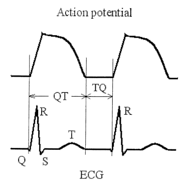
Figure 1 is a schematic graphic representation of the action potential in
cardiac
muscle summed up over its volume and the induced electrocardiogram (ECG)
recorded on a
human body surface.
Figure 2 is a block diagram of an apparatus for carrying out the present
method.
Figure 3 is an alternative block diagram of an apparatus for carrying out the
present
method.
Figure 4 is a block diagram of the processing steps for data acquisition and
analysis
of the present invention.
Figure 5 is a block diagram of the processing steps for an alternative
realization of
the data acquisition and analysis of the present invention.
CA 02533037 2006-O1-19
WO 2005/009234 PCT/US2004/020085
-11-
Figure 6 illustrates the experimental raw data and the processed data at
different data
processing steps ending with the RR-interval fluctuation hysteresis loops for
a healthy 50
year old male (SCIM(RR)TM=62).
Figure 7 illustrates the experimental raw data and the processed data at
different data
processing steps ending with the RR-interval fluctuation hysteresis loops for
a 58 year old
healthy male (SCIM(RR)TM =187).
Figure ~ illustrates the experimental raw data and the processed data at
different data
processing steps ending with the RR-interval fluctuation hysteresis loops for
a CAD male
patient (61 year old, SCIM(RR)TM =323).
Figure 9 illustrates a typical rapid peripheral nervous system and hormonal
control
adjustment of the RR interval as a result of an abrupt stop in exercise (that
is, an abrupt
initiation of a rest stage).
Figure 10 illustrates a typical slow (quasi-stationary) RR interval adjustment
measured
during gradually increasing and gradually decreasing cardiac stimulation.
Detailed Description of the Preferred Embodiments
The present invention is explained in greater detail below. This description
is not
intended to be a detailed catalog of all the different manners in which
particular elements of
the invention can be implemented, and numerous variations will be apparent to
those skilled
in the aut based upon the instant disclosure.
As will be appreciated by one of skill in the art, certain aspects of the
present
invention may be embodied as a method, data processing system, or computer
program
product. Accordingly, certain aspects of the present invention may take the
form of an
entirely hardware embodiment, an entirely software embodiment, or an
embodiment
combining software and hardware aspects. Furthermore, certain aspects of the
present
invention may take the form of a computer program product on a computer-usable
storage
medium having computer readable program code means embodied in the medium. Any
suitable computer readable medium may be utilized including, but not limited
to, hard disks,
CD-ROMs, optical storage devices, and magnetic storage devices.
Certain aspects of the present invention are described below with reference to
flowchart illustrations of methods, apparatus (systems), and computer program
products. It
will be understood that each block of the flowchart illustrations, and
combinations of blocks
in the flowchart illustrations, can be implemented by computer program
instructions. These
CA 02533037 2006-O1-19
WO 2005/009234 PCT/US2004/020085
-12-
computer program instructions may be provided to a processor of a general
purpose
computer, special purpose computer, or other programmable data processing
apparatus to
produce a machine, such that the instructions, which execute via the processor
of the
computer or other programmable data processing apparatus, create means for
implementing
the functions specified in the flowchart block or bloclcs.
Computer program instructions may also be stored in a computer-readable memory
that can direct a computer or other programmable data processing apparatus to
function in a
particular manner, such that the instructions stored in the computer-readable
memory produce
an article of manufacture including instruction means which implement the
function specified
in the flowchart block or blocks.
Computer program instructions may also be loaded onto a computer or other
programmable data processing apparatus to cause a series of operational steps
to be
performed on the computer or other programmable apparatus to produce a
computer
implemented process such that the instructions which execute on the computer
or other
programmable apparatus provide steps for implementing the functions specified
in the
flowchart block or blocks.
i. Definitions.
''A trend" on a data segment is a data set generally obtained from the raw
data
segment by smoothing. In a particular implementation herein a trend is
assessed as the
smoothest data set obtained by fitting the raw data on a data segment with a
lowest degree
polynomial (linear or quadratic, with the latter being used when the data set
encompasses a
single extremum, i.e., a minimum or a maximum). The total variation of the
trend is always
much smaller than the total variation of the raw data segment.
"A stationary data segment" is a data segment with a negligible variation of
the trend.
"A slow trend" is a trend with a small but not negligible variation. A trend
obtained
under the quasi-stationary protocol (see example 7) is a slow trend. A
duration of a stage
during which the data incorporating a slow trend are collected must be
approximately an
order of magnitude (e.g., at least about ten times) longer than the average
duration (~ 1
minute) of the heart rate adjustment after an abrupt stop of exercise from a
peak load rate
(typically from 120 to 150 beat/min) to the rest rate (typically from 50 to 80
beat/min).
"A fluctuation" of an RR interval on a data segment as used herein refers to a
set of
zero sum deviations from an RR slow trend corresponding to this particular
data segment. A
CA 02533037 2006-O1-19
WO 2005/009234 PCT/US2004/020085
-13-
traditional measure of fluctuations is the standard root-mean-square deviation
(STD). A
typical value of STD for RR interval fluctuations is of an order of magnitude
(e.g., at least
about ten times) smaller than the total variation of the RR interval trend
during the entire load
stage under quasi-stationary conditions.
"Cardiac ischemia" refers to a lack of or insufficient blood supply to an area
of
cardiac muscle. Cardiac ischemia usually occurs in the presence of
arteriosclerotic occlusion
of a single or a group of coronary arteries. Arteriosclerosis is a product of
a lipid deposition
process resulting in fibro-fatty accumulations, or plaques, which grow on the
internal walls of
coronary arteries. Such an occlusion compromises blood flow through the
artery, which
reduction then impairs oxygen supply to the surrounding tissues during
increased
physiological need -- for instance, during increased exercise loads. In the
later stages of
cardiac ischemia (e.g., significant coronary artery occlusion), the blood
supply may be
insufficient even while the cardiac muscle is at rest. However, in its earlier
stages such
ischemia is reversible in a manner analogous to how the cardiac muscle is
restored to normal
function when the oxygen supply to it returns to a normal physiological level.
Thus,
ischemia that may be detected by the present invention includes episodic,
chronic and acute
ischemia.
"Exercise" as used herein refers to voluntary skeletal muscle activity of a
subject that
increases heart rate above that fouund at a sustained stationary resting
state. Examples of
exercise include, but are not limited to, cycling, rowing, weight-lifting,
walking, ruuming,
stair-stepping, etc., which may be implemented on a stationary device such as
a treadmill or
in a non-stationary environment.
"Exercise load" or "load level" refers to the relative strenuousness of a
particular
exercise, with greater loads or load levels for a given exercise producing a
greater heart rate
in a subject. For example, load may be increased in weight-lifting by
increasing the amount
of weight; load may be increased in wallcing or running by increasing the
speed and/or
increasing the slope or incline of the walking or runniilg surface; etc.
"Gradually increasing" and "gradually decreasing" an exercise load refers to
exercise
in which the subject is caused to perform an exercise under a plurality of
different
sequentially increasing or sequentially decreasing loads. The number of steps
in the sequence
can be infinite so the terms gradually increasing and gradually decreasing
loads include
continuous load increase and decrease, respectively.
CA 02533037 2006-O1-19
WO 2005/009234 PCT/US2004/020085
-14-
"Intervening rest", when used to refer to a stage following increased cardiac
stimulation, refers to a stage of time initiated by a sufficiently abrupt
decrease in heart
stimulation (e.g., a sufficiently abrupt decrease in exercise load) so that it
evokes a clear
sympatho-adrenal response. Thus, an intervening rest stage is characterized by
a rapid
sympatho-adrenal adjustment (as further described in Example 8 below), and the
inclusion of
an intervening rest stage precludes the use of a quasi-stationary exercise (or
stimulation)
protocol (as further described in Example 9 below).
"Hysteresis" refers to a lagging of the physiological effect when the external
conditions are changed.
"Hysteresis curves" refer to a pair of curves in which one curve reflects the
response
of a system to a first sequence of conditions, such as gradually increasing
heart rate, and the
other curve reflects the response of a system to a second sequence of
conditions, such as
gradually decreasing heart rate. Here both sets of conditions are essentially
the same--i. e. ,
consist of the same (or approximately the same) steps--but are passed in
different order in the
course of time. A "hysteresis loop" refers to a loop formed by the two
contiguous curves of
the pair.
"Electrocardiogram" or "ECG" refers to a continuous or sequential record (or a
set of
such records) of a local electrical potential field obtained from one or more
locations outside
the cardiac muscle. This field is generated by the combined electrical
activity (action
potential generation) of multiple cardiac cells. The recording electrodes may
be either
subcutaneously implanted or may be temporarily attached to the sL~rface of the
skin of the
subject, usually in the thoracic region. An ECG record typically includes the
single-lead
ECG signal that represents a potential difference between any two of the
recording sites
including the site with a zero or ground potential.
"Pulse monitor" or "heart rate monitor" refers to a device that allows one to
measure
and to record the duration of each cardiac cycle during the monitored period
of time. Such a
device measures and records the time intervals between the instances when two
consecutive
caxdiac cycles have identical phases.
"Quasi-stationary conditions" refer to a gradual change in the external
conditions
and/or the physiological response it causes that occurs much slower than any
corresponding
adjustment due to sympathetic/parasympathetic and hormonal control. If the
representative
time of the external conditions variation is denoted by ieXt, and i;"t is a
representative time of
the fastest of the internal, sympathetic/parasympathetic and hormonal control,
then "quasi
CA 02533037 2006-O1-19
WO 2005/009234 PCT/US2004/020085
-15-
stationary conditions" indicates ie,~t » lint (e.g., 'text 15 at least about
five times greater than
dint)
"An abrupt change" refers to an opposite situation corresponding to a
sufficiently fast
change in the external conditions as compared with the rate associated with
sympathetic/parasympathetic and hormonal control-that is, it requires that
ie;~t « lint (e.g.,
ieXt is at least about five times less than iint). In particular, "an abrupt
stop" refers to a fast
removal of the exercise load that occurs during time shorter than lint ~ 20 or
30 seconds (see
Figure 9 below and comments therein).
"RR- data set" refers to a record of the time course of an electrical signal
comprising
action potentials spreading through cardiac muscle. Any single lead ECG record
incorporates
a group of tluee consecutive sharp deflections usually called a QRS complex
and generated
by the propagation of the action potential's front through the ventricles. The
time interval
between the cardiac cycles when observed via ECG (i.e., between the maxima of
the
consecutive R-waves) is called an RR-interval. Alternative definitions of
these intervals can
be equivalently used in the framework of the present invention. For example,
an RR-interval
can be defined as the time between any two similar points, such as the similar
inflection
points, on two consecutive R-waves, or any other way to measure cardiac cycle
length. An
ordered set of such interval durations simultaneously with the time instants
of their
beginnings or ends which are accumulated on a beat to beat basis or on any
given beat
sampling rate basis form an RR-interval data set. Thus, an RR- interval data
set will contain
two RR-interval related sequences f T~,1,T~,2,...,T~,n,} and {tl,t2,...,tn}.
"Cardiac cycle length data set" refers to a record of the time course of
consecutive
time intervals between the instances when two consecutive cardiac cycles have
identical
phases. A cardiac cycle length data set can be obtained either via ECG (RR-
interval data set)
or the pulse or heart rate monitor. The term "cardiac cycle length data set"
will be used
interchangeably with the term "RR-interval data set".
An "instantaneous heart rate" is defined as a reciprocal of a current RR
interval value
or, equivalently, as a reciprocal of a current cardiac cycle length.
In the following definitions, C[a, b] shall denote a set of continuous
functions f(t) on a
segment [a,b]. ~t,~, i=1,2,..., N, denotes a set of points from [a,b], i.e.
{tl}={ti. a<t; _< b,
i=1,2, ...,N~ and ~f(tz)}, where fE C[a, b] denotes a set of values of the
function f at the points
{t;}. In matrix operations the quantities i= f tl}, y=~f(it;)}, are treated as
column vectors. EN
CA 02533037 2006-O1-19
WO 2005/009234 PCT/US2004/020085
-16-
shall denote an N-dimensional metric space with the metric RN(x, y), xy E EN.
(RN(x, y) is said
b
to be a distance between points x and y.) A (total) variation ~j ~FJ is
defined for any
a
absolutely continuous function F from C[a, b] as the integral (a Stieltjes
integral)
~j [F(t)] - f ~ dF(t) ~ = f ~ F' (t) ~ dt . (D.1)
a n '
For a function F monotonic on segment [a, b] its variation is simply ~F(a)-
F(b)~. If a function
F(t) has alternating maxima and minima, then the total variation of F is the
sum of its
variations on the intervals of monotonicity. For example, if the points of
minima and maxima
are xl=a, x2, x;, ..., xh=b, then
b k-1
V [F(t)] _ ~ ~ F(x~ ) W'(x~+i ) ~ ~ (D.2)
a i=1
Fitting (best fitting): Let C [a, b] be a subset of C[a, b]. A continuous
function f(t),
fE C' [ca, b] is called the (best) fit (or the best fitting fisnetioh of class
C [a, b] with respect to
metric RN to a data set {x;,t;~ (i=1,2,..., N} if
RN(~tl)},{x;})= min (D.3)
f eC(cr,b]
The minimum value of RN is then called the e~f°ot° of the fit.
The functions f(t) from C [a, b]
will be called trial functions.
In most cases EN is implied to be an Euclidean space with an Euclidean metric.
The
error RN then becomes the familiar mean-root-square error. The fit is
performed on a subset
C [a, b] since it usually implies a specific parametrization of the trial
functions and/or such
constrains as the requirements that the trial functions pass through a given
point and/or have a
given value of the slope at a given point.
A smoother function (comparison of smoothness): Let f(t) and g(t) be functions
from
C[a, b] that have absolutely continuous derivatives on this segment. The
function f(t) is
synoothe~ than the function g(t) if
CA 02533037 2006-O1-19
WO 2005/009234 PCT/US2004/020085
-17-
b b
V [f(t)~ ~ V [g(t»~ (D.4)
a a
and
b b
V t~(t» ~ V [g'(t)>> (D.5)
a a
where the prime denotes a time derivative, and a strict inequality holds in at
least one of
relations (D.4) and (D.5).
A smoother set: A set ~x;,t;} (i=1,2,..., N} is smoothe~° than the set
~x~,t~} (j=1,2,...,
N} if the former can be fit with a smoother function f(t) of the same class
within the same or
smaller error than the latter.
"Smoothing" of a data set as used herein may be understood as follows: Let F
a.nd G
'10 be continuous functions on a N dimensional space with the values in a M
dimensional space.
A transformation of a data set (x,t)={x;,t;} (i=1,2,..., N} into another set
(y,i)=~y~,z~}
(j=1,2,..., M, N>_M} of the form
Y=F'(x), i=G(t)~ (D.6)
is called a sa~zoothing if the latter set is smoother than the former. Dne can
refer to ~y~,z~} as a
smoothed set. In practice, the functions F and G are linear and are
represented by uheN
matrices (N~~. According to the above definition a transformation of a data
set by filtering
(in the time or frequency domain) comprises smoothing.
A measure of a closed domain: Let S2 be a singly connected domain on the plane
(i,T) with the bovmdary formed by a simple (i.e., without self intersections)
continuous curve.
A measuoe M of such a domain S~ on the plane (i,T) is defined as the Riemann
integral
M = ~ f ,o(z,T)didT (D.7)
where p(i,T) is a nonnegative (weight) function on S2.
Note that when p(i,T)=1 the measure M of the domain coincides with its area,
A;
when p(i,T)=1/i2, the measure, M, has the meaning of the area, A; of the
domain S~' on the
transformed plane (f, T), where f--1/i can be understood as the heart rate
since the quantity i
has the meaning of RR-interval. [The domain SZ' is the image of domain S~
under the mapping
(i~~~(1/i,T).]
CA 02533037 2006-O1-19
WO 2005/009234 PCT/US2004/020085
-18-
"A trend" on a data segment is a data set generally obtained from the raw data
segment by low pass filtering under the restriction that the deviations from
the resulting trend
have a zero sum. In a particular implementation herein a trend is assessed as
the smoothest
data set obtained by fitting the raw data on the segment with a lowest degree
polynomial
(linear or quadratic, with the latter being used when the data set encompasses
a single
extremum, i. e. a minimum or a maximum). The total variation of the trend is
always much
smaller than the total variation of the raw data segment.
"A stationary data segment" is a data segment with a negligible variation of
the trend.
"A slow trend" is a trend with a small but not negligible variation. A trend
obtained
under the quasi-stationary protocol (see example 7) is a slow trend. A
duration of a stage
during which the data incorporating a slow trend are collected must be
approximately an
order of magnitude (e.g., at least about ten times) longer than the average
duration (-~- 1
minute) of the heart rate adjustment after an abrupt stop of exercise from a
peak load rate
(typically from 120 to 150 beat/min) to the rest rate (typically from 50 to 80
beat/min).
"A fluctuation" of RR interval on a data segment as used herein refers to a
set of zero
sum deviations from an RR slow treizd corresponding to this particular data
segment. A
traditional measure of fluctuations is the standard root-mean-square deviation
(STD). A
typical value of STD for RR interval fluctuations is of an order of magnitude
(e.g., at least
about ten times) smaller than the total vc~riatiosz of the RR interval trend
dL~ring the entire
load stage under quasi-stationary conditions.
Figure 1 illustrates the correspondence between the temporal phases of the
periodic
action potential (AP, upper graph, 20) generated inside cardiac muscle and
summed up over
its entire volume and the electrical signal produced on the body surface and
recorded as an
electrocardiogram (ECG, lower graph, 21). The figure depicts two regular
cardiac cycles.
During the upstroke of the action potential the QRS-complex is formed. It
consists of three
waves, Q, R, and S, which axe marked on the lower panel. The recovery stage of
the action
potential is characterized by its fall off on the AP plot and by the T-wave on
the ECG plot.
One can see that the time between consecutive R-waves (RR interval)
conveniently
represents the duration of a cardiac cycle, while its reciprocal value
represents the
corresponding instantaneous heart rate.
CA 02533037 2006-O1-19
WO 2005/009234 PCT/US2004/020085
-19-
2. Fluctuation analysis.
Finding the mean values of measured quantities has traditionally been the
center point
of data processing: In live systems, however, fluctuations, the deviations
from the mean
value, may carry the primary information about a subsystem, which interacts
with the rest of
the system. In the case of cardiac stress testing, the temporal heart rate
fluctuations provide
additional information on the state of ventricular muscle and more generally
on cardiac
function during exercise. The heart rate fluctuations or the fluctuations in
the cardiac cycle
length can be spoken of as the RR fluctuations. The RR fluctuation time series
must be
extracted from the original RR interval data sets.
2.1. Theoretical basis. The idea underlying the data processing algorithms
that allow
one to fmd the temporary fluctuations of the random process with non-
stationary mean value
and stationary increments (first differences) has been first laid by A.
I~olmogoroff (Soviet
Mathematics, Dolclady, 26:6-9 (1940); and 26:115-118(1940)) and later
developed by
Yaglom (Matematicheskii Sbomik, 37:141-196(1955)) for the case with stationary
higher
order differences. Let us denote for brevity the RR interval immediately
preceding a time
instant t by a single quantity, T(t). Measurements of T(t) result in a
discrete time series, which
is a sample of the stochastic process T(t), which can be divided into two
components, a
nonrandom component f(t) and a random component (fluctuations or physiological
and
physical noise) ~(t), so we have
T(t)=T(t)+8T(t), T(t)=<T>, <8T(t)>=0, ~ (2.1)
where the angle brackets denote ensemble averaging. The condition that the
ensemble
average of the fluctuations <8T5 is zero is of crucial importance and must be
preserved by
any consistent data processing procedure. We shall consider sufficiently short
segments of
data records such that the random component, 8T(t), can be considered as a
stationary
stochastic process with zero mean, and tiyne independent ynornents. For
brevity, we shall
denote {T~'~} by {Ti'} (k = 1, 2, ..., ll~. Let us consider a short segment of
data {Ti'} such
that the trend can be accurately represented by a low power polynomial, e.g, a
linear, or
quadratic, in the vicinity of the minimum. In the former case we represent the
sequence Tr' on
the segment by the expression
Tk =b(tk -tl)+c+~T~, (2.2)
where 8Ti' by definition is the k-th fluctuation if b and c are determined by
the requirement
that the error E is minimized:
CA 02533037 2006-O1-19
WO 2005/009234 PCT/US2004/020085
-20-
E = ~ (S T k )2 = min (2.3)
k=1 b,c
This condition determines the coefficients a and b and thereby a sequence of
the varying
trend values, T(tk)=b(tk-t,)+c, and the fluctuation time series, ST(tk)---~Ti'
for k=l, ,2 ...,
M. In the vicinity of the HR maximum, or RR interval minimum, one needs to use
a
parabolic fit for the trend and set
Tk =a(tk -tl)Z +b(tk -ti)+c+8Tk (2.4)
and determine the coefficients a, b and c by the similar requirement
E = ~ (b T k )2 = min (2.5)
k=i ~,b,~
One can easily check that one of the miumization equations, aElac=0, reduces
to the
requirement that
nr
~BTk =0, (2.6)
k=1
which, indeed, allows one to interpret the series {8TH'} as a series of
fluctuations, a stationary
random process with a zero mean value. It is also noteworthy that under the
condition of
quasi-stationarity the above constants a and b are sufficiently small so the
trend variation of
function T (t) is much smaller than its representative value on the segment.
In the linear case
it means that the following condition holds
b(tN-tk)<b(tN-tl)«c, (k=1,2,...,M). (2.7)
Similarly, in the quadratic case it means that
a(tN -t,~)2 < a(tN -tl)z « c, b(tN -t,) « c , ( k =1,2,...,M). (2.8)
2.2. An algorithm for finding and separating the trend and fluctuations. The
above ideas can be applied to the time series within a zzzovifzg, relatively
short time window
with the width determined by some additional requirements discussed below. Let
{(tk,Tk):
k=1,2, ...lV} be a set of raw data points obtained in the quasi-stationary
exercise test. The set
{Tk} is a shorthand notation for the RR-interval data set {T~'~} or an
equivalent cardiac cycle
length data set. The time instants {tk} are assumed to be equidistant, tk-
tk_1=is const, where
the time spacing is is in fact determined by the beat sampling rate, which is
equal to 1/is. We
define a kth time window with a given width (2m+1)is as a set of 2m+1 points
{(t~,T~): j=k nz,
CA 02533037 2006-O1-19
WO 2005/009234 PCT/US2004/020085
-21 -
k m+l, ..., k+m) that include and surround point (tk,Tk). Let us denote by
fk(t) a quadratic or
linear polynomial obtained by a linear regression such that (t, fk(t))
provides the best fit for
the data points {t~,T ~ within the window of a given width as defined by
Equations (2.3) or
(2.5) for a linear or quadratic polynomial function, fk, respectively. The
function fk(t)
describes the slow tend inside the window with the width M=2nz+1 as. The set
of
corresponding fluctuations 8Tj(m) within the window is defined by the equation
8Tj(m)=Tj-fk(tj), j=k-m,k-nz+1,...,k+m. (2.9)
For a polynomial fk(t) the standard deviation (STD) for this particular window
is given by the
equation
1 j=k+m
ak = -- ~ [Tj -.fk(tj)j2 (2.10)
2m j=k-ne
The error minimization corresponding to Equations (2.3) or (2.5) is equivalent
(at fixed value
of m) to minimization of the STD given by Equation (2.10). This procedure is
repeated for a
broad range of m values. Then an optimal value of zn is found by the
requirement that we
achieve the best accuf~acy (oy~ systerzzatic erv~oz° in the tt~eod). In
fact, this requires that 6~"
does not possess a clear trend as a function of the window width or a function
of rfz. In fact,
this requirement can be replaced by the condition that that the variation of
6~' / nz is
minimum as a function of nz.
6 "'
~k = min ~ (2.11 )
»>.»»,»,~;" m
The lower bound of m-values must be determined by the practical considerations
such as
robustness and stability of the results. Equation (2.11) defines the optimum
value of
In=moptimum. Thus optimized value of a'k(m=moptimum) is taken as the current
measure of
fluctuations for the given, kth window. The same value of ln=moptimum also
determines both
the trend value within given window centered on point tk, which should be
taken equal to the
value of fk(t~;) at the center of the window, i. e. fk(t~ As the next step we
shift the center of the
window one time-step further, to tk+i and proceed to evaluation of the trend
and 6~;+i in the
same way as at the just completed previous step. If N is the total size of the
sample (number
a
of data points) this procedure is performed N 2rn times and produces N 2nz
values of 6~;; the
respective slow trend values fk(tk).
CA 02533037 2006-O1-19
WO 2005/009234 PCT/US2004/020085
-22-
3. Testing methods.
The methods of the present invention are primarily intended for the testing of
human
subjects. Virtually any human subject can be tested by the methods of the
present invention,
including male, female, juvenile, adolescent, adult, and geriatric subjects.
The methods may
be carried out as an initial screening test on subjects for which no
substantial previous history
or record is available, or may be carried out on a repeated basis on the same
subj ect
(particularly where a comparative quantitative indicium of an individual's
cardiac health over
time is desired) to assess the effect or influence of intervening events
andlor intervening
therapy on that subject between testing sessions.
As noted above, the method of the present invention generally comprises (a)
collecting a first RR- interval data set from said subject during a stage of
gradually increasing
heart rate; (b) collecting a second RR- interval data set from said subject
during a stage of
gradually decreasing heart rate; (c) determining the trend in the first
cardiac cycle length.data
set and separating deviations from the trend (fluctuations) in the first
cardiac cycle length
data set; (d) determining the trend in the second cardiac cycle length data
set and separating
deviations from the trend (fluctuations) in the second cardiac cycle length
data set; (e)
comparing the fluctuations of the first cardiac cycle length data set to the
fluctuations of the
second cardiac cycle length data set at equal trend values of the cardiac
cycle length to
determine the difference between the fluctuation data sets; and (f) generating
from the
comparison of step (e) a measure of cardiac ischemia dining stimulation in the
subject,
wherein a greater difference between the first and second data sets indicates
greater cardiac
ischemia and lesser cardiac or cardiovascular health in the subject.
The stages of gradually increasing and gradually decreasing heart rate are
carried out
in a manner that maintains during both periods essentially or substantially
the same
stimulation of the heart by the peripheral nervous and hormonal control
systems, so that it is
the effect of cardiac ischemia rather than that of the external control which
is measured by
means of the present invention. This methodology can be carried out by a
variety of
techniques, with the technique of conducting two consecutive stages of
gradually increasing
and gradually decreasing exercise loads (or average heart rates) being
currently preferred.
The stage of gradually increasing exercise load (or increased average heart
rate) and
the stage of gradually decreasing exercise load (or decreased average heart
rate) may be the
same in duration or may be different in duration. In general, each stage is at
least 3, 5, 8, or
10 minutes or more in duration. Together, the duration of the two stages may
be from about
CA 02533037 2006-O1-19
WO 2005/009234 PCT/US2004/020085
- 23 -
6, 10, 16 or 20 minutes in duration to about 30, 40, or 60 minutes in duration
or more. The
two stages are preferably carried out sequentially in time-that is, with one
stage following
after the other substantially immediately, without an intervening rest stage.
In the alternative,
the two stages may be carried out separately in time, with an intervening
"plateau" stage
(e.g., of from 1 to 5 minutes) during which cardiac stimulation or exercise
load is held
substantially constant, before the stage of decreasing load is initiated.
The exercise protocol may include the same or different sets of load steps
during the
stages of increasing or decreasing heart rates. For example, the peals load in
each stage may
be the same or different, and the minimum load in each stage may be the same
or different.
In general, each stage consists of at least two or three different load
levels, in ascending or
descending order depending upon the stage. Relatively high load levels, which
result in
relatively high heart rates, can be used but are not essential. , An advantage
of the present
invention is that its sensitivity allows both exercise procedures to be
carried out at relatively
low load levels that do not unduly increase the pulse rate of the subject. For
example, the
method may be carried out so that the heart rate of the subject during either
the ascending or
descending stage (or both) does not exceed about 140, 120, or even 100 beats
per minute,
depending upon the condition of the subject. Of course, data collected at
heart rates above
100, 120, or 140 beats per minute may also be utilized if desired, again
depending upon the
condition of the subject.
For example, for an athletic or trained subject, for the first or ascending
stage, a first
load level may be selected to require a power output of 60 to 100 or 150 watts
by the subject;
an intermediate load level may be selected to require a power output of 100 to
150 or 200
watts by the subject; and a third load level may be selected to require a
power output of 200
to 300 or 450 watts or more by the subject. For the second or descending
stage, a first load
level may be selected to require a power output of 200 to 300 or 450 watts or
more by the
subject; an intermediate or second load level may be selected to require a
power output of
100 to 150 or 200 watts by the subject; and a third load level may be selected
to require a
power output of 60 to 100 or 150 watts by the subject. Additional load levels
may be
included before, after, or between all of the foregoing load levels as
desired, and adjustment
~ between load levels can be carried out in any suitable manner, including
step-wise or
continuously.
In a further example, for an average subject or a subject with a history of
cardiovascular disease, for the first or ascending stage, a first load level
may be selected to
CA 02533037 2006-O1-19
WO 2005/009234 PCT/US2004/020085
-24-
require a power output of 40 to 75 or 100 watts by the subject; an
intermediate load level may
be selected to require a power output of 75 to 100 or 150 watts by the
subject; and a third
load level may be selected to require a power output of 125 to 200 or 300
watts or more by
the subject. For the second or descending stage, a first load level may be
selected to require a
power output of 125 to 200 or 300 watts or more by the subject; an
intermediate or second
load level may be selected to require a power output of 75 to 100 or 1 SO
watts by the subject;
and a third load level may be selected to require a power output of 40 to 75
or 100 watts by
the subject. As before, additional load levels may be included before, after,
or between all of
the foregoing load levels as desired, and adjustment between load levels can
be carried out in
any suitable manner, including step-wise or continuously.
The heart rate may be gradually increased and gradually decreased by
subjecting the
patient to a predetermined schedule of stimulation. For example, the patient
may be
subjected to a gradually increasing exercise load and gradually decreasing
exercise load, or
gradually increasing electrical or pharmacological stimulation and gradually
decreasing
electrical or pharmacological stimulation, according to a predetermined
program or schedule.
Such a predetermined schedule is without feedback of actual heart rate from
the patient. In
the alternative, the heart rate of the patient may be gradually increased and
gradually
decreased in response to actual heart rate data collected from concurrent
monitoring of said
patient. Such a system is a feedback system. For example, the heart rate of
the patient may
be monitored during the test and the exercise load (speed and/or incline, in
the case of a
treadmill) can be adjusted so that the heart rate varies in a prescribed way
during both stages
of the test. The monitoring and control of the load can be accomplished by a
computer or
other control system using a simple control program and an output panel
connected to the
control system and to the exercise device that generates an analog signal to
the exercise
device. One advantage of such a feedback system is that (if desired) the
control system can
insure that the heart rate increases substantially linearly during the first
stage and decreases
substantially linearly during the second stage.
The generating step (~ may be carried out by any suitable means, such as by
generating curves from the data sets (with or without actually displaying the
curves), and then
(i) directly or indirectly evaluating a measure (e.g., as defined in the
integral theory) of the
domain (e.g., area) inside the hysteresis loop, a greater measure indicating
greater cardiac
ischemia in said subject, (ii) directly or indirectly comparing the shapes
(e.g., slopes or
derivatives thereof) of the curves, with a greater difference in shape
indicating greater cardiac
CA 02533037 2006-O1-19
WO 2005/009234 PCT/US2004/020085
- 25 -
ischemia in the subject; or (iii) combinations of (i) and (ii). Specific
examples are given in
Example 3-5 below.
The method of the invention may further comprise the steps of (e) comparing
the
measure of cardiac ischemia during exercise to at least one reference value
(e.g., a mean,
median or mode for the quantitative indicia from a population or subpopulation
of
individuals) and then (~ generating from the comparison of step (e) at least
one quantitative
indicium of cardiovascular health for said subject. Any such quantitative
indicium may be
generated on a one-time basis (e.g., for assessing the likelihood that the
subject is at risk to
experience a future ischemia-related cardiac incident such as myocardial
infarction or
ventricular tachycardia), or may be generated to monitor the progress of the
subject over
time, either in response to a particular prescribed cardiovascular therapy or
simply as an
ongoing monitoring of the cardiovascular physical condition of the subject for
improvement
or decline (again, specific examples are given in Example 3-5 below). In such
a case, steps
(a) through (f) above are repeated on at least one separate occasion to assess
the efficacy of
the cardiovascular therapy or the progress of the subject. A decrease in the
difference
between said data sets from before said therapy to after said therapy, or over
time, indicates
an improvement in cardiac health in said subject from said cardiovascular
therapy. Any
suitable cardiovascular therapy can be administered, including but not limited
to, aerobic
exercise, muscle strength building, change in diet, nutritional supplement,
weight loss,
smoking cessation, stress reduction, pharmaceutical treatment (including gene
therapy),
surgical treatment (including both open heart and closed heart procedl~res
such as bypass,
balloon angioplasty, catheter ablation, etc.) and combinations thereof.
The therapy or therapeutic intervention may be one that is approved or one
that is
experimental. In the latter case, the present invention may be implemented in
the context of a
clinical trial of the experimental therapy, with testing being carried out
before and after
therapy (and/or during therapy) as an aid in determining the efficacy of the
proposed therapy.
4. Testing apparatus.
Figure 2 provides an example of the apparatus for data acquisition, processing
and
analysis by the present invention. Electrocardiograms are recorded by an ECG
recorder, via
electrical leads placed on a subject's body. The ECG recorder may be, for
example, a
standard mufti-lead Holter recorder or any other appropriate recorder. The
analog/digital
converter digitizes the signals recorded by the ECG recorder and transfers
them to a personal
CA 02533037 2006-O1-19
WO 2005/009234 PCT/US2004/020085
-26-
computer, or other computer or central processing unit, through a standard
external
input/output port. The digitized ECG data can then be processed by standard
computer-based
waveform analyzer software, which identifies, in particular, R waves and their
timing. The
totality of such R wave timing instants translates into the RR interval data
,set, from which
cardiac or cardiovascular health indicium or other quantitative measure of the
presence,
absence or degree of cardiac ischemia can then be computed automatically in
the computer
through a program (e.g, Basic, Fortran, C++, etc.) implemented therein as
software,
hardware, or both hardware and software.
Figure 3 provides an example of an alternative apparatus for data acquisition,
processing and analysis by the present invention. The first two steps of data
acquisition are
performed by a Polar 5810 heart rate monitor (Polar Electro Inc., 370
Crossways Park Dr.,
Woodbury, NY 11797-2050). The actual monitor, Polar 5810 heart rate monitor,
incorporates
an analog-digital converter so its output is directly fed to a computer in
which the cardiac
cycle length data set is formed and stored. Using this data set, cardiac or
cardiovascular
health indicium or other quantitative measure of the presence, absence or
degree of cardiac
ischemia can then be computed automatically in the computer through a program
(e.g, Basic,
Fontran, C++, etc.) implemented therein as software, hardware, or both
hardware and
software.
Figures 4 and 5 correspond to two alternative data acquisition methods
represented in
Figures 2 and 3, respectively. These figures illustrate major steps of
digitized data
processing involved in the analysis of an RR data set collected from a subject
during there
and-back quasi-stationary changes in physiological conditions. The last seven
steps in
Figures 4 and Figure 5 are substantially the same while the initial steps
differ. As shown in
Figure 4, the digitized data collected from a mufti-lead recorder are stored
in a computer
memory for each lead as a data array (the 1St step). The size of each data
array is determined
by the durations of the ascending and descending heart rate stages and a
sampling rate used
by the waveform analyzer, which processes an incoming digitized ECG signal.
The
waveform analyzer software first detects major characteristic waves (Q,R,S and
T waves) of
the ECG signal in each lead. Then in each ECG lead it determines the timing of
each R wave
(the 2"d step). Using the data from the lead with the best data to noise
ratio, the time instants
of the R wave occurrence are determined as reference points to compute a set
of RR intervals
and a set of instantaneous heart rates (3'd step).
CA 02533037 2006-O1-19
WO 2005/009234 PCT/US2004/020085
-27-
Then, the application part of the software sorts the R.R intervals for the
ascending and
descending heart rate stages (the 4th step). The step includes computing by
the application
part of the software RR intervals separately for the ascending and descending
heart rate
stages effected by there-and-back gradual changes in physiological conditions
such as
exercise, pharmacological or electrical stimulation, etc. At the next, S~h,
step the application
software performs smoothing, filtering or data fitting, using exponential or
any other suitable
functions, in order to obtain a sufficiently smooth trend curve T~=F(t) for
each stage,
including the ascending and descending heart rate stages.
At the next, 6~', step the application part of the software determines
fluctuations as
deviations from the trend, 8TH=TRR-<T~> and generates a sufficiently smooth
curve
6~=6 ~ --- < b T~ > _ ~(t) to represent the standard deviations (STD) of
fluctuations as a
function of time during exercise.
At the 7th step these parametric representations, T~=F(t) and 8TH=~(t), are
used to
eliminate the time and generate or plot on the (<T~>,6~) plane a sufficiently
smooth
hysteresis loop parametrically represented by the pair of functions <T~>=F(t)
and 6~=~(t).
The next, 8~', step performed by the application part of the software can be
graphically
presented as closing the two branch hysteresis loop with an appropriate
interconnecting or
partially connecting line, such as a vertical straight line or a line
connecting the initial and
final points, in order to produce a closed hysteresis loop on the (<T~>,~~)-
plane. At the 9th
step the application software evaluates an appropriate measure of the domain
inside the
closed hysteresis loop. A measure, as defined in mathematical integral theory,
is a
generalization of the concept of an area and may include appropriate weight
fimctions
increasing or decreasing the contribution of different portions of the domain
into said
measure. The final, 10th, step of the data processing is that the application
software computes
indexes by appropriately renormalizing the said measure or any monotonous
functions of said
measure. The measure itself along with the indexes may reflect both the
severity of the
exercise-induced ischemia, as well as a predisposition to local ischemia that
can be reflected
in some particularities of the shape of the hysteresis loop and the curves
T~=F(t) and
6~=~(t). The results of all above-mentioned signal processing steps may be
used to
quantitatively assess cardiac ischemia and, as a simultaneous option,
cardiovascular system
health of a particular individual under evaluation.
CA 02533037 2006-O1-19
WO 2005/009234 PCT/US2004/020085
-28-
Instead of using the (T~,a~)-plane, a similar data processing procedure can
equivalently be performed on any plane obtained by a non-degenerate
transformation of the
(T~,6~)-plane, such as (f~,6~) where f~=<1 /T~> is the smoothed and/or
filtered heart
rate or the like. Such a transformation can be partly or fully incorporated in
the appropriate
definition of the said measure.
Figure 5 is similar to Figure 4 but corresponds to the alternative data
acquisition
method by a pulse monitor with digital output and also represents major steps
of the digitized
data processing involved in the analysis of a RR data set collected from a
subject during
there-and-back quasi-stationary changes in physiological conditions. The data
processing
steps by the application software (steps 3 through 9) are substantially the
same as the
respective steps 4 through 10 in Figure 4.
5. RR-interval monitoring with. blood pressure and/or pulse signals.
A quasi-stationary RR data set can be collected non-invasively not only via
measurements of a cardiac surface ECG but also by monitoring a blood pressure
andlor pulse
signals. In these cases, instead of the ECG recorder, a system for assessing
cardiac ischemia
may comprise pulse and/or blood pressure monitors, as discussed below.
A pulse monitor or pulse meter may be a suitable device, including but not
limited to
opto-electronic and phono or audio transducers attached to different parts of
a subject's body
(for example, to a finger as in finger plethysmography), if suchdevice
measures a heal-t rate or
pulse (HR). Preferably the device then computes RR-intervals equal to 1 /HR
and stores these
data in a computer memory in order to provide further RR-interval
computational analysis as
described herein.
A blood pressure monitor (e.g., a sphygmomanometer) can be any suitable
device,
including but not limited to a cuff, a stethoscope, or an automatic pressure
registering system
with a digital data storage module. Many such monitoring devices are
applicable even for
home use and typically contain all of the modules in one unit. An automatic
cuff inflation
monitor may also be included in the unit. Most units are portable and have a D-
ring cuff for
one-handed application. The cuff usually fits around the upper arm or the
wrist. These units
provide personalized cuff inflation and deflation. They automatically adjust
to changes in a
subject's blood pressure. Blood pressure monitoring with simultaneous
measurement of the
HR is convenient, easy to do and takes less than a minute per measurement. As
in the case
CA 02533037 2006-O1-19
WO 2005/009234 PCT/US2004/020085
-29-
above, the RR-intervals are equal to 1/ HR. Such apparatus may be easily
incorporated into a
method and apparatus of the present invention with suitable interfaces, in
accordance with
known techniques.
The options described above may be used separately or in parallel, including
in
parallel with ECG data, depending on experimental needs. Pulse meter ischemia
assessment
is expected to be more accurate than blood pressure monitoring since an RR
sampling
frequency (frequency of HR measurements) for a pulse meter is at least an
order of
magnitude higher (10 to 1 data point) than in the case of blood pressure
monitoring by an
automatic sphygmomanometer.
6. Conditions under which an abrupt stop exercise protocol can be considered
as
a guasi-stationary protocol.
An advantage of the present invention is, however, that it can be carried out
with an
"abrupt stop" exercise protocol as well as a gradually increasing/gradually
decreasing
exercise protocol. In the present invention, an "abrupt stop" exercise
protocol can be used for
any individual provided that the heart rate achieved prior to the stop is
sufficiently high for
that individual so that his or her recovery time is sufficiently long for an
appropriate
hysteresis to be measured.
In general, each stage of a gradually increasing and decreasing quasi-
stationary
exercise protocol is at least 3, 5, 8, or 10 minutes in duration. Each stage's
duration is
approximately an order of magnitude longer than the average duration (~ 1
minute) of heart
rate (HR) adjustment during an abrupt stop of the exercise between average
peal: load rate (~
120 - 150 beat/min) and average rest (~ 50 - 70 beat/min) heart rate values.
One should note that the ~ 1 minute HR adjustment period after an abrupt stop
of
exercise is typical only for healthy individuals and those with not more than
a relatively
moderate level of coronary artery disease. However, due to rapid development
of massive
exercise-induced ischemia for individuals with a pronounced coronary artery
disease level the
same adjustment period can be significantly longer - up to 10 minutes or more
in duration. In
these cases the HR deviations (described in the example 9 below) are small and
indicate that
such an ill patient remains under a significant physical stress even after an
abrupt stop of
exercise, without further exercise load exposure. In such cases a physician
must usually
interrupt the protocol prior to completion of a full quasi-stationary exercise
regimen.
However, under these conditions a recovery portion of the QT/RR hysteresis
loop, as well as
a loop formed after an abrupt stop of exercise, can be considered as the quasi-
stationary loop.
CA 02533037 2006-O1-19
WO 2005/009234 PCT/US2004/020085
-30-
Indeed, a slow heart rate recovery to a level equal to HR prior to the
exercise takes 3, 5, 8, 10
or more minutes in duration with small HR deviations and, therefore, still
satisfies the
underlying definition of a gradual exercise protocol.
Thus, for ill patients afflicted with coronary artery disease (CAD), an abrupt
stop of
exercise does not prevent the completion of the method of the present
invention, since due to
a prolonged HR recovery stage an indiciLUn of cardiac or cardiovascular health
can still be
calculated as a quasi-stationary loop index without a completion of a full
exercise protocol.
Note that typically a patient with a distinguished coronary artery disease
level, as
determined by physical exhaustion, shortness of breath, chest pain, and/or
some other clinical
symptom, is unable to exercise longer than 3, 5, 8 or 10 or more minutes at
even a low-level
power output of about 20 watts. A gradual recovery process followed the abrupt
stop of such
exercise in such patients satisfies the definition of a quasi-stationary
process as given herein.
Note that patients with moderate levels of coronary artery disease can
exercise longer, up to
minutes or more, and may be exposed to significantly higher exercise workouts
ranging
15 from 50 to 300 watts.
For healthy patients, an "abrupt stop" protocol can also be utilized in
carrying out the
present invention, for example when such patients achieve a peak level of
exercise close to
their maximmn level of physical exertion. Under such circumstances such
patients are similar
to patients afflicted with CAD, who reach such a threshold under much lower
exercise loads.
20 The present invention is explained in greater detail in the non-limiting
examples set
forth below.
EXAMPLE 1
Testing Apparatus
A testing apparatus consistent with Figure 2 was assembled. The
electrocardiograms
are recorded by an RZ152PM12 Digital EGG Holter Recorder (ROZINN ELECTRONICS,
INC.; 71-22 Myutle Av., Glendale, New York, USA 11385-7254), via 12 electrical
leads with
Lead-Lolc Holter/Stress Test Electrodes LL510 (LEAD-LOK, INC.; 500 Airport
Way, P.O.
Box L, Sandpoint, ID, USA 83864) placed on a subject's body in accordance with
the
manufacturer's instructions. Digital ECG data are transferred to a personal
computer (Dell
Dimension XPS TSOOMHz/Windows 98) using a 40 MB flash card (RZFC40) with a PC
700
flash card reader, both from Rozinn Electronics, Inc. Holter for Windows
(4Ø25); waveform
analysis software is installed in the computer, which is used to process data
by a standard
CA 02533037 2006-O1-19
WO 2005/009234 PCT/US2004/020085
-31-
computer based waveform analyzer software. The hysteresis loop for each tested
subject and
an indicium that provides a quantitative characteristic of the extent of
cardiac ischemia in said
subject are then computed manually or automatically in the computer through a
program
implemented in Fortran 90 as illustrated in Figure 4.
Experimental data were collected during an exercise protocol programmed in a
Landice L7 Executive Treadmill (Landice Treadmills; 111 Canfield Av.,
Randolph, NJ
07869). The programmed protocol included 20 step-wise intervals of a constant
exercise load
from 48 seconds to 1.5 minutes each in duration. Altogether these intervals
formed two
equal-in-dwation gradually increasing and gradually decreasing exercise load
stages, with
total duration varying from 16 to 30 minutes. For each stage a treadmill belt
speed and
elevation varied there-and-back, depending on the subj ect's age and health
conditions, from
1.5 miles per hour to 5.5 miles per hour and from one to ten degrees of
treadmill elevation,
respectively.
EXAMPLE 2
An alternative testing Apparatus
A testing apparatus consistent with Figure 3 was assembled. Experimental data
were
collected during an exercise protocol programmed in a Landice L7 Executive
Treadmill in
the way as described in Example 1. Instead of using a Digital ECG Hotter
Recorder, the
instantaneous heart rate during exercise was directly measured using a Polar
5810 heart rate
monitor (Polar Electro Inc., 370 Crossways Parlc Dr., Woodbury,NY 11797-2050).
The Polar
5810 heart rate monitor incorporates a~i analog-digital converter whose output
is directly fed
to a computer in which the data are stored as a digital array representing the
cardiac cycle
length data set. The hysteresis loop for each tested subject and an indicium
that provides a
quantitative characteristic of the extent of cardiac ischemia in said subject
are then computed
manually or automatically in the computer through a program implemented in
Fortran 90 as
illustrated in Figure 5.
EXAMPLES 3-5
Human RR-fluctuation Hysteresis Studies
These examples illustrate quasi-stationary ischemia-induced RR interval
fluctuation
hysteresis in different human subjects. These data indicate that the method
possesses a
potential for high sensitivity and high resolution.
CA 02533037 2006-O1-19
WO 2005/009234 PCT/US2004/020085
-32-
EXAMPLE 3
A Heart Rate Monitor Measurement of the Hysteresis Curve
and SCIMTM(RR) Score in Healthy Male Subject
The example was carried out on a SO year old male subject using the
alternative apparatus
and procedL~re described in Example 2 above. The subject exercised on a
treadmill
according to a quasi-stationary 20-minute protocol with gradually increasing
and gradually
decreasing exercise load. The test data obtained using alternative cardiac
cycle length
monitoring by a Polar 5810 heart rate monitor are displayed in Figure 3. The
upper panels,
Al, B1 and Cl, show raw data while the lower panels, A2, B2, and C2, show
smoothed and
partially or fully processed data at different processing stages. Panel Al in
Figure 6 displays
cardiac cycle length data sets ~T~(t~) f versus time, ~t~}, during the
exercise test. A beat
sampling rate with which cardiac cycle length was sampled was equal to 12
samples per
minute. Panel A2 in Figure 6 displays filtered and smoothed data from panel A1
as a
functional dependence <T~>=F(t). The smoothing procedure includes moving
averaging
with a two minute long moving window and is completed by the data fitting with
the third-
order polynomial functions within each two-minute long moving window. Panel B
1 in
Figure 6 displays the raw fluctuations of the cardiac cycle length, ~T~=T~-
<T~> versus
time during the exercise test. Panel B2 in Figure 6 displays filtered and
smoothed
characterization of the t7uctuation magnitude from the panel B1 as a
functional dependence
of the standard deviation, STD, versus time, 6~=~(t). Panel C1 in Figure 6
displays the
raw fluctuations versus the raw cardiac cycle length during the exercise test.
Panel C2 in
Figures 6 displays the hysteresis loop parametrically represented by the
dependencies
<T~>=F(t) and 6~=~(t) plotted in the respective panels A2 and B2. The vertical
lines
closing the loops (step 7 in Figure 5) are also shown in panels C2. The
subject had no
conventional ischemia-induced depression of the ECG-ST segments in similar ECG
stress
tests under the same quasi-stationary protocol, carried out separately on
multiple occasions.
The area of the closed hysteresis loop is relatively small as compared with
that shown in
Figure 8 and discussed in Example 5. Appropriate renormalization (see Example
7) of the
area results in the RR fluctuation ischemia index SCIMTM(RR) with the value of
62. This
value is significantly less than the value of SCIMTM(RR)=323 for the CAD
subject
considered in Example 5, below (see also Figure 8). The fact that the method
and apparatus
CA 02533037 2006-O1-19
WO 2005/009234 PCT/US2004/020085
- 33 -
of the present invention have precision, which is significantly higher than
the difference
between these two values, the method of the present invention allows one to
detect and
quantify ischemia with excellent resolution. This is also a clear indication
of high sensitivity
and specificity of the method and apparatus of the present invention.
EXAMPLES 4
A Holter Monitor Measurement of the Hysteresis Curve and
SCIMTM(RR) Score in Healthy Male Subject
The example illustrated by Figure 7 was carried out on a 58 years old male
subject using the
apparatus and procedure described in Example 2 above. The subject exercised on
a treadmill
according to a quasi-stationary 20-minute protocol with gradually increasing
and gradually
decreasing exercise load. The upper panels, Al, Bl and C1, show raw data while
the lower
panels, A2, B2, and C2, show partially or fully processed and smoothed data.
Panel Al in
Figure 7 displays RR interval data sets {T~(t~)} versus time, {t~), during the
exercise test. A
beat sampling rate, with which the waveform analyzer samples RR intervals was
equal to 15
samples per minute. The data presented in Figure 7 were obtained using left
precordial VS
leads. Panel A2 in Figure 7 displays filtered and smoothed data from the
respective panels
Al as a functional dependence <T~>=F(t). The smoothing procedure includes
moving
averaging with a two minute long moving window and is completed by the data
fitting with
the third-order palynomial functions within each two-minute long moving
window. Panel B1
in Figure 7 displays the RR interval fluctuations, STS=T,~-<T~>, versus time,
t, during the
exercise test. Panel B2 in Figure 7 displays filtered and smoothed
characterization of the
fluctuation magnitude from panel Bl as a functional dependence of the standard
deviation,
STD, versus time, 6~=~(t). Panel C1 in Figure 7 displays the raw fluctuations
versus raw
cardiac cycle length during the exercise test. Panel C2 in Figure 7 displays
the hysteresis
loop parametrically represented by the dependencies <T~>=F(t) and 6~=~(t)
plotted in the
respective panels A2 and B2. The vertical lines closing the loops (step 8 in
Figure 4) are also
shown in panels C2. The ROZINN software system detected no conventional
ischemia-
induced depression of the ECG-ST segments during test. The areas of the closed
hysteresis
loop is again relatively small as compared with that shown in Figure 8 and
discussed in
Example 5. Appropriate renormalization of the area (see Example 7) results in
the RR
fluctuation ischemia index SCIMTM(RR) with the value of 187. This value is
significantly
CA 02533037 2006-O1-19
WO 2005/009234 PCT/US2004/020085
-34-
less than the value of SCIM~(RR)=323 for the CAD subject considered in Example
5,
below (see also Figure 8). A comparison of this SCIMTM(RR) value with that
from Example
3 indicates that the method of the present invention allows one to
discriminate between
ischemia-induced hystereses in different subjects within a group which is sub-
threshold for
conventional ischemia detecting methods and techniques. A significant
difference between
the SCIMTM(RR) values of the present example and with that of Example 3 which
is equal to
125 indicates that the method possesses a good resolution even within a
conventionally sub-
threshold range of ischemic events. It also indicates that the method allows
one to
quantitatively differentiate the hystereses of the two subjects.
EXAMPLE 5
Hysteresis Curve for a Subject with ST Segment
Depression Observed During Exercise Test
The test was carried out on a 61-year-old male subject using the apparatus and
procedure described in Example 1 above. The subject exercised on a treadmill
according to a
quasi-stationary 20-minute protocol with a gradually increasing and gradually
decreasing
exercise load. A ST depression, indicating cardiac ischemia, was detected by
the ROZTNN
system during the test. Independently, the patient also had a positive
thallium ischemia stress
test result. Panel Al in Figure 8 displays cardiac cycle length (RR interval)
data set ,T~z(t~)f
versus time, {t~}, during the exercise test. A beat sampling rate with which
the waveform
analyzer sampled RR intervals was equal to 15 samples per minute. The data
were obtained
using left precordial V4 leads. Panel A2 in Figure 8 displays filtered and
smoothed data
from the respective panels A1 as a functional dependence, <T~>=F(t). The
smoothing
procedure includes moving averaging with a two minute long moving window and
is
completed by the data fitting with the third-order polynomial functions within
each two-
minute long moving window. Panel Bl in Figure 8 displays the raw RR interval
fluctuations, ~T~=T~-<T~>, versus time, t, during the exercise test. Panel B2
in Figure 8
displays a dependence of STD, ~~=~(t), representing filtered and smoothed
characteristics
of the fluctuation magnitude from panel B 1. Panel C 1 in Figure 8 displays
the raw
fluctuations, BTU, versus raw cardiac cycle length, T~, during exercise test.
Finally, panel
C2 in Figure 8 displays a hysteresis loop parametrically represented by the
dependencies
<T~>=F(t) and ~~=~(t) plotted in panels A2 and B2, respectively. Notice a
vertical line in
CA 02533037 2006-O1-19
WO 2005/009234 PCT/US2004/020085
-35-
panel C2 that closes the hysteresis loop and corresponds to step 8 in Figure
4. The area of the
closed hysteresis loop is relatively large as compared with those shown in
Figures 6 and 7
and discussed in Examples 3 and 4. Appropriate renormalization (see Example 7)
of the
area results in the RR fluctuation ischemia index SCIMTM(RR)=323. This value
is
significantly greater than the values of 62 and 187 obtained in Examples 3 and
4 above. All
the above cases demonstrate that the method of the present invention allows
one to
discriminate and quantitatively characterize the difference between (1) the
levels of ischemia
that can be detected by the conventional ST depression method and (2) the low
levels of
ischemia (illustrated in Figures 6 and 7) that are sub-threshold for the
conventional method,
which, therefore, can neither detect nor resolve such levels of ischemia or
perfusion
abnormality.
EXAMPLE 6
Fluctuation analysis method: an example of algorithm for determining
1 ~ the RR interval fluctuations and providing their characterization by the
moving STD
Let {(tk,T,;): k=1,2, ...N} be a set of data points (the time instants {ti;}
are equidistant,
t,;-ti;_1=const) obtained in the quasi-stationary exercise test as exemplified
in panels Al of
Figures fi through 8. The value of N in the above examples was about 400 and
slightly
varied from case to case. The data processing is essentially the same for the
data collected by
a Heart Rate Monitor, or by a regular Holter recorder and subsequently the
waveform
analyzer. The set {Tk} is a shorthand notation for the RR-interval data set
{T~'~} or an
equivalent cardiac cycle length data set. We define a k-th time m-window as a
set of 2na+1
points {(t~,T): j=k-m, k-m+1, ...,k+~7a} that include and surround the point
(tit,T~;). This in fact
must be done for a variety of m-values among which one particular value is
chosen as the
final algorithmic step described below. Let us denote by f~(t) a quadratic or
linear polynomial
obtained by a linear regression such that (t, fk(t)) provides the best fit for
the data points
ltl,T } within the window. The function also describes the slow ty~e~d inside
the window. The
set of corresponding fluctuations {Tj fk(t~)} is characterized by the standard
deviation within
the m-window given by the equation
1 ;=k+»>
ak' _ - ~ [T; -fk(t;)]2 (E.6.1)
2m ~_k_",
CA 02533037 2006-O1-19
WO 2005/009234 PCT/US2004/020085
-36-
This is repeated for various values of nz. Then the optimal value of m is
found from the
requirement that 6 k' does not include a trend, which reduces, in fact, to the
requirement that
the variation of 6 k' l ~ is minimum as a function of m.
6 "'
~K = min k (E.6.2)
n',u'>m"w, n2
The lower bound of nz-values is determined by the requirement that all the
calculations are
robust and stable. We have chosen the value of mm;" to correspond to a
45second long time
window. Thus, the optimized value of ~k is taken as the current measure of
fluctuations for
the given, loth window. Having evaluated such optimal standard deviation for
the RR
intervals within the k-th, time window via optimized equation (E.6.2), we
shift the window
one time-step further and proceed to evaluation of 6~;+1. Since N is the total
size of the
sample (number of data points), this procedure is performed N 2nz times and
produces N 2yn
values of 6~;; the respective slow trend values fk(tk).
EXAMPLE 7
1 ~ Calculation of a Quantitative
Indicium of Cardiovascular )EIealth
This example was carried out with the data obtained in Examples 3-5 above.
Figure
9 illustrates a comparative cardiovascular health analysis based on ischemia
assessment by
the method of the present invention. In this example an indicium of
cardiovascular health
(here designated the cardiac ischemia index (denoted by the acronym,
SCIMTM(RR)) was
designed, which was defined as a renormalized area, S, of a quasi-stationary
hysteresis loop
on the plane RR interval fluctuation versus mean RR interval. The
renormalization is done by
dividing the loop area, S, by the product [(T~)",a.~-(T~)m;")][(8T~)m~
(8T~)",;"j. For each
particular subject this factor provides a correction for individual
differences in the ranges of
RR intervals occurring during the tests under the quasi-stationary treadmill
exercise protocol.
Broad variations of the values of SCIMTM(RR) across different subjects far
exceed
experimental error and indicate that the method of the present invention
allows one to resolve
and quantitatively characterize different levels of cardiac and cardiovascular
health in a
region in which the conventional ST depression method is sub-threshold and is
unable to
detect any exercise-induced ischemia. Thus, unlike a rough conventional ST-
segment
depression ischemic evaluation, the method of the present invention offers
much more
CA 02533037 2006-O1-19
WO 2005/009234 PCT/US2004/020085
-37-
accurate assessing and monitoring of small variations of cardiac ischemia and
associated
changes of cardiac or cardiovascular health.
EXAMPLE 8
Illustration of Rapid Syxnpatho-Adrenal Transients
Figure 9 illustrates a typical rapid sympathetic/parasympathetic nervous and
hormonal adjustment of the RR (panels A and B) intervals to an abrupt stop
after 10 minutes
of exercise with increasing exercise load. Both panels depict temporal
variations of the RR
interval obtained from the right precordial lead V3 of the 12-lead mufti-lead
electrocardiogram. A sampling rate with which a waveform analyzer determined
RR intervals
was equal to 15 samples per minute. A human subj ect (a 47 years-old male) was
at rest the
first 10 minutes and then began to exercise with gradually increasing (during
10 minutes)
exercise load (the portion of panels A to the left from the RR minimtun). Then
at the peals of
the exercise load and the heart rate about 120 beat/min the subject stepped
off the treadmill in
order to initialize the fastest RR interval's adaptation to a complete abmpt
stop of the
exercise load. The subject rested sufficiently enough (13 minutes) in order to
insure that the
RR interval length had transitioned to a stationary, post-exercise average
value. Panel B
shows a blow up of the transitioning stage immediately after the abrupt stop
of the exercise.
The panel also includes the curve representing a data fit by a single
exponential fitnetion:
< T~ (t) >_ (TRU ).5~~~uay ~1- exp(-~, (t - tm~n )' (E.B.1 )
The recovery exponent, 1/~,=1/ln(5)=0.62 miri l, corresponds to the observed
recovery rate of
about O.lSs/min while the RR interval duration grows from 0.45s to 0.6s. Based
on the
above-described experiment, a definition for "rapid sympatho-adrenal and
hormonal
transients" or "rapid autonomic nervous system and hormonal transients" may be
given.
Rapid transients due to autonomic nervous system and hormonal control refer to
the
transients with the RR interval transition rate of 0.15s/min, which
corresponds to the heart
rate's rate of change of about 25 beat/min or faster rates of change in RR
interval duration in
response to a significant abrupt change (stop, reduction or increase) in
exercise load (or other
cardiac stimulus). The significant abrupt changes in exercise load are defined
here as the load
variations, which cause rapid variations in RR interval, comparable in size
with the entire
range from the exercise peals to the stationary average rest values.
CA 02533037 2006-O1-19
WO 2005/009234 PCT/US2004/020085
-38-
EXAMPLE 9
Illustration of a Quasi-Stationary Exercise Protocol
Figure 10 illustrates a typical slow (quasi-stationary) RR interval adjustment
measured during gradually increasing and gradually decreasing exercise load in
a right pre-
y cordial V3 lead of the 12 lead electrocardiogram recording. The sampling was
15 QT and RR
intervals per minute. A male, 47 year old subject exercised during two
consecutive 10 minute
long stages of gradually increasing and gradually decreasing exercise load.
Both QT and RR
intervals gradually approached the minimal values at about a peak exercise
load (peak heart
rate N120 beathnin) and then gradually returned to levels that were slightly
lower than their
initial pre-exercise rest values. The evolution of RR interval duration was
well approximated
by exponential fitting curves shown in gray. The ranges for the RR interval,
there-and-back,
time variations were 0.79s - 0.47s - 0.67s (an average rate of change
~0.032s/min or -~-6
beat/min): The standard root-mean-square deviation, 6, of the observed RR
interval lengths,
shown by black dots, from their exponential fits were of an order of magnitude
smaller than
the average difference between the corresponding peals and rest values dl~ring
the entire test
(6~0.03s). According to Figure 10 such small perturbations, when associated
with abrupt
heart rate changes due to physiological fluctuations or due to discontinuities
in an exercise
load, may develop and decay faster than in lOs, the time that is 60 times
shorter than the
duration of one 'gradual (ascending or descending) stage of the exercise
protocol. Thus,
because the evolution of the average values of RR interval occurs quite slowly
under the
quasi-stationary exercise protocol as compared with fast transients due to
sympathetic/parasympathetic and hormonal control, the hysteresis loop
practically does not
depend on the peculiarities of the transients.
Based on the above-described experiment a definition for a gradual, or "quasi-
stationary" exercise (or stimulation) protocol, can be quantitatively
specified: A quasi-
stationary exercise (or stimulation) protocol refers to two contiguous stages
(each stage 3, 5,
8 or 10 minutes or longer in duration) of gradually increasing and gradually
decreasing
exercise loads or stimulation, such as:
CA 02533037 2006-O1-19
WO 2005/009234 PCT/US2004/020085
-39-
1. Each stage's duration is approximately an order of magnitude (e.g., at
least about
ten times) longer than the average duration (~ 1 minute) of a heart rate
adjustment
during an abrupt stop of the exercise between average peak load rate (~ 120 -
150
beat/min) and average rest (~ 50 - 70 beat/min) heart rate values.
2. The standard root-mean-square deviations of the original RR interval data
set from
their smooth and monotonic (for each stage) fits are of an order of magnitude
(e.g., at
least about ten times) smaller than the average differences between peak and
rest RR
interval values measured during the entire exercise under the quasi-stationary
protocol.
As shown above (Figure 10) a gradual quasi-stationary protocol itself allows
one to
substantially eliminate abrupt time dependent fluctuations from a measured RR
interval data
set because these fluctuations have short durations and small amplitudes.
Their effect can be
even further reduced by fitting each RR interval data set for each stage with
a monotonic
function of time. As a result the fitted RR interval values during each
exercise stage can be
presented as a substantially monotonic and smooth function of time. A similar
conclusion can
be drawn for the time course of the averaged fluctuations represented by the
moving STD.
Presented on the (<TRR>,e'R~)-plane, these smooth dependencies give rise to a
hysteresis loop,
whose shape, area and other measures are quite similar to the hysteresis loops
presented in
Figures 6-8. Such hysteresis loops can provide an excellent measure of gradual
ischemic
exercise dependent changes in cardiac electrical conduction and can reflect
cardiac health
itself and cardiovascular system health in general.
The foregoing examples axe illustrative of the present invention and are not
to be
construed as limiting thereof. The invention is defined by the following
claims, with
equivalents of the claims to be included therein.