Note: Descriptions are shown in the official language in which they were submitted.
CA 02533538 2006-O1-23
WO 2005/008583 PCT/IL2004/000632
-1-
Method and system for identifying optimal image within a series of images that
depict a moving organ
FIELD OF THE INVENTION
This invention relates to medical image processing devices.
BACKGROUND OF THE INVENTION
Medical imaging devices are often used to image moving organs. Cardiac image
processing devices, in particular, are always used to image moving organs,
either the heart
(via ultrasound imaging for example), or the coronaries (via angiography for
example).
Many of these imaging processing devices are used to quantify the motion
either as an
indication by itself or as part of an image-processing algorithm.
An image processing device for Left Ventricle Analysis is used to evaluate
to Ejection Fraction, which is the percentage of the blood pumped out durlllg
each heartbeat.
Left Ventricle Analysis involves computing the Left Ventricle volume fiom an
angiogram
(taken from a tale-angio sequence of images). The Left Ventricle volume is
computed
once for the heart in its systolic phase and once for the heart in its
diastolic phase. Ejection
Fraction is estimated from the ratio of these volumes. Identifying the
systolic and diastolic
images is part of the LVA procedure.
Myocardium thickness and Heart Wall Motion are evaluated fiom Ultrasound
Images to indicate heart failure conditions. Eoth procedures, again, involve
the
identification of systolic and diastolic instances. Furthermore, quantifying
the object's
motion could be directly used for Wall Motion evaluation.
Infra-Vascular Ultrasound (IVUS) is a method of evaluating and analyzing
coronary defects by means of inserting an infra-vascular ultrasound device and
imaging the
vessel. IVUS measurements include measurements of the luminal vessel area.
Estimation
of the luminal area very much depends on the heart phase and results vary for
different
images depicting different stages within the cardiac cycle. Again, it is
useful to identify the
diastolic - or the minimal movement instance - in order to perform
measurements on the
optimal image.
CA 02533538 2006-O1-23
WO 2005/008583 PCT/IL2004/000632
-2-
CT, MRI and PET are also used to image the heart as well as the coronary
arteries. These methods use ECG triggering that synchronize image acquisition
to ECG
events (for example end diastole) in order to decrease motion artifacts that
decrease image
resolution and image quality, thus impairing the image result and consequently
clinical
assessments.
In the field of medical imagiilg, angiography is a gold standard for cardio-
vascular diagnostics. Conventional (2D) angiography, produced by C-Ann X-ray
equipment, applied during a catheterization procedure, provides a most
accurate modality
for evaluating vessel disease. Quantitative Coronary analysis is often applied
to measure
to vessel disease. Analysis is applied to a certain angiogram to measure
vessel dimensions;
the results are different, when derived from different angiograms, depicting
the vessel in
different instances of the heart cycle; QCA procedure recommends the use of
the end-
diastole image.
Three-Dimensional reconstruction of coronary vessels is also a method of
evaluating vessel disease from a procedure of conventional angiography. While
it is well
known and widely covered in the literature that 2D angiography has some
inherent
drawbacl~s, mainly presenting and measuring projected objects, which result in
inaccurate
measurements, methods are available for performing three-dimensional
reconstruction of
the arteries from the series of the two-dimensional images obtained. In order
to reconstruct
2o a three-dimensional image of the arteries, it is necessary to obtain at
least two two-
dimensional images of the arteries in the same phase of the heartbeat, for
example at
diastole. Therefore, image acquisition is usually synchronized to an E.C.G
signal. This
procedure involves simultaneous recordings of the video signal from the X-ray
camera and
the patient's E.C.G signal. This procedure of ECG gating suffers from many
drawbaclcs.
For example, the ECG signal, in many cases, is hard to correlate to a desired
state of the
coronaries. Fuuthermore, when reviewing recorded angiographic films, often the
E.C.G
signal is unavailable.
There are many additional cardiac and other medical procedures and
measurements that involve identifying instances within the object's movement
cycle and
also involve quantifying this movement.
Thus, it is desirable to quantify the organ movements. It is desirable to
identify
instances within the movement cycle. It is also evident that imaging a moving
organ poses
great difficulties for all modalities, impairing quantitative results and
clinical assessment.
CA 02533538 2006-O1-23
WO 2005/008583 PCT/IL2004/000632
-3-
SUMMARY OF THE INVENTION
It is an object of the invention to provide a method and system for imaging
moving organs and to quantify the organ movements.
The present invention provides several methods and systems that relate to the
evaluation of an organ motion.
According to the invention, there is provided a method for quantifying a
cyclic
motion within a series of images depicting a moving object subject to
composite motion
containing a cyclic component and a non-cyclic component of lower frequency
than the
cyclic component, the method comprising:
(a) computing the composite motion;
(b) computing the non-cyclic component as the integral of motion over a motion
cycle; and
(c) subtracting the non-cyclic component from the composite motion so as to
obtain the cyclic component.
The invention provides a novel method of evaluating the cyclic motion of am
organ from a series of images of any source and provides implementations of
such a
method that decrease or eliminate motion artifacts. Specifically, we present a
novel
method and system for selecting optimal images for the process of 3D
reconstruction of
the coronaries. We further provide a method and system for replacing the need
for ECG
2o Gating by an analysis of the heart movement.
A method for estimating the motion of an organ of a series of images comprises
the following operations. A medical imaging device acquires a series of images
presenting
an organ that is in motion. The motion is either of the organ changing shape
(eventually, in
a cyclic manner, regaining its original shape) or additionally of the organ
changing
location within the image (in angiography, for example, it is very common to
move the
patient's bed while imaging; as a result shifting the coronaries' location
within the image).
If a non-cyclic component is superimposed on the cyclic motion, the series of
images are
analyzed to separate the cyclic motion from the non-cyclic motion. Once these
two types
of motion are separated, the cyclic motion can be quantified. The quantified
motion can
3o now be used for direct measurements or can be investigated to identify
different events
within the motion cycle. In some implementations9 this investigation will
point to an
image that is optimal in the sense that it represents minimal motion and thus
yields
CA 02533538 2006-O1-23
WO 2005/008583 PCT/IL2004/000632
-4-
minimal or no motion artifacts. In other implementations, the cyclic motion
investigation
will point to several images, on which it is desired to perform an additional
procedure or
computation. We will specifically present two methods for quantifying the
organ's motion.
A preferred embodiment of this invention will include the following. We will
present a method and system for identifying the optimal image - (an image
depicting
minimal coronary movement) from a series of coronary cine-angiograms. We will
present
a method that separates the heart movement from other movements apparent in
the series
of angiograms. As a better alternative to the ECG Gating procedure, we suggest
to search,
using search algorithms such as are known in the art, the graph of the heart
motion for the
l0 end-diastole position, being the position that represents minimal movement.
We correlate
this end-diastole position to the appropriate angiogram. This selected
angiogram is the
optimal image to participate as an input to the procedure of three-dimensional
reconstruction of the coronaries. It is optimal in the sense that the 3D model
is most
representative of the vessels that are imaged. Thus, vessel measurements
derived from the
model axe most accurate.
BRIEF DESCRIPTI~N ~F THE DRAWINGS
In order to understand the invention and to see how it may be carried out in
practice, a preferred embodiment will now be described, by way of non-limiting
example
only, relating to determination of an optimal image within a series of images
depicting
heart motion, and with reference to the accompanying drawings, in which:
Fig. 1 provides an example that simply demonstrates that the integral of a
cyclic
motion, being a motion of an object that starts and ends in the same position,
is ~ero~
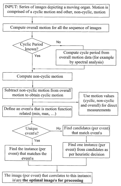
Fig. 2 is a flow chart showing the principal operations carried out in
accordance
with a general method according to the invention for identifying an optimal
image from a
series of images depicting a moving obj ect; and
Fig. 3 is a flow chart showing the principal operations carried out in
accordance
with a specific implementation of the method shown in Fig. 2.
CA 02533538 2006-O1-23
WO 2005/008583 PCT/IL2004/000632
-5-
DETAILED DESCRIPTION OF EXEMPLARY EMBODIMENTS
General principles
The invention will be described with particular reference to the determination
of
an optimal image within a series of images depicting heart motion possibly
containing
"noise" caused, for example, by shifting of the operating table on wluch a
patient is
disposed. Before doing so, some general algorithms will first be described.
Fig. 2 is a flow chart showing the principal operations carried out by a
method
according to the invention for identifying an optimal image from a series of
images that
depict a moving obj ect.
to Fig. 3 is a flow chart showing a preferred embodiment of such a method for
identifying optimal image from a series of coronary cine-angiography images;
this image
being the input for a three-dimensional reconstruction of a the coronaries.
A series of images is received as input to from any imaging source. These
images
depict a moving organ subj ected to two types of motions. The first is a
cyclic motion of
the object itself, meanng that, within a certain time frame, the object
restores its original
shape and position. The second is the motion of the object within the scene
(image),
meaning that the object changes position due to change in the imaging
position. In the
preferred embodiment of this method shown in Fig. 3, the images are a series
of coronary
angiographic images, obtained during a catheterization procedure. The cyclic
motion is the
heartbeat and the second motion could be, for example, movement of the C-ARM
table,
causing a shift of the imaged coronary vessels in the image.
First, the overall motion is computed for all sequence of images, using aaiy
method, for example optical flow or phase correlation.
If the cyclic period is unknown, then the cycle period is computed from the
overall motion data. ~ne method of doing so is by spectral analysis. The non-
cyclic
motion is computed, using overall motion and known or computed cycle period. A
preferred embodiment of a known cycle period is the period of a cardiac cycle
extracted
from analysis of the ECG signal.
The non-cyclic motion is subtracted from the overall motion to obtain the
cyclic
motion. In the preferred embodiment shown in Fig. 3, the heartbeat motion is
obtained by
subtractiizg the non-cyclic motion (mainly attributed to movement of the
patient's bed)
from the overall motion.
CA 02533538 2006-O1-23
WO 2005/008583 PCT/IL2004/000632
-6-
The motion values, especially those describing the cyclic motion, can now be
used for direct measurements, for example, for cardiac wall motion analysis.
To this end, there is defined an event related to the motion function. For
example,
in the preferred embodiment shown in Fig. 3, this event could be the miumum
instance,
identifying the image with least coronary motion, thus being the optimal image
for three-
dimensional reconstruction.
If the event is unique, then the instance (image) that matches this event is
found.
Otherwise, all matches for the event are found, and from this list of
candidates the one
instance that matches a heuristic rule is selected. In the preferred
embodiment, the event of
l0 least arteries motion could be unique if an approximation to the instance
is extracted by
analysis of the ECG signal (R peak is an approximation to the end-diastole
instance).
Otherwise, if such an approximation is not available, the event of least-
motion is not
unique, since it is matched by both end-systole and end-diastole. Thus, both
instances of
the least-motion event are found, and the end-diastole motion is identified by
the rule that
it depicts the arteries most relaxed, as opposed to least relaxed for end-
systole motion.
The image that correlates to the identified instance is the optimal image. For
example, as in the preferred embodiment, the image that correlates to the
event of least-
motion, most relaxed state of the arteries is optimal for three-dimensional
reconstruction.
Method for estimating the organ's motion
In the above embodiments, motion of the organ is computed from a series of
images (frames). Although the manner in wluch is this is done is not itself a
feature of the
invention, for the sake of completeness there vrill now be described ways in
which this can
be done.
First, we suggest an algorithm, where the number of images (frames) per cyclic
motion of the organ is lmown. This parameter is usually knov~m (for example,
the cardiac
cycle length is easily acquired via interfacing to the ECG unit in the
catheterization room).
Nevertheless, we will later obviate the need for knowing this parameter.
Let IMI, IM2... IM,t be n images that include, each, an organ that is in
cyclic
motion. Let rn be the number of images per cycle.
Any m+1 images, Il~II, IIeiIz... Il~m, IMm+i, f~rm a full cycle (for the sake
of
simplicity it will be assumed that m is an even number). Differences between
frames in
CA 02533538 2006-O1-23
WO 2005/008583 PCT/IL2004/000632
-7-
this sequence are attributed to the organ's cyclic motion, but are also
attributed to other
factors.
If ouy cyclic motion is present, the first and the last images in this
sequence must
be identical, IMl=IMm+i.
Difference between images representing composite motion can be computed, as
known in the art, by, for example, optical flow or by applying phase
correlation
computation to pairs of successive images IM; and IM;+i, iE f 1..m}. The
result of this
computation (for example the result of phase correlation) is described dX;
,dY; and p;,
where dX; and dY; are the shift between images (assuming a substantial part of
the same
to pattern is present in both images) in X and Y axes respectively and p; is
the correlation
grade. p; may be used to enhance the further described algorithms.
Let us define and compute the motion integration as:
C Yl ~ = Co~
X ~+i X t + ~t
CYf+~ ~=CY~ ~ CdY ~
meaning that in the first image, the motion integral is equal to zero. The
motion integral
for image i+1 is equal to the motion integral for image i plus sluft between
images i and
i+1 , as computed by the phase correlation.
It is mathematically understandable that the integration of a cyclic motion,
from
image IMl to IMm+i, is zero - if an object starts and ends in the same
position, then the
2o integration of the object movements (on X and Y axis) is zero (as shown in
Fig. 1). Thus,
if only cyclic motion is present, (Xm+i,Y",+i)=(Xi,Y;)=(~,~).
Let (XNC,YNC) be ~e integral of the non-cyclic motion,
~NC9YNC) _ ~m+l9Ym+1)s
This means that, given that the integral of cyclic motion is 0, (Xm+uYm+i)
represent the residual motion that is attributed to non-cyclic movement.
Assuming non-cyclic motion is consistent, or at the least that its frequency
is
lower than the cyclic motion frequency, we can subtract this motion from the
overall
motion:
(X;*,Y;*)=(X,,Y,)-(XNC,YNC)*(i-1)/m, i=1,2,. ..,m+1.
CA 02533538 2006-O1-23
WO 2005/008583 PCT/IL2004/000632
-g-
Thus, we obtain the following motion values. (XNC,YNC) are the values of the
non-cyclic motion, and (X;*,Y;*), i=1,2,...,nZ+l, are the values, per frame i,
of the cyclic
motion.
These values can now be used for direct measurements (such as cardiac wall
motion, for example), and can be used as input to further processing, as
further detailed
below.
We can obviate the requirement of knowing in advance the length of the cyclic
motion by means of direct computation. The most common method of doing so is
l0 performing spectral investigation, based on Fast Fourier Transform, applied
to a motion
graph for the entire sequence, to identify the frequency of the cyclic motion.
Method for obtaining the least motion image
In many applications, it is desirable to identify the image with least motion
(for
example, least heart motion or least coronaries motion). In cases where the
event of least
motion, within the motion cycle is unique, then the least motion image is
pointed to by the
minimum point on the motion differences graph.
Let:
D~>j (Xi 'I'j~z'+'Y ~jIZ
2o D;~ is motion differences graph. Least motion instance is the minimal
instance of
D;,~ function.
In other cases, where the least motion event is not unique within the motion
cycle
(for example, the cardiac cycle has two least motion instances - end-systole
and end-
diastole), we suggest the following method.
If an initial approximation for the least motion image is known (for example,
the
vicinity of the end-diastole image, within the cardiac cycle, is easily
identified by the R
peals, talcen from the ECG signal), then we will find the first extreme point,
which is most
distant to the approximated least motion image, meaning:
If IMF is the approximation for the least motion image, then the first extreme
point is DE>F = max {D;>F} for all i=l..na+1.
Least motion image - IMLM - is determined as most distant from image IME,
CA 02533538 2006-O1-23
WO 2005/008583 PCT/IL2004/000632
-9-
DLM,E = m~ f Da,E} for all i=l..m+1.
We can relieve the requirement for knowing in advance an approximation for the
least motion image by means of direct computation. If indeed the least motion
instance is
not unique, we can use heuristic criteria for distinction. For example, within
a sequence of
angiograms, depicting a cardiac cycle, it is easy to distinguish between the
end-systole
instance and the end-diastole instance, both representing least motion, since
the end-
diastole instance is identified by presenting the coronaries in maximal
spreading, while the
end-systole instance is identified by presenting the coronaries in minimal
spreading.
to
Preferred Embodiment.
We suggest a preferred embodiment for an application of three-dimensional
reconstruction of coronary vessels from a procedure of conventional
angiography. In order
to reconstruct a three-dimensional image of the arteries, it is necessary to
obtain at least
two two-dimensional images of the arteries in the same phase of the heartbeat,
for
example at end-diastole. Therefore, image acquisition is usually synchronized
to an ECG
signal. This procedure involves simultaneous recordings of the video signal
from the X
ray camera and the patient's ECG signal. We present here a novel method for
identifying
the end-diastole instance, equivalent to ECG-gating, without relying solely,
if at all, on the
2o ECG signal.
Let IMI, IMz... IM" be fZ images of a catheterization-acquired run.
Let na be the number of frames per cardiac cycle, either hnovyxi in advance or
computed as detailed in the above-described method for estimating the organ's
motion.
Let IM,~ be the approximate location of end-diastolic frame within the cycle,
either lcnown in advance or heuristically identified as detailed in the above-
described
method for obtaining the least motion image.
IMk_",i2, IMk_,"iz+i, IM k-m/2+1.. . IMk_",i2+m form a full cardiac cycle (for
the salve of
3o simplicity, let us presume that m is an even number). Differences between
frames in this
sequence are attributed to heart motion, but are also attributed to bed
motion, iodine
propagation and several other reasons. If only heart motion were present, the
first and the
CA 02533538 2006-O1-23
WO 2005/008583 PCT/IL2004/000632
-10-
last images in this sequence - IMk_m,2 and IMk_mi2+m - must be identical,
since the motion of
the heart is cyclic.
For the sane of simplicity, let us renumber the sequence as
IMI, IMZ... IMm, IMm+1.
As noted above, if only heart motion is considered then IMl=IMm+i. Also, the
end-diastolic renumbered frame is IM"~Z+u which is an approximation for the
least motion
frame.
to Apply Phase correlation computation to pairs of successive images IM; and
IM;+1,
iE ~ 1..m}. The result of the Phase correlation is described dX; ,dY; and p;,
where dX; and
dY; are the shift between images (assuming most of the same pattern - coronary
tree or
part of the coronary tree - is present in both images) in X and Y axis
respectively and p; is
the correlation grade. p; may be used to enhance the further described
algorithms.
Now, of all the reasons that attribute to the difFerences between successive
images, the most significant contributory factor to such differences -
sometimes more than
heart motion itself- is bed motion.
W tegration of cardiac motion, from image IMl to IMm+i, is zero -
(~m+~,Ym+~)=(X1,Y1)=(0,0).
Let (XB,YB) be the integral of the bed movement,
~B~YB) _ (~m+l~Ym+1),
meaning that, given that the integral of cardiac motion is zero, (gym+l,~m+i)
represents the
residual motion that is attributed to bed movement.
Assuming the bed movement is consistent (meaning the physician is moving the
bed in a general constant direction), or that, as a weaker constraint, the bed
movement is
slower than heartbeat, we can subtract this movement from the overall
movement:
(xt*,Y;*)=(XaYy-(XB,YB)*(i-1)~m, i=1,2,...,m+l.
3o The frame with minimum arterial motion is pointed by the extreme point on
, Curdle.
CA 02533538 2006-O1-23
WO 2005/008583 PCT/IL2004/000632
-11-
Let:
Dl>i - lx 1 'YJ ~Z + y
We can determine the end-systole point S, which is the one most distant from
the
approximated end-diastole point, meaiung:
Ds>"~2+i=max {D;>I,,i2+i}
Minimum motion point - end-diastole, ED - is determined as most distant from
systole point:
Ds>ED = max {Ds>J~.
Selecting the IMEO image per sequence of cine-angio images for the process of
three-dimensional reconstruction will provide the optimal result, in terms of
accuracy and
precision, for the reconstruction and for vessel analysis.
In the method claims that follow, alphabetic characters and Roman numerals
used to designate claim steps axe provided for convenience ouy and do not
imply any
particular order of performing the steps.
It will also be understood that the system according to the invention may be a
suitably programmed computer. Likewise, the invention contemplates a computer
program
being readable by a computer for executing the method of the invention. The
invention
further contemplates a machine-readable memory tangibly embodying a program of
instructions executable by the machine for executing the method of the
invention.