Note: Descriptions are shown in the official language in which they were submitted.
CA 02533670 2006-O1-25
WO 2004/010861 PCT/DK2003/000512
METHOD FOR CONVERTING VENOUS BLOOD VALUES TO ARTERIAL BLOOD VALUES,
SYSTEM FOR UTILISING SAID METHOD AND DEVICES FOR SUCH SYSTEM
The present invention relates to methods for converting venous blood values to
arterial
blood values. The invention also relates to an apparatus for performing the
method and
relates to uses of the apparatus, when performing the method.
BACKGROUND OF THE INVENTION
The assessment of acutely ill patients is a complex process involving
evaluation of the
patients numerous physiological systems, e.g. the pulmonary, metabolic, renal
and
circulatory systems. Much of the information necessary for this evaluation
comes from
analysis of the patients' blood. Blood samples can be taken from both arteries
and veins.
Arterial blood can be sampled either by placing an arterial catheter or
cannula in the
patient, or by performing an arterial puncture with a needle. Venous blood can
be sampled
from a cannula or a venous puncture at the periphery (peripheral venous
blood); from a
catheter placed in superior venal cava (central venous blood), or from a
pulmonary arterial
catheter placed in the pulmonary artery (mixed venous blood).
Placements of venous and arterial catheters are invasive procedures and
generally
restricted to high dependency departments. In addition catheterisation,
cannulation or
puncture of the arteries instead of the veins increases the risk of
complications such as
infection, hemorrhage, bleeding, thrombosis, emboli, neurological damage or
pseudo-
aneurysm formation. Sampling of arterial blood by arterial puncture is
generally considered
a more difficult procedure than sampling of venous blood through a venous
puncture.
Consequently, the routine sampling of arterial blood is generally restricted
to high
dependency environments. In other wards where patients are acutely ommited
e.g.
cardiology, abdominal surgery, thoracic surgery and medicine, routine sampling
of
peripheral venous blood is most common.
Many of the measurements taken from the blood, and used to assess the patient
state, are
similar in the venous and arterial blood samples. These included the
electrolytes and such
as sodium (Na), potassium (K), the haemoglobin concentration (Hb) and the
concentration
of abnormal forms of haemoglobin (e.g. carboxyhaemoglobin (COHb),
methylhaemoglobin
(MetHb)). However, the acid-base status of arterial and venous blood is not
the same,
regardless of the site of sampling. The acid-base status refers, in general,
to the following
measurements in blood; the pH, the pressure of oxygen (pOZ), the pressure of
carbon
dioxide (pCOZ), the bicarbonate concentration (HC03), the concentration of.
base higher
than a reference condition (base excess (BE)), the concentration of
bicarbonate at a
CA 02533670 2006-O1-25
WO 2004/010861 PCT/DK2003/000512
2
reference pCOz (standard bicarbonate SBC), the oxygen pressure (pOZ) and the
saturation
of haemoglobin with oxygen (S02) with p02 and SOZ often being referred to as
the
oxygenation status of blood . The variation in acid-base status between
arterial and venous
blood is due to oxygen removal from the blood and carbon dioxide addition due
to
metabolism at the tissues. In addition in patients with circulatory or
metabolic
abnormalities, the production of strong acid at the tissues due to anaerobic
metabolism
may also modify the acid-base status.
The acid-base status of arterial blood is used to assess the patients'
respiratory and
metabolic state. It has been argued (Adrogue et al., 1989a, 1989b; Brandi et
al., 1995;
Radiometer 1997) and to a large extent clinically accepted that venous blood
samples are
not adequate for assessing the acid/base and respiratory state of patients.
This is thought
to be particularly true for peripheral venous samples which "are not
recommended for
blood gas analysis as they provide little or no information on the general
status of the
patient" (Radiometer 1997).
In the intensive care unit placement of arterial catheters is routine practice
and an
assessment of the acid-base status can be obtained from the arterial blood. In
some other
hospital departments e.g. pulmonary medicine, or nephrology, arterial blood
gases are also
measured. However in other wards admitting acutely ill patients, e.g.
cardiology,
abdominal surgery, thoracic surgery and medicine, arterial samples are not
usually taken.
Usually a peripheral venous sample is taken and analysed in a central
laboratory. The
sample is usually taken aerobically, i.e. no attempt is made to ensure that
p02 and pCO~
remain constant during the sample procedure. Only a small amount of
information
concerning the acid-base status of the patient is measured in this sample i.e.
the standard
bicarbonate, SBC", and haemoglobin Hb". Other acid base parameters pH", carbon
dioxide
pressure (pC02"), base excess (BE~), oxygen saturation (SOZ") and oxygen
pressure (PO~~)
are not measured, and if measured would probably not reflect the true values
of venous
blood at this sample site given the aerobic nature of the sample.
US 6,334,065 describes a pulse oximeter providing simultaneous and non-
invasive oxygen
status at multiple sites of a patient. The pulse oximeter described measures
both arterial
and venous oxygen saturation at any specific tissue site of the patient. It is
mentioned that
a corresponding computation of arterial minus venous oxygen saturation is
advantageous
for oxygen therapy patients. However, also as mentioned, the pulse oximeter is
purely
noninvasive in its way of functioning limiting the values capable of being
derived.
US 3,874,850 describes an apparatus being an automatic blood sample analyzer
for
automatically measuring one or more unknown data or parameters of the blood
samples.
CA 02533670 2006-O1-25
WO 2004/010861 PCT/DK2003/000512
3
Based on the values measured, the apparatus comprises means for calculating a
number
of other parameters including acid-base status of the blood sample. The
analyzer may also
comprise means for photo-metrically measuring the hemoglobin contents of
samples of
blood. There is no computation of arterial blood values based on venous blood
samples.
SUMMARY OF THE INVENTION
The object of the invention is to provide a method for performing the
conversion of venous
blood values to arterial values, including the design of a sampling tube for
sampling
anaerobic venous blood and including a system for applying the method.
This object may be obtained by a method comprising the steps of:
- a) measuring arterial oxygenation,
- b) measuring and estimating values of venous blood acid/base status and
oxygenation
status of a venous blood sample taken anaerobically,
- c) converting the venous blood values by applying a mathematical model for
deriving
blood acid/base status and oxygenation status into estimated arterial blood
values.
The object may also be obtained by a method comprising the steps of:
- a) estimating arterial oxygenation,
- b) measuring and estimating values of venous blood acid/base status and
oxygenation
status of a venous blood sample taken anaerobically,
- c) converting the venous blood values by applying a mathematical model for
deriving
blood acid/base status and oxygenation status into estimated arterial blood
values.
The object may also be obtained by a method comprising the steps of:
- b) measuring and estimating values of venous blood acid/base status and
oxygenation
status of a venous blood sample taken anaerobically,
- a) measuring arterial oxygenation,
- c) converting the venous blood values by applying a mathematical model for
deriving
blood acid/base status and oxygenation status into estimated arterial blood
values.
The object may also be obtained by a method comprising the steps of:
- b) measuring and estimating values of venous blood acid/base status and
oxygenation
status of a venous blood sample taken anaerobically,
- a) estimating arterial oxygenation,
- c) converting the venous blood values by applying a mathematical model for
deriving
blood acid/base status and oxygenation status into estimated arterial blood
values.
CA 02533670 2006-O1-25
WO 2004/010861 PCT/DK2003/000512
4
The only differences between the above four methods are the order, in which
the different
steps are carried out and that the arterial oxygenation may either be measured
or
estimated. Additionally, both a measurement and an estimation may be
pertormed.
By means of mathematical models for the acid-base status of the body, venous
blood
sample values of acid-base status and of oxygen status together with pulse
oximetry may
be used to convert the venous blood values to corresponding arterial values.
Deriving of
blood acid/base status and oxygenation status into estimated arterial blood
values may be
performed either by estimation, or by calculation or by a combination of
estimation and
calculation.
We argue that parameters describing the venous acid-base chemistry should be
measured,
and describe a method whereby venous values can be combined with a
determination of
arterial oxygen saturation with a pulse oximeter to calculate predictions
(SBCaP, pHaP,
pCO2aP, BEap, pOZap, and SOzaP) of the corresponding arterial values, (SBCa,
pHa, pCO~a, BEa,
pO~a, and SO~a). This implies that the acid/base and respiratory status can be
assessed
without taking an arterial blood sample. To do so requires anaerobic sampling
of the
venous blood and this patent also describes the design of a sampling bottle
for this
purpose. This method will make acid/base and respiratory status available in a
large
number of patients without the cost, risk and inconvenience of taking an
arterial sample, in
particular in departments where arterial samples traditionally are only taken
rarely. Having
the acid/base and respiratory status available will make it easier to diagnose
different
types of respiratory and metabolic acidosis or alkalosis.
The assumptions of the models comprise that no acid is added between the
arterial blood
and the venous blood drawn, i.e. no anaerobic metabolism is taking place in
the
intermediate organ or tissue. It is known that this is not the case for haemo-
dynamically
unstable patients and for patients with severe chronic suffering.
In a first possible improved method, said measuring and analyzing comprises
the further
steps of:
- d) drawing an anaerobic venous blood sample,
- e) analysing said anaerobic venous blood sample for evaluating the acid/base
status of
the venous blood sample, and
- f) analysing said anaerobic venous blood sample for evaluating the
oxygenation status of
the venous blood sample.
CA 02533670 2006-O1-25
WO 2004/010861 PCT/DK2003/000512
In another possible improved method, said measuring and analyzing comprises
the further
steps of:
- d) drawing an anaerobic venous blood sample,
- f) analysing said anaerobic venous blood sample for evaluating the
oxygenation status of
5 the venous blood sample, and
- e) analysing said anaerobic venous blood sample for evaluating the acid/base
status of
the venous blood sample.
The only difference between the above two improved methods is the order in
comparison
with step d), in which the two other steps, i.e. step e) and step f), are
carried out.
In a possible further improved method, said method comprises the further step
of:
- g1) measuring the arterial oxygenation such as oxygen saturation, pressure
or
concentration by applying any suitable means for such measuring or estimation,
said
further step being performed at any time in relation to any of the steps of
claims 1-3.
In another further improved method, said method comprises the further step of:
- g2) estimating the arterial oxygenation such as oxygen saturation, pressure
or
concentration by applying any suitable means for such measuring or estimation,
said
further step being performed at any time in relation to any of the steps of
claims 1-3.
The only difference between the above two further improved methods is the
arterial
oxygenation either being measured or estimated. Additionally, both a
measurement and an
estimation may be performed, i.e. step g2) being performed additional to step
g1).
In an even further improved method, said method comprises the even further
step of
- h) simulating the blood acid/base status and oxygenation status of an
arterial blood
sample by use of mathematical modelling. Additional hereto, the method may be
still even
further improved by said method comprising the still even further steps of:
- i) mathematical modelling comprising simulated addition of oxygen, O2, to
and removal
of carbon dioxide, COZ, from the venous blood sample values in a ratio
determined by the
respiratory quotient,
j) said mathematical modelling being performed until the simulated oxygen
level is equal
to the arterial oxygenation level measured or estimated, and
- k1) calculating the acid/base status and the oxygenation of the arterial
blood by applying
the result of said modelling,
CA 02533670 2006-O1-25
WO 2004/010861 PCT/DK2003/000512
6
and alternative or additional hereto, said method comprising the still even
further steps of
- i) mathematical modelling comprising simulated addition of oxygen, Oz, to
and removal
of carbon dioxide, CO2, from the venous blood sample values in a ratio
determined by the
respiratory quotient,
j) said mathematical modelling being performed until the simulated oxygen
level is equal
to the arterial oxygenation level measured or estimated, and
- k2) estimating the acid/base status and the oxygenation of the arterial
blood by applying
the result of said modelling.
The only difference between the above two further improved methods is the
acid/base
status and the arterial oxygenation either being measured or estimated.
Additionally, both
a measurement and an estimation may be performed, i.e. step k2) being
performed
additional to step k1).
In an additional possible improved method, said method comprises a further
step of
- 11) measuring the arterial carbon dioxide level such as carbon dioxide
pressure, total
concentration or bicarbonate concentration) by applying any suitable means for
such
measuring or estimation, said further step being performed at any time in
relation to any
of the steps of claims 1-6.
In another additional improved method, said method comprises a further step of
- 12) estimating the arterial carbon dioxide level such as carbon dioxide
pressure, total
concentration or bicarbonate concentration) by applying any suitable means for
such
measuring or estimation, said further step being performed at any time in
relation to any
of the steps of claims 1-6.
The only difference between the above two further improved methods is that
arterial
carbon dioxide level may either be measured or estimated. Additionally, both a
measurement and an estimation may be performed, i.e. step 12) being performed
additional to step 11).
In a possible further improved method, said method comprises an even further
step of
- m) simulating the blood acid/base status and oxygenation status of arterial
blood sample
by use of modelling. Additional hereto, the method may be still event further
improved by
said method comprising the still even further steps of:
- n) mathematical modelling comprising simulated addition of Oz to and
removing CO~
from the venous blood sample values in a ratio determined by the respiratory
quotient,
CA 02533670 2006-O1-25
WO 2004/010861 PCT/DK2003/000512
7
- o) said modelling being performed until the simulated carbon dioxide level
is equal to the
arterial carbon dioxide level measured or estimated, and
- p1) calculating the acid/base status and the oxygenation of the arterial
blood by applying
the result of said modelling.
And alternative or additional hereto, said method comprising the still even
further steps of
- n) mathematical modelling comprising simulated addition of OZ to and
removing COZ
from the venous blood sample values in a ratio determined by the respiratory
quotient,
- o) said modelling being performed until the simulated carbon dioxide level
is equal to the
arterial carbon dioxide level measured or estimated, and
- p2) estimating the acid/base status and the oxygenation of the arterial
blood by applying
the result of said modelling.
The only difference between the above two further improved methods is the
acid/base
status and the arterial oxygenation either being measured or estimated.
Additionally, both
a measurement and an estimation may be performed, i.e. step p2) being
performed
additional to step pi).
The potential for use of venous blood samples to assess the status of acutely
ill patients in
various hospital departments is illustrated in figure 1 which graphically
shows the acute
blood samples in different patient groups at Aalborg Hospital in Denmark in
1999. Light
bars indicate arterial blood samples, dark bars indicate venous blood samples.
Three
different types of department can be identified within those treating acutely
ill patients. In
the first group arterial blood samples are taken frequently (70,000 per year
at Aalborg
Hospital, Denmark) (the population of Aalborg is approximately 160,000) and
often
analysed at the point of care. This group includes intensive care units,
departments of
anaesthesia and trauma units. In the second group arterial blood samples are
taken
regularly (2,000 arterial blood samples per year at Aalborg Hospital,
Denmark). This group
includes the departments of pulmonary medicine and nephrology. In the third
group
arterial blood samples are taken occasionally. This group includes for example
departments of cardiology, abdominal surgery, thoracic surgery and medicine.
In the departments of groups 2 and 3 venous blood samples are taken much more
frequently than arterial samples. Indeed, when taken in total, the number of
venous blood
samples taken in acutely ill patients actually exceeds the number of arterial
blood samples
(figure 1). It is in these departments that venous samples are usually
analysed in the
central laboratory where measurements of standard bicarbonate (SBC), total
haemoglobin
(Hb), and other blood values are taken, without a full blood gas analysis.
CA 02533670 2006-O1-25
WO 2004/010861 PCT/DK2003/000512
8
In order to test the strength and validity of the models it is therefore
necessary to test the
models for different groups of patients with varying haemo-dynamic conditions
and
accordingly different OZ and COZ conversion in the tissues.
Input for the mathematical models are venous values together with information
of the
arterial oxygenation as measured by means of as example a pulse oximeter.
In order to verify the validity of the models for converting venous blood
sample values to
arterial values the corresponding arterial values derived may be compared to
an arterial
blood sample drawn simultaneously with drawing of the venous blood sample.
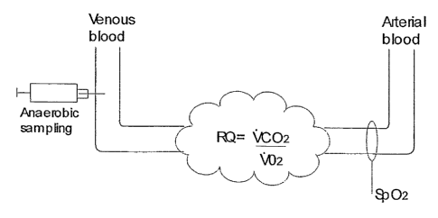
DESCRIPTION OF THE INVENTION
This section will be described in four parts. In part 1 the invention will be
described with
reference to the accompanying fig. 2 schematically showing a method for
performing the
prediction of arterial values from a venous blood sample.
In part 2 a design for a sampling bottle, capable of being used for anaerobic
sampling of
venous blood, is described. Anaerobic venous samples being required for the
method
described in step 1 (see part 1).
In part 3 two patient cases are described, both illustrating the potential use
of the method.
The first patient had a metabolic alkalosis due to potassium deficiency. In
that patient a
venous blood sample converted to arterial values would have revealed this
problem before
it developed into a crisis. The second example is a postoperative patient,
where an arterial
sample was actually available. This case is included to show that the
information that can
be derived from a venous sample converted into arterial values is equivalent
to the
information derived from the arterial sample. The case also shows that
conversion of
venous blood to arterial values is necessary: the calculated arterial values
showed that
arterial pCO2 was normal, despite the high venous value.
In part 4 it is shown that arterial values, calculated from the method of
converting venous
to arterial values, compare well with measured arterial values in 69 patient
cases,
including some categories of very ill patients. The accuracy of the converted
venous values
does not match what is obtained from an arterial sample, but is clearly
sufficient for a
clinical judgement to be made. As a minimum the arterialization method can be
seen as a
quite accurate screening method, that indicates when an arterial sample should
be taken.
CA 02533670 2006-O1-25
WO 2004/010861 PCT/DK2003/000512
9
Part 1. Conversion of venous blood values to arterial blood
The invention will be described with reference to the accompanying fig. 2
schematically
showing a method for performing the prediction of arterial blood acid-base
status values
from an anaerobically sampled venous blood sample.
Arterial blood gasses are, as an example, estimated as given in the 4 steps
below.
Step 1: An anaerobic venous blood sample is drawn and analysed using standard
blood
gas analysis technology (e.g. Radiometer, 1994) to provide a picture of the
acid/base
status of the venous blood (SBC", pH", pCOZ", BED, pOz" and SOZ").
Step 2: The arterial oxygen saturation is estimated or measured non-
invasively, possibly
by pulse oximetry.
Step 3: For a blood sample passing through the tissues from the arteries into
the veins,
the ratio of the amount of COz added (i.e. the rate of COa production (VCOz))
and OZ
removed (i.e. the rate of OZ utilisation (V02)), due to aerobic metabolism is
defined as the
respiratory quotient (RQ = VCOz/VOZ). RQ is often approximated by measurement
of
inspiratory and expiratory gases taken at the mouth, through the measurement
of
inspiratory oxygen (FiOz) and carbon dioxide (FiCO~) fraction and either end
tidal fractions
of oxygen (Fe'Oz) and carbon dioxide (Fe'COZ) or mixed expired fractions of
oxygen (FeOZ)
and carbon dioxide (FeC02) using the equations:
RQ = Fe'COz - FiCOZ or RQ = FeCO, - FiCOz
FiOZ - Fe'Oa FiOZ - FeOZ
Approximation of RQ by this method often gives values which can vary
substantially.
However, the true value of RQ at the tissues can only vary between 0.7-1.0,
being 0.7 in
aerobic metaboism of fat and 1.0 in aerobic metabolism of carbohydrate. In
this step a
mathematical model of blood acid/base and oxygenation status (e.g. Rees et al,
1996,
1997) is used to perform a simulation, where OZ is added and COZ removed from
the
venous blood in a ratio determined by a constant respiratory quotient, set to
be within the
physiologically possible range 0.7-1Ø This simulation is performed until the
simulated
oxygen saturation is equal to that estimated or measured in step 2, i.e. that
in arterial
blood.
Step 4: The model of blood acid/base and oxygenation status is then used to
calculate a
picture of the acid/base status and the oxygenation of the arterial blood
(SBCap, pHaP,
CA 02533670 2006-O1-25
WO 2004/010861 PCT/DK2003/000512
pCO2ap, BEap, pOzap and SOzap). This is possible as the simulated removal of
COZ and OZ from
venous blood at a fixed RQ ensures that when the simulated arterial
oxygenation matches
that measured, then the simulated values of other arterial acid-base variables
should also
match those measured.
5
For the purpose of testing the venous to arterial conversion method the
predictions of
arterial acid base status (SBCap, pHap, pCO~aP, BEap, pO2aP and SOzaP)
obtained from the
method can be compared against those measured (SBCa, pHa, pCO2a, BEa, pOza and
SOZa).
examples of which are given in sections 3 and 4.
The fundamental assumption contained in this method is that little or no
anaerobic
metabolism occurs across the tissue where the venous blood sample is taken. If
anaerobic
metabolism were present then this would result in two effects, the base excess
in the
arterial and venous blood would be different, and the strong acid produced by
this process
(H+) would bind with bicarbonate (HC03-) in the blood to form COz in the
following
reversible reaction
H~' + HC03- ~ COz + H20
The increase in COZ production by this reaction would mean that the apparent
VCOZ would
be increased without an increase in VOZ, meaning that conversion of venous
values to
arterial values using a constant RQ would not be correct . The degree of
anaerobic
metabolism depends upon the circulatory and metabolic state of the patient. In
a normal
well perfused peripheral limb it is unlikely that anaerobic metabolism occurs.
The quality of
perfusion of a limb can be assessed clinically by the presence of a clearly
recognizable
arterial pulse determined by palpation, a normal capillary response, and a
normal color
and temperature of the limb. Central or mixed venous blood is a mixture of
blood from
several sites and may therefore contain blood from an area of the body with
anaerobic
metabolism. The selection of the sample site is therefore important. In
section 3 the
validity of the method is tested for peripheral venous blood sampled from a
clinically
considered well perfused arm by comparing arterial values derived using the
method with
those obtained from an arterial blood sample drawn simultaneously with the
drawing of the
venous sample.
Part 2. Design of a sampling bottle capable of being used For anaerobic
samDlina of
venous blood.
The method of converting venous values describing the acid-base status of the
blood to
arterial values only applies if the venous blood samples are taken
anerobically, i.e. it is
CA 02533670 2006-O1-25
WO 2004/010861 PCT/DK2003/000512
11
ensured that the OZ and COZ pressure in the sample remains constant during and
after the
sampling procedure.
Currently, it is normal practice that only arterial samples are taken
anaerobically. These
are usually taken via a sampling syringe from a sampling connector (A) at the
sampling
site of an arterial catheter, cannula or needle, as illustrated in figure 3.
Arterial sampling
syringes are heparinized to prevent coagulation of the sample. After sampling
of the blood
the syringe is usually placed in a verticle position with the open end (B)
(figure 3)
uppermost, agitated and trapped air expelled using the plunger (C). This is
only possible
because the syringe is open to the environment, a lid being placed on the
syringe only
after expulsion of trapped air.
In principle, venous blood sampled using arterial sampling syringes could be
used in the
method of converting venous to arterial values described here. However, the
use of open
syringes increases the risk of infection of the person handling the blood. In
departments
routinely taking venous blood to assess the status of acutely admitted
patients, venous
blood samples are not usually taken using open syringes. Instead venous blood
samples
are taken using the sampling method illustrated in figure 4. A venous sampling
connector
(A) is attached to the venous sampling site. The connector includes a needle
(D), covered
with rubber so as to prevent leakage of blood except when pressure is applied
to the
rubber to expose the needle. The venous sampling bottle is sealed with a
sealing
membrane (E). Blood cannot enter or leave the bottle until the bottle is
pressed onto the
sampling connector. At this point the needle is exposed, pierces the sealing
membrane,
and a blood sample may be taken. Different sampling bottles often contain
chemicals for
specific conservation or analysis of the blood depending upon the parameters
to be
measured e.g. electrolytes, coagulation etc. However these sampling bottles
also contain
oxygen and/or carbon dioxide (typically air), which may diffuse into the blood
sample
altering its acid base status. In addition, since the sample bottle is closed
there is no
means to expel air which may enter the bottle during the sampling procedure.
Figure 5 illustrates an example of the invention according to claims 17-20
i.e. the design of
a sample bottle suitable for anaerobic sampling of venous blood.
The example design illustrates a sample bottle (B) with two heparinized
chambers B1 and
B2. Initially the two chambers are joined, as illustrated in figure 5(i). The
complete bottle
is then pressed on the sampling connector (A) and the plunger used to draw
blood, and
possibly air into both compartments. The sample bottle is then detached from
the sampling
connector as illustrated. in figure 5(ii) b and placed vertically with the
plunger facing
uppermost. By agitating the bottle and withdrawing the plunger further, any
air in
CA 02533670 2006-O1-25
WO 2004/010861 PCT/DK2003/000512
12
chamber B1 is drawn into chamber B2. The two chambers B1 and B2 are then
separated.
The rubber seals on the sampling needle (C) and the sealing membrane (D)
ensure no
leakage of blood. Chamber B1 contains only anaerobic venous blood, analysis of
which
may then be used in the arterial conversion algorithm. Chamber B2 contains air
and blood
and may be discarded.
The amount of air in the chambers can be further reduced by applying a partial
or
complete vacuum within the sample bottle prior to sampling. In addition if the
initial gas in
the sampling bottle contains inert gasses, and or OZ and COZ with pressures
adjusted to
typical venous values, then the effects of any residual gasses in the sampling
bottle will be
minimised.
Part 3 Clinical cases illustrating the potential use of the venous to arterial
conversion
method
This section describes two patient examples, the first with a metabolic
alkalosis due to
potassium deficiency. In this patient a venous blood sample converted to an
arterial value
would have revealed the problem before it developed into a crisis. The second
example is a
postoperative patient, where an arterial sample was actually available. This
case is
included to show that the information that can be derived from an venous
sample
converted to arterial values is equivalent to the information derived from the
arterial
sample. The case also shows that conversion of venous values to arterial
values is
necessary: the converted venous values show that arterial pCO~ is normal,
despite the
high venous value.
Case 1- Metabolic alkalosis due to potassium deficiency
A patient, age 60, male, was acutely admitted to the surgical department
complaining of
abdominal pain, and having vomited repeatedly over the past week. A peripheral
venous
sample was taken and analysed routinely, without a blood gas analysis, giving
a high
standard bicarbonate SBC" = 38 mmol/I, a slightly low haemoglobin Hb"= 7.0
mmol/I, and
a potassium value at the low end of the normal range K"= 3.6 mmol/I. The high
SBC
caused by loss of acid and potassium due to vomiting remained unnoticed for 3
days, at
which point the patients respiratory drive and cardiac function had
deteriorated to the
point of pulmonary odema, and an arterial blood gas was taken. Arterial blood
gas values
(pHQ=7.60, BEa= 18 mmol/I, pCOZ,a =6.0 kPa, SOZ,a =0.92) showed very severe
metabolic
alkalosis. The patient was then transferred to the intensive care unit, where
treatment for
this metabolic alkalosis proceeded for approximately two weeks.
CA 02533670 2006-O1-25
WO 2004/010861 PCT/DK2003/000512
13
For this patient, analysis of the peripheral venous blood gases on admission
might have
highlighted the severe alkalosis. In current clinical practice analysis of the
peripheral
venous blood gases are not generally accepted (Radiometer 1997). Conversion of
the
venous blood gas values to arterial values using the method included here
might then both
have highlighted the severe alkalosis before the patient reached a critical
state, and given
a clinically acceptable picture of the patient.
Case 2 - Post-operative coronary artery bypass patient
A patient, age 64, male, presented in the post operative intensive care unit
following
coronary artery bypass surgery. During the post operative period the patient
was
heamodynamically stable. An arterial catheter was present in this patient and
simultaneous
samples of arterial and peripheral venous blood were taken and analysed for
blood gases.
Venous blood values were SBC",= 23.7 mmol/I, pH",=7.29, pC02," =7.2 kPa, BED _
-0.3
mmol/I and SOa,". = 0.36. If interpreted directly these values would suggest
that the
patient had a respiratory abnormality causing a high pCOZ". However, when the
venous to
arterial conversion method was used to calculate arterial blood gas values a
relatively
normal pattern presented SBCap= 22.9 mmol/I, pHaP,= 7.35 pCOz,aP, = 5.8 kPa,
BEaP = -1.8
mmol/I and SO~,aP,=0.98 suggesting that the patient did not have a respiratory
abnormality. These converted venous values gave the same clinical picture as
arterial
values measured for comparison (SBCa,= 23.6 mmol/I, pHa= 7.37, pCOZ,a, = 5.5
kPa, BEa
- -1.1 mmol/I, and SOZ,a = 0.98), which were also within the normal range. The
information derived from the converted venous sample was therefore clinically
equivalent
to the information derived from the arterial sample. In this case an
interpretation of the
patient state could not be made from the venous blood without a conversion to
arterial
values since the conveted values showed that arterial pC02 was normal, despite
the high
venous value. If this patient had presented at the ward, without an arterial
catheter
conversion of venous blood to arterial values would have been necessary to
obtain the
correct clinical interpretation.
Part 4. Conversion of venous blood values to arterial values in 69 clinical
cases
This section describes the results of using the method for conversion of
venous to arterial
values. Peripheral venous blood samples were taken in 69 cases, and used to
measure
SBC", pH", pCO~,", BE", p02,~ and SOZ,". The method was then used to predict
arterial blood
values SBCaP, pHap, pCOz,aP, BEaP, pO2,vap and SOZ,aP. These arterial
predictions were then
compared with measurements of arterial blood SBCa, pHa, pCOZ,a, BEa and SOZ,a
taken
simultaneoulsy with the venous samples. Section 4.1 describes the patient
groups included
in this study including their severity of metabolic and respiratory disorders.
Section 4.2
describes the results of the venous to arterial conversion method. In this
section predicted
CA 02533670 2006-O1-25
WO 2004/010861 PCT/DK2003/000512
14
variables (SBCap, pHap, pCOZ,ap, BEap and SOZ,aP) are compared in turn to
measured arterial
values, and the accuracy and precision of the prediction quantified. Figures 6
through 9
illustrate Bland-Altman plots illustrating the mean of the measured and
predicted arterial
value plotted against the difference between the measured and predicted
arterial values.
Values of the mean difference between measured and predicted arterial values
and the
standard deviation, are also given in graphs 6-9 and in the following text.
4.1 Study population
Patients were studied from the following groups a) post operative coronary
artery bypass
patients, both haemodynamically stable and unstable; b) patients with sepsis,
both
haemodynamically stable and unstable; and d) Patients with chronic obstructive
lung
disease, both mechanically ventilated and spontaneously breathing. These
groups were
selected to represent a range of acid base status including metabolic and
respiratory
abnormalities, and presented with the values (median, range) pHa = 7.40, 7.24
to 7.54;
BEa =0.6 mmol/I, -6.9 to 19.7 mmol/I; SBCa = 25.0 mmol/I, 18.8 to 44.3 mmol/I;
pCO2,a=
5.68 kPa, 4.0 to 10.8 kPa. Patients also presented with a broad range of
arterio-venous
oxygen saturation difference (median, range) 0.15, 0.00 to 0.74. Arterial and
peripheral
venous blood samples were taken simultaneously with peripheral samples being
taken
from what were clinically considered well perfused arms. Results of these
groups are
presented here pooled.
4.2 Results
In this section we present a comparison of arterial values predicted using the
venous to
arterial conversion method (SBCap, pHaP, pCOZ,aP, BEaP and SOZ,aP) with
measured arterial
values (SBCa, pHa, pCOZ,a, BEa and S02,a).
pCOz,a versus pCO2,aP
Figure 6 illustrates a Bland-Altman plot of measured arterial carbon dioxide
pressure pC02
(pCOZ,a) versus that predicted using the venous to arterial conversion method
(pCOZ,aP).
The prediction of pCOZap can be seen as both accurate and precise (pC02,a -
pCOz,aP = -
0.10d0.32 kPa). In addition, errors in the prediction of pCOZ,aP are
clinically unimportant
when compared to the size of the arterial - venous pC02 difference pCO2,a -
pC02,~ _
0.6440.63 kPa.
SBCa VerUS SBCaP
CA 02533670 2006-O1-25
WO 2004/010861 PCT/DK2003/000512
Figure 7 illustrates a Bland-Altman plot of measured arterial Standard
Bicarbonate SBC
(SBCa) versus that predicted using the venous to arterial conversion method
(SBCaP). The
prediction of SBCap can be seen as both accurate and precise (SBCa - SBCAP =
0.17b'0.5
mmol/I). Since SBC changes with the addition of acid, the small bias of 0.17
mmol/I is
5 equivalent to the finding that the base excess changes by about 0.2 mmol/I
as the blood
flows through the tissues.
ABEa verSUS ABEaP
10 The major assumption in the venous to arterial conversion method is that no
significant
amount of strong acid is added to the blood as it passes through the tissues
across which
the arterial and venous blood samples are taken. To verify this, figure 8
illustrates a Bland-
Altman plot of measured arterial Base Excess BE (BEa) against that predicted
from the
arterial to venous conversion method (BEaP). BEa-BEap = 0.2 b' 0.5 mmol/I.
This implies
15 that 0.2 d 0.5 mmol/I acid is added when the blood is passing through the
tissues i.e an
insignificant amount.
pHa versus pHaP
Figure 9 illustrates a Bland-Altman plot of measured arterial pH (pHa) versus
that predicted
using the venous to arterial conversion method (pHaP). The prediction of pHaP
can be seen
as both accurate and precise (pHa - pHaP = 0.00840.013 ).
Possible 4roups of patients suitable for the invention.
The patient groups presented in section 4 reflect the testing of the method
where
simultaneous sampling of arterial blood is necessary for comparison with the
those
calculated by the method. When applying the method arterial samples would not
be taken.
The method may therefore be applied in all: normal subjects, patients, or
animals in which
a venous sample can be taken in combination with a measurement of arterial
oxygenation,
usually performed using a pulse oximeter. Whilst the method is tested here for
the
sampling of peripheral venous blood the method may also be applied to the
sampling of
central or mixed venous blood.
CA 02533670 2006-O1-25
WO 2004/010861 PCT/DK2003/000512
16
Reference List:
1. Rees SE, Andreassen S., Hovorka R, Summers R, Carson ER: Acid-base
chemistry of the
blood--a general model. Comput.Methods Programs Biomed. 1996; 51: 107-19
2. Rees S.E. , S. Andreassen, R Hovorka and E.R. Carson: A dynamic model of
carbon
dioxide transport in the blood. In: D.Linkens and E.R.Carson (Eds).
Proceedings of the
3rd International Federation of Automatic Control (IFAC) symposium onModelling
and
Control in Biomedical Systems, Elsevier, December 1997, pp 63-68.
3. Adrogue HJ, Rashad MN, Gorin AB, Yacoub J, Madias NE: Assessing acid-base
status in
circulatory failure. Differences between arterial and central venous blood.
N.EngI.J.Med.
1989; 320: 1312-6
4. Brandi LS, Giunta F, Pieri M, Sironi AM, Mazzanti T: Venous-arterial PC02
and pH
gradients in acutely ill postsurgical patients. Minerva Anestesiol. 1995; 61:
345-50
5. Radiometer Medical A/S: The Blood gas Handbook, 1997, pp 14-15
6. Radiometer Medical A/S: Blood Gas, Oximetry and Electrolyte Systems.
Reference
Manuel, 1994