Note: Descriptions are shown in the official language in which they were submitted.
CA 02533798 2006-O1-26
WO 2005/009257 PCT/IE2004/000103
1
"A Device"
Introduction
Accessing the abdominal cavity while preserving the abdominal wall as much as
possible is the aim of any surgical or exploratory procedure. Retraction
devices have
been used to this end. A retractor can help to expose an operative site and
minimise
the incision required to carry out the operation.
IO Minimally invasive surgery is an evolving surgical method that similarly,
attempts to
reduce the size of incisions required, in many cases dramatically. By using a
so-
called "keyhole" or cannula, the surgeon can gain access with instruments into
the
abdominal cavity to carry out an operation through a very small series of
holes in the
abdominal wall. Unlike in the case of "open surgery", primary retraction then
must
be accomplished by lifting the abdominal wall away from the abdominal viscera.
This is most often accomplished with the use of gas in a technique known, as
insufflation.
The use of a cannula to gain access as a means to see inside the abdomen or
introduce surgical instruments has existed since the Iate 19th century. A
cannula
comprises a rigid tube, which is inserted through the abdominal wall and is
held in
place by the tension of the abdominal wall itself around the inserted cannula.
The
tube must accommodate various thicknesses of abdominal wall and extend
significantly both inside and outside the abdomen to avoid slipping out of the
incision, and thereby causing gas pressure to escape.
The basic construction of a cannula, however, presents significant limitations
in
carrying out a surgical procedure. Some of these limitations are as follows.
CA 02533798 2006-O1-26
WO 2005/009257 PCT/IE2004/000103
2
1. A cannula is held in place, and thus prevents the escape of gas, by Tissue
tension.
This Tension can vary depending on the way the cannula is iniroduced or weaken
during the operation under normal surgical manipulation.
2. A cannula extends significantly into the abdominal cavity Taking up
precious
space and interfering with other instruments.
3. A cannula restricts the movement of instrumenis as they are rigid
siructures.
I0 4. A rigid cannula presents significant limitations on the design of the
instrument
which must be passed through the cannula.
5. A cannula takes up a significant space outside of the abdomen, shortening
the
effective length, and therefore reach, of the surgical instrument.
IS
This invention is directed towards providing a surgical device which will
address at
least some of these problems.
Statements of Invention
According to the invention there is provided an instrument access port
comprising: -
a retractor for retracting the sides of an incision;
the retracior comprising a distal member for insertion inio the incision, a
proximal member for location externally of the incision, and a retracting
member for extending between the distal member and the proximal member;
and
a valve for sealing around an instrument inserted through a retracted
incision;
CA 02533798 2006-O1-26
WO 2005/009257 PCT/IE2004/000103
-,
J
the valve being coupled to the retractor to define a low profile sealed
instrument access port.
In one embodiment of the invention the retractor is configured to retract the
sides of
a laparoscopic incision. Preferably the retractor is configured to retract the
sides of
an incision to a diameter substantially equal to a diameter of an instrument
to be
inserted through the retracted incision. Ideally the retractor is configured
to retract
the sides of an incision to a diameter substantially equal to a diameter of a
laparoscopic instrument to be inserted through the retracted incision.
The retractor may be configured to retract the sides of an incision to a
diameter of
less than 40mm, preferably between 3mm and 35mm, ideally between 5 mm and 12
mm.
In one case the retracting member is fixedly attached to at least part of the
proximal
member. Preferably the retracting member is movably coupled to the distal
member.
Ideally the retracting member is looped around the distal member. Most
preferably
the retracting member extends between the distal member and the proximal
member
in a two-layer arrangement. The retracting member may extend distally from the
proximal member to the distal member in a first layer and extends proximally
from
the distal member to the proximal member in a second layer, the first layer
being
located radially inwardly of the second layer.
In one case the r etractor member comprises a sleeve. The distal member may
comprise a ring. The proximal member may comprise a ring arrangement.
Preferably the proximal member comprises an inner ring part and an outer ring
part.
Ideally at least part of the retracting member is movably received between the
inner
ring part and the outer ring part.
:~ 0
CA 02533798 2006-O1-26
WO 2005/009257 PCT/IE2004/000103
4
In a preferred embodiment the valve is configured to seal around a
laparoscopic
instrument. Ideally the valve is configured to seal around an instrument
having a
diameter of less than 40 mm. Most preferably the valve is configured to seal
around
an instrument having a diameter of between 3 mm and 35 mm. In a particularly
preferred case the valve is configured to seal around an instrument having a
diameter
of between 5 mm and 12 mm.
The valve in one case comprises at least one sealing valve. Preferably the
valve
comprises a first sealing valve and a second sealing valve. Ideally the first
sealing
valve is located distally of the second sealing valve.
The sealing valve may comprise an iris valve. The sealing valve may comprise a
lip
seal. The sealing valve may comprise a duck-bill valve. Preferably the sealing
valve
is biased towards a closed, sealing configuration. Ideally the sealing valve
comprises
a biasing element to bias the sealing valve towards the closed, sealing
configuration.
The biasing element may comprises a coiled spring.
In a further embodiment the port comprises a coupling element for coupling at
least
part of the valve to the retractor. The coupling element may extend between
the
valve and the retractor to couple at Ieast part of the valve to the retractor.
In one case
the coupling element is substantially flexible to accommodate movement of the
valve
relative to the retractor while maintaining the coupling. Ideally the coupling
element
comprises a sleeve.
The coupling element comprises in one case a proximally extending portion of
the
retracting member.
The valve~may be engagable with the retractor to couple at least part of the
valve to
the retractor. Preferably the valve is engagable with the retractor in a snap-
fit
manner to couple at least part of the valve to the retractor. In one case the
valve and
CA 02533798 2006-O1-26
WO 2005/009257 PCT/IE2004/000103
the retractor comprise corresponding inter-engagement parts. Ideally the inter-
engagement parts comprise a male projecting part on one of the valve or the
retractor
and a corresponding female recess part on the other of the retractor or the
valve.
5 ~t least part of the valve may be engagable with at least part of the
proximal member
of the retractor. Preferably at least part of the valve is engagable with the
outer ring
part of the retractor.
Preferably the valve is sized for effecting a gas-tight seal with an
instrument no
IO larger than a laparoscopic instrument.
In another aspect the invention provides a cannula comprising: -
a proximal instrument insertion portion having a seal for sealingly engaging
I5 with an instrument shaft; and
a distal tubular portion defining an access channel for extension of an
instrument therethrough;
20 the proximal portion being movably coupled to the distal portion to
facilitate
relative movement between the proximal portion and the distal portion to
accommodate lateral movement of an instrument passing therethrough whilst
maintaining sealing engagement between the seal and an instrument shaft.
25 In one embodiment the cannula comprises a flexible coupling portion to
movably
couple the proximal portion to the distal portion. Preferably the coupling
portion is
substantially tubular. Ideally a longitudinal axis of the coupling portion is
substantially parallel to a longitudinal axis of the distal portion. The
coupling
portion may be concertinaed along at least part of the length of the coupling
portion.
30 Most preferably the coupling portion comprises a sheath.
CA 02533798 2006-O1-26
WO 2005/009257 PCT/IE2004/000103
6
The seal may be provided at a proximal end of the proximal portion. Ideally
the
proximal portion comprises a proximal opening through which an instrument may
be
inserted into the proximal portion, and the seal is provided at the proximal
opening.
In one case the seal comprises a lip seal.
According to a further aspect of the invention, there is provided a cannula
comprising: -
IO a proximal instrument insertion portion;
a distal tubular portion defining an access channel for extension of an
instrument therethrough; and
I5 a seal for sealingly engaging with an instrument shaft;
the, seal being movably coupled to the proximal portion to accommodate
lateral movement of an instrument passing therethrough while maintaining
sealing engagement between the seal and an instrument shaft.
In one embodiment the seal is located externally of the proximal portion. The
seal
may be located proximally of a proximal end of the proximal portion. Ideally
the
proximal portion comprises a proximal opening through which an instrument may
be
inserted into the proximal portion, and the seal is located proximally of the
proximal
opening.
In one case the seal comprises a lip seal.
In another embodiment the cannula comprises a flexible coupling portion to
movably
couple the seal to the proximal portion. Preferably the coupling portion is
CA 02533798 2006-O1-26
WO 2005/009257 PCT/IE2004/000103
7
substantially tu'ouLar. Ideally a longitudinal axis of the coupling portion is
substantially parallel to a longitudinal axis of the proximal portion. Most
preferably
the coupling portion is concertinaed along at Least part.of the Length of the
coupling
portion. The coupling portion may comprise a sheath.
In a further aspect, the invention provides a method of accessing a wound
interior
with an instrument , the method comprising the steps of: -
retracting the sides of an incision;
TO
sealing around an instrument; and
sealingly inserting the instrument through the retracted incision to access
the
wound interior.
I5
In one embodiment the incision is a laparoscopic incision. Preferably the
sides of the
incision are retracted to a diameter of Less than 40 mm. Ideally the sides of
the
incision are retracted to a diameter of between 3 mm and 35 mm. Most
preferably
the sides of the incision are retracted to a diameter of between 5 mm and 12
mm.
The sides of the incision may be retracted to a diameter substantially equal
to a
diameter of the instrument.
Preferably the instrument is a laparoscopic instrument. The instrument may
have a
diameter of Less than 40 mm. Ideally the instrument has a diameter of between
3 mm
and 35 mm. Most preferably the instrument has a diameter of between 5 mm and
12
mm.
In one case the method comprises the steps of: -
CA 02533798 2006-O1-26
WO 2005/009257 PCT/IE2004/000103
g
opening a seal to extend the instrument therethraugh; and
closing the seal around the instrument to seal around the instrument.
The seal may be opened by inserting the instrument through the seal. The seal
may
be opened before extending the instrument through the seal.
The method preferably comprises the step of creating the incision.
In one case the method comprises the step of mounting a retractor in the
incision.
Ideally the method comprises the step of coupling a seal to a retractor. Most
preferably the seal is coupled to the retractor by engaging the seal is
coupled to the
retractor by engaging the seal with the retractor.
According to the invention there is provided a wound retractor comprising:-
a retracting member for insertion into a wound opening; and
a proximal member for location externally of a wound opening;
the proximal member being movable relative to the retracting member to shorten
the
axial extent of the retracting member to laterally retract a wound opening.
In one embodiment the proximal member comprises an annular ring means.
2~
In one case the annular ring means comprises an inner ring and an outer ring
between
which the retracting member may be lead. One of the rings may define a
projection
for location in a complimentary recess of the outer ring with the retracting
member
located therebetween. The projection may be a relatively tight fit in the
recess to
CA 02533798 2006-O1-26
WO 2005/009257 PCT/IE2004/000103
9
grip the retracting member therebetween. In one arrangement the projection is
locatable in the recess in a snap-fit manner.
In one embodiment the inner ring defines the projection and the outer ring
defines
the recess.
Alternatively the outer ring defines the projection and the inner ring defines
the
recess.
IO In one embodiment the proximal member comprises one or more valves to
facilitate
sealed access of an object through the proximal member.
In an aspect of the invention the retractor comprises a distal member coupled
to a
distal end of the retracting member. The distal member may comprise an O-ring.
Alternatively the distal member comprises an annular disc. The distal member
may
be of a resilient material.
In one embodiment the retracting member is flared distally outwardly.
In one aspect the retractor comprises means to seal a retracted wound opening.
The
sealing means may be provided externally of a wound opening.
Typically, the sealing means is mountable to the proximal member. The sealing
means may comprise a cap.
In one embodiment the sealing means comprises one or more valves to facilitate
sealed access of an object through the sealing means.
In one arrangement the retracting member comprises a sleeve to line a wound
opening.
CA 02533798 2006-O1-26
WO 2005/009257 PCT/IE2004/000103
za
The invention also provides a method of retracting a wound opening, the method
comprising the steps of:-
providing a wound retractor comprising a retracting member, and a proximal
member;
inserting the retracting member into a wound opening;
locating the proximal member externally of the wound opening; and
moving the proximal member relative to the retracting member to shorten the
axial extent of the retracting member to laterally retract the wound opening.
In one embodiment the retracting member comprises a proximal portion located
proximally of the proximal member and a distal portion located distally of the
proximal member, and the method comprises the step of decoupling the proximal
portion from the distal portion after retraction of the wound opening.
The proximal portion may be decoupled from the distal portion by a cutting
action.
In one arrangement the proximal member comprises an inner ring and an outer
ring,
and the method comprises the step of snap-fitting the inner ring relative to
the outer
ring to grip the retracting member therebetween. The inner ring may be snap-
fitted
2S relative to the outer ring after retraction of the wound opening.
In one embodiment the step of snap-fitting the inner ring relative to the
outer ring
decouples the proximal portion of the retracting member from the distal
portion.
CA 02533798 2006-O1-26
WO 2005/009257 PCT/IE2004/000103
lI
In another aspect the method comprises the step of mounting the retracting
member
to an obturator, and the obturator is inserted into the wound opening to
insert the
retracting member into the wound opening.
Typically, the method comprises the step of sealing the retracted wound
opening.
According to the invention there is provided a medical device comprising:-
a retractor member comprising a distal portion for insertion through an
incision made
in a patient, and a proximal portion for extending from the incision and
outside of the
patient;
a distal member associated with the distal portion of the retractor member;
I5 a proximal member associated with the proximal portion of the retractor
member;
the retractor member being axially movable relative to the distal member to
draw the
proximal and distal members towards one another thereby shortening the axial
extent
of the retractor member between the proximal and distal members.
In one embodiment the retractor member comprises a sleeve member. The sleeve
member preferably extends around the distal member.
In one embodiment the distal member is a ring member such as a resilient ring
member, for example, an O-ring.
In one embodiment the proximal member is connected to the retractor member.
The
proximal member may be a ring member.
In one embodiment the sleeve member is of a pliable material.
CA 02533798 2006-O1-26
WO 2005/009257 PCT/IE2004/000103
I
In one arrangement the sleeve extends fram the proximal member. around the
distal
member and has a return section outside of the proximal member.
The return section may have a handle member such as wring member.
In one embodiment the device comprises a guide member.
The retractor member may extend between the guide member and the proximal
IO member.
The guide member may comprise a receiver for the proximal member.
The guide member may comprise a guide ring-receiving member.
The sleeve return section may be configured to provide an integral valve
member. In
this case the sleeve return section may be twisted to provide an iris valve.
In another embodiment the sleeve return section is mounted to the guide
member.
The sleeve return section may be extended into the opening defined by the
sleeve
member.
The device may comprise a lock for locking the guide member to the proximal
member. Typically the guide member is engagable with the proximal member to
provide the lock.
The guide member may be an interference fit with the proximal member.
CA 02533798 2006-O1-26
WO 2005/009257 PCT/IE2004/000103
13
In one embodiment of the invention the device includes a valve, such as an
iris-type
valve.
In one embodiment the device comprises a biassing member for biassing the
valve
into a desired position such as the closed position.
In one arrangement the device comprises a guide member located proximally of
the
proximal member and a biassing means is provided between the proximal member
and the guide member. The biassing means may comprise a spring such as a coil
IO spring.
In one embodiment a sleeve member extends between the proximal member and the
guide member and the biassing means is located around the sleeve. The sleeve
member may be an extension of the retractor member.
IS
In one embodiment the device comprises a release member for releasing the
device
from an incision. The release member may comprise an elongate member such as a
pull ribbon or string extending from a distal end of the device.
20 The release member may extend from the distal member.
In one embodiment the valve is located or locatable proximal of the proximal
member. A pliable material may be provided between the valve and the proximal
member. The pliable material may comprise a proximal extension of the
retractor
25 member.
In one embodiment the pliable material comprises a sleeve section.
In another embodiment the valve is a lip seal.
CA 02533798 2006-O1-26
WO 2005/009257 PCT/IE2004/000103
I4
The invention also provides a method for retracting an incision comprising the
steps
of:-
providing a device comprising a retractor member having a distal portion and a
proximal portion, a distal member associated with the distal portion and a
proximal
portion associated with the proximal portion;
inserting the distal member and the distal portion of the retractor member
through an
incision made in a patient; atzd
IO
pulling the retractor member axially relative to the distal member to draw the
proximal and distal members towards one another thereby shortening the axial
extent
of the retractor member between the proximal and distal members and retracting
the
incision.
According to the invention there is provided an access port comprising
a mounting element;
a sleeve of pliable material mounted to the mounting element, the sleeve
being twisted to define a normally closed access opening;
the sleeve being movable on insertion of an object such as an instrument or a
surgeon's hand to open the access opening whilst maintaining sealing
engagement with the object.
The mounting element may comprise a first ring element and a second ring
element
and the sleeve extends between the ring elements.
A biasing means to bias the sleeve to close the access opening may be
provided.
CA 02533798 2006-O1-26
WO 2005/009257 PCT/IE2004/000103
I5
The biasing means may be provided by pre-tensioning the sleeve to close the
access
opening.
In one embodiment the device comprises a spring element to bias the sleeve to
close
the access opening.
The spring element rnay extend between the first and second ring elements.
In one embodiment the spring element has opposite ends and at least one of the
ends
is attached to a ring element.
The invention also provides an access port comprising a device of the
invention.
According to one aspect the invention provides an assembly comprising a
retractor
and a device of the invention. The access port may be releasably mountable to
the
retractor.
The access port may be alternatively mounted to the retractor.
The invention also provides a method of performing surgery comprising the
steps
of:-
providing a device of the invention;
inserting an object such as an instrument or a hand into the device against
the
biasing of the sleeve whilst maintaining sealing engagement between the
sleeve and the obj ect.
CA 02533798 2006-O1-26
WO 2005/009257 PCT/IE2004/000103
16
The invention further provides a method of performing a surgical procedure
comprising the steps of providing a device of the invention and inserting an
object
into the device against the biasing of the sleeve whilst maintaining sealing
engagement between the sleeve and the object.
In one aspect the invention provides a medical device comprising a retractor
member
comprising a distal portion for insertion through an incision made in a
patient, and a
proximal portion for extending from the incision and outside of the patient;
IO a distal member associated with the distal portion of the retractor member;
a proximal member associated with the proximal portion of the retractor
member;
the retractor member being axially movable relative to the distal member to
draw the
IS proximal and distal members towards one another thereby shortening the
axial extent
of the retractor member between the proximal and distal members.
In one embodiment the retractor member comprises a sleeve member. The sleeve
member preferably extends around the distal member.
In one embodiment the distal member is a ring member such as a resilient ring
member, for example, an O-ring.
In one embodiment the proximal member is connected to the retractor member.
The
2~ proximal member may be a ring member.
In one embodiment the sleeve member is of a pliable material.
In one arrangement the sleeve extends from the proximal member, around the
distal
member and has a return section outside of the proximal member.
CA 02533798 2006-O1-26
WO 2005/009257 PCT/IE2004/000103
I7
The return section may have a handle member such as a ring member.
In one embodiment the device comprises a guide member.
The retractor member may extend between the guide member and the proximal
member.
The guide member may comprise a receiver for the proximal member.
IO
The guide member may comprise a guide ring-receiving member.
The sleeve return section may be configured to provide an integral valve
member. In
this case the sleeve return section may be twisted to provide an iris valve.
In another embodiment the sleeve return section is mounted to the guide
member.
The sleeve return section may be extended into the opening defined by the
sleeve
member.
The device may comprise a lock for locking the guide member to the proximal
member. Typically the guide member is engagable with the proximal member to
provide the Lock.
The guide member may be an interference fit with the proximal member.
In one embodiment of the invention the device includes a valve, such as an
iris-type
valve.
CA 02533798 2006-O1-26
WO 2005/009257 PCT/IE2004/000103
I8
In one embodiment the device comprises a biasing member for biasing the valve
into
a desired position such as the closed position.
In one arrangement the device comprises a guide member located proximally of
the
proximal member and a biasing means is provided between the proximal member
and the guide member. The biasing means may comprise a spring such as a coil
spring.
In one embodiment a sleeve member extends between the proximal member and the
IO guide member and the biasing means is located around the sleeve. The sleeve
member may be an extension of the retractor member.
In one embodiment the device comprises a release member for releasing the
device
from an incision. The release member may comprise an elongate member such as a
IS pull ribbon or string extending from a distal end of the device.
The release member rnay extend from the distal member.
In one embodiment the valve is located or locatable proximal of the proximal
20 member. A pliable material may be provided between the valve and the
proximal
member. The pliable material may comprise a proximal extension of the
retractor
member.
In one embodiment the pliable material comprises a sleeve section.
In another embodiment the valve is a lip seal.
The invention also provides a method for retracting an incision comprising the
steps
of:-
3~
CA 02533798 2006-O1-26
WO 2005/009257 PCT/IE2004/000103
19
providing a device comprising a retractor member having a distal portion and a
proximal portion, a distal member associated with the distal portion and a
proximal
member associated with the proximal portion;
inserting the distal member and the distal portion of the retractor member
through an
incision made in a patient; and
pulling the retractor member axially relative to the distal member to draw the
proximal and distal members towards one another thereby shortening the axial
extent
I0 of the retractor member between the proximal and distal members and
retracting the
incision.
The invention provides an access device for an incision comprising a retractor
for the
incision and a valve coupled to the retractor.
I5
The valve may be flexibly coupled to the retractor.
The invention also provides an introduction tool for introducing a distal ring
of a
retractor through an abdominal wall.
Brief Description of the Drawings
The invention will be more clearly understood from the following description
of
some embodiments thereof, given by way of example only, with reference to the
accompanying drawings, in which:-
Fig. A is a cross sectional view of an access port of the invention mounted in
an incision;
CA 02533798 2006-O1-26
WO 2005/009257 PCT/IE2004/000103
Fig. B is a cross sectional view of the port of Fig. I with an instrument
inserted;
Fig. C is a view similar to Fig. B;
5
Fig. Cl is a view comparable with Fig. C of a conventional cannula with the
same instrument in situ;
Fig. D is a cross-sectional, side view of a wound retractor according to the
10 invention, in use;
Fig. E is a perspective view of the retractor of Fig. 1 being inserted into a
wound opening;
15 Figs. F to H, K and L are cross-sectional, side views of the wound opening
being retracted using the retractor of Fig. D;
Fig. I is a plan view of the retractor and the wound opening of Fig. H;
20 Fig. K is a plan view of the retractor and the wound opening of Fig. K;
Figs. IVI and N axe views similar to Figs. H and K of a wound opening being
retracted in an alternative manner using the retractor of Fig. D;
Figs. O and P are cross-sectional, side views of a wound opening being
retracted using the retractor of Fig. D and an obturator;
Figs. Q and R are cross-sectional, side views of a wound opening being
retracted using the retractor and the obturator of Figs. O and P and a pusher;
CA 02533798 2006-O1-26
WO 2005/009257 PCT/IE2004/000103
2I
Fig. S is a cross-sectional, side view of the retractor of Fig. D and a
sealing
cap;
Figs. T and V axe perspective views of a distal end of other wound retractors
according to the invention;
Figs. W to Y are perspective views of an inner ring part of other wound
retractors according to the invention;
Fig. ~ is a crass-sectional, side view of another wound retractor according to
the invention;
Fig. I is a perspective view of a retractor according to the invention;
Fig. 2 is a cross sectional view of the device of Fig. 1;
Figs. 3 and 4 are perspective views illustrating the formation of the device
of
Figs. I and 2;
Figs. 5 and 6 are cross sectional views of Figs. 3 and 4 respectively;
Figs. 7 and 8 are perspective views illustrating the use of the device;
Figs. 9 and IO are cross sectional views illustrating the method of use of the
device;
Fig. II is a cross sectional view of another device according to the invention
in a configuration ready for use;
CA 02533798 2006-O1-26
WO 2005/009257 PCT/IE2004/000103
22
Fig. I2 is a perspective view of the device of Fig. I1 with a distal portion
inserted through an incision;
Fig. 13 is a cross sectional view of the device of Fig. Il with a distal
portion
inserted through an incision;
Fig. I4 is a cross sectional view of the device of Fig. 11 in use with an
incision retracted;
Fig. 15 is a perspective view of the device in the configuration of Fig. 14;
Fig. 16 is a perspective view of the device in situ with an excess sleeve
portion being removed;
Fig. I7 is a cross sectional view of the device in situ with an excess sleeve
portion extending bacl~ into the incision;
Fig. 18 is a perspective view of the device in situ with a excess sleeve
portion
being twisted;
Fig. I9 is a perspective view similar to Fig. 18 with the excess sleeve
portion
further twisted to provide an iris valve;
Fig. 20 is a cross sectional view of another device according_ to the
invention
in situ;
Fig. 2I is a cross sectional view of the device of Fig. 20 with an excess
sleeve
portion mounted to a guide member;
CA 02533798 2006-O1-26
WO 2005/009257 PCT/IE2004/000103
23
Fig. 22 is a cross sectional view of the device of Fig. 2I with the excess
sleeve portion inflated to provide an integral evening access part;
Fig. 23 is a perspective view of another retractor according to the invention
incorporating a release device;
Fig. 24 is a cross sectional view of the retractor of Fig. 23;
Fig. 25 is a perspective view illustrating the formation of the device of Fig.
23
Fig. 26 is a cross sectional view of the device in the configuration of Fig.
25;
Fig. 27 is a cross sectional view of the retractor of Figs. 23 to 26, in use;
Fig. 28 is a cross sectional view of the retractor of Figs. 23 to 27
illustrating
the operation of a release device;
Fig. 29 is a perspective view of another device according to the invention in
an insertion configuration;
Fig. 30 is a perspective view of the device of Fig. 29 in position in an
incision;
Fig. 3I is another perspective view of the device of Fig. 30 in another
configuration;
Fig. 32 is another view of the device of Fig 31 with an outer portion severed
and a valve being formed;
CA 02533798 2006-O1-26
WO 2005/009257 PCT/IE2004/000103
24
Fig. 33 is a view of the device of Fig. 32 with the valve closed;
Fig. 34 is a perspective view ~of another device similar to the device of
Figs.
29 to 33 with a valve closed;
Fig. 35 is a cross sectional view of the device of Fig. 34;
Fig. 36 is a perspective view of another device similar to the device of Figs.
29 to 33 incorporating a biasing means in an inserted configuration;
IO
Fig. 37 is another perspective view of the device of Fig. 36 in a retracting
configuration;
Fig. 38 is a perspective view of the device of Fig. 37 in another
configuration
IS and excess sleeve being removed;
Fig. 39 is a perspective view of the device of Fig. 38 with a valve closed;
Fig. 40 is a perspective view of the device of Fig. 39 with a valve partially
20 open;
Fig. 41 is a perspective view of the device of Fig. 39 with an object inserted
through the valve;
Z5 Fig. 42 is a perspective view of another device according to the invention;
Fig. 43 is a cross sectional view of the device of Fig. 42 in position in an
incision;
CA 02533798 2006-O1-26
WO 2005/009257 PCT/IE2004/000103
Fig. 44 is a cross sectional view of the device of Fig. 43 with an abject
extending therethrough;
Fig. 45 is a cross sectional view similar to Fig. 44 with an object offset
from
5 a longitudinal axis of the device;
Fig. 46 is a cross .sectional view of another device according to the
invention
on insertion into an incision;
10 Fig. 47 is a cross sectional view of the device of Fig. 46 with an incision
retracted;
Fig. d~8 and 49 are cross sectional views of the device of Fig. 47 showing the
formation of an iris valve;
I5
Fig. 49(a) is a cross sectional view of another device of the invention;
Fig. 49(b) is a plan view of another hand access device in a closed position;
20 Fig. 49(c) is a plan view of the device of Fig. 49(b) in an opened
position;
Fig. 49 (d) is a plan view showing the opening of the device of Figs. 49(b)
and
49 (c) ;
25 Figs. 49(e) and (f) are, respectively, plan and side views of the hand
access
device of Fig. 49(b) in a closed position;
Figs. 49 (g) and (h) are views similar to Fig. 49(e) and (f) with the device
in
an open position;
CA 02533798 2006-O1-26
WO 2005/009257 PCT/IE2004/000103
26
Fig. 49(i) is a cross sectional view of a hand access device with an ann in
position;
Fig. 49(j) is a view of a device similar to Fig. 49 (i) with a lip seal; and
Fig. 49 (k) is a view of a device similar to Fig. 49(i) with another lip seal.
Fig. 50 is a perspective ~ view of a hand access device .according to the
invention in use;
20 Fig. 51 is a perspective view of the device of Fig. 50 in use with a hand
being
pushed through the device;
Fig. 52 to 54 are side cross sectional views of the device of Figs. 50 and 51
with a surgeon's hand being progressively inserted through the device;
Figs. 54 (a) to 54 (d) are views illustrating an assembly of a hand access
device;
Figs. 55 (a) to (c) are, respectively, plan, side and side cross sectional
views
of the device of Figs. 50 to 54 in a closed configuration;
Figs. 56 (a) to (c) are views similar to Fig. 55 with the device partially
open;
Figs. 57 (a) to (c) are views similar to Fig. 55 with the device closed;
Fig. 58 (a) to 60 (c} are views similar to Figs. 55 (a) to 57 (c) of another
device according to the invention;
Fig. 61 is a cross sectional view of the device of Figs. 50 to 57 (c) mounted
on a retractor;
CA 02533798 2006-O1-26
WO 2005/009257 PCT/IE2004/000103
27
Fig. 62 is a cross sectional view of the device of Figs. 50 to 57 (c) being
mounted on another retractor.
Fig. 63 is a cross sectional view of the device, fully assembled to the
retractor
of Fig. 62.
Fig. 64 is a perspective view of another hand access device;
Fig. 65 is a perspective view of the device of Fig. 64 with a hand being
inserted;
Figs. 66 and 67 are perspective views of hand access devices;
Fig. 68 is a cross sectional view of the hand access device of Figs. 64 and 65
mounted on a retractor with excess retractor sleeve and a lip seal;
Fig. 69 is a cross sectional view of the device of Fig. 68 with an arm in
place;
Fig. 70 is a view of another arrangement similar to that of Figs. 68 and 69;
Fig. 71 is an exploded perspective view of an assembly of the invention
comprising a retractor and an iris valve;
Fig. 72 is a cross sectional view of the device of Fig, 71 assembled and in
position in an incision;
Fig. 73 is a top plan view of the device of Fig. 72 with the iris closed;
CA 02533798 2006-O1-26
WO 2005/009257 PCT/IE2004/000103
28
Fig. 74 is a reverse plan view of the device of Fig. 72 in the configuration
of
Fig. 73;
Fig. 75 is a top plan view of the device of Fig. 72 with the iris open;
Fig. 76 is a reverse plan view of the device of Fig. 72 in the configuration
of
Fig. 75;
Fig. 77 is an exploded perspective view of a valve of the invention;
IO
Fig. 78 is a top plan view of the assembled valve of Fig. 77 in a closed
configuration;
Fig. 79 is a cross sectional view of the valve of Fig. 78;
IS
Fig. 80 is a top plan view of the assembled valve of Fig. 77 in an open
configuration to receive an object;
Fig. 81 is a cross sectional view of the valve of Fig. 80;
Figs. 82 and 83 are respectively plan and cross sectional views of the closed
valve of Figs. 78 and 79;
Fig. 84 is an enlarged cross sectional view of the valve of Fig. 77;
Fig. 85 is a cross sectional view of an access port comprising a retractor
base,
a valve mounted to the base and a secondary seal for an object such as an
instrument;
CA 02533798 2006-O1-26
WO 2005/009257 PCT/IE2004/000103
~9
Figs. 86 to 88 are cross sectional views of the port of Fig. 85 showing the
insertion of an instrument;
Fig. 89 is a cross sectional view of another access port;
Fig. 90 is a cross sectional view of the port of Fig. 89 with an instrument in
position;
Fig. 91 is a cross sectional view of a further access port;
Fig. 9~ is a cross sectional view of the port of Fig. 9I with an instrument in
position;
Fig. 93 is a cross sectional view of another access port;
Fig. 94 is a cross sectional view of the port of Fig. 93 with an instrument in
position;
Fig. 95 is a perspective view of another valve and an associated mounting
ring;
Fig. 96 is a cross-sectional view illustrating mounting of the valve of Fig.
95
on a retractor;
Fig. 97 is a cross sectional view of the valve of Fig. 95 mourned on a
retractor;
Fig. 98 is a perspective view of a mounting ring for a valve;
CA 02533798 2006-O1-26
WO 2005/009257 PCT/IE2004/000103
Fig. 99 is a top perspective view of a cap and valve for use with the mounting
ring of Fig. 98;
Fig. 100 is an underneath perspective view of the cap and valve of Fig. 99;
5
Figs. 101 to 104 are cross sectional views of an access port incorporating the
mounting ring of Fig. 98 and the cap and valve of Figs. 99 and 100;
Figs. I05 to 108 are cross sectional views of another access port;
IO
Figs. 109 and l I0 are cross sectional views of a further access port;
Figs.111 [unused];
IS Figs. 112 and lI3 are cross sectional views of another access port;
Figs. 114 to 116 are cross sectional views of a further access port;
Figs. I17 to 120 are cross sectional views of another access port;
Fig. 121 is a view of an introducer tool according to the invention;
Figs. I22 to 124 are views of a retractor distal ring;
Figs. I25 to 127 are views of another introducer tool;
Figs. 128 and 129 are views of a further introducer tool;
Figs. 130 to I34 are crass sectional views of the tool of Figs. I28 and I29,
in
use;
CA 02533798 2006-O1-26
WO 2005/009257 PCT/IE2004/000103
3I
Figs. 135 and I36 are cross sectional views of another introducer tool, in
use;
Figs. 137 to 140 are cross sectional views of an introducer tool, in use;
Figs. I41 to 144 are crass sectional views of another introducer tool, in use;
Figs. I45 and 146 are cross sectional views of a cannula of the invention, in
use; and
IO
Figs. 147 to 149 axe cross sectional views of another cannula of the
invention,
in use.
Detailed Description
I5
Referring to Figs. A to C there is illustrated an access device of the
invention for an
incision a, for example in an abdominal wall b. The access device comprises a
retractor c for retracting the incision a, and a valve d coupled to the
retractor c. The
valve d may be flexibly coupled to the retractor c by a sleeve a of flexible
material.
20 The constrr.~ction of the various components and their attributes will be
explained in
detail below. In general, the access port is in this case used as a substitute
for a
conventional rigid tubular cannula x, which is illustrated in Fig. Ci.
The access port of the invention may be used to provide access to the
abdominal
25 cavity by an instrument f, which in this case has an operating element g,
such as a
surgical stapler, mounted at the distal end of a flexible shaft h.
It will be noted that the retractor c has a very low profile and is positively
retained in
the incision a against pull-out forces. Because of the low profile the
flexible shaft h
30 of the instrument f can begin bending immediately after entering the
abdominal
CA 02533798 2006-O1-26
WO 2005/009257 PCT/IE2004/000103
52
cavity, as illustrated in Figs B and C. The amount of free space required to
manipulate the instrument f is minimised. This is in contrast to a
conventional
cannula x of Fig. Ci, in which the rigid tube of the cannula x is extended
significantly into the abdomen to ensure that it remains anchored in the
abdomen,
otherwise gas pressure may cause it to become dislodged. Because of this
cannula
length extending into the abdomen, the shaft h of the instrument f cannot be
steered
until the steerable section has exited the cannula x. Thus, there are severe
limitations
on the use of such instruments using a conventional cannula x. These problems
are
overcome using the access port of the invention.
IO
Referring to Figs. D to S, there is illustrated a wound retractor 1 according
to the
invention. The retractor 1 comprises a proximal member 2 for location, in use,
externally of a wound opening 3, a retracting member 4 for insertion into the
wound
opening 3, and a distal member 5 coupled to a distal end of the retracting
member 4.
In this case, the retracting member 4 is provided in the form of a sleeve of
flexible,
polymeric film material which lines the sides of the wound opening 3 when the
retractor 1 is in use (Fig. D). The distal member 5 in this case comprises a
resilient
O-ring.
The proximal member 2 is provided, in this case, in the form of an annular
ring
means having an inner ring 6 and an outer ring 7 with the retracting member 4
lead
between the rings 6, 7. The inner ring 6 has a circular cross-section and the
outer
ring 7 defines a "C"-shaped recess. In this manner a projecting portion of the
inner
~5 ring o may be located in a snap-fit manner in the complimentary recess of
the outer
ring 7. The inner ring 6 is configured to be a relatively tight fit in the
recess of the
outer ring 7 to securely grip the retracting member 4 between the two rings 6,
7.
In use, a relatively small incision 8 is made in an abdominal wall 9 to form
the
wound opening 3. A typical length for the incision 8 is in the range of from
l2mm to
CA 02533798 2006-O1-26
WO 2005/009257 PCT/IE2004/000103
33
30mm. The resilient distal O-ring 5 is then manipulated into an elongate,
oblong
shape by squeezing the distal O-ring 5 to facilitate insertion of the distal O-
ring 5
through the wound opening 3 (Fig. E), until the distal O-ring 5 is fully
located within
the abdominal cavity 10 and the sleeve 4 lines the wound opening 3 (Fig. F).
The
sleeve 4 is then pulled upwardly to cause the distal O-ring 5 to engage with
the
internal surface of the abdominal wall 9 (Fig. G).
Next the proximal member 2 is threaded over the sleeve 4 with the sleeve 4
passing
between the inner ring 6 and the outer ring 7 and the inner ring etc. The
proximal
member 2 is then moved downwardly relative to the sleeve 4 by pulling the
sleeve 4
taut upwardly and pushing the proximal member 2 downwardly (Figs. H and T).
This
action of moving the proximal member 2 relative io the sleeve 4 shortens the
axial
extent of the portion of the sleeve 4 which lines the wound opening 3, and
thereby
results in lateral retraction of the wound opening 3, as illustrated in Figs.
J and K.
I5
The tight-fit arrangement of the inner ring 6 in the recess of the outer ring
7 ensures
that the sleeve 4 is securely gripped between the rings 6, 7. Thus the
proximal
member 2 acts as a Iock to maintain the wound opening 3 in the retracted
configuration illustrated in Figs. J and K.
The portion of the sleeve 4 proximally of the rings 6, 7 is thereafter surplus
to
requirements and may be removed, for example by cutting it away (Fig. L).
By engaging the internal surface of the abdominal wall 9, the distal O-ring 5
acts as
an anchor to maintain the retractor 1 in position in the wound opening 3,
during use.
An alternative method of using the wound retractor 1 to retract the wound
opening 3
is illustrated in Figs. M and N. In this case, the inner ring 6 and the outer
ring 7 are
moved downwardly relative to the sleeve 4 before the inner ring 6 is snap-
fitted into
CA 02533798 2006-O1-26
WO 2005/009257 PCT/IE2004/000103
34
position in the recess of the outer ring 7. The inner ring 6 is located above
the outer
ring 7.
The inner ring 6 is pushed downwardly, ~Thich causes the outer ring 7 to move
downwardly also, while pulling the sleeve 4 taut upwardly until the outer ring
7
engages the external surface of the abdominal wall 9. Further pushing of the
inner
ring 6 downwardly then causes the inner ring 6 to snap into position in the
recess of
the outer ring 7 securely gripping the sleeve 4 between the rings 6, 7. The
action of
the inner ring 6 snapping into position in the recess of the outer ring 7 may
be
I0 configured to cut the sleeve ~. for subsequent removal of the surplus
proximal portion
of the sleeve 4.
Referring to Figs. O to R there is illustrated another method of using the
wound
retractor I. In this case the retractor 1 is mounted to a blunt obturator 11
before
I5 insertion into the wound opening 3. The obturator II and the retractor I
are then
inserted together through the wound opening 3 until the distal O-ring 5 is
fully
located within the abdominal cavity 10 and the sleeve 4 lines the wound
opening 3
(Fig. O).
20 The distal O-ring 5 is engaged with the internal surface of the abdominal
wall 9, and
the proximal member 2 is moved downwardly relative to the sleeve 4 (Fig. P),
in a
manner similar to that described previously with reference to Figs. G to K.
The
obturator II may then be removed from the wound opening 3. The proximal
member 2 acts as a lock thereafter to maintain the wound opening 3 in the
retracted
25 configuration.
It has been found that the use of the obturator lI may assist in deployment of
the
wound retractor 1. In particular, retraction of the wound opening 3 by means
of the
sleeve 4 during the set-up procedure is not required when the obturator lI is
30 employed.
CA 02533798 2006-O1-26
WO 2005/009257 PCT/IE2004/000103
A sharp obturator could alternatively be employed in a similar manner to that
described previously with reference to Figs. O and P. A sharp obturator has
the
additional advantage that the initial incision ~ in the abdominal wall 9 could
be made
5 using the sharp abturator.
Figs. Q and R illustrate a further method of retracting the wound opening 3
using the
wound retractor 1, which is similar to the method described previously with
reference to Figs. O and P.
In this case, the retractor 1 is mounted to the obturator I1 before the inner
ring 6 is
snap-fitted into position in the recess of the outer ring 7. . A tubular
pusher I2 is
slidably mounted around the obturator 11 for engagement with the inner ring 6.
By pushing on the pusher 12 downwardly whale pulling the sleeve 4 taut
upwardly,
the rings 6, 7 are moved downwardly until the outer ring 7 engages the
external
surface of the abdominal wall 9. Further pushing of the pusher 12 downwardly
then
causes the inner ring 6 to snap into position in the recess of the outer ring
7, and
simultaneously causes cutting of the sleeve 4.
The sleeve 4 is thus securely gripped between the rings 6, 7 to maintain the
wound
opening 3 in the retracted configuration. Also the surplus proximal portion of
the
sleeve 4 which has been cut away may be removed.
The retractor I may include means to seal the retracted wound opening 3. For
example, Fig. S illustrates a sealing cap I3 releasably mounted to the
proximal
member 2 externally of the wound opening 3. The cap I3 may be temporarily
mounted to the proximal member 2 to maintain a gas-tight seal of the retracted
wound opening 3, for example to maintain pneumoperitoneum within the abdominal
cavity I0. If it is desired to access the abdominal cavity 10, and/or to
remove matter
CA 02533798 2006-O1-26
WO 2005/009257 PCT/IE2004/000103
36
from within the abdominal cavity 10, the cap I3 can be quickly and easily
removed
to reveal the retracted wound opening 3.
It will be appreciated that various other sealing means may alternatively be
provided
with the wound retractor I. For example, one or more valves may be included to
facilitate sealed access of an object, such as an instrument, through the
retracted
wound opening 3.
The distal end of the sleeve 4 may be flared distally outwardly towards the
distal O-
IO ring 20, as illustrated in the wound retractor 25 of Fig.T. This
arrangement enhances
the anchoring of the retractor 25 in position in the wound opening 3 with less
risk of
the distal O-ring 20 being pulled up through the wound opening 3, during use.
A variety of different configurations are possible for the distal member of
the wound
retractor within the scope of this invention. For example, the distal member
may be
a standard O-ring 21, as illustrated in the wound retractor 26 of Fig. LJ, or
the distal
member may be provided in the form of a flexible, annular disc 22, as
illustrated in
the wound retractor 27 of Fig. V. It has been found that the disc 22 provides
enhanced anchoring of the retractor 27 in position in the wound opening 3,
during
use.
In addition, a variety of different configurations are possible for the
proximal
member of the wound retractor within the scope of the invention. For example,
the
inner ring of the proximal member may be provided in the form of a standard O-
ring
30, as illustrated in Fig. W. Alternatively one or more valves, such as a Iip
seal 32,
may be provided as part of the inner ring 3I, as illustrated in Fig. X to
facilitate
sealed access of an object, such as an instrument, through the proximal
member. As
a further alternative, the proximal member may comprise a closed cap 33 (Fig.
Y) to
completely seal the retracted wound opening 3, for example, to maintain
pneumoperitoneum in the abdominal cavity I0.
CA 02533798 2006-O1-26
WO 2005/009257 PCT/IE2004/000103
37
It will be appreciated that the configuration of the proximal member 2 may be
reversed. For example, an inner ring 4I may define a "C"-shaped recess and an
outer ring 40 may have a circular crass-section, as illustrated in Fig. Z.
Referring to Figs. 1 to 10 thereof there is illustrated a medical device I
comprising a
retractor member provided by a sleeve 2, a distal member provided by a distal
ring 3
of resilient material such as an Q-ring and a proximal member provided by a
proximal ring 4 which may also be an O-ring.
The sleeve 2 is of any suitable material such as of pliable plastics film
material and
comprises a distal portion 5 for insertion through an incision 6, in this case
made in a
patient's abdomen 7, and a proximal portion 8 for extending from the incision
6 and
outside of the patient.
In this case the distal ring 3 is not fixed to the sleeve 2 but rather the
sleeve is led
around the ring 3 and is free to move axially relative to the distal ring 3
somewhat in
the manner of a pulley. The proximal ring 4 is fixed to the sleeve 2, in this
case at
the proximal inner end thereof. The sleeve 2 terminates in a handle or
gripping
portion which in this case is reinforced by a gripping ring 15.
To configure the retractor device according to the invention a sleeve 2 is
first
provided with the gripping ring 15 fixed at one end and the proximal ring 4
fixed at
the other end [Figs. 3, 5]. The distal ring 3 is then placed over the sleeve 2
as
illustrated in Figs. 4. and 6. The gripping ring I5 is then used to manipulate
the
sleeve 2 so that the sleeve 2 is folded back on itself into the configuration
of Figs. 1
and 2 in which the gripping ring I5 is uppermost. The sleeve extends from the
proximal ring 4 and the distal ring 3 is contained between inner and outer
layers 2a,
2b of the sleeve 2. The device is now ready for use.
CA 02533798 2006-O1-26
WO 2005/009257 PCT/IE2004/000103
38
The resilient distal ring 3 is scrunched up and inserted through the incision
6 with the
distal end 5 of the sleeve 2 as illustrated in Fig. -4. The sleeve 2 is then
pulled
upwardly in the direction of the arrows A in Figs. 8 to 10. On pulling of the
sleeve 2
upwardly the outer layer 2b is pulled up while the inner layer 2a is drawn
around the
proximal ring 3. This results in shortening the axial extent between the
proximal
ring 4 and the distal ring 3, tensioning the sleeve and applying a retraction
force to
the margins of the incision 6. The system appears to be self locking because
we have
observed that when tension is applied to the sleeve 2 and the pulling farce is
released
the rings 3, 4 remain in position with a retraction force applied. Frictional
IO engagement between the layers of the sleeve in this configuration may
contribute to
this self lockzng.
As the incision is being retracted the margins are also protected by the
sleeve. On
retraction, an access port is provided, far example for a surgeon to insert
his hand
I S andlor an instrument to perform a procedure.
Excess sleeve portion 20 outside the incision may, for example, be cut-away.
The retractor is suitable for a range of incision sizes and is easily
manufactured. It is
20 also relatively easy to manipulate, in use.
Referring now to Figs. 11 to 19 there is illustrated another device 50
according to the
invention which is similar to the device described above with reference to
Figs. 1 to
and like parts are assigned the same reference numerals. In this case the
device
25 comprises a guide rrfember 5I for the proximal ring 4. The guide member 51
is in
the form of an annular ring member with an inwardly facing C-shaped groove 52
which is sized to accommodate the ring 4 as illustrated. The outer layer of
the sleeve
2 is interposed between the ring 4 and the guide 51 to further control the
pulling of
the sleeve and thereby further controlling the application of the retraction
force. The
CA 02533798 2006-O1-26
WO 2005/009257 PCT/IE2004/000103
39
guide 51 also assists in stabilising the proximal ring 4. The use of the
device 50 is
illustrated in Figs. I2 to I5 is similar to that described above.
Referring to Fig. 16, it will be noted that in one case the excess sleeve
portion 20
may be cut-away.
Referring to Fig. 17, in this case the excess sleeve portion is inverted 60
into the
incision. In this configuration it may act as an organ retractor, or provide
the
surgeon with an open tunnel to work in.
IO
Referring to Figs. I8 and I9 in this case the excess sleeve portion is twisted
to form
an iris diaphragm valve 65.
In the embodiment illustrated in Figs. 20 to 22 a device 70 according to the
invention
I5 has an integral seal/valve 71. The device 70 is similar to that described
above with
reference to Figs. II to I9 and like parts are assigned the same reference
numerals.
In this case the guide member 50 has an outer groove 75 to receive the
gripping ring
I5 as illustrated in Figs. 2I. The excess sleeve portion 20 is folded out and
down
and the gripping ring I5 is engaged in the groove 75 to provide an air tight
seal. In
20 this configuration the excess sleeve may be inflated through an inflation
port 76 [Fig.
22] to provide an integral access valve 71. The valve may be used to sealingly
engage a hand, instrument or the like passing therethrough. The inflated
sleeve
portion defining the valve is evertable on passing an object therethrough.
25 F,.eferring to Figs. 23 to 28 there is illustrated another retractor 80
according to the
invention which is similar to the retractors described above and like parts
are
assigned the same reference numerals. In this case the retractor 80 has a
release
mechanism which in this case is provided by a release cord or ribbon 81 which
is
coupled at one end 82 to the inner ring 3 and terminates in an outer free end
83
30 which may be grasped by a user. the ribbon SI, on assembly, is Ied through
the gap
CA 02533798 2006-O1-26
WO 2005/009257 PCT/IE2004/000103
between the proximal ring 4 and the outer guide member 5I so that it is
positioned
between the ring 4 and the guide member. The ribbon 81 facilitates release of
the self
locked sleeve in the in-use configuration sited in an incision. Pulling on the
ribbon
81 pulls on the inner ring 3, allowing the ring 3 to be released from the
inner wall of
5 the incision to thereby release the device. The flexibility of the ring 3
facilitates this
movement.
The advantage of this arrangement is that a user can readily release the
device from
its self locked retracting configuration.
IO
Referring to Figs. 29 to 33 there is illustrated another device 90 according
to the
invention in which parts similar to those of the devices described above are
assigned
the same reference numerals. In this case the device 90 has a Iawer guide ring
51 for
the proximal ring 4. and an outer guide assembly provided by an upper guide
ring 91
IS and a second proximal ring 92 between which the sleeve 2 is led. The device
is used
to first retract an incision as described above. During this phase the outer
guide
assembly is conveniently external of the guide member 51 and proximal ring 4.
Tndeed, it may be completely detached from the sleeve 2 and subsequently
coupled
to the sleeve 2 at an appropriate stage such as when the incision is retracted
as
20 illustrated in Fig. 30. The outer guide assembly is then moved downwardly
towards
the incision as illustrated in Fig. 3 2. This may be achieved while pulling
the sleeve 2
upwardly. When the guide assembly is adjacent to the guide member 5I excess
sleeve length may be severed as illustrated in Fig. 32. By twisting the guide
assembly relative to the guide member 51 the sleeve 2 is twisted, closing down
the
25 lumen of the sleeve 2 and forming an iris type access valve 9S as
illustrated in Fig.
33. Tn this way a sealed access port is provided far hand and/or instrument
access
through the incision.
CA 02533798 2006-O1-26
WO 2005/009257 PCT/IE2004/000103
41
It will be appreciated that while reference has been made to an incision made
by a
surgeon the device may be applied for retraction of any opening such as a body
open mg.
Referring to Figs. 34 and 35 there is illustrated another retractor device 100
according to the invention which is similar to the device of Figs. 29 to 33
and Like
parts are assigned the same reference numerals. In this case a releasable lock
is
provided to maintain the access valve 95 closed. For interlocking, in this
instance
the upper guide ring 91 is an. interference fit with the lower guide ring 51.
Various
IO other locking arrangements may be used such as a screw threaded or bayonet
type
engagement, magnets, clips and the like.
Referring to Figs. 3~ to 4.1 there is illustrated another retractor device 110
according
to the invention which is similar to the device of Figs. 29 to 33 and like
parts are
assigned the same reference numerals. In this case the device incorporates a
biasing
means to bias an integral valve into a closed position. The biasing means is
in this
case provided by a coil spring 1I1 which is located around the sleeve between
the
guide rings 51, 91. In use, the device is used in a similar manner to the
device of
Figs. 29 to 33 except that on movement of the upper guide ring 91 downwardly
the
spring I l I also moves downwardly towards the lower guide ring 51, initially
into the
position illustrated in Fig. 38. Excess sleeve material may be removed at this
stage.
The spring lIl is tensioned as the upper ring 91 is rotated while pushing the
upper
ring 91 downwardly. The sleeve material between the two rings 51, 91 is
twisted,
forming an iris type valve 1I2 as illustrated in Fig. 39. To open the valve
112 to
pass an object such as an instrument, hard, ann or the like therethrough a
downward
force may be applied to push the upper ring 9I towards the Lower ring 51
against the
biasing of the spring. This configuration is illustrated in Fig. 40. When the
object is
inserted the upper ring member 91 is released, allowing the valve to close
around the
object. The operation of the device I IO will be readily apparent from Figs.
4I (a) to
4I (d) . In Fig. 41 (a) the valve 112 is illustrated in a closed resting
configuration.
CA 02533798 2006-O1-26
WO 2005/009257 PCT/IE2004/000103
42
Fig. 41 (b) shows the application of a downward force to open the valve 1I2.
An
object such as an instrument II3 is shown inserted through the open valve lI2
in
Fig. 4I (c). In Fig. 41 (d) the downward pressure on the upper ring 9I is
released
allowing the valve I 12 to close around the object II3.
S
Referring now to Figs. 42 to 45 there is illustrated another device I20
according to
the invention which has some aspects similar to the device of Figs. I1 to 18
and Iike
parts are assigned the same reference numerals. In this case the device has a
Iip seal
I2I. The lip seal I21 is provided by a membrane with a central aperture I22
through
which an object I23 such as an instrument is passed. The lip seal I21 is
located on
the sleeve 2 proximally of the guide ring 51 such that a proximal flexible
sleeve
section I25 is provided. This sleeve section I25 is very useful in
facilitating offset
movements of the object 123 as illustrated in Fig. 45. The sleeve section 125
accommodates movement of the object I23 whilst maintaining sealing engagement
IS between the Iip seal I2I and the object I23. It will be appreciated that
this feature,
as with several other features described above may be utilised in association
with
other constructions of wound protectorlretractors and access ports generally
other
than those illustrated in the drawings.
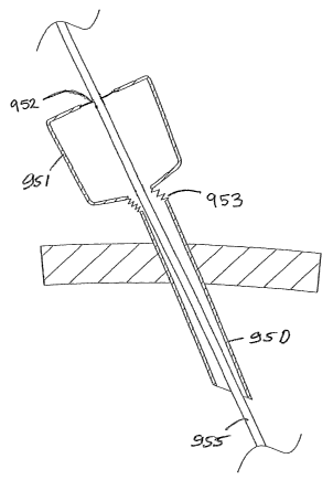
Referring to Figs. 46 to 48 there is illustrated another device I30 according
to the
invention which has some features similar to those of Figs. I1 to 15, Iike
parts being
assigned the same reference numerals. In this case the sleeve has a proximal
section
external of the wound when the device is in die retracting configuration. This
proximal sleeve section comprises a first portion I3I extending from the guide
ring
5I and a second portion I32 extending from the first portion I31. The second
portion 132 is defined between two spaced-apart iris rings 134, 135. It will
be noted
that the iris rings I34, 135 have engagement features such as projections and
grooves
for interengagement on assembly. The iris ring 134 also has an engagement
element,
in this case provided by a groove I37 for engagement on assembly with a
CA 02533798 2006-O1-26
WO 2005/009257 PCT/IE2004/000103
43
con-esponding engagement element of the guide ring 5I which in this case is
prow ided by .a projection I38.
The device is fitted as described above to retract an incision, leaving the
first and
second sleeve portions I31, 132 extending proximally. The first sleeve portion
13I
is redundant and can be removed or scrunched up on assembly of the first iris
ring
134 to the guide ring 138 as illustrated in Fig. 48. The second or upper iris
ring 135
is then rotated to twist the sleeve section 132 to form an iris-type seal as
illustrated in
Fig. 49. The iris ring 135 is engaged with the iris ring 134 as illustrated to
maintain
the valve closed.
Referring to Fig. 49(a) there is illustrated another device I40 according to
the
invention which has some aspects similar to the device of Figs. 46 and like
parts are
assigned the same reference numerals. In this case the iris rings 134, 135 are
used to
form an iris valve I41 which is proximally spaced from the guide ring 5I and a
flexible sleeve section I42 is thereby provided between the iris I41 and the
guide
ring 5I. This sleeve section 142 can act as a flexible cannula wall to permit
sealed
access of a cannula whilst facilitating lateral movement of the cannula
somewhat as
illustrated in Figs. 44 and 45.
Referring to Figs. 49 (b) to 49 (i) there is illustr ated a device according
to the
invention 150 comprising a first ring element 200, a second ring element 201
and a
sleeve 202 of pliable material with a first end mounted to the first ring
element 200
and a second end mounted to the second ring element 200. For ease of reference
the
ring elements 200, 201 have associated location markings 205, 206
respectively.
The sleeve 202 is twisted and has a normally closed access opening 207 and the
sleeve is movable on insertion of an object such as a surgeon's hand/arm 210
or an
instrument through the access opening 207.
CA 02533798 2006-O1-26
WO 2005/009257 PCT/IE2004/000103
44
As will be described in more detail below a biasing means is provided to bias
the
sleeve to close the access opening 207. The biasing may be provided by pre-
tensioning the sleeve, or by using a separate spring element.
Referring to Fig. 49(j) there is illustrated another device 160 which is
similar to the
device of Figs. 49(d) to (i) and like parts are assigned the same reference
numerals.
In this case the device has a lip-type seal I6I. Another device I65 with a
different
type of lip seal 162 is illustrated in Fig. 49(k).
Referring to Figs. 50 to 57 (c) there is illustrated an access port according
to the
invention for use in surgery comprising a first ring element 200, a second
ring
element 20I and a sleeve 202 of pliable material with a first end mounted to
the first
ring element 200 and a second end mounted to the second ring element 200. For
ease of reference the ring elements 200, 20I have associated location markings
205,
IS 206 respectively. The sleeve 202 is twisted and has a normally closed
access
opening 207 and the sleeve is movable on insertion of an object such as a
surgeon's
hand/arm 2I0 or an instrument through the access opening 207.
A biasing means is provided to bias the sleeve 202 to close the access opening
207.
The biasing may be provided by pre-tensioning the sleeve, or by using a
separate
spring element. In this case the spring element 2I5 is a strip of elastic
material 215
which is mounted at one end to the first ring 201. The elastic strip 215
causes the
rings to be biased into a rest position at which the opening 207 is closed. On
insertion of an object such as a surgeon's hand the entry force acts against
the
biasing of the elastic strip 215 and the rings 200, 201 rotate relative to one
another as
evidenced by the locator marks 205, 206. However, the opening is only
sufficient to
allow a specific sized object such as a hand and forearm .to be inserted
through the
sleeve whilst maintaining continuous sealing engagement between the sleeve and
the
object such as a surgeon's handlforearm, thus ensuring that there is no gas
leakage
and maintaining pneumoperitoneum. The device is very easily manufactured and.
CA 02533798 2006-O1-26
WO 2005/009257 PCT/IE2004/000103
most importantly, is extremely easy for a surgeon to use, as a sealed access
port is
provided through which a surgeon can easily insert his arm and forearm. It
will be
noted that the biasing ensures that the access opening substantially exactly
matches
the contours of the inserted object such as a hand/forearm and automatically
opens
5 and closes as required.
In another embodiment, as illustrated in Figs. 58 (a) to 60 (c), the spring
element
may be a coiled spring 220 which normally biases the rings in such a way as to
close
the opening.
I0
Referring to Fig. 61 the hand access device of Figs 50 to 60 is shown mounted
to a
retractor 230 such as a retractor as described above.
Referring to Figs 62 and 63 the access device is shown being mounted to
another
I5 type of retractor 240. In this case the first ring element 200 has a
circumferentially
extending groove 233 and an associated ring 234 with a retractor sleeve
section 235
accommodated therebetween to permit sliding action of the access device
relative to
the retractor sleeve section 235.
20 It will, however, be appreciated that the access devices of the invention
can be used
with any suitable retractor or other similar device.
Referring to Figs. 64 and 65 there is illustrated another access device which
is
similar to the device of Figs. 50 to 57 except that in this case the biasing
to close the
25 access port is provided by pre-tensioning the sleeve 240 and the surgeon,
on
insertion of an object such as his hand/arm acts to overcome the tension in
the sleeve
sufficient to allow hand insertion whilst still maintaining sealing engagement
to the
object such as the surgeon's hand/arm. This configuration will also be
apparent from
Figs 66 and 67. The twisted sleeve defining an iris is shown in Fig. 66 with a
strong
30 outer resilient material 245. As a surgeon inserts his hand the twist in
the sleeve 202
CA 02533798 2006-O1-26
WO 2005/009257 PCT/IE2004/000103
46
is transferred to the outer resilient material 245 with the applied force. In
Fig. 67 the
hand is removed for clarity, in reality on removal of the hand the system
v~lill revert
to the closed configuration of Fig. 66.
Referring now to Fig. 68 there is illustrated an assembly of a access device
of the
invention with a retractor 250 having an excess retractor sleeve section 251
provided
with an outer lip seal 252 for sealing engagement to the arm of a surgeon. The
excess retractor sleeve section may be used to externalise an organ during a
surgical
procedure. In Fig 69 a lip seal 255 is provided in a sleeve section 250
mounted to
the ring element 200. In Fig. 70 a lip seal 260 is provided on a separate
sleeve
section 26I.
Referring to Figs. 7I to 76 there is illustrated an assembly 500 of the
invention
which comprises a retractor 501 and an iris valve 502 releasably mounted to
the
I5 retractor 501. The retractor 501 is similar to the retractors described
above such as
with reference to Figs. 1 to I0. The iris valve 502 is similar to the iris
valves
described above such as with reference to Figs. 50 to 57(c).
The iris comprises the components within the chain bracket 510 in Fig. 71 and
the
retractor comprises the components within the chain bracket 520 in Fig. 71.
The iris 502 comprises a fixed outer iris ring member 5I1 and an inner
rotatable ring
member 512. The inner ring member 512 is in this case a snap fit and is free
to
rotate relative to the outer ring member 5I1. The snap fit engagement is
through an
annular rib 530 on the outer ring member 51I and a corresponding annular
groove
531 in the inner ring member 512. A flexible iris-forming sleeve 513 extends
between the inner and outer ring members 51I, 512. The sleeve 5I3 has a first
elasticated ring or band 514 at one end for anchoring in a corresponding
engagement
channel 5I5 in the inner ring member 512 and a second elasticated band 514 at
the
30' other end for anchoring in a corresponding engagement channel 517 in the
outer ring
CA 02533798 2006-O1-26
WO 2005/009257 PCT/IE2004/000103
47
member 511. Thus, one end of the iris-forming sleeve 513 is anchored to the
movable ring member 5I2 and the other end is anchored to the fixed ring member
512 so that rotation of the ring member 512 relative to the fixed ring member
511
will result in tWIStIIlg or untwisting of the sleeve, forming an iris valve.
The iris
valve is biased into a normally closed position (Figs. 72 to 74) by a spring
which in
this case is in the form of a strip of elastic material 5I8 having enlarged
head
portions 519, 52I at the ends thereof for location and engagement of one end
of the
spring 518 in a spring locating hole 522 in the fixed ring member 511 and for
location and engagement of the other end of the spring 518 in a spring
locating slot
IO 523 in the rotatable ring member 5I2. The spring 518 biases the iris-
forming sleeve
513 into the normally closed position. In insertion of an object such as a
surgeons
hand, the biasing force of the spring is counteracted causing partial opening
of the
iris valve whilst still remaining sealing engagement of the iris sleeve with
the object
passing therethrough. A twisting action of the object as it is being inserted
will aid
I5 overcoming of the spring biasing action, in some cases. The operation of
the iris is
described in more detail above.
The iris forming sleeve 513 has a length in the unassembled untwisted
configuration
of 71 that is preferably less than or equal to the diameter of the sleeve 513.
We have
20 found that this is advantageous in optimising the operation of the iris by
ensuring full
closure of the iris whilst ensuring that excess sleeve material, on twisting,
is
minimised.
The iris valve 502 is in this case releasably mounted to the retractor 501,
Thus, the
25 iris 502 may be used independently of the retractor 501 and vice versa. In
this
instance the iris valve is screw threadingly engagable with the retractor, the
outer
ring 5I1 of the iris having a thread 535 for connection to the retractor 501.
The
retractor 50I in tum has tabs 536 which project inwardly from a retractor top
ring
540 for engagement with the screw thread 535 of the outer ring 5I1. Any
suitable
30 interconnection may be provided.
CA 02533798 2006-O1-26
WO 2005/009257 PCT/IE2004/000103
48
The retractor 501 comprise a sleeve 552, a distal member provided by a distal
ring
553 of resilient material such as an O-ring and a proximal member provided by
a
proximal ring 554 which may also be an O-ring.
The sleeve 552 is of any suitable material such as of pliable plastics film
material
and comprises a distal portion 555 for insertion through an incision 556, in
this case
made in a patient's abdomen 557, and a proximal portion 558 for extending from
the
incision 556 and outside of the patient.
In this case the distal ring 553 is not fixed to the sleeve 552 but rather the
sleeve is
led around the ring 553 and is free to move axially relative to the distal
ring 553
somewhat in the manner of a pulley. The proximal ring 554 is fixed to the
sleeve
552, in this case at the proximal inner end thereof. The sleeve 552 terminates
in a
I5 handle or gripping portion which in this case is reinforced by a gripping
ring 565.
As described above with reference to Figs. I to I0, to configure the retractor
device
according to the invention a sleeve 552 is first provided with the gripping
ring 565
fixed at one end and the proximal ring 554 fixed at the other end [Figs. 3,
5]. The
distal ring 553 is then placed over the sleeve 552. The gripping ring 565 is
then used
to manipulate the sleeve 552 so that the sleeve 552 is folded back on itself
into the
configuration of Figs. 1 and 2 in which the gripping ring 565 is uppermost.
The
sleeve extends from the proximal ring 554 and the distal ring 553 is contained
between inner and outer layers of the sleeve 2. The device is now ready for
use.
The resilient distal ring 553 is scrunched up and inserted through the
incision 556
with the distal end 555 of the sleeve 552 as illustrated in Fig. 4. The sleeve
552 is
then pulled upwardly in the direction of the arrows A in Figs. 8 to 10. On
pulling of
the sleeve 552 upwardly the sleeve outer layer is pulled up while the sleeve
inner
Iayer is drawn around the proximal ring 553. This results in shortening the
a~;ial
CA 02533798 2006-O1-26
WO 2005/009257 PCT/IE2004/000103
49
extent between the proximal ring 554 and the distal ring 553, tensioning the
sleeve
552 and applying a retraction force to the margins of the incision 556. The
system
appears to be self locking because we have observed that when tension is
applied to
the sleeve 552 and the pulling force is released the rings 553, and 554 remain
in
position with a retraction force applied. Frictional engagement between the
layers of
the sleeve in this configuration may contribute to this self locking. As the
incision is
being retracted the margins are also protected by the sleeve. On retraction,
an access
port is provided, for example for a surgeon to insert his hand and/or an
instrument to
perform a procedure.
IO
In this instance the sleeve gripping ring 565 is led over the retractor top
ring 540 and
the gripping ring 565 is retained. outside of the top ring 540 as illustrated
in Fig. 72.
The retractor top ring 540 provides a guide member for the retractor proximal
ring
554. The guide member or top ring 540 is in the form of 'an annular ring
member
I5 with an inwardly facing C-shaped groove which is sized to accommodate the
ring
554 as illustrated. The outer layer of the sleeve 552 is interposed between
the ring
554 and the guide 540 to further control the pulling of the sleeve and thereby
further
controlling the application of the retraction force. The guide 540 also assist
in
stabilising the proximal ring 554.
Referring now to Figs. 77 to 84 there is illustrated a pinch valve for use
with the
access port of the invention. The pinch valve comprises a flexible cylindrical
film
sheath 800 which is twisted by a torsion spring 801 to form an iris-type
valve. The
spring 801 has spring arms 802, 803 at the free ends thereof which are
retained
within corresponding recesses 804 in finger handle parts 805, 806 of retaining
members 807, 808. The valve is normally in the closed position illustrated in
Figs.
78 and 79, in which the sleeve 800 is biased by the spring 801 into a closed
iris-
forming configuration. The handles 805, 806 can be readily gripped by a user
with
one hand and rotated against the biasing of the spring 801 causing the iris to
open as
illustrated in Figs. 80 and 8I, ready to receive an object such as an
instrument
CA 02533798 2006-O1-26
WO 2005/009257 PCT/IE2004/000103
therethrough. When the object has passed through the valve the finger handles
805,
806 are released, causing the iris to close and maintain gas pressure on the
patient
side of the valve.
5 because of the simple and compact open/ciose arrangement of the finger
handles
805, 806, it is possible for a user to open the iris using only an index
finger and a
thumb of one hand. This is a highly convenient means of operating the valve,
especially in the case of passing laparoseopic instruments through the valve.
IO The access port of Figs. 77 to 84 may also be used with a further seal such
as a lip
seal 810 which may be coupled to the top retaining member 807 as illustrated
in
Figs. 85 to 88. In these drawings the access port is shown coupled to a
retractor 81 I
located in an incision 813 in the abdomen 812 of a patient to create a low-
profile,
sealed instrument access port.
IS
The retractor 8I1 is preferable a retractor of the type described earlier.
In particular the retractor 811 for retracting the sides of the incision 813
comprises a
distal O-ring member I000 for insertion into the incision 813, a proximal 0-
ring
20 member 1001 far location externally of the incision 813, and a retracting
sleeve
member 1002 for extending between the O-rings 1000, 1001 to retract the sides
of
the incision 813 (Fig. 85).
The sleeve 1002 is fixedly attached to the proximal O-ring 1001, is looped
distally
25 around the distal O-ring 1000, and extends between the O-rings 1000, 1001
in a two-
layer arrangement.
The retractor 8II is particularly suitable for retracting the sides of a
laparoscopic
incision 8I3. Generally Iaparoscopic incisions are retracted to a diameter of
Iess
CA 02533798 2006-O1-26
WO 2005/009257 PCT/IE2004/000103
51
than 40 mm, preferably between 3 mm and 35 mm, and ideally between 5 mm and
I2 mm..
As illustrated in Figs. 87 and 88, the diameter of the retracted laparoscopic
incision
813 is substantially equal to the diameter of the laparoscopic instrument 8I4.
This is
possible because the walls of the retracting sleeve member 1002 are extremely
thin.
Thus the minimum amount of space is used up by the walls of the retractor 81I
enabling .the overall size of the laparoscopic incision 813 to be minimised.
The Lip seal 810 provides further sealing for an instrument 814 which may be
inserted through the pinch valve and the retractor 8I1, as illustrated in
Figs. 8& to 88.
In an alternative arrangement illustrated in Figs. 89 and 90, a lip seal 820
may be
connected to the retractor 811, such as by using excess sleeve material 822
from the
retractor 811. Other details of this embodiment are described above with
reference
to Figs. 77 to 84, and like parts are assigned the same reference numerals.
In another embodiment the access port does not have a secondary seal for the
instrument. Such an embodiment is illustrated in Figs. 91 and 92. Fasically
this
version involves a retractor 8I1, with a pinch valve arrangement as described
above
with reference to Figs. 77 to 74, attached directly thereto.
In some of the embodiments described above a valve is mounted directly to a
retractor base 8I1. ~t is possible to provide a flexible coupling between the
retractor
811 and the valve. For example, as illustrated in Figs. 93 and 94, such a
flexible
coupling is provided by a length of flexible sleeve 830 extending between the
retractor 8I1 and the valve 829. The flexible sleeve 830 may be formed by
excess
retractor sleeve material attached to the valve 829. The flexible nature of
the sleeve
830 accommodates movement of the valve 829 relative to the retractor 811 while
maintaining the gas-tight sealed coupling.
CA 02533798 2006-O1-26
WO 2005/009257 PCT/IE2004/000103
52
T'he access pori of the invention may be of modular construction. As
illustrated in
Figs. 95 to 97, a valve 840 may be mounted to a retractor base, such as to an
outer
ring part 844 of the retractor 8I1. The valve 840 may be of similar
construction to
the valve described previously with reference to Figs. 77 to 84, and like
parts are
assigned the same reference numerals. To facilitate ease of mounting, the body
of
the valve 840 and the retractor body 841 may have complementary interengagable
formations. In the embodiment illustrated, the retractor body 841 has a series
of
locating tabs 842 for corresponding slots 843 in the valve body. The assembly
will
be particularly apparent from Figs. 96 and 97.
Various means of attachment of a proximal assembly to a retractor base may be
provided. A proximal ring 845 illustrated in Fig. 98 may be attached to the
retractor
base BII.
A cap 850 is illustrated in Figs. 99 to 104. The cap 850 in this case has an
integral
duck-bill valve 851 through which an operating cable 852 may be passed. An
operating device or instrument such as a surgical stapler 853 may be attached
to the
cable 852, and the cap 850 may be mounted to the retractor proximal ring 845,
as
illustrated in Figs. 101 to 104. The cap 850 may be releasably mounted to the
proximal ring 845 using suitable complementary formations such as projecting
ribs
846 on the proximal ring 845 and corresponding ledges 854 on the cap 850. With
the stapler 853 or other device in the abdominal space insufflation may be
used and
the stapler 853/device can be used laparoscopically.
1n a further embodiment of the invention as illustrated in Figs. I05 to IIO a
valve
860 may be coupled to the retractor 811 in such a way as to facilitate a
flexible joint
therebetween. For example, a fixed length sleeve 862 may extend between an
outer
proximal ring 863 of the retractor 811 and the valve 860. Excess sleeve
material 864
from the retractor 8II may pass up through the valve 860. The valve 860 may be
CA 02533798 2006-O1-26
WO 2005/009257 PCT/IE2004/000103
53
pushed down and the excess sleeve pulled up to firmly lock the base retractor
8I I in
the incision. Excess sleeve material 864 may be cut-away and removed, if
desired.
The sleeve material 864 allows the instrument to tilt as illustrated in Fig.
110 without
compromising the valve seal to the shaft of the instrumentlobject 814.
As illustrated in Figs. 112 and I13 a spring 867 may be provided between the
valve
860 and the retractor proximal ring 863 for more controlled flexibility.
Referring now to Figs. 114 to 116 another modular system is illustrated in
which a
valve 870 is releasably mounted to a retractor 81I. The retractor 8II may have
a
proximal ring 871 with a recess 872 to receive the valve 870. An instrument
shaft
814 can readily pass through the valve 870 and retractor 811. At least a
section 873
of the shaft 814 can be bent or steered almost immediately distal of the
retractor.
IS Referring now to Figs. II7 to 120 any suitable valve 880 may be coupled to
a
retractor 811 using excess sleeve material 881 from the retractor 811. The
valve 880
may be pulled upwardly to deploy the base retractor 811. The excess sleeve
material
881 provides a flexible neck which facilitates easy introduction of objects
such as an
instrument 883, even one having a bent shaft (Fig. 119). As illustrated in
Fig. 120
such an arrangement also facilitates additional instrument reach by allowing
the
valve 880 to be moved closer to the base retractor 811.
The access ports of the invention can be used in a number of ways. In one
method
the retractor is used as described above, the distal inner ring being inserted
into an
incision, the outer ring being slid to controllably radially expand the
incision. The
retractor may then be locked in position. If necessary, the outer ring can be
moved
further downwardly to create a larger incision.
CA 02533798 2006-O1-26
WO 2005/009257 PCT/IE2004/000103
S~
In some arrangements an instrument may be bent manually outside the body and
the
bent instrument is delivered through the access port to readily access the
operative
site.
In a further embodiment an instrument is inserted into the access port and the
surgeon uses the abdominal wall itself to bend the instrument and then insert
the bent
section further into the abdomen.
The access ports of the invention have at least some of the following
advantages:
Controlled Radial Expansion
I. Greater access using smaller incision
2. Can vary incision size as need be (e.g. specimen removal during lap coli.)
Greater Sealing Capabilities
I. No gas leakage from the wound margins
2. Cannot be inadvertently pulled out of the incision
3. Will seal any incision and never require secondary sealing method (suture,
Hassan port, etc.)
Eliminate Intra-abdominal Profile
1. Gives bacle more working space in the abdomen (critical in pelvic surgery)
2. Perineal access for operations such as Radical Prostatectomy.
1 rotection of ~TOUnd from Infection and Cancer Seeding
I. Tight seal with no "chimney stack" effect
2. Upon removal all areas of potential contamination are isolated from the
incision
~2c~duced Extra-abdominal Profile
1. Will increase the effective working length of an instrument
CA 02533798 2006-O1-26
WO 2005/009257 PCT/IE2004/000103
2, Greater working are outside the abdomen
Increase the freedom of movement of conventional laproscopic instruments
5 The retractor of the invention may be inserted through the abdominal wall as
described below. An initial thin incision 900 may be made in the abdominal
wall
907 and an inner distal ring 901 of the retractor may be attached to an
insertion tool
902 as illustrated in Fig. 121. The ring 90I is flexible and can be stretched
or bent as
illustrated for ease of insertion through the incision 900. The ring 901 may
be
I0 retained in the stretched/bent insertion configuration using locating
grooves 903 in
the insertion tool 902. Alternatively or additionally as illustrated in Figs.
122 to I24
the ring 90I may be split into a number (in this case 4) of sections 905 with
an inner
thread 906 passing between and linking the sections 905. The ring 901 can be
bent
as illustrated to reduce the profile in the insertion configuration. The
system is
IS biased so that the ring 90I re-forms into the circular configuration once
released on
insertion.
In some cases (Figs. 125 to I27) the ring 90I may be inserted through the
incision
using a blunted or round-nosed obturator tool 910.
Alternatively as illustrated in Figs. 12& and 129 the ring 910 may be inserted
using
an obturatorltrocar tool 9I1 with a leading cutting blade 912. In this case,
as
illustrated in Figs. 130 to 134, the tool 911 itself makes an incision in the
abdominal
wall, allowing the distal ring 9I0 of the retractor to be delivered and
deployed,. as
illustrated.
In some cases, as illustrated in Figs. I35 and 136 the insertion tool 9I0 may
have a
stop 9I5 thereon to limit the extent by which the tool can project into the
patient.
The stop 915 may be fixed, or adjustable in position. The adjustment of the
stop 9I5
may be used to facilitate different thicknesses of abdomen. Such adjustment
could
CA 02533798 2006-O1-26
WO 2005/009257 PCT/IE2004/000103
5s
be achieved using any suitable means such as a screw thread or ratchet system.
The
adjustment may be rendered automatic by using a spring loaded type system.
An alternative insertion tool 920 is illustrated in Figs. 137 to 140. In this
case the
leading end 92I of the tool 920 is blunted and is inserted through a pre-made
incision
900. The distal ring 901 of the retractor is retained in a groove 922 at the
distal end
of the tool 920.
In an alternative embodiment illustrated in Figs. 141 to 144 the introducer
tool 920
has an integral blade 925 which is lined up to the desired location and the
tool 920 is
pushed through to make a leading incision in the abdominal wall 907.
Another possible solution to the problem presented by a conventional rigid
cannula
as described above is provided by an access device illustrated in Figs. 145
and 146
which has a distal hollow tubular section 950 and a proximal instrument
insertion
section 951 with a lip seal 952 for sealingly engaging with an instrument
shaft 955,
which are movably coupled together by a flexible tubular sheath section 953.
The distal section 950 defines an access channel for extension of an
instrument 955
therethrough. The flexible section 955 facilitates relative movement between
the
sections 950, 951 to accommodate lateral movement of the instrument 955 while
maintaining the seal between the lipseal 952 and the instrument 955.
This access device allows greater manoeuvrability on insertion of an
instrument 955.
The flexible section 953 may be concertinaed to enhance the flexing action. As
illustrated the lip seal 952 is located at the proximal opening of the
proximal section
95I.
CA 02533798 2006-O1-26
WO 2005/009257 PCT/IE2004/000103
57
In particular, if the instrument 955 is tilted to the side, as illustrated in
Fig. I46, the
flexible section 953 permits lateral movement of the proximal section 95I with
the
instrument 955. By effectively following the lateral movement of the
instrument
955, this ensures that no leakage gap occurs between the instrument 955 and
the lip
seal 952 and thus the pneumoperitoneum within the abdominal cavity is
maintained.
In this manner the access device of Figs. 145 and I46 provides a solution to
the
leakage problems encountered by conventional cannula when an instrument is
tilted
to the side.
Another possible solution is provided by an access device illustrated in Figs.
147 to
149 which has an external lip seal 952 movably connected to the proximal
section
95I by a flexible sheath section 956 upstanding proximally from a proximal end
of
the proximal section 95I. This arrangement also accommodates lateral movement
of
the instrument 955 while maintaining the seal.
In conventional rigid cannula systems, if the trocar and/or instruments is
tilted to one
side a Ieak path is developed through the seal. The systems of Figs. I45 to
1.49 avoid
this problem.
The invention is not limited to the embodiments hereinbefore described, with
reference to the accompanying drawings, which may be varied in construction
and
detail.
z5