Note: Descriptions are shown in the official language in which they were submitted.
CA 02534515 2006-01-31
A SURGICALLY IMPLANTABLE INJECTION PORT HAVING AN
ABSORBABLE FASTENER
[0001] Field of the Invention
[0002] The present invention has application in conventional endoscopic and
open surgical
instrumentation as well as application in robotic-assisted surgery. The
present invention
has even further relation to adjustable surgically implantable bands, such as
gastric bands
for the treatment of obesity.
[0003] Background of the Invention
100041 The percentage of the world's population suffering from morbid obesity
is steadily
increasing. Severely obese persons are susceptible to increased risk of heart
disease,
stroke, diabetes, pulmonary disease, and accidents. Because of the effect of
morbid
obesity to the life of the patient, methods of treating morbid obesity are
being researched.
[0005] Numerous non-operative therapies for morbid obesity have been tried
with virtually no
permanent success. Dietary counseling, behavior modification, wiring a
patient's jaws
shut, and pharmacological methods have all been tried, and failed to correct
the
condition. Mechanical apparatuses for insertion into the body through non-
surgical
means, such as the use of gastric balloons to fill the stomach have also been
employed in
the treatment of the condition. Such devices cannot be employed over a long
term,
however, as they often cause severe irritation, necessitating their periodic
removal and
hence interruption of treatment. Thus, the medical community has evolved
surgical
approaches for treatment of morbid obesity.
[0006] Most surgical procedures for treatment of morbid obesity may generally
be classified as
either being directed toward the prevention of absorption of food
(malabsorption), or
restriction of stomach to make the patient feel full (gastric restriction) The
most common
malabsorption and gastric restriction technique is the gastric bypass. In
variations of this
technique, the stomach is horizontally divided into two isolated pouches, with
the upper
1
CA 02534515 2012-07-27
pouch having a small food capacity. The upper pouch is connected to the small
intestine,
or jejunum, through a small stoma, which restricts the processing of food by
the greatly
reduced useable stomach. Since food bypass much of the intestines, the amount
of
absorption of food is greatly reduced.
[0007] There are many disadvantages to the above procedure. Typically the
above mentioned
procedure is performed in an open surgical environment. Current minimally
invasive
techniques are difficult for surgeons to master, and have many additional
drawbacks.
Also, there is a high level of patient uneasiness with the idea of such a
drastic procedure
which is not easily reversible. In addition, all malabsorption techniques
carry ongoing
risks and side effects to the patient, including malnutrition and dumping
syndrome.
[0008] Consequently, many patients and physicians prefer to undergo a gastric
restriction
procedure for the treatment of morbid obesity. One of the most common
procedures
involves the implantation of an adjustable gastric band. Examples of an
adjustable
gastric band can be found in U.S. Patents 4,592,339 issued to Kuzmak; RE 36176
issued
to Kuzmak; 5,226,429 issued to Kuzmak; 6,102,922 issued to Jacobson and
5,601,604
issued to Vincent. In accordance with current practice, a gastric band is
operatively
placed to encircle the stomach. This divides the stomach into two parts with a
stoma in-
between. An upper portion, or a pouch, which is relatively small, and a lower
portion
which is relatively large. The small partitioned portion of the stomach
effectively
becomes the patients new stomach, requiring very little food to make the
patient feel full.
[0009] Once positioned around the stomach, the ends of the gastric band are
fastened to one
another and the band is held securely in place by folding a portion of the
gastric wall over
the band and closing the folded tissue with sutures placed therethrough
thereby
preventing the band from slipping and the encircled stoma from expanding.
Gastric
bands typically include a flexible substantially non-extensible portion having
an
expandable, inflatable portion attached thereto. The inflatable portion is in
fluid
2
CA 02534515 2012-07-27
communication with a remote injection site, or port. Injection or removal of
an inflation
fluid into or from the interior of the inflatable portion is used to adjust
the size of the
stoma either during or following implantation. By enlarging the stoma, the
patient can
eat more food without feeling as full, but will not lose weight as fast. By
reducing the
size of the stoma, the opposite happens. Physicians regularly adjust the size
of stoma to
adjust the rate of weight loss.
[0010] For most fluid injection ports for the above described bands are
attached underneath the
skin to the fascia of a patient. Such ports are often provided with suture
holes and the
port is sutured to the tissue. However, alternative means of attaching the
port to the
patient, such as using integral hooks, can be used as well. Such other means
for attaching
the port to a patient are described in commonly assigned and copending U.S.
Patent
Numbers: 7,061,714 issued June 13, 2006; 7,374,557 issued May 20, 2008.
[0011] However, many of the prior art fasteners could cause patient
discomfort, including pain.
It is well known that once the port is placed a fibrotic capsule begins to
grow over the
port until it is completely enclapsuled. The rate at which the fibrotic
capsule grows
varies from patient to patient, but generally surgeons agree that the port is
fully
encapsulated after 2 months. Once the port has been captured by the fibrotic
capsule,
there is no longer a need for the port to be fastened with sutures or other
types of
fasteners. In fact, it would be desirable if these additional fastening means
were no
longer part of the port system so as to not cause patient discomfort.
[0012] Summary of the Invention
In accordance with the present invention, there is provided an implantable
surgical
injection port having an undeployed position, and a deployed position wherein
it is
attached to tissue. The port includes a housing having a closed distal end, an
open
proximal end and a fluid reservoir therebetween. The port further includes a
needle
penetrable septum attached to the housing about the opening. The port even
further
3
, . CA 02534515 2012-07-27
includes at least one attachment mechanism mounted to the housing for
initially attaching
the port to tissue wherein the attachment mechanism is made from a
bioabsorbable
material. The attachment mechanism takes the form of an arcuate hook having a
length
extending substantially greater than 900 about said pivot point.
Detailed Description of the Drawings
[0013] The novel features of the invention are set forth with particularity
in the appended claims.
The invention itself, however, both as to organization and methods of
operation, together
with further objects and advantages thereof, may best be understood by
reference to the
following description, taken in conjunction with the accompanying drawings in
which:
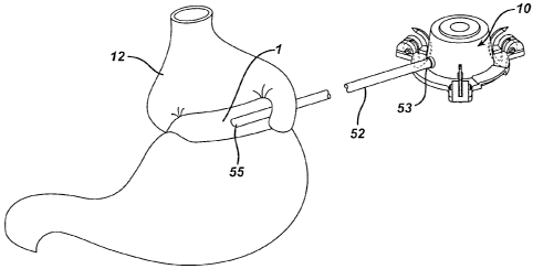
[0014] Figure 1 is a perspective view of a surgically implantable fluid port
made in accordance
with the present invention, showing the port attached to an adjustable gastric
band.
[0015] Figure 2 is a perspective view of a surgically implantable fluid port
made in accordance
with the present invention.
[0016] Figure 3 is a cross section of the port shown in Figures 1 and 2,
taken along line 3-3 in
Figure 1.
[0017] Figure 4 is a view similar to that of Figure 3 but showing the fluid
port implanted within
a patient.
[0018] Detailed Description of the Invention
[0019] Referring now to the drawings wherein like numerals indicate the same
elements
throughout the views, as stated above there is shown in Figure 1 an adjustable
gastric
band 1 of the type described in the above mentioned incorporated references.
Band 1 is
implanted within a body of a patient to surround the stomach 12. The
inflatable portion
of the band is in fluid communication with injection port 10 via a catheter
tube 52. Tube
52 has a proximal end 53 attached to the port 10 and a distal end 55 attached
to adjustable
4
. . CA 02534515 2012-07-27
gastric band 1. Port 10 can be used for a wide range of devices in the medical
field and
not only for gastric bands. For example the port can also used for vascular
access for
drug delivery.
[0020] As seen from Figures 2 and 3, surgically implantable injection port 10
includes a housing
12. Housing 12 can be made from any number of materials including stainless
steel,
titanium, or polymeric materials. Housing 12 has a distal back portion or
closed distal
end 14 and a perimeter wall portion 16 extending proximally from the back
portion 14 at
an angle. Wall portion 16 defines a proximal opening or open proximal end 18,
and a
fluid reservoir 20 between opening 18 and back portion 14. The port includes a
needle
penetrable septum 22 attached to the housing about the opening 18 so as to
cover the
opening and seal the reservoir 20. Septum 22 can be made from any number of
materials
including silicone. Septum 22 is preferably placed in a proximal enough
position such
that the depth of the reservoir 20 is sufficient enough to expose the open tip
of a needle,
such as a Huber needle, so that fluid transfer can take place. Septum 22 is
preferably
arranged so that it will self seal after being punctured by a needle and the
needle is
withdrawn. In one embodiment, the septum is made from silicone which is under
compression when attached to the housing. Port 10 further includes a catheter
tube
connection member 30, in fluid communication with reservoir 20.
[0021] Port 10 is implanted into a patient and attached to the fascia just
below the skin of the
patient, so that fluid can be inserted and withdrawn from the inflatable
portion with a
syringe. As seen from the figures, port 1 includes one or more attachment
mechanisms
70, taking the form of an arcuate hook. However, for purposes of this
invention, the
attachment mechanism could take the form of alternative means such as using
suture.
Some of these other means for attaching the port to a patient are described in
commonly
assigned and copending U.S. Patent Numbers: 7,061,714 issued June 13, 2006;
7,374,557
issued May 20, 2008.
5
CA 02534515 2006-01-31
[0022] As seen from the figures, port 1 includes one or more attachment
mechanisms 70. The
figures herein show three attachment mechanisms all substantially identical
and equally
spaced from each other. Attachment mechanisms 70 are mounted to the housing 12
at a
pivot point 80 along an outer periphery 13 of the housing 12. As seen from the
figures,
attachment mechanisms 70 are arcuate hooks pivotable with respect to the
housing.
Attachment mechanisms 70 have an arcuate length L extending substantially
greater than
90 , and preferably at least 180 about the pivot point. Implantable surgical
injection port
has an undeployed position, shown as a solid line in Figure 3, and a deployed
position,
shown as the phantom line in Figure 3 and in Figure 4, wherein the port is
attached to
tissue. Attachment mechanisms 70 is preferably made from a bioabsorbable
material
including, but not limited to, one or more of the following either alone or in
combination:
iron, polydioxanone, polyglactin and/or poliglecaprone.
100231 Attachment mechanism 70 has a fixed end 72 pivotally attached to the
housing 12 at
pivot point 80. The design allows a surgeon to use forceps and drive the
fastener through
the tissue until the free end 74 rests against the flat 75. In this way the
patient is
protected from the sharp end of the tip. Attachment mechanism 70 also includes
a free
end 74 which has a sharp or pointed configuration. Housing 12 further includes
at least
one recessed portion 15 along its distal end 14. Recessed portion 15 is
designed to
receive the free end 74 of attachment mechanisms 70 when the port 1 is in its
deployed
position. This design prevents any exposure of the sharp free end to tissue
after the port
has been implanted.
100241 The above described 180 hook or attachment mechanisms provide
advantages over prior
90 or less hooks. As seen from Figure 4, the above described attachment
mechanism
allows the hook to engage a greater area of tissue, and allows for two locking
points,
entry into and then out of the fascia. This provides for better sacrament of
the port to the
tissue. Further no "sharp" is exposed to the patient. A further advantage of
the fastener
configuration is that the fastener follows a constant radius when pushing
through the
tissue. By maintaining a constant radius the fastener never induces a
compressive force
6
CA 02534515 2012-07-27
onto the fascia. This should minimize pain because the fastener is not
"compressing or
squeezing" nerves.
[0025] In practice, the physician would create an incision in the skin 110 of
a patient to expose
the fascia according to well known surgical techniques. Thereafter, as seen
from Figure
4, the port 1 could be placed against the fascia 100 of the patient with the
port in its
undeployed position. Thereafter, the physician could rotate, manually or
otherwise, the
attachment mechanism substantially greater than 90 and preferably at least
1800 so that
the hook enters and then exits the fascia. The design allows a surgeon to use
forceps and
drive the fastener through the tissue until the free end 74 rests against the
flat 75. In this
way the patient is protected from the sharp end of the tip. This could be done
for each
attachment mechanism on the device. Thereafter, the catheter tube 52 would be
connected to connection member 30, and the patient is sewn up.
[0026] It will become readily apparent to those skilled in the art that the
above invention has
equally applicability to other types of implantable bands. For example, bands
are used
for the treatment of fecal incontinence. One such band is described in U.S.
Patent
6,461,292. Bands can also be used to treat urinary incontinence. One such band
is
described in U.S. Patent Publication No. 2003/0105385. Bands can also be used
to treat
heartburn and/or acid reflux. One such band is described in U.S. Patent
6,470,892.
Bands can also be used to treat impotence. One such band is described in U.S.
Publication No. 2003/0114729.
[0027] While preferred embodiments of the present invention have been shown
and described
herein, it will be obvious to those skilled in the art that such embodiments
are provided
by way of example only. Numerous variations, changes, and substitutions will
now occur
to those skilled in the art without departing from the invention. For example,
as would be
apparent to those skilled in the art, the disclosures herein have equal
application in
robotic-assisted surgery. In addition, it should be understood that every
structure
described above has a function and such structure can be referred to as a
means for
performing that function.
7
CA 02534515 2006-01-31
robotic-assisted surgery. In addition, it should be understood that every
structure
described above has a function and such structure can be referred to as a
means for
performing that function. Accordingly, it is intended that the invention be
limited only
by the spirit and scope of the appended claims.
8