Note: Descriptions are shown in the official language in which they were submitted.
CA 02534632 2012-03-09
WO 2005/038459 PCMIS20041025708
=
BRIDGED ELEMENT FOR DETECTION
OF A TARGET SUBSTANCE
=
Technical Field
This document pertains generally to sensor devices, and more
particularly, but not by way of limitation, to detection 94 analysis of a
target
substance.
Background
Previous efforts to detect enalytes, such as biological agents, pathogens,
bacteria, viruses, fmigi, molecules and toxins are relatively cumbersome, time-
consuming, and require significant technical expertise to operate. For
example,
one technique generally requires the incubation of samples on Petri plates
over
an extended period of several days. Another technique involves the use of dyed
antibodies selected to identify the presence of specific pathogenic bacteria.
in addition, some systems require that the target biological molecules
(= undergo an amplification procedure which is prone to errors and requires a
high
level of teclmical. skill. Purdy:more, amplification sometimes cannot
determine
the concentration of a target biological agent and are not practical for use
in the
field.
Some systems fail to detect natural or engineered changes in biological
agents, are known to generate false positive errors and are sensitive to
testing
conditions. Some devices for the detection of biological molecules (such as
=
DNA sequences or proteins) require a large number of target molecules to
=
CA 02534632 2012-03-09
J.)
WO
2005/0314459 PC11US20414/0257118
operate effectively. Accordingly, the target molecules must be amplified, and
in
some instances tagged which prevents further use of the template molecules.
Brief Description Of The Drawings
In the drawings, which are not necessarily drawn to scale, like numerals
describe substantially similar components throughout the several views. Like
numerals having different letter suffixes represent different instances of
substantially similar components. The drawings illustrate generally, by way of
example, but not by way of limitation, various embodiments discussed in the
present document.
fig. 1 illustrates a flow chart for a method of detecting a target molecule.
Fig. 2 illustrates a cantilever detector.
Fig. 3 illustrates a portion of a cantilever structure.
Fig. 4 illustrates a graph of displacement as a function of time.
Figs. 5.A. and 5B illustrate a cantilever detector system.
Pig, 6 illustrates a graph of current as a function of voltage.
Figs. 7A and 7B illustrate measured pararneters.as a function of time.
Fig. 8 illustrates a shift in resonance.
Fig. 9 illustrates a flow chart for a method of preparing a template
molecule.
Figs. 10 and 11 illustrate flow charts for methods of detecting a target
moleeele,
Fig. 12 schematically illustrates an array of cantilevers in a system.
Fig. 13 illustrates an example of a portable detector.
Detailed Description
The following detailed description includes references to the
accompanying drawings, which form a part of the detailed description. The
drawings show, by way of illustration, specific embodiments in which the
invention may be practiced. These embodiments, which are also referred to
herein as "examples," are described in enough detail to enable those skilled
in
the art to practice the invention. The embodiments may be combined, other =
embodiments may be utilized, or structural, logical and electrical changes may
be tnade: = = _ The
following detailed description is, therefore, not to be taken in a limiting
sense,
2
CA 02534632 2012-03-09
14 =
WO 2005/03$159 PCTAIS7004/02570
rather, the scope of the claims should be given the broadest possible
interpretation
consistent with the description as a whole.
In this document, the terms "a" or "an" are used, as is common in patent
documents, to include one or more than one, In this document, the term "or" is
used to refer to a nonexclusive or, unless otherwise indicated,
The accompanying drawings that form a part hereof, show by way of
illustration,
and not of limitation, specific embodiments in which the subject matter may be
practiced. The embodiments illustrated are described in sufficient detail to
enable those skilled in the art to practice the teachings disclosed herein.
Other
embodiments may be utilized and derived therefrom, such that structural and
logical substitutions and changes may be made,. It will be clear to
any
person skilled in the art that modifications of and adjustments to the
foregoing
embodiments, not shown, are possible. The scope. of the claims should not be
limited by the embodiments set forth in the examples, but should be given the
broadest possible interpretation consistent with the description as a whole.
Such embodiments of the inventive subject matter may be referred to herein,
individually or collectively, by the term "invention" merely for convenience
and
without intending to voluntarily limit the ; 'application to any single
invention or inventive concept if more than one is in fact disclosed. Thus,
although specific embodiments have been illustrated and described herein, it
should be appreciated that any arrangement calculated to achieve the same
purpose may be substituted for the specific embodiments shown. This document
is intended to cover any and all adaptations, or variations, or combinations
of
various embodiments. Combinations of the above embodiments, and other
embodiments not specifically described herein, will be apparent to those of
skill
in the art upon reviewing the above description.
3
CA 02534632 2006-02-02
WO 2005/038459
PCT/US2004/025708
Introduction
Molecules are affected by changes in their environment. For example, a
single stranded deoxyribonucleic acid (ssDNA) will respond to the introduction
of its complementary ssDNA. Hybridization of one DNA strand with its
complementary strand results in a reduction in overall length, as well as a
change
in DNA conductive properties. The changes are directly proportional to the
fidelity of match between the two DNA strands with even a single nucleotide
mismatch having a measurable effect. Analysis of the changes permits
identification of various pathogens and allows differentiating between
specific
strains.
In one example, a microelectromechanical (MEMS) structure is used to
measure molecular changes associated with hybridization. For example, a
mobile element in a MEMS chip is bridged by a selected single-strand DNA
fragment. A complementary fragment is detected and identified based on a
measurement of the deflection of the element and a measurement of conductivity
resulting from hybridization. In one example, signal processing is used to
interpret the hybridization events as detection and identification. The
correlation
of length and conductivity change to DNA strand homology is used to
discriminate between known and variant pathogens, and between benign and
virulent strains.
In addition, a voltage applied after hybridization causes the pathogen
DNA strand, or target molecule, to be released from the template molecule. The
current at which the target molecule releases can also provide information to
identify the target molecule. In addition, by releasing the target molecule,
the
sensor can be prepared for an additional detection event. Furthermore, the
integrity of the sensor is tested by conducting a low-level current through
the
template molecule prior to a sensing event to verify continuity. In one
example,
an array of DNA-bridged MEMS sensors allows simultaneous multiplexed
detection of numerous viral or bacterial pathogens, or enables the measurement
of concentration of a single pathogen.
In addition, changes in the resonance of the template molecule are used
for identifying a sample that binds to the template molecule. In one example,
a
4
CA 02534632 2006-02-02
WO 2005/038459
PCT/US2004/025708
movable end of a cantilever is bridged to a structure by a template molecule.
The resonance frequency of the cantilever system (with the template molecule
bridge) will change upon hybridization of the sample with the template
molecule. The degree of homology can be determined by the magnitude and
direction of the shift in amplitude or frequency.
Exemplary Method
Fig. 1 illustrates exemplary procedure 100 for detecting and identifying a
target substance. The target substance, in one example, includes a single
strand
DNA fragment.
As used herein, the target molecule and the template molecule are coined
terms and the molecules are related in the manner of their binding together.
Accordingly, a particular sensor uses a first ssDNA strand as a template
molecule and a second ssDNA strand is a target molecule, another sensor can
use the first ssDNA strand as the target molecule and the second ssDNA strand
as the template molecule.
Other combinations of binding partners are also contemplated. For
example, either the template molecule or the target molecule can include
nucleic
acid molecules (e.g. oligonucleotides, including ss-DNA or RNA referred to as
ss-RNA), proteins and carbohydrates. A template molecule comprising a single
strand of DNA may hybridize with a complementary strand of DNA to form a
double stranded DNA (ds-DNA). In addition, a template molecule including a
protein may bind to a target molecule that also includes a protein (through a
protein-protein recognition), a nucleic acid (through protein-nucleic acid
recognition) or a carbohydrate (through protein-carbohydrate recognition). In
addition, a template molecule including nucleic acid may bind to a target
molecule including a nucleic acid using DNA (through nucleic acid-nucleic acid
recognition) or a carbohydrate (through nucleic acid-carbohydrate
recognition).
Furthermore, a template molecule including a carbohydrate may bind to a target
molecule including a carbohydrate (through carbohydrate-carbohydrate
recognition). In general, template molecule-target molecule combinations can
be
described as a lock-and-key mechanism that allows certain molecules to bind
only with other molecules.
5
CA 02534632 2006-02-02
WO 2005/038459
PCT/US2004/025708
At 105, the sample to be analyzed is collected. The sample, which
potentially includes the target molecule, can be in a gas, liquid or solid
form. At
110, the sample is prepared for analysis, which, in one example, includes
filtering of the sample. At 115, the sample is delivered to the sensor for
analysis.
Sample delivery, in one example, includes routing the sample using a
microfluidic pump, valve, channel, reservoir or other structure. At 120, the
sample is introduced to one or more sensors for possible detection and
identification. In various examples, detection and identification include
monitoring for a change in length or position, a change in a force, a change
in
electrical conductivity or resistivity, determining a signal level for
disbonding a
sample from the template molecule and determining a shift in resonance. At
125, the collected data is processed to detect and identify the sample.
Processing
the data, in various examples, includes comparing an output signal with stored
data where the stored data includes a look-up table which correlates a target
molecule with a template molecule.
Other procedures are also contemplated. For example, a sensor integrity
test may be performed before exposing the sensor to the sample by monitoring
various parameters.
Exemplary Cantilever Sensor
Fig. 2 illustrates sensor 200 according to one example. Substrate 215
provides a structure or reference stage upon which the cantilever is
fabricated.
Base 210 is affixed to one end of cantilever 205A and elevates cantilever 205A
above substrate 215. In one example, cantilever 205A has dimensions of
approximately 200 m in length by 20 m in width and 1 Am in thickness. A
portion of template molecule 220 is affixed to a free end of cantilever 205A.
The figure illustrates template molecule 220 as a linear element having
one end bonded to the free end of cantilever 205A and another end bonded to a
portion of substrate 215. The space between cantilever 205A and substrate 215
is bridged by template molecule 220.
In the figure, template molecule 220 is shown at a time where no
complementary binding partner has bonded and cantilever 205A is shown in a
relaxed or unloaded state. Alternative positions for cantilever 205A are
illustrated in dotted lines. Cantilever 205B, for example, is illustrated at a
time
6
CA 02534632 2006-02-02
WO 2005/038459
PCT/US2004/025708
when a binding partner has associated with template 220. Cantilever 205B has
been displaced by distance D1 below the position shown by cantilever 205A.
Template molecule 220 is associated with a binding partner of low affinity.
Cantilever 205C illustrates a time when a different binding partner has
associated with template 220. Cantilever 205C has been displaced by distance
D2 below the position shown by cantilever 205A. Cantilever 205C represents
the case when template molecule 220 is associated with a binding partner with
greater affinity than the binding partner represented with cantilever 220B.
Displacement of cantilever 205A is detected, in one example by an
optical detection system. In the figure, optical source 230 projects light
beam
250 on a surface of cantilever 205A which is reflected, as shown by ray 245A,
and detected by cell 240A of optical sensor 235. Cantilever 205B reflects
light,
as shown by ray 245B which is detected by cell 240B and cantilever 205C
reflects light, as shown by ray 245C which is detected by cell 240C. Sensor
235
is illustrated as having three cells, however, more or less are also
contemplated.
In one example, optical source 230 includes a laser or other source of
collimated
light.
Other means for detecting displacement or resonance of cantilever 205A
are also contemplated. In one example, a piezoelectric element provides an
electrical signal as a function of deflection of cantilever 205A. The
piezoelectric
element includes a piezoelectric material that is bonded to, or integrated
with, a
surface of cantilever 205A, base 210, or other structure.
In one example, a measure of capacitance is used to determine
displacement or resonance. For example, a conductive layer of a cantilever
structure serves as a capacitor plate. Capacitance between the conductive
layer
of the cantilever and another conductor varies with the distance between the
conductors. Thus, a measure of capacitance can provide displacement and
resonance data. In various examples, the conductive layer of the cantilever is
electrically isolated from other conductive layers of the cantilever.
In one example, a magnetic or electric field is used to determine the
displacement or resonance of a cantilever structure. Relative motion between a
magnet and a conductor provides a signal used to determine displacement or
7
CA 02534632 2006-02-02
WO 2005/038459
PCT/US2004/025708
resonance. In addition, a strain gauge affixed to a cantilever provides
displacement and resonance information.
Exemplary Cantilever Structure
Fig. 3 illustrates an example of a sensor structure fabricated using
Directed Template Circuitry (DiTC) construction. Directed template circuitry
uses microelectromechanical systems, self assembled monolayers (SAMs), and
DNA hybridization. Using lithography, thin films of various materials,
including metals such as silver (Ag), chromium (Cr), gold (Au) and carbon, are
patterned in micron size dimensions. Self assembled monolayers allow
selectively immobilizing template molecules on a MEMS surface. In addition,
proteins and other biomolecules can be immobilized onto surfaces such as gold
using SAMs. Moreover, target analytes can be detected using amperometric
methods and SAMs on electrodes.
In one embodiment of the directed template circuit, SAMs technology is
used to apply a monolayer of the protein streptavidin on gold which is layered
on
chromium. Streptavidin is immobilized on the gold electrode surface based
upon binding the protein to a biotinylated disulde monolayer on the gold
surface.
The same biotin based chemistry is then used to bind between
approximately 20 and 100 base oligonucleotides primers specifically designed
and synthesized to hybridize to the single-stranded DNA template bridge as
shown in Fig. 8. The directed template circuitry primers direct the
orientation
and positioning of the ssDNA template bridge, i.e. the left-hand primer.
In one example, a single-strand DNA (ssDNA) template is bound using
oligonucleotides primers in a manner that bridges an electronic MEMS based
circuit. Hybridization to target DNA derived from the microorganism being
identified causes a reduction in distance between cantilever arms.
Manufacturing of MEMS microchip devices using directed template
circuitry entails, briefly, a gel photopolymerization technique to produce
micromatrices of polyacrylamide gel pads separated by a hydrophobic glass
surface. In one example, DNA oligonucleotides are applied to the gel pads and
tested for proper positioning and orientation by fluorescence microscopy and
exonuclease digestion.
8
CA 02534632 2012-03-09
wo 2005/038459
PCT/11S2004/025708
Other methods can be used to attach the template molecule to the contact
points in a manner that aligns the template molecule for detection and
identification of a target molecule. For example, bonds established using gold-
Streptavidin and sulfur group/biotin are also contemplated.
In one example, the pruners are designed and synthesized to hybridize to
a template molecule comprising a single stranded DNA molecule. Furthermore,
the primers arc arranged and oriented so that the template molecule will have
a
desired orientation and position. More particularly, the primers ensure that a
selected portion (at or towards a first end) of the template molecule is bound
to
one surface of the cantilever and that a selected portion (at or towards a
second
end) of the template molecule is bound to another surface of the cantilever.
In
various examples, the ends of the template molecule are keyed to a specific
portion of the cantilever structure (one way alignment) or not keyed (two way
alignment),
Performance - Displacement
In one example, the strength of a DNA strand, and the length, is
dependent upon base composition, sequence and the environment. A measurable
biophysical phenomenon occurs when a single strand of DNA interacts with its
complementary strand. In particular, the average free reduction in DNA length
upon hybridization with its complements is approximately 40%. DNA
nucleotide sequence and composition can be correlated with shuctural and other
_biophysical parameters,
Pig. 4 illustrates a relationship between force amplitude and displacement
of a surface of cantilever 205A. For a particular cantilever, the measured
performance can be used to identify a complementary binding partner, If a
cantilever is exposed to a sample molecule that is not a completely
complementary, then results will be different.
= Fig. 4 graphically illustrates the strength of a biological molecule for
one
example. For the data presented, a molecule is tethered between the cantilever
= 30 tip and a substrate. The tethering is accompliShed by adding a
functional group
to the ends of the template asDNA for attachment to the cantilever on one end
of
the sequence and to the substrate at the other end. Exempla'y combinations
include gold-Thiol bonds and biotin-streptavidin bonds.
9
CA 02534632 2006-02-02
WO 2005/038459
PCT/US2004/025708
Analysis of data to establish the molecular tensile strength is presented in
the figure. In one example, the sensor structure is fabricated such that the
template molecule is held in slight tension, however, a neutral or near zero
tension is also contemplated. With the template molecule held in such a
manner,
a binding partner is introduced. For a template molecule of ssDNA, a suitable
binding partner is the complementary ssDNA strand. The subsequent binding
(or hybridization) of the template molecule with the target molecule provides
a
measurable change in a physical parameter or characteristic. The cantilever is
able to detect (and measure) displacement and the change in length of the
molecule due to hybridization.
The figure illustrates cantilever displacement relative to hybridization of
a template ssDNA with a target ssDNA. As noted, the cantilever deformed by
approximately 10.2 run after exposing the template ssDNA with a genetically
matching (or complementary) target ssDNA. This experimental approach was
repeated on more than 100 different biological molecules.
In the figure, standard target represents a complementary ssDNA strand
with 100% homology with the template molecule (strand). Other targets are
illustrated at 2%, 19% and 38% variant from the 100% homologous
complementary ssDNA strand. As used herein, the term variant denotes a target
ssDNA containing random base pair substitutions relative to the 100%
homologous complementary ssDNA strand. The gradations noted in the figure
illustrate that variant ssDNA molecules are also detectable and identifiable
using
the present system.
Exemplary Dual Cantilever Detector
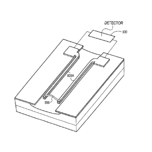
Figs. 5A and 5B illustrate views of cantilever based sensor 500. Fig. 5A
illustrates template molecule 545 bridging, or linking, the free ends of dual
cantilevers 505A. Cantilevers 505A are electrically coupled to detector
circuit
530 via conductors 550 and connection plates 535 disposed at stabilized ends
of
cantilevers 505A. Connection plates 535 are bonded to layer 510 which is
disposed atop layers 515 and layer 520. Layers 510, 515 and 520, in one
example, are comprised of Si3N4, Si, Si3N4 each having a thickness of
approximately 50 nm, 150 run and 250 nm, respectively. Cantilevers 505A are
suspended above sample channel 540 formed in layer 515. Cantilevers 505A, in
CA 02534632 2006-02-02
WO 2005/038459
PCT/US2004/025708
one example, are formed of layer 555 (gold) and layer 560 (chromium) having
thicknesses of approximately 40 urn and 5 urn, respectively.
In Fig. 5A, cantilevers 505A are bridged by a template molecule of
ssDNA 545 under little or no tensile forces. Detector circuit 530, in one
example, provides an electrical current to detect the level of conductivity.
It will
be appreciated that conductivity is the reciprocal of resistance and in one
example, a resistance is determined. In one example, an impedance value is
determined. In Fig. 5B, bridge 565 represents a hybridized dsDNA formed by
the combination of the template molecule (ssDNA) and the target molecule
(ssDNA). As illustrated in Fig. 5B, cantilevers 505A are convergently
deflected.
Detector circuit 530 generates a measurement of conductivity and upon
hybridization of the dsDNA strand, reflects a measured increase in
conductivity.
In various examples, a test circuit and a reset circuit are provided in
detector circuit 530. The test circuit is configured to provide a current to
template molecule 545 to establish that the template molecule is properly
affixed
to the cantilever arms. For example, a series combination of a current source,
sensor and a resistor will indicate an expected current flow if the sensor and
template molecule are properly configured. Deviations from an expected current
level may indicate that the template molecule or the sensor is not properly
configured for sample testing.
A reset circuit of the detector circuit includes a driving circuit for
disbonding the target molecule from the template molecule in preparation for
another detection and identification event. In one example, this entails
providing
a ramping voltage to the template molecule and monitoring for a peak current.
In one example, this entails providing a ramping current to the template
molecule and monitoring for a peak voltage. The peak voltage, or current, will
coincide with a denaturing or disassociating event of the template molecule
and
the target molecule.
Fig. 6 illustrates an example of current required to induce denaturation of
tethered DNA from a complementary strand. The reset circuit, or other means of
providing a denaturation current can remove the complementary strand from a
sensor site, thereby readying it for a new sensing event. In one example, a
11
CA 02534632 2006-02-02
WO 2005/038459
PCT/US2004/025708
sensor disposed in a flow stream can be used for successive and separate
sensing
events, thus, enabling continuous operation of the sensor.
, In the figure, denatration current is indicated on the ordinate and applied
voltage appears on the abscissa. The difference in current magnitude, as
illustrated, provides a means for discerning variations from a complementary
target molecule.
A high degree of proportionality is noted between the amount of current
required to force denaUiration and the degree of mismatch of the template and
target ssDNA strands regardless of whether the variation occurred on one
region
of the genetic sequence or was spread out over a number of different locations
along the sequence of the target.
Figs. 7A and 7B illustrate a manual process of increasing the voltage to
force denaturation followed by a reduction in the voltage back to sensing
levels
to allow another hybridization to occur. As illustrated in Fig. 7A, a number
of
sensing events are noted. Reset signals are illustrated in Fig. 7B, as
corresponding to those sensing events. A denaturation current provides a means
of resetting the sensor. The denaturation current appears consistent, both
during
the ssDNA and after hybridization (dsDNA) states.
In the figure, a complementary strand was introduced at approximately
15 seconds from the start of the data acquisition followed by an immediate
sensing event. After several seconds, the voltage was manually increased to
approximately 4 volts, resulting in denaturation of the double stranded DNA.
The voltage was manually reduced to 3 volts, and the system was thus reset for
another sensing event.
Resonance Example
Fig. 8 includes graphical data 800 illustrating how resonance can be used
to identify and detect a target molecule using a bridged template molecule.
In the figure, frequency is plotted on the abscissa and amplitude on the
ordinate. The cantilever structure, or other suspended structure is driven to
oscillate using an excitation signal. In various examples, the excitation
signal is
provided by a magnetic, piezoelectric or acoustical member disposed near the
movable structure. In the figure, curve 805 illustrates an example wherein
the
template molecule resonates at an initial frequency of F2 and with an initial
12
CA 02534632 2006-02-02
WO 2005/038459
PCT/US2004/025708
amplitude of A2. After exposing the template molecule to the target molecule,
the structure resonates with a frequency of F1 and with an amplitude of Al.
Frequency difference AF and amplitude difference AA are indicative of the
degree of homology and therefore, allow detection and identification of the
target molecule. For example, it is believed that a target molecule with a
higher
percentage of match with the template molecule will exhibit a greater change
in
either or both of the amplitude and the frequency.
The figure illustrates a reduction in both the amplitude and the frequency.
However, in other examples, either or both of the amplitude and frequency may
exhibit an increase or a decrease.
In one example, resonance of the mobile portion of the MEMS device
allows detection and identification. In one example, an end of the cantilever
includes a magnetic material and an alternating current passed through a coil
disposed under the cantilever causes the cantilever to vibrate at the
frequency of
the alternating current. In one example, the dimensions of the cantilever and
the
alternating current are selected to maximize the output. For example, if the
alternating current is near the natural frequency of the cantilever, the
response of
the structural system will be maximized. The stiffness of the structural
system is
changed when the ssDNA, or other template molecule, is tethered between the
end of the cantilever and the substrate base. Either or both the amplitude of
the
displacement of the oscillating system and the oscillation frequency will
differ
from that of the system with the free cantilever end. Upon introduction of a
target molecule (such as an analyte or the ssDNA complement to the template
ssDNA), the amplitude of the displacement and frequency of the oscillating
system will change. The amount of the change will be proportional to the
degree
of homology between the template and target molecules since the stiffness of a
dsDNA strand is greater than the sum of the stiffnesses of two independent
ssDNA. Thus, in addition to the detection of the presence of the analyte, the
change in amplitude and frequency can be used to measure the degree of
homology of the ssDNA molecules when compared to that of a complementary
match.
13
CA 02534632 2006-02-02
WO 2005/038459
PCT/US2004/025708
Exemplary Preparation
Fig. 9 illustrates a flow chart of method 900 according to one example.
In the figure, a cantilever is formed on a base at 905. Structures other than
a
cantilever are also contemplated, including, for example, a circular or
helical
structure having one supported end and a free end. In addition, a disc-shaped
or
rectangular structure is also contemplated with a movable center region and a
perimeter affixed to a base structure in the manner of a drum head. In one
example, semiconductor fabrication techniques are used for the formation of
the
cantilever on the base.
At 910, a bonding material, or primer, is applied to the cantilever and to
the base structure or substrate. The primer is selected to assure that the
template
molecule is affixed with proper alignment and orientation. In various
examples,
the primer includes gold and streptavidin.
At 915, the template molecule is bridged between the substrate and the
cantilever.
In one example, method 900 is performed by a manufacturer in preparing
a sensor for a particular application.
Exemplary Detection and Identification
Figs. 10 and 11 illustrate testing methods 1000 and method 1100,
respectively. The methods illustrated, as well as other methods, can be
implemented using a computer, or other control circuitry, coupled to a sensor.
In
one example, the method is executed using manual control of the sensor.
In Fig. 10, the sample is prepared at 1005. Sample preparation, in
various examples, entails filtration, purification, amplification and other
procedures to ready the sample for analysis.
At 1010, the template molecule is analyzed to establish one or more
parameters to serve as a baseline. In one example, this entails verifying that
the
template molecule is properly aligned and positioned by verifying a current
level
through the template molecule. In addition, the conductivity or resistivity,
resonant amplitude and frequency for the template molecule alone is measured.
In one example, the physical position of the template molecule is measured.
At 1015, the sample is exposed to the template. In one example, this
entails injecting a sample, which possibly includes the target molecule, into
a
14
CA 02534632 2006-02-02
WO 2005/038459 PCT/US2004/025708
channel or reservoir of the test apparatus. The channel or reservoir is in
communication with the template molecule.
At 1020, the template molecule is analyzed to generate a physical
parameter corresponding to the exposed template molecule. The exposed
parameter, in various examples, includes measuring a change in a position, or
displacement, measuring a change in alignment, measuring conductivity or
resistance, measuring a denaturing current and measuring changes in resonance.
=
Other physical parameters are also contemplated, including those based on a
color or optical property of the combination of the template and target
molecules.
At 1025, a query is presented to determine if a difference is noted
between the baseline and the parameter after exposure of the sensor to the
target
molecule. If a change in the physical parameter, or a difference, is noted,
then
processing continues at 1030 where the sample is identified. The existence of
a
difference is indicative of detection of the template molecule.
As noted elsewhere in this document, the degree of homology is
indicative of the match between the template molecule and the target molecule.
Other binding pairs are also contemplated and proximity to a complete match
can be correlated to the difference noted in the physical parameters.
In one example, a memory coupled to a processor of the present subject
matter includes stored data in the form of a look-up table. The stored data
provides a correlation between the differences or changes noted in a physical
parameter and the degree of homology.
If the query at 1025 yields a negative answer, then processing continues
to 1035 where an output signal is generated. The output signal, in various
embodiments, includes a measure of the difference or change noted, the degree
of homology or the identification of the target molecule.
At 1040, the template molecule is cleared of any remaining target
molecule or sample material, or reset, by applying an electrical excitation to
the
template molecule and inducing denaturation or disbonding.
In one example, following 1040, the method returns to 1005 for detection
and identification of an additional sample.
CA 02534632 2006-02-02
WO 2005/038459
PCT/US2004/025708
Fig. 11 illustrates method 1100 which includes a serial testing of a
sample. It will be appreciated that other orders of testing are also
contemplated
as well as parallel testing. For example, measurement of a change in resonant
frequency, resonant amplitude and conductivity can be performed concurrently.
In method 1100, the initial conditions, or baseline, established at 1105.
At 1110, the displacement of the sensor, due to the template molecule
hybridizing with the target molecule, is determined. At 1115, a shift in
resonance is determined. The shift may correspond to a change in the resonant
frequency or the resonant amplitude. At 1120, the conductivity of the template
molecule with target molecule is determined. At 1125, a disbonding current, or
heat level, is determined by monitoring for a peak signal.
Fig. 12 illustrates an array of cantilever sensors fabricated on substrate
1205 having common base 1210. The cantilevers, some of which are labeled
1240A, 1240B, 1240C and 1240D are affixed to base 1210 on one end and
tethered by template molecules to contact points 1245A, 1245B, 1245C and
1245D, respectively, on a surface of substrate 1205. The template molecules,
in
one example, are of identical composition and provide a level of redundancy
for
testing. In one example, at least two template molecules are different and are
tailored to detect and identify different target molecules.
Each cantilever, such as 1240A, is coupled to controller 1215 by
electrical conductors 1235A and 1235B using multiplexer 1220. Controller
1215 selectively applies testing current, voltage, drive signals or other
signals to
enable each cantilever to detect and identify a target molecule. Power source
1225, of controller 1215 provides a constant or ramped voltage or current for
excitation. In one example, power source 1225 provides a denaturing current or
voltage. In one example, power source 1225 provides a drive signal to excite
resonance in each cantilever.
Interface 1230 is coupled to controller 1215 and provides data entry and
data output. In various examples, interface 1230 includes a display, a touch
sensitive screen, a keyboard, a keypad, a mouse or other pointer control, an
audio transducer, a storage device, a printer, a network connection (for
example,
a wide area network such as the Internet, or a local area network such as an
intranet), an electrical connector or a wireless transceiver.
16
CA 02534632 2006-02-02
WO 2005/038459
PCT/US2004/025708
Fig. 13 illustrates exemplary portable device 1300 according to one
embodiment. In the figure, display 1305 provides visual prompts and data
corresponding to analysis of a target molecule and device condition. User
accessible controls and data entry points include power button 1310, reagent
cut
1315, sample input 1320 and controls 1325. Other controls and data entry
devices are also contemplated. In one example, a permeable surface on device
1300 allows a user to deliver a sample using a *inge or other injection
device.
In one example, a port on a surface of 1300 includes a reservoir to receive a
sample.
Device 1300 is illustrated as a portable, battery operated device,
however, other embodiments are also contemplated, including for example, a
desk-top unit with accommodations for receiving a sample and providing an
output.
Example
In addition to measurable changes in length or force, the capacity of
DNA structures to conduct an electrical current is related to content,
sequence,
length, and bridged circuit chemical environment. In one example, conductivity
is related to levels of guanine/cytosine.
In one example, a 121 base pair (bp) Bacillus genomic DNA sequence
was isolated from genomic, plasmid, and lambda viral DNA. Data indicates
consistent results using numerous variables with regard to DNA properties
(length, sequence composition) and analysis conditions (redox, pH, salt,
denaturant, and hybridization accelerant controls) and other DNA (single and
double stranded), molecules and genetic variants isolated from Bacillus as
well
as E. coli DNA.
The subject fragment was isolated from Bacillus genomic DNA by
restriction endonuclease digest and ligated into a pUC 13 plasmid cloning
vector
that was transformed into an E. coli host as the parental strain. A library of
random genetic point mutations were created along the length of the plasmid
insert and isolates with inserts that varied from the parental by 2%, 19%, and
35% were sequenced and used for further analysis. Both parental and variant
inserts were excised from the host plasmid and the 5' and 3' ends were
chemically modified with a thiol and biotin groups. Single strand DNA was
17
CA 02534632 2006-02-02
WO 2005/038459
PCT/US2004/025708
isolated using affinity chromatography (based upon the biotin modifications)
and
attached between streptavidin and gold coated conductive atomic force
microscopy (AFM) tips and stages. The AFM tip is initially undeflected. In one
example, the change in effective length of a single strand of DNA as it
hybridizes to its complementary strand is a reduction of 40%.
AFM tip displacement was observed within seconds of introducing the
DNA complement (in hybridization solution) or genetic variants to the tethered
DNA template. The experimental results indicated a consistent tip displacement
(<0.5% COY) as a function of the degree of mismatch between template and
complement. It is postulated that base-pair mismatches did not contribute to
overall structure helical formation, thus reducing the reduction of molecule
length.
Electrical conductivity was determined utilizing conductive AFM tips.
The same 121 bp fragment was tethered between AFM tip and stage and the
DNA complement was introduced. Electrical current (in nanoamps) was
measured as a function of time at a fixed voltage.
Prior to the introduction of the complementary DNA, the applied voltage
resulted in a consistent, or baseline, current (of approximately 0.3 nA)
passing
through the tethered ssDNA. Treatment of the tethered ssDNA with DNA
nuclease resulted in a loss of this baseline current. Heat treated nuclease
did not
result in a loss of current.
Measurable current through a sensor provides a verification of sensor
function (sensor self-test) since electric current will flow if the ssDNA
remains
tethered to the MEMS mobile elements. Following hybridization between the
tethered ssDNA and its complementary strand, an increase in current appears as
noted in the figures. hi addition, the figures illustrate a relationship
between
measured current and the degree of match between the strands. After
hybridization, the conducted current remains relatively consistent as long as
the
voltage is applied to the sensor site.
After hybridization, the applied voltage was increased and the conducted
current correspondingly increased to a peak level. Denaturation of the
tethered
ssDNA and the complementary strand occurred at a potential of approximately
4.1 volts for this particular 121 bp fragment. The amount of current
associated
18
CA 02534632 2015-09-25
with denaturation varied with the degree of match between the tethered ssDNA
and the complementary strand. Concurrent with the drop in current, the AFM tip
returned to an undeflected position. It is postulated that the higher level of
current infuses sufficient energy into the hybridized strands such that they
can no
longer remain hybridized although other mechanisms or factors may be
responsible for this phenomena. The base pair mismatches present in the
variants appear to introduce insulating locations along the strands, thereby
proportionately reducing conductance.
In one example, the selection of a template molecule, such as particular
DNA, to bridge a circuit or cantilever structure affects DNA specificity,
length,
sequence, composition and conductivity. In one example, detection and
identification specificity is enhanced by selecting ssDNA from multiple
genetic
regions of a pathogen for tethering. In one example, a longer DNA segment
provides increased specificity (DNA sequence that is unique to the specific
biological agent target). In one example, a shorter DNA segment allows
selection of regions of high guanine (G) and cytosine (C) content. GC overall
composition and sequence provides insulating characteristics of adenine (A)
and
thymine (T). High AT content in DNA leads to inflexibility and overall
molecular curvature depending upon the relative positioning of AT-rich regions
(i.e. in phase with the helical rotation). In various embodiments, selected
DNA
segments were greater than 40% GC, or greater than 60% GC. DNA segment
length was therefore less than 500 base-pair (bp), or less than 150-200 base
pair
(bp). Determination of DNA GC composition, sequence, and specificity was
determined using commercially and publicly available software such as PubMed
BLAST. Other software is available to correlate first
order molecular parameters to higher order features (i.e. flexibility,
curvature).
In one example, the template molecule is selected using a software algorithm.
Additional Examples
In one example, the sensor includes a suspended member which includes
a cantilever. The template molecule is affixed to a contact point, at least
one of
which is located on the suspended member. The cantilever, in various examples,
is curved, circular or web shaped. In one example, the suspended member is a
rotary member that turns about an axis. As a rotary member, the contact point
is
19
CA 02534632 2006-02-02
WO 2005/038459
PCT/US2004/025708
displaced along an arc when the template molecule binds to the target
molecule.
In one example, one end of the rotary member rotates while another remains
stationary or rotates in an opposite direction or through a smaller range and
the
phase difference between the two ends of the rotary member provides a
difference signal that is used to discern the target molecule.
In one example, the template molecule has more than one binding site
specific to a target molecule. In one example, the target molecule has
multiple
binding sites, each of which is specific to a different target molecule. In
one
example, the target molecule has multiple binding sites, each of which is
specific
to a single target molecule.
In one example, multiple contact points are provided on a sensor and the
template molecule binds to two or more of multiple contact points. For
example,
a double-ended template molecule can bind to a sensor having two, three or
more contact points. As another example, a three-ended template molecule can
bind to a sensor having two, three, four or more contact points.
In one example, an output signal is generated as a function of a change in
a measure of a physical parameter. Physical parameters include structural as
well as electrical parameters. Exemplary structural parameters include
positional changes such as physical displacement, resonant frequency, resonant
amplitude, physical alignment or orientation of a contact point and a
reference
point, a force exerted on an axis, heat generated and optical changes
including
color. Other physical parameters are also contemplated.
In one example, an output signal is generated as a function of a change in
a measure of an electrical parameter. Exemplary electrical parameters include
impedance, conductivity, resistivity, inductance, capacitance. In addition, an
electrical parameter can be described as an output signal in the presence of
an
input signal. For example, a change in current conducted in a template
molecule
can result in a change in voltage. In addition, a change in voltage applied to
the
template molecule can result in a change in a current. Other driving signals
can
also be applied and measured responses can be used to generate an output
signal.
Other electrical parameters are also contemplated.
In one example, a physical parameter includes a measurement of
electrical conductivity. Electrical conductivity is a measure of the flow of
CA 02534632 2006-02-02
WO 2005/038459
PCT/US2004/025708
electrons in a material. Electrical conductivity is the reciprocal of
resistivity, or
resistance, and in one example, the monitored physical parameter includes
resistivity.
In one example, a ssDNA template molecule, bound via oligonucleotides
primers, bridges an electronic MEMS based circuit. Hybridization to a target
molecule (such as DNA) is derived from the microorganism being identified and
causes a reduction in the distance between the cantilever elements.
In various embodiments, a comparator or Wheatstone bridge is used to
detect, identify and compare voltage levels, current levels, conductivity or
other
parameters.
In one example, denaturing of the template molecule is performed by
applying heat to the template and target molecules. A level of heat is
quantified
by measuring a current, voltage or wattage. In one example, a difference in
the
level of heat is correlated to the identity of the target molecule.
In one example, a single sensor site includes a tethered, single strand
DNA (ssDNA) bridging a mobile element on a micro electromechanical system
(MEMS) chip. In one example, hundreds or thousands of such sites are placed
on a single chip. The tethered ssDNA is selected to hybridize with a
complementary strand extracted from a bioagent of interest. The resulting
hybridization both changes the physical length of the tethered molecule, and
changes the conductivity of the tethered molecule. The changes are measured in
a MEMS system at a high signal-to-noise ratio. The degree of change is related
to the degree of match between the tethered and bioagent DNA strands. Thus,
the degree of variance (specificity) of the bioagent can be measured. After
detection, identification and discrimination are confirmed, the bioagent DNA
strand is expelled from the tethered strand by increasing the current flow
through
the molecule, thus resetting the sensor for subsequent sensing events. Sensor
, viability is verified through a self-test since a tethered ssDNA (absent
its
complement) is able to conduct a measurable amount of current.
The physical parameter changes are proportional to the fidelity of match
between the template molecule and the target molecule (or DNA strands) A
single nucleotide mismatch yields a measurable change, thus enabling
21
CA 02534632 2006-02-02
WO 2005/038459
PCT/US2004/025708
identification of various pathogens and differentiation of subtle variations
between specific strains.
In one example, the template molecule includes ssDNA. In one example,
specific DNA regions of B. anthracis are propagated and functionalized. The
sensor can be bridged by DNA from any biological agent (bacteria, virus, or
fungi).
In one example, four (4) 150-200 base pair (bp) segments of Bacillus
anthracis (Ames) are selected for use as DNA bridge templates. In one example,
for the purpose of testing the systems ability to discriminate between
strains,
alternative sequences to one of the templates was designed. The variants
differ
from the parent molecule by random nucleotide substitutions to generate 2%,
10%, and 20% variants. Template molecule selection is based upon calculated
specificity, conductivity parameters, and flexibility. Species and strain
specific
segments were chosen from 16S rRNA fingerprint and virulence genes.
In one example, the four selected 150-200 bp templates and variants were
synthesized through commercially available DNA synthesis facilities. The 150-
200 bp DNA templates and variants were synthesized in ¨50 bp ssDNA
fragments. Hybridization and ligation steps were used to create full length
150-
200 bp templates. The templates were ligated into an appropriate plasmid
cloning vector and a library of the DNA bridge templates were generated in
preparation of large-scale plasmid production. Optionally, the selected 150-
200
bp template candidates are excised by restriction endonuclease digests or PCR
amplified and subcloned from Bacillus anthracis (Ames) DNA.
In one example, DNA bridge templates were covalently attached to AFM
and MEMS surfaces via biotin-streptavidin and thiol-gold bonds. The plasmid
borne templates were restriction endonuclease excised, and 5-prime/3-prime
biotin/thiol end-labeled with commercially available kits. In one example, the
templates were PCR amplified using biotin and thiol labeled primers.
In one example, DNA templates and variants were verified for sequence
integrity through commercially available subcontracted DNA sequencing
services. The specificity of the each of the molecules was verified through
standard Southern screening against genomic DNA of Bacillus anthracis strains
(i.e. Ames, Sterne, A2012, 1055, Vollum, Kruger) and anthrax simulants (i.e.
B.
22
CA 02534632 2006-02-02
WO 2005/038459
PCT/US2004/025708
globigii, B. cereus, B. subtilis, B. thuringiensis) purchased commercially
from
the American Type Culture Collection or acquired through a material transfer
agreement or other collaborators.
In one example, atomic force microscopy (AFM) was used to measure
specific physical properties (i.e. displacement and conductivity) of the B.
anthracis and variant ssDNA fragments. AFM tips and stages were coated with
gold and streptavidin and the thiol/Biotin end labeled DNA bridge templates
were attached. AFM tip displacement and material electrical properties were
measured prior to, during, and after hybridization with complementary and
variant ssDNA molecules and used as input into MEMS device design. In one
example, reagents to control hybridization (pH buffers, salts), denaturation,
hydrolysis and nucleotide oxidation were selected.
In one example, MEMS fabrication techniques are used for the
construction of the sensor chips. In one example, fabrication entails
deposition
of thin films of material onto a substrate, application of a patterned mask
onto
the material using photolithographic methods and selective etching of the film
using the resulting developed mask. Deposition of the material onto the
substrate (silicon wafers) is accomplished by chemical reaction-based
approaches (chemical vapor deposition, epitaxy, electrodeposition, or thermal
oxidation) or by physical reaction-based approaches (evaporation, sputtering
or
casting). Removal of materials is accomplished through etching techniques.
Thus, the circuitry for the device, using the application of patterned
photolithographic masks, is constructed using appropriate application of
insulating and conducting material layers. The fabrication facility
incorporates
the chip into packaging which, in one example, includes a ceramic or plastic
housing for the chip that includes the pinned interface for attachment to a
printed
circuit board (PCB).
In one example, upon fabrication of the MEMS chips, the DNA bridge
templates are generated and tested. In one example, the MEMS mobile elements
(cantilever end) includes thiol/gold and biotin/streptavidin covalent bonds.
Single molecule attachment was accomplished by electrostatic attraction. In
one
example, the device applies a 5 volt electrical potential across the gap
between
the mobile MEMS elements where the tethered ssDNA is desired, in series with
23
CA 02534632 2006-02-02
WO 2005/038459
PCT/US2004/025708
a 10 MO resistor. The ssDNA molecule is attracted to the resulting electrical
field. When in close proximity, the biotin-streptavidin bonds on the substrate
base is formed, and the gold-thiol bonds is formed on the free end of the
cantilever. When one molecule attaches in this manner, the potential across
the
gap is reduced due to the resistance in series in the circuit. Thus, a single
ssDNA molecule attaches at each site. Excess DNA that does not bridge across
the MEMS circuits is removed by DNA exonuclease digests. The circuit is
stored in DNA stabilizing buffers (i.e., 300 mM NaC1, 10 mM Na citrate and 5
mM EDTA).
In one example, current measurement in the nano-amp range is
performed using integrated circuit amplifiers. A multiplexing integrated
circuit
(IC) amplifier and other electronics and processing are used to display the
results
of the sensing events. Analog signals taken from the MEMS chip are amplified
and converted into digital signals.
In one example, a printed circuit board includes accommodations for the
attachment of the MEMS chip and the IC chip. The board also includes a
dedicated main processing chip used to perform calculations and control
electrical operations on the PCB. The PCB contains electrical interfaces for
the
display and the battery, as well as the menu buttons. In one example, a
program
executed by the main processor uses the digital signals output from the IC to
provide display. The user interface includes controls to display and to set
parameters that determine the display characteristics, the threshold detection
values, battery level, on/off and pathogen molecule purging control.
In one example, a plastic housing contains the printed circuit board (and
attached bio- and IC-chips), sample flow paths, LCD display and control
buttons. In one example, an interior walls of the housing contain ledges and
slots to contain electronic components and preclude shifting inside the
housing.
In one example, the housing includes one or more flow paths for introduction
and removal of the reagents and sample.
In one example, electrical conduction through the sensor sites is greater
than 0.2 nA using AFM electronics. In one example, the integrated circuit
provides analog signal amplification >10 mV peak-to-peak.
24
CA 02534632 2006-02-02
WO 2005/038459
PCT/US2004/025708
In one example, the sensor is configured to detect and identify prepared
genomic DNA of Bacillus anthracis strains and anthrax simulants (i.e. B.
globigii, B. cereus, B. subtilis, B. thuringiensis), chemical and/or
mechanically
(sonicated) disrupted inactivated whole cell and spores of the previously
mentioned anthrax strains and simulants, DNA and whole cells/spores in the
presence of common Contaminants and interferants such as postal dust, soil
components, other chemical mixes, and mixed consortia of microorganisms.
In one example, the system includes signal amplification, processing and
display sufficient to detect and identify a target molecule. In one example,
electronic controls include on/off, mode, display control, battery life, self-
test
and reset functions. In one example, the system includes one or more flow
passages to deliver a prepared sample to the surface of a sensor configured to
identify a single predetermined pathogen, simulants or DNA target variants in
a
disruption solution.
In one example, the sample is collected outside of the system using
surface wipe or batch air collector.
The present system includes a method and apparatus for determining the
presence, the identity or quantity of a target substance comprising biological
or
chemical analytes. In one example, an electronic circuit including at least
one
deflectable arm of a bio-electronic cantilever is surface treated to
facilitate
binding to a template polymer molecule that undergoes a measurable change in a
physical parameter in response to environmental changes, such as the presence
of a target molecule associated with a target substance to be detected. For
example, a change in the physical configuration or dimensions of the template
molecule is translated into a deflection of a cantilever arm. In one example,
a
change in the electrical characteristics across the template molecule is
detected
by hybridizing a single-stranded DNA bridge template to its complimentary
strand. Such changes in physical properties or parameters are measured to
provide information related to the presence and identity of a substance of
interest. In one example, a number of such circuits bridged by similar
template
molecules are provided and information related to the concentration or
quantity
of the target substance can be obtained.
CA 02534632 2006-02-02
WO 2005/038459
PCT/US2004/025708
In one example, a microcircuit with geometry tailored for use within a
biological agent detection device is provided for detecting the presence of
target
and related biological agents or substances.
As used herein, a template molecule may be any molecule that will bind,
or is likely to respond, to the presence or bind to a biological agent or a
component of a biological agent. Accordingly, a template molecule may
comprise a naturally occurring or synthetically formed biological molecule
that
will, or is capable of, selectively responding to, or binding to, a target
molecule
, associated with the target substance to be detected. In one example, the
template
molecule includes an antibody, protein, nucleic acid, carbohydrate,
glycoprotein
or a polymer. In one example, the specific template molecule selected for
detecting the presence of a particular target molecule (or species or genus of
target molecule) is selected such that the template molecule responds to, or
binds
only with the exact target molecule or a related target molecule. In one
example,
a differential electric current or physical displacement results from a
biomolecule biomolecule recognition between the template and target.
Accordingly, the relationship between a template molecule and a target
molecule
may be that of complementary strands of nucleic acids, including ribonucleic
acids (RNA) and deoxyribonucleic acids (DNA) and derivative molecules.
Further examples of the relationship between the template molecule and the
target molecule include nucleic acid ¨ nucleic acid recognition, protein ¨
protein
recognition, protein ¨ nucleic acid recognition, protein ¨ carbohydrate
recognition, nucleic acid ¨ carbohydrate recognition, and carbohydrate ¨
carbohydrate recognition.
In one example, a biodetection device containing the described circuit
including a template molecule spanning a gap between two surfaces, is
provided.
In one example, the two surfaces are movable relative to one another. When the
template molecule is exposed to, or bound to, a target molecule, the template
molecule undergoes a dimensional change, altering the distance between the two
surfaces. Accordingly, a biodetection device in accordance with such an
embodiment of the present invention can signal the presence of a target or
related biological agent or substance when a change in the distance between
the
two surfaces is detected.
26
CA 02534632 2006-02-02
WO 2005/038459
PCT/US2004/025708
In one example, the amount by which the distance between the surfaces
is altered is indicative of the molecule exposed to, or bound to, the template
molecule. For instance, a target molecule that is an exact match for the
template
molecule (i.e., an "exact target molecule") may result in shortening the
distance
between the points of the template molecule interconnected to the two surfaces
by an amount that is greater than the shortening that occurs when the template
molecule is bound to a molecule that is related to but not an exact match for
the
template molecule. Accordingly, by measuring the amount by which the
distance between the two surfaces has changed, information related to the
identity of the molecule bound to the template molecule is obtained.
In one example, a biodetection device containing the described circuit
capable of measuring the conductivity across a template molecule is provided.
In particular, a template molecule is interconnected to first and second
electrodes, such that it spans the gap between the two electrodes. When the
template molecule is bound to a target molecule, the conductivity between the
electrodes is altered. Accordingly, by detecting a change in the conductivity
between the electrodes, the presence of a target molecule or related molecule
can
be detected. Furthermore, the amount by which the conductivity between the
electrodes changes is indicative of the molecule bound to the template
molecule.
For example, an exact target molecule will cause a greater change in the
observed conductivity between the electrodes than will a target molecule bound
to the template molecule that is related but not identical to the exact target
molecule.
In accordance with an embodiment of the present invention, a detection
device that can be reused, without requiring the replacement of components, is
provided. In particular, by heating the template molecule, the target or
related
molecule can be unbound from the template molecule. In accordance with an
embodiment of the present invention, heating of the template molecule is
accomplished by passing a current across the template molecule (and the bound
molecule). Furthermore, the process of unbinding the target molecule from the
template molecule can be used to obtain information related to the identity of
the
target molecule. In particular, the current applied across the template
molecule
(and target molecule) may be steadily increased or increased in steps, until a
27
CA 02534632 2006-02-02
WO 2005/038459
PCT/US2004/025708
sudden change in the conductivity is observed, which indicates that the target
molecule has been dissociated from the template molecule. Because the current,
and therefore heat, necessary to unbind the target molecule is related to how
closely matched the target molecule is to the template molecule, the amount of
current required to unbind the target molecule is an indication of the
closeness of
the match between the bound molecule and the target molecule. For example, an
exact target molecule would be expected to require more energy to unbind it
from the template molecule than would a molecule that is not identical to the
target molecule.
In one example, a biological agent detection device containing the
described circuit combining a number of detection mechanisms or techniques is
provided. For example, a detection device may determine the presence of a
target biological agent by detecting a dimensional change experienced by a
template molecule, by detecting a change in the conductivity across a template
molecule, or by determining the amount of current required to unbind a target
molecule from the template molecule. Furthermore, information for identifying
the target molecule may be provided using such mechanisms or techniques.
In accordance with an embodiment of the present invention, a method for
detecting target substances or analytes by detecting a change in a physical
dimension associated with a template molecule is provided. According to such a
method, a template molecule that undergoes a change in physical dimension
when bound to a target molecule is exposed to a suspected biological agent or
target substance (i.e., a substance suspected of containing a target
molecule).
The suspected biological agent may be derived from a gaseous, liquid, or solid
medium. If the exact target molecule or a related molecule binds to the
template
molecule, the resulting dimensional change in the template molecule is
detected,
and the change reported. In accordance with a further embodiment, the method
includes measuring the amount by which the physical dimension of the template
molecule has changed.
In one example, a method for detecting a target substance by sensing a
change in the conductivity associated with the template molecule in the
presence
of the analyte is provided. A template molecule capable of selectively binding
to
an exact target molecule or related target molecule is exposed to a suspected
28
CA 02534632 2006-02-02
WO 2005/038459
PCT/US2004/025708
biological agent. According to the method, the conductivity across the
template
molecule is monitored. Upon becoming bound to a target molecule, the resulting
change in the conductivity across the template molecule is detected, and that
change is reported. In one example, the change in conductivity is measured.
In one examplp, a method for detecting the presence of a suspected
biological agent by determining the amount of energy required to unbind a
target
molecule from a template molecule is provided. An electrical current is passed
across the template molecule ¨ target molecule pair. Furthermore, the
amplitude
of the current may be increased, until a sudden change in the conductivity
across
the template molecule is observed, indicating that the target molecule has
become unbound from the template molecule. Furthermore, the current at which
the target molecule is unbound from the template molecule is used to
characterize or identify the target molecule that was bound to the template
molecule.
The present system relates to the detection and identification of
biological analytes. According to the present invention, target biological
molecules are detected by sensing a change in a template biological molecule.
The change in the template biological Molecule may include a change in a
physical dimension of the template molecule, a change in the electrical
conductivity observed across the template molecule, and/or the energy required
to dissociate a target molecule from the template molecule. The magnitude of
the change in a physical dimension, change in conductivity, or amount of
energy
required to dissociate a target molecule from a template molecule, may be
measured to determine the degree of homology between the target molecule and
the template molecule. In a further aspect, the present invention provides a
detection method and apparatus that does not require the replacement of
components in order to make multiple readings.
In one example, an electronic circuit is bridged by a template molecule
including a biological component or a representation of the biological
component. In one example, the circuit includes a MEMS-based structure
bridged by a nucleic acid molecule, such as a DNA molecule, or a molecule that
physically and chemically represents a single strand DNA molecule. In one
example, the MEMS circuit senses and responds to the motion and conductivity
29
CA 02534632 2015-09-25
of a bridged DNA molecule as it hybridizes with its complimentary, or near
complimentary DNA strand.
The following describes the selection and design of biological
components of the bio-electronic circuit.
As used herein, biological detection and identification device specificity
refers to the ability of the system to specifically and accurately identify a
particular genus, species and strain of target biological agent. In the case
of
DNA-based biological detectors/identifiers, the term specificity sometimes
refers
to the ability of the DNA components of the system to specifically compliment
and hybridize to DNA isolated from the biological agent. In order to enhance
detection and identification specificity, here, multiple (in one example, more
than three), biological agent DNA segments are selected. The DNA segment
selection is based upon the calculated length, specificity, conductivity
parameters and flexibility of the molecule to bridge the MEMS circuit. Longer
DNA segments tend to retain greater specificity (DNA sequence that is unique
to
the specific biological agent target), and yet shorter DNA segments allow
selection of regions of high guanine (G) and cytosine (C) content. GC overall
composition and sequence is related to the insulating characteristics of
adenine
(A) and thymine (T). In addition, high AT content in DNA leads to
inflexibility
and overall molecular curvature, depending upon the relative positioning of AT-
rich regions (i.e. in phase with the helical rotation). Thus, selected DNA
segments may be greater than 40% GC, or greater than 60% GC. In one
example, DNA segment length is less than 500 base-pair (bp), or less than 1.50-
200 base pair (bp). Determination of DNA GC composition, sequence,
flexibility, curvature, and specificity is determined through a number of
privately, commercially, and publicly available software such as PubMed
BLAST. In one example, four DNA template
segments are 100% homologous to Bacillus anthracis (Ames) and show lesser
homology to other Bacillus species and strains. Species and strain specific
segments have been chosen from 16S rRNA fingerprint and virulence genes. In
this embodiment, microorganisms outside of the Bacillus genus fall below
accurate detection and identification thresholds. In one example, a matrix
includes DNA-bridged MEMS having DNA components that have specificity to
CA 02534632 2006-02-02
WO 2005/038459
PCT/US2004/025708
other biological agents, and, in one example, is able to continuously monitor
for
the presence of agents simultaneously.
The following describes the production of the biological components of
the bio-electronic circuit.
In one example, specific DNA regions of the targeted biological agent
are selected, generated, mass produced and chemically modified for the sake of
adherence to a MEMS circuit. In addition, variants of the DNA regions are also
generated and produced for the purpose of testing the proposed circuit for
discrimination capabilities. In one example, a variant is a DNA molecule that
differs from the selected DNA template in nucleotide composition and sequence
by 2% to 30%. In one example, four selected 150-200 bp segments are either
synthesized, PCR amplified, or sub-cloned from an actual targeted biological
agent. High fidelity DNA synthesis is generally limited to ¨50 bp ssDNA
fragments that then require hybridization and ligation steps in order to
create the
desired full length 150-200 bp DNA templates. The completed DNA templates
are then attached directly to MEMS lead surfaces if the 5-prime and 3-prime
¨50
bp synthesized fragments were specifically labeled with attachment ligands.
Alternatively the completed 150-200 bp templates are ligated into a plasmid
cloning vector and a library of the DNA bridge templates is generated in
preparation for mass production. In one example, the selected DNA regions
chosen to bridge the MEMS circuit leads may be excised by restriction
endonuclease digests or PCR amplified, and sub-cloned from the targeted
biological agent.
The following describes verification of biological component integrity.
In one example, DNA circuit bridge templates and variants are verified
for sequence integrity. Sequence integrity refers to the actual nucleotide
sequence as compared to the desired sequence. DNA sequencing methods will
reveal the exact nucleotide sequence of the molecules intended to be labeled
and
attached to the MEMS lead surfaces. The present subject matter is sensitive to
single base pair mismatches, thus, any sequence variation should be accounted
for.
The following describes testing and analysis of biochemical and physical
features of the biological component.
31
CA 02534632 2006-02-02
WO 2005/038459
PCT/US2004/025708
In one example, specific ssDNA molecules are selected, mass produced,
and used to bridge the mobile elements of a MEMS circuit. The selection of the
specific ssDNA molecules is based upon a number of biophysical features such
as length, nucleotide composition and sequence, flexibility and mechanical
motion, and conductivity parameters. The calculated motion and conductivity
parameters are verified by atomic force microscopy (AFM) which can measure
specific physical properties (i.e. molecular motion as determined by tip
displacement and conductivity) of the ssDNA bridge template and variant
ssDNA fragments. AFM tips and stages coated with gold and streptavidin
according to published procedures are bridged by thiol/Biotin end labeled DNA
bridge templates. AFM tip displacement and material electrical properties are
measured prior to, during and subsequent to hybridization with complimentary
and variant ssDNA molecules as input into MEMS device design.
Operational, chemical and temperature environments can be considered
for specific ssDNA bridge templates. For example, reagents to control the
effects of the user-defined operational environment, operational temperature,
and
DNA specific hybridization (pH buffers, salts), denaturation, hydrolysis and
nucleotide oxidation can have an effect. In one example, operational reagents
include:
a. Salts, pH, temperature
b. Hydrolysis control: Conductivity through aqueous environments may
induce hydrolysis that could affect conductivity through the media. Control of
this effect through the addition of appropriate reagents.
c. Oxidation control: Conductivity through DNA may induce oxidative
damage particularly to the guanine residues. Control of this oxidative damage
through the addition of antioxidants (i.e ascorbic acid, citric acid).
d. DNA thermal stability factors (i.e. 0.5 ¨ 3 molar betains (N,N,N,-
trim.ethylglycine; (Rees et al., Biochem., (1993) 32:137-144).
e. Denaturing reagents (i.e. 2-4 molar tetraethyl acetate, urea, chaotropic
salts (i.e. trichloroacetate, perchlorate, thiocyanates and fluoroacetates),
or
glycerol, formamide, formaldehyde, and dimethylsulfoxide (DMSO).
f. Hybridization accelerants to enhance DNA to DNA hybridization
through molecular exclusion phenomenon. Exemplary accelerants include
32
CA 02534632 2006-02-02
WO 2005/038459
PCT/US2004/025708
mixtures of acetate salts and alcohols, certain amines (spermine, spermidine,
polylysine) 0.1 ¨ 0.5 molar detergents (dodecyl trimethylammonium bromide,
and cetyltrimethylammonium bromide) and specific small proteins such as single
stranded binding protein.
The following describes verification of DNA specificity.
In one example, DNA circuit bridge templates and variants are verified
for sequence specificity. Software analysis of the selected DNA fragment may
demonstrate the specificity of the fragment for one region of one strain of a
targeted biological agent which may be confirmed through bioagent specificity
screening. An example of these methods includes Southern screening in
which various restriction digested fragments of the bioagent target genome
(and
any other suspected related species) are electrophoretically resolved and
transferred to a solid substrate (i.e. nylon or nitrocellulose). The fixed
genomic
DNA fragments may then be incubated with labeled (i.e. fluorescent or
radioactive) DNA bridge template DNA. Under appropriate conditions (i.e.
temperature, pH, and salt concentration) the template DNA will hybridize to a
single fragment (assuming the genomic DNA restriction digests did not cut the
fragment). Multiple sites of DNA template hybridization to the genomic DNA ,
may entail modification of conditions in the biodetection device or selection
of a
new template DNA.
The following describes DNA bridge template end labeling.
In one example, synthesized, PCR amplified, or cloned DNA fragments
(selected to bridge MEMS circuit leads) are attached to MEMS surfaces by any
of the various methods concerning DNA attachment to organic or inorganic
surfaces. In one example, orientation-specific immobilization is achieved when
unique chemical moieties on the DNA bridge template termini and MEMS lead
surface are cross linked. Commercially available chemical moiety-specific
crosslinkers are generally based on nucleophilic substitution chemistry. This
chemistry generally involves a direct displacement of a leaving group by an
attacking nucleophile. In one example, the MEMS circuit leads include leads
coated with gold and streptavidin respectively. In one embodiment, ssDNA
bridge templates are covalently attached to AFM and MEMS surfaces via biotin-
33
CA 02534632 2006-02-02
WO 2005/038459
PCT/US2004/025708
streptavidin and thiol-gold bonds. The DNA fragments are 5-prime/3-prime
biotin/thiol end-labeled with commercially available kits or by any other
means
of labeling or functionalizing the 5' and 3' ends of DNA. Attachment
chemistries can include, but is not limited to amino groups (such as N-hydroxy-
succinimidyl esters), polyethylene glycols, carbodiimide, thiol groups (such
as
maleimide or a-haloacetyl), organo-silane groups, or biotin-streptavidin. In
one
example, DNA fragments are synthesized with 5' and 3' biotin or streptavidin
modified nucleotides, or PCR amplified with biotin/streptavidin labeled
primers
from genomic or plasmid borne DNA targets.
The following describes an example of MEMS fabrication.
In one example, microelectromechanical systems (MEMS) refers to
technology utilizing small mobile structures constructed on the millionth of a
meter (micron) scale. These structures are made through the use of a number of
tools and methodologies, similar to that used in the fabrication of integrated
circuits (IC). MEMS devices, in one example, include combinations of
mechanical elements and electrical elements, and, upon fabrication, are placed
in
a pinned packaging that allows attachment through a socket on a printed
circuit
board (PCB).
The following describes MEMS layered construction.
In general, fabrication of a MEMS device involves the deposition of thin
films of material onto a substrate, the application of a patterned mask onto
the
material using photolithographic methods and the selective etching of the film
using the resulting developed mask. Deposition of the material onto the
substrate (usually silicon wafers) can be accomplished by chemical reaction-
based approaches (chemical vapor deposition, epitaxy, electrodeposition, or
thermal oxidation) or by physical reaction-based approaches (evaporation,
sputtering or casting). Each of which varies in speed, accuracy and process
cost;
the applied material can be from a few nanometers to about 100 microns.
Application of the pattern involves placing a photosensitive material on the
surface, locating the patterned mask over the surface (typically with the aid
of
alignment marks on the surface and mask), and exposing the photosensitive
material through the mask. Depending on the process used, either the positive
or
the negative of the exposed material can be removed, leaving the pattern on
the
34
CA 02534632 2006-02-02
WO 2005/038459
PCT/US2004/025708
substrate material. Other procedures includes preparing the surface,
developing
the photosensitive film and cleaning the result.
MEMS element manufacture may be performed using micro fabrication
techniques. In one example, lithographic techniques are employed in
fabrication
using semi-conductor manufacturing procedures, such as photolithographic
etching, plasma etching or wet chemical etching, on glass, quartz or silicon
substrates.
The removal of materials is typically accomplished through wet etching,
in which the material is dissolved away when immersed in a chemical solution,
or dry etching, in which material is removed in a process essentially opposite
physical reaction-based deposition. As with the various approaches to
deposition, speed, accuracy and cost vary with the approach. In one example,
etching of "deep" pockets from a substrate is performed with side wall aspect
ratios at 50 to 1.
The MEMS device can be fabricated with one or more mobile elements,
across which will be attached the template molecule of ssDNA. The mobile
element, for example a cantilever beam, can be constructed so that both the
beam
and the substrate beneath the free end of the beam contain at least one
conductive layer. Thus, the circuitry for the device, using patterned
photolithographic masks, can be constructed using appropriate application of
insulating and conducting material layers. In one example, the geometry allows
for flow of sample from one DNA bridged MEMS to the next and re-circulating
to enhance probability of contact and to conserve reagents. One embodiment
includes an electronic circuit constructed on a support composed of such
materials such as, but not limited to, glass, quartz, silicon, and various
polymeric
substrates, e.g. plastics.
In one example, various insulating layers are provided on the substrate.
In one example, solid material amenable to supporting and responding to the
described molecular properties (i.e. conductivity and motion) are used to
130 construct the device. Although the figures in this disclosure may
depict a flat
positioning of the circuit, other embodiments include other orientation (i.e.
vertical, etc.).
CA 02534632 2006-02-02
WO 2005/038459
PCT/US2004/025708
One example includes additional planar elements(s) which overlay the
channels and reservoirs to enclose and seal to form conduits. This additional
planar surface is attached by adhesives, thermal bonding, or natural adhesion
in
the presence of certain charged or hydrophilic substrates.
In one example, sample collection and preparation is completed outside
the device. Samples are collected from surfaces using swabs or pads or
collected
in air or liquid by aspiration through filters, liquid traps or chromatography
resins. Those samples are prepared by the addition of the reagents to disrupt
the
biological agent, release the target molecules and prepare those molecules for
sensing by the device. The prepared sample are then be introduced into the
device through a flow channel, reservoir or duct by syringe, pipette,
eyedropper
or other such manual or automated means.
One example includes air sample acquisition and preparation into the
device itself for automatic operation through the presence of an on-board fan
aspiration system. One example includes automated liquid sample collection
and preparation into the device. Disruption of the samples can be provided by
mechanical means, such as sonication techniques.
In one example, the sample is delivered through a flow path incorporated
into the device to the surface of the biochip. The device includes reservoirs
of
reagents for sample preparation, as well as for system flushing, device
calibration and waste material collection. In one example, the device includes
means of pumping materials to and from these reservoirs. In one example, the
device recirculates reagents through the system if no positive sensing event
has
occurred and the reagent remains of suitable purity.
In one example, oligonucleotide sequences are layered upon the leads to
aide in the positioning and orientation of DNA bridge templates. In general,
the
oligonucleotide directed hybridization of DNA across a circuit is used to
orient
and position the single stranded DNA (ssDNA) bridge template. In one
example, the ssDNA bridge template is bound to the MEMS leads via any
method of attachment, including thiol or biotin mediated bonds.
Multiple DNA bridged MEMS circuits can be positioned on a single chip
in a matrix or array geometry in order to enable simultaneous detection and
identification of multiple pathogens from the same sample. In one example, the
36
CA 02534632 2006-02-02
WO 2005/038459
PCT/US2004/025708
matrix includes multiple repeats of identical DNA bridged MEMS in order to
enable concentration measurement of target DNA molecules as a function of the
number of 'stimulated' circuits per volume of sample.
The following describes biological bridging of MEMS mobile elements.
After the physical MEMS device containing the mobile elements and the
circuitry is fabricated, the single ssDNA molecule of interest (the template
molecule) is attached to the device. In one example have a cantilever beam,
the
ssDNA is attached from the free end of the cantilever to the substrate base
beneath the cantilever. In one example, the surfaces are prepared such that
the
functionalized ends of the template ssDNA will attach to the surface. The
ssDNA is functionalized with a thiol group on one end (that has a high
affinity
for a gold surface) and biotin on the other end (that has a high affinity for
a
streptavidin-coated surface). Thus, if gold is used on the conductor on the
bottom surface of the cantilever, the thiol-functionalized end of the ssDNA
will
attach to it. A gold surface on the substrate below the free end of the
cantilever
will also be exposed. Before introduction of the ssDNA template to be bound,
biotin is electrostatically deposited on the gold surface on the substrate.
Streptavidin is introduced over the wafer, which bonds to the biotinylated
surface on the substrate. Accordingly, the surfaces of the MEMS device are
prepared to bind the ssDNA template.
The following describes electrostatic trapping of a bridged molecule
template which, in one example, includes attachrnent of a single ssDNA
molecule across the gap between the free end of the cantilever and the
prepared
substrate base beneath it. In one example, an electrical potential is applied
across the gap, in series with a large resistor. The ssDNA molecule is
attracted
to the resulting electrical field. When in close proximity, the biotin-
streptavidin
bonds on the substrate base are formed, and the gold-thiol bonds are formed on
the free end of the cantilever. As soon as one molecule attaches in this
manner,
the potential across the gap is vastly reduced due to the resistance in series
in the
circuit. Thus, only one ssDNA molecule will attach at each site. Field
assisted
attraction of ssDNA to MEMS lead arms ¨ device to aide in single molecule
attachment. Electrostatic trapping of single conducting nanoparticles between
nanoelectrodes. Appl. Phys. Lett. 71(9).
37
CA 02534632 2006-02-02
WO 2005/038459
PCT/US2004/025708
The following describes removal of excess template molecules.
In some cases, even though only one strand has attached across the gap, a
number of ssDNA may be tethered to either the gold or the biotinilated
surface.
These "strays" have the potential of undesirably binding target pathogen ssDNA
sequences. In one example, the device is treated with an exonuclease, to
remove
all ssDNA not tethered at both ends, thus eliminating the potential of binding
events at other than sensor sites.
In one example, the system incorporates additional mobile elements,
bridged with either synthetic or biological molecules, that act as reference
or
baseline controls for the elimination of mechanical, electrical or chemical
background effects such as temperature, pressure, motion, stray voltage,
induced
electrical fields, and/or sample chemical contaminants. In one example, a
device
includes thousands of sensor and reference sites on one MEMS chip. The sensor
sites may all include biological elements to detect the presence of a single
bioagent, or may include a variety of biological elements to enable
simultaneous
detection of numerous bioagents on a single biochip.
The following describes integration of the MEMS circuit into signal
amplification, processing, and display systems. Measuring current in the nano-
amp range entails integrated circuit amplifiers. In one example, multiplexing
and integrated circuit (IC) amplifiers and other electronics and processing
are
used to display the results of the sensing events. An integrated circuit is
used to
amplify analog signals from the MEMS chip and convert them into digital
signals. In one example, IC fabrication incorporate the IC into packaging that
allows pinned attachment of the IC to a printed circuit board (PCB). In
addition
to providing attachment of the MEMS and IC chips, the PCB includes a main
processor to perform calculations and control electrical operations on the
PCB.
The PCB includes an electrical interface for the display and the battery, as
well
input/output to the menu buttons. In one example, the processor is programmed
to use the digital signals output from the IC to provide display. The user
interface includes direct controls of the display, and controls to set
parameters
that determine the display characteristics, the threshold detection values,
battery
level, on/off and bioagent molecule purging control. In one example, the PCB,
38
CA 02534632 2006-02-02
WO 2005/038459
PCT/US2004/025708
display, user interface, battery and input/output are integrated into a
plastic or
metal housing.
The following describes testing and analysis of an exemplary system.
One example entails sample collection, processing, and delivery to the
electronic
circuitry.
a. Sample Collection: The procedure for air, liquid, or solid sample
collection are based on the target molecule source and instrument working
environment. For example, particles of appropriate size, mass, or charge may
be
isolated and collected by filtration and/or mass spectrometry methods.
Chemical
or biological agents trapped on filters can be eluted by sample preparation
reagents and delivered to the instrument. In one example, instrument
aspiration
technology is employed to draw or force air samples through sample preparation
reagents that subsequently are delivered to the detection chip.
b. Sample Preparation: In one example, air, liquid, or solid samples are
prepared externally to the detection device or internally via automatic sample
preparation technologies. Specific steps and reagents for sample preparation
depend upon the source and target molecules to be detected and identified.
Chemical, thermal, and/or mechanical means are used to rupture biological cell
contents and release target molecules. Similar means are used to prepare
previously prepared organic or inorganic sources of target molecules. In
general,
disruption of biological cells requires detergents that solubilize lipid
membranes,
enzymatic digestion of proteins associated with cell membranes or target
molecules, and various denaturants that modify or otherwise prepare the target
molecules for detection and identification. Mechanical means include
sonication
or agitation, alone or in the presence of, disruptive beads. In one example,
filtration or chromatography methods are used to purify the target molecules
based upon size, hydrophilicity, charge, or ligand affinity. In one example,
the
system is resistant to target source associated components and other
environmental contaminants (i.e. salts, dusts, solvents or reagents
specifically
added to inhibit proper detection or identification. In one example, the
system is
sensitive trace amounts of target molecule associated with the sample. In one
example, the system detects and identifies specific DNA fragments found in, on
the surface of, or other wise associated with the DNA source (i.e. microbe,
39
CA 02534632 2006-02-02
WO 2005/038459
PCT/US2004/025708
animal cell, virus). In one example, the double stranded DNA is fragmented and
denatured.
c. Sample delivery to microchip.
d. Detection, identification and discrimination occurs.
e. Result displayed on device.
Exemplary applications for the present system include real time
detection, identification, discrimination, and concentration measurement of
components derived from sources including, but not limited to animals,
bacteria,
viruses, fungi, plants, archaea, found in soil, water, or air. Exemplary
components include, but are not limited to organic (i.e. composed of nucleic
acids, amino acids, or carbohydrates, etc.) or inorganic (i.e. metals,
inorganic
phosphates, etc.) related to human, plant, or animal pathogens, components and
sources of concern to food safety, components and sources of concern to
medical
diseases, genetic sequences associated with predisposition of diseases, pre-
symptomatic diagnosis of diseases in plants and animals, laboratory diagnostic
tool of listed components or sources, a laboratory tool to monitor gene
expression of specific RNA and identification of specific animals or persons
through polymorphism ID.
In addition to DNA-DNA interaction, other alternatives are
contemplated, including:
Protein ¨ Protein
1) Prion protein interaction with other Prions. Bovine spongiforrn
encephalopathy (BSE, also known as mad cow disease), is a neurodegenerative
disease in cattle and ingestion of infected meat products causes Creutzfeldt-
Jakob Disease (CJD and vCJD) in humans. Variations of BSE have been
identified in animal species and all have been classified as transmissible
spongiform encephalopathies (TSEs). BSE in cattle, or CJ]) in humans, results
when a neural protein called a prion changes shape from its normal form to a
misfolded infectious form. The mis-folded infectious prions then induce other
prion proteins to also mis-fold. The present system can detect physical
conformation of a normal prion to the infectious form. In one example, a
normal
prion protein is attached via surface exposed cysteine or histidine residues
to
gold, nickel or platinum coated MEMS surfaces.
CA 02534632 2006-02-02
WO 2005/038459
PCT/US2004/025708
2) Antibody ¨ Antigen interaction. In one example, the mobile elements
of
a MEMS circuit are bridged with a naturally produced or synthetic
representation of the antigen binding site of an antibody that is specific to
an
antigen from a particular biological or inorganic agent. In one example, the
amino acid molecule representing the antigen binding site is attached to the
mobile MEMS surfaces via similar chemistry noted above for prions.
Interaction of bio-agent antigens with bridged antigen binding sites induces a
structural change in the bridged element thus creating a measurable signal.
Protein ¨ Carbohydrate (glycoprotein formation)
1) interactions between extra cellular carbohydrate epitopes and receptor
proteins can be detected and analyzed. Examples of such biological processes
include viral and bacterial infections. One exemplary model is the Jack bean
(Canavalia ensiformis) protein concanavalin A (con A) with mannose sugars
(Acta Crystallogr.,Sect.D 50 pp. 847 (1994)). One example includes bridging
the mobile elements of a MEMS circuit with Con A via histidine or cysteine
residues. Glucose containing samples bind and alter the dimensional
configuration of the Con A protein thus generating a signal to the system. The
present system including such a circuit can detect the presence and
concentration
of glucose residues in the case of diabetes control.
2) The glycolipid globotriaosyl ceramide, expressed on kidney cell surfaces
acts as the receptor to the Shiga-like toxin 1 (SLT1), a member of the two-
component bacterial toxins, through binding interactions between the protein's
pentameric 'B' binding subunit.
Carbohydrate ¨ Carbohydrate
Cell to cell interactions and cell adhesion forces are sometimes
associated with carbohydrate interactions with other carbohydrates or
glycoproteins (J Cell Biol. 2004 May 24;165(4):529-37. 2004 May 17).
Glycosphingolipid (GSL) co-interaction appears to play a role in mouse
melanoma cell adhesion. One exemplary system includes the creation of
glycoprotein structures with GSL binding sites for the purpose of monitoring
cancer cell growth.
41
CA 02534632 2006-02-02
WO 2005/038459
PCT/US2004/025708
In one example, a chemical and biological detection/identification system
entails sample collection, sample processing and delivery, sample analysis
technology, and signal processing and output.
Strength of a DNA strand and length are dependent upon base
composition, sequence, and environment. On average, the diameter of dsDNA is
20 Angstroms (A), and the distance between adjacent nucleotides 3.4 A or
approximately 34 A for one full helical rotation. The dsDNA helix demonstrates
two grooves; the minor groove (-12 Angstroms) and major groove (-22
Angstroms). The distance between adjacent nucleotides of ssDNA however is
approximately 5.84 angstroms.
MOLECULAR CONTRACTION
The first physical phenomenon is related to the helical shape assumed by
double
stranded DNA (dsDNA). A single strand of DNA (ssDNA) is generally a linear
physical structure of a length equal to approximately 5.84 Angstroms per
nucleotide base. (The individual building blocks (bases) comprising a DNA
strand are guanine (G), adenine (A), thymine (T), and cytosine (C)). The
alignment and bonding of one strand of DNA with its complimentary strand (a
process referred to as hybridization), results in a helical-shaped dsDNA
structure
with a reduced distance between nucleotide bases of 3.4 Angstroms. Therefore
whereas 50 bases of ssDNA is approximately 292 Angstroms, the same number
of nucleotides of dsDNA is only 175 Angstroms (17.5 nm), or an expected
average reduction in DNA length of approximately 40% (dependent upon base
composition, sequence, and environment). This reduction in length is a
consistent and physically measurable event.
Additionally, the amount of overall change in length of the molecule is
related to the degree of match between the first (template) ssDNA and the
second (compliment or target), attaching strand. Under appropriate conditions,
less than perfectly matched DNA strands will also hybridize. However, there is
a proportionately lesser effect on length reduction. Thus, if the change in
length
is measured, and the maximum change in length due to a perfect match is
known, the degree of variation of the genetic sequence between a target ssDNA
and an introduced ssDNA can be determined. Environmental conditions that
demand exact matches include low salt conditions and high temperatures (20C
42
CA 02534632 2006-02-02
WO 2005/038459
PCT/US2004/025708
A&T, 40C G&C). Conversely, hybridization between in-exact matched DNA
strands (or genetic variants) is possible by raising the salt conditions and
lowering the temperature.
The interaction of one ssDNA molecule with another and the resulting changes
in physical conformation is critical to the function of the invention. The
overall
strength of strand-to-strand and subunit link bonding is a result of the
combined
attractive forces between individual units. These attractive forces include
hydrogen bonding, base stacking interactions, and hydrophobic interactions
that
force bases into the interior and phosphates to the exterior. It is also
important to
realize that the exact measure of bonding energy is dependent upon neighboring
nucleotides and nature of the environment (pH, temperature, ionic strength,
etc.).
Hydrogen bonding adds 4-7 kcal/mole, base stacking adds 3.8-14.6 kcal/mole,
and the covalent bond energies of phosphodiester linkages (80-100 kcal/mole)
(referecences). Experimental and theoretical studies confirm that the average
base composition and sequence dsDNA tensile strength is approximately 5 x10-
12 Newtons (kg/sec2) (referecences). This force corresponds roughly to the
weight of a single bacterium; nevertheless the techniques just mentioned are
delicate enough to apply such forces accurately.
CONDUCTIVITY
A second physiological phenomenon related to the recombination of two
matching ssDNA is that there occurs a marked increase in conductivity of the
ssDNA subsequent to hybridization. A number of research studies have proven
that ssDNA is capable of conducting electricity' . The conductivity of the
ssDNA
is highly dependent on the makeup of the genetic sequence, with guanine and
cytosine (G and C) tending to act as conductors while adenine and thymine (A
and T) tending to act more as insulators (referecences).
The increase in conductivity subsequent to hybridization can be as much
as 100X that of ssDNA for appropriately G-C rich sequences. Debate exists in
the literature as to the mechanism that allows this remarkable increase, be it
electron tunneling through the base pair region or electron hopping in the
sugar
phosphate backbone; regardless of the mechanism, the increase is measurable
and well above any signal-to-noise concerns.
43
CA 02534632 2006-02-02
WO 2005/038459
PCT/US2004/025708
As with the change in molecule length, the increase in conductivity is
proportional to the degree of match between a target ssDNA and a potential
matching ssDNA. The greater the mismatch between the ssDNA molecules, the
less the increase in conductivity subsequent to hybridization.
VOLTAGE INDUCED DENATURATION
A third physiological phenomenon is related to the separation of a
dsDNA into the two complimentary ssDNAs. Southern hybridization is a well-
proven laboratory approach to control the hybridization and denaturation of
DNA molecules (referecences). By controlling the environmental variables such
as salinity and temperature, one can force hybridization or denaturation of
the
DNA molecules. Denaturation can also be effected by the passing of sufficient
current through the dsDNA. Again, the ultimate mechanism is not completely
understood; however, the phenomenon has been repeatedly observed
(referecences).
As with the changes in molecular length and conductivity, the current required
to
force denaturation of the dsDNA into the two component ssDNA is proportional
to the degree of match between the component ssDNAs. The greater the match,
the greater the current required to result in denaturation.
DNA Template Production
Bacillus subtilis Genomic DNA preparation: Bacillus subtilis
(Ehrenberg) Cohn strain 168, (American Type Culture Collection (ATCC)
#27370) was cultured in 100 ml sterilized ATCC media #265 composed of 12.5
g heart infusion broth (BD #238400), 5.4 g nutrient broth (BD #234000), and
2.5
g yeast extract (BD # 212730) per liter. The cultures were grown for 15 hours
at
30 C with shaking at 140 rpm. Genomic DNA was purified from the cultures
according to an adaptation of a standard procedure (Marmur, J. 1961. A
procedure for the isolation of deoxyribonucleic acid from microorganisms. J.
Mol. Biol. 3:208-218). Briefly, 100 ml bacillus cultures were centrifuged at
4000xg for 10 minutes. Pellets were resuspended and incubated for 1 hour at
37 C in 9.5 ml TB (10mM Tris (tris hydroxyl-aminomethane EM #9210), 1mM
EDTA (ethylene diamide tetra-acetic acid EM #4010)); 0.5 ml 10% SDS
(sodium dodecyl sulfate EM#DX2490-2); and 50 microliters 20 mg/ml
proteinase K (EM #24568-3). After incubation, 1.8 ml of 5M NaCl (sodium
44
CA 02534632 2006-02-02
WO 2005/038459
PCT/US2004/025708
chloride VWR #6430-1) is added and the solution mixed thoroughly followed by
the addition of 1.5 ml CTAB/NaC1 solution (10% CTAB (hexadecyltrimethyl
ammonium bromide VWR 80501-950) in 0.7 M NaCl)) and incubated at 65 C
for 20 minutes. The solution is extracted with an equal volume of chloroform
(EM #CX1054-6)/isoamyl alcohol (Calbiochem #80055-544) and spun for 10
minutes at 6000xg at 22 C. The aqueous phase was extracted with phenol (EM
#PX0511-1)/chloroform/isoamyl and spun for 10 minutes at 6000xg at 22 C.
DNA in the aqueous phase was precipitated by the addition of 0.6 volumes of
isopropyl alcohol (VWR 3424-7) and spun for 10 minutes at 6000xg at 4 C.
The precipitate was washed with 70% (v/v) ethanol and resuspended in 4 ml TE.
The concentration was determined by spectrophotometer absorbance at 260nm,
and adjusted to 100 g/ml. 200 1 of ethidium bromide (EM #4310) and 4.3 g of
CsC1 (cesium chloride EM #3030) were added per 4 nil of DNA. The solution
was spun for 4 hours at 300,000xg at 15 C. The genomic DNA band was
visualized by UV light and removed by syringe needle. The ethidium bromide
was extracted with CsCl-saturated isopropanol, and the CsC1 removed by
overnight dialysis in 2 liters TE at 4 C. The DNA was precipitated with 0.6
volumes isopropyl alcohol and stored at -70 C.
PCR amplification of Bacillus subtilis 16S rRNA: The 16S rRNA gene
of B. subtilis was PCR amplified from the genomic DNA prepared above
according to published methods (H.-J. Bach,l, D. Errampalli, K. T., Leung, H.,
Lee, A,. Hartmann, J., T. Trevors, and J. C. Munch. 1999. Specific Detection
of
the Gene for the Extracellular Neutral Protease of Bacillus cereus by PCR and
Blot Hybridization. Applied and Environmental Microbiology, p. 3226-3228,
Vol. 65, No. 7). Amplification of DNA was carried out with the GeneAmp PCR
System 9600 (Perkin-Elmer, Norwalk, Conn.). 50 1 samples contained 50 ng of
template B. subtilis genomic DNA, 25 picomole of each primer (Forward: 5'-
gggtttgatcctggctcag-3'; Reverse: 5'- acggttaccttgttacgactt ¨ 3'), 0.2 mM
deoxynucleotide triphosphates (Boehringer, Mannheim, Germany), 2 units of
Taq/AmpliTaq DNA polymerase (Promega), 5 1 of 10x reaction buffer (EM),
and 3 mM MgC12(EM). The PCR program was as follows: hot start cycle of
94 C for 5 min and 80 C for 4 min; one cycle of 94 C for 2 min, 64 C for 1
min,
and 72 C for 2 min; 30 cycles of 94 C for 30 seconds, 64 C for 30 seconds, and
CA 02534632 2006-02-02
WO 2005/038459
PCT/US2004/025708
72 C for 45 seconds; and a final extension at 72 C for 10 minutes. Amplified
PCR products were resolved by gel electrophoresis with 0.8% agarose (Promega
LMP #V2831) in TAE buffer (40 mM Tris-acetate [pH 7.6], 1 mM Na2EDTA).
The approximately 1500 bp PCR product was excised, and extracted from the
agarose using AgarACES (Promega) digestion method. (Note: the primers notes
above have been specifically designed by Dr. Albert as tools to PCR amplify
thel6S rRNA nucleotide sequence from numerous bacteria).
Cloning of Bacillus subtilis 16S rRNA: The 16S rRNA PCR product
produced in the step above was ligated into plasmid cloning vector pGem -T
Easy (Promega) according to manufacturer's recommended procedure. Briefly,
75 ng PCR product was added to 5 Al 2x rapid ligation buffer (Promega), 1 pi
(50 ng) vector DNA, and 1 pi (3 Weiss units) T4 DNA ligase. The reaction
solution was incubated at 22 C for one hour. The ligation reaction product was
transformed into JM109 competent cells (Promega) according to the following
procedure: 2 pl of ice cold ligation reaction was gently added to 50 p,1 of
ice cold
competent cells in a sterile 1.5 ml tube. The tube was incubated on ice for 20
minutes, followed by 50 seconds at 42 C, and returned to ice for 2 minutes.
950
p,1 of 22 C sterile SOC medium was added and the tubes incubated at 37 C for
1.5 hours shaking at 150 rpm. The transformed cells were plated on
LB/ampicillin/IPTG/X-Gal plates. And the plates incubated at 37 C for 20
hours. White colonies were subsequently screened for inserts by the plasmid
miniprep procedure outlined below.
Plasmid miniprep procedure: Plasmids containing inserts of B. subtilis
16S rRNA nucleotide sequence were quickly purified from the JM109 E. co/i
hosts by a modification of the technique of Blin & Doly in Nucleic Acids
Research 7:1513-1523 (1979). Twenty white colonies, representing candidate
insert/vector transformed clones, were each picked by sterile pipette tip into
2 ml
LB/ampicillin broth in a sterile capped test tube and incubated for 16 hours
at
37 C shaking at 200 rpm. The cultures were each added to a sterile 1.5 ml
microcentrifuge tube and centrifuged at 4000xg for 5 minutes at 4 C. The
pellets were resuspended in 180 pi Solution I plus 20 pl 5 mg/ml lysozyme and
incubated at 22 C for 5 minutes. 400 p,1 of solution II was added by inversion
x
5 and the solution was incubated on ice for 5 min. 300 p.1 ice cold solution
III
46
CA 02534632 2006-02-02
WO 2005/038459
PCT/US2004/025708
was added and the tube contents vortexed and incubated on ice for 10 minutes.
The samples were centrifuged at 10,000xg for 2 minutes at 22 C, and the
supernatant transferred to a new sterile 1.5 ml microcentrifuge tube
containing
500 1 (0.6 volumes) of isopropanol to precipitate the DNA. The samples were
vortexed and centrifuged at 10,000xg for 5 minutes at 22 C and the pellets
were
resuspended in 200 ill of TB.
Screening transformants for 16S rRNA gene inserts: 5 1 of the plasmid
preparation were restriction digested with Not I (Promega) according to
manufactures direction, and plasmid and excised insert fragments were resolved
by 1% agarose electrophoresis to identify colony isolates containing the
desired
pGem-B.subtils rRNA plasmid constructs. Clone candidates identified by
electrophoretic analysis as harboring the appropriate plasmid construct were
cultured in 500 ml LB/ampicillin media, aliquots were stored in 70% glycerol
at
-70 C, and large scale plasmid preps were conducted to establish a stock of
cultures and DNA.
Alternative approaches toward generating the same 121 bp bridge
template: Two alternatives toward generating the identical 121 bp nucleotide
sequence are provided below.
1) Subcloning from B. subtilis DNA. Frequently, cloned fragments of
microbial DNA can be obtained from personal or commercial sources.
Plasmids containing large inserts of B. subtilis DNA that include the
desired 121 bp fragment could be digested with the restriction enzymes
Aat II and Sph I. The digest would release a 171 bp fragment that could
be cloned into any appropriate cloning vector such as the pGem-T
(Promega) Aat II / Sph I sites. , The desired 121 bp fragment could be
subcloned from this plasmid according to the PCR procedure indicated
above.
2) Synthesis of the desired bridge templates. The desired oligonucleotide
bridge templates may be synthesized on any commercially available
system such as an ABI 392 DNA synthesizer. Standard phosphoramidite
chemistry would be utilized resulting in a dimethoxy trityl protecting
group on the 5' end. The molecules would be purified by C18 reverse
phase HPLC (25 mM NH 4 OAc, pH 7, 5-25% CH 3 CN over 30 min)
47
CA 02534632 2006-02-02
WO 2005/038459
PCT/US2004/025708
followed by deprotection in 80% acetic acid for 15 min. Following
deprotection, the oligonucleotides would be purified a second time on the
same C18 column by reversed-phase I-IPLC (25 mM NH 4 OAc, pH 7,
2-20% CH 3 CN over 30 min). Synthesized DNA quantities would be
determined UV¨visible absorption spectroscopy using the following
extinction coefficients for single-stranded DNA: (260 rim, M ¨1 cm ¨1)
adenine (A) = 15,000; guanine (G) = 12,300; cytosine (C) = 7400;
thymine (T) = 6700. Since most synthesis procedures loose fidelity
beyond 70-90 bases, the 121 bp bridge template discussed here would be
synthesized in at least two fragments. Specifically, as shown in step 1 of
the research plan, four ¨60 base single stranded nucleotides would be
synthesized, and hybridized and ligated to each other in the appropriate
manner. The product would then be ligated into a cloning vector.
Preparation of 121 bp circuit bridge template by PCR amplification: The
121 bp segment of the B. subtilis 16S rRNA gene was PCR amplified from the
pGem-B.subtilis rRNA plasmid construct. 50 .1 samples contained 50 ng of Sal
I digested template plasmid DNA, 25 picomole of each primer (Forward: 5'-
CGAGCGGCCGCCTGGGCTACACACGTGC -3'; Reverse: 5'-
CGACCGCGGCCAGCTTCACGCAGTCG ¨ 3'), 0.2 mM deoxynucleotide
triphosphates (Boehringer, Mannheim, Germany), 2 units of Taq/AmpliTaq
DNA polymerase (Promega), 5 Ill of 10x reaction buffer (EM), and 3 mM
MgC12(EM). The PCR program was as follows: hot start cycle of 94 C for 5 min
and 80 C for 4 mM; one cycle of 94 C for 2 min, 64 C for 1 mM, and 72 C for 2
min; 30 cycles of 94 C for 30 seconds, 64 C for 30 seconds, and 72 C for 45
seconds; and a fmal extension at 72 C for 10 minutes. Amplified PCR products
were resolved by gel electrophoresis with 1.5% agarose (Promega LMP #V2831)
in TAE buffer (40 mM Tris-acetate [pH 7.6], 1 mM Na2EDTA). The
approximately 121 bp PCR product was excised, extracted from the agarose
using AgarACE (Promega) digestion method. The purified product was
ligated to the Not I / Sac II site of pGem vector, transformed in JM109, and
121
bp insert containing plasmid was produced as described above.
DNA Bridge Template Specificity Verification: The 121 bp B. subtilis
fragment cloned by the procedure described above was verified for sequence
48
CA 02534632 2006-02-02
WO 2005/038459
PCT/US2004/025708
integrity through commercially available subcontracted DNA sequencing
services. It was determined that the 121 bp insert nucleotide sequence was: 5'-
ctgggctacacacgtgctacaatggacagaacaaagggcagcgaaaccgcgaggttaagccaat
Cccacaaatctgtictcagtteggatcgcagtctgcaactcgactgcgtgaagctgg-3', and is an exact
match to 16S rRNA Bacillus subtilis subspecies subtilis strain 168 (Entrez
PubMed accession #NC000964).
The specificity of the 121 bp insert was verified through standard
Southern screening against genomic DNA of Bacillus subtilis and other Bacillus
species (i.e. B. globigii, B. cereus, B. subtilis, B. thuringiensis) purchased
commercially from the American Type Culture Collection. Southern
hybridization screening involved the following procedure. Bacillus species
genomic DNA, generated as described above, was restriction digested with
restriction enzymes Hind III, EcoR I, and Pst I which do not digest the 121 bp
insert. The fragments generated by the digests were resolved on 0.8% agarose
in
TAE and stained with ethidium bromide. The gel was UV treated at 260 nm for
5 minutes on a transilluminator, subsequently soaked in 0.2 M HC1 for 7
minutes. The gel was then soaked in base solution (1.5M NaCl, 0.5M NaOH
for 45 minutes, followed by the neutralizing solution (5 M Tris-HC1, 3M NaC1,
pH 7.4) for 90 minutes. In standard Southern configuration (Southern, E.M.
(1975) Detection of specific sequences among DNA fragments separated by gel
electrophoresis. J. Mol. Bio1.98, 503-517). Briefly the treated gel is
sandwiched
between absorbent Whatman 3MM filter paper and a DNA binding '
nitrocellulose (SS), such that 20x SSPE (3 M NaCl, 0.2 M NaH2PO4, 20 mM
EDTA, pH 7.0) is wicked through filter paper, and through the gel as it
transfers
the gel DNA onto the binding membrane. The genomic DNA was allowed to
transfer for 16 hours at room temperature. The membrane was removed and
soaked in 5x SSPE for 30 minutes and baked in an 80 C vacuum oven a few
hours until the filter was dry. The membrane was then probed for fragments
that
would hybridize to the above 121 bp cloned template fragment labeled according
to manufactures direction AlkPhose-DIRECT(Pharmacia). Color development
of the probed membrane clearly indicated that the probe was specific to B.
subtilis, and to only a single site within the B. subtilis genome.
49
CA 02534632 2006-02-02
WO 2005/038459
PCT/US2004/025708
Generation of point mutant variants: Genetic variants of the 121 bp
nucleotide sequence were created by published methods (Molecular Biology:
Current Innovations and Future Trends. Eds. A.M. Griffin and H.G.Griffin.
ISBN 1-898486-01-8. 1995 Horizon Scientific Press, PO Box 1, Wymondham,
Norfolk, UK). Specifically, 0.5 pmole pGem-B.subtilis plasmid template DNA
was added to a PCR cocktail containing, in 25 ul of lx mutagenesis buffer: (20
mM Tris HC1, pH 7.5; 8 mM MgCl2; 40 ug/ml BSA); 20 pmole each of T7 and
SP6 primers (Promega), 250 uM each dNTP, 2.5 U Taq DNA polymerase, 2.5 U
of Taq Extender (Stratagene). The PCR cycling parameters were 1 cycle of: 4
min at 94 C , 2 min at 50 C and 2 min at 72 C; followed by 5-10 cycles of 1
min at 94 C, 2 min at 54 C and 1 min at 72 C. The parental template DNA and
the linear, mutagenesis-primer incorporating newly synthesized DNA were
treated with Dpni (10 U) and Pfu DNA polymerase (2.5U). This resulted in the
DpnI digestion of the in vivo methylated parental template and hybrid DNA. Pfu
DNA polymerase removed the Taq DNA polymerase-extended base(s) on the
linear PCR product. The reaction was incubated at 37 C for 30 min and then
transferred to 72 C for an additional 30 min. 115 ul mutagenesis buffer
containing 0.5 mM ATP was added and the solution mixed. 4 units T4 DNA
ligase was added to10 ul of the solution in a new microfuge tube and incubated
for 90 min at 37 C. The solution is transformed into competent JM109 E. coli
as
indicated above and plated on LB/ampicillin for 16 hours at 37 C. Plasmid
DNA was generated from individual isolates and the nucleotide sequence
determined as indicated above. Isolates demonstrating 2%, 19%, and 35% base
mutation were saved for further study.
End labeling of 121 bp templates: DNA bridge templates will be
covalently attached to AFM and MEMS surfaces via biotin-streptavidin and
thiol-gold bonds. The plasmid containing the 121 bp 16S rRNA B. subtilis
fragment was restriction digested with Not I and Sac II to release the insert
which was gel purified on 0.8% LMP agarose in TAE buffer as described above.
The 5'-ends and 3'-ends of the molecules were respectively labeled with thiol
and biotin chemistry using commercially available (Pierce #89818) end-labeling
kits and according to published procedures (B. A. Cormolly and P. Rider,
CA 02534632 2006-02-02
WO 2005/038459
PCT/US2004/025708
Nucleic Acids Res. 13, 4485 (1985). AND A. Kumar, S. Dvani, H. Dawar, G.
P. Talwar, ibid. 19, 4561 (1991).
Measurement of DNA Physical Properties: Atomic force microscopy
(AFM) is typically used to assess the topography of a surface at the molecular
level. The AFM system includes a cantilever arm with a sharp tip protruding
orthogonal to the longitudinal axis of the ann. The arm is lowered until the
tip
comes into contact with the surface, and then is pulled along the surface
measuring changes in surface dimensions. The movement of the cantilever tip is
measured; the process is repeated over enough of the sample surface to
characterize the topography. Most commonly, the tip deflection is measured
using a reflected laser approach.
In this work, AFM was used for sake of either measuring the force
/required to break the bonds along the axis of a single or double stranded DNA
molecule, to measure the displacement of the AFM tip as a result of the motion
of bound DNA molecules.
Experimental measurements of resistance to force applied to a molecule
tethered between an AFM tip and stage: A molecule loosely tethered between
tip and stage is initially compressed in standard AFM fashion until a positive
force is recorded. As the tip is subsequently lifted from the surface, the
molecule becomes taut resulting in a negative force recording until the
distance
between tip and stage exceeds maximum molecular length at which point the
molecule breaks.
Atomic force microscopy (AFM) was used to accurately measure specific
physical properties (i.e. displacement and conductivity) of the B. subtilis
121 bp
insert and variant DNA fragments. The AFM was operated according to
prescribed chemistry and methodology. AFM tips and stages were coated with
gold and streptavidin according to published procedures, and the thio1/13iotin
end
labeled DNA bridge templates generated above were attached (RM
Zimmermann and EC Cox. 1994. DNA stretching on functionalized gold
surfaces. Nucleic Acids Research, Vol 22, Issue 3 492-497). Prior to
attachment, the end-labeled DNA thiol group was deprotected overnight with
0.04 M DTT, 0.17 M phosphate buffer, pH 8Ø Repeated extractions with ethyl
acetate to remove excess DTT was performed just prior to attachment in 10 mM
Si
CA 02534632 2006-02-02
WO 2005/038459
PCT/US2004/025708
HEPES, 5 mM EDTA buffer, pH 6.6. Gold treated AFM tips were submerged in
the DNA solution for 2 hours at room temperature and dried under a nitrogen
stream. The DNA-bound AFM tips were mounted in the AFM, and the reaction
chamber flooded with SPE (0.1M sodium phosphate pH 6.6, 1mM EDTA, and
1M sodium chloride) to allow for the formation of covalent biotin-streptavidin
bonds.
AFM tip displacement and material electrical properties were measured
prior to, during, and subsequent to hybridization with complementary and
variant ssDNA molecules. Reagents that controlled hybridization (pH buffers,
salts), denaturation, hydrolysis and nucleotide oxidation were examined.
Experiment concerning tip displacement from fixed state: The length of
a single strand of tethered DNA is reduced as a result of hybridization with
its
complimentary strand. Under these experimental conditions, the length
reduction of the tethered ssDNA results in a measurable displacement of the
AFM tip toward the stage.
RESULTS
This work described here actually encompassed molecular and AFM
research on approximately 80 molecules ranging in size from 83 bp to 2854 bp
in length. The DNA was derived from plasmid vectors, lambda virus, E. coli
genome, and B. subtilis genome DNA. Although the most intensive studies
were conducted on the 121 bp fragment described above, the results were
similar
across all molecules. Initial studies measured the reduction of length of the
single strand of 121 bp fragment attached at both ends to the AFM tip and
stage
according to methods discussed earlier. AFM tip displacement was observed
within seconds of pipetting 4-5 [il of dilute (1-2 molecules per 'al) single
stranded 121 bp fragments, or denatured plasmid containing the 121 bp insert.
Fig. 4 shows how the 121 bp fragment reduced in length by approximately 10
urn.
The exact length of a strand of DNA is dependent upon base
composition, sequence, and environment. On average, the diameter of dsDNA is
20 Angstroms (A), and the distance between adjacent nucleotides is 3.4 A or
approximately 34 A for one full helical rotation. The dsDNA helix demonstrates
two grooves; the minor groove (-12 Angstroms) and major groove (-22
52
CA 02534632 2006-02-02
WO 2005/038459
PCT/US2004/025708
Angstroms). The distance between adjacent nucleotides of ssDNA however is
approximately 5.84 angstroms. Therefore whereas 121 bases of ssDNA is
approximately 701 Angstroms, the same number of nucleotides of dsDNA
should be approximately 411Angstroms, or an expected average reduction in
DNA length of approximately 40%. Therefore, if the 121 bp strand tethered in
the AFM were allowed to twist freely, the tip should have been displaced by as
much as 29 nm.
The separate introduction of DNA molecules that differed from the
parental nucleotide sequence by 2%, 19%, and 35% (variants) demonstrated a
measurable difference in tip displacement. If AFM tip displacement in these
studies was due to double strand DNA helical formation with a length less than
that of the single strand DNA, then it would follow that hybrid molecules
composed of less complementary strands would demonstrate less tip
displacement.
Fig. 4 illustrates the 121 bp DNA bridge template was tethered, and held
at slight tension between AFM tip and stage as discussed in the text, for a
time
prior to approximately 15 seconds. The tip, held at a steady state position of
approximately 71 nm moved approximately 10 nm upon introduction of the
template's complementary DNA strand. Separate introduction of molecules that
varied from the parental by 2%, 19%, and 35% resulted in a measurable
difference in tip displacement. Graph lines depict less than 0.3% deviation
over
5 measurements each.
AFM cantilever surfaces were gold coated to allow the conductance of
electrical current through molecules of study. By applying a potential between
the cantilever and the stage, a corresponding current was measured through the
bridged 121 bp ssDNA template. For an applied voltage of approximately one
volt, the current measured through the ssDNA template was approximately 0.3
nA. When a perfectly complimentary target ssDNA was introduced the
measured current at the same voltage rose to approximately 2.1 nA.
These results confirm the behavior related to ssDNA/dsDNA
conductivity discussed earlier. The conductivity of the DNA molecule was also
confirmed to be dependent on the composition and sequence of the DNA
molecule. Specifically, adenine (A) and thymine (T) nucleotides act as
53
CA 02534632 2006-02-02
WO 2005/038459
PCT/US2004/025708
insulators whereas guanine (G) and cytosine (C) nucleotides are better
conductors.
Similar results OMIT when target ssDNA strands are introduced that are
an engineered variant with a known degree of variation from the template. The
same variant target ssDNA molecules used in the deformation studies were
introduced, with the conductivity response measured accordingly. Of note is
the
high degree of proportionality between the conductivity increase of the
molecules upon hybridization and the degree of mismatch of the template and
target ssDNA strands, providing experimental proof of the specificity
phenomenon outlined earlier. The results were consistent whether the variation
occurred on one region of the genetic sequence, or was spread out over a
number
of different locations along the sequence of the target.
Once the amount of conducted current was measured through the
dsDNA, the applied voltage was manually increased across the AFM cantilever
and the substrate, resulting in increased conductance through the dsDNA. The
voltage was increased until denaturation of the dsDNA occurred. A plot of
results for such experiments are shown in Fig. 6. At the point of
denaturation,
the current conducted dropped back down to the level associated with ssDNA.
From Fig. 6, there was a high degree of proportionality between the
amount of current required to force denaturation and the degree of mismatch of
the template and target ssDNA strands. As with the prior studies, the results
were consistent whether the variation occurred on one region of the genetic
sequence or was spread out over a number of different locations along the
sequence of the target.
Another experiment conducted with the AFM involved increasing the
voltage to force denaturation, and then reducing the voltage back to sensing
levels to allow another hybridization to occur. As can be seen in Fig. 7, the
AFM successfully performed a number of sensing events, and validated the use
of the current sensing of hybridization/forced denaturation by increased
current
as a means of resetting the biosensor. Of note in Fig. 7 is the consistency of
the
measured current, both during the ssDNA and after hybridization (dsDNA)
states, throughout the multiple sensing events. While the experiment depicted
in
Fig. 7 shows four discrete sensing events, a number of the experiments were
54
CA 02534632 2006-02-02
WO 2005/038459
PCT/US2004/025708
actually conducted through hundreds of sensing events, with no notable
degradation of the signal and no significant change in the measured currents
before and after sensing.
The AFM allows investigation and verification of the phenomena
outlined in the prior section. The sensing events involved selected attributes
of a
dedicated biodetection device. For example, AFM cantilever tips are
constructed using the same approach and techniques as used device
manufacturing, the sensor device involving the AFM utilized one ssDNA as a
sensing site.
Research plan to synthesize a 121 bp DNA bridge template:
Step 1. Synthesis of molecule fragments (A+, A-, B+, and B-) with linkers
(small case)
(A+) = 5'-CTGGGCTACACACGTGCTACAATGGACAGAACAAAGGGCAGCGAAACCGCGAGGTTAAGCCAATCC
(B+) = 5'-CACAAATCTGTTCTCAGTTCGGATCGCAGTCTGCAACTCGACTGCGTGAAGCTGGgcatg
(A-) = 3'- tgcaGACCCGATGTGTGCACGATGTTACCTGTCTTGTTTCCCGTCGCTTTGGCGCT
(B-) = 3'-
CCAATTCGGTTAGGGTGTTTAGACAAGAGTCAAGCCTAGCGTCAGACGTTGAGCTGACGCACTTCGACC
Step 2. Hybridization of molecule fragments (A+ to A-, and B+ to B-)
(A+/A-) - Heat to 95 Celsius, slow cool to room temperature
CTGGGCTACACACGTGCTACAATGGACAGAACAAAGGGCAGCGAAACCGCGAGGTTAAGCCAATCC
tgcaGACCCGATGTGTGCACGATGTTACCTGTCTTGTTTCCCGTCGCTTTGGCGCT
(B+/B-)- Heat to 95 Celsius, slow cool to room temperature
CACAAATCTGTTCTCAGTTCGGATCGCAGTCTGCAACTCGACTGCGTGAAGCTGGgcatg
CCAATTCGGTTAGGGTGTTTAGACAAGAGTCAAGCCTAGCGTCAGACGTTGAGCTGACGCACTTCGACC
Step 3. Ligate fragments A+/A- to B+TB-
(A+ /A- ) - In lx ligation buffer plus T4 DNA ligase at room
temperature
CTGGGCTACA----ACCGCGAGGTTAAGCCAATCCCACAAATCTGTT---GTGAAGCTGGgcatg
tgcaGACCCGATGT----TGGCGCTCCAATTCGGTTAGGGTGTTTAGACAA---CACTTCGACC
CA 02534632 2006-02-02
WO 2005/038459
PCT/US2004/025708
The resulting 121 bp molecules can then be ligated into the Aat II / Sph I
cloning sites
of pGem-T.
Solutions:
Solution I: 50 mM Glucose (0.9% w/v); 25 mM Tris pH 8, 10 mM EDTA pH 7.5
Solution II: 0.2 N NaOH, 1% SDS
Solution III: 2.7 M potassium acetate to pH 4.8 with glacial acetic acid.
SOC Medium = (per 100 ml: 2 grams Bactoe-tryptone (BD), 0.5 grams yeast
extract
(BD), 1 ml 1 molar NaC1, 0.25 ml 1 molar KC1, 1 ml 2 molar Mg+2 stock, 1 ml 2
molar
glucose)
Mg+2 stock = 1 molar MgC12, 1 molar MgSO4
LB/ampicillintIPTG/X-Gal plates = per liter add 15 grams agar (BD), 10 grams
Bacto -tryptone (BD), 5 grams yeast extract (BD), and 5 grams NaCl; adjust pH
with
NaOH; autoclave, cool to 50 C; add ampicillin to 100 micrograms per ml, IPTG
to 0.5
milimolar, and 80 micrograms per milliliter X-Gal. Pour 30-35 ml of medium per
85
mm plate, let agar harden at 22 C, and store 4 C.
Abbreviations:
ml:millileter, g:gram, mg:milligram, ng:nanogram,
M:molar (moles/liter), mM:millimolar,
(BD = Becton, Dickinson and Company, Franklin Lakes, NJ)
(EM = EMD Chemical Inc., Gibbstown, NJ)
(VWR = VWR International, West Chester, PA)
(Calbiochem/Novabiochm Corp, San Diego, CA)
(Promega = Promega Corporation Madison, WI)
(Pierce = Pierce Biotechnology Inc., Rockford, IL)
All chemical and biological detection/identification systems must
incorporate (A) sample collection, (B) sample processing and delivery, (C)
sample analysis technology, and (D) signal processing and output (Fig. 1). In
56
CA 02534632 2006-02-02
WO 2005/038459
PCT/US2004/025708
one example, the microchip, or series of microchips, is composed of thousands
of DNA sensitive cantilevers called micro-electromechanical systems (MEMS)
arranged in a manner to allow for the detection, identification, and
concentration
flux of multiple pathogens (multiplex).
Each well on a microchip may contain a single or hundreds of cantilever-
based circuits. Each circuit is composed of some form of mobile element
bridged by preferably a biological molecule such that when the molecule
interacts with other molecules, the associated motion causes the cantilevers
to
move. Numerous geometries are possible such as a single cantilever suspended
over a stage as depicted in the AFM drawings, a single rotating disc or other
shape, or two cantilever arms that move relative to each other as depicted in
figure 10. A matrix of these bridged cantilevers, or wells of these
cantilevers,
could be constructed such that on one axis redundant identical circuits
measure
the concentration of a single biological agent depending on the number of
circuits that respond to the presence of the bio-agent. Each row of these
cantilevers, or wells of these cantilevers, could be dedicated to a different
biological agent. Each chip would also contain a significant number of
reference
cantilevers to respond to background levels of chemical, mechanical, or other
environmental 'noise'. A biodetection/identification device could include
chips
that are dedicated toward a particular array of biological agents. For
example,
one device may contain a chip with cantilevers bridged by molecules that would
only react with agents associated with homeland security. Other devices may
contain bridged cantilevers that only respond to food safety, or agents
important
to the medical or agricultural industries.
As the biological molecule responds to changes in its environment, the
mobile elements will deflect, and that subsequent deflection is measured. The
measurement of motion in MEMS devices is well documented. One way to
measure the deformation is to reflect a laser beam off a surface on the
deformable element. As the molecule responds, the element moves, and the
laser beam is subsequently deflected to different receptor locations. The
change
in the reflected beam is measured by the different receptor locations and
correlated to the amount of physical response exhibited by the molecule.
57
CA 02534632 2006-02-02
WO 2005/038459
PCT/US2004/025708
The present sensitivities are about 10 angstroms for displacement and 5
pico-Newtons for force, but improvements as the size of the device shrinks are
expected. The smallest transistor-probe structure reported has dimensions of
3x2
microns x 140 rim. Stanford's Thomas Kenny reported on the use of slender
cantilevers in atomic force microscopes to measure forces at the attonewton
(10-
18 newton) level.
Several alternate methods for measuring the deformation of the mobile
MEMS elements also exist. One method is to include a layer of piezoelectric
material on the deformable elements themselves. Another involves adding a
mass of magnetic material at the end of a mobile MEMS element and measuring
the change in magnetic field as the mobile element is moved by the biological
element's responses. Yet another involves measuring changes in the capacitance
across the gap bridged by the biological element as it is moved by the
biological
element.
The mobile elements of the MEMS device shall also comprise a circuit,
including the template molecule. Thus, a voltage can be applied across the
mobile elements, and a resulting current will pass through the ssDNA. The
increase in conductivity through the circuit subsequent to hybridization will
be
measured by sensing the increase in current flow, amplifying that signal and
converting the result to digital output for processing.
During normal operation, when a sensor site is in its ready state (a
detection event has not yet occurred), the voltage across the mobile elements
will
be set to a "sensing" level. This level is high enough to allow measurable
current, but too low to be near the denaturation limit. This current seems to
have
the additional benefit of drawing target ssDNA to the sensing site, most
likely
through electrophoresis.
Passing a current through the ssDNA has an additional benefit. Since the
ssDNA is capable of passing a small amount of current by itself (prior to
hybridization), the device has an inherent self-test. If the ssDNA template is
damaged, broken, or comes loose from the mobile MEMS elements, the circuit is
no longer complete. Thus, the ability of each sensor site to be in a ready
state for
a sensing event is able to be validated. If a specific site is found to be
58
CA 02534632 2012-03-09
.======= = == 46v . =
1
WO 2005141311459
pcvosaooverine
inoperable, that signals from that site can be removed in software from
inclusion
in future calculations for pathogen presence and concentration calculations.
Having the circuit completed by the template ssDNA will also allow the
= ability to measure the third phenomenon noted earlier: effecting
denaturation
using electrical current. The applied voltage can be increased to a level that
results in sufficient current to denature the dsDNA. This provides the sensor
with the ability to reset itself, in that after a sensing event occurs, the
pathogen
attached to the template portion of the device can be repelled. The repelled
saDNA, target is swept away in the material flowing through the sensor, the
voltage is lowered hack down to its sensing level, and the sensor site is then
ready for another sensing event
The size of these elements is extremely small, such that potentially
thousands of sensor sites could be located on a MEMS chip the size of a penny.
Thus, a number of virulence gene regions could be included on a given chip for
a
specific pathogen, and additionally, a number of pathogens could be, included
on
a given chip as well.
In various examples, the sensor of the present subject matter is coupled to
a detector, In one example, the detector includes an electrical circuit and is
referred to as a detector circuit. In one example, the detector utilizes non-
electrical means for discerning a physical displacement or resonance
condition.
In the absence of a modifier, the term detector includes both electrical and
non-
electrical detectors.
rt is to be understood that the above description is intended to be
illustrative, and not restrictive. For. example, the above-described
embodiments,
or aspects thereof, may be used in combination with each other. Many other
embodiments will be apparent to those of skill in the art upon reviewing the
above description. _
_
= In the appended claims; the terms "including"
and "in which" are used as the plain-English equivalents of the respective
twins
= "comprising' and "wherein." Also, in the following claims, the terms
"including" and "comprising" are open-ended, that is, a system, device,
article, -
or process that includes elements in addition to those listed after such a
term in a
59
CA 02534632 2012-03-09
= A. = = == = =
111
"
WO 2(14)5/(138459
PCT/U5204/025708
claim are still deemed to fall within . that claim. Moreover, in the
following claims, the terms "first," "second," and "third," etc. are used
merely as
labels, and are not intended to impose numerical requirements on. their
objects.
=