Note: Descriptions are shown in the official language in which they were submitted.
CA 02535068 2006-02-07
WO 2005/018726 PCT/US2004/026004
VALVED CATHETER
BACKGROUND
1. Technical Field
The present disclosure generally relates to medical catheter apparatus and,
more
particularly, to a multiple lumen catheter apparatus that facilitates bi-
directional fluid flow.
2. Description of the Related Art
Some known catheters are tubular, flexible medical devices for administration
of
fluids (withdrawal, introduction, etc.) with cavities, ducts, vessels, etc. of
a body.
Typically, catheter devices are inserted with the cavity of a body via a
sheath, stylet, trocar,
etc.
These catheter devices may be employed for administration of fluids that
includes
the simultaneous introduction and withdrawal of fluid for applications such
as, surgery,
treatment, diagnosis, etc. For example, in one particular hemodialysis
application, blood is
withdrawn from a blood vessel for treatment by an artificial kidney device and
the treated
blood is introduced back into the blood vessel. Various known catheter devices
have been
employed for simultaneous withdrawal and introduction of fluid with a body.
Some devices
use two separate needles or catheters. These devices, however, require two
separate
punctures with the associated discomfort, possibility for infection, and
consequent trauma to
the blood vessels. Other devices employ dual lumen catheters to facilitate bi-
directional
fluid flow whereby one lumen performs withdrawal of blood and the other lumen
introduces
treated blood to the vessel.
The above mentioned catheter devices, however, typically require clamping of
the
tubular portions or lumens when fluid administration is not being performed.
This type of
structure can result in several drawbacks. For example, blood can remain in
the lumen
causing thrombosis in the line and/or at the tip of the device. This results
in a flow
restriction that can significantly reduce flow rate. Further, the clamps of
these catheter
devices may fail and/or may cause damage or deformation to the extension
lines,
particularly in those devices employed for extended periods of use, such as
chronic
1
CA 02535068 2012-03-01
catheters. Failure may result in undesirable blood evacuation, heparin
leakage, etc.
Moreover, devices employing clamps are generally bulky and cumbersome.
Therefore, it would be desirable to overcome the disadvantages and drawbacks
of
the prior art with a catheter apparatus that facilitates bi-directional fluid
flow by
employing a multiple lumen body having a valve configuration that prevents
thrombosis.
It would be desirable if such a catheter apparatus included a multiple valve
configuration
that prevents undesirable blood evacuation and anti-coagulant leakage. It
would be
highly desirable if the catheter apparatus had a smaller relative design to
achieve the
principles of the present disclosure. It is contemplated that the catheter
apparatus and its
constituent parts are easily and efficiently manufactured and assembled.
SUMMARY
Accordingly, a catheter apparatus is provided that facilitates bi-directional
fluid
flow by employing a multiple lumen body having a valve configuration that
prevents
thrombosis and may break up fibrin sheath to overcome the disadvantages and
drawbacks of the prior art. The apparatus comprises a tubular body defining a
longitudinal axis and having proximal and distal ends, the tubular body
defining first
and second longitudinal lumens terminating in respective first and second
ports adjacent
the distal end of the tubular body; first and second adapters adjacent the
proximal end of
the tubular body in fluid communication with the first and second lumens
respectively of
the tubular body, at least the first adapter including a first valve normally
biased to
substantially seal an interior of the first adapter and being movable to
substantially open
the interior to permit flow of fluid; and a pusher member connected to the
first valve and
extending within the tubular body, the pusher member having a distal tip
dimensioned to
substantially seal at least one of the first and second ports of the tubular
body when in a
closed position of the distal tip, the pusher member being movable upon
movement of
the first valve to an open position of the distal tip to thereby open the at
least one of the
first and second ports to permit flow of fluid therethrough. Desirably, such a
catheter
apparatus includes a multiple valve configuration that prevents blood
evacuation and
anti-coagulant leakage. Most desirably the catheter apparatus has a smaller
relative
design to achieve the principles of the present disclosure. The catheter
apparatus is easily
and efficiently manufactured and assembled. An embodiment of the invention
described
2
CA 02535068 2012-03-01
in the present disclosure may resolve one or more related-disadvantages and
drawbacks.
experienced in the prior art.
In one particular embodiment, a dialysis catheter is provided with a tip that
moves in and out of the catheter, to expose an arterial lumen and a venous
lumen, for use
and sealing when not in use. The motion of the tip results from attaching a
blood line to
the device whereby a male luer fitting pushes on a push rod of the device, as
will be
discussed.
Some of the advantages of the catheter device of the present disclosure
include
the arterial and/or venous lumens being scaled from blood contact when not in
use. This
configuration minimizes thrombosis and heparin leakage. Further, when the
catheter
device is not being employed, clamps are not required to prevent leakage.
Thus, blood
evacuation risk is minimized.
In another embodiment, the catheter device of the present disclosure includes
a
dual lumen catheter used for transdennal catheter related procedures,
including
hemodialysis. Upon attachment of appropriate blood lines, the device includes
a
normally closed arterial lumen that can be opened at-the tip of the device to
allow blood
flow into the device. A
2a
CA 02535068 2006-02-07
WO 2005/018726 PCT/US2004/026004
normally closed valve within the hub of the device, including a luer fitting,
can be opened to
allow blood flow. The venous lumen may be similarly actuated. It is
contemplated that the
venous lumen is normally open at the tip of the device.
Other advantages of the catheter device include a reduced size that results in
increased subject comfort. The tip of the catheter device allows for
aspiration through an
angle of 360 degrees. This facilitates a plurality of orientations and
prevents positional
occlusion. The tip of the device is axially movable relative to the lumens
thereby disrupting
fibrin sheath formation.
In one particular embodiment, the catheter apparatus includes a tubular body
having
a distal end. The body defines a first lumen and a second lumen. The first
lumen includes a
first adapter extending to a proximal end thereof. The first adapter includes
a first valve
biased to seal the proximal end. The first lumen defines a first lateral port
and the second
lumen defines a second lateral port adjacent the distal end of the body. A
push rod is
connected to the first valve for corresponding movement therewith and extends
to a tip
disposed adjacent the distal end of the body. The tip includes a first member
extending into
the first lumen and a second member extending into the second lumen such that
the first
member seals the first lateral port and the second member seals the second
lateral port in a
closed position of the tip. The first valve is engageable such that fluid
communication is
established between the proximal end of the first lumen and the first lumen,
and the push
rod causes the first and second members to move to an open position of the tip
whereby
fluid communication is established between the first lateral port and the
first lumen, and the
second lateral port and the second lumen.
The first lumen and the second lumen may be disposed in a substantially
parallel
orientation along at least a portion of the body. The first lumen may be
configured for fluid
flow in a first direction and the second lumen may be configured for fluid
flow in a second
opposite direction. The first lumen may be configured for venous blood flow
and the
second lumen may be configured for arterial blood flow. Each of the first
lumen and the
second lumen can have a substantially D-shaped configuration. The push rod is
slidably
mounted within the body and disposed between the first lumen and the second
lumen. A
portion of the push rod may be coaxially mounted with the first adapter.
The tip can include a pointed distal head. The tip may include a reverse
umbrella
valve that includes the first and second members such that the first and
second members are
slidable within the first and second lumens, respectively. Movement of the tip
can cause the
3
CA 02535068 2006-02-07
WO 2005/018726 PCT/US2004/026004
first member to move out of alignment with the first lateral port and the
second member to
move out of alignment with the second lateral port.
A luer fitting may be mounted with the proximal end of the first lumen. The
luer
fitting has a pusher that is connected with the first valve. A luer fitting
may be mounted
with the proximal end of the second lumen. The luer fitting has a pusher that
is connected
with the second valve. The proximal end of the first lumen may be configured
for
attachment to a fluid line for introduction of fluid into the first lumen and
the first lateral
port may be configured for expulsion of the fluid. The second lateral port may
be
configured for introduction of fluid into the second lumen and a proximal end
of the second
lumen may be configured for expulsion of the fluid to a receiving fluid line.
In an alternate embodiment, the first adapter defines a valve housing that
supports
the first valve and a spring that biases the first valve to seal the proximal
end. The second
lumen may include a second adapter extending to a proximal end thereof. The
second
adapter may include a second valve biased to seal the proximal end of the
second lumen.
The second adapter may define a valve housing that supports the second valve
and a spring
that biases the second valve to seal the proximal end.
The body desirably includes a valve configuration for simultaneously
establishing
fluid communication between the proximal end of the first lumen and the first
lumen, and
between the first lateral port and the first lumen, and between the second
lateral port and the
second lumen.
In another alternate embodiment, the catheter apparatus includes a tubular
body
having a distal end. The body defines a first lumen and a second lumen in a
substantially
coaxial orientation along at least a portion of the body. The first lumen
includes a first
adapter extending to a proximal end thereof. The first adapter has a first
valve biased to
seal the proximal end. The first lumen defines a first port and the second
lumen defines a
second port adjacent the distal end of the body. The first lumen has a portion
that is
connected to the first valve for corresponding movement therewith and the
first lumen
extends to the first port such that the first port seals the second port in a
closed position
thereof. The first valve is engageable such that fluid communication is
established between
the proximal end of the first lumen and the first lumen, such engagement
further causing the
first port to move to an open position whereby fluid communication is
established between
the second port and the second lumen.
4
CA 02535068 2006-02-07
WO 2005/018726 PCT/US2004/026004
BRIEF DESCRIPTION OF THE DRAWINGS
The objects and features of the present disclosure, which are believed to be
novel,
are set forth with particularity in the appended claims. The present
disclosure, both as to its
organization and manner of operation, together with further objectives and
advantages, may
be best understood by reference to the following description, taken in
connection with the
accompanying drawings, as set forth below.
FIG. 1 is a perspective view of a catheter apparatus, in accordance with the
principles of the present disclosure;
FIG. 2 is a top view of the catheter apparatus shown in FIG. l;
FIG. 3 is a side cross-sectional view, in part elevation, of the catheter
apparatus in a
closed position taken along line A-A of FIG. 2;
FIG. 3A is a cross-sectional view of the catheter apparatus taken along line B-
B of
FIG. 2;
FIG. 4 is an a side cross-sectional view, in part elevation, of the catheter
apparatus in
an open position taken along line A-A of FIG. 2;
FIG. 5 is an enlarged perspective cutaway view of a proximal end of the
catheter
apparatus;
FIG. 6 is a perspective half-section view of the proximal end shown in FIG. 5
in a
sealed configuration;
FIG. 7 is a perspective half-section view of the proximal end shown in FIG. 5
in a
non-sealed configuration;
FIG. 8 is a perspective view of an alternate embodiment of the catheter
apparatus, in
accordance with the principles of the present disclosure;
FIG. 9 is a top perspective view of the catheter apparatus shown in FIG. 8;
FIG. 10 is a side view of the catheter apparatus shown in FIG. 8;
FIG. 11 is a front view of the catheter apparatus shown in FIG. 8;
FIG. 12 is an enlarged perspective half section view of the catheter apparatus
shown
in FIG. 8, in a closed position;
FIG. 13 is an enlarged perspective half section view of the catheter apparatus
shown
in FIG. 8, in an open position;
5
CA 02535068 2006-02-07
WO 2005/018726 PCT/US2004/026004
FIG. 14 is a perspective view of another alternate embodiment of the catheter
apparatus, in accordance with the principles of the present disclosure;
FIG. 15 is a front view of the catheter apparatus shown in FIG. 14;
FIG. 16 is a top view of the catheter apparatus shown in FIG. 14;
FIG. 17 is a side cross-sectional view, taken along line A-A of FIG. 16, of
the
catheter apparatus; and
FIG. 18 is an enlarged perspective half section view of the portion of the
catheter
apparatus shown in FIG. 14.
DETAILED DESCRIPTION OF EXEMPLARY EMBODIMENTS
The exemplary embodiments of the catheter apparatus and methods of use
disclosed
are discussed in terms of medical catheters for the administration of fluids
(withdrawal,
introduction, etc.) with the body of a subject and more particularly, in terms
of a catheter
apparatus that facilitates bi-directional fluid flow by employing a multiple
lumen body
having a valve configuration that prevents thrombosis and fibrin sheath
formation. It is
envisioned that the present disclosure may be employed with a range of
catheter
applications including surgical, diagnostic and related treatments of
diseases, body ailments,
etc. of a subject. It is further envisioned that the principles relating to
the catheter apparatus
disclosed include employment with various catheter related procedures, such
as, for
example, hemodialysis, cardiac, abdominal, urinary, intestinal, etc., in
chronic, acute, etc.
applications. It is contemplated that the catheter apparatus can be used for
administration of
fluids such as, for example, medication, saline, bodily fluids such as, blood,
urine, etc.
In the discussion that follows, the term "proximal" will refer to the portion
of a
structure that is closer to a practitioner, while the term "distal" will refer
to the portion that
is further from the practitioner. As used herein, the term "subject" refers to
a human patient
or other animal. According to the present disclosure, the term "practitioner"
refers to a
doctor, nurse or other care provider and may include support personnel.
The following discussion includes a description of the catheter apparatus,
followed
by a description of an exemplary method of operating the catheter apparatus in
accordance
with the principles of the present disclosure. Reference will now be made in
detail to the
exemplary embodiments of the present disclosure, which are illustrated in the
6
CA 02535068 2006-02-07
WO 2005/018726 PCT/US2004/026004
accompanying figures. Turning now to the figures wherein like components are
designated
by like reference numerals throughout the several views and initially to FIGS.
1 and 2, there
is illustrated a catheter apparatus 20, in accordance with the principles of
the present
disclosure.
The components of catheter apparatus 20 are fabricated from materials suitable
for
medical applications, such as, for example, polymerics or metals, such as
stainless steel,
depending on the particular catheter application and/or preference of a
practitioner. Semi-
rigid and rigid polymerics are contemplated for fabrication, as well as
resilient materials,
such as molded medical grade polypropylene. One skilled in the art, however,
will realize
that other materials and fabrication methods suitable for assembly and
manufacture, in
accordance with the present disclosure, also would be appropriate.
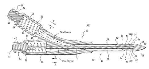
Catheter apparatus 20 includes a tubular body 22 having a distal end 24.
Tubular
body 22 is elongated and has a cylindrical outer surface. It is contemplated
that tubular
body 22 may be variously dimensioned and attachable to other medical devices.
It is further
contemplated that the outer surface of tubular body 22 may have various
configurations,
such as, for example, rectangular, elliptical, polygonal, etc.
Referring to FIGS. 3-8, tubular body 22 defines a first lumen such as, for
example,
venous lumen 26 and a second lumen such as, for example, arterial lumen 28.
Venous
lumen 26 and arterial lumen 28 each have a substantially D-shaped or semi-
circular
configuration. Venous lumen 26 includes an inner surface 27 having a
substantially planar
portion 27A and a substantially arcuate portion 27B, as shown in FIG. 3A.
Arterial lumen
28 includes an inner surface 29 having a substantially planar portion 29A and
a substantially
arcuate portion 29B. Lumens 26, 28 are elongated with tubular body 22 and
inner surfaces
27, 29 are configured to facilitate fluid flow within lumens 26, 28. It is
envisioned that
lumens 26, 28 may have various configurations, such as, for example,
cylindrical,
rectangular, elliptical, polygonal, etc.
Venous lumen 26 is configured for fluid flow, such as, for example, venous
blood
flow, in a first direction, as shown by arrows A. Arterial lumen 28 is
configured for fluid
flow, such as, for example, arterial blood flow in a second opposite
direction, as shown by
arrows B. The first and second lumens may be configured for various forms of
fluid flow in
various directions and orientations, according to the requirements of a
particular catheter
application.
7
CA 02535068 2006-02-07
WO 2005/018726 PCT/US2004/026004
Lumens 26, 28 may be uniformly dimensioned or include alternative dimensional
cross sections within tubular body 22, such as, narrow and broad portions,
converging
surfaces, undulating surfaces, etc. according to the particular flow
indications and/or flow
rate requirements. It is contemplated venous lumen 26 and arterial lumen 28
may extend
alternative lengths. It is further contemplated that tubular body 22 may
include one or a
plurality of lumens. It is envisioned that the first lumen may include the
arterial lumen and
the second lumen may include the venous lumen.
Venous lumen 26 and arterial lumen 28 are disposed in a substantially parallel
orientation adjacent a distal portion 30 of tubular body 22. Distal portion 30
may extend
various lengths and may include portions of tubular body 22 that are in a non-
parallel
orientation. It is also contemplated that venous lumen 26 and arterial lumen
28 may be
spaced apart.
Venous lumen 26 includes a first adapter, such as, for example, tubular venous
adapter 32 that extends to a proximal end 34 thereof. Venous adapter 32
defines a valve
housing 36 adjacent proximal end 34. Valve housing 36 has a cylindrical
configuration to
facilitate support of a first valve 38 and a spring 40 that biases first valve
38, in a
substantially proximal direction as shown by arrow C, to seal proximal end 34.
Spring 40
may be fixedly mounted to an inner surface of valve housing 36. A first luer
fitting 42 is
mounted with proximal end 34. First luer fitting 42 includes a first pusher 44
that is
connected with first valve 38. It is contemplated that first pusher 44 may be
separately
formed from first valve 38 and disposed for engagement therewith.
Spring 40 expands, via a spring force thereof, to engage first valve 38,
forcing first
valve 38 in the direction shown by arrow C. As first valve 38 moves, a surface
46 of first
valve 38 engages a surface 48 of proximal end 34. This engagement creates a
fluid tight
seal between first valve 38 and proximal end 34. The seal prevents inflow of
fluids into
venous lumen 26 and prevents leakage of fluids therefrom. First valve 38,
being connected
to first pusher 44, causes first pusher 44 to move in the direction shown by
arrow C, and
protrude from proximal end 34 for engagement with a venous blood line 50, as
will be
discussed. It is contemplated that the attachment of venous blood line 50 with
proximal end
34 is configured for introduction of fluid into venous lumen 26.
First luer fitting 42 is configured for attachment to venous blood line 50.
Venous
blood line 50 includes a pusher component 52 that engages first pusher 44 to
facilitate fluid
8
CA 02535068 2006-02-07
WO 2005/018726 PCT/US2004/026004
communication between venous blood line 50 and venous lumen 26. Venous blood
line 50
may be attached via luer connection, threaded connection, snap on, clips, etc.
Venous blood line 50 is attached to first luer fitting 42 such that pusher
component
52 engages first pusher 44, causing movement of first pusher 44 in a
substantially distal
direction, as shown by arrow D. The portion of first pusher 44 protruding from
proximal
end 34 is engaged by pusher component 52 as venous blood line 50 is attached
to proximal
end 34. The movement of first pusher 44 causes first valve 38 to overcome the
bias of
spring 50 and allow movement of first valve 34 in the direction shown by arrow
D.
Surface 46 of first valve 38 disengages from surface 48 of proximal end 34.
The
fluid tight seal is interrupted, thereby opening proximal end 34 to establish
fluid
communication between proximal end 34 and venous lumen 26.
Conversely, as venous blood line 50 is removed from proximal end 34, pusher
component 52 disengages from first pusher 44. Spring 40 re-expands, forcing
first valve 38
in the direction shown by arrow C. Surface 46 engages surface 48 to create the
fluid tight
seal between first valve 38 and proximal end 34. It is contemplated that valve
housing 36
may have various geometric configurations such as, rectangular, elliptical,
polygonal, etc. It
is further contemplated that spring 40 may alternatively include resiliently
biasing structure
such as, a resilient arm, pneumatic, hydraulic, magnetic force, etc. and may
be electronically
or manually controlled. First valve 38 may be oriented to engage various
portions of
proximal end 34. It is envisioned that first valve 38 may be monolithically
formed or
integrally connected to first pusher 44, or may include other valve structure,
such as, slit
valves, threaded, umbrella valves, diaphragm valves, etc.
Venous lumen 26 includes a first lateral port 54 disposed adjacent distal end
24 of
tubular body 22. First lateral port 54 includes an opening 55 that is
configured for fluid
flow. First lateral port 54 may be variously dimensioned and configured, such
as, for
example, rectangular, elliptical, polygonal, etc. Opening 55 is defined by the
thickness of a
wall portion 56 of tubular body 22 adjacent thereto. First lateral port 54 may
include
adapters, clips, etc. to facilitate fluid flow and/or attachment to other
structure. It is
contemplated that first lateral port 54 is configured for expulsion of fluid
from venous
lumen 26.
Arterial lumen 28 includes a first adapter, such as, for example, tubular
.arterial
adapter 58 that extends to a proximal end 60 thereof. Arterial adapter 58
defines a valve
9
CA 02535068 2006-02-07
WO 2005/018726 PCT/US2004/026004
housing 62 adjacent proximal end 60. Valve housing 62 has a cylindrical
configuration to
facilitate support of a second valve 64 and a spring 66 that biases second
valve 64, in a
substantially proximal direction as shown by arrow E, to seal proximal end 60.
Spring 66
may be fixedly mounted to an inner surface of valve housing 62. A second luer
fitting 68 is
mounted with proximal end 60. Second luer fitting 68 includes a second pusher
70 that is
connected with second valve 64.
It is contemplated that second pusher 70 may be separately formed from second
valve 64 and disposed for engagement therewith. It is further contemplated
that proximal
end 60 is configured for expulsion of fluid to a receiving fluid line. It is
envisioned that one
or both of lumens 26, 28 may include no adapters, one or a plurality of
adapters, such as, for
example, an embodiment whereby venous lumen 26 has a valved adapter and
arterial lumen
28 does not have a valved adapter.
Spring 66 expands, via a spring force thereof, to engage second valve 64,
forcing
second valve 64 in the direction shown by arrow E. As second valve 64 moves, a
surface
72 of second valve 64 engages a surface 74 of proximal end 60. This engagement
creates a
fluid tight seal between second valve 64 and proximal end 60. The seal
prevents inflow of
fluids into arterial lumen 28 and prevents leakage of fluids therefrom. Second
valve 64,
being connected to second pusher 70, causes second pusher 70 to move in the
direction
shown by arrow E, and protrude from proximal end 60 for engagement with an
arterial
blood line 76, as will be discussed.
Second luer fitting 68 is configured for attachment to arterial blood line 76.
Arterial
blood line 76 includes a pusher component 78 that engages second pusher 70 to
facilitate
fluid communication between arterial blood line 76 and arterial lumen 26.
Arterial blood
line 76 may be attached via luer connection, threaded connection, snap on,
clips, etc.
Arterial blood line 76 is attached to second luer fitting 68 such that pusher
component 78 engages second pusher 70, causing movement of second pusher 70 in
a
substantially distal direction, as shown by arrow F. The portion of second
pusher 70
protruding from proximal end 60 is engaged by pusher component 70 as arterial
blood line
76 is attached to proximal end 60. The movement of second pusher 70 causes
second valve
64 to overcome the bias of spring 66 and allow movement of second valve 64 in
the
direction shown by arrow F.
CA 02535068 2006-02-07
WO 2005/018726 PCT/US2004/026004
Surface 72 of second valve 64 disengages from surface 74 of proximal end 60.
The
fluid tight seal is interrupted, thereby opening proximal end 60 to establish
fluid
communication between proximal end 60 and arterial lumen 28.
Conversely, as arterial blood line 76 is removed from proximal end 60, pusher
component 78 disengages from second pusher 70. Spring 66 re-expands, forcing
second
valve 64 in the direction shown by arrow E. Surface 72 engages surface 74 to
create the
fluid tight seal between second valve 64 and proximal end 60. It is
contemplated that valve
housing 62 may have various geometric configurations such as, rectangular,
elliptical,
polygonal, etc. It is further contemplated that spring 66 may alternatively
include resiliently
biasing structure such as, a resilient arm, pneumatic, hydraulic, magnetic
force, etc. and may
be electronically or manually controlled. Second valve 64 may be oriented to
engage
various portions of proximal end 60. It is envisioned that second valve 64 may
be
monolithically formed or integrally connected to second pusher 70, or may
include other
valve structure, such as, slit valves, threaded, umbrella valves, diaphragm
valves, etc.
Arterial lumen 28 includes a second lateral port 80 disposed adjacent distal
end 24
of tubular body 22. Second lateral port 80 includes an opening 82 that is
configured for
fluid flow. Opening 82 may be variously dimensioned and configured, such as,
for
example, rectangular, elliptical, polygonal, etc. Opening 82 is defined by the
thickness of a
wall portion 84 of tubular body 22 adjacent thereto. Second lateral port 80
may include
adapters, clips, etc. to facilitate fluid flow and/or attachment to other
structure. It is
contemplated that second lateral port 80 is configured for introduction of
fluid into arterial
lumen 28.
A push rod 88 is connected to first valve 38 within valve housing 36. Push rod
88 is
slidably supported by a central lumen 90 (FIG. 3A) of tubular body 22 and
extends to a
pointed distal tip 92 disposed adjacent distal end 24. Central lumen 90 is
disposed between
venous lumen 26 and arterial lumen 28, and extends to distal end 24. Push rod
88 is
mounted with central lumen 90 such that the portion of push rod 88 disposed
with venous
adapter 32 is coaxially mounted therewith.
Push rod 88 is associated with first valve 38 for corresponding slidable
movement
therewith. For example, as first valve 38 is forced proximally in the
direction shown by
arrow C, discussed above, push rod 88 is similarly forced in the direction
shown by arrow
C. Further, as first valve 38 is forced distally in the direction shown by
arrow D, discussed
above, push rod 88 is similarly forced in the direction shown by arrow D. Tip
92 is
11
CA 02535068 2006-02-07
WO 2005/018726 PCT/US2004/026004
movable corresponding to the movement of first valve 38, as facilitated by
push rod 88.
The slidable movement of push rod 88 causes corresponding slidable movement of
a valve
94 that includes tip 92, as will be discussed.
Tip 92 has a proximal portion 96 and a distal portion 98. Proximal portion 96
includes a first member 100 extending into venous lumen 26 and a second member
102
extending into arterial lumen 28. First lateral port 54 is disposed
proximally, with tubular
body 22, relative to second lateral port 80. Thus, first member 100 extends a
greater
dimensional length than second member 102 to seal first lateral port 54 and
second lateral
port 80, as will be discussed.
First member 100 extends, in a proximal direction, a greater depth within
venous
lumen 26 relative to the depth of extension of second member 102 within
arterial lumen 28.
It is envisioned that second member 102 may extend a greater depth within
lumens 26, 28
than first member 100, or alternatively, first member 100 and second member
102 may
extend the same depth.
First member 100 includes an arcuate portion 104 that conforms to the
correspondingly configured arcuate portion 27B of inner surface 27 of venous
lumen 26,
adjacent first lateral port 54. Arcuate portion 104 engages arcuate portion
27B to facilitate
slidable movement of first member 100 relative to venous lumen 26. It is
contemplated that
arcuate portion 104 sealingly engages arcuate portion 27B via interference
using an O-ring
type thin malleable surface, an umbrella type valve surface, etc. It is
envisioned that first
member 100 may have a D-shaped/semicircular cross-section, or may have a wall
portion
that includes arcuate portion 104.
Second member 102 includes an arcuate portion 106 that conforms to the
correspondingly configured arcuate portion 29B of inner surface 29 of arterial
lumen 28,
adjacent second lateral port 80. Arcuate portion 106 engages arcuate portion
29B to
facilitate slidable movement of second member 102 relative to arterial lumen
28. It is
envisioned that arcuate position 106 sealingly engages arcuate portion 29B via
interference
using an O-ring type thin malleable surface, an umbrella type valve surface,
etc. It is
contemplated that second member 102 may have a D-shaped/semicircular cross-
section, or
may have a wall portion that includes arcuate portion 106. It is further
contemplated that
first member 100 and second member 102 may be monolithically formed with tip
92, or
alternatively, may be integrally assembled with tip 92 and fabricated from
dissimilar
materials.
12
CA 02535068 2006-02-07
WO 2005/018726 PCT/US2004/026004
Distal portion 98 of tip 92 includes a pointed distal head 99. Distal head 99
facilitates disposal of tubular body 22 within a body vessel and may be
employed with a
guidewire, sheath, etc. It is envisioned that distal head 99 may be employed
with a stylet,
tunneler, trocar, etc. to tunnel tubular body 22 under the skin of a subject
(not shown). It is
contemplated that distal head 99 may be variously configured or,
alternatively, distal
portion 98 may include a blunt tip. It is contemplated that tip 92 allows for
aspiration
through an angle of 360 degrees. This configuration facilitates disposal of
distal end 24 of
tubular body 22 in a plurality of orientations and prevents positional
occlusion.
As push rod 88 moves distally, as shown by arrow C, or proximally, as shown by
arrow D, first member 100 and second member 102, extending from tip 92,
similarly move
in a distal direction and a proximal direction. Such movement facilitates
corresponding
movement of valve 94, which includes tip 92, between a closed position (FIG.
3) and an
open position (FIG. 4).
In the closed position, tip 92 flushly engages distal end 24 of tubular body
22. First
member 100 extends a sufficient depth within venous lumen 26 such that arcuate
portion
104 spans across first lateral port 54. Arcuate portion 104 flushly engages
first lateral port
54 and the adjacent portions of arcuate portion 27B of venous lumen 26 to
close off first
lateral port 54 and create a fluid tight seal therewith. Similarly, second
member 102 extends
a sufficient depth within arterial lumen 28 such that arcuate portion 106
spans across second
lateral port 80. Arcuate portion 106 flushly engages second lateral port 80
and the adjacent
portions of arcuate portion 29B of arterial lumen 28 to close of second
lateral port 80 and
create a fluid tight seal therewith.
As push rod 88 moves in the distal direction, as shown by arrow D, first
member
100 and second member 102 are caused to slidably move relative to venous lumen
26 and
arterial lumen 28, respectively. First member 100 slides out of alignment with
first lateral
port 54. Second member 102 slides out of alignment with second lateral port
80.
In the open position, first member 100 disengages from first lateral port 54
and the
adjacent portions of arcuate portion 27B of venous lumen 26 to interrupt and
open the fluid
tight seal of first lateral port 54, thereby facilitating fluid communication
between first
lateral port 54 and venous lumen 26.
Similarly, second member 102 disengages from second lateral port 80 and the
adjacent portions of arcuate portion 29B of arterial lumen 28 to interrupt and
open the fluid
13
CA 02535068 2006-02-07
WO 2005/018726 PCT/US2004/026004
tight seal of second lateral port 80, thereby facilitating fluid communication
between second
lateral port 80 and arterial lumen 28.
As push rod 88 is caused to move back in the proximal direction, as shown by
arrow
C, first member 100 and second member 102 are caused to slidably move relative
to venous
lumen 26 and arterial lumen 28, respectively. First member 100 reseals first
lateral port 54,
as discussed, and second member 102 reseals second lateral port 80, as
discussed, such that
valve 94, which includes tip 92, is again disposed in the closed position. It
is contemplated
that valve 94, including tip 92, may be releasably locked or permanently fixed
in the open
position and/or the closed position via detents, clips, etc. mounted adjacent
distal end 24,
adapters 32, 58 or along other portions of tubular body 22. This configuration
advantageously facilitates desirable fluid flow rates and may break up
thrombus or fibrin
sheath formation. Further, the structure and methods illustrated for achieving
the principles
of the present disclosure also advantageously prevent undesirable fluid
evacuation to further
prevent thrombus formation on an innersurface of tubular body 22.
Referring to FIGS. 3 and 4, in use, a catheter apparatus 20, similar to that
described,
is assembled, properly sterilized and otherwise prepared for storage, shipment
and use in a
hemodialysis procedure. A practitioner (not shown) manipulates distal end 24
of tubular
body 22 such that pointed distal head 99 of tip 92 can enter a body cavity of
a subject (not
shown). Distal end 24 is inserted within a blood vessel of the subject.
Catheter apparatus
20 is employed for administration of fluids that includes the simultaneous
introduction of
venous blood flow and withdrawal of arterial blood flow. Catheter apparatus 20
is inserted
with the blood vessel of the subject such that blood is withdrawn, via
arterial blood flow in
a first direction, from the blood vessel for treatment by an artificial kidney
device (not
shown) and the treated blood is introduced back into the blood vessel, via
venous blood
flow in a second opposite direction.
Initially, valve 94, which includes tip 92, is in the closed position. First
member 100
extends within venous lumen 26 such that arcuate portion 104 spans across
first lateral port
54. Arcuate portion 104 flushly engages first lateral port 54 and the adjacent
portions of
arcuate portion 27B of venous lumen 26 to close off first lateral port 54 and
create a fluid
tight seal therewith. Similarly, second member 102 extends within arterial
lumen 28 such
that arcuate portion 106 spans across second lateral port 80. Arcuate portion
106 flushly
engages second lateral port 80 and the adjacent portions of arcuate portion
29B of arterial
lumen 28 to close of second lateral port 80 and create a fluid tight seal
therewith.
14
CA 02535068 2006-02-07
WO 2005/018726 PCT/US2004/026004
Surface 46 of first valve 38 engages surface 48 of proximal end 34 to create a
fluid
tight seal between first valve 38 and proximal end 34, as discussed. First
pusher 44
protrudes from proximal end 34. Surface 72 of second valve 64 engages surface
74 of
proximal end 60 to create a fluid tight seal between second valve 64 and
proximal end 60.
Second pusher 70 protrudes from proximal end 60.
Venous blood line 50 is attached to first luer fitting 42 such that pusher
component
52 engages first pusher 44, causing movement of first pusher 44 in a
substantially distal
direction, as shown by arrow D, overcoming the bias of spring 50. Surface 46
of first valve
38 disengages from surface 48 of proximal end 34 and the fluid tight seal is
interrupted,
thereby opening proximal end 34 to establish fluid communication between
proximal end
34 and venous lumen 26. Venous blood flow is introduced to catheter apparatus
20 through
proximal end 34.
Arterial blood line 76 is attached to second luer fitting 68 such that. pusher
component 78 engages second pusher 70, causing movement of second pusher 70 in
a
substantially distal direction, as shown by arrow F, overcoming the bias of
spring 66.
Surface 72 of second valve 64 disengages from surface 74 of proximal end 60
and the fluid
tight seal is interrupted, thereby opening proximal end 60 to establish fluid
communication
between proximal end 60 and arterial lumen 28. Arterial blood flow may be
received by
arterial blood line 76.
As first valve 38 is forced distally in the direction shown by arrow D,
discussed
above, push rod 88 is similarly forced in the direction shown by arrow D.
Valve 94, which
includes tip 92, is movable corresponding to the movement of first valve 38,
as facilitated
by push rod 88. As push rod 88 moves in the distal direction, first member 100
and second
member 102 are caused to slidably move relative to venous lumen 26 and
arterial lumen 28,
respectively. First member 100 slides out of alignment with first lateral port
54. Second
member 102 slides out of alignment second lateral port 80.
Valve 94, which includes tip 92, moves to the open position. First member 100
disengages from first lateral port 54 and the adjacent portions of arcuate
portion 27B of
venous lumen 26 to interrupt and open the fluid tight seal of first lateral
port 54, thereby
facilitating fluid communication between first lateral port 54 and venous
lumen 26. Thus,
venous blood flow is introduced to the blood vessel of the subject via venous
lumen 26.
Second member 102 disengages from second lateral port 80 and the adjacent
portions of
arcuate portion 29B of arterial lumen 28 to interrupt and open the fluid tight
seal of second
CA 02535068 2006-02-07
WO 2005/018726 PCT/US2004/026004
lateral port 80, thereby facilitating fluid communication between second
lateral port 80 and
arterial lumen 28. Thus, arterial blood flow is withdrawn from the blood
vessel and
received by arterial lumen 28 for receipt by arterial blood line 76.
In the event that the practitioner desires to discontinue administration of
fluids with
the subject, valve 94, which includes tip 92, may be returned to the closed
position. Venous
blood line 50 is removed from proximal end 34 to recreate the fluid tight seal
between first
valve 38 and proximal end 34. Arterial blood line 76 is removed from proximal
end 60 to
recreate the fluid tight seal between second valve 64 and proximal end 60.
Push rod 88 is caused to move back in the proximal direction, as shown by
arrow C.
First member 100 reseals first lateral port 54 and second member 102 reseals
second lateral
port 80 such that valve 94, which includes tip 92, is again disposed in the
closed position.
Referring to FIGS. 8-13, an alternate embodiment of the present disclosure is
shown
that includes a catheter apparatus 220. Catheter apparatus 220 includes a
tubular body 222
having a distal end 224. Tubular body 222 is elongated and has a cylindrical
outer surface.
Tubular body 222 defines a first lumen such as, for example, venous lumen 226
and
a second lumen such as, for example, arterial lumen 228. Venous lumen 226 and
arterial
lumen 228 are in a substantially coaxial orientation, with a longitudinal axis
x, along a distal
portion 230 of tubular body 222. Venous lumen 226 and arterial lumen 228 each
have a
substantially tubular configuration that facilitate fluid flow. It is
envisioned that lumens
226, 228 may have various configurations, such as, for example, cylindrical,
rectangular,
elliptical, polygonal, etc.
Venous lumen 226 is configured for fluid flow, such as, for example, venous
blood
flow, in a first direction, as shown by arrows AA. Arterial lumen 228 is
configured for fluid
flow, such as, for example, arterial blood flow in a second opposite
direction, as shown by
arrows BB. The first and second lumens may be configured for various forms of
fluid flow
in various directions and orientations, according to the requirements of a
particular catheter
application.
Lumens 226, 228 may be uniformly dimensioned or include alternative
dimensional
cross sections within tubular body 222, such as, narrow and broad portions,
converging
surfaces, undulating surfaces, etc. according to the particular flow
indications and/or flow
rate requirements. It is contemplated venous lumen 226 and arterial lumen 228
may extend
alternative lengths. It is further contemplated that tubular body 222 may
include one or a
16
CA 02535068 2006-02-07
WO 2005/018726 PCT/US2004/026004
plurality of lumens. It is envisioned that the first lumen may include the
arterial lumen and
the second lumen may include the venous lumen.
Venous lumen 226 includes a first adapter, such as, for example, tubular
venous
adapter 232 that extends to a proximal end 234 thereof. Venous adapter 232
defines a valve
housing 236 adjacent proximal end 234. Valve housing 236 has a cylindrical
configuration
to facilitate support of a first valve 238 and a spring 240 that biases first
valve 238, in a
substantially proximal direction as shown by arrow CC, to seal proximal end
234. Spring
240 may be fixedly mounted to an inner surface of valve housing 236. A first
luer fitting
242 is mounted with proximal end 234. First luer fitting 242 includes a first
pusher 244 that
is connected with first valve 238.
Spring 240 expands, via a spring force thereof, to engage first valve 238,
forcing
first valve 238 in the direction shown by arrow CC. As first valve 238 moves,
a surface 246
of first valve 238 engages a surface 248 of proximal end 234. This engagement
creates a
fluid tight seal between first valve 238 and proximal end 234. The seal
prevents inflow of
fluids into venous lumen 226 and prevents leakage of fluids therefrom. First
valve 238,
being connected to first pusher 244, causes first pusher 244 to move in the
direction shown
by arrow CC, and protrude from proximal end 234 for engagement with a venous
blood line
250, as will be discussed.
First luer fitting 242 is configured for attachment to venous blood line 250.
Venous
blood line 250 includes a pusher component 252 that engages first pusher 244
to facilitate
fluid communication between venous blood line 250 and venous lumen 226.
Venous blood line 250 is attached to first luer fitting 242 such that pusher
component 252 engages first pusher 244, causing movement of first pusher 244
in a
substantially distal direction, as shown by arrow DD. The portion of first
pusher 244
protruding from proximal end 234 is engaged by pusher component 252 as venous
blood
line 250 is attached to proximal end 234. The movement of first pusher 244
causes first
valve 238 to overcome the bias of spring 250 and allow movement of first valve
234 in the
direction shown by arrow DD.
Surface 246 of first valve 238 disengages from surface 248 of proximal end
234.
The fluid tight seal is interrupted, thereby opening proximal end 234 to
establish fluid
communication between proximal end 234 and venous lumen 226. Conversely, as
venous
blood line 250 is removed from proximal end 234, pusher component 252
disengages from
17
CA 02535068 2006-02-07
WO 2005/018726 PCT/US2004/026004
first pusher 244. Spring 240 re-expands, forcing first valve 238 in the
direction shown by
arrow CC. Surface 246 engages surface 248 to create the fluid tight seal
between first valve
238 and proximal end 234.
Venous lumen 226 defines a first port 254 disposed adjacent distal end 224 of
tubular body 222. First port 254 includes an opening 255 that is configured
for fluid flow.
First port 254 may be variously dimensioned and configured, such as, for
example,
rectangular, elliptical, polygonal, etc. First port 254 may include adapters,
clips, etc. to
facilitate fluid flow and/or attachment to other structure. It is contemplated
that first port
254 is configured for expulsion of fluid from venous lumen 226.
Arterial lumen 228 includes a first adapter, such as, for example, tubular
arterial
adapter 258 that extends to a proximal end 260 thereof. Arterial adapter 258
defines .a valve
housing 262 adjacent proximal end 260. Valve housing 262 has a cylindrical
configuration
to facilitate support of a second valve 264 and a spring 266 that biases
second valve 264, in
a substantially proximal direction as shown by arrow EE, to seal proximal end
260. Spring
266 may be fixedly mounted to an inner surface of valve housing 262. A second
luer fitting
268 is mounted with proximal end 260. Second luer fitting 268 includes a
second pusher
270 that is connected with second valve 264.
Spring 266 expands, via a spring force thereof, to engage second valve 264,
forcing
second valve 264 in the direction shown by arrow BE. As second valve 264
moves, a
surface 272 of second valve 264 engages a surface 274 of proximal end 260.
This
engagement creates a fluid tight seal between second valve 264 and proximal
end 260. The
seal prevents inflow of fluids into arterial lumen 228 and prevents leakage of
fluids
therefrom. Second valve 264, being connected to second pusher 270, causes
second pusher
270 to move in the direction shown by arrow BE, and protrude from proximal end
260 for
engagement with an arterial blood line 276, as will be discussed. Second luer
fitting 268 is
configured for attachment to arterial blood line 276. Arterial blood line 276
includes a
pusher component 278 that engages second pusher 270 to facilitate fluid
communication
between arterial blood line 276 and arterial lumen 226.
Arterial blood line 276 is attached to second luer fitting 268 such that
pusher
component 278 engages second pusher 270, causing movement of second pusher 270
in a
substantially distal direction, as shown by arrow FF. The portion of second
pusher 270
protruding from proximal end 260 is engaged by pusher component 270 as
arterial blood
line 276 is attached to proximal end 260. The movement of second pusher 270
causes
18
CA 02535068 2006-02-07
WO 2005/018726 PCT/US2004/026004
second valve 264 to overcome the bias of spring 266 and allow movement of
second valve
264 in the direction shown by arrow FF.
Surface 272 of second valve 264 disengages from surface 274 of proximal end
260.
The fluid tight seal is interrupted, thereby opening proximal end 260 to
establish fluid
communication between proximal end 260 and arterial lumen 228. Conversely, as
arterial
blood line 276 is removed from proximal end 260, pusher component 278
disengages from
second pusher 270. Spring 266 re-expands, forcing second valve 264 in the
direction shown
by arrow EE. Surface 272 engages surface 274 to create the fluid tight seal
between second
valve 264 and proximal end 260.
Arterial lumen 228 includes a second port 280 disposed adjacent distal end 224
of
tubular body 222. Second port 280 includes an opening 282 that is configured
for fluid
flow. Opening 282 may be variously dimensioned and configured, such as, for
example,
rectangular, elliptical, polygonal, etc. Second port 280 may include adapters,
clips, etc. to
facilitate fluid flow and/or attachment to other structure. It is contemplated
that second port
280 is configured for introduction of fluid into arterial lumen 228.
A portion of venous lumen 226, such as, for example, push rod portion 288 is
connected to first valve 238 within valve housing 236 for corresponding
movement
therewith. Push rod portion 288 is slidably mounted within tubular body 222
and extends to
first port 254. First port 254 seals second port 280 in a closed position of a
valve
configuration including first port 254 and second port 280.
Push rod portion 288 is connected with first valve 238 for corresponding
slidable
movement therewith. Push rod portion 288 includes openings 289 that facilitate
fluid
communication between proximal end 234 and venous lumen 226. Openings 289 may
be
variously configured such as, slots, vents, circular, polygonal, etc. Push rod
portion 288
may be attached to first valve 238 by various means, such as, for example,
adhesive, clips,
etc., may be monolithic therewith, or spaced apart therefrom.
For example, as first valve 238 is forced proximally in the direction shown by
arrow
CC, discussed above, push rod portion 288 is similarly forced in the direction
shown by
arrow CC. Further, as first valve 238 is forced distally in the direction
shown by arrow DD,
discussed above, push rod portion 288 is similarly forced in the direction
shown by arrow
DD. First port 254 is movable corresponding to the movement of first valve
238, as
facilitated by push rod portion 288.
19
CA 02535068 2006-02-07
WO 2005/018726 PCT/US2004/026004
As push rod portion 288 moves proximally, as shown by arrow CC, or distally,
as
shown by arrow DD, first port 254 similarly moves in a proximal direction and
a distal
direction. Such movement facilitates corresponding movement of the valve
configuration
that includes first port 254 and second port 280, between a closed position
(FIG. 12). and an
open position (FIG. 13).
In the closed position, first port 254 engages second port 280 to close off
second
port 280 and create a fluid tight seal therewith. As push rod portion 288
moves in the distal
direction, as shown by arrow DD, first port 254 is caused to slidably move
relative to distal
end 224 and second port 280. In the open position, first port 254 disengages
from second
port 280 to interrupt and open the fluid tight seal of second port 280,
thereby facilitating
fluid communication between second port 280 and arterial lumen 228.
As push rod portion 288 is caused to move back in the proximal direction, as
shown
by arrow CC, first port 254 is caused to slidably move relative to distal end
224 and second
port 280. First port 254 reseals second port 280, as discussed, such that the
valve
configuration that includes first port 254 and second port 280 is again
disposed in the closed
position. It is contemplated that the valve configuration that includes first
port 254 and
second port 280 may be releasably locked or permanently fixed in the open
position and/or
the closed position via detents, clips, etc. mounted adjacent distal end 224,
adapters 232,
258 or along other portions of tubular body 222. This configuration
advantageously
facilitates desirable fluid flow rates and may prevent thrombosis and fibrin
sheath
formation. Further, the structure and methods illustrated for achieving the
principles of the
present disclosure also advantageously prevent undesirable fluid evacuation
and enhance
comfort to a subject. It is envisioned that one or both of lumens 226, 228 may
include no
adapters, one or a plurality of adapters, such as, for example, an embodiment
whereby
venous lumen 226 has a valved adapter and arterial lumen 228 does not have a
valved
adapter.
Referring to FIGS. 12 and 13, in use, a catheter apparatus 220, similar to
that
described, is assembled, properly sterilized and otherwise prepared for
storage, shipment
and use in a hemodialysis procedure. A practitioner (not shown) manipulates
distal end 224
of tubular body 222 for connection to a body cavity of a subject (not shown).
Distal end
224 is inserted within a blood vessel of the subject. Catheter apparatus 220
is employed for
administration of fluids that includes the simultaneous introduction of venous
blood flow
and withdrawal of arterial blood flow. Catheter apparatus 220 is inserted with
the blood
CA 02535068 2006-02-07
WO 2005/018726 PCT/US2004/026004
vessel of the subject such that blood is withdrawn, via arterial blood flow in
a first direction,
from the blood vessel for treatment by an artificial kidney device (not shown)
and the
treated blood is introduced back into the blood vessel, via venous blood flow
in a second
opposite direction.
Initially, the valve configuration that includes first port 254 and second
port 280 is in
the closed position. Surface 246 of first valve 238 engages surface 248 of
proximal end 234
to create a fluid tight seal between first valve 238 and proximal end 234, as
discussed. First
pusher 244 protrudes from proximal end 234. Surface 272 of second valve 264
engages
surface 274 of proximal end 260 to create a fluid tight seal between second
valve 264 and
proximal end 260. Second pusher 270 protrudes from proximal end 260.
Venous blood line 250 is attached to first luer fitting 242 such that pusher
component 252 engages first pusher 244, causing movement of first pusher 244
in a
substantially distal direction, as shown by arrow DD, overcoming the bias of
spring 250.
Surface 246 of first valve 238 disengages from surface 248 of proximal end 234
and the
fluid tight seal is interrupted, thereby opening proximal end 234 to establish
fluid
communication between proximal end 234 and venous lumen 226 as facilitated by
openings
289. Venous blood flow is introduced to catheter apparatus 220 through
proximal end 234
and into the blood vessel of the subject via venous lumen 226.
Arterial blood line 276 is attached to second luer fitting 268 such that
pusher
component 278 engages second pusher 270, causing movement of second pusher
2.70 in a
substantially distal direction, as shown by arrow FF, overcoming the bias of
spring 266.
Surface 272 of second valve 264 disengages from surface 274 of proximal end
260 and the
fluid tight seal is interrupted, thereby opening proximal end 260 to establish
fluid
communication between proximal end 260 and arterial lumen 228. Arterial blood
flow may
be received by arterial blood line 276.
As first valve 238 is forced distally in the direction shown by arrow DD,
discussed
above, push rod portion 288 is similarly forced in the direction shown by
arrow DD. First
port 254 is movable corresponding to the movement of first valve 23 8, as
facilitated by push
rod portion 288. The valve configuration that includes first port 254 and
second port 280
moves to the open position. First port 254 disengages from second port 280 to
interrupt and
open the fluid tight seal of second port 280, thereby facilitating fluid
communication
between second port 280 and arterial lumen 228. Thus, arterial blood flow is
withdrawn
21
CA 02535068 2006-02-07
WO 2005/018726 PCT/US2004/026004
from the blood vessel and received by arterial lumen 228 for receipt by
arterial blood line
276.
In the event that the practitioner desires to discontinue administration of
fluids with
the subject, the valve configuration that includes first port 254 and second
port 280 may be
returned to the closed position. Venous blood line 250 is removed from
proximal end 234
to recreate the fluid tight seal between first valve 238 and proximal end 234.
Push rod
portion 288 is caused to move back in the proximal direction, as shown by
arrow CC,
thereby sealing second port 280. Arterial blood line 276 is removed from
proximal end 260
to recreate the fluid tight seal between second valve 264 and proximal end
260.
Referring to FIGS. 14-18, another alternate embodiment of the present
disclosure is
shown that includes a catheter apparatus 420, similar to those described.
Catheter apparatus
420 includes a tubular body 422 having a distal end 424. Distal end 424
includes a cap 425
that is mounted with tubular body 422. Cap 425 is separately formed and
configured for
assembly with tubular body 422 via threaded engagement. It is contemplated
that cap 425
may be assembled by various attachment such as, for example, adhesive,
interference or
friction, snap engagement, etc.
Tubular body 422 defines a venous lumen 426 and an arterial lumen 428. Venous
lumen 426 and arterial lumen 428 are in a substantially side by side
orientation along a
distal portion 430 of tubular body 422. The distal end of venous lumen 426
extends a
greater length relative to the distal end of arterial lumen 428 for connection
to the body
cavity of a subject. As such, the distal end of arterial lumen 428 is recessed
from the distal
end of venous lumen 426. Distal portion 430 may include a valve configuration,
similar to
those described herein with regard to FIGS. 1-13.
Venous lumen 426 includes a tubular venous adapter 432 that extends to a
proximal
end 434 thereof. Venous adapter 432 defines a valve housing 436, including
valve
components, and a luer fitting 442, similar to those described herein with
regard to FIGS. 1-
13. The valve components of valve housing 436 are biased to create a fluid
tight seal with
proximal end 434.
First luer fitting 442 is configured for attachment to a venous blood line
(not
shown). The venous blood line is attached to first luer fitting 442 to
overcome the bias of
the valve components of valve housing 436. The fluid tight seal is
interrupted, thereby
opening proximal end 434 to establish fluid communication between proximal end
434 and
22
CA 02535068 2006-02-07
WO 2005/018726 PCT/US2004/026004
venous lumen 426. Conversely, as the venous blood line is removed from
proximal end
434, the bias of the valve components of valve housing 436 recreates the fluid
tight seal
with proximal end 434. Venous lumen 426 defines a first port 454 disposed
adjacent distal
end 424 that is configured for fluid flow.
Arterial lumen 428 includes a tubular arterial adapter 458 that extends to a
proximal
end 460 thereof. Arterial adapter 458 defines a valve housing 462, including
valve
components, and a second luer fitting 468, similar to those described herein
with regard to
FIGS. 1-13. The valve components of valve housing 462 are biased to create a
fluid tight
seal with proximal end 460.
Second luer fitting 468 is configured for attachment to an arterial blood line
(not
shown). The arterial blood line is attached to second luer fitting 468 to
overcome the bias
of the valve components of valve housing 462. The fluid tight seal is
interrupted, thereby
opening proximal end 460 to establish fluid communication between proximal end
460 and
arterial lumen 428. Conversely, as the arterial blood line is removed from
proximal end
460, the bias of the valve components of valve housing 462 recreates the fluid
tight seal
with proximal end 460. Arterial lumen 428 defines a second port 480 disposed
adjacent
distal end 424 that is configured for fluid flow.
In use, catheter apparatus 420, is inserted with the blood vessel of the
subject.
Tubular body 422 is then reverse tunneled under the skin of a subject (not
shown) away
from an insertion site to another exit site of the body of the subject.
Tubular body 422 is
sized as desired and cap 425 is threaded for assembly with tubular body 422.
Blood is
withdrawn employing arterial lumen 428, via arterial blood flow in a first
direction, from
the blood vessel for treatment by an artificial kidney device (not shown) and
the ' treated
blood is introduced back into the blood vessel employing venous lumen 426, via
venous
blood flow in a second opposite direction. Catheter apparatus 420 is employed
for
administration of fluids that includes the simultaneous introduction of venous
blood flow
and withdrawal of arterial blood flow.
It will be understood that various modifications may be made to the
embodiments
disclosed herein. Therefore, the above description should not be construed as
limiting, but
merely as exemplification of the various embodiments. Those skilled in the art
will
envision other modifications within the scope and spirit of the claims
appended hereto.
23