Note: Descriptions are shown in the official language in which they were submitted.
CA 02535129 2006-02-07
WO 2005/016134 PCT/US2004/014575
GASTROINTESTINAL LAVAGE SYSTEM
Technical Field
This invention relates upper and lower GI (GI) endoscopy. More specifically,
this invention
is used in conjunction with an endoscope to enable GI lavage and vacuuming
thus making
endoscopy safer and more clinically effective.
Background Art
Since its inception in the latter 1960's flexible GI endoscopy; first with a
miniature film
camera, then with fiber optic bundles to carry the incident image and finally
culminating in
video chip technology, has evolved from a mechanical curiosity to a proven
life-saving
system for diagnosis and therapy. Many structures previously considered to be
beyond
diagnostic evaluation and therapeutics by any modality short of surgery are
now open to the
GI endoscopist: esophagus, stomach, duodenum, small intestine, biliary tree
and
pancreatic system are now accessible without use of an open surgical
technique.
Endoscopic techniques can treat and palliate conditions such as stopping of GI
bleeding,
polyp removal, gallstone removal, stricture dilation, foreign body removal,
endoscopic stint
placement for GI obstruction and a host of other procedural capabilities too
numerous to
mention. The lesser morbidity and mortality with endoscopic procedures allows
their use
on sicker, more debilitated patents, increasing the clinical utility of
endoscopic therapy.
While endoscopic diagnosis and therapy has opened many new avenues of GI care,
some
of its design strengths produce some of its greatest limitations. The
endoscope is a flexible
tube less than 1 cm in diameter, generally possessing one hollow bore through
which
instruments for injection of dye or haemostatic materials, biopsy and
polypectomy
attachments, and haemostatic probes are passed. At the same time this bare is
used for
suctioning of secretions. The bore is approximately 2.6 to 2.8 mm in diameter
and not
conducive to removal of larger diameter foreign material or highly viscous
substances such
as clotted blood. Cleansing the endoscopic visual field to remove food, blood,
secretions
impacted food bolus or foreign body is often impossible using this small bore.
A larger bore
conduit is needed to correct this shortcoming. Since the endoscope itself
really cannot be
CA 02535129 2006-02-07
WO 2005/016134 PCT/US2004/014575
modified to rectify this shortcoming, an appliance to be used in conjunction
with the
endoscope would be the preferable alternative.
Frequently, certain appliances are used in conjunction with endoscopy to aid
in its use for
diagnosis and therapy. A long flexible fenestrated tube with an open proximal
end is often
used to clear the upper GI tract of blood, food, foreign materials and
secretions. This
device uses a vacuum system for the removal of these materials and either
gravity flow or
pressure system to instill lavage fluid into the GI tract to help remove these
materials. This
system cannot be used simultaneously with an endoscope but must be used
independently
and inserted blindly into the upper GI tract. A blind insertion may cause
trauma to the
respiratory tree with sometimes disastrous results. Repeated removal and
replacement of
the endoscope and lavage tube may also stimulate retching which can induce
vomiting,
aspiration of gastric contents and even severe bleeding, both serious
complications. The
least number of removal and replacement of tubes against the pharynx and
esophagus, the
better. Further complicating the process of alternating lavage and endoscopy
is the fact
that rapid bleeding in the GI tract is frequently too rapid and too difficult
to clear to the point
that an endoscope may be effectively used. A lavage tube may cause a fleeting
improvement in the visualization of a bleeding site, but from the time of
removal of the
lavage tube to the time of reintroduction of the endoscope, continued bleeding
can obscure
both normal enteric architecture as well as the pathologic process
precipitating the problem.
A system which allowed simultaneous lavage and endoscopy would circumvent all
these
problems.
Removal of foreign bodies from the upper GI tract is another common problem
with
potentially serious consequences. Foreign bodies may produce pain, bleeding
and
obstruction in the GI tract. Endoscopic treatment of this problem may be
hampered by the
need to frequently remove blood, food and secretions either prior to or at the
same time as
endoscopic evaluation and therapy is performed. Endoscopic therapy must take
into
consideration not only proper preparation and endoscopic technique for foreign
body
removal, hemostasis, stricture and dilation but also safety issues such as the
prevention of
aspiration of GI contents and foreign bodies into the respiratory tree, and
prevention of
laceration of the duodenum, stomach, esophagus and pharynx. Complicating this
issue is
that while a flexible polymer plastic over tube through which the endoscope
and foreign
CA 02535129 2006-02-07
WO 2005/016134 PCT/US2004/014575
bodies can be inserted and removed thus protecting surrounding structures, no
system yet
exists which can perform the endoscopic function while simultaneously
possessing the
capability to lavage out blood, food and secretions which can complicate
foreign body
removal. This increases the level of difficulty in their removal as well as
increases potential
complications in the removal process, such as aspiration and perforation. The
ability to
simultaneously see the GI tract while removing a foreign body would be a
tremendous
advantage over the system presently used.
Treatment of acute lower GI bleeding is complicated by the frequent
obscuration of the
bleeding site by blood, mucus and stool thus seriously limiting the utility of
therapeutic
colonoscopy for the acute treatment of lower GI bleeding. If blood, secretions
and stool
could be effectively and simultaneously removed while performing the
colonoscopy,
colonoscopy would be a more effective acute diagnostic and therapeutic
interventional
system than it is at the present time. This would save time in that a
colonoscopy could be
performed without the need to perform preceding colonic cleansing using either
oral
osmotic cathartics or enema which takes both precious time and involves
increased risk in
the patient who is semiconscious and hypotensive. The logistics in the typical
medical
center favors endoscopic intervention over the more involved and potentially
more risky
interventional angiography.
A system is needed which can cleanse and remove blood, foreign material,
secretion and
stool from the upper and lower GI tracts while simultaneously performing
diagnostic and
therapeutic endoscopy for hemostasis and removal of impacted material. The
system must
be capable of protecting GI structures from trauma and the respiratory system
from
aspiration of secretions, blood and foreign material.
There have been a number of developments in this area. US Patent Number
4,795,424
(Burner) concerns an apparatus for controlled irrigation of the natural
cavities of the human
body. It is characterized by a correcting means for equalizing the measuring
signal, which
represents the pressure, which is emitted by a pressure gauge according to the
pressure
losses and according to the differential pressure between it and the
measurement of the
pressure in the intervention area which corresponds to an organ. These
correcting means
include a correcting circuit adapted to effect a correction according to said
pressure
differential and a correcting circuit adapted to take into account the
pressure loss, namely
CA 02535129 2006-02-07
WO 2005/016134 PCT/US2004/014575
the flow rate of the fluid injected through the inner duct of the endoscope
and the cross
section of this duct.
This is an electrical system, designed principally for urologic endoscopy, for
monitoring flow
of fluid into and out of a small space by monitoring changes in pressure in
the system.
This is not a lavage system but a pressure-monitoring device for fluid
entering and exiting a
space. This could be used in the bladder or uterus for surgery to gauge fluid
flow - it has
nothing to do whatever with lavage of blood, food, dilation of strictures and,
removal of
foreign bodies. This is for measuring/monitoring and not effecting a change.
US Patent Number 5,030,202 (Harris) discloses a lavage apparatus comprising a
lavage
fluid supply apparatus, a first catheter having a distal end for positioning
in the uterus of an
animal and a proximal end for communication with the lavage fluid supply, a
pair of bilateral
catheters connected to the proximal end of the first catheter for extending
into respective
horns of the uterus, and catheter locating means for locating the catheters in
the uterus.
The first catheter defines respective supply and drain lumens. The bilateral
catheters each
define respective lumens for fluid communication between the supply lumen and
the interior
of the respective uterine horn. The lavage fluid supply apparatus may include
a pump for
supplying lavage fluid under pressure. The supply may also include a lavage
fluid storage
tank for containing a volume of fluid, and a heater to maintain the
temperature of the stored
fluid within a predetermined range. For use in a gravid uterus, a drain tube
may be
provided at the distal end of the first catheter for reaching into the depths
of the uterus.
Further, vacuum drainage apparatus may be provided to drain lavage fluid from
the uterus.
If desired, the lavage fluid supply may be utilized for purposes other than
uterine lavages
such as, for example, supplying fluids for intravenous infusion. It cannot be
used for
simultaneous endoscope and lavage in either the upper or lower
gastrointestinal tract.
Neither is it portable or disposable. ~ne cannot effectively dilate and remove
foreign
material with this device.
US Patent Number 5,620,408 (Vennes et al.) discloses an endoscopic over-tube
for
receiving and guiding medical instrumentation into the upper alimentary canal
of a patient.
The endoscopic over-tube has a bite block for insertion and retention in the
mouth of the
patient. The endoscopic over-tube also has a flexible protective sheath
engaging the bite
block and adapted for insertion into the upper alimentary canal of the
patient. The sheath
CA 02535129 2006-02-07
WO 2005/016134 PCT/US2004/014575
includes a stiffened region that insulates the posterior wall of the patient's
pharynx from
medical instrumentation as the medical instrumentation is intubated through
the sheath and
into the patient's alimentary canal.
This tube is solely used to help an upper endoscopic bypass direct control of
the pharynx
by the endoscope protecting against irritation and gagging and possibly
protecting the
upper airway. The system is short and too rigid to be used for lavage. It
cannot be sealed
for lavage purposes. It cannot be moved when it is properly inserted. The new
arrangement system is not as useful for lavage, hemostasis, foreign body
removal, and
dilation. It could not be used to simultaneously endoscope and lavage a
patent, removing
either blood, food or other foreign materials solely and expeditiously to
accomplish the
same procedure.
US Patent Number 5,827,177 (Oneda et al.) discloses an endoscope sheath
assembly
usable within an elongated flexible endoscope to isolate a portion of the
endoscope from an
external environment during a therapeutic or diagnostic endoscopic procedure.
The sheath
assembly has a sheath with an elongated flexible endoscope tube, a plurality
of working
bores extending the endoscope tube, and a low-friction, fabric isolating
sleeve positioned
within the endoscope tube and surrounding portions of the working bores. The
isolating
sleeve includes first and second sleeve lumens formed therein through which
respective
working bores extend. The coefficient of friction between the isolating sleeve
and the
working bores is less than the coefficient of friction between the working
bores themselves
so that working bores are frictionally isolated from each other, from the
endoscope tube,
and from the insertion tube to reduce resistance to articulation of the
endoscope.
This is an endoscope with distensible elastomeric working bore within the
endoscope tube
for passage of instruments during diagnostic or therapeutic endoscopy. It is
just a modified
endoscope with the same failing described above: the bores are inadequate for
removal of
food, blood clots, passage of rigid dilators and removal of foreign bodies,
let alone lavage
and endoscopic treatment simultaneously.
US Patent Number 5,876,379 (Harben) discloses an endoscope for use with an
elastomeric
sheath for performing an endoscopic procedure. The endoscope includes a body,
an
insertion tube extending away from the body and terminating at a distal end
portion, and a
CA 02535129 2006-02-07
WO 2005/016134 PCT/US2004/014575
sheath retainer connected to the distal end portion of the insertion tube for
releasably
retaining the sheath on the insertion tube in a substantially fixed position
during the
endoscopic procedure. The sheath is an elastomeric member movable between a
radially
expanded position and a contracted position, and the sheath is biased toward
the
contracted position. The sheath retainer is a generally C-shaped member having
a pair of
retaining tabs spaced apart from each other and extending away from an outer
surface of
the insertion tube's distal end portion to define a working bore retaining
area there between.
The retaining tabs are positioned and sized to engage a portion of the sheath
at its distal
end portion and to prevent the distal end portion of the sheath from moving
axially relative
to the distal end portion of the insertion tube, particularly during an
endoscopic procedure.
This is not a lavage system but an endoscope covered by a tight-fitting
elastomer
membrane keeping the visual system of the endoscope in close proximity to a
biopsy-utility
bore. This is just an endoscope tube composed of two pieces covered by an
elastomeric
sheath. It has the same failings of contemporary endoscopes: no ability to
lavage, dilate or
remove foreign bodies.
US Patent Number 6,149,633 (Maaskamp) discloses a system which utilizes an
endoscope
and irrigation fluid during surgery within a body orifice, and in which
irrigation fluid is
withdrawn via a principal path into a suction canister, there is a
substantially constant
suction and withdrawal rate from the operative site to limit absorption of
irrigation fluid within
the patient, because a bifurcated flow path that leads to the suction canister
not only from
the endoscope but also from a drain bag receiving overflow from the body
orifice via a
tailored drape includes a substantially greater flow impedance in the drain
bag path. The
common suction line joined to the endoscope line and the drain bag line draws
a flow
through the endoscope that predominates, to maintain substantially constant
withdrawal of
irrigation fluid via that path, and substantially eliminates the possibility
of loss of suction.
This is a system to meter fluid into body cavities during laparoscopy and not
endoscopy. It
measures fluid going into the cavity and fluid coming out of the cavity to
prevent fluid
overload of patient during surgery. It has nothing to do a lavage of GI
bleeding, dilation of
stricture and removal of foreign bodies. It is solely a fluid-monitoring
system.
CA 02535129 2006-02-07
WO 2005/016134 PCT/US2004/014575
US Patent Number 6,187,346 (Neuwirth) discloses a method and composition for
effecting
necrosis of a tissue lining of a mammalian body cavity, particularly a uterine
endometrium,
by introducing an applicator comprising a hysteroscope housing a first and a
second
catheter connected to a catheter into the uterus, distending the uterus by
introducing C02
gas under pressure, delivering a silver nitrate paste to the endometrium
through the first
catheter and allowing the paste to remain a sufficient amount of time to
substantially
cauterize the entirety of the tissue lining, particularly the endometrium and
delivering an
aqueous sodium chloride solution to the uterus through the second catheter
thereby
neutralizing the silver nitrate and rinsing the uterine cavity.
This system is a hysteroscope with two bores to one apply a silver nitrite
paste to the lining
of the uterus/endometrial and the second to supply a sodium chloride solution
to the silver
nitrite-treated area to neutralize it. This system is used to effect necrosis
and chemical
cauterization of tissue, neutralize the cauterizing substance and rinse
tissue. It is not an
addition to an endoscope for simultaneous lavage of blood, foreign material,
dilation of
stricture and removal of foreign bodies. It cannot be adapted to any endoscope
of popular
design. The system has the same faults as described in GI endoscopes
previously
described : the bores are too small, it is non-flexible, and has no ability to
remove foreign
bodies or dilate strictures. It is a low flow system, with no ability to
endoscopically perform
hemostasis, biopsy and tissue removal while simultaneously lavaging the cavity
the system
has been introduced into. All foreign material goes back through the bore
which may close.
Alternatively, the foreign material just runs out of the uterus into the
vagina where it is either
suctioned or just spills out of the vagina into a receptacle. This is not an
option in the
upper GI tract due to the fear of aspiration of fluid into the lungs.
US Patent Number 6,203,493 (Ben-Haim) discloses a locatable endoscope
attachment
including an attachment connectable to an insertion tube portion of an
endoscope for
determining the endoscope's position; and one or more sensors, fixedly
positioned with
respect to the attachment, which are used for determining the positions of the
one or more
sensors. Preferably, when the attachment is fixedly attached to the endoscope,
the one or
more sensors are distanced from elements of the endoscope which interfere with
determining the positions of the one or more sensors. This system places a
magnetic field
position sensor on the surface of an endoscope to determine its location in
the intestinal
CA 02535129 2006-02-07
WO 2005/016134 PCT/US2004/014575
tract. It has nothing to do at all with endoscopic lavage or has any real
direct
therapeutic/interventional application.
US Patent Number 6,322,495 (Snow et al.) discloses a system to place an
intestinal
feeding tube visually through the abdominal wall of the subject and direct it
down the GI
tract. It is solely for visualizing the position of the proximate end of the
system for purposes
of placement. It is designed to not work with or need a regular endoscope and
has no
capabilities for lavage, hemostasis, foreign body removal, etc.
US Patent Number 6,428,510 (Kadan) discloses a system for performing a single
portal
diagnostic needle arthroscopy and lavage comprised of a hand piece having
valves for
irrigation and suctioning and a diagnostic cannula attached to the hand piece.
An
arthroscope is passed through the hand piece and the diagnostic cannula and
allows the
procedure to be performed through a single port of entry into the interior of
the joint. The
system includes a mobile cart with a camera and light system and a high-
resolution
monitor. An air compressor is the pressure source powering the individually
controlled
irrigation pumps which drive the irrigation fluid through an irrigation hose
to the hand piece
while a vacuum suction console provides suction for collection of fluid in
canisters mounted
on the mobile cart. The system includes a biopsy cannula that can be easily
exchanged for
the diagnostic cannula using an exchange rod. The biopsy cannula is comprised
of a pair
of piggyback bores, one for visualization, diagnosis, irrigation and suction,
while the other is
for insertion of a biopsy instrument or other surgical devices. The biopsy
instrument is
comprised of a thin, approximately 1 mm flexible shaft connected to a forceps
configured
jaw at the distal end for performing surgical procedures in the joint
compartment. A ring
handle configuration at the instruments proximal end controls the jaws. Both
the diagnostic
cannula and biopsy cannula have couplings that include an auxiliary valve for
introducing
medication and drugs into the joint compartment as well as for removal of
sterile synovial
fluid.
While a well-designed system for arthroscopy biopsy and cleaning of joint
spaces, this
design does not apply to a lavage system to be used via the GI tract. The
system cannot
be used for dilation or foreign body removal. The arthroscope and lavage
system are really
not separable and due to this and its construction a system using the diagram
no matter
CA 02535129 2006-02-07
WO 2005/016134 PCT/US2004/014575
how scaled up would have functional problems and clogging with mucus, blood,
food and
other foreign material. This will obviate its effectiveness.
US Patent Number 6,461,294 (Oneda et al.) discloses an apparatus and methods
for
attaching and forming enclosed inflatable members on an endoscope assembly
with a
disposable sheath are disclosed. In one embodiment, an apparatus includes a
flexible and
resilient cuff member that is positioned on the outer surface of the
disposable sheath and
sealably and fixedly bonded to the sheath cover material at the cuff edges to
form an
annular space capable of being inflated. The inflatable member formed thereby
is inflated
through a lumen internal to the sheath that has an opening into the interior
annular space.
In another embodiment, the annular space may be divided into separate
inflatable lobes. In
still another embodiment, the cuff member is a flexible and resilient member
that is
substantially toroidal in shape that is positioned on the outer surface of the
sheath. In a
further embodiment, the inflatable member is formed from an excess length of
sheath cover
material disposed on the disposable sheath. A single reentrant fold of sheath
material is
formed with an edge that is sealably and fixedly bonded to the sheath cover
material to
form an annular space capable of inflation. In alternate embodiments, the
excess length of
cover material may be used to form members with dual reentrant folds that
comprise
inflatable members with single and dual inflatable lobes. '
This is an inflatable outer elastomeric sheath to hold a bronchial endoscope
in proper
position for bronchial biopsy. It has nothing to do with endoscope lavage,
etc.
US Patent Number 6,520,951 (Carillo, Jr. et al.) discloses a single operator
exchange biliary
catheter having a common distal lumen. The biliary catheter includes an
elongate shaft
having a proximal portion defining an ancillary lumen and a distal portion
defining a
common guide wire and ancillary lumen. The common distal lumen reduces the
profile of
the distal portion of the shaft. The elongate shaft also includes a proximal
guide wire port
disposed between the proximal end of the shaft and the distal end of the shaft
to facilitate
single operator use. A seal may be disposed adjacent the proximal guide wire
port to
thereby seal the port. Preferably, the shaft includes a single lumen distal
portion and a
bitumen proximal portion. The single lumen distal portion of the shaft may be
curved and
may include a tapered or spherically shaped distal tip.
CA 02535129 2006-02-07
WO 2005/016134 PCT/US2004/014575
This is an easily exchanged biliary catheter used to stint bile duct
strictures for trist
obstruction. It is more easily placed and exchanged. It has nothing to do with
endoscopic
lavage, foreign body retrieval, dilation and cannot be used simultaneously
with an
endoscope.
The systems presently in use and described above are inadequate due to certain
inherent
disadvantages:
A) The only bores presently available in all eridoscopes commonly used in
clinical
practice are woefully too narrow to accommodate very viscous or large solid
materials,
making their removal impossible.
B) No system exists specifically adapted for endoscopes, either upper or
lower, to allow
simultaneous instillation of solubilizing lavage fluid with its subsequent
removal by a
vacuum system while performing endoscopic visualization and manipulation of GI
structures.
C) Foreign bodies, large chunks of meat or vegetables cannot be removed by
lavage
systems in general use. Nor do these systems allow for endoscopic
visualization of the
bolus while it is being removed.
D) Flexible tubes used to remove harmful foreign bodies by producing a
shielded bore
through which the material is removed without risk of injury to surrounding
structures (GI
and respiratory) do not have lavage capability.
E) No endoscopic lavage system is as yet produced which can function with or
without
the endoscope in place.
F) No such system such as described in E) exists which can be used to dilate
strictures
in the GI tract using either flexible or balloon dilators.
G) No endoscopic lavage system exists which can be used concomitantly with
endoscopic visualization of the lower GI tract (i.e., colon). The system would
not only
remove blood and mucus from the GI tract, but also any and all fecal material
which may be
obscuring the vision of the endoscopist.
to
CA 02535129 2006-02-07
WO 2005/016134 PCT/US2004/014575
H) No such system described in G) exists which may be used independently of
the
endoscope to draw gas and fluids from the lower GI tract. This could be very
important in
the treatment of such conditions as ileus or volvulus, where temporary colonic
decompression by colonoscope is frequently used for acute intervention but
prolonged
decompression might be of greater clinical advantage.
I) The use of laser therapy to open areas of the GI tract closed by cancer
sometimes
produces smoke from the burned flash which can both occlude the view of the
procedural
area and also produce a noxious smell. An endoscopic vacuum system would be
useful for
the removal of offending gases and help to maintain a clear operative field.
No such
system is presently available.
Development of a system which can cleanse and remove blood, foreign material,
secretion
and stool from the upper and lower GI tracts while simultaneously performing
diagnostic
and therapeutic endoscopy for hemostasis and removal of impacted material
represents a
great improvement in the field of endoscopy and satisfies a long felt need of
the
gastroenterologist.
Disclosure of Invention
The present invention is an apparatus having a tube section with a proximal
end and,
connected to the tube, an expanded end section or housing with a distal end.
The
expanded end section is preferably made of plastic and adapted for insertion
of an
endoscope. The tube section is flexible and adapted to slide into the GI tract
and the
expanded end section is rigid. Preferably, the apparatus has a central bore
there through
and the expanded end section has two peripheral bores which communicate with
the
central bore within the expanded end section. The central bore is large enough
to loosely
receive an endoscope tube. There is a collar around the central bore at the
distal end
which adapts the end section to form an air- and water-tight seal with the
head of an
endoscope. The expanded end section also has fittings designed to connect
tubing to the
peripheral bores.
In an alternative embodiment, the tube has a central bore there through and
the expanded
end section or housing is hollow. The central bore is large enough to loosely
receive an
11
CA 02535129 2006-02-07
WO 2005/016134 PCT/US2004/014575
endoscope tube. There is a collar at the distal end which adapts the end
section to form an
air- and water-tight seal with the head of an endoscope. The collar has a
central bore of
the same size as and axially aligned with the central bore through the tube,
The expanded
end section also has fittings designed to connect tubing to the hollow center.
The tube may be fenestrated at its proximal end.
This apparatus is used by: sliding an endoscope, tube first, into the central
bores) until the
endoscope head forms an air- and water-tight seal with the central bore
through the collar;
connecting a lavage tube to one fitting; connecting a vacuum tube to the other
fitting;
inserting the endoscope and the apparatus into a GI tract; and manually
controlling the
apparatus with visual feedback from the endoscope to apply lavage and vacuum
where and
as needed within the GI tract.
A cap adapted to form an air- and water-tight seal to the collar may also be
provided. In
this case the apparatus is used by: turning off the lavage and vacuum;
removing the
endoscope from the central bore(s); capping the collar and thus the central
bores) with the
cap; and turning on the lavage and vacuum.
Alternatively, the apparatus may be used by: turning off the lavage and
vacuum; removing
the endoscope from the central bore(s); attaching a lavage or vacuum tube to
the central
bore(s); and turning on the lavage and vacuum thus producing a high volume
lavage
system capable of removal of solids and liquids.
Several objectives and advantages of this present invention are:
A) A system to provide optimum removal of blood, food, secretions and other
material
from the GI tract leaving the bores of the endoscope free to be used for thier
primary role
as a conduit for diagnostic and therapeutic devices.
B) A system which will allow for simultaneous instillation of solubilizing
lavage fluid with
subsequent removal by vacuum extraction while performing endoscopy.
C) A system which can be used for removal of foreign bodies, large chunks of
meat or
vegetable material.
12
CA 02535129 2006-02-07
WO 2005/016134 PCT/US2004/014575
D) A system to remove harmful foreign bodies from the GI tract without injury
to the GI
tract of the patient or respiratory tract as well.
E) A system capable of being used with or without the endoscope.
F) A system capable of use in the lower GI tract simultaneously incorporating
lavage with
endoscopic examination and manipulation.
G) A system capable of being used independently of the endoscope in the lower
GI tract
for removal of solids, liquids and gas for treatment of ileus or volvulus
H) A system to remove gas, blood and secretions from the GI tract while
performing laser
therapy.
An appreciation of the other aims and objectives of the present invention and
an
understanding of it may be achieved by referring to the accompanying drawings
and
description of a preferred embodiment.
Brief Description of Drawings
Figure 1 is side view of this invention completely straight.
Figure 1A is a partial side view of the distal end of an alternate embodiment
of this
invention.
Figure 2 is a view of this invention from the distal end.
Figure 3 is a view of this invention from the proximal end.
Figure 4 is an end view of the plug for plugging the large central bore.
Figure 5 is a side view of this invention with the endoscope fully inserted.
Figure 6 is a view of this invention turned about 180o with the endoscope
fully inserted
illustrating the flexibility of the invention.
Figure 7 is an enlarged view of the area outlined at 7 on Figure 5.
13
CA 02535129 2006-02-07
WO 2005/016134 PCT/US2004/014575
Best Mode for Carrying Out Invention
While the present invention is described herein with reference to illustrative
embodiments
for particular applications, it should be understood that the invention is not
limited thereto.
Those having ordinary skill in the art and access to the teachings provided
herein will
recognize additional modifications, applications, and embodiments within the
scope thereof
and additional fields in which the present invention would be of significant
utility.
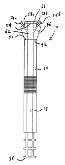
The preferred embodiment 10 of this invention is described in Figures 1
through 7. The
invention 10 comprises a long tube 14 with a large central bore 18 through it.
The tube 14
is constructed of a flexible, solvent, and acid-resistant plastic polymer. The
central bore 18
is large enough to loosely contain the tube 22 of an endoscope 26. A loose fit
is defined to
mean that there is sufficient space 30 between the endoscope tube 22 and side
34 of the
bore 18 to permit: injection of lavage fluid by a lavage system all the way to
the proximal
end 38 of the tube 14 and into the GI tract; and removal of this lavage fluid
plus blood,
mucus, particles, etc. from the GI tract by vacuum action.
The distal end 42 of the tube 14 is attached to a housing 46. The housing 46
is constructed
of a hard, solvent, and acid-resistant plastic polymer. Attachment can be by
any means
that provide air and water tight joints. Alternatively, the housing 46 and
tube 14 can be
fabricated integrally. The housing 46 is the same diameter as the tube 14 at
the proximal
end 50 but of larger diameter at the distal end 54. There are three bores 58a,
58b, 62
through the housing 46. The large central one 62 mates with the central bore
18 through
the tube 14 at the proximal end 50 and terminates in a rubber collar 66 at the
distal end 54.
This collar 66 provides an air- and water-tight seal when the endoscope head
70 is fully
advanced into the housing 46. This collar 66 is also designed to mate with
vacuum and
lavage tubing of an appropriate size with an air and water-tight seal.
The two, smaller, peripheral bores 58a, 58b intersect the large central bore
62 within the
housing 46 and terminate in fittings 74a, 74b at the distal end 54. The large
bores 18, 62
through the tube 14 and housing 46 are adapted to loosely receive the tube 22
of an
endoscope 26. The fittings 74a, 74b are adapted to mate with externally
supplied fluid and
vacuum lines.
14
CA 02535129 2006-02-07
WO 2005/016134 PCT/US2004/014575
In an alternative embodiment, the tube 14 has a central bore 18 there through
and the
housing 46 has a hollow center 48. The central bore 18 is large enough to
loosely receive
an endoscope tube. There is a collar 66 at the distal end 54 which adapts the
housing to
form an air- and water-tight seal with the head 70 of an endoscope. The collar
66 has a
central bore 68 of the same size as and axially aligned with the central bore
18 through the
tube 14. The housing 46 also has fittings 74a, 74b designed to connect tubing
to the hollow
center 48.
The reason the housing 46 expands in diameter from the proximal 50 to distal
54 end is to
provide room for attachment of tubing to the fittings 74a, 74b.
The flexible tube 14 is uniquely constructed in three basic regions. The first
and most
proximal region 78 is a smooth with a number of fenestrations 82 beginning a
short
distance behind the proximal end 38 of the tube 14. This segment 78 is
critical to the
function of the system 10 as it allows easy entrance and egress of blood, food
and foreign
semisolids and lavage fluid even while the endoscope 26 is in position.
Frontal and radial
positioning of openings 82 allows not only an easier passage of fluids but
makes it less
likely that the covering of a few of the openings by GI mucosa or extraneous
material would
seriously impede the function of the invention 10.
The second segment 86 of the tube 14 is accordion-pleated to allow for both
optimal
flexibility and strength so as to prevent collapse of the bore 18 within the
tube 14 while it is
flexed or under vacuum (negative pressure). This pleating is constructed as to
not
compromise the desired inner diameter of the tube 14. The pleating allows the
invention to
flex backwards up to almost 180o as illustrated in Figure 6.
The third segment 90 of the tube 14 is again smooth.
A separate polymer plastic cover 94, as illustrated in Figure 4, for the
endoscope docking
collar 66 allows the invention to be used without the endoscope 26 in place.
The cover is
designed to provide a water- and air-tight seal.
The peripheral bores 74a. 74b are used for vacuum extraction of blood, food
and sections,
and instillation of lavage fluid even without the endoscope 26 in place due to
a now larger
effective bore in the flexible tube 14 and housing 46 in the absence of the
endoscope 26.
is
CA 02535129 2006-02-07
WO 2005/016134 PCT/US2004/014575
In light of the previous descriptions, a number of advantages of this system
10 for its use
with and without the endoscope become obvious:
A) The system 10 allows for removal of viscous secretions, semisolids and
liquids without
having to use the inadequately small bores of the endoscope 26 itself.
B) The system 10 will provide a system 10 heretofore unavailable to the GI
endoscopist
for simultaneous lavage and endoscopic visualization and treatment.
C) Removal of larger-sized material than currently possible can be
accomplished with this
system 10.
D) The ability to remove and replace the endoscope 26 into the system 10 while
still in
place anatomically will allow it to act as a protecting shield against damage
of surrounding
GI structures when foreign bodies are removed from the GI tract. This system
10 will also
protect the upper airway as well preventing dangerous aspiration of material
into the lungs.
E) This system 10 may be used for lavage of foreign material in the absence of
an
endoscope.
F) This system 10 in modified form can be used with colonoscopy for
visualization of the
lower GI tract for cleansing of stool and blood to aid in endoscopic diagnosis
and treatment.
No other system 10 exists with this capability.
G) No other system 10 exists which can be placed endoscopically and used
independently of the endoscope 26 to draw gas and fluids from the lower GI
tract providing
the only such system 10 for simultaneous endoscopic diagnosis and treatment,
along with
decompression useful in treatment of certain common disorders. The system 10
may be
left in place without the endoscope 26 to allow for continued drainage and
decompression,
a technique heretofore impossible to perform.
H) This system 10 can be used in conjunction with endoscopic laser therapy to
remove
smoke, blood and burned tissue while simultaneously performing the therapy. No
other
system 10 has this capability.
16
CA 02535129 2006-02-07
WO 2005/016134 PCT/US2004/014575
The manner of use of the invention with a GI endoscope 26 is a new therapeutic
approach,
but straightforward in concept. The proximal end of the endoscope 26 is
introduced
through the docking collar 66 and the whole endoscope 26 advanced forward
toward the
proximal end 38 of the flexible tube 14. The endoscope 26 is advanced until
the flexible
segment of the endoscope handle 70 makes a snug air- and water- tight contact
with the
docking collar 66. A lavage hose is attached to one fitting 74a and a vacuum
hose is
attached to the other fitting 74b and the system 10 is now ready for use. The
lavage
system can be either a gravity flow bag-type infusion system or operator
controlled
peristaltic infusion pump. The vacuum system may use either wall type vacuum
unit or
portable (Gomco) type vacuum pump.
The complete system 10 is then inserted either orally or rectally (upper
endoscope or
colonoscope) in the conventional fashion. The exposed end of the endoscope 26
makes
an excellent vehicle for the introduction of both the endoscope 26 and the
invention 10.
Once advanced into the GI tract (beyond the upper esophagus/sphincter or
colon) the
invention 10 can be readily applied to remove blood, food, mucus, foreign
materials and
stool. Should the volume of viscous secretions, semisolids and liquids be of
large
magnitude, the endoscope 26 may be removed and the docking collar 66 sealed by
the
cover 94 and the invention 10 used without the presence of the endoscope 26.
Alternatively, by connecting the docking collar 66 to a large tube, the
central bores 18, 62,
68 can be used for introduction of even larger quantities of lavage fluid and
extraction of
even larger particles by vacuum.
The invention 10 is advanced while attached over the endoscope 26 but may be
removed
without the endoscope 26 being in place. The invention 10 allows for easy
reinsertion of
the endoscope 26 while it is in position in the GI tract. Extraction and
reinsertion capability
is important not only for high volume lavage and extraction but also removal
of potentially
hazardous materials and foreign objects which may cause direct injury to the
GI tract as
well as possible aspiration into and injury to the pulmonary tract. With this
invention 10
objects of almost 3/ inch can be removed.
When used with a laser the invention 10 allows use of the vacuum system to
draw out
fumes from burned tissue as well as blood, mucus, feces and secretions,
producing a
17
CA 02535129 2006-02-07
WO 2005/016134 PCT/US2004/014575
clearer field of vision for the endoscopist. The same vacuum system used for
fluid
extraction can be used for fume removal as well.
Accordingly, the reader will see that this system 10 allows for several
enhancements in the
performing of GI endoscopy. It is a unique system 10 which is presently not in
existence
and is capable of:
A) Optimal removal of blood, food, secretions and other materials from the GI
tract,
leaving the bores of the endoscope 26 free to be used for their primary role
as conduits for
diagnostic and therapeutic devices.
B) Use concomitantly with endoscopy, allowing instillation of lavage fluid and
removal of
waste while performing endoscopy.
C) Removal of foreign bodies, meat and vegetable materials from the GI tract
with rapid
and easy insertion and removal of the endoscope.
D) Removal of harmful fluids and foreign bodies from the GI tract while
preventing injury
to the GI tract and pulmonary system.
E) Use as an instrument for lavage with or without endoscope in place.
F) Use in the lower GI tract (color and lower ileum) to perform simultaneous
lavage and
clearance of materials (blood, stool, mucus, undigested food) while performing
endoscopic
examination and manipulation.
G) Use in lower GI tract independently of the endoscope for removal of solids,
liquids and
gas for medical treatment of volvulus and ileus.
H) Use for removal of gas, blood and secretions from the GI tract while
performing laser
therapy.
The system 10 presented is unique from all other GI lavage systems in that it
can be
performed simultaneously with diagnostic and therapeutic endoscopy. Present
endoscopic
and lavage system function independently both physically and temporally with
respect to
endoscopic evaluation and therapy and lavage. No system exists which can do
both at the
same time, a definite benefit to the utility of endoscopy as well as its safe
performance.
is
CA 02535129 2006-02-07
WO 2005/016134 PCT/US2004/014575
The present system is designed to be used with upper and lower endoscopes
commonly
used in clinical practice.
The following reference numerals are used on Figures 1 through 7:
This invention
5 14 Flexible tube
18 Bore through flexible tube
22 Tube portion of endoscope
26 Endoscope
30 Space between wall of bore and endoscope tube
10 34 Wall of bore
38 Proximal end of flexible tube
42 Distal end of flexible tube
46 Housing
48 Hollow center of housing in alternate embodiment
50 Proximal end of housing
54 Distal end of housing
58a,b Peripheral bores in housing
62 Central bore through housing
66 Docking collar
68 Central bore through docking collar in alternate embodiment
70 Endoscope head
19
CA 02535129 2006-02-07
WO 2005/016134 PCT/US2004/014575
74a,b Tube connection fittings
78 Proximal portion of tube
82 Fenestration
86 Medial, pleated portion of tube
90 Distal portion of tube
94 Sealing cap
Thus, the present invention 10 has been described herein with reference to
particular
embodiments for a particular application. Those having ordinary skill in the
art and access
to the present teachings will recognize additional modifications, applications
and
embodiments within the scope thereof.
It is therefore intended by the appended claims to cover any and all such
applications,
modifications and embodiments within the scope of the present invention.