Note: Descriptions are shown in the official language in which they were submitted.
CA 02535450 2006-02-10
WO 2005/014091 PCT/DE2004/001646
1
Method and Arrangement for Respiratory Support of a Patient as well as
Airway Prosthesis and Catheter
The invention concerns a method and an arrangement for respiratory support of
the
patient, as well as an airway prosthesis and a catheter for use herein.
To allow the body to take up oxygen and release carbon dioxide, both
components
of the respiratory bronchial system must function. The lung as a gas
exchanging
organ and the respiratory pump as a ventilation organ which transports air
into the
lung and back out again. The correct function of the respiratory pump requires
the
respiration centre in the brain, central and peripheral nerves, the bony
thorax and
the respiratory musculature, as well as clear, stable airways.
In certain illnesses, there is a long-term overuse or exhaustion of the
respiratory
pump. A typical illness is lung emphysema with flat diaphragm, and inability
to
contract. In
CA 02535450 2009-07-21
2
lung emphysema, the airways are usually extremely limp and collapsed. Due to
the
flattened, overstretched diaphragm, the patient cannot inhale deeply enough.
Due to the
collapsed airways, the patient is also unable to exhale sufficiently. This
leads to
insufficient respiration with oxygen undersupply and increased carbon dioxide
levels in
the bloodstream, which is also known as ventilatory insufficiency.
Treatment of inhalatory weakness is often done with a respirator. The so-
called home
respiration is artificial respiration to support or completely unburden the
respiratory
pump.
Respiration can take place non-invasively via a tube and a nose or mouth mask
which
the patient can put on and take off by himself as required. However, this
restricts free
breathing and the patient's ability to speak. Furthermore, a blocked tracheal
cannula can
be inserted into the trachea. This also means that the patient can no longer
speak.
In invasive respiration, this is usually carried out via a tracheostoma. This
is a surgically
created opening in the trachea. Via the opening, a finger-thick catheter with
a blocking
balloon is inserted into the trachea and connected to a respirator. This
enables
sufficiently deep respiration, but hinders the patient in speaking. Aside from
respiration,
there is transtracheal oxygen administration via thinner catheters.
Corresponding
suggestions can be found In US Patent No. 5,181,509 or US Patent No.
5,279,288. In
this manner, the patient receives high-dosed oxygen in a continuous stream
with a
fixedly adjusted frequency. The oxygen is regulated manually via a regulator.
It is not
possible to adapt to the patient's natural respiration process. Respiration is
not
deepened. The catheter end which is introduced into the airway may also lead
to
irritation and local trauma to the surrounding tissues by striking the trachea
due to the
movement of respiration, or the surrounding tissues are dried out by the jet
stream.
CA 02535450 2009-07-21
3
So-called "Montgomery-T tubes" which are placed within the trachea are also
known.
Through these, the patient can receive oxygen via the segment of the T which
is directed
to the outside. Furthermore, the patient can suction off his secretions
himself when
required. The patient can breathe freely and speak if the front segment is
closed.
However, artificial respiration is not possible through the "Montgomery-T
tube", since the
air which is introduced escapes upwards into the mouth and/or pharynx.
Based on the state of the art, the invention therefore has the task of
providing a more
efficient process for respiratory support for a patient, and to create an
arrangement for
this purpose which can also be carried by the patient and is safe to use.
Furthermore,
the invention aims at an airway prosthesis and a catheter which enables
respiratory
support that is synchronized with the patient's spontaneous respiration
without negatively
affecting the ability to speak.
The solution of the method section of the task consist in that the patient's
spontaneous
respiration is recorded by sensors, and an additional amount of oxygen is
administered
at the end of a respiratory process. This can take place in the form of an
oxygen burst
via a jet catheter from an oxygen reservoir. Herein, there is synchronization
of respiratory
support with the patient's natural respiration. Respiratory depth which is
reduced due to
overwork or exhaustion of the respiratory pump is thus compensated. Due to the
additional oxygen quantity, respiration is kept at sufficient levels. Oxygen
undersupply
and increased carbon dioxide in the blood are thereby avoided.
For practical purposes, the additional oxygen quantities have a volume between
25 ml
and 150 ml.
If desired, the patient's exhalation process can also be slowed by a counter-
flow as
needed. This is always recommended when the patient's airways are collapsible,
that is,
they collapse during respiration, which can extremely hinder the exhalation
process. This
is prevented in that a counter-flow is applied during exhalation, keeping the
airways open
and preventing their collapse.
A representational solution to the problem on which the invention is founded
includes an
oxygen pump which can be connected to an oxygen source, as well as an airway
prosthesis which, if applicable, can be connected via a catheter with the
further use of a
supply tube. The outflow end of the catheter forces the oxygen flow into a jet
character.
CA 02535450 2009-07-21
4
This may, for instance, be accomplished by a reduction of the cross-section.
In principle,
the end of the catheter may also be equipped with a jet nozzle. Furthermore,
the
invention intends sensors to record the patient's spontaneous respiration.
These sensors
are linked with a control unit for activation of the oxygen pump. The airway
prosthesis
possesses a tubular support body with a connector for the catheter. The
support body
and the integrated catheter are dimensioned so that the patient can breathe
and speak
freely, without restriction. The main respiration takes place through the
larger inner
lumen of the airway prosthesis. Spontaneous respiration, coughing and speaking
are not
hindered. Furthermore, the support body includes at least two sensors which
are part of
the arrangement.
The airway prosthesis is implanted in the airway of the patient. A small
airway incision is
made to provide access for the catheter to the outside. The catheter can be
led directly
into the support body with one end via the connector. It is also possible to
connect the
catheter to the connector externally via a coupling mechanism.
The sensors serve to record the patient's spontaneous respiration. Various
respiration
sensors, such as respiration flow sensors or pressure sensors, can be used.
Thermistors
are particularly advantageous. These are semi-conductor components with
temperature-
dependent resistance. The temperature dependency of the resistance forces is
used to
record the inhalation and exhalation processes, since the exhaled air in the
lung is
naturally warmer within the airway than the inhaled air.
In accordance with the present invention, a sensor is applied to the internal
wall of the
supporting body. The other sensor is arranged on the external wall of the
support body
or embedded within the support body itself.
A bridge circuit is provided for compensation of the recorded measurement
value
differences between the internal and external sensors. This double arrangement
can be
used to equalize environmental influences, such as temperature variations etc.
In accordance with the present invention, the catheter end which is located
within the
support body is largely positioned parallel to its longitudinal axis and
provided with a jet
nozzle at its end. This may be a separate nozzle. However, the jet nozzle may
also be
designed in the form of a reduction in cross-section at the end of the
catheter. In this
manner, the air or oxygen flow which is introduced via the catheter can be
aimed in the
CA 02535450 2009-07-21
direction of the lungs, and this can be accomplished with a laminar flow. The
oxygen is
prevented from escaping into the mouth or pharyngeal space. The support body
which
receives the catheter end or end piece prevents dehydration of the surrounding
tissues.
Trauma to the airway and/or surrounding tissues, e.g. through movements of the
catheter end, is furthermore avoided.
The oxygen pump is functionally structured as a piston pump. The use of a
cylinder with
a double-action piston or a movable membrane is particularly preferable. Such
an
oxygen pump excels due to its compact construction. Furthermore, reliable
adjustment of
the supplied oxygen quantity is possible in supporting both the exhalation
process and
the inhalation process. Since the maximum quantity of air per jet lift is
limited by the
cylinder size, overinflation of the lung with consequential baro trauma is
also prevented.
Within the framework of the arrangement as per the invention, it is possible
to use two
catheters, wherein one jet catheter is used to support the inhalation process,
and the
other catheter is used for precisely slowing the exhalation process. A
catheter can also
be constructed with a double lumen. The double-lumen catheter provides
separate
channels for the administration of oxygen in the inhalation process and in the
exhalation
process.
The security of the arrangement is increased through the provision of
additional
respiration sensors. These, too, are sensors which record the patient's
spontaneous
respiration. These may, for instance, be affixed to the patient's chest so
that
spontaneous respiration can be monitored through a thorax impedance
measurement.
Sound or flow measurement at the patient's mouth or nose is also a
possibility.
Inhalation or exhalation support is provided by equalizing the recorded
signals from the
airway and the further respiration sensors in a control unit and sending
corresponding
signals to the oxygen pump. The additional respiration sensors guarantee
redundant
construction and contribute to the security of the arrangement.
Self-reliant protection is desired for the airway prosthesis as per the
present invention.
This possesses a tubular support body with a connector for a catheter, with at
least two
sensors which are arranged on the support body. The airway prosthesis excels
in its
ability to allow measurement of the patient's respiration. This permits
synchronization of
external respiratory support with the patient's own respiration.
CA 02535450 2009-07-21
6
A sensor is advantageously mounted on the internal wall of the support body.
Thermistors are regarded as particularly suitable within the framework of the
invention.
By linking the thermistors via a bridge circuit, it is possible to compensate
for
temperature differences between the Internal and external thermistors. This
double
arrangement of the sensors in the bridge circuit compensates for environmental
influences, such as temperature variations, or also differences which may be
caused by
secretions coming into contact with the internal sensor, thereby producing
localized
cooling or warming.
It is furthermore advantageous if the catheter end is placed within the
support body so
that it is parallel to the longitudinal axis of the support body. This results
in directional
provision of the oxygen flows in the direction of the bronchial tract, with
laminar flow
conditions.
Furthermore, the catheter of the present invention has an outflowing end which
includes
at least one sensor. It is functional to provide at least two sensors in this
location in
order to be able to carry out the compensation of measurement values within a
bridge
circuit.
Such a catheter can be introduced into a support body from the outside, such a
support
body may, for instance, consist of the well-known "Montgomery-T-Stent". The
catheter is
introduced from the externally accessible segment of the T segment so that
respiration
can be supported via the catheter.
Furthermore, the end of the catheter has a jet nozzle. As already described
above, this
can, for instance, be provided by a reduction in the cross-section of the end.
However, it
may also consist of a separate jet nozzle.
The end of the catheter is preferably bent. In this manner, the end which is
introduced
into the airway or support body is naturally oriented into the direction of
the bronchial
tract, parallel to the longitudinal axis of the support body.
According to further broad aspect of the present invention there is provided
the use of a
transtracheal catheter for supplementing respiratory volume of a spontaneously
breathing patient comprising: a) inserting the transtracheal catheter, having
a first end
and a second end, into an airway of the patient such that the second end is
adapted to
terminate in the trachea of the patient, wherein .the inserted transtracheal
catheter
CA 02535450 2009-07-21
7
permits spontaneous patient breathing while inserted into the airway of the
patient;
b) determining a spontaneous inspiration process and a spontaneous expiration
process
of the patient; and c) activating a delivery mechanism based on the
determining step to
deliver a supplemental gas volume through the transtracheal catheter and into
the
patient's lungs synchronized with a portion of the patient's spontaneous
inspiration
process.
According to still further broad aspect of the present invention there is
provided an open
system ventilation apparatus for supplementing respiration of a spontaneously
breathing
patient, comprising: a) a gas delivery mechanism connected to an oxygen
source; b) a
transtracheal catheter having a first end and a second end, the first end
connected to the
gas delivery mechanism and the second end adapted and configured for
transtracheal
insertion into the patient airway without obstructing the patient's
spontaneous respiration
phases such that the second end terminates in the trachea of the patient; c)
at least
one respiration sensor in communication with the transtracheal catheter and
adapted to
sense the spontaneous respiration phases of the patient; and d) a control unit
in
communication with the at least one respiration sensor, the control unit
adapted and
configured to control the gas delivery mechanism to deliver a supplemental
volume of
gas to the transtracheal catheter in synchrony with a portion of the patient's
spontaneous
breathing pattern.
According to a still further broad aspect of the present invention there is
provided a
tracheal prosthesis comprising: a tubular support body having a first end and
a second
end and a lumen therebetween, wherein the tubular support body is sized and
configured to terminate within and along a portion of the trachea without
occluding the
tracheal airway while permitting the spontaneous breathing of a patient
through the
lumen; a connector on the tubular support body between the first end and the
second
end, the connector configured to attach to a catheter; a catheter having a
first end and a
second end and a lumen therebetween wherein. the first end is connected to the
connector so that the lumen of the catheter is aligned along the tubular
support body
lumen and toward the second end of the tubular support body; and at least one
respiration detection sensor coupled to the tubular support body, wherein the
at least
one respiration detection sensor is in communication with the lumen of the
tubular
support structure without being in line with the lumen of the catheter.
CA 02535450 2009-07-21
7a
According to a still further broad aspect of the present invention, there is
provided a
catheter for delivering ventilation to a patient comprising: an elongate body
having a first
end, a second end and a lumen therebetween wherein the first end is adapted
and
configured for connection to an outlet so that gas flowing from the outlet
moves through
the lumen; the second end of the elongate body is adapted and configured for
insertion
transtracheally into a trachea of a patient so that the second end may be
inserted into
the trachea without occluding the tracheal airway of the patient such that the
second end
terminates in the trachea of the patient; and at least one respiratory sensor
positioned
on the elongate body without being in the path of the gas flow through the
lumen.
According to a still further broad aspect of the present invention there is
provided the use
of a wearable ventilation system for supplementing a patient's spontaneous
breathing,
comprising: determining the patient's spontaneous breathing by a respiration
sensor
which measures intra-tracheal airflow; delivering a supplemental volume to the
patient
via a transtracheal catheter having a first end and a second end that does not
substantially obstruct the patient's airway and the second end is adapted to
terminate in
the trachea of the patient wherein the supplemental volume is delivered in
synchrony
with a portion of the patient's inspiratory and/or expiratory spontaneous
breath phase;
and providing mobility to the patient by performing the delivering step with
the wearable
ventilation system that is configured to be worn by the patient.
The invention is described in further detail by the attached drawings. The
following are
shown:
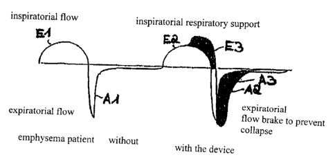
Figure 1 The upper body of a patient who is wearing a respiratory support
arrangement as per the invention.
Firgure 2 A diagram showing the respiratory flow of an emphysema patient,
with
and without respiratory support.
Figure 3 A technically simplified representation of an airway prosthesis as
per the
invention.
CA 02535450 2006-02-10
WO 2005/014091 PCT/DE2004/001646
8
Figure 4 A further embodiment of an airway prosthesis.
Figure 5 Also, in the schema, an oxygen pump belonging into the arrangement
as per the invention, depicting control of the air flow, as well as a control
unit.
Figure 6 The end section of a catheter as per the invention, and
Figure 7 the catheter placed into a support body as in figure 6.
Figure 1 uses P to indicate a patient suffering from lung emphysema, with
overwork
and exhaustion of the respiratory pump. This renders the patient unable to
inhale
deeply enough. The exhalation process is furthermore obstructed by limp and
collapsing airways.
Such a respiration process with inhalation (inspiratorial flow) and exhalation
(expiratorial flow) is shown in figure 2 in the left half of the image. The
inhalation
curve is identified as El, while the exhalation curve is identified with Al.
To support and unburden the respiratory pump, the patient's spontaneous
respiration is recorded by sensors, and an additional quantity of oxygen is
administered to the lungs at the end of an inhalation process. This
respiration flow is
further clarified in figure 2 in the right half of the image. The additional
quantity of
oxygen increases the respiration volume during inhalation as shown in curve E2
by
the differential volume which is darkened in on the upper curve, and
identified as E3.
The additional oxygen quantity may possess a volume between 25 ml and 150 ml.
The patient's exhalation process is furthermore slowed by a counter-flow. This
causes the respiratory flow during exhalation to shift as shown in the curve
which is
identified as A2. This resistance, which specifically counteracts the
exhalation flow,
prevents airway collapse during exhalation. This process enlarges the
exhalation
volume by the volume which is also darkened in, and identified as A3.
CA 02535450 2006-02-10
WO 2005/014091 PCT/DE2004/001646
9
This process consequently prevents insufficient respiration with oxygen
undersupply
and increased carbon dioxide levels in the bloodstream. The patient P is
significantly
more stressable and mobile, as well as feeling less or no respiratory
distress.
The arrangement which is intended to provide respiratory support to the
patient P
includes an oxygen pump 1 which can be connected to an oxygen source (see
figure 5) and an airway prosthesis 2, 3 (see figures 3 and 4). In accordance
with
figure 1, the oxygen pump 1 is part of a compact mobile respiration unit 4.
The
oxygen pump 1 and the airway prostheses 2 and 3 are connected via a catheter
5.
As figures 3 and 4 show, each airway prosthesis 2 and 3, respectively,
possesses a
tubular support body 6 with a connector 7 for the catheter 5. Two sensors 8, 9
are
assigned to the support body 6 in the form of thermistors for the purpose of
recording
the patient's spontaneous respiration. Herein, a sensor 8 is fastened to the
internal
wall 10 of the support body 6, while the other sensor 9 is located at the
outside wall
11 of the support body 6. The sensors 8, 9 are connected with a control unit
12 for
activating the oxygen pump 2. The control unit 12 is schematically shown in
figure 5
with its entries and exits. As already mentioned, the sensors 8, 9 are
thermistors,
that is, temperature dependent resistors. These are linked in a bridge circuit
within
the arrangement, so that the compensation of measurement values between the
inner sensor 8 and the outer sensor 9 takes place in response to environmental
influences.
It is furthermore shown in figure 1 that further respiration sensors 13, 14
are
intended. These are likewise sensors for recording the spontaneous respiration
of
the patient P. Equalization of the measurement values recorded by the sensors
8
and 9, as well as 13 and 14, provide a precise depiction of the respiratory
process of
the patient P. Security against erroneous measurements or failure of one of
the
sensors 8, 9 as well as 13, 14 is furthermore improved.
In the airway prosthesis 2 as per figure 3, the jet catheter 5 can be
introduced into
the support body 6 via the connector 7. The end of the jet catheter 15 which
is
located within the support body 6
CA 02535450 2006-02-10
WO 2005/014091 PCT/DE2004/001646
is guided / redirected parallel to the longitudinal axis L of the support
body. The data
conduits of the sensors 8, 9 for the control unit 12 are identified as 16 and
17. These
run within the catheter 5. At the outflow end 15, the jet catheter 5 is
designed as a
jet nozzle 25. This can be accomplished by a reduction of the catheter cross-
section. This increases the speed of the oxygen flow at the exit of the
catheter 5,
directing it into the direction of the bronchial tract. The diameter of the
support body 6
is dimensioned with a lumen which is sufficiently large so that the patient P
can
breathe and speak freely even with the integrated catheter 5.
In the airway prosthesis 3 as per figure 4, a separate coupling 18 is provided
at the
connector 7 to connect the catheter 5 to the airway prosthesis 3. In this
case, within
the support body 6 and parallel to the longitudinal axis L, a fixed length
segment 19
is intended as a catheter end, wherein the oxygen flow is directed into the
direction of
the bronchial tract via a jet nozzle 26.
The oxygen pump 1 is schematically shown in figure 5. It involves a cylinder
pump
with a double-action piston 20 which is arranged within a cylinder 27. The
arrangement possesses a total of four valves Vito V4. Oxygen is supplied out
of an
external oxygen reservoir via the connector 21. The switching conditions of
the
valves Vito V4, as well as the incoming and outgoing supply lines, are
identified by
the letters a to g.
In respiratory support, the function of the oxygen pump 1 within the
arrangement is
as follows:
When the valve V1 from c to a are open (b to c closed) and the valve V2 from b
to e
is open (e to d closed), the piston 20 at the image level moves to the left,
and
oxygen flows through the outlet 22 and the jet catheter 5 to the patient P.
The
additional quantity of oxygen E3 is administered during the inhalation process
of the
patient P.
When the valve V1 from b to c (c to a closed) is open, and the valve V2 from e
to d
is open (b to e closed), the piston 20 at
CA 02535450 2012-12-10
It
the image level moves to the right, and oxygen flows out in the direction of
the valve
V3. The valve V3 is connected to the outside air via an outlet 23. If the
valve V3
from d to g is open, the oxygen flows without an expiration resistor: This
means that
the exhalation process is not slowed by a counter-flow.
If the valve V3 from d to g is closed, and is open from d to f, the oxygen
flows in the
direction via the supply line 24 to the outlet 23 and the catheter 5 to be
administered
to the patient P during the exhalation process, as well as slowing the
respiratory
flow. The counter-flow prevents airway collapse and keeps the airways open.
This
enables deeper exhalation.
In the supply line 24 of the arrangement, the valve V4 is also switched,
allowing
variable adjustment of the flow-through (f to a). This may preferably consist
of a
proportional valve with pulse width modulation.
Figure 6 shows a catheter 28 with a long, flexible tube 29 and an outflow end
31
which is angled through the use of a bent segment 30. The end includes two
sensors 32, 33 to record the spontaneous respiration of a patient P. The
sensors 32,
33 preferably consist of thermistors. The representation of data cables has
been
omitted for the sake of simplicity. These run through the catheter 28 or the
catheter
wall. 34 identifies a stop.
It is furthermore recognizably shown that the end 31 of the catheter 28 is
provided
with a jet nozzle 35. Within the jet nozzle 35, the flow cross-section is
reduced
relative to the cross-section of the catheter, so that the exit speed of the
supplied
oxygen is increased.
The catheter 28 may be introduced into a support body 36, as shown in figure
7.
The support body 35 is located within the airway of a patient P. The
connection to
the outside is provided via a connector 37.
The support body 36 may consist of a customary "Montgomery-T-Stent".
CA 02535450 2006-02-10
WO 2005/014091
PCT7DE2004/001646
12
List of reference symbols
1 - Oxygen pump
2 - Airway prosthesis
3 - Airway prosthesis 4
Respirator
- -Catheter
6- Support body 7
Connector
8 - Sensor
9 - Sensor
Internal wall, front 6 11
External wall, front 6 12
Control unit
13 - Respiration sensor
14 - Respiration sensor
15- End, front 5
16- Data cable
17- Data cable
18- Coupling
19- Length segment
20- Piston
21 - Connector
22 - Outlet
23- Outlet 24
¨ supply line
25- Jet nozzle
26- Jet nozzle
27- Cylinder
28- Catheter
29- Tube
30 - Bend
31 - End, front 28
32- Sensor 33 -
Sensor
CA 02535450 2006-02-10
WO 2005/014091
PCT/DE2004/001646
13
34- Stop
35- Jet nozzle
36- Support body
37 - Connector
P- Patient
El ¨ Inhalation curve
E2 - Inhalation curve
E3 - Volume Al ¨
Exhalation curve A2 ¨
Exhalation curve A3 -
Volume
VI - Valve
V2- Valve
V3- Valve
V4- Valve
L- Longitudinal axis, front 5
a - line
b- line
c- line
d - line
e - line
f- line
g - line