Note: Descriptions are shown in the official language in which they were submitted.
CA 02535454 2006-02-10
1
Curved positioning and insertion instrument for
inserting a guide wire into the femur
The invention relates to a curved positioning and
insertion instrument for inserting a guide wire into
the femur for precise bone opening.
Precise bone opening requires a reliable reference in
position and direction which a surgeon with a drill or
cutter can use as a guide. It is known that guide
wires which are inserted (drilled or tapped) into the
femur prior to drilling in order to guide the drill
safely are used for this purpose.
US-A-6074392 describes such a curved positioning and
insertion instrument for inserting a guide wire into
the femur, comprising a curved guide tube having a
distal end for placing on the trochanter and a proximal
end for pushing in a guide wire, comprising a retaining
arm for the connection between the guide tube and a
handle. This design has been chosen because the
insertion of a guide wire along a curve is advantageous
with regard to the anatomy of the human femora, and the
tissue in the affected region is therefore impaired to
a lesser extent during an operation.
US-A-6074392 therefore describes an instrument design
comprising a key element which consists of a curved
guide tube for a curved guide wire. This guide wire is
CA 02535454 2006-02-10
2
inserted into the bone and serves as a guide path for a
hollow and flexible drill. The guide wire is mounted
with the aid of a positioning and insertion instrument,
the curved guide tube being mounted by means of a
support part on a handle which can be properly held by
the surgeon. The exact positioning of the guide tube
is implemented by means of X-rays or by direct
inspection. After positioning, the guide wire is
hammered in along the longitudinal axis of the bone or
driven in in another manner. Thereafter, the insertion
instrument is removed and the flexible drill or cutter
is passed over the guide wire. After drilling is
complete, everything is removed.
However, the weak point of this solution is that there
is no particular possibility for correction if the X-
ray image shows that the guide wire is not optimally
positioned.
In a completely different design, for example according
to US-A-5951561 a guide instrument which has the
possibility of correction of guide wires is also
described. However, this design consists of a bulky
sheath which receives a rotary cylindrical component
comprising a plurality of discs each having a plurality
of holes, which have to be oriented concentrically with
one another in a complicated production process and in
addition provide no possibility for tapping a guide
wire along a curve into the femur. To this extent,
this design offers no improvement for a design
CA 02535454 2006-02-10
3
according to the generic type.
It is now the object of the invention to permit
corrections by means of a simple device, the intention
being to impair the tissue in the affected region as
little as possible. For this reason, as already noted
above, the person skilled in the art will not at all
have used US-A-5951561 as prior art for achieving the
present object. It is also comprehensible that US-A-
6074392 published in 1998 is itself a further
development of US-A-5624447 published in 1997, while
US-A-5951561 was published in the same year as US-A-
6074392 and therefore followed another parallel route
for setting guide wires.
Further but less relevant documents relating to the
prior art are US-A-4466429, US-A-3439671, US-A-5135527,
US-A-5112336, US-A-4712541, US-B1-6273892.
In order to achieve the object, the inventor now
presents a novel positioning and insertion instrument
which comprises a handle which is arranged by means of
a retaining arm on a curved guide tube. At the end
thereof there is a positioning hook with, if
appropriate, a guide bore which is oriented at an angle
of 6° - 30°, preferably 7° - 20°, in particular
about 8°,
relative to the guide tube. In addition to this bore,
various correction bores are present, according to an
improved further development. Furthermore, a T-handle
is required when the instrument according to the
CA 02535454 2006-02-10
4
invention is used. The invention thus makes it
possible to insert at least one guide wire which
subsequently guides an opening drill. Instead of a T-
handle or a motorised drilling or tapping drive, it is
also possible to provide an impact head so that the
guide wire can be hammered in.
According to the invention, during use in practice, the
guide tube is, if appropriate, first loaded with a
reference guide wire. The handle of the positioning
and insertion instrument is then aligned by visual
inspection approximately parallel to the longitudinal
axis of the femur and with the extension of the
intramedullary canal thereof. The positioning hook is
placed on the trochanter and the nose of the hook is
forced by pressure in the medial direction into the
muscle (gluteus medius), the reference guide wire being
inserted parallel to the longitudinal axis of the
femur. The instrument is removed. Thereafter, the
guide tube of the positioning and insertion instrument
is loaded with an opening guide wire and, according to
this method of use, the guide bore in the positioning
hook is pushed over the reference guide wire and thus
once again placed on the trochanter. Because the guide
bore in the positioning hook is set at about 8° the
positioning and insertion instrument becomes aligned
accordingly in order to give the opening guide wire the
correct angle. This procedure is carried out because
it is more difficult to estimate a certain angle
visually than a parallelism. If, however, the
CA 02535454 2006-02-10
reference guide wire were to happen to fit the first
time, it would also be possible to use it as an opening
guide wire. As a rule, however, it is expected that a
second guide wire has to be set as the opening guide
5 wire.
A number of embodiments without a guide bore dispenses
with the insertion of the reference guide wire and
ensures the placing of the positioning and insertion
instrument at the correct site and angle by the
particular characteristics of its positioning hook
which are adapted to the anatomical conditions. The
design of these hooks is therefore chosen so that, when
gently pressed into the medial muscle and when placed
on the trochanter, the hook comes to rest in the
optimum position for insertion of the opening guide
wire in the majority of applications. The nose is
designed so that it can be pressed into the medial
muscle while protecting soft tissue. Its thickness is
about 0.5-lOmm, preferably about 1-5mm and in
particular about 2.5mm. The length between the centre
of the guide tube and the tip of the nose of the
positioning hook may vary, depending on the soft tissue
situation, between about 10-30mm, preferably about 15-
25mm, and is in particular about 20mm. The correction
bores have a diameter of about 1-5mm, preferably about
1.8-3.6 and in particular about 3.3mm. The receptacle
for the guide tube is about 3-20mm, preferably about 6-
lOmm and in particular about 8mm.
CA 02535454 2006-02-10
6
If the opening guide wire is also inserted into the
trochanter, it is approximately at an optimum angle of
about 8° relative to the reference guide wire in the
frontal plane at its insertion point. X-ray images can
now confirm whether the opening guide wire is in the
correct position for implementing the opening.
However, if it is, for example, too close to the
lateral cortex there is according to the invention the
possibility of a correction with the aid of the
positioning and insertion instrument. This is then
pushed with one of its correction bores over the
reference or opening guide wire and a further,
previously loaded, opening guide wire is inserted in a
new position into the trochanter. The old opening
guide wire which was not correctly positioned can then
be removed. If required, such a correction measure can
be made on the basis of the only set reference guide
wire by directly pushing a correction bore, instead of
the guide bore, onto the reference guide wire.
Further developments of the invention are indicated in
the figures and in the dependent patent claims. Claim
10 describes a novel method improved compared with the
prior art and intended for placing an opening guide
wire.
The list of reference numerals is part of the
disclosure.
The invention is explained in more detail schematically
CA 02535454 2006-02-10
7
and by way of example on the basis of figures.
The figures are described in relation to one another
and as a whole. Identical reference numerals denote
identical components, and reference numerals with
different indices indicate functionally identical or
similar components.
Fig. 1 schematically shows the positioning and
insertion instrument having a T-handle, guide wires and
an opening drill;
Fig. 2 schematically shows the positioning and
insertion instrument with loaded guide wire;
Fig. 3a schematically shows the positioning hook of the
positioning and insertion instrument with its guide and
correction bores and the nose for retention in the
medial muscle in frontal view or anterior/posterior
view.
Fig. 3b schematically shows the positioning hook of the
positioning and insertion instrument with its guide and
correction bores and the nose for retention in the
medial muscle in view A according to fig. 3a;
Fig. 4 schematically shows the positioning and
insertion instrument with guide wire and T-handle,
placed on the trochanter and ready for drilling, in
frontal view or anterior/posterior view;
CA 02535454 2006-02-10
8
Fig.5 schematically shows the positioning and insertion
instrument loaded with the opening guide wire and
pushed over the reference guide wire;
Fig. 6 schematically shows the positioning and
insertion instrument with T-handle, loaded with the
opening guide wire, moved over the reference guide wire
and placed again on the trochanter and ready for
drilling;
Fig. 7 schematically shows the opening drill pushed
over the opening guide wire and with T-handle ready for
drilling;
Fig. 8 schematically shows an X-ray image of the femur
with inserted guide wire;
Fig. 9 schematically shows an X-ray image of the femur
with inserted reference guide wire and opening guide
wire to be inserted;
Fig. 10 schematically shows an X-ray image of the femur
with inserted reference guide wire and inserted opening
guide wire;
Fig. 11 schematically shows an X-ray image of the femur
with inserted opening guide wire and without or
optionally with removed reference guide wire;
CA 02535454 2006-02-10
9
Fig. 12 schematically shows an X-ray image of the femur
with non-optimally positioned first opening guide wire
and second opening guide wire for correction;
Fig. 13 schematically shows an X-ray image of the femur
with correctly set opening guide wire and
Fig. 14 schematically shows an X-ray image of the femur
with opening bore.
Fig. 15a schematically shows the positioning hook of
the positioning and insertion instrument without guide
bore and with its correction bores and the nose for
retention in the medial muscle in frontal view or
anterior/posterior view;
Fig. 15b schematically shows the positioning hook of
the positioning and insertion instrument without guide
bore and with its correction bores and the nose for
retention in the medial muscle in view A according to
fig. 15a;
Fig. 16a schematically shows the positioning hook of
the positioning and insertion instrument without guide
bore and with its correction bores and the nose for
retention in the medial muscle in frontal view or
anterior/posterior view;
Fig. 16b schematically shows the positioning hook of
the positioning and insertion instrument without guide
CA 02535454 2006-02-10
1~
bore and with its correction bores and the nose for
retention in the medial muscle in view A according to
fig. 16a;
Fig. 17a schematically shows the positioning hook of
the positioning and insertion instrument without guide
bore and with its correction bores and the nose for
retention in the medial muscle in frontal view or
anterior/posterior view;
Fig. 17b schematically shows the positioning hook of
the positioning and insertion instrument without guide
bore and with its correction bores and the nose for
retention in the medial muscle in view A according to
fig. 17a;
Fig. 18a schematically shows the positioning hook of
the positioning and insertion instrument without guide
bore and with its correction bores and the nose for
retention in the medial muscle in frontal view or
anterior/posterior view;
Fig. 18b schematically shows the positioning hook of
the positioning and insertion instrument without guide
bore and with its correction bores and the nose for
retention in the medial muscle in view A according to
fig. 18a;
Fig. 19a schematically shows the positioning hook of
the positioning and insertion instrument without guide
CA 02535454 2006-02-10
11
bore and with its (additional) correction bores and the
nose for retention in the medial muscle in frontal view
or anterior/posterior view;
Fig. 19b schematically shows the positioning hook of
the positioning and insertion instrument without guide
bore and with its (additional) correction bores and the
nose for retention in the medial muscle in view A
according to fig. 19a.
Fig. 1 shows the individual components which as a rule
are required in using the instrument according to the
invention. The positioning and insertion instrument 1
consists of a curved guide tube 2, at the distal end of
which a positioning hook 3 is arranged. The curvature
of the guide tube 2 describes a radius of about 300 to
800mm, preferably 500 to 700mm, in particular about
600mm. The positioning hook 3 is formed in such a way
that it is retained on the tip of the greater
trochanter and the medial muscle. In the region of the
proximal end of the guide tube 2, mounted approximately
perpendicularly thereto, is a retaining arm 4 which
then curves in order to run approximately parallel to
the guide tube 2. A handle 5, the longitudinal axis of
which is approximately parallel to a tangent to the
guide tube 2 in the region of the positioning hook 3,
is present at the end of the retaining arm 4.
Furthermore, fig. 1 shows two guide wires 50 which are
such that they can be inserted into the guide tube 2
and can be used either as reference guide wire 50a or
CA 02535454 2006-02-10
12
as opening guide wire 50b. In addition a hollow
opening drill 20 is shown, which is formed in such a
way that it can be passed over a guide wire 50 and can
be operated by a T-handle 30. Finally, fig. 1 shows
the above mentioned T-handle 30 with a lever 32 and a
grill chuck, which fits both onto the guide wires 50
and on to the opening drill 20. Of course, motor-
driven drilling and tapping drives are of course also
possible as an alternative to the T-handle.
Fig. 2 shows the positioning and insertion instrument 1
with a guide wire 50 loaded into the guide tube 2. The
guide wire 50 can be inserted at the proximal or distal
end of the positioning and insertion instrument l,
which leads to at least temporary curvature of the
guide wire 50.
Fig. 3a and fig. 3b show the positioning hook 3 of the
positioning and insertion instrument 1, comprising a
guide bore 6, which is set at an angle of about 8° in
the nose 11 relative to the guide tube 2 and the
central bore thereof. In addition to this bore,
various correction bores are present, in particular an
anterior/posterior correction bore 7, a medial
correction bore 8, a posterior/anterior correction bore
9 and a lateral correction bore 10.
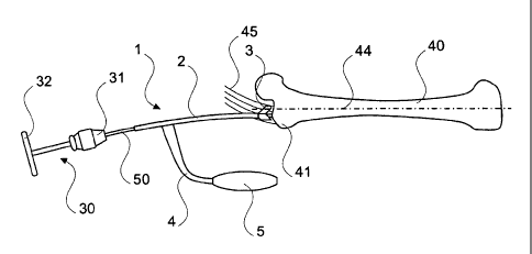
Fig. 4 shows the positioning and insertion instrument 1
with a guide wire 50 lowered into the guide tube 2,
which instrument is placed on the greater trochanter 41
CA 02535454 2006-02-10
13
of a femur 40. The characteristics of the positioning
hook 3 ensure proper non slip positioning on the
surface of the trochanter 41 and in the medial muscle
45. The handle 5 of the positioning and insertion
instrument 1 is aligned parallel to the longitudinal
axis of the femur 40 and parallel to the extension of
the intramedullary canal thereof. The T-handle 30 is
shown in the position in which it is capable of
inserting the guide wire 50.
Fig. 5 shows the positioning and insertion instrument 1
which is just being pushed along its guide bore 6 on
the reference guide wire 50a already inserted into the
trochanter 41 of the femur 40. The positioning and
insertion instrument 1 or its guide tube 2 is loaded
with an opening guide wire 50b.
Fig. 6 shows the positioning and insertion instrument 1
which has been pushed through the guide bore 6 over the
reference guide wire 50a already inserted into the
trochanter 41 of the femur 40. The positioning and
insertion instrument 1 is correctly positioned
corresponding to the angle of about 8° in the frontal
plane of the guide bore 6, in order to insert the
already loaded opening guide wire 50b. The T-handle 30
is shown in the position in which it is capable of
inserting the opening guide wire 50b.
Fig. 7 shows the opening drill 20 when it has been
pushed over the inserted opening guide wire 50b. The
CA 02535454 2006-02-10
14
T-handle 30 is shown in the position in which it is
capable of inserting the opening drill 20 into the
trochanter 41 of the femur 40.
Fig. 8 shows an X-ray image which shows the positioning
hook 3 of the positioning and insertion instrument 1
and a guide wire 50 loaded in the guide tube 2 and
inserted into the trochanter 41 of the femur 40.
Proper positioning by means of the characteristics of
the positioning hook 3 on the surface of the trochanter
41 is evident. The guide wire 50 has been inserted
parallel to the longitudinal axis of the femur 40 and
in the intramedullary canal thereof.
Fig. 9 shows an X-ray image which shows the positioning
hook 3 of the positioning and insertion instrument 1,
which has been pushed via its guide bore 6 over the
reference guide wire 50a already inserted into the
trochanter 41 of the femur 40. The positioning and
insertion instrument l, is optimally correctly
positioned, corresponding to the angle of about 8°, in
the frontal plane of the guide bore 6, in order to
insert the already loaded opening guide wire 50b.
Fig. 10 shows an X-ray image which shows the reference
guide wire 50a and opening guide wire 50b inserted into
the trochanter 41 of the femur 40. The desired
insertion angle 42 of about 8° between reference guide
wire 50a and opening guide wire 50b is clearly
recognisable.
CA 02535454 2006-02-10
Fig. 11 shows an X-ray image which shows the remaining
opening guide wire 50b optionally after removal of the
reference guide wire 50a. In this example, the opening
5 guide wire 50b is located too close to the lateral
cortex to implement the opening and has to be
corrected.
Fig. 12 shows an X-ray image which shows the correction
10 of the opening guide wire 50b. A second opening guide
wire 50b is inserted into the trochanter 41 parallel to
the guide wire 50b.
Fig. 13 shows an X-ray image which shows the corrected
15 opening guide wire 50b after removal of the first
opening guide wire 50b. It is now located optimally
for implementing the opening by means of opening drill
20.
Fig. 14 shows an X-ray image which shows the
implemented opening 43 of the trochanter 41 of the
femur 40.
Fig. 15a and fig. 15b show the positioning hook 3 of
the positioning and insertion instrument 1, which
positioning hook has no guide bore, comprising various
correction bores, in particular an anterior/posterior
correction bore 7, a medial correction bore 8, a
posterior/anterior correction bore 9 and a lateral
correction bore 10. In comparison with the variant
CA 02535454 2006-02-10
16
shown in fig. 3a and fig. 3b, the nose 11 is also
markedly thinner.
Fig. 16a and fig. 16b show the positioning hook 3 of
the positioning and insertion instrument 1, which
positioning hook has no guide bore, comprising various
correction bores, in particular an anterior/posterior
correction bore 7, a medial correction bore 8, a
posterior/anterior correction bore 9 and a lateral
correction bore 10. In comparison with the variant
shown in fig. 15a and fig. 15b, the nose 11 tapers
somewhat more sharply and is longer.
Fig. 17a and fig. 17b show the positioning hook 3 of
the positioning and insertion instrument 1, which
positioning hook has no guide bore, comprising various
correction bores, in particular an anterior/posterior
correction bore 7, a medial correction bore 8, a
posterior/anterior correction bore 9 and a lateral
correction bore 10. This variant differs in comparison
with the variants shown in fig. 15a and fig. 15b, fig.
16a and fig. 16b, substantially through a smaller
cross-sectional area. The correction bores are
enclosed by the outer contour.
Fig. 18a and fig. 18b show the positioning hook 3 of
the positioning and insertion instrument 1, which
positioning hook has no guide bore, comprising various
correction bores, in particular an anterior/posterior
correction bore 7, a medial correction bore 8, a
CA 02535454 2006-02-10
17
posterior/anterior correction bore 9 and a lateral
correction bore 10. In comparison with the variant
shown in fig. 17a and fig. 17b, the nose tapers
somewhat more sharply and is longer. This variant
differs in comparison with the variants shown in fig.
15a and fig. 15b, fig. 16a and fig. 16b, substantially
through a smaller cross-sectional area. The correction
bores are enclosed by the outer contour.
Fig. 19a and fig. 19b show the positioning hook 3 of
the positioning and insertion instrument 1, which
positioning hook has no guide bore, comprising various
correction bores, in particular an anterior/posterior
correction bore 7, a medial correction bore 8, a
posterior/anterior correction bore 9, a lateral
correction bore 10 and four additional correction
bores.
CA 02535454 2006-02-10
18
List of reference numerals
1 - Positioning and insertion instrument
2 - Guide tube
3 - Positioning hook
4 - Retaining arm
- Handle
6 - Guide bore
7 - Anterior/posterior correction bore
8 - Medial correction bore
9 - Posterior/anterior correction bore
Lateral correction bore
-
11 Nose
-
12 Additional correction bore
-
Opening drill or cutter
-
T-handle
-
31 Drill chuck
-
32 Lever
-
Femur
-
41 Trochanter
-
42 Angle
-
43 Opening
-
44 Femur axis
-
Medial muscle
-
Guide wire
-
50a - Reference guide wire
50b - Opening guide wire
50b1 - Further opening guide wire