Note: Descriptions are shown in the official language in which they were submitted.
CA 02535502 2006-02-10
WO 2005/019416 PCT/US2004/022403
METHOD AND DEVICE FOR MONITORING LOSS OF BODY FLUID AND
DISLODGMENT OF MEDICAL INSTRUMENT FROM BODY
FIELD OF THE INVENTION
[01] The present invention is in the field of methods and devices for alerting
medical personnel of the leakage of blood or other fluids from a medical
instrument
insertion site into the body and of the dislodgment of the medical instrument
from the
insertion site.
BACKGROUND OF THE INVENTION
[02] A"fistula needlC'is a large bore needle, commonly 14 to 17 gauge, which
is
bonded to a section of medical grade tubing used to connect the fistula needle
to an
extracorporeal blood circuit for use in hemodialysis.
[03] Hemodialysis is one of the primary treatments for patients with kidney
failure.
These life-sustaining treatments typically require 3 to 4.5 hours each and may
occur
three or more times a week. However, due to differences in protocols,
techniques,
or varying patient needs, some hemodialysis treatment may last six hours or
even
overnight.
[04] The most common access to the vascular system during hemodialysis, for
chronic patients, is through use of a large gauge needle inserted through the
skin
into an arterial/ventricular graft, an implanted shunt or an implanted
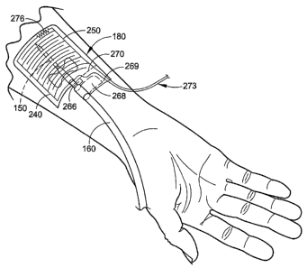
receptacle.
[05] During a treatment, the patients' blood is processed by a filtering
device
commonly called a dialyzer or hemodialyzer. The blood travels to and from this
filtering device through an extracorporeal blood circuit by the action of a
blood pump.
Every hemodialysis unit is required to have certain alarms (AAMI RD5- 3.3.6)
that
monitor conditions throughout a treatment to insure patient safety. These
alarms
CA 02535502 2006-02-10
WO 2005/019416 PCT/US2004/022403
2
include temperature, dialysate pressure, transmembrane pressure, blood circuit
pressure, conductivity, blood leak, and blood circuit air embolism protection.
[06] Blood travels out from the body through the arterial fistula needle to an
arterial
bloodline. The arterial blood is then pumped through the bloodline, into and
through
the filtering device, and returned to the body through the venous bloodline
attached
to the venous fistula needle. As used herein, the term 'Yenou~' it is intended
to mean
'returning to the body' and the term "arterial' is intended to mean "coming
from the body'.
These fistula needles are commonly taped in place on the patients skin near
and
around the access site.
[07] One or both of the fistula needles are occasionally dislodged or removed
from
the access site during a treatment. Some examples of how this hazardous
situation
may occur unintentionally include the bloodlines getting caught on the
treatment
chair during a positional change such.as reclining from a seated position or
siting up
from a reclined position. Dislodgment may also occur when clothing or blankets
brush against the fistula needles and tapings during normal movements.
Sometimes
someone moving past catches the bloodline with a foot, a walker, a wheelchair
or a
cart. It may even happen when the tape on a patient simply comes off, due to
dry or
- sweaty-skin, and the needles slip out. Intentional removal of the needles)
during
treatment is also not unheard of, requiring many of the more mentally or
emotionally
unstable patients to be restrained during treatments. Other patients move
around
frequently and the many little tugs on the bloodline and tapings, and the
constant
pulling eventually loosens that tapings to the point that they come off and
the fistula
needle falls out.
CA 02535502 2006-02-10
WO 2005/019416 PCT/US2004/022403
3
[08] Should the venous needle become partially dislodged during a treatment,
the
patients' blood may infiltrate (into the surrounding tissue areas), usually
causing
great pain, or it may leak out around the needle entry site, or a combination
of both.
[09] Should the venous needle become completely dislodged during a
hemodialysis treatment, the patients' blood is not being returned and the
blood is
effectively drained out. With typical blood pump ranges of 50 to 650 ml/min.,
the
blood loss may be very rapid. This situation requires an immediate medical
intervention response to prevent severe patient injury or death, by
exsanguination.
Obviously, even the most observant and dedicated of medical staff could not
possibly watch each patient all of the time.
j~0,j Currently, the primary device to monitor for a venous needle dislodgment
is
the venous pressure monitor (VPM). Under certain circumstances, VPM is not a
dependable indicator for a venous needle dislodgment because the VPM may not
"see" a change beyond the standard alarm limit range (50 ml/min.). This may be
exacerbated when the alarm limits are not set"centered'around the varying
average
pressure. Significantly, the VPM will often fail to register a sufficient
pressure
change (to set off an alarm) due to the inherent"back pressurd'developed in a
venous blood line by the resistance of the viscous blood-traVeling through the
relatively small orifice of the fistula needle.
('??j One attempt to solve the partial dislodgment problem is offered by Shaw
in his
U.S. Patent Nos. 3,618,602 and 4,010,749. These basically use the increase in
skin
temperature to determine the presence of an infiltration. This solution has
limitations
in that it is rather slow to respond as it is dependent on the reaction of the
body to
the problem. Additionally, it fails to address the present concern of
dislodgment.
CA 02535502 2006-02-10
WO 2005/019416 PCT/US2004/022403
4
While an infiltration is painful, may require surgery to correct and could
even result in
the loss of the limb, it is not immediately life threatening.
[12] One proposed solution to the complete dislodgment problem is attempted in
U.S. Patent No. 6,077,443, entitled "Method and device for monitoring a
vascular
access during a dialysis treatment", issued to Goldau, Rainer, which monitors
the
impulses (natural or added) detectable in an extracorprial blood circuit. This
method
has not experienced commercial success, or widespread utilization. It is
believed
this may be because the pressures illustrated appear to be on a very still
patient,
which is not a realistic assumption throughout a four-hour or longer
treatment. Even
very small arm movements can set off the VPM without dislodgment of a needle
because of the natural pressures inherent in the needle as described above.
[13] There are a number of sensor designs that use the inherent conductivity
of
blood and other fluids to set off an alarm, most commonly used in a diaper to
indicate a soiled condition.
[14] A"System for use in detection of electrically conductive fluid~'was
suggested
~in U.S. Patent No. 5,790,036, issued to Fisher, et al., which uses the
inherent
r
conductivity of body fluids and wastes to set off an alert in a diaper. This
arrangement does not adequately protect a patient as it could have a sensor
failure
or disconnect without alerting the staff of the sensor failure or
disconnection.
Additionally, the device does not explicitly provide for compliance with the
nonisolated patient connection requirements of Safe Current Limits for
Eletromedical
Apparatus as required by Applicable Document 2.3.
[15] U.S. Patent Number 5,779,657 to Daneshvar entitled "Nonstretchable wound
cover and protector" shows and describes a simple blood leak detector. The
soiling
of a gauze pad with blood would complete a circuit, allowing an alarm to
sound. As
CA 02535502 2006-02-10
WO 2005/019416 PCT/US2004/022403
in the case of the Fisher unit, Daneshvar's unit fails to alarm in the case of
a sensor
failure. Daneshvar's unit also is not compliant with the nonisolated patient
connection
requirements for electrically sensitive patients. Additionally, whether
Daneshvar's
unit will alarm depends on the absorbency of the gauze, which may be
compromised
due to being saturated by non-conductive fluids or by compression, or coated
by
certain medical gels, pastes or ointments.
[16J The devices described in WO 99/24145 and U.S. Published Patent
Application 2002/0198483 A1 attempt to detect a separation of the
extracorporeal
circuit. However, neither of these allow for an unobstructed view of the
access site.
Another problem they share is that they are designed for use as an integral
part of a
dialysis machine. As such, they are specifically not designed for stand-alone
use.
Of greater concern is the failure of any of these devices to fail in a safe
manner. If
the unit fails for some reason, such as a dead battery, the protection is lost
and the
staff is not aware of it. While both of these references indicate thafi they
determine
needle dislodgment, they really are mere variations on the wet diaper sensor
idea in
that they only detect blood or other conductive fluids. They are not actually
determining the needle position. With some of the newer implanted tubing and
other
new types of vascular accesses, there is very little bleeding when a needle is
removed, and, hence, limited opportunity for success in alerting the staff in
the case
of a rapid needle withdrawal, such as when a bloodline is caught by a passing
foot,
or when a mentally unstable patient intentionally removes the needle.
Additionally,
in applications other than dialysis, the underlying region or substrates may
not have
a sufficient positive relative pressure to force out blood, body fluid or
liquid to wet the
sensor.
CA 02535502 2006-02-10
WO 2005/019416 PCT/US2004/022403
6
[17] The reusable sensors described in the references mentioned above and
elsewhere in the prior art involve the myriad of problems and costs associated
with
reusing soiled medical equipment including, but not by way of limitation,
sterilization,
clean storage, verification of the absence of the sterilant prior to use,
reused devices
being reused only by the original patient, quality problems due to subjective
assessments, etc.
SUMMARY OF THE INVENTION
[18] The present invention involves a device for monitoring loss of body fluid
and
dislodgment of a medical instrument from an access site of the body. The
device
involves a system that is designed for use as a critical medical monitor,
provides the
requisite electrical isolation, and provides an unobstructed view of the
access site.
The device incorporates a sterile and disposable sensor that fails-to-safe
such that a
loss of protection results in an alarm, can be reset in the case of small
leaks, can be
tested "in-sit~f, without extra test equipment, alarms if the patch is removed
or
damaged in use, can stand alone and is not be required to be integrated into
the
alarm circuits of common, existing hemodialysis units, monitors the placement
of the
needle relative to its insertion point, has a monitored power supply, and
alarms in the
intentional removal of needle by patient.
[19] Another aspect of the invention involves a hemodialysis site sensor
attachable
to at least one of a blood line and a fistula needle for alerting medical
personnel of
the leakage of blood from an access site where the fistula needle enters into
a
patienfs body and dislodgment of the fistula needle from the access site. The
hemodialysis site sensor includes a base membrane layer made of a medical-
grade,
biocompatible material and including an upper side, a lower side adherable to
skin of
CA 02535502 2006-02-10
WO 2005/019416 PCT/US2004/022403
7
the patient, and one or more holes disposed therein to allow the passage of
blood
and vapor therethrough; a first membrane layer including an upper side, a
lower
side, and one or more holes disposed therein to allow the passage of blood and
vapor therethrough; a third membrane layer including an upper side and a lower
side; an electrical connection adapted to be electrically coupled to an
analytical
circuit and including a first sensing array disposed between the upper side of
the
base membrane layer and the lower side of the first membrane layer, a second
sensing array disposed between the upper side of the first membrane layer and
the
lower side of the third membrane layer, and resistively connected to the first
sensing
array; a disconnection mechanism attachable to at least one of the blood line
and the
fistula needle and severing the electrical connection upon dislodgment of the
fistula
needle from the access site; and wherein an electrical signal sent through the
electrical connection changes when blood contacts at least one of the sensing
arrays
or the electrical connection is severed by the disconnection mechanism,
causing the
analytical circuit to actuate an alarm notifying medical personnel of partial
or total
dislodgment of the fistula needle from the access site.
[20] Another aspect of the invention involves a method of alerting medical
personnel of partial -and total dislodgment of a fistula needle from an access
site
where the fistula needle enters into a patienfs body during hemodialysis. The
method includes providing a site sensor including a base membrane layer made
of a
medical-grade, biocompatible material and including a lower side adherable to
skin
of the patient at the access site, and one or more holes disposed therein to
allow the
passage of blood and vapor therethrough, a top membrane layer, an electrical
connection including one or more resistively connected sensing arrays disposed
between the base membrane layer and the top membrane layer, and a
CA 02535502 2006-02-10
WO 2005/019416 PCT/US2004/022403
8
disconnection mechanism attachable to at least one of the blood line and the
fistula
needle and severing the electrical connection upon dislodgment of the fistula
needle
from the access site; providing an analytical circuit in electrical
communication with
the electrical connection; sending a signal from the analytical circuit to the
site
sensor and receiving the signal from the site sensor with the analytical
circuit, the
signal traveling through the one or more resistively connected sensing arrays
of the
electrical connection; partially dislodging the fistula needle from the access
site
causing blood to contact the one or more resistively connected sensing arrays
and
the signal sent from the analytical circuit to change; completely dislodging
the fistula
needle from the access site causing the disconnection mechanism to sever the
electrical connection and the signal sent from the analytical circuit to
change;
determining with the analytical circuit whether the signal changed outside of
a
predetermined range; and actuating an alarm with the analytical circuit if the
signal
changed outside of a predetermined range.
[21] A still further aspect of the invention involves a method of alerting
medical
personnel of a problem during hemodialysis. The method includes providing an
active, fail-to-safe site sensor for a fistula needle at an access site during
hemodialysis, for a needle, or for another skin penetrating rriedical device;
and
automatically alerting medical personnel of a problem during hemodialysis
using the
active, fail-to-safe site sensor during at least the following: failing of the
active, fail-
to-safe site sensor; insufficient powering to the active, fail-to-safe site
sensor; partial
fistula needle dislodging from the access site; and complete needle dislodging
from
the access site.
CA 02535502 2006-02-10
WO 2005/019416 PCT/US2004/022403
9
[22] Further objects and advantages will be apparent to those skilled in the
art
after a review of the drawings and the detailed description of the preferred
embodiments set forth below.
BRIEF DESCRIPTION OF THE DRAWINGS
[23] FIG. 1 is a front elevational view of a human and illustrates multiple
exemplary
vascular and peritoneal access sites.
[24] FIG. 2A is front perspective view of an embodiment of a dialysis
treatment
chair in a normal position.
[25] FIG. 2B is right perspective view of the dialysis treatment chair of FIG.
2A and
illustrates the chair in a reclined position.
[26] FIG. 2C is right side elevational view of the dialysis treatment chair of
FIG. 2A
and illustrates the chair in a fully reclined, Trendlenburg position.
[27] FIG. 3 is a front perspective view of an embodiment of a hemodialysis
unit
connected to a dialysis patient sitting in a dialysis treatment chair.
[23] FIG. 4 is an exploded perspective view of an embodiment of a site sensor
of
the system for monitoring loss of body fluid and dislodgment of a medical
instrument
from an access site of the body.
[29] FIG. 5A is a cross-sectional view of the site sensor taken along lines 5A-
5A of
FIG. 4.
[30] FIG. 5B is a cross-sectional view of the site sensor taken along lines 5B-
5B of
FIG. 4.
[31] FIG. 6 is a perspective view of the site sensor of FIG. 4 applied to a
patients
arm with a bloodline of an extracorprial blood circuit and conductive wires
shown
extending from the site sensor.
CA 02535502 2006-02-10
WO 2005/019416 PCT/US2004/022403
[32] FIG. 7 is an electrical schematic of an embodiment of an analytical
circuit and
enunciator that may be used with the site sensor.
[33] FIG. 8 is a perspective view of an alternative embodiment of a site
sensor
applied to a patienfs arm with a fistula needle/bloodline of an extracorprial
blood
circuit and conductive wires shown extending from the site sensor.
[34] FIG. 9 is a top plan view of another embodiment of a site sensor.
[35] FIG. 10 is a top plan view of a further embodiment of a site sensor.
DETAILED DESCRIPTION OF PREFERRED EMBODIMENTS
[36] With reference to FIGS. 1-10, an embodiment of a system and method for
monitoring a vascular access site of a patient 105 during a hemodialysis
treatment
and alerting the medical staff in the event of a needle dislodgment will be
described.
[37] Although the system will be described in conjunction with monitoring a
vascular access site during a hemodialysis treatment, the system may be used
for
monitoring any penetration or access site through the skin with respect to
blood,
body fluids, and/or medical fluids that may leak around the skin penetration
site
and/or dislodgment of a needle relative to the skin penetration or access
site.
Further, the system may be used for monitoring -access of the vascular system,
access to sub-dermal or other implanted devices, access of the peritoneal
cavity,
access of internal organs, monit~ring trans-dermal exudation, other usage
where an
alert to the leakage of blood, body fluids, medical fluids, liquids or other
fluids may be
desired, or other usage where an alert to the separation of a medical device
or
instrument from an access site of the human body may be desirable.
[38] Before describing the system in detail, hemodialysis and some of the
equipment used during hemodialysis will first be described.
CA 02535502 2006-02-10
WO 2005/019416 PCT/US2004/022403
11
[39] Hemodialysis is one of the primary treatments for patients 105 with
kidney
failure. These life-sustaining treatments typically require 3 to 4.5 hours
each and
may occur three times a week. The most common access to the vascular system,
for chronic patients, is through use of a large gauge needle inserted through
the skin
into an arterial/ventricular graft, an implanted shunt or an implanted
receptacle at
one of the common sites 110 (FIG. 1 ) used for access to the vascular system
of the
patient 105. While the illustrated sites in FIG. 1 are not exhaustive, these
sites are
the normal locations for accessing the blood supply for use in hemodialysis
and
apheresis as well as where the interventional radiologist accesses the primary
blood
vessels for access to the heart. Several of these locations are viable for
infusion
therapy as well.
[40] The use of medical tape and a patch of sterile gauze covering the
peritoneal
insertion site 120 should be noted because this is an example of a site that
may be
monitored by the system in an alternative embodiment for any leakage,
bleeding, or
oozing which would indicate infection.
[41] During a treatment, the blood of the patient 105 is processed by a
hemodialysis unit 130 (FIG. 3). The blood travels to and from the hemodialysis
unit
130 through bloodlines 140 of an extracorporeal blood circuit by the action of
a blood
pump in the hemodialysis unit 130.
[42] - Blood travels out from the body through an arterial fistula needle 150
(FIG. 6)
to an arterial bloodline 160, which may be connected to the arterial fistula
needle 150
by a leader and appropriate luer lock fittings. The arterial blood is then
pumped
through the arterial bloodline 160, into and through the hemodialysis unit
130, and
returned to the patient 105 through a venous bloodline attached to a venous
fistula
needle.
CA 02535502 2006-02-10
WO 2005/019416 PCT/US2004/022403
12
[43] During the dialysis process, the patient 105 normally rests in a typical
dialysis
treatment chair 170 (FIGS. 2A-3) in one of three positions: 1 ) a normal
sitting
position (FIG. 2A), 2) a reclined sitting position (FIG. 2B), and 3) a fully
reclined or
Trendelenburg sitting position (FIG. 2C). The fully reclined position or
Trendelenburg sitting position shown in FIG. 2C is used to lower the head of
the
patient 105 below the level of the heart and is used when the blood pressure
of the
patient 105 gets too low ('crashing') and staff is attempting to prevent the
patient 105
from blacking out. It is important to note the many corners of the chair 170
and other
features of the chair 170 that provide opportunities for the needle 150 to be
pulled on
when changing from one sitting position to another. Bloodlines 140 may get
caught
on the treatment chair 170 during a positional change such as reclining from a
seated position or sitting up from a reclined position, causing one or both of
the
fistula needles to become partially or completely dislodged or removed from
the
access site during dialysis. Dislodgment may also occur when clothing or
blankets
brush against the fistula needles and tapings during normal movements by the
patient 105.
[44] It is also important to notice that, as with most chairs, the sides of
the chair
170 are covered, which often-greatly-increases the time between when the
needle
150 is dislodged and when it is noticed by the medical staff by observation of
a
growing puddle of blood on the floor under the expiring patient 105. The
presence of
sides on dialysis chairs 170 also causes the problem of clothing or blankets
tending
to bunch up in the lower corners of the chair 170, soaking up blood that is
leaking-
again extending the time between when the needle 150 is dislodged and when it
is
noticed by the medical staff.
CA 02535502 2006-02-10
WO 2005/019416 PCT/US2004/022403
13
[45] The drawing of FIG. 3 illustrates how the bloodlines 140 dangle between
the
chair 170 and the hemodialysis unit 130 during the typical dialysis process,
and the
danger this represents due to the fistula needles and bloodlines 140 being
accidentally caught and pulled from the patient 105 during dialysis due to the
length
of the bloodlines and their potential for interference with the medical
attendants that
serve the patient 105 undergoing dialysis. In certain situations, such as when
a
dialysis treatment station is positioned in a corner, the hemodialysis unit
130 may be
on the side of the patient 105 opposite the patient's access to the chair 170.
In this
case, the bloodlines 140 actually cross over the patient 105, significantly
increasing
the risk of having the bloodlines 140 being inadvertently caught on something
or
pulled on. Sometimes someone moving past catches the bloodlines 140 with a
foot,
a walker, a wheelchair or a cart.
[46] Should the venous needle become partially dislodged during dialysis, the
blood of the patient 105 may infiltrate (into the surrounding tissue areas),
usually
causing great pain, or it may leak out around the needle entry site, or a
combination
of both.
[47] Should the venous needle become completely dislodged during a
hemodialysis treatment, the blood of the patient 105 is not being returned and
the
blood is effectively drained out. With typical blood pump ranges of 50 to 650
ml/min., the blood loss may be very rapid. This situation requires an
immediate
medical intervention response to prevent severe patient injury or death by
exsanguination.
[48] With reference to FIGS. 4-7, the system will first be generally described
followed by a detailed description of the elements of the system. The system
generally includes a site sensor 180 (FIGS. 4-6), an analytical circuit 190
(fig. 7), and
CA 02535502 2006-02-10
WO 2005/019416 PCT/US2004/022403
14
an enunciator 200 (FIG. 7). A electrical signal is sent from the analytical
circuit 190
to the active site sensor 180, where the signal passes through one or more
sensing
arrays of the site sensor 180, and back out to the analytical circuit 190. The
analytical circuit 190 monitors the return signal. Any change of this returned
signal
outside of a designated range produces an alarm output, which is coupled to
the
enunciator 200, for alerting medical personnel. The site sensor 180 includes a
disconnection mechanism that disconnects the site sensor 180 from the
analytical
circuit 190 upon sufficient pull of the bloodline 160. This changes the
returned signal
to the analytical circuit 190, producing an alarm output that is sent to the
enunciator
200 for alerting medical personnel.
[49] Each of main elements of the system will now be described in more detail
in
turn below:
Site Sensor:
[50] With reference to FIGS. 4-6, the embodiment of the site sensor 180 shown
includes a base membrane layer 210, a second membrane layer 220, and a third
membrane layer 230. A first conductive layer or sensing array 240 is disposed
between the base membrane layer 210 and the second membrane layer 220, and a
second conductive layer or sensing array 250 is disposed betweeri the second
membrane layer 220 and the third membrane layer 230. Although the sensing
arrays 240, 250 are shown separated by the single second membrane layer 220,
in
an alternative embodiment, the sensing arrays 240, 250 may be separated by
more
than one layer and/or may have additional sensing layers between them.
[51 ] The base membrane layer 210 is made up of one or more appropriate
medical-grade, biocompatible materials, and have a rectangular shape as shown
or
may have a different shape. The same may be true for the other membrane layers
CA 02535502 2006-02-10
WO 2005/019416 PCT/US2004/022403
220, 230. An underside of the base membrane layer 210 may be completely or
partially coated or circumscribed with a coating of medical-grade,
biocompatible
adhesive 252 (FIGS. 5A, 5B) covered by a removable sheet to allow for the
attachment of the site sensor 180 to the prescribed area on the patient 105.
The
adhesive may include one or more adhesives of difFerent adhesive strengths to
facilitate controlled separation of the site sensor 180 along prescribed lines
270 or at
the connector of the site sensor 180 in a manner to be described device when a
dislodging stress is applied to the bloodline 160 to interrupt the signal
being
transmitted through the site sensor 180 and cause an alarm condition. The site
sensor 180 may also be held in place by any number of methods, as known to
those
skilled in the art, in conjunction with or in place of the medical-grade,
biocompatible
adhesive. The locational stability of the site sensor 180 is of paramount
importance
to the functional benefit of the system. The site sensor 180 in every case
must be
firmly attached to the fistula needle 150 and/or the bloodline 160. This
attachment
may be made during manufacturing, attached at time of use, or sometime in
between.
[52] In an alternative embodiment, the base membrane layer 210 may carry one
or
more electrodes somewhat below the base membrane layer 210 to indicate
positive
contact with the skin of the patient 105. An exemplary electrode that may be
used
for this purpose is a silver/ silver-chloride type as is used in a
plethysmograph or an
ElectroCardioGram (ECG) lead set; however, other types of electrodes, as is
known
to those skilled in the art, and/or additional sensors may be used. The one or
more
electrodes and/or sensors may be coupled to the first sensing array 240 or
have
another electrical connection for the purpose of determining actual physical
contact
with the skin of the patient 105. The first sensing array 240 may, if placed
below the
CA 02535502 2006-02-10
WO 2005/019416 PCT/US2004/022403
16
base membrane layer 210 or in the absence of the base membrane layer 210,
provide a biorhythm output (e.g., an ECG waveform may be transmitted to the
analytical circuit 190) that is indicative of a good and stable connection
with the
patient 105. An additional advantage of this is that if the patient expires,
thus ending
the waveform, an alarm at the enunciator 200 may be actuated.
(53] The base membrane layer 210 may have holes 260 to allow for the free
passage of perspiration, blood, other liquids and/or vapors through the layer.
One of
more of these holes 260 may be of the same or varying sizes, shapes,
quantities and
regional densities. The base membrane layer 210 (and the other membrane layers
220, 230) may have a perforation 270, or other preconditioning treatment,
along a
prescribed path to facilitate the destruction of the site sensor 180 along
prescribed
lines in the case of a physical force against it from a pull on the bloodline
160 or
fistula needle, again resulting in an alarm state. The perforation 270 may
terminate
in a generally rectangular cut-out 266, which delineates a pull-over adhesive
tab 268
of a detachment section 269. The adhesive tab 268 may include an adhesive
covered by removable sheet on its upper surface. The pull-over adhesive tab
268
and the perforation 270 combine to form a dislodgment mechanism. In
alternative
embodiments, the dislodgment mechanism may have alternative configurations,
different elements, and/or greater or fewer elements. With reference to FIG.
6, in
use, bloodline 140 and/or fistula needle 150 is placed through the cut-out
266, over
the adhesive tab 268 and under the rest of the site sensor 180, the removable
sheet
is removed from the adhesive tab 268, exposing the adhesive, and the adhesive
tab
268 is folded over the bloodline 140 and/or fistula needle 150 and adhered to
the
upper surface of the third membrane layer 230 so that the bloodline 140 and/or
fistula needle 150 is firmly attached to the tab 268 such that a pull on the
bloodline
CA 02535502 2006-02-10
WO 2005/019416 PCT/US2004/022403
17
140 would separate the conductive wires 272 or connection 284 and detachment
section 269 from the rest of the site sensor 180 along the perforation 270,
thus
interrupting the conductive path and causing the analytical circuit 190 to
cause the
actuation of an alarm at the enunciator 200.
[54J An upper side of the base membrane layer 210 may be used as a base for
the
first sensing array 240 to be placed, painted, deposited, adhered to, or
otherwise
disposed on. The first sensing array 240 may be held between the base membrane
layer 210 and the second membrane layer 220, and include a plurality of
conductive
wires or traces 272. One of conductive wires 273 (FIG. 6) connecting the site
sensor
180 and the analytical circuit 190 may be connected to the first sensing array
240 at
a connection point 274 (FIG. 4) at one end of the first sensing array 240. One
or
more resistors 276 may be located at an opposite end of the first sensing
array 240
and may be used to resistively connect the first sensing array 240 to the
second
sensing array 250 through a hole 278 in the second membrane layer 220. The
firsfi
sensing array 240 may have a configuration and be positioned so as to evenly
cover
the area between the base membrane layer 210 and the second membrane layer
220. The first sensing array 240 may cover some, all, or none of the holes 260
in the
base membrarie layer 210: The first sensing array 240 may be embedded in one
of
the layers 210, 220, or may be simply attached to or held in place between the
layers
210, 220. The first sensing array 240 may be of one polarity or made up of
multiple
leads with several polarities.
[55] The second membrane layer 220 may also have holes 280, one or more of
which may be of the same or varying sizes, shapes, quantities and regional
densities.
CA 02535502 2006-02-10
WO 2005/019416 PCT/US2004/022403
18
[56] These holes 280 may or may not line up with the holes of any other layer.
The top of the second membrane layer 240 may be used as a base for the second
sensing array 250 to be placed, painted, deposited, adhered to, or otherwise
disposed on. The second sensing array 250 may be held between the second
membrane layer 220 and the third membrane layer 230, and include a plurality
of
conductive wires or traces 282. One of the conductive wires 273 connecting the
site
sensor 180 and the analytical circuit 190 may be connected to the second
sensing
array 250 at a connection point 284 at one end of the second sensing array 250
by
soldering or any other well-known electric connection manner. These connectors
may be of a calibrated strength or holding ability. The second sensing array
250 is
connected to the one or more resistors 276 at an opposite end of the second
sensing
array 250. The second sensing array 250 may have a configuration and be
positioned so as to evenly cover the area between the base membrane layer 210
and the second membrane layer 220. The second sensing array 250 may cover
some, all, or none of the holes 280 in the second membrane layer 220. The
second
sensing array 250 may be embedded in one of the layers 220, 230, or may be
simply
attached to or held in place between the layers 220, 230. The second sensing
array
250 may be of one polarity or made up of multiple leads with several
polarities.
[57] The sensing arrays 240, 250 are preferably resistively connected through
the
one or more resistors 276, allowing for the fail-to-safe feature of the
present
invention, which will be described in more detail below. In alternative
embodiments,
the wires or traces of both arrays 240, 250 may be made of a resistive
material.
[58] Although the sensing arrays 240, 250 have been described as having a
configuration and being positioned so as to evenly cover the area between the
layers
210, 220, 230, the coverage of the areas by the sensing arrays 240, 250 may
not
CA 02535502 2006-02-10
WO 2005/019416 PCT/US2004/022403
19
necessarily be even. In alternative embodiments, differing amounts of coverage
in
differing areas may occur. For example, a space may be located near the center
of
the site sensor 180 to allow penetration through the site sensor 180 without
damaging, disturbing, or contacting any of the sensing arrays 240, 250.
[59] The third membrane layer 230 is also made of one or more appropriate
medical-grade, biocompatible materials. The third membrane layer 230
preferably
does not include holes like the base membrane layer 210 and the second
membrane
layer 220, but is preferably made of one or more appropriate materials to
allow for
the transpiration of perspiration and vapor, but not blood or other liquids.
This
selectively or semi-permeable layer 230, together with the seal formed by the
adhesive of the base membrane layer 210, ensures that the sensing arrays 240,
250
will come into contact with any blood or liquids that may be coming from the
protected area.
[60] ~uter edges of the membrane layers 210, 220, 230 may be sealed, glued or
bonded to one-another in any manner as known to those skilled in the art.
[61 ] The site sensor 180 is preferably transparent in all of the one or more
layers
above the needle entry site.
[62] With reference to FIG. 8, an alternative embodiment of the site sensor
180 is
shown where conductive leads 273 connect with the site sensor 180 at
detachment
corner sections 292 near an end of the site sensor 180 opposite the detachment
section 269 and wrap around the limb of the patient. A cinching mechanism 300
may be used to cinch the conductive leads 273 snugly against the limb to help
to
make the physical location and security of the site sensor 180 against the
limb more
stable. The bloodline 160 is secured to the site sensor 180 by the adhesive
tab 268
of the detachment section 269 in a manner similar to that described above with
CA 02535502 2006-02-10
WO 2005/019416 PCT/US2004/022403
respect to FIGS. 4-6 so that the force that would dislodge the fistula needle
150
causes the adhesive tab 268 and detachment section 269 to tear away from the
site
sensor 180 along the perforation 270 and interrupt the conductive path which
would
be detected by the analytical circuit 190, producing an alarm. Similarly, the
force
that would dislodge the fistula needle 150 (or movement of the patienfs arm)
may
cause the site sensor 180 and one or both of the detachment corner sections
292,
which are adhered strongly to the patient's skin, to separate along one or
both
perforations 294. This would interrupt the conductive path which would be
detected
by the analytical circuit 190, producing an alarm. In alternative embodiments,
one or
both of the perforations 276, 294 of the site sensor 180 may have a straight,
rectilinear, or curvilinear configuration other than that shown.
[63] In an alternative embodiment where the site sensor 180 is not used, one
could simply wrap a wire similar to the conductive leads 290 around the limb
of the
patient and secure the wire fio the fistula needle 150 to provide for an
indication of
needle dislodgment without leak detection because pulling on the bloodline 160
causes the wire to sever and interrupt the conductive path which would be
detected
by the analytical circuit 190, producing an alarm. In this embodiment, it
would be
desirable to secure the wire onto the limb with medical tapes, patches or
manner
known to those skilled in the art.
[64] In the immediate following paragraphs, features that may be part of one
or
more of the implementations of the system or site sensors 180, 310, 350
(hereinafter
'Ijite sensor 180') described herein are indicated.
[65] For example, in one or more implementations of the system, the system may
include one or more of the following. The entire system is contained in a
single unit.
One or more of the site sensor 180, the analytical circuit 190 and the
enunciator 200
CA 02535502 2006-02-10
WO 2005/019416 PCT/US2004/022403
21
are integrated with each other. The system is used for monitory blood/fluid
leakage
and/or needle dislodgment of at an access site of a human or animal. The site
sensor 180, the analytical circuit 190 and the enunciator 200 are connected to
each
other with any mechanical connection device. The site sensor 180, the
analytical
circuit 190 and the enunciator 200 are connected to each other with any
electrical
connection device. The site sensor 180, the analytical circuit 190 and the
enunciator
200 are connected to each other with any hollow fiber or solid fiber device.
The site
sensor 180, the analytical circuit 190 and the enunciator 200 are connected
via any
telemetering type equipment. The site sensor 180, the analytical circuit 190
and the
enunciator 200 are connected via any optical/photonic type equipment. The site
sensor 180, the analytical circuit 190 and the enunciator 200 are connected
via any
combination of equipment type. The site sensor 180 and enunciator 200 are
controlled by a separate, distinct controller. The site sensor 180 has' a
series of tabs
at the edges that have the connective areas of the conductive traces or leads.
The
site sensor 180 has a series of tabs at the edges that have the connective
areas of
the conductive traces or leads with fold-over tabs that complete the circuit
such that
all tabs are folded over (with self adhesive contacts) thus continuing the
circuit ,
terminating at the one corner or tab where the actual connector leading to the
analytical circuit 190 or enunciator 200 (or a combination of the two) is
located. The
site sensor 180 has a series of conductive rings that surround the exterior of
the site
sensor 180 such that a connector could be attached at any part of the edge of
the
site sensor 180 and make full contact with the required leads of the sensing
array.
The site sensor 180 utilizes a stereo or other style plug with as many
discrete
contacting areas as necessary. The site sensor 180 has a contact or connector
at
each level which may or may not be interconnected to each other (allows for
various
CA 02535502 2006-02-10
WO 2005/019416 PCT/US2004/022403
22
levels to be active). The site sensor 180, sensor/analytical circuit, or
sensor/analytical circuit /enunciator assembly is self-adhesively attached to,
or in
position on, the monitored site on the subject. The site sensor 180,
sensor/analytical circuit, or sensor/analytical circuit/enunciator assembly
which is
self-adhesively attached to, or in position on, the monitored site on the
subject has
an adhesive that is varied in its relative adhesive strength in order to
facilitate
destruction of the site sensor 180 along prescribed paths. The site sensor
180,
sensor/analytical circuit, or sensor/analytical circuitlenunciator assembly is
attached
to, or in position on, the monitored site on the subject with one or more of
(a) an
arrangement of material(s), fibers, plastics, tubing, straps, or other useful
product or
device, tied, connected, bonded or otherwise joined so as to hold the site
sensor
180, sensor/analytical circuit, or sensor/analytical circuit/enunciator
assembly in
contact with, or in position on, the monitored site of the subject; (b) an
arrangement
of adhesive tapes which may or may not be directly connected to the site
sensor
180, sensor/analytical circuit, or sensor/analytical circuit/enunciator
assembly in
contact with, or in position on, the monitored site of the subject; (c) a
clamp
arrangement so as to physically hold the site sensor 180, sensor/analytical
circuit, or
sensor/analytical circuit/enunciator assembly in contact with, or in position
on, the
monitored site of the subject; (d) any combination of the methods (a), (b),
and/or (c)
so as to hold the site sensor 180, sensor/analytical circuit, or
sensor/analytical
circuit/enunciator assembly in contact with, or in position on, the monitored
site of the
subject. The power supply and analytical circuit 190 are contained within a
dialysis
unit. The power supply and analytical circuit 190 are contained within an
apheresis
unit. The power supply and analytical circuit 190 are contained within an
infusion
unit. The power supply and analytical circuit 190 are contained within a
medical
CA 02535502 2006-02-10
WO 2005/019416 PCT/US2004/022403
23
instrument or other medical device. The power supply and analytical circuit
190 are
contained within an infusion pump unit. The site sensor 180 is connected to
the
analytical circuit 190 and/or enunciator assembly with a conductive wire, set
of wires,
coiled wire set or any other form of conductive wiring or cable as know to
those
skilled in the art. The site sensor 180, sensor/analytical circuit, or
sensor/analytical
circuit/enunciator assembly is attached to seat, bed, mattress, float,
cushion, gurney,
wheelchair, or any other physical device for support of the patient. The site
sensor
180, sensor/analytical circuit, or sensor/analytical circuit/enunciator
assembly is
attached to the floor, ceiling, wall, post, column, bar, or any other physical
structure
on, around or near the patient.
[66] In one or more implementations of the site sensors 180, 310, 350, the
site
sensors 180 may include one or more of the following. There may be other
numbers
of layers of material and varying areas covered by one or more sensing arrays,
which may have their own electrical connector, so that differing volumes of
liquid are
required to generate an alarm. There may be concentric areas of sensing arrays
to
allow for multiple levels of protection or to allow resetting of the system
without
having to use a new site sensor 180. The site sensor 180 may include a test
section
to allow for functional verification of the site sensor 180. The site sensor
180 may
allow for oblique andlor perpendicular piercing of the site sensor 180 by one
or more
needles and may allow for the attachment of the one or more needles in the
manner
shown and described above. Such an embodiment would be ideal for use with the
devices implanted in the upper chest or clavicle area such as the Vasca
device, for
such items as drainage tubing as used in many surgeries, and for use with
indwelling
catheters and central line catheters and the like. The space or gap between
the
layers 210, 220, 230 may be varied or function as the calibrated variable in
CA 02535502 2006-02-10
WO 2005/019416 PCT/US2004/022403
24
determining how much blood or other fluid is required to set off the alarm in
the site
sensor 180. One or more drain holes may be included in the site sensor 180 to
allow
blood or other occluding fluid that may have set off the alarm to be cleared.
After the
fistula needle 150 has been repositioned, a press on the top of the site
sensor 180
would cause the displacement of the blood or occluding fluid out of the one or
more
drain holes, which may have a valve or other flow control device,. and allow
the
continued use of the site sensor 180 without having to replace it. This would
be of
significant advantage in the case of a larger sized site sensor 180 such as a
site
sensor 180 for larger wound coverings or for monitoring shunts such as those
used
in radiographic heart studies (angiograms, angioplasty, etc.). Alternatively,
the site
sensor 180 may include a '~uer locl~' ar other tubing or syringe connector to
allow the
use of nonconductive sterile water or even air to rinse the site sensor 180
out in-situ.
In such an embodiment, the third membrane layer 230 may have one or more vent
holes to facilitate this.
[67] In one or more implementations of the site sensor 180, the site sensor
180
may include one or more of the following. The site sensor 180 is a switch. The
site
sensor 180 is disposable. The site sensor 180 is reusable. The site sensor 180
has
a limited life cycle or number of uses. The site-sensor 180 is active. The
site sensor
180 is passive. The site sensor 180 is electronic. The site sensor 180 is
photonic.
The site sensor 180 is chemical. The site sensor 180 is mechanical. The site
sensor 180 is reactive to any contact, stress, temperature, light, odor,
chemical,
electrical potential, or any other measurable physical property. The site
sensor 180
is reactive to one or more of contact, stress, temperature, light, odor,
mechanical,
chemical, electrical or electronic property, and any other measurable physical
property. The site sensor 180 reacts in the absence of any one of contact,
stress,
CA 02535502 2006-02-10
WO 2005/019416 PCT/US2004/022403
temperature, light, odor, electrical or electronic property, mechanical,
chemical,
optical or any other physical property of the site sensor 180 being monitored.
The
site sensor 180 is comprised of various layers of hydrophobic and/or
hydrophilic
materials. The site sensor 180 is comprised of various layers of transparent,
medical-grade, biocompatible materials. One or more of the layers of the site
sensor
180 has penetrations, holes, openings, or a path or paths through it or them
which
would allow liquid underneath to freely flow through the various layer(s). The
various
penetrations, holes, openings, path or paths of the site sensor 180 are in any
number, size, shape, origin, concentration, paucity, permeability, durability
or
function. The various penetrations, holes, openings, path or paths of the site
sensor
180 are differing in number, size, shape, origin, concentration, paucity,
permeability,
durability or function. The various penetrations, holes, openings, path or
paths of the
one or more site sensors 180 are differing in number, size, shape, origin,
concentration, paucity, permeability, durability or function vary from layer
to layer.
The membrane layer is reactive to the presence or absence of liquid or vapor
and
can alter, adjust, moderate, amplify, augment or otherwise vary the size,
shape or
other feature of the holes, openings, path or paths or any other route through
the
membrane.- The site sensor 180 is a combination of opaque materials. The site
sensor 180 contains an area that is perforated, thinned, weakened or otherwise
made so as to direct force (a shearing force) along a predetermined path on,
along,
across, over or otherwise through the site sensor 180 so as to ensure that the
patch
is separated from itself, or bisected, thus changing the electrical,
mechanical,
chemical, optical, sonic or any other monitored physical property of the site
sensor
180 due to a physical force being applied to it. The site sensor 180 contains
the
aforementioned perforated, thinned, weakened or force-directing section
wherein
CA 02535502 2006-02-10
WO 2005/019416 PCT/US2004/022403
26
that section may or may not cross the entirety of the site sensor 180. The
perforated, thinned, weakened or force-directing sections of the one or more
layers
may be different at each layer. The site sensor 180 contains the
aforementioned
perforated, thinned, weakened or force-directing section wherein that section
may
not be in a straight line but rather in an angle, circle or other shape as it
may be
desired. The site sensor 180 contains the aforementioned perforated, thinned,
weakened or force-directed section wherein that section is at a corner or at
one of
the corners or edges of the site sensor 180. The active sensing area of the
layers is
made of wire, traces, various conductive material, metals, painted traces,
liquid
conductive applications, sputtered deposition, vapor deposition build up, MEMs
production, photolithography, or other electrical connection production
method. The
site sensor 180 has a conductive phase or array that may or may not dissolve
in the
presence of a liquid or vapor. The site sensor 180 is in any shape, thickness,
or
cuvature as may be desirable for application to differing areas of the body.
The
active sensing area of the differing levels is of differing sizes and / or
shapes. The
specified area of the site sensor 180, throughout its layers, has a region
where there
is no active sensing area to allow for penetration through the site sensor 180
itself by
a needle or-other-device or observation or other monitor access. The site
sensor
180 has included in it any additional site sensor 180 that is able to
determine the
actual physical contact with the body being monitored. The site sensor 180 has
included in it the ability to sense the degree of actual physical contact with
the body
being monitored using a applied sensing method such as, but not limited to
optical,
thermal, and sonic. The site sensor 180 contains wet, dry or both wet and dry
components. The site sensor 180 contains any of the known types of"dry jell
products. The site sensor 180 contents and construction may be monolithic or
of
CA 02535502 2006-02-10
WO 2005/019416 PCT/US2004/022403
27
discrete components. The site sensor 180 is comprised entirely of, or has as
components, membranes or layers that are permeable by vapors. The site sensor
180 includes vapor permeable membrane layers where the membrane layers are of
differing and/or variable permeability. The site sensor 180 layers are sewn,
bonded,
connectored, sealed, fused, adhesively attached, glued, melted together or
connected by any other method known to those skilled in the art. The perimeter
of
the site sensor 180 is surrounded by an area of absorbent material. The site
sensor
180 includes layers of absorbent materials. The site sensor 180 monitors any
physical property that can be measured or gauged. The site sensor 180 is
comprised of thermistors, thermal transducers, or thermal detectors to provide
output
to the analytical circuit. The site sensor 180 uses exothermic or endothermic
chemicals) to enhance the responsiveness of the thermistors, thermal
transducers,
or thermal detectors. The site sensor 180 uses a hydrophilic product in
conjunction
with a reed switch or other mechanical switch that would be caused to change
states
due to pressure from the filling of the hydrophilic materials applying
pressure against
the switch. The hydrophilic switch may or may not be encased inside of a semi-
rigid
covering for the purpose of containing and/or directing the pressure towards
the
switch. The site sensor 180 contains a reed switch or other style of physical
switch
that is entirely encased in a non-conductive covering which may be of any
appropriate material or fabric. The site sensor 180 uses a resonate frequency
to
determine the status of the site sensor 180. The site sensor 180 uses the
electronic
determination of the resonate frequency and its stability and range for the
purpose of
determining the status of the site sensor 180 and is resettable around a new
frequency in the case of a partial occlusion of the site sensor 180 or a
slight
movement of the site sensor 180 area. The site sensor 180 has different
chemicals
CA 02535502 2006-02-10
WO 2005/019416 PCT/US2004/022403
28
or other discrete sensors applied to the differing layers to aid in the
differentiation of
what may or may not be coming into contact with the site sensor 180. The site
sensor 180 utilizes two or more electrodes and the measurement therebetween to
determine contact with the body. The site sensor 180 utilizes a single
electrode to
determine contact with the body. The site sensor 180 utilizes the separation
or
spacers between the various layers of the site sensor 180 to control or
calibrate the
amount of blood or fluid required to activate an alarm condition. The site
sensor 180
has a drain hole in one or more areas of the site sensor 180 to allow the
occluding
blood or fluid to be drained ofF. The site sensor 180 has one or more holes in
a top
layer of non-vapor permeable membrane or a limited permeable membrane which
would allow the aforementioned holes to be covered with a finger and pressure
applied to the site sensor 180 to force blood out of a drain hole in order to
reset the
site sensor 180. The site sensor 180 has a luer fitting or other appropriate
fitting to
allow a liquid or gas or vapor to be infused in order purge the internal
spaces of the
site sensor 180. The site sensor 180 has a suction port to allow the vacuum or
suction removal of liquid, blood or occluding vapors. The site sensor 180
contains
various chemical-determining sensors which would allow the determination of
what
liquid is contacting the site sensor 180. The site sensor 180 uses the various
optical
properties of differing liquids to determine what liquid is contacting the
site sensor
180. The site sensor 180 uses a hydrophilic inner layer which has a capacitive
or
inductive site sensor 180 to determine the presence of liquid. The capacitive
or
inductive element is external to the sensor 180 itself. The site sensor 180
uses a
piezo-electric crystal or device that changes the pressure of a liquid in a
hydrophilic
pad contained by a rigid or semi-rigid container or package. The site sensor
180
CA 02535502 2006-02-10
WO 2005/019416 PCT/US2004/022403
29
uses electro-active powders that produce an electrical potential or current
when
wetted.
[68J In one or more further implementations of the site sensor 180, the site
sensor
180 may include one or more of the following. The input to the site sensor 180
is an
electrical type sensor, a mechanical sensor, a chemical sensor, an optical
sensor, or
any other type of sensor. The input to the site sensor 180 is a direct current
(DC)
voltage potential. The input to the site sensor 180 is an alternating current
(AC)
voltage potential. The input to the site sensor 180 is an amplitude modulated
(AM)
signal. The input to the site sensor 180 is a frequency modulated (FM) signal.
The
input to the site sensor 180 is a pulse width modulated signal. The input to
the site
sensor 180 is a light source (of any wavelength). The input to the site sensor
180 is
part of the electromagnetic spectrum. The input to the site sensor 180 is a
thermal
change. The input to the site sensor 180 is a mechanical force. The input to
the site
sensor 180 is an electrochemical change. The input to the site sensor 180 is
any
combination of inputs. The sensor input is sent to a computer file. The sensor
input
is sent to an electronic storage or media device. The sensor input is
displayed on a
computer monitor. The sensor input is displayed on a medical device's user
interface. The input to the site sensor 180 is different from the output. The
site
sensor 180 operates in multiple or singular modalities. The site sensor 180
operation may change modalities.
[69] In one or more additional implementations of the site sensor 180, the
site
sensor 180 may include one or more of the following. The output from the site
sensor 180 is electrical, mechanical, chemical, thermal, optical, or any other
type of
output. The output from the site sensor 180 is a direct current (VDC) voltage
potential. The output from the site sensor 180 is an alternating current (VAC)
CA 02535502 2006-02-10
WO 2005/019416 PCT/US2004/022403
voltage potential. The output from the site sensor 180 is an amplitude
modulated
(AM) signal. The output from the site sensor 180 is a frequency modulated (FM)
signal. The output from the site sensor 180 is a pulse width modulated signal.
The
output from the site sensor 180 is a light source (of any wavelength). The
output
from the site sensor 180 is part of the electromagnetic spectrum. The output
from
the site sensor 180 is a mechanical force. The output from the site sensor 180
is an
electrochemical change. The output from the site sensor 180 is any combination
of
outputs. The site sensor 180 output is different from the input. The sensor
input is
different from the output. The sensor output is sent to a computer file. The
sensor
output is sent to an electronic data storage or media device. The sensor
output is
displayed on a computer monitor. The sensor output is displayed on a medical
device's user interface. The sensor output is variable depending on which
layers are
responding or providing an output or where a variance is detectable. The
sensor
output is variable or progressive or regressive depending on the amount of
liquid
detected by the site sensor 180.
Analytical Circuit/Enunciator:
[70] With reference to FIG. 7, an electrical schematic of an embodiment of the
analytical circuit 190 is shown. The analytical circuit 190 illustrated is a
standard
comparator that compares the value of an electrical output signal, in one
embodiment, sent through the conductive wires 273 to the site sensor 180 to
the
value of an input signal received through the conductive wires 273 from the
site
sensor 180. This allows for the fail-to-safe feature of the present invention
because
if a difFerence between the output signal and the input signal is outside a
designated
range or if there is no return signal as in the case of detachment of the
detection
CA 02535502 2006-02-10
WO 2005/019416 PCT/US2004/022403
31
section 269 from the site sensor 180, the analytical circuit 190 responds to
that
change by sending an output signal to the enunciator 200 to be actuated or
causing
one or more switches to be closed to actuate the enunciator 200. The
analytical
circuit 180 and the enunciator 200 may be powered by a power supply 302.
Although the analytical circuit is show as including a comparator, in
alternative
embodiments, other analytical circuits may be used to provide the fail-to-safe
feature
of the present invention. Further, hardware that may perform the functions
described
herein include, but not by way of limitation, an application specific
integrated circuit
(ASIC), a set of wired logic circuits, and a hardwired circuit of electrical
components,
e.g., transistors, capacitors, and resistors. Further, hardware and software
may be
used to perform the functions described herein. Examples of hardware and
software
that may perform the functions described herein include, but not by way of
limitation,
a programmed computer and an application specific computer. The analytical
circuit
190 is powered by a power supply.
[71] In one or more implementations of the analytical circuit 190, the
analytical
circuit 190 may include one or more of the following. The analytical circuit
190
provides electrical isolation in compliance with the nonisolated patient
connection
requirements of Safe Current Limits for Electromedical Apparatus as required
by
Applicable Document 2.3. The analytical circuit 190 is part of the sensor 180,
310,
350. The analytical circuit 190 is part of the enunciator 200. The analytical
circuit
190 is part of a sensor/enunciator assembly. The analytical circuit 190 may
include
a reset and/or mute button to reset the analytical circuit 190 in the event of
an alarm
by the enunciator 200 and/or mute the alarm of the enunciator 200.
[72] In one or more implementations of the enunciator 200, the enunciator 200
may include one or more of the following. The input to the enunciator 200 is
the
CA 02535502 2006-02-10
WO 2005/019416 PCT/US2004/022403
32
output of an electrical type sensor, a mechanical sensor, a chemical sensor,
an
optical sensor, or any other type of sensor. The input to the enunciator 200
is a
direct current (DC) voltage potential. The input to the enunciator 200 is an
alternating current (AC) voltage potential. The input to the enunciator 200 is
an
amplitude modulated (AM) signal. The input to the enunciator 200 is a
frequency
modulated (FM) signal. The input to the enunciator 200 is a pulse width
modulated
signal. The input to the enunciator 200 is a light source (of any wavelength).
The
input to the enunciator 200 is part of the electromagnetic spectrum. The input
to the
enunciator 200 is a thermal change. The input to the enunciator 200 is a
mechanical
force. The input to the enunciator 200 is an electrochemical change. The input
to
the enunciator 200 is any combination of inputs. The input to the enunciator
200 is
different from the output. The enunciator 200 operates in multiple or singular
modalities. The enunciator 200 operation may change modalities. The output
from
the enunciator 200 is electrical, mechanical, chemical,, thermal, optical, or
any other
type of output. The output from the enunciator 200 is a direct current (VDC)
voltage
potential. The output from the enunciator 200 is an alternating current (VAC)
voltage
potential. The output from the enunciator 200 is an amplitude modulated (AM)
signal. The output from the enunciator 200 is a frequency i~nodulated (FM)
signal.
The output from the enunciator 200 is a pulse width modulated signal. The
output
from the enunciator 200 is a light source (of any wavelength). The output from
the
enunciator 200 is part of the electromagnetic spectrum. The output from the
enunciator 200 is a mechanical force. The output from the enunciator 200 is an
electrochemical change. The output from the enunciator 200 is any combination
of
outputs. The sensor output is enunciator 200 is different from the input. The
enunciator input is different from the output. The enunciator output is sent
to a
CA 02535502 2006-02-10
WO 2005/019416 PCT/US2004/022403
33
computer file. The enunciator output is sent to an electronic data storage or
media
device. The enunciator output is displayed on a computer monitor. The
enunciator
output is displayed on a medical device's user interface. The enunciator
output is
variable depending on which layers are responding or providing an output or
where a
variance is detectable. The enunciator output is variable or progressive or
regressive depending on the amount of liquid or rate of change detected by the
site
sensor 180. The output of the enunciator 200 goes to any electronic data
storage
device. The independent power supplies for each component may differ from each
other. The enunciator 200 output is visual. The enunciator output is audible
at any
volume or frequency. The enunciator output is vibration. The'enunciator output
is
an electronic signal. The enunciator output is an optical/photonic signal. The
enunciator output is any part of the electromagnetic spectrum. The enunciator
200
itself is of any shape or size. The enunciator output is any combination of
outputs.
The site sensor 180, the analytical circuit 190 and the enunciator 200 are
connected
to each other with any physical connection device.
[73] In one or more implementations of the power supply 302 of the analytical
circuit 190, the power supply 302 may include one or more of the following.
The
analytical circuit 190 is powered by an external power supply. The analytical
circuit
190 is powered by an internal power supply. The analytical circuit 190 is
powered by
solar energy. The analytical circuit 190 is powered by a combination of
external and
internal or solar power supplies. The analytical circuit 190 is powered by a
combination of differing power supplies which may be internal or external or
both.
The power supply is a disposable battery. The power supply is a rechargeable
battery. The power suppl~s rechargeable battery is recharged from the medical
device it is attached to, such as an infusion pump or dialysis unit. The power
supply
CA 02535502 2006-02-10
WO 2005/019416 PCT/US2004/022403
34
is a "Cap-Batter' or other power storage device. The power supply is a
proprietary or
custom battery of varying shapes or voltages or outputs. The power supplies
are
independent for each component. The power supply has a redundant or a "back-
u~'
arrangement. The power supply is monitored for a low-battery condition. The
power
supply is monitored for trends in capacity. The power supply is monitored for
trends
in capacity and indication given as to expected capacity on current cycle
andlor
remaining cycles before performance is considered to be unacceptable. The
state of
the power supply is monitored and status is displayed on a user interface such
as an
indicator light, graphical user interface, monitor (CRT, flat panel, etc.), or
any other
format as know to those skilled in the art. The power supply is a current
storage
device.
Method of use:
[74] With reference to FIGS. 4-7, the system will now be described in use. An
electrical signal is sent from the analytical circuit 190 to the active site
sensor 180.
This signal passes from the first sensing array 240 of the site sensor 180
through the
resistor 276 to the second sensing array 250, and back out to the analytical
circuit
190. The analytical-circuit 190-monitors the return signal. Any change of this
returned signal outside of a designated range produces an output, which is
sent to
the enunciator 200 to actuate an alarm. Monitored conditions that would cause
the
returned signal to be outside of the designated range include when partial
venous
needle dislodgment occurs, and when complete venous needle dislodgment occurs.
[75] During partial venous needle dislodgment (not exclusively infiltrating),
blood
leaking around the fistula needle 150 or otherwise will flow through the base
membrane layer 210, contacting the first sensing array 240, and through the
second
CA 02535502 2006-02-10
WO 2005/019416 PCT/US2004/022403
membrane layer 220 thereby coming into contact with the second sensing array
250.
Because the sensing arrays 240, 250 are resistively connected and the signal
input
from the analytical circuit 190 provides a constant value through the system
(allowing
for a fail-to-safe feature), the change in resistance in the sensing arrays
240, 250
caused by the blood contact alters the signal so that the returned signal to
the
analytical circuit 190 is outside of the designated range. The analytical
circuit 190
responds to this condition by energizing the enunciator 200, causing the
alarm.
[76] During complete venous needle dislodgment, the tab 268 that wraps around
the bloodline 160 transfers the pulling force causing the dislodgment to the
perforation 270 or otherwise weakened area, allowing the detachment section
169 to
separate from the rest of the site sensor 180 and cutting the connection
between the
conductive wires 273 and the sensing arrays 240, 250 so that the electrical
circuit is
opened. Severing the electrical circuit eliminates the return signal to the
analytical
circuit 190 so that the difference between the sent signal and returned signal
(or lack
thereof) is outside of the designated range determined by the analytical
circuit 190 .
The analytical circuit 190 responds to this condition by energizing the
enunciator
200, causing the alarm.
Thermal Site Sensor:
[77] With reference to FIG, 9, an alternative embodiment of a site sensor 310
is
shown. In this embodiment, one or more sensing arrays 320 include a plurality
of
thermistors 330 spaced along the sensing array 320 in a space between a base
membrane layer and a second membrane layer (and possibly other membrane
layers). The thermistors 330 are disposed within an exothermic chemical 340
within
the space between the base membrane layer and the second membrane layer (and
CA 02535502 2006-02-10
WO 2005/019416 PCT/US2004/022403
36
possibly other membrane layers). The underside of the base membrane layer is
adhesively coated near its periphery and has holes that would allow the free
passage of vapor, blood, or other fluid therethrough. The exothermic chemical
340
releases its thermal potential when the blood or other fluid contacts it. The
resulting
thermal change warms the thermistors 330, causing the thermistors 330 to
change
their resistive values in correlation to the amount of thermal change. This
change in
resistance is detected by the analytical circuit 190 which produces an alarm.
The
site sensor 310 includes a detachment section 269 similar to the detachment
section
269 described above with respect to FIG. 6.
[73] With reference to FIG. 10, another embodiment of a site sensor 350 is
shown.
The site sensor 350 may include one or more sensing arrays 360 comprised of
one
or more optical fibers 370 disposed in a space between a base membrane layer
and
a second membrane layer (and possibly other membrane layers). Similar to the
embodiments shown and described above, the underside of the base membrane
layer is adhesively coated near its periphery and has holes that would allow
the free
passage of vapor, blood or other fluid therethrough. The one or more optical
fibers
of the sensing array 360 may be connected to a light source. The exterior of
the one
or more optical fibers may be treated to encourage adhesion of blood or other
fluid
thereto. Light from the light source passes through the one or more optical
fibers
between the base membrane layer and the second membrane layer. When blood or
other fluid passes through the holes of the base membrane layer and occludes
the
space between the base membrane layer and the second membrane layer and
contacts the one or more optical fibers, the light transmission through the
one or
more optical fibers is affected. This is detected by the analytical circuit,
causing an
alarm to be produced. In an alternative embodiment, light may pass through a
block
CA 02535502 2006-02-10
WO 2005/019416 PCT/US2004/022403
37
or other optical unit that would be occluded in the presence of blood or other
liquid,
and the analytical circuit may detect this. In another embodiment, a light
path may
be altered by the physical presence of blood or liquid in the space between
layers,
altering an optical picture, which is detected by the analytical circuit 190
and sets off
the alarm.
[79] It will be readily apparent to those skilled in the art that still
further changes
and modifications in the actual concepts described herein can readily be made
without departing from the spirit and scope of the invention as defined by the
following claims.