Note: Descriptions are shown in the official language in which they were submitted.
CA 02535524 2009-08-24
APPARATUS AND METHOD FOR LIQUID SAMPLE PARTITIONING
BACKGROUND
Technical Field
[0002] The present disclosure relates to methods for the quantification of
biological material in a sample, and to devices for partitioning and holding
the biological
material during quantification.
Discussion of Related Art
100031 The determination and enumeration of microbial concentration is an
essential part of microbiological analyses in many industries, including
water, food,
cosmetic, and pharmaceutical industries. The classical methods of detection
and
quantification of biological material are performed using semi-solid nutrient
agar medium
(e.g. pour plate method, membrane filtration) or liquid nutrient medium (e.g.
the most
probable number method). If a pour plate method is being performed, the sample
being
tested for microbial contamination is first dispensed in a Petri-dish. Then 15
ml of the
appropriate nutrient medium is poured over the sample. The Petri-dish is then
left to
solidify at room temperature for approximately 20 minutes and then incubated
at a
specific temperature for a specific time, and any resulting colonies are
counted.
Drawbacks for the pour plate method include bacterial colonies, which may be
too small
or overlapping each other for counting and paiticulate matter in the samples,
which may
also interfere with counting. For the membrane filtration method, the required
volume of
sample is filtered through a membrane of a very small pore size to non-
specifically trap
bacteria. The membrane is then placed on a prepared solid medium, which
supports the
growth of the target bacteria. The medium is then incubated at a specific
temperature for
CA 02535524 2006-02-10
WO 2005/021157 PCT/US2004/027659
a specific time, and any resulting colonies are counted. Drawbacks of membrane
filtration include particulate matter other than bacteria in the sample (e.g.,
a waste water
sample) may clog the membrane making it unusable and bacterial colonies may be
too
small or overlapping each other making it difficult to count.
[0004] Improved methods using solid-base nutrient medium to support microbial
growth for microbial detection and quantification include READIGEL (3M
Microbiology Products, St. Paul, Minnesota), which uses a special chemically
treated
Petri-dish. The sample is inoculated into a growth medium and poured into the
plate.
The sample/medium mixture is solidified 20 minutes after it comes into contact
with the
chemicals coated in the plates. Alternatively, PETRIFILM (3M Microbiology
Products,
St. Paul, Minnesota), which is an adhesive tape-like material having a coated
media
deposited thereon may also be used. This arrangement forms a thin layer of
growth
media that hydrolyzes and gels upon contact with liquid samples. A cover piece
helps to
disburse the sample inoculums and also acts as a cover for incubation. These
methods
offer improvement over the pour plate and membrane filtration methods in that
these
methods are easier to perform. However, these methods suffer the same
limitations as
those of pour plate and membrane filtration methods as described above.
[0005] The most probable number method (MPN) is well known and described,
for example, in Recles et al., "Most Probable Number Techniques" published in
"Compendium of Methods for the Microbiological Examination of Foods", 3rd ed.
1992,
at pages 105-199, and in Greenberg et al., "Standard Methods For the
Examination of
Water and Wastewater" (8th ed. 1992).
[0006] Microbial quantification devices and methods using the MPN method are
commercially available. Devices and Methods such as Quanti-Tray and Quanti-
Tray
2000 (IDEXX Corporation, Westbrook, Maine) are used for microbial
quantification for
drinking water, surface water, and waste water samples. A detailed disclosure
of these
tests may be found in Naqui et al. U.S. Patent Nos. 5,518,892; 5,620,895; and
5,753,456.
To perform these tests the separate steps of adding the sample/reagent to the
device and
then sealing the device with a separate sealing apparatus are required before
the
incubation period. These methods and devices offer a significant improvement
over the
traditional multiple tube fermentation techniques in terms of their ease of
use and also
allow for accurate quantification of microorganisms in the sample. However,
devices of
-2-
CA 02535524 2006-02-10
WO 2005/021157 PCT/US2004/027659
this type may require an instrument to distribute the sample/medium mixture
into each
individual compartment and are more applicable for enumerating microbial
populations in
the microaerophilic condition.
[0007] Croteau et al. also describe a method and device for quantification of
biological material in a sample using the MPN method in U.S. Patent Nos.
5,700,655;
5,985,594; and 6,287,797. The device uses a flat horizontal incubation plate
and the
surface is divided into a plurality of recessed wells. The liquefied
sample/medium
mixture is poured onto the surface of the device and after gentle mixing the
sample/medium mixture is distributed into the recessed wells and held in the
well by
surface tension. The plate is then incubated at a specific temperature for a
specific time
until the presence or absence of the biological material is determined.
Pierson et al. in
U.S. Patent No. 6,190,878, entitled "Methods and Devices for the Determination
of
Analyte in a Solution", disclose devices using a flat horizontal surface,
which is divided
into a plurality of recessed wells. Others have one or more surfaces with
reagent islands
immobilized thereon. Each well or wells or reagent islands are sized and
shaped, and
formed of a suitable material to hold the aliquot within the well or reagent
islands by
surface tension. These devices offer improvement over the gel-based methods
for
microbial enumeration by providing the benefit of easy result interpretation
and higher
counting ranges. These methods and devices potentially may have some
disadvantages.
Sample inoculation may be hampered by air bubbles, which form in the wells
during the
inoculation of samples and requires a pipetting step.
SUMMARY
[00081 The present invention provides methods and devices for detecting and
quantification of the presence and absence of biological materials,
microorganisms, and
analytes in a liquefied sample solution. The invention makes use of "capillary
flow",
wherein a liquefied sample can be partitioned into discrete compartments
through
capillary channels. The present invention overcomes deficiencies of the prior
art by
providing devices and methods, which significantly reduce the amount of hands-
on time
and do not require skilled laboratory personnel to perform or interpret the
assay.
[0009] In one aspect, the invention features a method for the quantification
of
target microorganisms by providing a target-microbe free incubation device to
partition
-3-
CA 02535524 2006-02-10
WO 2005/021157 PCT/US2004/027659
an aqueous or liquefied biological sample into discrete compartments. The
device
generally comprises a sample landing area, at least one capillary channel, and
at least one
recessed compartment each having a venting mechanism to allow functional
capillary
flow to take place. Each capillary channel is adapted to transport liquefied
sample from
the sample-landing zone to the recessed compartment. Preferably, each channel
is either
made of a material or treated with material suitable to facilitate capillary
flow and has a
geometry that also facilitates capillary flow. Each compartment is designed to
hold an
aliquot of sample/medium mixture for the detection of the biological material.
[00101 The device may be used in combination with a specific microbiological
medium for determining the presence or amount of a specific type of biological
material
in a test sample. The microbiological medium is used to facilitate growth and
to indicate
the presence of target microorganisms. Depending on the test being performed
different
media may be utilized to detect different target microorganisms. The choice of
the testing
medium will depend on the biological material to be detected. The testing
medium
preferably only detects the presence of the biological material sought to be
quantified, and
preferably does not detect the presence of other biological material likely to
be in the
sample. The medium also preferably causes some visible or otherwise sensible
change,
such as color change or fluorescence, if the biological material sought to be
detected is
present in the sample. Generally, no positive response is detected in the
absence of the
target microorganisms. For example, Townsend et al., U.S. Patent Nos.
6,387,650 and
6,472,167, describes a medium for the detection of bacteria in food and water
samples.
Alternatively, the medium of Edberg (US Patent Nos. 4,925,789; 5,429,933; and
5,780,259) or other microbiological media that are not based on the Edberg
Defined
Substrate Technology media may be used to determine and quantify the amount
of total
coliforms and Escherichia coli in the devices of this invention. Also, the
medium of
Chen et al., U.S. Patent No. 5,620,865, may be used to detect enterococci in a
sample
using this invention.
100111 In a preferred embodiment, the medium is deposited into the sample
landing area. Upon inoculation of a liquefied sample, the medium is
reconstituted and
mixed with the sample to form a sample/medium mixture and is partitioned into
the
recessed or reaction compartment through the adapted capillary channels via
capillary
flow. The medium may also be deposited in the capillary channels and/or the
recessed or
-~-
CA 02535524 2006-02-10
WO 2005/021157 PCT/US2004/027659
reaction compartments. The sample is partitioned through the adapted capillary
channels
to be mixed with the medium to form a sample/medium mixture. The device is
then
incubated to allow the detection of target biological material. The recessed
or reaction
compartment or compartments may contain a plurality of media, and different
compartments may contain different medium or different combinations of
different
media, so that numerous assays may be performed on a single device. In another
embodiment, the sample may be mixed with the medium to form a liquefied
sample/medium mixture before inoculating onto the sample landing area of the
device
and then partitioned into the recessed or reaction compartment through
capillary flow.
[0012] In one preferred embodiment, the device is constructed of plastic
material
through injection molding techniques and alternatively it may be constructed
through
other means. In a preferred embodiment, the plastic material is polystyrene. A
preferred
embodiment of the device is circular in shape; however, any suitable geometric
configuration can be used such as rectangular, oval, or other. The reaction
compartment
may be of uniform size with each compartment having the capacity to hold a
predetermined volume of the liquid. The reaction compartments may be round,
teardrop,
or other shaped geometry. The capillary channel may be adapted by treating
with a
capillary flow enhancing coating to enhance the capillarity of the liquid in
the channel. In
a particular embodiment, the capillary flow enhancing coating is corona
treatment or
other surface treatment to enhance the capillarity of the channels.
[0013] According to one aspect of the present disclosure, a device for
partitioning
a liquefied sample into discrete volumes is provided. The device includes a
bottom
member; a top member disposed adjacent the bottom member; and at least one
channel
member disposed between the top and bottom members. The at least one channel
member is at least partially defined by the top and bottom members and having
first and
second end portions. The first end portion has an opening to receive liquid
and the
second end portion has a reaction compartment and an associated vent opening.
Accordingly, when the liquefied sample is introduced to the first end portion,
capillary
action assists in causing the liquefied sample to travel from the first end
portion to the
second end portion and at least a portion of the liquefied sample is caused to
remain in the
reaction compartment.
-5-
CA 02535524 2006-02-10
WO 2005/021157 PCT/US2004/027659
[0014] In an embodiment, the top and bottom members of the device may have a
central region for receiving a liquefied sample, and a plurality of channel
members extend
radially outward from the central region. Accordingly, when a liquefied sample
is
disposed in the central region, the sample flows into each channel member and
portions
of the liquefied sample become disposed in each reaction compartment of each
channel
member.
[0015] Desirably, at least one channel member is treated in a manner to
enhance
capillary flow of a liquid. More desirably, only the channel members are
treated in a
manner to enhance capillary flow of a liquid.
[0016] It is envisioned that the top member and the bottom member are made
from polymethylpentene, polystyrene, polyester, or PETG.
[0017] In one embodiment, a medium is desirably disposed in a portion of the
device. More desirably, the medium is disposed in each reaction compartment.
The
medium may be disposed in each channel and/or in the central region.
[0018] In another embodiment, the invention features a device having its
capillary
channels and target reaction compartments constructed by stacking two or more
layers of
plastic films. At least one or more surfaces of these plastic films are
hydrophilic to
promote or facilitate capillary flow of the liquefied sample. The lamination
of the plastic
films is achieved by using a pressure sensitive adhesive, a heat activated
adhesive, a
pressure sensitive transfer adhesive or a heat sensitive transfer adhesive.
The layers of
plastic films and adhesives comprise a hydrophilic top layer, a hydrophobic
frame having
at least one capillary channel, and a plastic backing layer. Preferably, the
plastic material
of the hydrophilic top layer is selected from clear polystyrene, polyester
(PE),
Polymethylpentene (PMP), or PETG, or any other clear plastic material. The
hydrophobic frame layer, which forms at least a portion of the capillary
channels, is made
from material selected from the group consisting of polystyrene, polyester,
PETG, or
other similar polymers. The plastic backing layer can be a hydrophilic or
hydrophobic
plastic layer. It is preferably made of polystyrene, polyester (PE), PETG, or
other
material.
[0019] The device generally includes a sample landing zone, at least one
capillary
channel and at least one reaction compartment located within the capillary
channel and
6-
CA 02535524 2006-02-10
WO 2005/021157 PCT/US2004/027659
each having a venting mechanism to facilitate the capillary flow. The sample
landing
area may be hydrophilic or hydrophobic in nature. Preferably, it is
hydrophobic in nature
to repel the liquefied sample or liquefied sample/medium mixture into the
capillary
channels and further to prevent the liquid from flowing back. Each capillary
channel is
adapted to partition a liquid sample from the sample-landing zone to the
reaction
compartment. Each compartment is designed to hold an aliquot of sample/medium
mixture for the detection of the biological material.
[0020] In an alternative embodiment, the device may further include an
absorbent
pad at the bottom to absorb excess liquid or liquefied sample/medium mixture.
The
absorbent material can be a die cut polyester foam, polyether foam or
cellulose acetate,
cotton fiber or absorbent material of other nature. Alternatively, an
absorbent pad of like
material may also be placed in the device cover or on top of the top layer of
plastic film to
absorb excess liquid or liquefied sample/medium mixture and aid
humidification.
[0021] In a further preferred embodiment, a housing container is provided to
hold
and house the layers of plastic films. In one preferred embodiment, the layers
of plastic
films are held tightly in place by at least two (2) ribs on the inner diameter
of the
container bottom. In another embodiment, the housing container, is made of
snug-fit top
and bottom halves, and is used to hold and house the layers of plastic films.
[0022] In yet another preferred embodiment, the device is constructed through
an
injection mold technique by having the distribution channels and recessed
wells molded
directly on the bottom half of the housing container. One layer of the plastic
film is
laminated on top of the distribution channels and recessed wells to form
capillary
channels and target reaction compartments. The plastic film may be hydrophilic
to
promote or facilitate capillary flow of the liquefied sample. The plastic film
may be
selected from a pressure sensitive adhesive film or a heat activated adhesive
film.
Alternatively, the capillary channel may be adapted to enhance the capillarity
of the liquid
in the channel. The channel may be treated with a capillary flow enhancing
coating. In a
particular embodiment, the capillary flow enhancing coating is corona
treatment or other
surface treatment to enhance the capillarity of the channels. Preferably, the
plastic
material of the top layer is selected from clear polystyrene, polyester (PE),
Polymethylpentene (PMP), or PETG, or any other clear plastic material. The
hydrophobic frame layer molded directly on the bottom of the housing container
is made
-7-
CA 02535524 2006-02-10
WO 2005/021157 PCT/US2004/027659
from material selected from the group consisting of polystyrene, polyester,
PETG, or
other similar polymers.
[0023] In another aspect, this invention provides a method of detecting one or
more target analyte(s) or microorganism(s) in a test sample including the
steps of: 1)
contacting the test sample with the medium capable of detecting the presence
of target
biological material in the sample landing area; 2) partitioning the
sample/medium mixture
in through at least one capillary channel via capillary flow into the discrete
reaction
compartment(s); 3) subjecting the test device to reaction parameters which
allow the
development of a sensible signal; and 4) determining the presence of and
enumerating the
amount of target analyte(s) or microorganism(s).
[0024] In another aspect, the invention provides a method of detecting one or
more target analyte(s) or microorganism(s) in a test sample including the
steps of: 1)
providing a device, which comprises the structure of at least one sample
landing area, at
least one capillary channel, and at least one reaction compartment deposited
with one or
more media capable of detecting the presence of target biological material; 2)
adding the
test sample to the sample landing area of the device; 3) partitioning the test
sample
through the at least one capillary channel via capillary flow into at least
one discrete
reaction compartment(s); 4) subjecting the test device to reaction parameters
which allow
the development of a sensible signal; and 5) determining the presence of and
enumerating
the amount of target analyte(s) or microorganism(s).
[0025] In yet another aspect, the invention provides a method of detecting one
or
more target analyte(s) or microorganism(s) in a test sample, which includes
the steps of:
1) selecting and mixing a test medium suitable for detecting the target
analyte(s) or
microorganisms with the test sample to create a test solution; 2) providing a
device,
which includes one or more sample landing area(s), at least one partitioning
channel
having a substantially capillary structure, and at least one reaction
compartment, which is
capable of holding a predetermined amount of test solution; 3) adding the test
solution to
the device for a time sufficient to partition the test sample into the
reaction compartments;
and 4) subjecting the device in reaction parameters which allow the detection
of the
presence of and the enumeration of target analyte(s) and microorganism(s). In
another
embodiment, the providing step may further include a determining means which
includes
a medium (or use reagent) which produces a sensible signal that signifies the
presence of
-~-
CA 02535524 2006-02-10
WO 2005/021157 PCT/US2004/027659
or the amount of target analyte(s) or microorganism(s). In another embodiment,
the
allowing step may include subjecting the device to reaction parameters
sufficient to allow
development of the reagent. Another step may be added to the method including
observing the determining, or a step of determining the presence of or the
amount of
target analyte(s) or microorganism(s), or a step of determining the quantity
of target
analyte(s) or microorganism(s).
BRIEF DESCRIPTION OF THE DRAWINGS
[0026] The foregoing advantages and features of the presently disclosed
apparatus
and methods for liquid sample testing will become more readily apparent and
may be
understood by referring to the following detailed description of illustrative
embodiments,
taken in conjunction with the accompanying drawings, in which:
[0027] FIG. 1 is a perspective view of one illustrative embodiment of a liquid
sample testing apparatus constructed in accordance with the present
disclosure;
[0028] FIG. 2 is a perspective view of two of the testing apparatus of FIG. 1
shown in a stacked configuration;
[0029] FIG. 3 is an enlarged partial view of a portion of a leg of the testing
apparatus of FIG. 1;
[0030] FIG. 4 is an enlarged partial view of an alternative leg configuration;
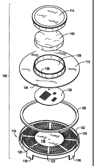
[0031] FIG. 5 is a perspective view with parts separated showing the various
individual components of the testing apparatus of FIG. 1;
[0032] FIG. 6 is a top plan view of a multi-welled base of the testing
apparatus of
FIG. 1;
[0033] FIG. 7 is a partial cross-section view of the multi-welled base taken
along
section line 7-7 of FIG. 6;
[0034] FIG. 8 is a cross-sectional view of the assembled liquid sample testing
apparatus of FIG. 1;
-9-
CA 02535524 2006-02-10
WO 2005/021157 PCT/US2004/027659
[0035] FIG. 9 is a top plan view of an alternative embodiment of a multi-
welled
base;
[0036] FIG. 10 is a top plan view of a further alternative embodiment of a
multi-
welled base;
[0037] FIG. 11 is a partial cross-sectional view taken along section line 11-
11 of
FIG. 10;
[0038] FIG. 12 is a perspective view of another illustrative embodiment of a
liquid sample testing apparatus constructed in accordance with the present
disclosure;
[0039] FIG. 13 is a cross-sectional view of the assembled liquid sample
testing
apparatus of FIG. 12;
[0040] FIG. 14 is a perspective view of another illustrative embodiment of a
liquid sample testing apparatus constructed in accordance with the present
disclosure;
[0041] FIG. 15 is a perspective view with parts separated showing the various
individual components of the testing apparatus of FIG. 14;
[0042] FIG. 16 is a cross-sectional view of the assembled liquid sample
testing
apparatus testing apparatus of FIG. 14;
[0043] FIG. 17 is a cross-sectional view with parts separated of the liquid
sample
testing apparatus testing apparatus of FIG. 14;
[0044] FIG. 18 is a perspective view of a further alternative illustrative
embodiment of a liquid sample testing apparatus constructed in accordance with
the
present disclosure;
[0045] FIG. 19 is a perspective view with parts separated of the testing
apparatus
of FIG. 18;
[0046] FIG. 20 is a top plan view of a base of the liquid sample testing
apparatus
of FIG. 18;
-10-
CA 02535524 2006-02-10
WO 2005/021157 PCT/US2004/027659
[0047] FIG. 21 is a perspective view of a further alternative illustrative
embodiment of a liquid sample testing apparatus constructed in accordance with
the
present disclosure;
[0048] FIG. 22 is a perspective view with parts separated of the liquid sample
testing apparatus of FIG. 21;
[0049] FIG. 23 is a top plan view of a frame element which forms capillary
channels of the testing apparatus of FIG. 21;
[0050] FIG. 24 is a perspective view of a further alternative illustrative
embodiment of a liquid sample testing apparatus constructed in accordance with
the
present disclosure;
[0051] FIG. 25 is a perspective view of a further alternative illustrative
embodiment of a liquid sample testing apparatus constructed in accordance with
the
present disclosure; and
[0052] FIG. 26 is a perspective view of yet another alternative illustrative
embodiment of a liquid sample testing apparatus constructed in accordance with
the
present disclosure.
DETAILED DESCRIPTION OF PREFERRED EMBODIMENTS
[0053] Referring now in specific detail to the drawings, in which like
reference
numerals identify similar or identical elements throughout the several views,
the
following detailed description will focus on specific exemplary embodiments of
testing
apparatus and methods. It is to be understood that the apparatus and methods
disclosed
herein may be adapted for use in testing for quantification of biological
material as may
be desired or necessary for a given application. Accordingly, the presently
disclosed
apparatus and methods are applicable to any biological material that it
presents at any
level in a liquefied sample (provided that one or more units of the material
can be
detected), and to any applicable testing medium. As used herein, a "liquefied
sample"
includes, but is not limited to, any sample that is a liquid or a sample that
has been
processed to act as a liquid.
-11-
CA 02535524 2006-02-10
WO 2005/021157 PCT/US2004/027659
[0054] Referring now to FIGs. 1-5, one illustrative embodiment of a testing
apparatus specifically configured and adapted to achieve quantification based
MPN
methods is shown generally as disc assembly 100. In general, operation of the
various
test apparatus embodiments disclosed herein are based on capillary fluid
dynamics to
achieve an acceptable division and distribution of the liquefied sample into
separate
targeted compartments described in greater detail herein, without external
forces from
human manipulations. The end result is to yield visual binary signals for the
quantitative
detection of biological materials based on MPN.
[0055] Disc assembly 100 includes as its major structural components, a base
110,
a lid 112 and a cap 114 which are assembled to form an integrated unit. Each
of these
components are preferably made from a durable material which provides
sufficient
structural strength such that a number of disc assemblies 100 may be stacked
on top of
each other as described in greater detail below. Examples of such material
include but are
not limited to acrylic, and polystyrene.
[0056] Base 110 includes a series of legs 116 formed to extend downwardly from
the bottom of the base and spaced around the periphery thereof. Each disc 100
is
preferably provided with four legs 116 (only three legs 116 being seen in
FIGs. 1 and 2).
However, it is also contemplated that fewer or more than four legs may be
utilized. Each
of legs 116 may be flared outward to provide additional stability when resting
disc 100 on
a flat surface or on top of other discs 100. As an additional measure of
stability, each leg
116 includes a notch or stepped end 116a, FIG. 3, to facilitate stacking of
multiple discs
100 on top of each other as shown in FIG. 2. Stepped end 116a also prevents
lateral
movement of stacked discs relative to each other.
[0057] It is also contemplated that in environments where additional stability
is
desired or necessary, active retention of adjacently stacked members with
respect to each
other could also be provided by way of a retention mechanism. This may be
useful, for
example, in mobile applications or for tests performed where it is necessary
or desirable
to index the adjacent stacked discs 100 with respect to each other. In
particular, where
more than one media is utilized to perform multiple tests at the same time,
disc
assemblies 100 could be indexed to align the corresponding media of the wells
of
adjacently stacked disc assemblies 100. To facilitate indexing of adjacent
stacked disc
assemblies 100, indicia (not shown) can be provided on each disc assembly 100
to
-12-
CA 02535524 2006-02-10
WO 2005/021157 PCT/US2004/027659
properly orient the discs relative to each other. Alternatively, the retention
mechanism
could be formed such that stacking of adjacent disc assemblies is only
possible in one
orientation of respectively stacked disc assemblies 100.
[0058] One example of a retention mechanism is shown in FIG. 4, wherein a
detent mechanism is formed between the inner surface of stepped portion 11 6a
and the
corresponding outer surface of base 116 by having a protruding portion such as
bump
116b formed on the inside surface of stepped portion 116a to be aligned with a
complementary shaped depression such as a detent 116c formed on the outer
surface of
base 110. In this manner, when discs 100 are stacked on top of each other the
detent
mechanism would function to actively retain the adjacent discs from vertical
or horizontal
movement. Other types of retention mechanisms, for example, tabs and slots,
hook and
loop fasteners, snaps, friction fit complementary shaped surfaces, or the
like, could also
be used to maintain the relative positioning of a stacked series of discs 100.
[0059] Referring to FIGs. 5-8, base 110 further includes a central sample
receiving well 118 and a plurality of individual radially arranged capillary
channels 120
formed on the upper surface. Each of capillary channels 120 is in fluid
communication at
a first end with central well 118 at a uniform height above the bottom of
central well 118
as best shown in FIG. 7. In this manner, a fluid sample poured into central
well 118 first
spreads evenly across the entire well surface and must rise to the level of
the capillary
channels 120 along the perimeter wall of central well 118. Thus, fluid will be
distributed
evenly to enter each of the capillary channels 120 substantially
simultaneously. A
plurality of target wells 122 are formed one each in fluid communication with
respective
capillary channels 120.
[0060] As best shown in FIGs. 7 and 8, target wells 122 are deeper than
central
well 118 and capillary channels 120 and may be formed in various geometrical
shapes.
For example, target wells 122 as shown in FIG. 4 have a somewhat teardrop or
pear-
shaped opening having a rounded inner end, straight side walls, are narrower
at their
juncture with capillary channels 120 and broadening to a rounded outer end.
Target wells
122 have a rectangular cross-sectional configuration. Target wells 122 may
also be
formed in other geometrical configurations. For example, both the opening and
cross-
sectional profile of target wells 122 may be of different shapes such as,
elliptical, circular,
or polygonal.
-13-
CA 02535524 2006-02-10
WO 2005/021157 PCT/US2004/027659
[0061] As shown in FIG. 6, target wells 122 are arranged in multiple groupings
uniformly around base 110. For example, as shown in FIG. 6, target wells 122
are
arranged in eight groups of nine wells each for a total of 72 independent
target wells to
achieve quantification based MPN methods. It is contemplated that different
groupings
of target wells 122 may be used depending upon the test being performed. For
example,
as shown in the embodiment of FIG. 9, base 210, which is similar to base 110,
has eight
groups of five target wells 122 each, fewer target wells 122 may form each
grouping in
order to visually space each group. Alternatively, it may be desired to have a
maximum
target wells per disc 100, as shown for example in the embodiment of FIG. 10,
wherein
base 310 is shown having no distinguishable well groups but rather a
continuous series of
target wells 122.
[0062] In each of the base embodiments 210 and 310 there is also illustrated
an
alternative capillary channel construction from that of the embodiment of
FIGs. 1-8. In
particular, instead of a single depth capillary channel as shown for channels
120, each of
bases 210 and 310 are provided with capillary channels formed to include
different
sections having different depths. Channel sections furthest away from central
wells 218,
318 are of a greater depth than sections closer to central wells 218, 318. As
shown in
FIG. 11, which is illustrative of base 310, each of capillary channels 320
includes stepped
sections 320a and 320b extending radially away from central well 318 and are
in fluid
communication with target well 322. Each target well 322 is formed a distance
radially
away from central well 318 nearer to the periphery of base 310.
[0063] Referring once again to FIGs. 6-8, base 110 further includes an
overflow
well 124 which is in fluid communication with each of target wells 122 by way
of
individual run-off channels 126 extending radially outwardly from each target
well 122.
An absorbent ring 128 is disposed in overflow well 124 to absorb any excess
sample
liquid flowing into well 124 from each of the individual target wells 122.
Alternatively,
as shown in the embodiments of FIGs. 9 and 10, base 210, 310 are formed
without an
overflow well. Excess sample in each of these embodiments is absorbed by an
absorbent
pad disposed in the cap of each of those embodiments.
[0064] A medium to facilitate growth of the target microorganism is placed in
the
base. Depending on the test being performed different media may be utilized to
detect
different microorganisms. The choice of testing medium will depend on the
biological
14-
CA 02535524 2006-02-10
WO 2005/021157 PCT/US2004/027659
material to be detected. The testing medium must be a medium, which will
detect the
presence of the biological material sought to be quantified, and preferably
not detect the
presence of other biological material likely to be in the medium. It must also
be a
material, which will cause some visible or otherwise sensible change, such as
color
change or fluorescence, if the biological material sought to be detected is
present in the
sample.
[0065] In one embodiment, the medium is in a powder form to simplify the
overall manufacturing process. The powder may be deposited directly into the
sample
landing area in the central 118 such that the medium immediately dissolves in
the sample
when the sample is poured into disc assembly 100. In alternative embodiments,
other
rapid medium dispersion methods may be utilized, for example, as shown in FIG.
5, a
porous solids-containment material, such as medium retention and dispersion
bag 130
may be used to retain the powdered medium and prevent movement of the medium
during
movement of the device, such as during shipping. Medium dispersion bag 130 may
function in an analogous manner to that of a tea bag, wherein the material of
the bag is
porous to permit flow-through of fluids. However, the size of the pores formed
in the
material making up bag 130 is preferably sized to retain the medium until
dissolved by
the fluid sample.
[0066] Still other rapid medium dispersion devices and techniques are
envisioned,
for example, quick dissolve tablets, water-permutable seals, etc.
[0067] A further alternative approach is to dispense the medium into each
target
compartment 122 directly. In each of the above-noted medium placement
embodiments,
the medium forms an integrated part of the device as shipped, thereby
eliminating the
need for a separate medium package and the separate step of preparing the
medium.
[0068] Lid 112 is configured and dimensioned to cover base 110 and is sealed
to
an upper horizontal rim 132 formed along the outer perimeter of base 110 by
suitable
techniques, for example by ultrasonic welding. A vent hole 134 is formed
through lid
112 and is located thereon to be positioned above and in fluid communication
with
overflow well 124 when lid 112 is secured to base 110. Vent hole 134 is sized
to provide
sufficient venting when a sample is poured into disc assembly 100 so as to
prevent back
-15-
CA 02535524 2006-02-10
WO 2005/021157 PCT/US2004/027659
pressure from impeding the capillary flow action of the sample through
capillary channels
120.
[0069] Lid 112 is further provided with a collar 136, which extends upwardly
from lid 112 and defines an opening 138 through the lid. Cap 114 is configured
and
dimensioned to fit over collar 136 to form a sliding seal contact therewith.
Alternatively,
the inside of cap 114 and the outside of collar 136 could be provided with
mating threads
to facilitate threaded securing of cap 114 to lid 112.
[0070] An absorbent pad 140 is configured and dimensioned to be retained
within
cap 114, for example by a friction fit. In this manner, after a sample has
been poured
through opening 138 and the cap 112 is placed securely on collar 136, any
excessive
water sample remaining in central well 118 will be absorbed and retained by
pad 140.
This will assist in preventing cross-contamination or "cross-talk" between the
individual
capillary channels 120 and, therefore, individual target wells 122. It is
envisioned that the
assembly of the various embodiments described herein may be accomplished by
way of
manual assembly, semi-automatic assembly and fully automated assembly.
[0071] Referring to FIGs. 12 and 13, another illustrative embodiment of a
water
testing apparatus constructed in accordance with the present disclosure is
shown generally
as disc assembly 400. For purposes of clarity only the structural components
of disc
assembly 400 are shown. Some or all of the previously described additional
elements
may also be incorporated into disc assembly 400 and are not repeated herein.
Disc
assembly 400 differs from disc assembly 100 in that cap 414 is formed from a
pliable
material such as rubber to permit the user to push down on the cap after it is
placed over
the sample "S". This plunging action displaces the volume of air contained
below the cap
and assists to force the sample through channels 420 and into target wells
422.
[0072] Base 410 also illustrates an embodiment wherein legs are not provided
so
that multiple bases 410 may be placed flat on a horizontal surface.
Alternatively, base
410 may be provided with legs as disclosed above for base 110.
[0073] Referring now to FIGs. 14-17, a further alternative embodiment of a
water
sample testing apparatus is shown generally as disc assembly 500. As with the
previous
disc assembly embodiment 100-400 structure which is similar to that of
previous
embodiments is labeled similarly except that each element is numbered in the
500 series.
-16-
CA 02535524 2006-02-10
WO 2005/021157 PCT/US2004/027659
Accordingly, those features, which are substantially similar to or the same as
previous
features noted on the previously described embodiments are labeled herein but
are not
necessarily separately recited with respect to the embodiment of disc assembly
500.
[0074] Lid 512 is formed with fill opening 538 formed therein, but does not
include a collar member about the periphery thereof. Instead a series of vent
holes are
formed in lid 512 close to opening 538. As shown in FIG. 16, vent holes 534
are in fluid
communication with capillary channel section 520b to provide venting when cap
512 is
removed from lid 512. Upon placement of cap 514 in lid 512 vent holes 534 are
sealed
off to prevent additional infiltration of air during the incubation period.
This arrangement
is particularly beneficial when it is important to have test conditions that
ensure that no
additional air is introduced into target wells 522.
[0075] Referring to FIGs. 18-20, a further alternative embodiment of the
presently
disclosed water sample testing apparatus is shown generally as test device
600, which is
substantially similar to the previous embodiments in many respects. The
principle
difference of test device 600 is that it is formed in a generally rectangular
configuration.
In all other aspects, test device 600 is similar to the previously described
embodiments
and may be constructed to include the various alternative features previously
described
herein.
[0076] The method of using each of the above-described embodiments is
substantially similar and will now be described. Where differences between
embodiments exist, they will be noted. Briefly, to conduct a liquefied sample
test, such
as a water sample test, a user removes the cap and pours approximately 1 ml to
approximately 5 ml of water sample into the center well, replaces the cap,
inverts the test
device once to absorb excessive sample left in the center well, and incubates
the test
device at the required temperature and for the time required by the particular
test. Results
are obtained by the enumeration of positive targets and comparing enumerated
positives
to a MPN table.
[00771 When the sample is poured in to the center well, the powder medium is
dissolved upon contact with the water sample to achieve a proper sample-medium
mixture. When the height of the sample in the center well reaches the height
of the
-17-
CA 02535524 2006-02-10
WO 2005/021157 PCT/US2004/027659
capillary channels, the sample-media mixture flows to the wells located at the
outer edge
of the test device.
[0078] The device may be left in the inverted position or may be returned to
the
original upright position for the incubation period. As previously noted, for
those
embodiments which facilitate it, where multiple tests are to be conducted
simultaneously,
the individual devices may be stacked upon each other due to the uniquely
advantageous
structure of the base with the stepped legs formed thereon.
[0079] FIGs. 21-23 illustrate a further alternative embodiment of a liquid
sample
testing apparatus for the quantification of target microorganisms, which is
shown
generally as test device 700. Briefly the operational portion of test device
700 includes a
multiple layer assembly of plastic films which are held together as a unit,
for example by
a transfer adhesive and are enclosed in a hydrophobic container such as a two-
part
transparent dish having a top portion 702a which fits over a bottom portion
702b. The
multiple-layer film assembly includes a top hydrophilic layer 710, a
hydrophobic frame
712 which includes at least one capillary channe1720 formed therein, and a
plastic
backing layer 714.
[0080] Preferably, top layer 710 is made of clear polyester (PE) material with
a
hydrophilic surface to facilitate passage of the liquid sample being tested
through top
layer 710 and into hydrophobic frame 712. Alternatively, top layer 710 may be
made
from any other clear plastic material with a hydrophilic surface. Furthermore,
the top
layer 710 can be hydrophilic and have a heat or pressure sensitive adhesive
coated on the
same side facing the frame 712. This configuration can eliminate the need to
use a
transfer adhesive or other means of bonding to put the two parts together.
[0081] Hydrophobic frame 712, which forms the capillary channel structure, is
preferably made from material selected from the group consisting of
polystyrene,
polyester, and PETG. A sample-landing zone 716 is defined in the central
portion of
frame 712. Capillary channels 720 are formed in hydrophobic frame 712 and are
enclosed from top and bottom when top layer 710 and plastic backing layer 714
are
adhered to hydrophobic frame 712, for example by a transfer adhesive. Each
capillary
channel is in fluid communication with the sample-landing zone 716 and is
adapted to
partition liquid sample from sample-landing zone 716 to the recessed
compartment.
-1~-
CA 02535524 2006-02-10
WO 2005/021157 PCT/US2004/027659
Capillary channels 720 may be formed in various clustered arrangements or in a
continuous arrangement as described with respect to the previous embodiments.
[0082] As shown in FIG. 23, fifty capillary channels 720 are arranged in
groups
of five. Each of capillary channels 720 includes a reaction well 722 are
formed in
hydrophobic frame 712. The capillary channels 720 and reaction wells 722 may
be
configured and dimensioned as shown or in any of the previously described
configurations and dimensions set forth with respect to the other embodiments
illustrated
and described herein.
[0083] Reaction wells 722 are formed to include at least one recessed
compartment, which is in fluid communication with a venting slot 724 disposed
radially
outwardly therefrom to facilitate the capillary flow. Each reaction well 722
is configured
and dimensioned to hold an aliquot of sample/medium mixture for the detection
of the
targeted biological material.
[0084] The plastic backing layer 714 is hydrophobic plastic layer. It is
preferably
made from polyester or other similar material. Plastic backing layer 714
includes a series
of holes 726 formed therethrough, each hole being preferably spaced radially
such that
upon assembly of the layers, holes 726 are positioned one each, in between the
groups of
capillary channels 720 (see FIG. 24). A central hole 728 is formed to align
centrally with
the sample-landing zone 716. Together holes 726 and 728 facilitate passage of
excess
sample through to the bottom of device 700.
[0085] In an alternative embodiment, the device may further include an
absorbent
pad 730, which is positioned below the multi-layer plastic assembly inside
bottom disc
portion 702a to absorb any excess liquid sample. The absorbent material may be
a die cut
polyester foam, polyether foam, cotton, or a cellulose acetate or other
suitable absorbent
material. The absorbent pad containing excessive liquid samples also acts as a
humidifying source to prevent the assay in the assembly 700 from drying out
during
incubation.
[0086] In use, the top disc portion 702a is removed from device 700 and an
inoculating volume of approximately 3.5 ml of liquid sample is introduced into
sample
landing zone 716 and top portion of disc 702a is replaced to close device 700.
The total
time for introduction of the sample should be approximately 5 seconds. The
sample fills
-19-
CA 02535524 2006-02-10
WO 2005/021157 PCT/US2004/027659
the landing zone 716 and is drawn by capillary action into capillary channels
720 and fills
each of reaction wells 722. Excess sample is absorbed by pad 730 as it either
travels
through holes 726, 728 or through venting slots 724.
[0087] FIG. 24 illustrates a further alternative embodiment of a liquid sample
testing apparatus for the quantification of target microorganisms, which is
shown
generally as test device 800. The operational portion of test device 800 is
similar to that
of test device 700 in that it also includes a multiple layer assembly of
plastic films, which
are held together as a unit, and are enclosed in a hydrophobic container such
as a two-part
transparent dish having a top portion 802a, which fits over a bottom portion
802b. The
multiple-layer film assembly includes a top hydrophilic layer 810 having a
sample
receiving hole 816 formed therethrough, a hydrophobic frame 812 which includes
at least
one capillary channel 820 formed therein, and an absorbent pad backing layer
830.
Hydrophobic frame 812 may be formed by suitable techniques such as injection
molding
or heat stamping. Furthermore, the top layer 810 can be both hydrophilic and
heat or
pressure-sensitive achieve coated on the same side facing the frame 812. This
configuration can eliminate the usage of transfer achieve or other means of
bonding to put
the two parts together.
[0088] Test device 800 does not include, however, a backing layer like plastic
backing layer 714 of test device 700. Instead, vent holes 826 and central hole
828 are
formed in the central region of hydrophobic frame 812. As with the various
previous
embodiments, capillary channels 820 may be formed in various clustered
arrangements or
in a continuous arrangement as described with respect to the previous
embodiments. The
use of test device is the same as that for test device 700 and will not be
addressed in detail
again. Furthennore, the top layer 810 can be both hydrophilic and heat or
pressure-
sensitive achieve coated on the same side facing the frame 812. This
configuration can
eliminate the usage of transfer achieve or other means of bonding to put the
two parts
together.
[0089] FIGs. 25-26 illustrate a further alternative embodiment of a liquid
sample
testing apparatus for the quantification of target microorganisms, which is
shown
generally as test device 900. The operational portion of test device 900
includes the
distribution channels and recessed compartments molded directly onto a bottom
half 901
of test device 900 through the injection mold technique. As with the various
previous
-20-
CA 02535524 2009-08-24
embodiments, capillary channels and target reaction compartments are formed by
placing
a plastic film 903 on top of bottom half 901 of device 900. Plastic film 903
can have
either a heat or a pressure-sensitive adhesive coated on the same side facing
bottom half
901 of device 900. An absorbent ring 904 may be attached on top of plastic
film 903 to
absorb the excess liquid or liquefied sample/medium mixture. Alternatively, as
shown in
FIG. 26, a plastic ring 905 may be attached on top of plastic film 903 to
contain the liquid
sample or liquefied sample/medium mixture before distributing into the
capillary
channels and target reaction compartments through the capillary action. In
addition, as
seen in FIG. 26, an absorbent pad 906 is attached on a top half 902 of device
900 to
absorb the excess liquid or liquefied sample/medium mixture. The use of test
device 900
is the same as that for previous embodiments and will not be addressed in
detail again.
Example 1: Bacterial Detection and Enumeration Device for
Heterotrophic Bacteria in Water
[0090] The following is an example of how the present invention provides a
method of detecting and enumerating heterotrophic bacteria in water samples.
The device
used in this assay is constructed according to the drawing illustrated in
Figure 26. The
medium of Townsend and Chen (U.S. Patent Nos. 6,387,650 and 6,472,167)
is provided and deposited in the
capillary channels and reaction compartments. The medium includes the
following
components: a source of amino acids and nitrogen mixture (2.5 gram/liter); a
source of
vitamin mixtures (1.5 gram/liter); sodium pyruvate (0.3 gram/liter); magnesium
sulfate
(0.5 gram/liter); fast green dye (0.002 gram/liter); buffer components (4.4
gram/liter); and
a mixture of enzyme substrates (0.105 gram/liter).
100911 The results of this example were evaluated against an Intemational
Standard Method ISO 6222 (Water Quality- Enumeration of Culturable Micro-
organisms - Colony Count by Inoculation in a Nutrient Agar Culture Medium).
Data
were analyzed using the statistical method described in the ISO Method 17994
(Water
Quality - Criteria for establishing the equivalency of two microbiological
methods).
Results are reported in Table I, below. A total of 368 water samples were
analyzed and
incubated at or about 37 C for approximately 48 hours and a total of 339 water
samples
were incubated at or about 22 C for approximately 72 hours. An aliquot of
about 3.5mL
- 2] -
CA 02535524 2006-02-10
WO 2005/021157 PCT/US2004/027659
of each water sample was added to the sample-landing area of a respective
device. Each
water sample automatically distributed, through capillary action, into all the
reaction
compartments within few seconds. The device was then incubated at or about 37
C for
approximately 48 hrs or at or about 22 C for approximately 72 hrs. Bacterial
concentrations in the water sample were determined by examining the number of
reaction
compartments exhibiting a fluorescent signal under a UV lamp (366,,,,,). The
number of
bacteria present in the sample was then determined based on MPN statistics.
The
statistical analysis of the data based on ISO Method 17994 (Water Quality -
Criteria for
establishing the equivalency of two microbiological methods) is set forth in
Table I.
Table I.
ISO Method 17994 Statistical Analysis Comparison
between the present invention and ISO Method 6222
37 C for 48 hrs 22 C for 72 hrs
N 368 339
Mean % RD 9.9 16.3
U 10.3 12.1
LO -0.5 4.2
HI 20.2 28.3
N = Number of Samples
RD (Relative Difference) means the difference between two results A
(invention) and B (ISO Method
6222) measured in the relative (natural logarithmic) scale. The value of RD is
expressed in percent
according to RD% = 100 =[ln (A) - ln (B)].
U (Expanded Uncertainty) is derived from the standard uncertainty of the mean
by using the coverage
factor K = 2. To evaluate the result of the comparison the "confidence
interval" of the expanded uncertainty
around the mean is calculated by computing the limits: LO (Lower Limit) =
(Mean %RD) -(U) and HI
(Upper Limit) = (Mean %RD) +(U). It is desirable to achieve an average
performance that is either
quantitatively equivalent or higher than the reference method. In such cases,
the "One-sided Evaluation"
method is used and two methods are determined to be "no different" when - 10 <-
LO <- 0 and HI > 0.
When LO is greater than zero, it means that the method of the present
invention is more sensitive than the
reference method.
(0092] The results reported in Table I indicate that the device and method
according to the present invention can detect and enumerate heterotrophic
bacteria in
water samples and is equivalent or better than the standard reference method.
-22-
CA 02535524 2009-08-24
Example II: Bacteria] Detection and Enumeration Device for
Enterococcus Batceria
100931 The following is another example of detecting and enumerating
microorganisms using the present invention. The device used in this assay is
constructed
according to the drawing illustrated in FIG. 26. The medium of U.S. Patent No.
5,620,865 to Chen, et al.,
(which is practiced by IDLXX's commercial EnterolertT"' medium, a medium for
the
detection of Enterococcus bacteria in a sample) is provided and deposited in
the capillary
channels and reaction compartments. A known level, as determined by the
Typicase Soy
Agar supplemented with 5% sheep blood, of Enterococcusfeacalis ATCC 35667 was
inoculated into a device of this invention (Table iI). Results indicated that
the
concentration of E. faecalis'ATCC 35667 determined by the FIG. 26 device is
statistically
equivalent to those determined by the TSA with 5% sheep blood plate count
method.
Table II
TSA/5% Sheep Blood Fig. 26 Device
Replicate 1 22 24.5
Replicate 2 16 13.5
Replicate 3 14 29.3
Replicate 4 16 17.1
Replicate 5 22 15.5
Average 18 20.1
Standard Deviation 3.7 6.7
[0094] While the invention has been particularly shown and described with
reference to the preferred embodiments, it will be understood by those skilled
in the art
that various modifications in form and detail may be made therein without
departing from
the scope and spirit of the invention. Accordingly, modifications such as
those suggested
above, but not limited thereto, are to be considered within the scope of the
invention.
-23-