Note: Descriptions are shown in the official language in which they were submitted.
CA 02535663 2009-09-09
NEUROTOXIC AMINO ACID OR NEi7ROTOXIC DERIVATIVE THEREOF ASSOCIATED WITH
NEUROLOGI
CAL DISORDERS
Field of the Invention
The present invention relates to screening for neurological disorders.
Specifically, the
invention relates to screening for neurological disorders in a subject by
analyzing a tissue
sample from the subject to determine the presence of neurotoxic amino acids or
neurotoxic
derivatives thereof associated with neurological disorders. In particular, the
present invention
relates to methods for diagnosing a neurological disorder in a subject, or
predicting the
likelihood of developing a neurological disorder in a subject, by determining
the levels of D-
N-methylamino-L-alanine (BMAA) or a neurotoxic derivative thereof, in a tissue
sample
obtained from the subject. Further, the invention relates to screening
environmental samples
for a neurotoxic amino acid or neurotoxic derivative thereof associated with
neurological
disorders. Further, the invention relates to inhibiting neurological
disorders.
Background of the Invention
A unique neurological disease initially identified among the Chamorro people
of
Guam by Kurland and Mulder (1954) is characterized by a combination of
symptoms
including stooped posture, a blank expressionless face, dementia, slow
shuffling movement, a
resting tremor that stops upon deliberate action, slow movements, and muscle
atrophy that
results in muscles dipping down in the hand. In some clinical manifestations,
patients have
clinical symptoms indistinguishable from amyotrophic lateral sclerosis (ALS).
Other patients
have Parkinsonism features combined with dementia (Parkinsonism Dementia
Complex,
PDC). In still others, only dementia is observed. Some patients also have both
ALS and PDC.
Neuropathologically, all clinical forms of the disease result in a specific
feature,
neurofibri lary tangles, found in the cortex and in the spinal cord. Because
the disease has
aspects that resemble amyotrophic lateral sclerosis (ALS), Parkinson's disease
(PD) and
Alzheimer's disease (AD), this disease is known as amyotrophic lateral
sclerosis-
Parkinsonism dementia complex of Guam (ALS-PDC) and is also known as lytico-
bodig.
1
CA 02535663 2006-02-13
WO 2005/019830 PCT/US2003/039202
Summary of the Invention
The present invention provides methods of screening a subject having or at
risk of
having a neurological disorder by analyzing a tissue sample from the subject
to determine the
presence of a neurotoxic amino acid, or neurotoxic derivative thereof,
associated with the
neurological disorder. The neurotoxic amino acid or neurotoxic derivative
thereof can be a
glutamate receptor agonist such as (3-N-methylamino-L-alanine (BMAA), or 0 -N-
oxalyl-
amino-L-alanine (BOAA). In a tissue sample, protein-bound neurotoxic amino
acid or
neurotoxic derivative thereof can be analyzed, free (unbound) neurotoxic amino
acid or
neurotoxic derivative thereof can be analyzed, or both protein-bound and free
neurotoxic
amino acid or neurotoxic derivative thereof can be analyzed in a sample. In a
tissue sample,
protein-bound BMAA, free BMAA, or both protein-bound BMAA and free BMAA can be
analyzed. The subject may have symptoms of a neurological disorder, or may be
asymptomatic for a neurological disorder, or may have been identified as being
at risk for
developing a neurological disorder. The neurotoxic derivative may be any
derivative having
neurotoxic activity, such as a carbamate adduct or metabolite of the
neurotoxic amino acid.
The present invention provides methods of screening a subject having or at
risk of
having a neurological disorder by analyzing a tissue sample from the subject
to determine the
presence of a neurotoxic amino acid or neurotoxic derivative thereof
associated with the
neurological disorder, wherein the presence of a detectable level of a
neurotoxic amino acid
or neurotoxic derivative thereof indicates a neurological disorder. Methods of
the invention
can be used to detect neurological disorders including a neurofibrillary
tangle disorder (NFT
disorder) such as amyotrophic lateral sclerosis-Parkinsonism dementia complex
(ALS-PDC),
Alzheimer's disease, or progressive supranuclear palsy (PSP), a movement
disorder such as
Parkinson's disease, or a motor neuron disease such as amyotrophic lateral
sclerosis (ALS).
The present invention provides methods of screening a subject having or at
risk of
having a neurological disorder by analyzing a tissue sample from the subject
to determine the
presence of a neurotoxic amino acid or neurotoxic derivative thereof
associated with the
neurological disorder, wherein the methods can be used to predict the
likelihood of
developing a neurological disease, and/or to predict the latency period prior
to onset of the
neurological disorder, and/or to predict the severity of the neurological
disorder. Methods of
the present invention can be practiced using tissue samples including, but not
limited to,
neurological tissue or non-neurological tissue. Neurological tissue can be
associated with the
2
CA 02535663 2006-02-13
WO 2005/019830 PCT/US2003/039202
central nervous system (CNS), including brain tissue or cerebral-spinal fluid
(CSF), or may
be associated with the peripheral nervous system (PNS). Non-neurological
tissue can be
keratinous tissue including but not limited to, hair, skin, nail, including
fingernail or toenail,
feather, claw, hoof, or horn. Non-neurological tissue can be non-keratinous
tissue including
but not limited to, hood, serum, saliva, or urine.
The present invention provides methods for screening an environmental sample
to
determine if the environmental sample is associated with a neurological
disorder, by
analyzing the environmental sample to determine the presence of a neurotoxic
amino acid or
neurotoxic derivative thereof associated with the neurological disorder. The
neurotoxic amino
acid or neurotoxic derivative thereof can be a glutamate receptor agonist such
as a methylated
alanine, in particular, BMAA. Suitable environmental samples include water
and/or food
items or sources.
The present invention provides methods for screening an environmental sample
to
determine if the sample is associated with a neurological disorder, by
detecting neurotoxic
amino acid or neurotoxic derivative thereof producing cyanobacteria in the
environmental
sample. The neurotoxic amino acid or neurotoxic derivative thereof can be a
glutamate
receptor agonist such as a methylated alanine, in particular, BMAA. Methods of
the
invention are suitable for detecting cyanobacteria producing the neurotoxic
amino acid or
neurotoxic derivative thereof, including cyanobacteria of the genus Nostoc
and/or Anabena.
Suitable environmental samples include water and/or food items or sources.
The present invention provides methods for inhibiting a neurological disorder
in a
subject by reducing levels of a neurotoxic amino acid or neurotoxic derivative
thereof
associated with the neurological disorder, in particular by releasing the
neurotoxic amino acid
or neurotoxic derivative thereof from an endogenous reservoir. The neurotoxic
amino acid or
neurotoxic derivative thereof can be a glutamate receptor agonist such as a
methylated
alanine, in particular, BMAA.
The present invention provides methods inhibiting a neurological disorder in a
subject
by increasing the cellular concentration of a neuroprotectant compound that
blocks
interaction of a neurotoxic amino acid or neurotoxic derivative thereof
associated with the
neurological disorder with a target molecule. The neurotoxic amino acid or
neurotoxic
derivative thereof can be a glutamate receptor agonist such as a methylated
alanine, in
3
CA 02535663 2006-02-13
WO 2005/019830 PCT/US2003/039202
particular, BMAA. The neuroprotectant compound can be glutamic acid. An agent
that ,
binds or chelates the neurotoxic amino acid or neurotoxic derivative thereof,
can be included.
The present invention further provides kits for screening a subject having or
at risk of
having a neurological disorder, wherein the kits include a means for obtaining
a tissue sample
from the subject and a means for analyzing the tissue sample to determine the
presence ofa
neurotoxic amino acid or neurotoxic derivative thereof associated with the
neurological
disorder. The kit may include means for determining the presence of a
glutamate receptor
agonist such as a methylated alanine, in particular BMAA. The kit may include
means for
analyzing protein-bound BMAA, free BMAA, or both protein-bound BMAA and free
BMAA in the sample. The kit may include means for obtaining and analyzing a
plurality of
tissue samples from the subject. The tissue samples may include a sample of a'
tissue in
which a neurotoxic amino acid or neurotoxic derivative thereof is known to
accumulate and a
sample of a tissue in. which neurotoxic amino acid or neurotoxic derivative
thereof is known
to not accumulate. The tissue samples may include a sample of at least two
distinct tissues in
which a neurotoxic amino acid or neurotoxic derivative thereof is known to
accumulate. The
kit may include means for performing repeated screening of the subject.
Brief Description of Drawings
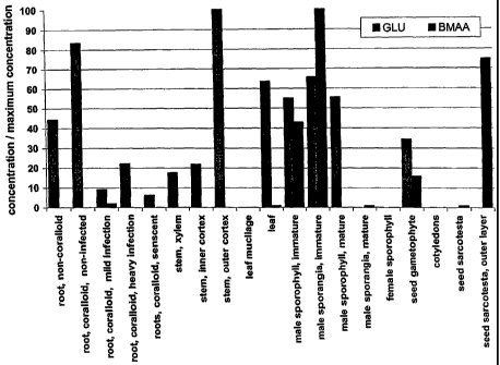
Figure 1 shows concentrations of BMAA and glutamic acid (GLU) in Cycas
micronesia Hill, normalized by dividing the maximum concentration of each
amino acid,
which permits a comparison of relative abundance throughout the plant; values
below 9 gg/g
cannot be seen in this figure as these values are too small relative to the
maximum
concentration.
Table 1 shows BMAA and GLU concentrations in various tissues of Cycas
naicronesia Hill; concentrations are expressed as g/g.
Table 2 shows BMAA concentrations in samples of cycad tissues, cyad flour, and
flying fox tissues.
Table 3 shows levels of free and protein-associated BMAA in tissue samples
from the
superior frontal gyrus of patients from Chamorro and Canadian populations.
4
CA 02535663 2006-02-13
WO 2005/019830 PCT/US2003/039202
Detailed Description of the Invention
The present disclosure provides methods for screening for neurological
disorders.
Methods as provided herein can be used to diagnose or predict neurological
disorders in a
subject, to screen for environmental factors associated with neurological
disorders, and to
inhibit neurological disorders in a subject.
The present invention provides methods for screening a subject for
neurological
disorders by analyzing a tissue sample from the subject to determine the
presence of a
neurotoxic amino acid or neurotoxic derivative thereof associated with the
neurological
disorder. The present invention further provides methods for screening
environmental
samples to determine the presence of a neurotoxic amino acid or neurotoxic
derivative
thereof associated with neurological disorders. The phrase "to determine the
presence of a
neurotoxic amino acid or neurotoxic derivative thereof' or "determining the
presence of a
neurotoxic amino acid or neurotoxic derivative thereof' or a similar phrase,
includes not only
determining the presence or absence of detectable levels of a neurotoxic amino
acid or
neurotoxic derivative thereof, but also includes quantifying the levels of a
neurotoxic amino
acid or neurotoxic derivative thereof detected in a sample. Thus, in a
particular embodiment,
"determining the presence of a neurotoxic amino acid or neurotoxic derivative
thereof' in a
sample can include determining the level of the neurotoxic amino acid or
neurotoxic
derivative thereof and can further include determining whether the level of
neurotoxic amino
acid or neurotoxic derivative thereof in the sample is elevated or decreased
in comparison
with the levels detected in other samples.
Screening includes but is not limited to, diagnosing or predicting
neurological
disorders in a subject by analyzing a tissue sample from the subject.
Screening may be
carried out on a subject having a neurological disorder, or may be carried out
on a subject at
risk of having a neurological disorder, or may be carried out on a subject
having no known
risk of having a neurological disorder. Screening further includes analyzing
environmental
samples to determine actual or potential exposure of a subject to a neurotoxic
amino acid or
neurotoxic derivative thereof associated with a neurological disorder.
As provided herein, neurotoxic amino acids or neurotoxic derivatives thereof
associated with neurological disorders include, but are not limited to, non-
protein amino acids,
excitatory amino acids, amino acid analogs, amino acid metabolites, carbamate
adducts of
CA 02535663 2006-02-13
WO 2005/019830 PCT/US2003/039202
amino acids, and conjugates of amino acids. In one embodiment, one or more
neurological
disorders can be screened- for in a subject by determining the presence of (3-
N-methylamino-
L-alanine (BMAA) in a sample of tissue obtained from the subject. In another
embodiment,
one or more neurological disorders can be screened for by determining the
presence of (S )-2-
amino-3-(3-hydroxy-5-methylisoxazol-4-yl) propionic acid (AMPA) in a sample of
tissue
obtained from the subject. In yet another embodiment, one or more neurological
disorders
can be screened for in a subject by determining the presence of (3 -N-oxalyl-
amino-L-alanine
(BOAA, also.described as S -(-)-B-N -oxalyl-,l3-diaminopropionic acid) in a
sample of tissue
obtained from the subject. It is understood that methods for determining of
neurotoxic amino
acid or neurotoxic derivative thereof include, when necessary, methods for
distinguishing the
neurotoxic isomer from the nonneurotoxc isomer of the -same compounds, -e.g.,
for
distinguishing neurotoxic L-BOAA from non-neurotoxic D-BOAA.
Neurotoxic amino acids of the present invention can be non-protein amino acids
including but not limited to, (3-alanine (3-alanine), 4-aminobutyrate (GABA),
3-cyanoalanine
((3-cyanoalanine), 2-aminobutyric acid, 2-methylene-4-aminobutyric acid, 3-
methylene-4-
aminobutyric acid, 2-aminoisobutyric acid, 5-aminolevulinic acid, 2-amino-4-
methylhexanoic acid (homoisoleucine), 2-amino-4-methylhex-4-enoic acid, 2-
amino-4-
methylhex-5-ynoic acid, 2-amino-3-methylpentanoic acid, 2-aminoadipic acid, 4-
ethylideneglutamic acid, 3-aminoglutaric acid, 2-aminopimelic acid, N4-
ethylasparagine, N4-
methylasparagine, erythro-4-methylglutamic acid, 4-methyleneglutamic acid. 4-
methyleneglutamine, N5-methylglutamine, N5-ethylglutamine (theanine), N5-
isopropylglutamine, 2-amino-4-(aminoxy)butyric acid (canaline), 2,4-
diaminobutyrate, N4-
acetyl-2,4-diaminobutyrate, N4-lactyl-2,4-diaminobutyrate, N4-oxalyl-2,4-
diaminobutyrate,
2,3-diaminopropionic acid, N3-acetyl-2,3-diaminopropionic acid, N3-methyl-2,3-
diaminopropionic acid, N3-oxalyl-2,3-diaminopropionic acid, N6-acetyllysine,
N6-
methyllysine, N6-trimethyllysine (laminine), ornithine (2,5-diaminopentanoic
acid),
saccharopine (N6-(2'-glutamyl)lysine, 2,6-diaminopimelic acid, N4-(2-
hydroxylethyl)asparagine, erythro-3-hydroxyaspartic acid, 4-hydroxyarginine, 4-
hydroxycitrulline, threo-4-hydroxyglutamic acid, 3,4-dihydroxyglutamic acid, 3-
hydroxy-4-
methylglutamic acid, 3-hydroxy-4-methyleneglutamic acid, 4-hydroxy-4-
methylglutamic
acid, 4-hydroxy-glutamine, N5-(2-hydroxyethyl)glutamine, 5-hydroxynorleucine,
threo-4-
hydroxyhomoarginine, homoserine, O-acetylhomoserine, O-oxalylhomoserine, 0-
phosphohomoserine, 4-hydroxyisoleucine, 5-hydroxymethylhomocysteine, threo-3-
6
CA 02535663 2006-02-13
WO 2005/019830 PCT/US2003/039202
hydroxyleucine, 5-hydroxyleucine, 2-hydroxylysine, 4-hydroxylysine, 5-
hydroxylysine, N6-
acetyl-5-hydroxylysine, N6-trimethyl-5-hydroxylysine, 4-hydroxyornithine,
mimosine, 4-
hydroxynorvaline, 5-hydroxynorvaline, 2-amino-4,5-dihydroxypentanoic acid, 2-
amino-4=
hydroxypimelic acid, 4-hydroxyvaline, 0-acetylserine, 0-phosphoserine,
pipecolic,acid
(piperidine-2-carboxylic acid), 3-hydroxypipecolic acid, trans-4-
hydroxypipecolic acid, trans-
5-hydroxypipecolic acid, 5-hydroxy-6-methylpipecolic acid, 4,5-
dihydroxypipecolic acid,
trans-3-hydroxyproline, trans-4-hydroxyproline, trans-4-hydroxymethylproline,
azetidine-2-
carboxylic acid, N-(3-amino-3-carboxypropyl)azetidine-2-carboxylic acid, 4,5-
dehydropipecolic acid (baikiain), 3-amino-3-carboxypyrrolidone (cucurbitine),
2-(cyclopent-
2'-enyl)glycine, 5-hydroxytryptophan, albizziine (2-amino-3-ureidopropionic
acid),
arginosuccinic acid, canavinosuccinic acid, citrulline, homoarginine,
homocitrulline,
indospicine, O-ureidohomoserine, 6-hydroxykynurenine, 3-(4-
aminophenyl)alanine, 3-(3-
aminomethylphenyl)alanine, 3-(3-carboxyphenyl)alanine, 3-carboxytyrosine, 3-(3-
hydroxymethylphenyl)alanine, 3-(3-hydroxyphenyl)alanine, 3-(3,4-
dihydroxyphenyl)alanine
(L-DOPA), 2-(phenyl)glycine, 2-(3-carboxyphenyl)glycine, 2-(3-carboxy-4-
hydroxyphenyl)glycine, 2-(3-hydroxyphenyl)glycine, 2-(3,5-
dihydroxyphenyl)glycine, 4-
aminopipecolic acid, guvacine, 2-amino-4-(isoxazolin-5-one)-2-yl)butyric acid,
lathyrine, or
tetrahydrolathyrine. (Spencer and Berman, 2003, in, Plant Toxins and Human
Health, CABI,
pp 1-23). The present disclosure provides sufficient guidance for one of skill
in the art to
identify a neurotoxic non-protein amino acid of the present invention.
Neurotoxic derivatives of non-protein amino acids include but are not limited
to
metabolites, carabamate adducts, analogs, and other amino acid derivatives
having neurotoxic
activity. In accordance with one aspect, neurotoxic derivatives are carbamate
adducts
(carbamates) of neurotoxic amino acids. In one embodiment, neurotoxic
derivatives of the
prsent invention are carbamate adducts of BMAA, including a-N-carboxy-(3-N-
methylamino-
L-alanine (BMAA-a-NCO2) and/or (3-(N-carboxy-N-methyl)-amino-L-alanine (BMAA-
(3-
NCO2), (Brownson et al., 2002, JEthnophaf macol 82: 159-167; Myers and Nelson,
1990, J
Biol Chem 265:10193-10195). In accordance with another aspect, neurotoxic
derivatives
include the neurotoxic isomer of a neurotoxic amino acid, although it could
alternately be
understood that the neurotoxic isomer is the neurotoxic amino acid in a
particular
embodiment. Neurotoxic derivatives may also be methylated, carbamylated, or
hydroxylated
metabolites, or metabolites conjugated to sugars, lipids, or proteins. It is
understood that the
methods provided herein are suitable for determining neurotoxins associated
with
7
CA 02535663 2006-02-13
WO 2005/019830 PCT/US2003/039202
neurological disorders, and may provide a robust measurement of neurotoxin
even when the
compound being measured is not necessarily. the compound or compounds acting
in vivo in a
particular subject. In one embodiment, the present disclosure provides methods
for
determining BMAA levels in tissue samples and environmental samples, and these
methods
generate robust results even when these methods do not distinguish whether
BMAA, or a
derivative such as a carbamate adduct of BMAA (e.g., (BMAA-a-NCO2 or R-(N
carboxy-N-
methyl)-amino-L-alanine (BMAA-(3-NCO2) is the most active compound in a
particualr
embodiment.. TJ,e methods presented herein are robust, and can be further
refined by one of
skill in the art, according to the particular circumstances of a particular
embodiment.
In accordance with another aspect, the present invention provides methods for
screening environmental samples for neurotoxic amino acids or neurotoxic
derivatives
thereof associated with neurological disorders. Screening environmental
samples for
neurotoxic amino acids or neurotoxic derivatives thereof includes, but is not
limited to,
screening to determine actual or potential exposure of a subject to neurotoxic
amino acids or
neurotoxic derivatives thereof associated with neurological disorders, and
screening to
identify environmental samples contaminated with neurotoxic amino acids or
neurotoxic
derivatives thereof associated with neurological disorders. In one embodiment,
the present
invention provides methods for determining BMAA levels in environmental
samples
including water samples or food items.
In accordance with yet another aspect, the present invention provides methods
for
inhibiting neurological disorders in a subject by reducing levels of a
neurotoxic amino acid or
neurotoxic derivative thereof associated with neurological disorders, e.g., by
draining
endogenous reservoirs of the neurotoxic amino acid or neurotoxic derivative
thereof.
Inhibiting includes, but is not limited to, treating existing neurological
disorders or preventing
neurological disorders. In one embodiment, the present invention provides
methods for
draining endogenous reservoirs of BMAA or derivatives thereof in a subject.
In accordance with another aspect, the invention provides methods for
inhibiting a
neurological disorder in a subject by interfering with the interaction between
a neurotoxic
amino acid or neurotoxic derivative thereof and its target molecule. In
particular, the
invention provides methods for inhibiting a neurological disorder by
increasing the cellular
concentration of a neuroprotectant compound that blocks interaction of a
neurotoxic amino
acid or neurotoxic derivative thereof with a target molecule. In one
embodiment, the
8
CA 02535663 2006-02-13
WO 2005/019830 PCT/US2003/039202
neurotoxic amino acid or neurotoxic derivative thereof is BMAA or a BMAA
derivative, and.
the neuroprotectant compound is glutamic acid or a glutamic acid analog.
Agents that bind
neurotoxic amino acids or neurotoxic derivatives thereof can be included to
sequester
neurotoxic amino acid or neurotoxic derivative thereof released from
endogenous reservoirs.
Chelating agents can be included to chelate metal ions released when
neurotoxic amino acids
or neurotoxic derivatives thereof are released from endogenous reservoirs.
As provided herein, a subject may be any organism suitable for practicing the
methods of the present invention. In particular, a subject is a mammal, more
particularly a
primate, even more particularly a human. In one embodiment, a subject is an
experimental
animal that is exposed to a neurotoxic amino acid or neurotoxic derivative
thereof associated
with neurological disorders. Such experimental animals include, but are not
limited to, a
mouse, rabbit, rat, bat, pig, sheep, cow, monkey, ape, or other animal
suitable for research on
neurological disorders. In one embodiment, methods of the present invention
are carried out
using an experimental animal for which an animal model of one or more
neurological
diseases exists. In another embodiment, methods of the present invention are
carried out
using an experimental animal as part of developing an animal model of one or
more
neurological diseases. In yet another embodiment, methods of the present
invention are
carried out using an experimental animal in which the effects of exposure to a
neurotoxic
amino acid or neurotoxic derivative thereof associated with neurological
disorders are
measured by studies of brain chemistry, structure, or function. In one
embodiment, a subject
is a human. In another embodiment, a subject is a human suffering from one of
more
neurological disorders. In another embodiment, a subject is a human who is
asymptomatic
for one or more neurological disorders. In another embodiment, a subject is a
human who
has been identified as being at risk for developing a neurological disorder.
In yet another
embodiment a subject is a human who is known or suspected of having been
exposed to at
least one neurotoxic amino acid or neurotoxic derivative thereof associated
with neurological
disorders.
In accordance with one aspect of the present invention, methods are provided
for
analyzing tissue samples from a subject, or environmental samples used in
environmental
screening, for one or more forms of neurotoxic amino acids or neurotoxic
derivatives thereof
associated with neurological disorders. Methods include analysis of free
(e.g., unbound,
cytosolic, circulating) forms of neurotoxic amino acids or neurotoxic
derivatives thereof
9
CA 02535663 2006-02-13
WO 2005/019830 PCT/US2003/039202
associated with neurological disorders, protein-bound forms of neurotoxic
amino acids or
neurotoxic derivatives thereof associated with neurological disorders (e.g.,
bound to proteins
or incorporated into proteins), or conjugated forms of neurotoxic amino acids
or neurotoxic
derivatives thereof associated with neurological disorders (e.g., conjugated
to sugars or
lipids). One of skill in the art can determine what forms of neurotoxic amino
acid or
neurotoxic derivative thereof are present in a sample, and can further
determine which forms
are of diagnostic or predictive interest for a given embodiment. In one
embodiment, tissue
samples are analyzed for one or more forms of BMAA. BMAA can exist in a free
(unbound)form in a tissue, or can exist in a protein-bound form, where it may
be incorporated
into a protein or it may be otherwise associated with a protein. In one
embodiment, both free
and protein-bound BMAA levels are determined. In another embodiment; only free
BMAA
levels are determined. In another embodiment, only levels of protein-bound
BMAA are
determined.
In accordance with another aspect, methods of the invention can be practiced
using
any tissue sample obtained from a subject, provided the'tissue sample can be
analyzed to
determine the presence of a neurotoxic amino acid or neurotoxic derivative
thereof associated
with a neurological disorder. In one embodiment, a tissue sample may be
analyzed to
determine the presence of BMAA and if BMAA is present, to determine the amount
of
BMAA. Amounts of free BMAA and/or protein-bound BMAA may be quantified,
according
to the nature of the tissue sample and the question to be answered in a
particular embodiment.
In some embodiments, it may be desirable to determine both free and protein-
bound BMAA
levels. In other embodiments, it may be desirable to determine only free BMAA
levels. In
other embodiments, it may be desirable to determine only protein-bound BMAA
levels.
Tissue samples may be obtained from a living subject, or may be obtained from
a preserved
specimen, including stored tissue, biopsy and/or autopsy samples, or museum
specimens.
Stored tissue may be frozen tissue, histological specimens, tissue dried on
solid storage media,
or other forms of stored tissue. Suitable tissue samples include but are not
limited to
neurological tissue or non-neurological tissue. Neurological tissue can be
associated with the
central nervous system (CNS), including brain tissue or cerebral-spinal fluid
(CSF), or may
be associated with the peripheral nervous system (PNS). Non-neurological
tissue can be
keratinous tissue including but not limited to, hair, skin, nail, including
fingernail or toenail,
feather, claw, hoof, or horn. Non-neurological tissue can be non-keratinous
tissue including
but not limited to, bood, serum, saliva, or urine. In one embodiment, hair
samples are
CA 02535663 2006-02-13
WO 2005/019830 PCT/US2003/039202
analyzed to determine the level of protein-bound BMAA. In another embodiment,
skin is
analyzed to determine BMAA levels. In one embodment, skin is analyzed to
determine free
BMAA levels and protein-bound BMAA. In another embodiment, skin is analyzed to
determine only free BMAA levels. In another embodiment, skin is analyzed to
determine
only protein-bound BMAA levels. In yet another embodiment brain tissue is
analyzed to
determine BMAA levels. In yet another embodiment, samples of cerebrospinal
fluid (CSF)
are analyzed to determine the BMAA levels. Brain or CSF tissue may be analyzed
to
determine the levels of protein-bound BMAA, free BMAA, or both protein-bound
and free
BMAA, wherein protein-bound BMAA may be bound to neuroproteins or to other
proteins.
Screening for neurological disorders
The present invention provides screening methods for neurological disorders.
As
provided herein, neurological disorders (also known as neurologic disorders,
or neurologic
diseases,. or neurological diseases) are disorders that involve the central
nervous system
(brain, brainstem and cerebellum), the peripheral nervous system (including
cranial nerves),
and the autonomic nervous system (parts of which are located in both central
and peripheral'
nervous system). It is understood that neurological disorders may have complex
etiologies,
such that one or more environmental or genetic factors may contribute to
development of a
neurological disorder in a subject. Neurological disorders include well-
characterized '
disorders or syndromes such as Alzheimer's disease or Parkinson's disease, or
may be signs
(e.g., aphasia) or symptoms (e.g., tremors) that are observed in multiple
disorders. It is
further understood that the development of a neurological disorder in a
subject may be due to
one factor or a combination of factors. Likewise, it is understood that a
particular
neurological disorder in a subject may be due to different factors or
different combinations of
factors that resulted in the same neurological disorder in other subjects.
Screening methods
as provided herein are suitable for screening for neurological disorders
wherein one or more
environmental or genetic factor may play a part.
Screening methods include but are not limited to, methods for diagnosing one
or more
neurological disorders in a subject, methods for predicting the likelihood of
developing one
or more neurological disorders in a subject, methods for predicting the
severity of a
neurological disorder in a subject, and methods for determining exposure of a
subject to
neurotoxic amino acids or neurotoxic derivatives thereof associated with
developing
neurological disorders. Methods of the present invention include methods for
carrying out
11
CA 02535663 2006-02-13
WO 2005/019830 PCT/US2003/039202
repeated testing to generate time series data on the presence and levels of
neurotoxic amino
acids or neurotoxic derivatives thereof in a subject, and/or the presence and
levels of
neurotoxic amino acids or neurotoxic derivatives thereof in environmental
samples.
In accordance with one aspect, methods are provided for diagnosing one or more
neurological disorders in a subject. Methods include correlating the presence
or absence of a
neurotoxic amino acid or neurotoxic derivative thereof in tissue samples from
a subject, with
other physical or psychological determinations relevant to assessing
neurological disorders.
Methods further include correlating the levels of a neurotoxic amino acid or
neurotoxic
derivative thereof measured in one or more tissue samples from a subject, with
other physical
or psychological determinations relevant to assessing neurological disorders.
In one
embodiment, tissue samples are obtained from a subject diagnosed as having a
neurological
disorder, BMAA levels are determined, and these results are compared with
other physical or
psychological measurements of the subject, as part of a method for diagnosing
one or more
neurological disorders. Methods of invention can likewise be practiced to
refine or confirm a
diagnosis of one or more neurological disorders, or to exclude other possible
diagnoses.
In one embodiment, tissue samples are obtained from a subject suspected of
having a
neurological disorder, BMAA levels are determined, and these results are
compared with
other physical or psychological measurements of the subject, as part of a
method for
diagnosing one or more neurological disorders. As disclosed in the Example 4
and Table 3
below, elevated levels of BMAA were found in brain tissue of six Chamorros
suffering from
ALS-PDC (lytico-bodig) at the time of their death. As further disclosed in
Example 4,
elevated levels of BMAA were also found in brain tissue of Canadian patients
diagnosed as
suffering from Alzheimer's Disease (AD) at the time of death.
In yet another embodiment, BMAA levels are measured in tissue samples from a
subject who is currently asymptomatic for one or more neurological disorders.
As disclosed
in Example 4 and Table 3 below, elevated levels of BMAA were found in brain
tissue of a
Chamorro patient who was asymptomatic for ALS-PDC at the time of death. In a
futher
embodiment, BMAA levels are measured in tissue samples from a subject who is
currently
asymptomatic for one or more neurological disorders, as part of a method for
identifying
subjects at risk of developing a neurological disorder, who may be in need of
additional
monitoring.
12
CA 02535663 2006-02-13
WO 2005/019830 PCT/US2003/039202
In accordance with another aspect, methods are provided for determining the
severity
of one or more neurological disorders in a subject. Without wishing to be
limited by this '
theory, one indicator of the severity of a neurological disorder is the level
of a neurotoxic
amino acid or neurotoxic derivative thereof measured in a tissue sample from a
subject. In
one embodiment, the BMAA levels are measured in a tissue sample from a subject
diagnosed
as having, or suspected of having, one or more neurological disorders, where
higher BMAA
levels are correlate with a more severe neurological disorder.
In accordance with another aspect, methods are provided for predicting the
likelihood
of developing a neurological disease. Methods include correlating the levels
of,a neurotoxic
amino acid or neurotoxic derivative thereof measured in one or more tissue
samples, with
other physical or psychological determinations relevant to assessing
neurological disorders.
Methods of the present invention further include correlating the levels of a
neurotoxic amino
acid or neurotoxic derivative thereof measured in one or more tissue samples
from a subject,
with genetic analysis of the subject to determine the likelihood of developing
a neurological
disease. Genetic analysis includes analysis of family history and/or
genotyping tissue
samples, as part of a method for determining the likelihood of developing a
neurological
disease. Without wishing to be limited by this theory, the likelihood of a
subject developing
a neurological disorder shows a direct correlation with the presence of
neurotoxic amino acid
or neurotoxic derivative thereof measured in a tissue sample from a subject.
As disclosed in
the Example 4 below, elevated levels of BMAA were found in brain tissue of six
Chamorros
suffering from ALS-PDC (lytico-bodig). As further disclosed in Example 4,
elevated levels
of BMAA were also found in brain tissue of individuals who died from
Alzheimer's Disease.
Accordingly, in one embodiment, BMAA levels are determined in tissue samples
from
subjects having symptoms of one or more neurological disorders. In another
embodiment,
BMAA levels are determined in tissue samples from subjects asymptomatic for
neurological
disorders.
In accordance with another aspect, methods are provided for predicting the
severity of
a neurological disease in a subject considered to be at risk for developing
one or more
neurological disorders. Without wishing to be limited by this theory, levels
of BMAA in a
tissue sample from a subject are understood to correlate directly with the
severity of a
neurological disorder once it develops in the subject. Methods of the
invention therefore
include correlating the levels of a neurotoxic amino acid or neurotoxic
derivative thereof
13
CA 02535663 2006-02-13
WO 2005/019830 PCT/US2003/039202
measured in one or more tissue samples, with other physical or psychological
determinations
relevant to predicting the severity of a neurological disorder. Methods of the
present
invention further include correlating the levels of a neurotoxic amino acid or
neurotoxic
derivative thereof measured in one or more tissue samples from a subject, with
genetic
analysis of the subject, to predict the severity of a neurological disease in
a subject considered
likely to develop one or more neurological disorders. Genetic analysis
includes analysis of
family history and/or genotyping tissue samples.
In accordance with another aspect, methods are provided for longitudinal
studies of
neurological disorders by taking tissue samples at repeated intervals over a
period of time and
BMAA levels are determined in each tissue sample, providing time series data
on BMAA
levels useful for longitudinal studies. BMAA levels were measured over time in
a subject
suffering from progressive supranuclear palsy (PSP). In yet another
embodiment, BMAA
levels in tissue samples from a subject are repeatedly measured over a period
of time, in order
to determine the level of BMAA release over time, providing data useful for
predicting the
likelihood and/or timing and/or severity of future onset of one or more
neurological disorders.
The invention provides' methods for screening neurological disorders including
but
not limited to, Parkinson's disease (PD), Alzheimer's disease (AD),
progressive supranuclear
palsy (PSP), amyotropic lateral sclerosis (ALS), and the neuropathological
disease known as
ALS-PDC (or, lytico-bodig disease). The teachings of the present disclosure
provide
sufficient guidance to identify other neurological disorders for which the
present invention
provides screening methods: one of skill in the art can practice the methods
of the present
invention to determine the levels of a neurotoxic amino acid or neurotoxic
derivative thereof
in tissue samples from a subject, then compare these levels with other indicia
of neurological
disease in the subject, and ascertain whether a correlation exists between
levels of the
neurotoxic amino acid or neurotoxic derivative thereof, and indicia of a
particular
neurological disease.
In accordance with one aspect, methods as provided herein are suitable for
screening
for neurodegenerative disorders with neurofibrillary tangles (known as
neurofibrillary tangle
disorders or NFT disorders), including but not limited to argyrophilic grain
disease,
Alzheimer's disease, ALS-PDC of Guam, corticobasal degeneration, mytonic
dystrophy,
Pick's disease, postencephalitic parkinsonism, primary progressive aphasia,
progressive
supranuclear palsy (PSP), and subacute sclerosis panencephalitis. These
disorders are
14
CA 02535663 2006-02-13
WO 2005/019830 PCT/US2003/039202
generally characterized by neurofibrillary degeneration (NFD) leading to
intraneuronal
accumulation of pathological tau proteins into abnormal filaments and
sometimes called
"tauopathies." Different NFT disorders have distinct tau pathologies (that is,
tau protein
isoforms and distribution in brain). In accordance with one aspect of the
invention, levels of
a neurotoxic amino acid or neurotoxic derivative thereof, e.g., BMAA, are
measured in a
tissue sample from a subj ect known or suspected to be suffering from an NFT
disorder. In
accordance with another aspect, levels of neurotoxic amino acid or neurotoxic
derivative
thereof, e.g., RMAA, are measured in a tissue sample from a subject who is
asymptomatic for
an NFT disorder. In accordance with another aspect, levels of modified acids,
e.g., BMAA,
are measured in a tissue sample from a subject who is asymptomatic for an NFT
disorder but
is considered to be at risk for developing an NFT disorder, e.g, based a
family history of NFT
disorders, or based on known or suspected exposure to environmental factors
associated with
NFT disorders. Analysis of brain tissue according to the methods of the
present invention
permits comparison of BMAA levels with other factors including, but not
limited to ,
identifying whether NFTs are present, identifying which tau protein isoforms
are present, and
investigating the distribution pattern of tau protein and/or NFTs in the brain
of the subject.
In one embodiment, BMAA levels are measured in brain tissue of a subject known
or
suspected to be suffering from ALS-PDC, which has a distinct tau pathology
from other NFT
disorders. In another embodiment, BMAA levels are measured in brain tissue of
a subject
known or suspected to be suffering from Alzheimer's disease, which has a
distinct tau
pathology from other NFT disorders. In another embodiment, BMAA levels are
measured in
brain tissue of a subject known or suspected to be suffering from progressive
supranuclear
palsy (PSP) and/or corticobasal degeneration, which have a distinct tau
pathology from other
NFT disorders. In another embodiment, BMAA levels are measured in brain tissue
of a
subject known or suspected to be suffering from Pick's disease, which has a
distinct tau
pathology from other NFT disorders. In another embodiment, BMAA levels are
measured in
brain tissue of a subject known or suspected to be suffering from myotonic
dystrophy, which
has a distinct tau pathology from other NFT.
In one embodiment, BMAA levels in tissue samples from subjects diagnosed as
suffering from Alzheimer's disease are determined according the methods of the
present
invention. In another embodiment, BMAA levels are determined in tissue samples
from
subjects who are asymptomatic for Alzheimer's disease. In yet another
embodiment, BMAA
CA 02535663 2006-02-13
WO 2005/019830 PCT/US2003/039202
levels in tissue samples from subjects who are asymptomatic for Alzheimer's,
but are
suspected to be at risk of developing Alzheimer's disease, are determined
according to the
methods of the present invention.
In accordance with another aspect, methods as provided herein are useful for
distinguishing between neurological disorders and/or screening individuals
having a
neurological disorder for additional neurological disorders. In one
embodiment, an
individual with Down's syndrome, a neurological disorder caused by trisomy of
chromosome
21 and characterized by symptoms including NFTs, is screened for neurotoxic
amino acid or
neurotoxic derivative thereof as provided herein. In one embodiment,
detecting, the presence
of BMAA in a subject suffering from Down's syndrome can be used to identify a
subject at
risk of developing a neurological disorder associated with a neurotoxic amino
acid or
neurotoxic derivative thereof. In another embodiment, detecting the presence
of BMAA in a
subject suffering from Down's syndrome can be used to distinguish between
multiple
neurological disorders in a subject. In another embodiment, detecting the
presence of BMAA
in a subject suffering from Down's syndrome can be used to distinguish between
possible
causes (etiologies) of a sign or symptom of a neurological disorder:
In accordance with another aspect, methods as provided herein are useful for
screening for dementias including but not limited to Alzheimer's disease (AD),
Lewy body
dementia (LBD, also called dementia with Lewy bodies (DLB)) and vascular
dementia. In
accordance with another aspect, methods as provided herein are useful for
screening for
movement disorders including but not limited to Parkinson's disease (PD),
dystonias
(sustained involuntary muscle contractions), Huntington's disease
(Huntington's chorea),
multiple system atrophy, progressive supranuclear palsy, corticobasal
degeneration,
dyskinesias, essential tremor, hereditary spastic paraplegia, myoclonus,
restless legs
syndrome, Rett syndrome, spasticity, Sydenham's chorea, Tourette's syndrome,
and Wilson's
disease. In accordance with another aspect, methods as provided herein are
useful for
screening for motor neuron diseases (MND) including but not limited to
amyotrophic lateral
sclerosis (ALS), progressive muscular atrophy (muscular dystrophy (MD)), and
postpolio
syndrome. In accordance with yet another aspect, methods as provided herein
are useful for
screening for amyotrophic lateral sclerosis/parkinsonism-dementia complex of
Guam
(ALS/PDC, also known as lytico-bodig).
16
CA 02535663 2006-02-13
WO 2005/019830 PCT/US2003/039202
It is understood that methods as provided herein are suitable for screening
for
neurological disorders regardless of whether any signs or symptoms of
neurological disorders
are present. As disclosed in the Example 4 below, elevated levels of BMAA were
found in
brain tissue from one Chamorro who was asymptomatic for-ALS-PCD, while another
asymptomatic Chamorro did not have detectable BMAA levels. This result is
consistent with
the observation that n'eurofibrillary tangles have been observed in brain
tissue of certain
Chamorros who did not show symptoms of ALS-PDC.
It is further understood that methods as provided herein are suitable for
screening for
neurological disorders regardless of whether a particular neurological
disorder can be
diagnosed. Because distinct disorders often share similar signs and symptoms
(e.g., tremors,
dementia, aphasia), methods of the present invention may be suitable as part
of an initial
screening for neurological disease, wherein the results of the initial
screening are relied upon
for determining what further tests are needed for a thorough assessment. For
example,
subjects with ALS-PDC can have symptoms similar to Alzheimer's disease or
Parkinson's
disease, or both diseases, and although ALS-PDC is considered a separate
disorder, it is also
possible for a subject with ALS-PDC to also suffer from Alzheimer's disease or
Parkinson's
disease. Accordingly, measurement of BMAA levels in a subject may aid in
identifying
which neurological disorders are present are contributing to the signs and
symptoms observed
in the subject.
Neurological disorders include, but are not limited to: acquired epileptiform
aphasia;acute disseminated encephalomyelitis; adrenoleukodystrophy; agenesis
of the corpus
callosum; agnosia; Aicardi syndrome; Alexander disease; Alpers' disease;
alternating
hemiplegia; Alzheimer's disease (AD); amyotrophic lateral sclerosis (ALS);
amyotrophic
lateral sclerosis/parkinsonism-dementia complex of Guam (ALS/PDC);
anencephaly;
Angelman syndrome; angiomatosis; anoxia; aphasia; apraxia; arachnoid cysts;
arachnoiditis;
Arnold-Chiari malformation; arteriovenous malformation; Asperger syndrome;
ataxia
telangiectasia; attention deficit hyperactivity disorder; autism; autonomic
dysfunction; Batten
disease; Behcet's disease; Bell's palsy; benign essential blepharospasm;
benign focal
amyotrophy; benign intracranial hypertension; Binswanger's disease;
blepharospasm; Bloch-
Sulzberger syndrome; brachial plexus injury; brain abscess; brain injury;
brain tumor; spinal
tumor; Brown-Sequard syndrome; Canavan disease; carpal tunnel syndrome (CTS);
causalgia;
central pain syndrome; central pontine myelinolysis; cephalic disorder;
cerebral aneurysm;
17
CA 02535663 2006-02-13
WO 2005/019830 PCT/US2003/039202
cerebral arteriosclerosis; cerebral atrophy; cerebral gigantism; cerebral
palsy; Charcot-Marie-.
Tooth disease; Chiari malformation; chorea; chronic inflammatory demyelinating
polyneuropathy (CIDP); chronic pain, chronic regional pain syndrome; Coffin
Lowry
syndrome; coma, including persistent vegetative state; congenital facial
diplegia; corticobasal
degeneration; cranial arteritis; craniosynostosis; Creutzfeldt-Jakob disease;
cumulative
trauma disorders; Cushing's syndrome; cytomegalic inclusion body disease
(CIBD);
cytomegalovirus infection; dancing eyes-dancing feet syndrome; Dandy-Walker
syndrome;
Dawson disease; De Morsier's syndrome; Dej erine-Klumpke palsy; dementia;
dermatomyositis; diabetic neuropathy; diffuse sclerosis; dysautonomia;
dysgraphia; dyslexia;
dystonias; early infantile epileptic encephalopathy; empty sella syndrome;
encephalitis;
encephaloceles; encephalotrigeminal angiomatosis; epilepsy; Erb's palsy;
essential tremor,;-
Fabry's disease; Fahr's syndrome; fainting; familial spastic paralysis;
febrile seizures; Fisher
syndrome; Friedreich's ataxia; Gaucher's disease; Gerstmann's syndrome; giant
cell arteritis;
giant cell inclusion disease; globoid cell leukodystrophy; Guillain-Barre
syndrome; HTLV-1
associated myelopathy; Hallervorden-Spatz disease; head injury; headache;
hemifacial spasm;
hereditary spastic paraplegia; heredopathia atactica polyneuritiformis; Herpes
zoster oticus; '
Herpes zoster; Hirayama syndrome; holoprosencephaly; Huntington's disease;
hydranencephaly; hydrocephalus; hypercortisolism; hypoxia; immune-mediated
encephalomyelitis; inclusion body myositis; incontinentia pigmenti; infantile
phytanic-acid
storage disease; infantile Refsum disease; infantile spasms; inflammatory
myopathy;
intracranial cyst; intracranial hypertension; Joubert syndrome; Kearns-Sayre
syndrome;
Kennedy disease; Kinsbourne syndrome; Klippel Feil syndrome; Krabbe disease;
Kugelberg-
Welander disease; kuru; Lafora disease; Lambert-Eaton myasthenic syndrome;
Landau-
Kleffner syndrome; lateral medullary (Wallenberg) syndrome; learning
disabilities; Leigh's
disease; Lennox-Gastaut syndrome; Lesch-Nyhan syndrome; leukodystrophy; Lewy
body
dementia; lissencephaly; locked-in syndrome; Lou Gehrig's disease (ALS);
lumbar disc
disease; Lyrne disease, neurological sequelae; Lytico-Bodig syndrome (ALS-
PCD);
Machado-Joseph disease; macrencephaly; megalencephaly; Melkersson-Rosenthal
syndrome;
Menieres disease, meningitis; Menkes disease; metachromatic leukodystrophy;
microcephaly;
migraine; Miller Fisher syndrome; mini-strokes; mitochondrial myopathies;
Mobius
syndrome; monomelic ainyotrophy; motor neurone disease; Moyamoya disease;
mucopolysaccharidoses; multi-infarct dementia; multifocal motor neuropathy;
multiple
sclerosis; multiple system atrophy with postural hypotension; muscular
dystrophy;
myasthenia gravis; myelinoclastic diffuse sclerosis; myoclonic encephalopathy
of infants;
18
CA 02535663 2006-02-13
WO 2005/019830 PCT/US2003/039202
myoclonus; myopathy; myotonia congenita; narcolepsy; neurofibromatosis;
neuroleptic
malignant syndrome; neurological manifestations of AIDS; neurological sequelae
of lupus;
neurological sequelae of Lyme disease; neuromyotonia; neuronal ceroid
lipofuscinosis;
neuronal migration disorders; Niemann-Pick disease;. O'Sullivan-McLeod
syndrome, occipital
neuralgia; occult spinal dysraphism sequence; Ohtahara syndrome;
olivopontocerebellar
atrophy; opsoclonus'myoclonus; optic neuritis; orthostatic hypotension;
overuse syndrome;
paresthesia; Parkinson's disease (PD); paramyotonia congenita; paraneoplastic
diseases;
paroxysmal attacks; Parry Romberg syndrome; Pelizaeus-Merzbacher disease;
periodic
paralyses; peripheral neuropathy; persistent vegetative state; pervasive
developmental
disorders; photic sneeze reflex; phytanic acid storage disease; Pick's
disease; pinched nerve;
pituitary tumors; polymyositis; porencephaly; post-polio syndrome;
postherpetic neuralgia;
postinfectious encephalomyelitis; postural hypotension; Prader-Willi syndrome;
primary
lateral sclerosis; prion diseases; progressive hemifacial atrophy; progressive
multifocal
leukoencephalopathy; progressive sclerosing poliodystrophy; progressive
supranuclear palsy
(PSP); pseudotumor=cerebri; Ramsay-Hunt syndrome; Ramsay Hunt syndrome Type I;
Ramsay Hunt syndrome Type II; Rasmussen's Encephalitis; reflex sympathetic
dystrophy
syndrome; Refsum disease - infantile; Refsum disease; repetitive motion
disorders; repetitive
stress injuries; restless legs syndrome; retrovirus-associated myelopathy;
Rett syndrome;
Reye's syndrome; Saint Vitus Dance; Sandhoff disease; Schilder's disease;
schizencephaly;
septo-optic dysplasia; shingles; Shy-Drager syndrome; Sjogren's syndrome;
Soto's syndrome;
spasticity; spina bifida; spinal cord injury; spinal cord tumors; spinal
muscular atrophy; Stiff-
Person syndrome; stroke; Sturge-Weber syndrome; subacute sclerosing
panencephalitis;
subcortical arteriosclerotic encephalopathy; Sydenham chorea; syncope;
syringomyelia;
tardive dyskinesia; Tay-Sachs disease; temporal arteritis; tethered spinal
cord syndrome;
Thomsen disease; thoracic outlet syndrome; tic douloureux; Todd's paralysis;
Tourette's
syndrome; transient ischemic attack; transmissible spongiform
encephalopathies; transverse
myelitis; traumatic brain injury; tremor; trigeminal neuralgia; tropical
spastic paraparesis;
tuberous sclerosis; vasculitis including temporal arteritis; Von Hippel-Lindau
Disease (VHL);
Wallenberg's syndrome; Werdnig-Hoffinan disease; West syndrome; Williams
syndrome;
Wilson's disease; Zellweger syndrome.
19
CA 02535663 2006-02-13
WO 2005/019830 PCT/US2003/039202
Screening for environmental factors associated with neurological disorders.
In accordance with one aspect, methods are provided for screening for
environmental
factors associated with neurological disorders. Environmental factors
associated with
neurological disorders include, but are not limited to, a neurotoxic amino
acid or neurotoxic
derivative thereof, e.g., BMAA. Screening as provided herein includes, but is
not limited to,
testing environmental samples to determine actual or potential exposure of a
subject to a
neurotoxic amino acid or neurotoxic derivative thereof associated with
neurological disorders.
An environmental sample may be obtained from material that is ingested, e.g. a
water sample
or a food sample. An environmental sample may be material that is
deliberately, ingested,
e.g., water used for drinking, or plants or animals that are part of the food
supply or food
chain. Alternately, an environmental sample may be obtained from material that
is
incidentally ingested, e.g., material from an organism whose contents or
secretions become
associated with other ingested material, such as cyanobacterial symbionts
present in plants
used for food, or cyanobacteria in water used for washing or drinking.
In one embodiment, the BMAA levels in environmental samples are measured to
determine the actual of potential exposure of a subject to BMAA. Measurements
of BMAA
levels in environmental samples leads to a determination of potential or
actual exposure. to
BMAA, and these measurements can be used to predict the likelihood that
neurological
disorders will develop in a subject exposed to these environmental samples. It
is understood
that BMAA in cycad tissues and other plant tissues, is produced by
cyanobacterial symbionts
and taken up by the cycads and other organisms that feed on cycads (Example
3). Numerous
samples from an archive of cyanobacteria have been tested for the ability to
produce BMAA,
and nearly all strains tested produce BMAA. In light of the discovery of
symbiotic
cyanobacteria as the source of BMAA in cycads (Example 3), coupled with the
near ubiquity
of cyanobacteria in soil and water, and the discovery that many cyanobacterial
strains
produce BMAA, it is proposed that BMAA may be present in many environments.
Accordingly, methods of the present invention may further include screening
environmental
samples for the presence of cyanobacteria in addition to screening for
particular factors such
as BMAA.
In accordance with another aspect, an environmental sample is water known to
contain cyanobacteria. In another embodiment, an environmental sample is water
suspected
of containing cyanobacteria. In another embodiment, an environmental sample is
water
CA 02535663 2006-02-13
WO 2005/019830 PCT/US2003/039202
whose contents are unknown. In another embodiment, an environmental sample may
be an
food animal that ingests cyanobacteria-containing water, e.g., a fish, bird,
deer, or
domesticated animal. In another embodiment, an environmental sample may be
lichen or
moss or liverworts that contain or live in symbiosis with cyanobacteria.
In another embodiment, an environmental sample may be a marine or freshwater
alga
or a marine or freshwater fungus that contain or live in symbiosis with
cyanobacteria. In
another embodiment, an environmental sample may be a marine or freshwater
invertbrate that
contains or lives in symbiosis with cyanobacteria. In another embodiment, an
environmental
sample may be a stromatolite, or a petrochemical deposit, or a mineral deposit
left by
cyanobacteria. In another embodiment, an environmental sample may be a food
animal that
ingests a plant, lichen, moss, alga, marine invertebrate, that contain
cyanobacteria or a
stromatolite, petrochemical deposit, or mineral deposit left by cyanobacteria,
e.g. a reindeer,
caribou, deer, moose, marine or freshwater fish, bird, reptile, or
domesticated animal.
In accordance with another aspect, an environmental sample is screened to
determine
if the sample is associated with a neurological disorder, by detecting the
presence of
cyanobacteria that produce a neurotoxic amino acid or neurotoxic derivative
thereof, in the
environmental sample. By screening environmental samples'to detect
cyanobacteria that
product neurotoxic amino acids or neurotoxic derivatives thereof, it is
possible to determine
actual or potential exposure of a subject to environmental factors associated
with a
neurological disorder. Neurotoxic amino acids or neurotoxic derivatives.
thereof, e.g., BMAA,
have been found in many cyanobacteria strains of genera including, but not
limited to, Nostoc
and Anabena.
In accordance with another aspect, a plurality of environmental samples is
tested to
determine the levels of neurotoxic amino acids or neurotoxic derivatives
thereof associated
with neurological disorders, at different levels throughout a food chain.
Without wishing to
be limited by this theory, biomagnification of factors associated with
neurological disorders,
e.g., BMAA, can occur by accumulation of a factor in tissues of organisms at
different
trophic levels, with the result that consumption of an organism from a higher
trophic level
may give a much higher exposure to a neurotoxin than consumption of an
organism from a
lower trophic level. In one embodiment, a plurality of environmental samples
is tested in a
food chain, including cycad coralloid roots, cycad leaves, cycad seeds, and
tissue samples
from flying foxes (bats) known to eat cycad seeds. In another embodiment, a
plurality of
21
CA 02535663 2006-02-13
WO 2005/019830 PCT/US2003/039202
environmental samples is tested in a food chain, including water, aquatic
plants, *food animals.
that ingest the water or aquatic plants, e.g., fish birds, a wild or
domesticated animal, and
carnivores that ingest plant-eating animals. In one embodiment, a plurality of
environmental
samples can be tested to determine whether a factor such as BMAA is found in a
particular
food chain. After testing a plurality of environmental samples, levels of a
neurotoxic amino
acid or neurotoxic derivative thereof can be compared and analyzed for
evidence of
accumulation or biomagnification in the food chain.
In accordance with a further aspect, a tissue sample from a subject is also
tested, in
addition to testing environmental samples for a neurotoxic amino acid or
neurotoxic
derivative thereof associated with neurological diseases. This provides
methods for
determining accumulation or biomagnification of environmental factors
(neurotoxic amino
acids or neurotoxic derivatives thereof) in a food chain and correlating
levels of these
environmental factors in each step of the food chain with the frequency or
severity of
neurological disorders in subjects that consume material from various trophic
levels of the .
food chain. In one embodiment, a tissue sample from a subject with symptoms
of, or a
diagnosis of, a neurological disorder is tested for a neurotoxic amino acid or
neurotoxic
derivative thereof associated with neurological diseases. In another
embodiment, a tissue
sample from a subject asymptomatic for a neurological disorder is tested for a
neurotoxic
amino acid or neurotoxic derivative thereof associated with neurological
diseases. This
aspect of the present invention provides a powerful tool for linking
neurological disorders
with exposure to environmental factors that are known or suspected to be
associated with
neurological disorders. As shown in Example 4 below, elevated BMAA levels were
detected
in brain tissues of subjects who died of ALS-PDC after known exposure to food
sources that
were known or suspected to contain BMAA-that is, the subjects who died of ALS-
PDC
were Chamorros who had eaten a traditional Chamorro diet at some time in
their'life.
Without wishing to be limited by this theory, these results are congruent with
the results
presented in Example 2 below, showing high concentrations of BMAA in specimens
of flying
foxes, a traditional Chamorro food, leading to the prediction by the inventors
that
consumption of a single flying fox would result in a dose of BMAA equivalent
to the dose
obtained by eating 174 - 1,014 kg of processed cycad flour. In addition,
elevated BMAA
levels were detected in one Chamorro subject who was asymptomatic for ALS-PDC
and died
of other causes. Without wishing to be limited by this theory, it should be
noted that this
result is congruent with the report by Forman et al. on a study of 30
Chamorros (Forman et al.,
22
CA 02535663 2006-02-13
WO 2005/019830 PCT/US2003/039202
2002, Am JPathol 160: 1725-1731), which found neurofibrillary tangles in brain
tissue of
both affected (ALS-PDC) and unaffected (asymptomatic) Chamorros. In contrast,
anothe'r'
Chamorro subject who was asymptomatic for ALS-PDC and died of other causes,
did not
have detectable BMAA levels in brain tissue.
Another aspect of the invention provides methods for detecting environmental
contamination by environmental factors associated with neurological disorders.
Surprisingly,
elevated BMAA levels were found in brain tissue of non-Chamorro (Canadian)
subjects who
had suffered from Alzheimers disease (see, Examples below) and in a non-
Chamorro
(Canadian) suffering from progressive supranuclear palsy (PSP). In accordance,
with this
aspect of the invention, elevated BMAA in brain tissue of these Alzheimer's
disease patients,
and the elevated BMAA in tissue samples from a PSP patient, indicated that
these subjects
had been exposed to environmental sources of BMAA at some time in their life.
These
results suggested that bioaccumulation of cyanobacterial BMAA may occur
through different
food chains in other areas. Since the frequency of illness in a population
exposed to
neurotoxins is a function of dose, even low levels of progressive neurological
disorders might
be related to exposure to low concentrations of BMAA in water supplies
contaminated by
cyanobacteria. Accordingly, environmental screening as provided herein can be
carried out
to investigate possible environmental sources of BMAA or other environmental
factors
associated with neurological disorders. Environmental screening as provided
herein can be
carried out to prevent or minimize exposure of other subjects to BMAA or other
environmental factors associated with neurological disorders, thereby
decreasing the risk of
developing a neurological disorder associated with BMAA or other factors.
In accordance with a further.aspect, the present invention can be used to
protect
subjects from exposure to environmental factors associated with neurological
disorders by
developing assays and assay kits for such factors. In one embodiment, assays
are provided to
test food samples, including plant or animal matter, for BMAA. In another
embodiment,
assays are provided to test water supplies for BMAA. In yet another
embodiment, assays kits
are provided for environmental screening for BMAA, where kits include
materials for
practicing methods of the invention to test water supplies, food supplies, and
other
environmental samples, to protect subjects from exposure to BMAA. In
accordance with
another aspect, assays and assay kits for BMAA can be used for public health
purposes, e.g.,
23
CA 02535663 2006-02-13
WO 2005/019830 PCT/US2003/039202
to indicate contamination of a water supply or food source with cyanobacteria
that produce
BMAA.
Reservoirs of neurotoxic amino acids or neurotoxic derivatives thereof
associated
with neurological disorders
Neurotoxic amino acids or neurotoxic derivatives thereof may accumulate in one
or
more endogenous reservoirs in a subject. BMAA is of natural origin, unlike
certain other
environmental, factors associated with neurological disorders, e.g., mercury
or PCBs.
Protein-bound BMAA has been found in various tissues, suggesting possible
incorporation
during protein synthesis or through association with a carrier protein.
Earlier reports
indicated that 90% of injected BMAA is not eliminated'from either urine or
feces in rats,
suggesting that BMAA accumulates in subjects, particularly'in mammals. These
findings, in
combination with the epidemiological observation of a period of latency
associated with
ALS-PDC, suggest an endogenous neurotoxic reservoir from which BMAA may be
released
over time, probably as a result of protein metabolism. Without wishing to be
bound by this
theory, the BMAA reservoir may function as a "slow toxin" causing damage in a
subject
through at least five different possible neuropathological routes: (1)
incorporation of non-
protein amino acids such as BMAA may alter tertiary folding of neuroproteins,
altering their
biological activity; (2) protein-associated BMAA may form dimers that
covalently bind metal
ions, which could result in a protein punctuated with reactive non-protein
amino acid
complexes that alter ionic balance in neuronal cells, generate free radicals,
or even catalyze
deleterious chemical processes; (3) capture and release of metal ions such as
those of Zn2+
Cue+, or Caa+ by BMAA complexes may interfere with the proper function of NMDA
and
AMPA receptors; (4) BMAA incorporation may truncate proteins before completed
synthesis
or collapse proteins after release from the ribosome, where such truncation of
protein
synthesis is characteristic of many of the tauopathies (NFT disorders); and
(5) BMAA may be
slowly released in free form through protein metabolism in the brain, serving
as an agonist at
AMPA, NMDA, and other neuroreceptors. The latter activity may effectively
translate a
single ingestion, or episodic ingestions, of BMAA into a highly prolonged,
constant low level
exposure of BMAA within the superior frontal gyrus, possibly resulting in
neuron death via
excitotoxicity. Etiologically, such prolonged low-level exposure may not
produce acute
disease, such as has been observed in animal models, but instead might result
in both the
latency and progressive nature typified by ALS-PDC among the Chamorro people.
Protein-
24
CA 02535663 2006-02-13
WO 2005/019830 PCT/US2003/039202
associated BMAA in endogenous reservoirs may therefore be the hypothesized
"slow toxin" .
of ALS-PDC.
A study was carried out to determine whether BMAA is associated with proteins
in
the food chain. As shown in the Examples below, protein-bound BMAA was
measured by
removing all free amino acids from samples of cyanobacteria, cycad seed
tissue, flying fox
(bat) hair and skin, and human brain tissue. After all free amino acids were
removed, the
protein fraction was hydrolyzed. The hydrolyzed proteins were then tested for
BMAA.
Protein-bound BMAA was found in all tissues tested. Without wishing to be
limited by this
theory, this finding of protein-bound BMAA in all tissues suggests possible
incorporation
during protein synthesis, or through association with a carrier protein.
The results disclosed herein indicate that BMAA, which originates with
cyanobacteria,
accumulates in plant and animal tissues that become part of the food chain. In
particular,
these results shows that BMAA of cyanobacterial original accumulates in the
Guam food
chain, where it is biomagnified by flying foxes who consume BMAA-containing
cycad seeds
and accumulate BMAA, and may be further biomagnified when Chamorro. people
eat, flying
foxes containing large amounts of BMAA, with the result that BMAA accumulation
in brain
tissue is associated with the ALS-PDC neurological disorder among the Chamorro
people.
The brain tissue in which BMAA was detected exhibited intercellular
neurofibrillary
tangles, extracellular neurofibrillary tangles and cell loss. In one Lytico-
Bodig (ALS-PDC)
patient, no unbound BMAA was found in the brain tissue, but more than 1 mg/g
BMAA was
recovered from the protein-bound fraction. In all other patients, there was
roughly a 60-130
fold greater quantity of protein-bound BMAA compared to the BMAA recovered
from the
free amino acid pool (free BMAA). This suggests that the rate of amino acid
flux between
the protein-bound BMAA and free BMAA, varies between individuals, and may be
subject to
nutritional status, genetic proclivities, age, endocrine function, or
idiopathic differences.
Without wishing to be limited by this theory, protein-bound BMAA represents
the BMAA
reservoir for a subject, and maybe the more robust indicator in screening for
neurological
disorders. The relative amounts of BMAA in the protein-bound form (e.g., in
the
"endogenous neurotoxic reservoir") and in the unbound form in the free amino
acid pool
should be compared with clinical manifestations of neurological disorders, to
determine the
dose/duration relationship.
CA 02535663 2006-02-13
WO 2005/019830 PCT/US2003/039202
The possibility of alternative pathways for bioaccumulation of cyanobacterial
BMAA
in other parts of the world is supported by the finding of protein-associated
BMAA in brain
tissue of Alzheimer's patients from Canada. As shown in Table 3, high levels
of protein-
bound BMAA (149-1190 gg/g) were found in frontal cortex tissue of all six
Chamorrow
patients who had died from ALS-PDC. Frontal cortex tissue from five of six
Chamorro
patients who had died from ALS-PDC also had high levels of free BMAA (3-10
g/g). In
addition, significant amounts of free and protein-bound BMAA was found in one
asymptomatic Chamorro patient who did not die of ALS-PDC, consistent with
previous
findings of Chamorros who exhibited no clinical manifestations of ALS-PCD, but
who
showed signficant neuroanatomical pathologies when autopsied. Significant
concentrations
of BMAA were found in the frontal gyrus of brain cortex of two Canadian
patients who were
diagnosed as suffering from from Alzheimer's disease at thelime of their
death. In the same
study, brain tissue of thirteen Canadian patients who did not have a diagnosis
of Alzheimer's
disease and died of other causes, did not have detectable levels of BMAA. The
unexpected
finding of BMAA in Canadian patients suffering from Alzheimer's disease, which
is a
disorder with a tau pathology distinct from that of ALS-PDC, suggests that
BMAA is present
and may accumulate in other food chains, finally accumulating in human
subjects in a
reservoir from which BMAA may be released over time. Since there was no
indication that
the Canadian Alzheimer's patients ever lived in Guam or consumed a Chamorro
diet, the
MDAA found in their brains must ultimately be traced to a non-cycad source.
Accordingly,
the present invention provides methods for determining exposure and
biomagnification of
environmental factors associated with neurological disorders, including
methods for
identifying the vectors of biomagnification.
Inhibiting neurological disorders: treatment and/or prevention
In accordance with yet another aspect, the present invention provides methods
for
inhibiting neurological disorders in a subject. In accordance with one aspect,
neurological
disorders are inhibited by reducing levels of a neurotoxic amino acid or
neurotoxic derivative
thereof associated with neurological disorders. In accordance with another
aspect,
neurological disorders are inhibited by reducing the toxic effect of a
neurotoxic amino acid or
neurotoxic derivative thereof associated with neurological disorders. In
accordance with one
aspect, neurological disorders are inhibited by interfering with the
interaction of a neurotoxic
amino acid or neurotoxic derivative thereof with target molecules. It is
understood that
26
CA 02535663 2006-02-13
WO 2005/019830 PCT/US2003/039202
neurological disorders can be inhibited by treating one or more existing
disorders, or by
treating early symptoms or signs of disorders, or by preventing the onset of
one or more
disorders, or by preventing the progression (worsening) of one or more
disorders.
In accordance with one aspect, levels of a neurotoxic amino acid or neurotoxic
derivative thereof can be reduced by releasing the neurotoxic amino acid or
neurotoxic
derivative thereof from an endogenous reservoir in the subject. The invention
further
provides methods for minimizing damage from releasing a neurotoxic amino acid
or
neurotoxic derivative thereof from endogenous reservoirs including,~but not
limited to,
providing neuroprotectant compounds that interfere with the interaction of the
neurotoxic
amino acid or neurotoxic derivative thereof with a target molecule, or
providing compounds
to bind and inactivate the neurotoxic amino acid or neurotoxic derivative. In
one
embodiment, a neurological disorder can be inhibited by releasing ("draining")
BMAA from
endogenous reservoirs, to prevent accumulation in a subject. In one
embodiment, a subject is
infused with a monoclonal antibody against BMAA when BMAA is released from the
reservoir. In another embodiment, the subject is infused with glutamate as a
neuroprotectant
compound when BMAA is released from the reservoir. In a further embodiment, a
subject is
infused with metal chelating compounds to absorb metal ions released when BMAA
is
released from the reservoir.
In accordance with another aspect, the toxic effects of a neurotoxic amino
acid or
neurotoxic derivative thereof associated with a neurological disorder are
reduced by adding
neuroprotectant compounds that interfere with the interaction of the
neurotoxic amino acid or
neurotoxic derivative thereof with target molecules, thereby diluting the
effective level of the
neurotoxic amino acid or neurotoxic derivative thereof. In one embodiment, the
toxic effects
of BMAA are reduced by increasing the intracellular levels of glutamic acid
(possibly ionized
as glutamate) or glutamic acid homologs, such that the effective pool of BMAA
is diluted and
target molecules are protected. In this embodiment, glutamic acid or glutamate
functions as a
neuroprotectant compound. In a further embodiment, chelating agents are added.
Kits for screening for neurotoxic amino acids or their derivatives
The present invention furthe provides kits for screening a subject having or
at risk of
having a neurological disorder, wherein the kits include a means for obtaining
a tissue sample
from the subject and a means for analyzing the tissue sample to determine the
presence of a
27
CA 02535663 2006-02-13
WO 2005/019830 PCT/US2003/039202
neurotoxic amino acid or neurotoxic derivative thereof associated with the
neurological
disorder. Means for obtaining tissue samples are known in the art. Means for
analysing a
tissue sample to determine the presence of a neurotoxic amino acid or
neurotoxic derivative
thereof are known in the art; non-limiting embodiments are disclosed herein.
The kit may
include means for determining the presence of a glutamate receptor agonist
such as a
methylated alanine, in particular BMAA. The kit may include means for
analyzing protein-
bound BMAA, free BMAA, or both protein-bound BMAA and free BMAA in the sample.
In
accordance with one aspect, kits of the present invention include "control"
samples of one or
more neurotoxic amino acids being determined, to facilitate both detection and
quantification
of each neurotoxic amino acid or neurotoxic derivativethereof being determined
in the sample.
In one embodiment, a kit includes thin layer chromatography (TLC) plates, such
that tissue
samples and control samples can be spotted on plates, separated by solvent
migration, and
determined.
In accordance with one aspect of the invention, a kit may include means for
analyzing
a plurality of tissue samples from the subject. In one embodiment, the tissue
samples may
include a sample of a tissue in which a neurotoxic amino acid or neurotoxic
derivative thereof
is known to accumulate and a sample of a tissue in which neurotoxic amino acid
or
neurotoxic derivative thereof is known to not accumulate, thereby permitting a
determination
of whether neurotoxic amino acids have accumulated in certain tissues. In
another
embodiment, the tissue samples may include a sample of at least two distinct
tissues in which
a neurotoxic amino acid or neurotoxic derivative thereof is known to
accumulate, permitting
a determination of the relative levels of accumulation in different tissues.
In accordance with
another aspect of the invention, a kit may include means for performing
repeated screening of
the subject. In one embodiment, the subject is screened at repeated intervals
that may stretch
over days, months, or years. Kits of the present invention can be used in
longitudinal studies
as described above.
28
CA 02535663 2006-02-13
WO 2005/019830 PCT/US2003/039202
EXAMPLES
Example 1. Distribution of BMAA in Cycas micronesica Hill.
The concentrations of BMAA and glutamic acid (GLU) were measured in different
cycad tissues. Other known nitrogenous neurotoxins, including the carbamate
precursors
DAB, DAP, and ODAP (also known as BOAA) were also measured.
BMAA, GLU, DAB, DAP, and ODAP (BOAA) were measured in wild seeds of
Cycas micronesica Hill collected from Guam, and in various tissues' from
living specimens of
Cycas micronesica Hill of known provenance in the Fairchild Tropical Gardens,
the
Montgomery Botanical Center, and the National Tropical Botanical Garden.
Herbarium
tissue from the National Tropical Botanical Garden was also analyzed, as BMAA
has been
found to -be stable in dried mammal specimens of great age (Banack & Cox,
2003).
BMAA and GLU were quantified from free amino acid extracts of cycad tissues
following the techniques of Kisby, Roy & Spencer (1988) with minor
modifications.
Aqueous or trichloroacetic acid sample extracts were derivatized with 6-
aminoquinolyl-N-
hydrozysuccinimidyl carbamate (ACQ) following standardized protocols (Cohen &
Michaud,
1993). Free amino acids were separated by reverse phase separation on a
gradient HPLC
system (Waters 717 Automated Injector, Waters 1525 Binary Solvent Delivery
System and
Waters Nova-Pak C18 column, 300 mm x 3.9 mm) at 37 C. Individual compounds
were
eluted from the column with a gradient elution of 140 mM sodium acetate, 5.6
mM
triethylamine, pH 5.2 and 60% acetonitrile (Cohen & Michaud, 1993). The
identities of the
BMAA peak and the GLU peak were confirmed by comparison to authenticated
standards
and were re-verified by modified gradient elution. The concentration of BMAA
and, GLU in
samples was determined by fluorescence detection (Waters 2487 Dual-1
Fluorescence
Detector) with excitation at 250 nm and emission at 395 nm with concurrent UV
detection
(Waters 2488 UV detector) at 254 nm. Detection of the ACQ-derivatized BMAA was
dependent on concentration and comparison of equal amounts of BMAA and a
norleucine
internal standard resulting in a mean response of 51.2%. These data may be
indicative of
internal quenching of the derivative, but did not significantly affect sample
quantification as
the percentage response was consistent across the quantifiable concentration
range. The
limits of detection (LOD) and limits of quantification (LOQ) were determined
by a
29
CA 02535663 2006-02-13
WO 2005/019830 PCT/US2003/039202
concentration gradient of an authenticated standard (Sigma Chemical Co., St.
Louis, MO).,
The LOD and the LOQ were 0.00013 gmoles and 0.013 gmoles respectively per
injection for
all analyses. As shown in Table 1 below, for purposes of data interpretation,
all sample
analyses were quantified within the range of the LOQ, or were reported as not
present.
Authenticated standards for DAB (2,4-diamino butyrate), DAP (2,3-
diaminopropionate) and
BOAA ((3-N-oxalyl-amino-L-alanine) were also used to identify the
corresponding HPLC .
peaks for presence or absence of these compounds, but no attempt was made to
quantify their
concentrations. Breeze scientific software (Trinity Consultants Inc., Dallas
TX)was used to
control system operation and collect and analyze data.
As shown in Table 1, BMAA and GLU occurred in various cycad tissues, as did
compounds corresponding to the neurotoxins DAB, DAP, and BOAA. GLU was found
in
substantially higher quantities than BMAA in all tissues.
Table 1. BMAA and GLU concentrations ( g/g) in Cycas micronesica Hill tissues.
BMAA GLU
Sample mean mean
Tissue size (gg/g) (gg/g) BOAA DAP DAB.
root, non-coralloid 3 - 45,922 t
root, coralloid, non-infected 2 - 86,900 t
root, coralloid, mild infection 2 37 9,449 t
root, coralloid, heavy infection 2 2 22,988 t t
roots, coralloid, senescent infection 1 - 6,435 -
senescent
stem, inner cortex 3 - 22,781 -
stem, outer cortex 1 - 103,919 -
stem, xylem 3 - 18,323 -
Leaf 3 13 65,953
leaf mucilage 1 - - -
male sporophyll, immature 1 663 57,334 -
male sporophyll, mature 1 - 57,819 -
male sporangia, immature 2 1546 68,202
male sporangia, mature 2 11 - t
CA 02535663 2006-02-13
WO 2005/019830 PCT/US2003/039202
Table 1. BMAA and GLU concentrations ( g/g) in Cycas micronesica Hill tissues.
female sporophyll 2 - - - t
seed sarcotesta 3 9 - - - -
seed sarcotesta, outer integument 3 1161 - j- - -
layer
seed gametophyte 3 240 35,700 j- - -
Samples were analyzed by HPLC and compared with amino acid standards.
Legend:
t= trace amounts
- = not detectable
For purposes of comparison, the relative concentrations of BMAA and -GLU were
normalized by dividing the concentration for each molecule by the maximum
concentration
found (Figure 1). The highest concentrations of BMAA were found in plant
reproductive
tissues. Concentrations of GLU were similar in all tissues tested, and showed
no clear pattern
of distribution. Although GLU appeared to be distributed throughout the plant
without any
apparent pattern, BMAA was concentrated in the male and female reproductive
tissue where
it may act as a deterrent to herbivory. The high concentration of BMAA in the
outer layer of
the sarcotesta showed that any animal that forages on cycad sarcotesta (e.g.,
flying foxes)
would be exposed to high cumulative doses of BMAA over time. Other neurotoxic
compounds were detected but not quantified in various parts of the cycad
tissues including
BOAA, DAB, DAP (Table 1).
Example 2. Biomagni acation of cycad neurotoxins in flyin foxes (bats) of
Guam.
BMAA levels were measured in tissues of Cycas micronesica Hill from Guam, and
tissues of Pteropus mariannus mariannus, an indigenous flying fox (bat) of
Guam. Because
Pteropus mariannus mariannus is now highly endangered, BMAA levels were
measured in
skin tissue from museum specimens of three flying foxes that were collected
five decades ago
in Guam, preserved as dried study skins, and deposited at the Museum of
Vertebrate Zoology
(MVZ), at the University of California, Berkeley.
Seeds of Cycas micronesica Hill collected from Guam, and samples of processed
(washed, detoxified) cycad flour specimens collected from Guam (Dr. J.C.
Steele, 1987-1988)
were analyzed for their BMAA content. Traditional prepartion of cycad flour by
the
31
CA 02535663 2006-02-13
WO 2005/019830 PCT/US2003/039202
Chamorros commonly involves siaking the gametophytes of the seeds of Cycas
Micronesia
Hill for about 3 weeks with changes of water every 2-3 days.
BMAA was detected using high performance liquid chromatography (HPLC), and
results were confirmed with thin layer chromatography (TLC) and gas
chromatography-mass
spectroscopy (GC-MS). For BMAA analysis, free amino acid extracts of flying
fox and,
cycad tissues were prepared. Tissue samples were rehydrated for 30 minutes
with water or
trichloroacetic acid (mean tissue prep 80 mg/ml 32 SD), macerated, and
filtered. Extracts
were derivatized with 6-aminoquinolyl-N-hydrozysuccinimidyl carbamate
(ACQ).following
standardized protocols. Free amino acids were separated by reverse phase
separation on a
gradient HPLC system (Waters 717 Automated Injector, Waters 1525 Binary
Solvent
Delivery System and Waters Nova-Pak C18 column, 300 mm x 3.9 mm) at 37'C.
Individual,
compounds were eluted from the column with a gradient elution of 140 mM sodium
acetate,
5.6 mM triethylamine, pH 5.2 (mobile phase A) and 60% acetonitrile (mobile
phase B) with a
flow rate of 1.0 ml/min.9 Gradient conditions were as follows: initial = 100%
A, 2.0 min
90% A curve 11, 5.0 min = 86% A curve 11, 10.0 min = 86% A curve 6,.18.0 min
73% A
curve 6, 30.0 min = 60% A curve 10, 35.0 min = 40% A curve 6, 39.0 min 10% A
curve 6,
followed by a wash with 100% B for 5 minutes and reequilibration for 5 minutes
at 100% A.
BMAA peak identity was confirmed by comparison to a commercial standard (Sigma
B-107;
>94% pure) and was re-verified by modified gradient elution. The concentration
of BMAA
in samples was determined by the fluorescent tag using a Waters 2487 Dual-1
Fluorescence
Detector, with excitation at 250 nm and emission at 395 nm. Detection of the
ACQ-
derivatized BMAA was dependent on concentration and quantification was
accomplished
with comparison of equal amounts of BMAA and a norleucine internal standard
(representing
a single mid-range concentration) resulting in a mean response of 51.2%. These
data
expressed the average response of values for several experiments and depict
the efficiency of
the derivatization protocol and the relative ratio between BMAA and the
internal standard.
The results may have been indicative of internal quenching of the
derivativized compound,
but did not significantly affect sample quantification as the percentage
response was
consistent across the quantifiable concentration range. The limits of
detection (LOD, defined
as the lowest concentration of an analyte in a sample that can be detected
though not
necessarily quantitated) and limits of quantification (LOQ, the concentration
within the linear
range of the calibration curve relating absorbance to concentration) were
determined by a
concentration gradient of an authenticated standard (Sigma Chemical Co., St.
Louis, MO).
32
CA 02535663 2006-02-13
WO 2005/019830 PCT/US2003/039202
The LOD was 0.000 13 moles per injection and LOQ was 0.013 moles per
injection for all
analyses. For data interpretations, all sample analyses were quantified within
the range of the
LOQ or were reported as not detected (ND) (Table 1). Breeze scientific
software (Trinity
Consultants Inc., Dallas TX) was used to control system operation and collect
and analyze
data.
To confirm the identity of BMAA in HPLC fractions, TLC was carried out using
HPLC fractions and BMAA standards (BMAA, Sigma B-107; Methionine, Aldrich 15,
169-
6). Briefly, 0.5 min HPLC fractions of derivatized standards and tissue
samples were
collected and pooled, and then were concentrated in a Savant speed-vac
concentrator and
spotted on channels of TLC plates. TLC was carried out using glass-backed 250
m
analytical layer silica gel plates (20 x 20 cm) with a mobile phase of 60ml
butanol : 15m1
glacial acetic acid : 25 ml 0.5 N NaCl. After drying, the BMAA on plates were
visualized
with a 365 nm ultra-violet light. Finally, GC-MS of HPLC fractions containing
peaks
identified as BMAA confirmed the presence of BMAA at 02.1 m/z for both the
Sigma
Standard compound and a sample isolated from flying fox (bat) tissue (MVZ
114607).
33
CA 02535663 2006-02-13
WO 2005/019830 PCT/US2003/039202
Table 2: BMAA in samples of cycads, cycad flour, and flying foxes
Concentration
Species Sample (ug/g)
Gametophyte 240
Sarcotesta 9
Cycas outer integument
micronesica of sarcotesta 2,657 Concentration g/g)
Based on Based on
Kisby et al. Duncan et alb
1992 1990
Merizo village 3' 18 73
Cycad seed Agat village 8 1 4
flour Yigo village ND 5 8
Equivalent mean dose in kg of cycad flour
Based on Based on
Kisby et.al. Duncan et al. Current
1992 1990 calculation
114607 7,502 690 104 1,014
Pteropus
mariannus 114606 1, 879 173 26 1 254
(dried skin) 114609 1,287 118 18 174
* Mean concentration reported, based on published values with reported sample
sizes ranging
from 1 to 4, found in Kisby et al., 1992, Neurology, 42:1336:1340 and Duncan
et al., 1990, Neurology
40:767-772.
As shown in Table 2, flying fox skin tissue contained elevated quantities of
BMAA
(1,287 to 7,502 g/g), in contrast to the sarcotesta of cycad seeds which had
a mean BMAA
concentration of 9 tg/g. However, the outermost integument of the seed had
extraordinarily
high concentrations of BMAA up to 2,657 gg/g. These results showing the
abundance of
BMAA in the fifty-year old museum specimens of flying fox (bat) tissues,
demonstrated that
the Chamorro people who consumed this once-abundant flying fox species
unwittingly
ingested high doses of BMAA. For example, consumption of MVZ flying fox
specimen
#114607, assuming a fresh weight of 500g and uniform distribution of BMAA
throughout the
specimen, resulted in the ingestion of 3,751 mg of BMAA, which is comparable
to
34
CA 02535663 2006-02-13
WO 2005/019830 PCT/US2003/039202
consuming 1,014 kg of processed cycad flour. Table -2 further shows
comparative values of
BMAA concentrations in cycad flour samples from published reports.
Differences. in the
values in Table 2 reflect different extraction methods, differences in the
analytic
methodology, and values that were not adjusted for efficiency of BMAA
recovery.
Exams lp e 3. Cyanobacterial neurotoxins: cyanobacterial on in ofBMAA.
BMAA was quantified in 200 mg samples of actively growing cyanobacteria
isolated
from infected coralloid roots of Cycas nzicronesia Hill, cycad tissues; Azolla
plants (collected
near Hanapepe, Kauai); Gunnera plants (collected from Mt. Wailaleale, Kauai).
All samples
were homogenized twice in 0.1 N trichloroacetic acid and centrifuged at 15,800
x g for 3 min
to precipitate proteins and extract free amino acids. Protein-bound BMAA was
released by
hydrolysis of the precipitate in 6N HCl under nitrogen for 24 hours, followed
by
centrifugation and ultrafiltration to remove sediment. An aliquot of the
hydrolysis extract
was then freeze-dried to complete dryness and resuspended in 20 mM HCl for
derivatization.
Sample extracts were derivatized with 6-aminoquinolyl-N-hydroxysuccinimidyl
carbamate
and amino acids quantified via HPLC separation as described above.
The presence of BMAA in the samples, as well as the identity and purity of the
BMAA peak in the HPLC fractions, was verified by liquid chromatography mass
spectroscopy (LC-MS) using an Agilent 1100 HPLC coupled with a variable
wavelength
diode array detector (DAD) and an SL single quadrapole MS with an atmospheric
pressure
ionization source (API) using the electrospray ionization interface (ESI).
Compounds were
separated on a Waters SymetryShield RP 18 column heated at 30 C with a linear
gradient
elution of CH3CN (10 40%) in water. Nitrogen gas was purified and supplied to
the ESI
interface with a nebulizing pressure of 35 psi and two distinct modes were
used for detection
of compounds within the MS. The DAD detected compounds at 254 mn with a full
spectral
scan from 200 600 nm and 0.5 nm resolution within a semi micro flow cell. The
initial signal
was determined in positive scan mode with a 100 600 Da range at 50V fragmentor
voltage, at
a gain of 1.0 V. BMAA was identified through selective ion monitoring (SIM) in
the positive
ion mode with a dwell time of 45 msec and a 70 V fragmentor voltage. For both
signals, the
capillary voltage was 4 kV and the electron multiplier voltage gain was 4 V.
The cycle time
was 0.82 sec/cycle, split 50% for each of the two MS signals.
CA 02535663 2006-02-13
WO 2005/019830 PCT/US2003/039202
Cyanobacterial origin of BMAA
Cyanobacterial symbionts were isolated from infected coralloid roots of Cycas
nzicronesica Hill, harvested from three accessioned specimens (vouchered
specimens of
known provenance) growing in the National Tropical Botanical Garden, Kalaheo,
Kauai, and
grown as an axenic culture with repeated passages. For analysis of cycad root
tissues
infected with cyanobacterial symbiots, soil-borne bacteria were removed from
root tissues of
Cycas inicronesica Hill by surface-sterilizing roots immersion in a solution
of 70% ethanol
for 3 min, followed by a 30 min immersion in 1.6 % sodium hypochlorite with 2
drops of
surfactant and 3 sequential washes with sterile deionized water. Surface-
sterilized root
explants (1-2 cm long) were excised and and cultured onto standard BG-1'1
medium, pH 7.1
solidified with gellam gum (Sigma). Root explant cultures were incubated in a,
controlled
environment room with a 16 h photoperiod at a light intensity of 35-45
mole/m2/s' and
temperatures of 25-30 C. After 7-10 days of culture, proliferation of colonies
of the
cyanobacterial symbiont of the root explants was clearly visible. Serial
subculture of
individual cyanobacterial colonies ensured the absence of residual BMAA from
root tissue.,
To assess the effects of amino acids on cyanobacterial growth, BG-11 medium
was
supplemented with glutamate or glutamine (0, 126, or 250 gmol/L);
cyanobacterial ,growth
was increased two-fold by supplementation with these amino acids. Histological
verification
of culture purity was conducted prior to chemical analyses. The cyanobacterial
colonies.
appeared to be generally devoid of heterocysts, and prolific filamentous
growth was observed.
Results. BMAA was not detected in non-infected cycad roots, but was abundant
in
coralloid roots infected by the cyanobacterial symbiont Nostoc, where
coralloid roots with
mild infections had 37 gg/g BMAA and coralloid roots with heavy infections had
2 g/g
BMAA. Axenic cultures of Nostoc isolated from coralloid roots were found to
have 0.3 gg/g
of BMAA. In non-root cycad tissues, BMAA is concentrated in cycad seeds (which
are eaten
by flying foxes), with 9 gg/g BMAA in the fleshy sarcotesta, and up to 2,657
g/g BMAA in
the outer integument layer of the sarcotesta. See also, results in Table 1.
Additional studies of two unrelated plant species with cyanobacterial
symbionts were
carried out to determine that cyanobacterial symbionts were a source of BMAA
in plant hosts.
Azolla filiculoides, a floating fern in rice paddies with a cyanobacterial
symbiont, had 2 g/g
of BMAA. Gunnera kauaiensis, a large-leafed angiosperm with a cyanobacterial
symbiont,
had 4 g/g of BMAA in petiolar tissue. Levels of BMAA in the protein pellet
(protein-bound
36
CA 02535663 2006-02-13
WO 2005/019830 PCT/US2003/039202
BMAA) were about 240-fold higher than the level fo BMAA quantified as free
amino acid.
These results confirmed that BMAA of cyanobacterial origin may be found in
many
environments and may be ingested in many food chains.
Example 4. BMAA in brain tissues
BMAA levels were measured in 200 mg samples of the superior frontal gyrus of
brains of eight (8) Chamorro patients in Guam, and fifteen (15) Canadian
patients. Tissues
were providedby~Dr. Patrick McGeer of the University of British Columbia,
Vancouver,
B.C., Canada. Autopsied tissues from patients were fixed in paraformaldehyde
prior to
storage in 15% buffered sucrose maintenance solution, where the time interval
between death
and autopsy varied from 4 hours to 5 days. Familial relationships, clinical
histories, and
histochemical characteristics of the Chamorro patients had been disclosed
previously
(McGeer et al, 1997, Neurology 49, 400-409). In addition, autopsied tissued
from Canadian
patients was provided, including two samples from two (2) Canadian patients
who were
clinically diagnosed. with Alzheimer's disease prior to death, and thirteen
(13) Canadian
patientswho died of natural causes other than progressive neurodegenerative
diseases.
Tissues were homogenized twice in 0.1 N trichloroacetic acid and centrifuged
at
15,800 x g for 3 min to precipitate proteins and extract free amino acids.
Protein-bound
BMAA was released by hydrolysis of the precipitate at 110 C in constant
boiling 6N HCl for
24 hours. Particulate matter was removed from a 500 l aliquot by
ultrafiltration (Ultrafree-
MC, Millipore Corp.) at 15,800 x g and the resulting extract was freeze-dried.
Amino acids
were resuspended in 20 mM HCI, applied to a Sep-Pac C18 cartridge equilibrated
with
sequential washes of 100% methanol, 50% methanol in water, and a gradient of
borate buffer :
acetronitrile (0.5M borate : 0-60% CH3CN) at 20% increments. BMAA in sample
extracts
was derivatized with 6-aminoquinolyl-N-hydroxysuccinimidyl carbamate following
standardized protocols (Banack et al., 2003, Neurology 61: 387-389). Amino
acids were
quantified via HPLC separation as described above (Example 3). The presence of
BMAA in
the samples, as well as the identity and purity of the BMAA peak in the HPLC
fractions, was
verified by liquid chromatography mass spectroscopy (LC-MS) using an Agilent
1100 HPLC
coupled with a variable wavelength diode array detector (DAD) and an SL single
quadrapole
MS with an atmospheric pressure ionization source (API) using the electrospray
ionization
interface (ESI). Compounds were separated on a Waters SymetryShield RP 18
column
heated at 30 C with a linear gradient elution of CH3CN (10 40%) in water.
Nitrogen gas was
37
CA 02535663 2006-02-13
WO 2005/019830 PCT/US2003/039202
purified and supplied to the ESI interface with a nebulizing pressure of 35
psi and two'
distinct modes were used for detection of compounds within the MS. The DAD
detected'
compounds at 254 rim with a full spectral scan from 200 600 nm and 0.5 rim
resolution within
a semi micro flow cell. The initial signal was determined in positive scan
mode with a 100
600 Da range at 50V fragmentor voltage. BMAA was identified using the
extracted ion
chromatogram in which the molecular ion peak was confirmed.
Results. As shown in Table 3, high levels of protein-bound BMAA (149-1190
gg/g)
were found in frontal cortex tissue of all six Chamorrow patients who had died
from ALS-
PDC. Frontal cortex tissue from five of six Chamorro patients who had died
frgm ALS-PDC
also had high levels of free BMAA (3-10 g/g). In addition, significant
amounts of free and
protein-bound BMAA was found in one asymptomatic Chamorro patient who 'did not
die of
ALS-PDC, consistent with previous findings of Chamorros who exhibited no
clinical
manifestations of ALS-PCD, but who showed signficant neuroanatomical
pathologies when
autopsied. Significant concentrations of BMAA were found in the frontal gyrus
of brain
cortex of two Canadian patients who were diagnosed as having died from
Alzheimer's
disease. Frontal cortext tissues of the other thirteen Canadian patients, all
of whom died of
other causes, did not have detectable levels of BMAA.
Table 3. Quantification of free and protein-associated BMAA in superior
frontal gyrus
tissue of Chamorro and Canadian patients
Free Protein-bound
Age at BMAA BMAA
Clinical Diagnosis at Death Nationality Death Gender ( g/g) ( g/g)
PDC (Lytico-bodig) Chamorro 60 M ND 1190
PDC (Lytico-bodig) Chamorro 69 M 6.7 644
ALS (Lytico-bodig) Chamorro 68 F 10.1 610
PDC (Lytico-bodig) Chamorro 77 M 7.0 736
PDC (Lytico-bodig) Chamorro 60 M 9.1 149
PDC (Lytico-bodig) Chamorro 67 F 3.3 433
Asymptomatic Chamorro 41 M 4.8 82
Asymptomatic Chamorro 61 M ND ND
38
CA 02535663 2006-02-13
WO 2005/019830 PCT/US2003/039202
Table 3. Quantification of free and protein-associated BMAA in superior
frontal gyrus
tissue of Chamorro and Canadian patients
Alzheimer's Canadian - - 3.4 220
Alzheimer's Canadian - - 9.7 264
Metastic cancer Canadian 39 F ND ND
Heart failure Canadian 62 M .ND ND
Cancer of the esophagus Canadian 69 M ND ND
Chronic obstructive Canadian 80 M ND ND
pulmonary disease
Lymphoma Canadian 60 F ND ND
Cancer of the thyroid Canadian 86 F ND ND
Heart failure Canadian 89 M ND ND
Cancer of the pancreas Canadian 76 M ND ND
Chronic obstructive Canadian 89 M ND ND
pulmonary disease
Heart failure Canadian 71 F . ND ND
Acute heart attack Canadian 80 F ND ND
Chronic heart failure Canadian 87 F ND ND
Aortic aneurysm Canadian 85 F ND ND
Various modifications can be made to the preferred embodiments without
departing
from the spirit and scope of the invention as defined in the appended claims.
39