Note: Descriptions are shown in the official language in which they were submitted.
CA 02535797 2006-02-14
WO 2005/018490 PCT/US2004/026112
ARTICULATING SPINAL FIXATION ROD AND SYSTEM
Bacl~~round of the Invention
Field of the Invention
The present invention relates generally to medical devices and, more
particularly, to
systems for aligning and llllplalltlllg ol-thopedic fixation or stabilization
implants within the
body. In one application, the present invention relates to minimally invasive
procedures
and devices for implanting posterior instrumentation.
Description of the Related Art
The human vertebrae and associated connective elements are subject to a
variety of
diseases and conditions which cause pain and disability. Among these diseases
and
conditions are spondylosis, spondylolisthesis, vertebral instability, spinal
stenosis and
degenerated, herniated, or degenerated and herrliated intervertebral discs.
Additionally, the
vertebrae and associated connective elements are subject to injuries,
including fractures and
torn ligaments and surgical manipulations, including laminectomies.
The pain and disability related to these diseases, conditions, injuries and
manipulations often result from the displacement of all or part of a vertebra
from the
remainder of the vertebral column. A variety of methods have been developed to
restore
the displaced vertebrae or portions of displaced vertebrae to their normal
position and to fix
them within the vertebral column. For example, open reduction with screw
fixation is one
currently used method. The surgical procedure of attaching two or more parts
of a bone
with pins, screws, rods and plates requires an incision into the tissue
surrounding the bone
and the drilling of one or more holes through the bone parts to be joined. Due
to the
significant variation in bone size, configuration, and load requirements, a
wide variety of
bone fixation devices have been developed in the prior art. In general, the
current standard
of care relies upon a variety of metal wires, screws, rods, plates and clamps
to stabilize the
bone fragments during the healing or fusing process. These methods, however,
are
associated with a variety of disadvantages, such as morbidity, high costs,
lengthy in-patient
hospital stays and the pain associated with open procedures.
Therefore, devices and methods are needed for repositioning and fixing
displaced
vertebrae or portions of displaced vertebrae which cause less pain and
potential
complications. Preferably, the devices are implantable through a minimally
invasive
procedure.
-1-
CA 02535797 2006-02-14
WO 2005/018490 PCT/US2004/026112
Summar~of the W vention
hl accordance with one aspect of the present invention, a system is provided
for the
minimally invasive implantation of posterior fixation hardware. The system
generally
includes at least two bone anchors with transverse portals and loclcing
members. The
system also generally includes a linking rod for linking two or more bone
anchors through
their respective portals. The rod is provided with at least one angularly
adjustable joint. In
many clinical situations, the rod is provided with more than one angularly
adjustable joint.
The system may also include a driver for inseuting the bone anchor into a bone
and locking
the angularly adjustable joint with the locking member. In one embodiment, an
insertion
tool is provided for the insertion of the linkage rod. The bone anchors, the
linkage rod and
the joints may be fixed by the loclcing of the loclcing members on the bone
anchors, to
subcutaneously form a prosthesis.
hl another aspect of the invention, an implantable fixation rod comprises a
first
segment, having a proximal end and a distal end and a second segment, having a
proximal
end and a distal end. A joint is positioned between the first segment and the
second
segment. The joint is convertable between a first state in which the first and
second
segments are movable with respect to each other, and a second state in which
the first and
second segments are fixed with respect to eachother.
W another aspect of the present invention, a method is provided for the
minimally
invasive implantation of posterior fixation hardware. In one embodiment, the
method
comprises the insertion of a first bone anchor, having a loclcing member and a
transverse
portal into a first vertebral body. A second bone anchor, having a locking
member and a
transverse portal, is inserted into a second vertebral body. The first and
second vertebral
bodies may be adjacent to each other, or separated by one or more other
vertebral body or
bodies. A linlcage rod with at least one angularly adjustable joint is
inserted through the
portals of both bone anchors. The locking member of each bone anchor is then
locked,
Fxing the position of at least one of the angularly adjustable joints, and
securing the liucage
rod within the bone anchor, to form a prosthesis.
In accordance with another embodiment of the present invention, the method
further
comprises the insertion of another bone anchor with a transverse portal and a
locking
member into another vertebral body. This latter vertebral body may be adjacent
to either or
both of the first and second vertebral bodies, or separated from both the
first and second
_2_
CA 02535797 2006-02-14
WO 2005/018490 PCT/US2004/026112
vertebral bodies. The linkage rod is inserted tluough the transverse portals
of the bone
anchors to form the prosthesis.
hz accordance with another embodiment of the present invention, the method
additionally includes the placement of one or more gv.iide wires. A guide wire
may be
inserted into a bone to define a path for the insertion of a bone anchor.
Another guide wire
may be threaded through the portals of two or more bone anchors, to guide the
insertion of
the linkage rod.
hz any of the foregoing systems and methods, the guide wire may be replaced or
supplemented by a flexible guide tube. In such implementations of the
invention, the bone
anchor and/or the linkage rod may be advanced through the interior of the
guide tube.
Further features and advantages of the present invention will become apparent
to
those spilled in the art in view of the detailed description of preferred
embodiments which
follows, when considered together with the attached drawings.
Brief Description of the Drawings
Figure 1 is an overview of a system for minimally invasive posterior spinal
fixation
according to one embodiment of the present invention.
Figure 2 is an exploded view of the bone anchor and the driver of Figure 1.
Figure 2A is an enlarged view of the circled area in Figure 2.
Figure 2B illustrates a locking cap and its complementary inner adapter
according to
yet another embodiment.
Figure 2C illustrates a coimector, a locking cap and its complementary inner
adapter
according to yet another embodiment.
Figime 2D illustrates an angiilarly adjustable connector with rotation limits
according to another embodiment.
Figure 2E is a cross-sectional view of an angularly adjustable cormector with
rotation limits positioned within a head of a bone anchor according to another
embodiment.
Figimes 2F-2H illustrate another embodiment of a connector.
Figures 2I-2L illustrate another embodiment of a connector.
Figure 3 is a side view of view of the system for minimally invasive posterior
spinal
fixation illustrated in Figure 1, with the fixation rod detached from its
insertion tool.
-3-
CA 02535797 2006-02-14
WO 2005/018490 PCT/US2004/026112
Figure 3A is an unassembled side view of the fixation rod of Figure 3.
Figure 3B is an assembled side view of a portion of the fixation rod of Figure
3
showing the range of angular adjustment.
Figure 3C is an assembled top view of a portion of the fixation rod of Figure
3B
showing the range of angular adjustment.
Figure 4 is a side view of another embodiment of a system for minimally
invasive
posterior spinal fixation illustrated, with the fixation rod detached from its
insertion tool.
Figure 4A is another view of the system of Figure 4.
Figure 4B is an unassembled side view of the fixation rod of Figure 4.
Figure 4C is a side view of another embodiment of a system for minimally
invasive
posterior spinal fixation illustrated, with the fixation rod detached from its
insertion tool.
Figure 5 is another view of the system for minimally invasive posterior spinal
fixation illustrated in Figure l, with the linkage rod detached from its
insertion tool.
Figure SA is an enlarged view of the circled area in Figure 4.
Figure 6 is another view of the insertion tool of the system for minimally
invasive
posterior spinal fixation illustrated in Figure 1.
Figures 7-12 illustrate the use of positioning tools to position a guide wire
into a
vertebral body.
Figures 13-14 illustrate the use of a dilation balloon catheter to dilate a
tissue tract.
Figures 15-20 illustrate the positioning of a sheath adjacent to a veutebral
body.
Figures 21-23 illustrate a drill used to create an opening in a vertebral body
to
receive a bone anchor.
Figures 24-25 illustrate advancing a bone anchor over the wire towards a
vertebral
body.
Figures 26-27 illustrate a bone anchor and the driver used to insert the bone
anchor
into a vertebral body.
Figures 28-31 illustrate the use of the driver to insert a bone anchor into a
vertebral
body.
Figure 32 illustrates two bone anchors positioned in two adjacent veuebral
bodies.
Figure 33 is a side elevational perspective view of a guidewire positioned
through
two adj acent bone anchors.
Figure 34 illustrates an alignment device for positioning a guidewire though a
bone
-4-
CA 02535797 2006-02-14
WO 2005/018490 PCT/US2004/026112
anchor in accordance with one aspect of the present invention.
Figure 35 illustrates a flexible obtuator for positioning within the arcuate
arm of the
alignment device.
Figure 36 illustrates a first aligmnent device coupled to first bone anchor,
and a
second alignment device coupled to a second bone anchor.
Figure 37 illustrate a guidewire capture device, for positioning within the
arcuate
arm on an alignment device.
Figure 38 illustrates the first and second aligmnent devices, with a guidewire
advancing from the first alignment device towards the capture device carried
by the second
alignment device.
Figure 39 is an illustration as in Figure 38, after the guidewire has entered
the
giiidewire capture device and traversed the curved ann on the second aligmnent
device.
Figure 40 is a side view of a lincage rod positioned over a guidewire.
Figure 41 is an illustration as in Figure 32, with the lincage rod positioned
within
the first and second bone anchors.
Figure 42 is an illustration as in Figure 32, with a driver in position to
loclc the first
bone anchor to the linl~age rod.
Figure 43 is an illustration as in Figure 32, with a portion of the driver
tool
proximally retracted.
Figure 44 is an illustration as in Figure 43, with the driver tool retracted,
the first
and second bone anchors loclced onto the lineage rod.
Figure 45 is an illustration as in Figure 44, with the insertion tool
decoupled from
the linl~age rod.
Figure 46 is an illustration as in Figure 45, with the insertion tool and the
guidewire
removed from the linltage rod, illustrating a formed in place one level
posterior fusion
device in accordance with the present invention.
Fig-~.ire 47 is an illustration as in Figure 45, showing a two level fusion or
fixation
device, percutaneously assembled in accordance with the present invention.
Detailed Description of the Preferred Embodiments
Although the application of the present invention will be disclosed primarily
in the
context of a spinal fixation procedure, the systems and methods disclosed
herein are
-5-
CA 02535797 2006-02-14
WO 2005/018490 PCT/US2004/026112
intended for use in a wide variety of medical applications where the minimally
invasive
implantation of an attachment, bullring, brace, support, fixation or other
prosthesis may be
desirable. The systems and methods disclosed herein may find also utility in a
variety of
medical procedures where it is desirable introduce an implant into the body in
a flexible
configuration and thereafter convert the implant to a substantially rigid
configuration (e.g.,
splinting or stabilizing a broken or fractured bone).
One advantage of the prosthesis formation described in the various embodiments
of
the present invention is the ability to access a treatment site through
minimally invasive
pathways, while allowing the formation of a relatively larger prosthesis at
the treatment site.
In one embodiment, various components of a prosthesis axe inserted into a
patient through
minimally invasive pathways, then joined to fomn a single prosthesis. This is
facilitated by
providing a linkage rod with angularly adjustable joints, which provide leeway
or angular
adjustability as the linkage rod is tlueaded through a plurality of bone
anchors. Afterwards,
the joints in the linkage rod may be locked to fix or set the liu~age rod in a
desired
configuration.
A corollary advantage of several embodiments is the ability to unlock and
adjust
joints in the linkage rod, to set the prosthesis in other desirable
configurations during or
even after its implantation and formation. The prosthesis may thus be adjusted
in
subsequent procedures.
The systems and methods for spinal fixation according to various embodiments
of
the present invention minimize procedure morbidity by avoiding open surgical
cutdowns or
other invasive access procedures. The basic percutaneous access, bone screw
construction
and implantation methods, and methods and structures for percutaneously
positioning a
fixation rod across bone screws, all of which are useful in the practice of
the present
invention, are disclosed in Unted States Patent Application Serial No.
091747,066, entitled
Percutaneous Vertebral Fusion System, to Teitelbaum, filed December 21, 2000;
United
States Patent Application Serial No. 09/943,636 to Shaolia.n et al., entitled
Fonnable
Orthopedic Fixation System, filed August 29, 2001; United States Patent
Application Serial
No. 09/976,459 to Teitelbaum et al., entitled Fonnable Orthopedic Fixation
System with
Cross-Linking, filed October 10, 2001; and United States Patent Application
Serial No.
10J161,554 to Shaolian et al., entitled Formed in Place Fixation System with
Thermal
Acceleration, filed May 31, 2002; United States Patent Application Serial No.
10!~, ,
-6-
CA 02535797 2006-02-14
WO 2005/018490 PCT/US2004/026112
filed June 13, 2003 under attorney docket number VLINI~.021A andentitled
System and
Method for Minimally Invasive Posterior Fixation, the disclosures of all of
which are
hereby incorporated in their entireties by reference herein.
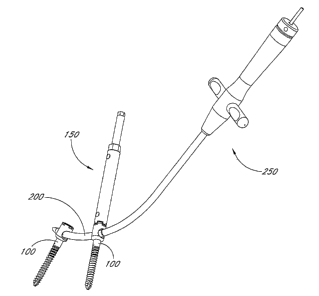
An overview of a system for minimally invasive posterior spinal fixation
according
to one embodiment of the present invention is provided in Figure 1. The system
includes at
least two and optionally three or four or more bone anchors 100 and an
articulating jointed
linkage rod 200. In Figure l, the bone anchors are shown connected by the
jointed linlcage
rod 200. The system may also include a driver 150, shown engaging one of the
bone
anchors 100, and an insertion tool 250, shown comlected to the linkage rod
200. Although
these components will be described primarily in the context of a single
liu~age rod
connected to two bone anchors, a normal fusion application will typically
involve the
implantation of two liucage rods, each carried by two or more bone anchors,
bilaterally
symmetrically mounted on the spine as is well understood in the art.
Figure 2 shows an exploded view of the bone anchor 100 and the driver 150. The
bone anchor 100 is provided with threads 102 by which it is screwed into a
vertebral body.
A locking cap 106 is disposed within the head 108 of the bone anchor 100.
The driver 150 comprises an outer adapter 152 concentrically arranged around
an
imler adapter 154. Either adapter may be freely rotated with respect to the
other. The outer
adapter 152 is adapted to engage the head 108, to screw the bone anchor 100
into a bone.
The inner adapter 154 is adapted to engage the locking cap 106, to secure the
fixation rod
200 within the head 108. In one embodiment, the hexagonal proximal end 156 of
the outer
adapter 152 allows torque to be applied to the outer adapter 152 by means of a
wrench, a
spanner or another tool. Similarly, the hexagonal proximal end 158 of the
inner adapter
154 allows torque to be applied to the inner adapter 154.
Releasable, rotational engagement between the driver and the bone anchor may
be
accomplished in any of a variety of ways. W the illustrated embodiment, the
distal end the
inner adapter 154 is provided with at least one surface for cooperating with a
complimentary surface on the proximal end of the bone anchor 100, for
transmitting torque
from the inner adapter 154 to the bone anchor 100, to enable transmission of
torque from
the imzer adapter 154 to locking cap 106. Similarly, the distal end of the
outer adabter 152
is provided with at least one surface for cooperating with a complimentary
surface on the
proximal end of the bone anchor 100, for transmitted torque from the outer
adapter 152 to
CA 02535797 2006-02-14
WO 2005/018490 PCT/US2004/026112
the bone anchor 100 to enable credible engagement between the bone anchor 100
and the
vertebral body.
W one embodiment, the bone anchor 100, its locking cap 106, and the firmer
adapter
154 are all provided with a central axial lumen tluough which a guide wire 190
may pass.
Figure 2A is an enlarged view of the circled area in Figure 2, showing the
proximal
head 108 of the bone anchor 100 and the distal ends of the outer adapter 152
and the imler
adapter 154. The locking cap 106 is shown outside the head 108.
A transverse portal 116 extends tluough the head 108 along an axis
approximately
perpendicular to the central axis of the bone anchor 100. While the transverse
portal 116 of
the head 108 is illustrated as circular, it may be of different shapes in
other embodiments,
depending upon the cross sectional shape of the fixation rod (e.g. oval,
elliptical,
rectangular, square, etc.). The diameter of the transverse portal 116 is
generally larger than
the diameter of the corresponding portions of the fixation rod 200 such that
before the
locking cap 106 is tightened at least a portion of the fixation rod 200 may be
inserted
through the portal 116. W the illustrated embodiment, the portal 116 includes
a race or
groove 114 within the head 108. The groove 114 is preferably configured to be
slightly
larger than the diameter of the corresponding portions of the fixation rod 200
but yet have a
smaller diameter than the portal 116. W other embodiments, the groove 114 may
be
eliminated from the transverse portal 116.
Figure 2B is similar to Figure 2A above, and illustrates an inner adapter 154'
and a
locking cap 106' according to another embodiment. h1 one embodiment, the inner
adapter
154' is provided with a Torx distal end 158' which is adapted to engage a
complementary
Torx opening 120' at the top of the loclcing cap 106'. Any of a variety of
complementary
surface structures may be used, as will be understood in the art in view of
the disclosure
herein.
W yet another embodiment, the head 108 of bone anchor 100 may also be provided
with an angularly adjustable connector 104 as shown in Figure 2C. The
connector 104 may
be disposed within the head 108 beneath the locking cap 106. W one embodiment,
the
corrector 104 is spherical with an aperture 110 extending theretluough, and a
gap 112 in its
circumference, such that it is approximately C-shaped when viewed along the
central axis
of the aperture 110. The aperture 110 is adapted for the insertion of the
lincage rod (not
shown), and has a diameter slightly larger than that of the liucage rod. One
skilled in the
_g_
CA 02535797 2006-02-14
WO 2005/018490 PCT/US2004/026112
art will understand that the connector 104 can be provided in a variety of
suitable shapes.
In one embodiment, the connector 104 is seated on the race or groove 114 which
may be provided in the head 108 as described above. In Sllch e111bOd1111e11tS,
the groove 114
is preferably provided with a complementary surface to the spherical exterior
surface of the
connector 104. The connector 104 may rotate on any axis within the head 108 of
the bone
anchor (or bone screw) 100. The locking cap 106 may be threaded into the head
108 to
lock the connector 104 against the lil~l~age rod 200, by compressing the
groove 114, fixing
the connector 104 within the head 108. The bottom of the loclcing cap 106 may
be
provided with a concave surface (not shown) which is complementary to the
spherical
exterior surface of the connector 104.
While tile aperture 110 of the connector 104 is illustrated as circular, they
may be of
different shapes in other embodiments, depending upon the cross sectional
shape of the
fixation rod (e.g. oval, elliptical, rectangular, square, etc.). The diameter
of the transverse
portal 116 is generally smaller than the outside diameter of the uncompressed
corniector
104 but greater than the inside diameter of the aperture 110. Before the
locking cap 106 is
tightened, the connector 104 may rotate on any axis within the head 108 to
accommodate
different entrance angles for the fixation rod. Thus the central axis of the
aperture 110 and
the central axis of the transverse pol-tal 116 may be coaxial or angularly
offset.
In one embodiment, the threading of the loclcing cap 106 into the head 108
compresses the connector 104, decreasing the width of the gap 112 and reducing
the cross
sectional area of the aperture 110. This secures a linkage rod (not shown)
extending
through the transverse portal 116 of the bone anchor 100 within the apel-ture
110. The
tightening of the locking cap 106 into the head 108 also fixes the rotational
position of the
connector 104 within the head 108.
Figure 2D illustrates an alternate connector 104'. Similar to the connector
104
described above, the connector 104' is provided with an aperture 110' having a
longitudinal
axis and a gap 112'. The spherical exterior surface of the colmector 104' is
provided with
one or two or three or more surface stnlctures such as projections or
indentations 111. The
indentations 111 receive complementary surface structures such as projections
provided
within the head 108 of the bone anchor 100 to limit the degree of rotation of
the connector
104' within the head 108. For example, Figure 2E illustrates an exemplary
embodiment
wherein the complementary surface stwcture comprises a pin 101 that may be
laser welded
-9-
CA 02535797 2006-02-14
WO 2005/018490 PCT/US2004/026112
or otherwise coupled to or integrally formed with the screw head 108. As
described above,
the pin 101 interacts with the indentation 111 to limit the degree of rotation
of the
comlector 104' within the head 108. In one specific embodiment, the connector
104' is
limited to about 30 degrees of rotation on any axis within the head 108, from
the
longitudinal axis through the transverse portal 116. In other embodiments, the
connector
104' may be limited to a range of up to about 60 degrees of rotation from the
longitudinal
axis. In one embodiment, the connector 104' is limited to no more than about 5
degrees or
about 10 degrees of rotation on any axis from the longitudinal axis. In
general, the rotation
of the connector 104' is limited such that the aperture will always be exposed
through
transverse portal 116 to the linkage rod 200.
Figure 2F illustrates a comlector 104" according to another embodiment.
Similar to
the connectors 104 and 104' described above, the connector 104" is provided
with an
aperture 110" and one or more compressible gaps 112". The gaps 112" are
provided with a
compressible material which compresses when the loclcing cap 106' tightens the
connector
104" against the groove 114 within the head 108. Compressible material,
including any of
a variety of compressible polymeric materials lcnown in the medical device
arts can be used
according to several embodiments of the present invention. One skilled in the
art will
appreciate that other suitable flexible or compressible materials may also be
used. W
addition, any of a variety of metal (stainless steel, titanium, etc.)
connectors 104 may be
configured such that the aperture 110 is moveable from a first, large cross-
section, for
receiving a liu~age rod 200 therethrough, to a second, reduced cross section
for loclcing the
linkage rod 200 in place. This may be accomplished by providing opposing
components
forming the side wall of the connector 104 with any of a variety of
interloclcing structures
such as ramp and pawl ratchet structures, or sliding fit structures which
permit a reduction
in the diameter in the aperhtre 110 under compressive force from the locking
cap 106.
In an alternate embodiment, portions or all of the connector 104 comprise a
compressible media such as an open cell foam, closed cell foam or solid
compressible
material. Structures comprising polyethylene, PEEK, nylon, and other polymers
laiown in
the medical arts may be utilized, depending upon the construction and desired
compressibility. In general, the combination of material and the structure of
the connector
104 is sufficient to allow angular adjustment of the longitudinal axis of the
aperture 110, to
accommodate various entrance angles of the lincage rod 200. After the lincage
rod 200 has
-10-
CA 02535797 2006-02-14
WO 2005/018490 PCT/US2004/026112
been positioned within the aperture 110, rotational and/or axial movement of a
locking
element such as loclcing cap 106 functions to both prevent axial movement of
the linlcage
rod 200 within the aperture 110, as well as prevent further angular adjustment
of the
longitudinal axis of the aperture 110 with respect to the longitudinal axis of
the bone anchor
100.
Figures 2G-2H illustrate the connector 104", the aperture 110", the gaps 112",
and a
compressible or foldable membrane or line 115 in greater detail. Figure 2F is
an isometric
view of the connector 104". Figure 2G is a front plan view of the corrector
104" viewed
along the central axis of the aperture 110". Figvtre 2H is the corresponding
side pla~z view.
In the embodiment illustrated in Figures 2F-2H, the compressible link is
formed by
grinding, laser etching, molding or otherwise forming a recess such as a V-
shaped channel
113 that leaves a thin link 115 which folds flat when the connector 104" is
compressed.
One of ordinary skill in the art will understand that compressible materials
a~ld structures
can be provided in a variety of suitable shapes and forms.
In one embodiment, the apertures 110' and 110" have a tendency to return to
their
original diameters even after the connectors 104 and 104', respectively, are
compressed by
the locking cap 106 against the groove 114 within the head 108. This tendency
results from
the resiliency of the metal, alloy or other material used to malce the
correctors 104 and
104'. The use of compressible material, such as V-shaped channels 113 in the
gaps 112" of
the connector 104", reduces or eliminates this tendency and may allow a
lineage rod (not
shown) to be more firmly secured within the aperture 110". One skilled in the
art will
understand that the connectors 104 and 104' can be made from lower resiliency
materials
wluch can also reduce or eliminate the tendency of apertures 110' and 110" to
return to their
original diameters.
Figures 2I-L illustrate another embodiment of a comzector 104"' according to
another embodiment. In this embodiment, the connector 104"' is provided with
an
aperture 110 and an indentation 111 as described. A top portion of the
connector 104"' is
provided with a compressible material or foldable linlc 117, which in
comprises a series V-
shaped channels formed into the body of the connector 104"'. hi the
illustrated
arrangements, the channels comprise a series of 40 degree V-shaped chamlels
119 formed
on the outer surface of the connector 104"' and 20 degree V-shaped channels
121 on the
imzer surface of the comiector 104"'. In a similar manner, a foldable link 123
is provided
-11-
CA 02535797 2006-02-14
WO 2005/018490 PCT/US2004/026112
on a lower portion of the connector 104"'. W this embodiment, as the loclcing
cap 106 is
tightened the top and bottom pouions of the comlector 104" are defomned and
laterally
depressed so as to secure the fixation rod within the aperture 110.
Further details and additional embodiments of a bone anchor utilizing a
comiector
104 can be found in co-pending United States Patent Application Serial No. 10/
, ,
filed June 13, 2003, under Attorney Docket No. VLINI~.021A and entitled System
and
Method for Minimally W vasive Posterior Fixation, which was incorporated by
reference
above.
As discussed above with reference to Figure 2, in one embodiment, the outer
adapter 152 is adapted to engage the head 108, and the inner adapter 154 is
adapted to
engage the locking cap 106. W the illustrated embodiment, projections 156 on
the distal
end of the outer adapter 152 are adapted to engage complementary projections
118 on the
head 108 of the bone anchor 100. The hexagonal distal end 158 of the firmer
adapter 154 is
adapted to engage a complementary hexagonal opening 120 at the top of the
loclcing cap
106.
Although specific interlocking relationships between the driver 150 and the
bone
anchor 100 are illustrated herein, the present inventors contemplate a variety
of
modifications. For example, the male-female relationship between the driver
and the
implant may be reversed, for either or both of the inner adaptor 154 and outer
adapter 152.
In addition, each of the inner adapter 154 and outer adapter 152 is provided
with a surface
structure for enabling rotational engagement with a coiTesponding component on
the
implant. Although this may be conveniently executed using corresponding
hexagonal male
and female components, any of a variety of alternative structures may be
utilized in which a
first surface on the inner adapter 154 or outer adapter 152 cooperates with a
second,
complementary surface on the corresponding aspect of the bone anchor 100, for
allowing
rotational engagement, followed by axial decoupling.
With reference now to Figures 3 and 3A, the jointed fixation rod 200 will now
be
described in more detail. The fixation rod 200 preferably includes a first
segment 204, a
second segment 206 and a central lumen 202, which is configured to receive a
guidewire as
will be explained in more detail below. The first and second segments 204, 206
are
coupled together by an axzgularly adjustable joint 208a.
As will be explained in more detail below, the fixation rod 200 may be
provided
-12-
CA 02535797 2006-02-14
WO 2005/018490 PCT/US2004/026112
with one or more joints 208a. The joints 208a provide the fixation rod 200
with a degree of
flexibility that allows the fixation rod 200 to travel tluough a nonlinear,
disjointed and/or
curved path. This is particularly advantageous for inserting the fixation rod
200 through the
transverse portals 116 of a plurality of bone anchors 100. For example, it is
generally
difficult to align the transverse portals 116 of a plurality of bone anchors
100 with each
other because the surfaces of the spine are typically non-planar and non-
uniform. As such,
each bone anchor 100 may extend from the spine at a different angular
orientation and/or
height. It is particularly difficult to align the portals between an anchor
positioned in the LS
vertebra and an anchor the S 1 vertebra (i.e. sacrum).
It is therefore difficult to thread a straight or even carved fixation rod
through the
transverse portals 116 of more than one bone anchor 100. W a non-minimally
invasive
procedure, the surgeon may measure the degree of non-alignment between the
bone anchors
100 and bend the fixation device and/or adjust the position of the bone anchor
in the spine.
However, in a minimally invasive procedure, such adjustments are impractical
because they
cause prolonged expose of the patient and the use to fluoroscopical radiation.
Although a particular configuration of an articulating joint will be described
in
detail below, any of a variety of structures may be utilized in implementing
the present
invention. W general, the implantable fixation rod will have at least a first
segment and a
second segment which are angularly adjustable with respect to each other while
the rod is in
a first state, and fixed with respect to each other when the rod is in a
second state. This
permits, for example, percutaneous introduction to a treatment site within a
patient along a
nonlinear path while the rod is in the first state. The rod may then be
converted to the
second state such that it exhibits a sufficient rigidity to produce the
desired clinical result.
This may be rigid fixation of a first bone or bone fragment with respect to a
second bone or
bone fragment.
The first and second segments will generally be separated by an interface, at
which a
first attachment or interface surface on a first segment is in contact with a
second,
complementary attaclnnent or interface surface on the second segment. One
implementation of this type of interface is a ball and socl~et type joint,
described below.
Another type of interface may be formed, for example, by a leaf spring type
structure, or
other axial element stntctures, such as two or more generally axially
extending elements
which are moveable with respect to each other to permit flexion but can be
loclced relative
-13-
CA 02535797 2006-02-14
WO 2005/018490 PCT/US2004/026112
to each other such as by lateral compression.
The articulating joint can be transformed from the movable state to the fixed
state in
any of a variety of ways. One convenient mariner of fixation is responsive to
lateral
compression which may be applied, for example by an axially movable component
(e.g., a
threaded shaft such as a set screw) carried by the bone anchor. Thus in a ball
and socket
type structure where a male component fits within a complementary female
component, a
surface on the outer female component may be flexed or compressed laterally
inwardly
against a complementary surface on the male component to provide fixation.
Either or both
complementary surfaces may be provided with friction enhancing surface
structures such as
ridges, roughening or micropitting, as will be appreciated by those of slcill
in the art in view
of the disclosure herein.
With reference to Figures 3 and 3A, the joint 208a comprises a first surface
210
formed on the first segment 204 and a second complementary surface formed on
the second
segment 206. W the illustrated embodiment, the first surface comprises a
socket or recess
214 and the second surface comprises a ball 220 so as to form of a ball joint
between the
adjacent ends of the first and second segments 204, 206. The first segment 204
includes a
soclcet portion 210, which comprises a generally cylindrical outer surface 212
that is
configured to extend within the transverse portal 116 of the bone anchor 100.
The proximal
end of the soclcet portion 210 includes the generally spherical socket or
recess 214. At least
one gap 216 is provided in the soclcet portion 210. In the illustrated
embodiment, the at
least one gap 216 extends from one side of the outer surface 212, through the
central lumen
202 and to the opposite side of the outer surface 212 and therefore divides
the socket
portion 210 generally in half. Compression of the gap 216 reduces the cross-
sectional
diameter of the soclcet 214 and fixes the angular position of the joint 208a
as will be
explained in more detail below.
The socket portion 214 is configured to receive the ball 220 or spherical
protrusion
that is provided on the distal end of the second segment 20G. In one
embodiment, the ball
220 is configured such that it may be press-fitted into the soclcet 214. That
is, the soclcet
214 defines an opening 222 that in a relaxed state is smaller than the maximum
diameter of
the ball 220. In this manner, as the ball 220 is inserted into the socket 214,
the gap 216
expands to increase the diameter of the opening 222 and allow insertion of the
ball 220 into
the socket 218. Preferably, once in place, axial movement of the ball 220 with
respect to
-14-
CA 02535797 2006-02-14
WO 2005/018490 PCT/US2004/026112
the socket 214 is limited while at least limited angular adjustment of the
ball 220 with
respect to the socket 216 is permitted. This may be accomplished by providing
the soclcet
216 with a slightly larger diameter than the ball 220 and/or configuring the
joint 208a such
that the friction between the ball 220 and the soclcet 214 permits angular
adjustment. In
this mamler, the angular orientation between the first and second segments
204, 206 may be
adjusted.
In the illustrated embodiment, the second segment 206 may be adjusted to any
of a
variety of angular orientations defined within a cone having a vertex v
positioned generally
at the center of the socket 214 and the ball 220. The angle a (see Figures 3B
and 3C)
represents the angular adjustment between the two segments and is defined
primarily by the
interference between the proximal end of tile soclcet portion 204 and a neclc
224 on the
proximal end of the ball 220. This angle may be increased by decreasing the
diameter of
the neck 224. It should be appreciated that the maximum angle adjustment
between the
longitudinal axes b, c of the first and second segments is generally half of
the angle a of the
vertex.
Depending upon the environment of use, the angle a of the vertex is preferably
within the range of about 15 to 90 degrees and the angle a between the
longitudinal axis of
the second segment 206 with respect to the first segment 204 may be rotated to
any angle
orientation within such cone. In one embodiment, the angle a of the vertex
within the range
of about 15 to 30 degrees for joints 208 positioned at the hunbar levels and
within the range
of about 45 to 90 degrees for joints 208 positioned at the LS and Sl levels.
In another
embodiment, in the lumbar levels, the rod 200 may be fixed (e.g., formed
without joints)
while in the LS and S 1 levels the vertex of the j oints 208 may be in the
range of about 45 to
90 degrees.
~ne skilled in the art will understand that in other embodiments the
illustrated ball
joint may be replaced with any of a variety of other angularly adjustable
structures such as
hinges or other sliding structures that provide angular adjustment. For
example, the shape
of the socket and/or the ball may be modified in several different ways and
still provide the
angular adjustability described above. In one particular embodiment, the
angular
adjustability may be modified and/or limited. This may be accomplished by
providing
spherical exterior surface of the ball 220 with one or two or three or more
surface structures
such as projections or indentations. The indentations receive complementary
surface
-15-
CA 02535797 2006-02-14
WO 2005/018490 PCT/US2004/026112
structures such as projections provided within the socket 214 to limit the
degree of rotation
of the ball 220 within the socket 214 and/or the plane through which angular
orientation
may be adjusted. For example, in one embodiment, the first and second segments
may be
angularly adjusted only through one plane (e.g., a horizontal plane).
In the illustrated embodiment, the socket portion 210 is configured to fit
within the
transversal portal 116 within the head 108 (see Figures 2-2B) or through the
aperture 110 in
the corrector 104 (see Figure 2C). The portal 116 or aperture 104 is
preferably provided
with a complementary surface to the cylindrical exterior surface 212 of the
socket portion.
210 As the socket portion 210 is inseuted through the head 108, the angular
orientation of
the second segment 206 with respect to the first segment 204 may be adjusted
as the ball
220 rotates with respect to the socket 214. The locking cap 106 may be
tlueaded into the
head 108 to loclc the angular orientation between the first and second
segments 204, 206, by
acting against the outer surface of the socket portion 210 or the connector
105 and fixing
the ball 220 within the socket 214. The bottom of the locking cap 106 may be
provided
with a concave surface (not shown) which is complementary to the spherical
exterior
surface 212 of the soclcet portion 210 or the connector.
In one embodiment, the threading of the locking cap 106 into the head 108
compresses the socket portion 210, decreasing the width of the gap 216 and
reducing the
cross sectional area of the soclcet 214. This secures the ball 220 within the
socket 214 and
fixes the angular orientation of the first segment 204 with respect to the
second segment
206. In the embodiments which use a connector 104, the locking cap also fixes
the angular
position of the corrector 104 within the head 108. In some embodiments, the
socket 214
and/or the ball 220 may be roughened, etched (e.g., mechanical, electrical,
photo, chemical
etc.) and/or coated with material to increase the friction between these
components. In this
mamler, the locking force between the socket 214 and the ball 220 may be
enhanced. Such
techniques may also be applied to the connector 104 and the outer surface of
the first
segment 204.
h1 the illustrated embodiment, the locking cap 106 also fixes the axial
position of
the socket portion 210 within the bone ailchor 100. However, in modified
embodiments
this may be accomplished by a separate device (e.g., a set screw).
In general for lumbar applications, in the loclced position, the fixation rod
200 will
preferably exhibit a static compression within the range of from about 120 to
about 200
-1 G-
CA 02535797 2006-02-14
WO 2005/018490 PCT/US2004/026112
lbs., and, more preferably greater than about 150 lbs and the rod will
preferably exhibit a
static torsion within the range of from about 15 to about 25 inch pounds, and,
more
preferably in excess of about 20 inch pounds. The rods will preferably reach
at least about
million cycles, at 5 Hz. In general for cervical applications, in the locked
position, the
5 fixation rod 200 will preferably exhibit a static compression within the
range of from about
30 to about 100 lbs., and, more preferably greater than about 80 lbs and the
rod will
preferably exhibit a static torsion within the range of from about 10 to about
20 inch
pounds, and, more preferably in excess of about 15 111Ch pounds. The rods will
preferably
reach at least about 5 million cycles, at 5 Hz. Each of these parameters may
be measured in
accordance with the protocols described in the American Society for Testing
and Materials
(ASTM) designation F 1717-96, a copy of which is incorporated in its entirety
herein by
reference.
As mentioned above, the socket portion 210 and the con esponding gap 216
formed
in the socket portion preferably have a length of approximately 10 to 30
millimeters. This
provides the joint 208a with a working range in which the locking cap 106 can
be used to
fix the angular orientation of the joint 208a. That is, the locking cap 106
can be used to fix
the angular orientation of the joint 208a as long as a least a portion of the
socket portion
210 is positioned in the head 108 such that the loclcing cap 106 may compress
the gap 216.
For a one level application, typically two bone anchors 100 are inserted into
adjacent vertebrae. In such an application, the fixation rod 200 preferably
includes two
joints 208a, 208b. As shown, in Figure 3A, the second joint 208b may be formed
between
a proximal end of the second segment 206 and a distal end of a third or end
segment 230.
In the illustrated embodiment, the proximal end of the second segment 206
includes a
spherical protrusion or ball 220, which may be configured as described above.
The end
segment 230 includes a socket portion 210 configured as described above and
including a
socket 214 to receive the ball 220 of the second segment 206.
As can be seen Figure 3, the first segment 204 of the lil~l~age rod 200 may be
provided with a tapered distal end 232. The tapered distal end 232 may be
machined and
be an integral part of the segment 204, may be molded integrally with the
soclcet portion
210 or may be separately formed and attached to the linkage rod 200. In one
implementation, the tapered end 232 may be a polymeric component such as
nylon, HDPE,
PEBAX or other materials known in the art. The tapered tip 232 facilitates
advance of the
-17-
CA 02535797 2006-02-14
WO 2005/018490 PCT/US2004/026112
lil~lcage rod 200 through the transverse portal 116. In other embodiments, the
distal end 232
may be blunt or ball shaped to minimize the protntding portion of the rod 200
from the
portal 116 of the distal most anchor 100. In certain application, such
embodiments
advantageously reduce interference between the distal end of the rod 200 and
the S 1 body.
With continued reference to Figure 3, the end segment 230 may include a
hexagonal
proximal end 234. The hexagonal proximal end 234 may be connected to the
insertion tool
as will be explained in more detail below.
The length of the linkage rod 200 in a device intended for use in a human
adult one
level lumbar or lumbar-sacral fusion, will generally be in the range from
about 30mm to
about 90m1n and have a generally circular cross-section with an average
diameter within the
range of about S.Smm to about 9mm. In such an embodiment, the first segment
204 and the
end segment 230 will generally have a length within the range of from about
lOmm to
about 401nm. The gaps 216 will generally have width within the range of about
O.Smm to
l.5mm and a length within the range of from about 9 lnln to about 291n1n. The
socket
portions 210. will generally have a length within the range of from about l
Omm to about
30mm. The second segment 206 will generally have a length Wlthlll the range of
about
lOmm to about lOmm. The socket 214 and the ball 220 may have a diameter within
the
range of about S.Omm to 7.Omm.
I11 a two level application, three bone anchors 100 are typically inserted
into
adjacent vertebra. In such an application, the fixation rod 200' preferably
includes four
joints 208a, 208b, 208c, 208d. See Figures 4, 4A and 4B. As shown, in Figure
4B, the four
joints 208a-d may be provided by adding a fourth or intermediate segment 238
and an
additional second segment 240, which is configured as described above. The
immediate
segment 240 may include two sockets 214 positioned at the distal and proximal
ends of the
segment 240 and a gap 216 that extends through the entire length of the
segment 240. The
outer surface 212 is configured to fit within a bone anchor (see Figure 4A)
such that
tightening the loclcing cap 106 compresses both of the sockets 214 in the
intermediate
segment 238 and thereby fixes the corresponding joints 208b, 208c.
A linkage rod 200' in a two-level device intended for use in a human adult
lumbar
or lumbar-sacral fusion will generally have a length within the range of from
about 70mm
to about 120m1n and a generally circular cross-section with an average
diameter within the
range of about S.Omm to about 9.Omm. In such an embodiment, the first segment
204 and
-18-
CA 02535797 2006-02-14
WO 2005/018490 PCT/US2004/026112
end segment 230 will generally have a length within the range of from about
lOnun to
about 40mm. The gaps will 216 will generally have a length within the range of
from about
9mm to about 29mm and the socket portions 210 will generally have a length
within the
range of from about lOmm to about 30mm. The second and intermediate segments
206,
240 will generally have a length within the range of from about l Omm to about
40mm. The
socket 214 and the ball 220 may have a diameter within the range of about
S.Omm to
7.Omm.
In another embodiment of the linlcage rod 200 intended for two level fusion
for use
in the treatment of thoracic and cervical segments of the spine, the rod 200
has a length of
about 100mm to 240mm and a generally circular cross-section with an average
diameter of
in the range of from about 3mm to about 4mm. In such an embodiment, the first
segment
204 and end segment 230 will generally have a length within the range of from
about l Omm
to about 40mm. The gaps will 216 will generally have a length within the range
of from
about 9mm to about 29mm and the soclcet portions 216 will generally have a
length within
the range of from about lOmm to about 30mm. The second and intermediate
segments 206,
240 will generally have a length within the range of from about l0mm to about
40mm. The
socket 214 and the ball 220 may have a diameter within the range of about Smm
to 7mm.
Figure 4C illustrates a modified embodiment of the fixation rod 200". This
embodiment is particularly suited for a level two device for use in sacral-
lumbar fusion. In
this embodiment, the rod 200" includes at least one joint 208a, and preferably
two joints
208a, 208b, between the distal and intermediate bone anchors 100 while the
fixation rod
200" is fixed (i.e., formed without joints) between the intermediate and
proximal anchors
100. As such, in this modified embodiment, the proximal or end portion 230'
may be
elongated as compared to the embodiment of Figure 3 such that it can extend
through the
intermediate and proximal anchors 100. The end portion 230' may be
substantially straight,
partially curved or curved depending upon the clinical application. In this
embodiment, the
locking cap 106 in the proximal anchor 100 merely secures the rod 200" within
the
transverse portal 116.
hi the embodiments described above, the cross sectional area of the rod 200,
which
may be expressed as a diameter in a circular cross sectional implementation,
may be varied
depending upon the desired structural integrity of the finished implant. The
anchors 100
will have a diameter of in the range of from about 3.Smm to about 4mm and a
length in the
-19-
CA 02535797 2006-02-14
WO 2005/018490 PCT/US2004/026112
range from about lOrnrn to about SSmm.
h1 modified embodiments, the gaps 216 in the fixation rod 200 may be provided
with a compressible material which compresses when the locking cap 106
tightens the
cylindrical portion within the head 108. Compressible material, including any
of a variety
of compressible polymeric materials known in the medical device arts can be
used
according to several embodiments of the present invention. One slcilled in the
art will
appreciate that other suitable flexible or compressible materials may also be
used. In
addition, any of a variety of metal (stainless steel, titanium, etc.)
connectors may be
configured such that the socket 214 is moveable from a first, large cross-
section, for
allowing movement of the ball 220 therethrough, to a second, reduced cross
section for
locking the angular position of the ball 220. This may be accomplished by
providing
opposing in the socket portion 210 any of a variety of interlocking structures
such as ramp
and pawl ratchet structures, or sliding fit structures which permit a
reduction in the
diameter in the socket 214 under compressive force from the loclcing cap 106.
h1 a modified embodiment, poutions or all of the socket portion 210 comprise a
compressible media such as an open cell foam, closed cell foam or solid
compressible
material. Strictures comprising polyethylene, PEED, nylon, and other polymers
lmown in
the medical arts may be utilized, depending upon the construction and desired
compressibility. In general, the combination of material and the stricture of
the socket 214
is sufficient to allow angular adjustment of the longitudinal axis of ball 220
and the socket
214 to provide the linkage rod 200 with an angularly adjustable joint 208.
After the socket
portion 210 has been positioned within the transverse portal 116, rotational
and/or axial
movement of a locking element such as locking cap 106 functions to both
prevent axial
movement of the linkage rod 200 within the aperture 116, as well as prevent
further angular
adjustment of the joint 208.
Tn one embodiment, the sockets 214 have a tendency to return to their original
diameters even after the cylindrical portions 210, respectively, are
compressed by the
loclcing cap 106 within the head 108. This tendency results from the
resiliency of the metal,
alloy or other material used to make the cypindrical. The use of compressible
material,
such as V-shaped channels in the gaps 216, reduces or eliminates this tendency
and may
allow a linlcage rod 200 and the joint 208 to be more firmly secured. One
skilled in the art
will understand that the sockets 214 can be made from lower resiliency
materials wluch can
-20-
CA 02535797 2006-02-14
WO 2005/018490 PCT/US2004/026112
also reduce or eliminate the tendency of soclcets 214 to return to their
original diameters.
In Figure 5, the linkage rod 200 is shown positioned within two adjacent bone
anchors 100, and released from the insertion tool 250. The insertion tool 250
is provided
for the insertion of the lineage rod 200 into the bone anchors 100. The
insertion tool 250
comprises an am 252 and a handle 254. W the illustrated embodiment, the am 252
is
curved to facilitate insertion of the lineage rod 200 into the bone anchors
100 within a
patient along a curved tissue tract which passes through the aperture 110 of
at least each of
a first bone anchor and a second bone anchor. However, it should be
appreciated that in
modified embodiments, the arm 252 may be of a different shape (e.g., straight)
and be
inserted through a tract of a different shape.
A central control line 256 (shown mostly in phantom) such as a torque
transmission
tube, rod or cable extends through an axial lumen of the insertion tool 250,
and terminates
at a control such as a knob 258 at the proximal end of the insertion tool 250.
A screw (not
shown) threaded into a tumzel 260 extending along a radius of the lmob 258 may
be used to
secure the control line 256 within the knob 258. The control line 256 is
provided with a
threaded distal tip 262. Rotating the knob 258 thus rotates the control line
256 and its
threaded distal tip 262 to engage or disengage the linkage rod 200.
In one embodiment, both the lineage rod 200 and the control line 256 are
provided
with a central axial lumen for the passage over a guide wire.
Figure 5A is an enlarged view of the circled area in Figure 5, showing the
distal end
of the outer adapter 152, the bone anchor 100, the linkage rod 200, and the
distal end of the
arm 252 of the insertion tool. The linkage rod 200 is shown fixed within the
head 108 of
the bone anchor 100.
As mentioned above, lineage rod 200 is provided with a hexagonal proximal end
234 adapted to engage a complementary hexagonal socket (not shown) in the
distal end of
the arm 252 of the insertion tool. In some embodiments, alteriative
complementary surface
structures may be provided on the linkage rod 200 and the arm 252 to
rotationally fix their
orientation with respect to one another. W the illustrated embodiment, the
hexagonal
proximal end 234 is provided with a dimple 235 adapted to engage a
complementary nub
(not shown) within the hexagonal socket (not shown) in the distal end of the
arm 252 of the
insertion tool. The dimple 235 and nub (not shown) fix the axial orientation
of the linlcage
rod 200 with respect to the arm 252. The tlueaded distal tip 262 of the
control line 256 may
-21-
CA 02535797 2006-02-14
WO 2005/018490 PCT/US2004/026112
be threaded into a complementary threaded hole 237 in the hexagonal proximal
end 234 of
the lineage rod 200, enabling the linlcage rod 200 to be detachably secured to
the a~-m 252
of the insertion tool. The threaded distal tip 262 may be threaded into the
threaded hole
206 by rotating the knob (not shown) at the proximal end of the insertion
tool. Untlueading
the tlueaded distal tip 262 from the tlueaded hole 206 allows the linl~age rod
200 to be
released from the insertion tool 250.
With continued reference to Figure SA, in the illustrated embodiment, the
outer
adapter 152 is provided with an opening 160 extending along a diameter for
fluoroscopic or
other visualization of the rotational orientation of the outer adapter 152, to
align the portal
116 of the bone anchor 100 engaged by the outer adapter 152. Towards this end,
the axis of
the opening 160 is preferably arranged at a right angle to the axis of the
portal 116 as shown
in Figure SA. To visualize the axial position of the outer adapter 152 and the
bone anchor
100, the inner adapter 154 may be temporarily retracted so that it does not
block the
opening 160. W another embodiment a translucent marlcer may be installed in
opening 160
for fluoroscopic or other visualization of the outer adapter 152.
Alternatively, any of a variety of other indicium of the rotational
orientation of the
bone anchor 100 may be provided. For example, the complementary surface
structures
between the proximal end of the bone anchor 100 and the distal end of the
insertion tool
250 may be configured to only allow coupling between the two components in a
predetermined rotational orientation. In this construction, visual indicia may
be provided
on a portion of the insertion tool 250 (e.g. "T" handle, painted or etched
marlcings or other
indicium) which remains external to the patient, to allow direct visual
observation of the
rotational orientation of the longitudinal axis of the transverse portal 116.
Figure 6 illustrates the described insertion tool from another angle. The
lcnob and
its attached central cable have been removed for clarity. The hexagonal
soclcet 264 adapted
to engage the hexagonal proximal end (not shown) of the linlcage rod, as
described above, is
ShOWIl. The nub 266 adapted to engage the dimple (not shown) on the hexagonal
proximal
end (not shown) of the linkage rod is also shown.
In several embodiments, the components of the bone anchor, the linkage rod,
the
driver, and the amn of the insertion tool may be made of titanium, stainless
steel or any
other suitable metals, alloys, or material. The handle of the insertion tool
is preferably
made of a suitable non-slip material. The selection of these materials for the
manufacture
-22-
CA 02535797 2006-02-14
WO 2005/018490 PCT/US2004/026112
of the components and devices described in the above embodiments would be
lmown by
those slcilled in the art. .
Methods for the minimally invasive implantation of posterior fixation hardware
according to embodiments of the present invention are disclosed in the context
of a spinal
fixation procedure with reference to Figures 7-47. Additional details
concerning the
illethOd are disclosed in the copending patent applications incorporated by
reference
previously herein. Although the methods and instruments of the present
invention ca.n be
utilized in an open surgical procedure, the present invention is optimized in
the context of a
percutaneous or minimally invasive approach. Thus, the method steps which
follow and
those disclosed in the copending patent applications incorporated by reference
herein are
intended for use in a trans tissue approach. However, to simplify the
illustrations, the soft
tissue adjacent the treatment site is not illustrated in the drawings
discussed below.
W Figures 7 and 8, a trocar 300 is inserted through a tissue tract and into a
vertebral
body 310. The trocar 300 comprises a sharp-tipped rod 308 (shown in Figure 16)
attached
to a proximal or top half handle 302. The sharp-tipped rod 308 is arranged
concentrically
within a catmula 304, which is attached to the bottom half handle 306 of the
trocar 300.
The top half handle 302 and the bottom half handle 306 of the trocar 300 are
screwed
together for initial use, as shown in Figures 7-8. The trocar 300 is inserted
through the
slcin, muscle and other tissues of the patient into the vertebral body 310.
Figure 9 shows the bottom half handle 306 with the attached cannula 304
embedded
in the vertebral body 310. The top half handle (not shown) has been unscrewed
and set
aside from the bottom half handle 306. In Figure 10, a guide wire 312 is
inserted into the
vertebral body 310 via the bottom half handle 306 and the cammla 304.
W Figure 11, the bottom half handle 306 and the cannula 304 are removed from
the
vertebral body 310. Preferably, the guide wire 312 remains in place in the
vertebral body
310.
Figure 12 shows the guide wire 312 in the vertebral body 310 after the bottom
half
handle 306 and the cammla 304 are removed.
Figures 13-14 show one embodiment of use in which an inflatable tissue
expander
for enlarging the tissue tract is used. W Figure 13, a balloon catheter 314
carrying a balloon
31G is advanced over the guide wire 312 towards the vertebral body 310. In
Figure 14, the
balloon 316 is inflated to dilate the tissues adjacent the access pathway to
the vertebral
-23-
CA 02535797 2006-02-14
WO 2005/018490 PCT/US2004/026112
body 310. This provides an enlarged path for the inseution of a sheath as
described below.
In Figure 15, a guide tube 322 is advanced over the guide wire 312 into the
vertebral
body 310. As shown in Figure 16, in one embodiment, the guide tube 322 may be
approximately the same diameter as the cannula 304 of the trocar 300, allowing
the guide
tube 322 to be inserted into the opening in the vertebral body 310 created
earlier by the
trocar 300. The guide tube 322 acts as a stable rail over which a tapered
dilation cylinder
324 may be advanced against the vertebral body 310.
W Figures 16-17, a tapered dilation cylinder 324 is advanced over the guide
tube
322 against the vertebral body 310. In one embodiment, the tapered dilation
cylinder 324
may be approximately the same diameter as the inflated dilation balloon 316
discussed
above with reference to Figures 13-14. The tapered dilation cylinder 324 is
used to occupy
the path created by the dilation balloon, and facilitates the insertion of a
sheath. fii an
alternate sequence, the dilation cylinder 324 is provided without a tapered
distal end, and is
distally advanced into position directly over the inflatable balloon.
hi Figures 18-20, a sheath 320 is advanced over the tapered dilation cylinder
324
against the vertebral body 310. The sheath 320 occupies the path created by
the dilation
balloon. Afterwards, the guide tube 322 and the tapered dilation cylinder 324
are removed.
As shown in Figure 20, the guide wire 312 preferably remains in the vertebral
body 310
after the placement of the sheath 320.
W Figures 21-23, a drill 330 having a rotatable distal tip 332 is advanced
over the
guide wire 312 and through the sheath 320. The drill 330 drills an opening
(not shown) in
the vertebral body 310 adapted for the inseuion of a bone anchor 100.
Afterwards, the drill
330 is removed. In Figures 24-25, the bone anchor 100 is advanced over the
guide wire
312 and through the sheath 320 towards the vertebral body 310.
hi Figures 24 and 25, a bone anchor 100 is advanced over the wire 312 and
through
the sheath 320 into engagement with the vertebral body 310. Although the
insertion tool
250 is not illustrated, the bone anchor 100 may be coupled to the insertion
tool 250 prior to
the step of advancing the bone anchor 100 into contact with the vertebral body
310.
Figures 26 and 27 show the outer adapter 152 and the imier adapter 154 of the
driver 150, as well as a bone anchor 100, with the the locking cap 106
disposed within the
head 108 of the bone anchor 100. The interrelation of these components have
been
described in detail above with reference to Figures 2 and 2A. The outer
adapter 152
-24-
CA 02535797 2006-02-14
WO 2005/018490 PCT/US2004/026112
illustrated in Figures 26-28 additionally comprises a pivot hole 153 which
extend tluough a
diameter of the outer adapter 152. The pivot hole 153 is adapted for the
attaclunent of a
guide wire insertion device 400 described in fiu-ther detail below. In Figure
28, these
components are shown aiTanged over a guide wire 190.
h1 Figure 28, the driver 150 (comprising the outer adapter 152 and the imler
adapter
154 ) is advanced over the guide wire 312 until the driver 150 engages the
bone anchor 100.
W Figures 29 and 30, torque is applied to the outer adapter 152 to screw the
bone anchor
100 into the vertebral body 310. In Figure 31, the driver 150 is removed,
leaving the bone
anchor 100 in place, with the longitudinal axis of the portal 116 aligned
approximately
parallel with the longitudinal axis of the spine. The sheath 320, discussed
above with
reference to Figures 18-25, while not shown in the steps discussed with
reference to Figures
28-31, may nonetheless be used to shield the driver from adjacent tissue in
these steps, as
will be understood by those slcilled in the art.
W Figure 32, a second bone anchor 340 has been inserted into another vertebral
body 350. While bone anchors 100 and 340 are shown inserted into adjacent
vertebral
bodies 310 and 350, respectively, the system and methods for minimally
invasive spinal
fixation according to the embodiments of the present invention are also
applicable to
nonadjacent vertebral bodies. For example, a first bone anchor may be
positioned in a first
vertebral body as has been described above. A second bone anchor may be
positioned in a
second vertebral body, spaced apart from the first vertebral body by one or
more
intervening third vertebral bodies. The first and second bone anchors may
thereafter be
connected by the implantation of a linkage rod 200. Alternatively, a third
bone anchor may
be positioned in a third vertebral body, positioned in between the first and
second vertebral
bodies to produce, for example, a three level fusion system as will be
discussed.
Preferably, after the bone anchors are in place, a guidewire 368 (see Figure
33 is
advanced through the transverse portals 118 of the of bone anchors 100 and
340. Various
methods of inserting guide wires are known in the art and the invention is not
limited to an
particular method. Instead, various methods and devices for inserting a guide
wire known
to those skilled in the art may be used in accordance with the present
invention.
Figures 34-40 illustrate a particularly advantageous guide wire insertion
device 400
according to one embodiment. The guide wire insertion device comprises a
handle 410 and
a hollow access needle 450. The handle 410 is detachably joined to the outer
adapter 152
-25-
CA 02535797 2006-02-14
WO 2005/018490 PCT/US2004/026112
of the driver 150. The handle 410 is forked at its proximal end 412. Each fork
is provided
with a pivot pin 414, which engages the pivot hole 153 (Figure 28) of the
outer adapter 152.
The forked proximal end 412 of the handle 410 may be spread slightly to allow
the pivot
pins 414 to engage the pivot hole 153. The handle 410 5W111gS Oll 1tS pivot
pins 414 at the
pivot hole 153 of the outer adapter 152 of the driver 150 to insert the access
needle 450
through the transverse portal 116 of the bone anchor 100.
A hollow access needle 450 is attached to the distal end 416 of the handle
410. In
one embodiment, the access needle 450 is disposed within an opening 418 at the
distal end
416 of the handle 410. A screw (not shown) may be threaded through a screw
hole 420 at
the distal end 416 of the handle 410 to tighten the access needle 450 within
the opening
418. The lengthwise position of the access needle 450 within the opening 418
is therefore
adjustable to allow the access needle 450 to be aimed through the transverse
portal 116 of
the bone anchor 100. In one embodiment, the access needle 450 may be aimed
such that it
passes tluough the transverse portal 116 at a point lower (towards the threads
102 in Figl~re
2) than the center of the transverse portal 116 because obstructions
encountered during the
in vivo insertion of the access needle 450 may deflect the needle 450 towards
the inside of
its curvature and the center of the traxisverse portal 116.
In several embodiments, the sharp, tapered distal end 452 of the access needle
450
terminates at an opening 454. W one embodiment, the access needle 450 is
provided with
threaded proximal end 456, the purpose of which is described in further detail
below.
Figure 35 illustrates a flexible obturator 500 of the guide wire insertion
device 400
according to one embodiment. The obturator 500 comprises a tubing 502, a
threaded cap
504 on its proximal end and a plug 506 on its distal end. The tubing 502 is
sized such that
it fits snugly within the hollow access needle 450 and occupies the length of
its lumen. The
cap 504 can be made with a threaded luer connector which may be tightened onto
the
threaded proximal end 456 of the access needle 450. The plug 506 may be formed
from an
adhesive, for example, Loctite 3104, etc. The obturator 500 occupies the lumen
of the
access needle 450, and minimizes the collection of tissue or other matter
within the access
needle 450 as it is advanced through the patient.
Fig~.ire 36 shows a first guide wire insertion device 400 joined to a first
outer adapter
152 engaging a first bone anchor 100 and a second guide wire insertion device
400' joined
to the outer adapter 152' engaging a second bone anchor 340. W one embodiment,
both
-26-
CA 02535797 2006-02-14
WO 2005/018490 PCT/US2004/026112
handles 410 and 410' are pivoted with respect to outer adapters 152 and 152'
to advance
access needles 450 and 450' tluough the patient's tissues and towards the
transverse poutals
116 of bone anchors 100 and 340, respectively. Figure 36 also shows an
obturator 500
according to one embodiment being inserted into the access needle 450 of the
guide wire
insertion device 400 as described above with reference to Figure 35.
Preferably, the
obturator 500 is inserted into the access needle 450 and threaded onto its
threaded proximal
end 456 before the access needle 450 is inserted into the patient. Lilfewise,
another
obturator 500 may be inserted into the access needle 450'.
In one embodiment of the present invention, the guide wire insertion device
400
additionally comprises a guide wire snare or capture device 530, illustrated
in Figure 37.
The guide wire capture device 530 comprises an imzer tubing 532 located
coaxially within
an outer tubing 534. The inner tubing 532 is provided with an inner half cone
536 and the
outer tubing 534 is provided with an outer half cone 538. The imler half cone
536 may be
furled and retracted within the outer tubing 534. Lil~ewise, the outer half
cone 536 may be
furled to ease its insertion into and navigation through the hunen of the
hollow access
needle 450. Inner half cone 536 may be rotationally oriented with respect to
outer half
cone 538 to form the conical funnel 540 of the guide wire capture device 530,
as illustrated
in Figure 38. When a guide wire contacts the conical fennel 540 of the guide
wire capture
device 530, the guide wire is directed into the lumen 542 of the inner tubing
532. The
guide wire capture device 530 also additionally comprises a handle 544 in the
illustrated
embodiment. .
W Figure 39, the access needle 450 has been advanced through the transverse
portal
116 of bone anchor 100, and access needle 450' has been advanced through the
transverse
portal 116 of bone anchor 340. The guide wire capture device 530 is inserted
through the
lumen of the access needle 450, and its conical funnel 540 is deployed. A
guide wire 368 is
inserted through the lumen of the access needle 450' and advanced towards the
conical
funnel 540 of the guide wire capture device 530. When the guide wire 368
contacts the
conical funnel 540, the guide wire 368 is directed into the lumen 542 of the
inner tubing
532 of the guide wire capture device 530.
W Figure 40, the guide wire 368 is advanced through the lumen 542 of the inner
tubing 532 until it extends past the handle 544 of the guide wire capture
device 530 As
mentioned above, various methods of inserting guide wires are lcnown in the
art and the
_27_
CA 02535797 2006-02-14
WO 2005/018490 PCT/US2004/026112
invention is not limited to the methods disclosed herein. W stead, any method
of inserting a
guide wire lmown to those sleilled in the art may be used in accordance with
the present
invention. Following placement of the guide wire 368, the first insertion
device 400 and
second insertion device 400' rnay be removed.
W a modified embodiment, only the first the guide wire insertion device 400
may be
used to insert the guidewire 368 through the first bone anchor 100, The
guidewire 368 may
thereafter be pushed through subsequent bone anchors and tissue using
traditional
teclmiques with or without the aid of the insertion device 400.
A flexible or curved bone drill (not shown) may be advanced along the guide
wire
368 to clear a path between the transverse portals 116 of bone anchors 100 and
340. In one
embodiment, the bone drill arm carrying the drill bit is provided with a
ceutain degree of
flexibility to allow it to travel along the arcuate, discontinuous and/or non-
linear course of
the guide wire 368. The bone drill is removed from the g~.iide wire 368 after
a path has
been cleared between transverse portals 116 of bone anchors 100 and 340.
In Figure 41, a linleage rod 200 and its insertion tool 250 are shown arranged
over
the guide wire 368. The linleage rod 200 and insertion tool 250 are described
above with
reference to Figures 3-3A. The lineage rod 200 and insertion tool 250 in the
embodiment
illustrated in Figure 41 are provided with slightly different indexing
features than the
linleage rod and insertion tool described with reference to Figures 3-3A.
Referring again to
Figure 41 the lineage rod 200 is provided with one or more bumps 220 on its
hexagonal
proximal end 202. The bumps 220 are complementary with one or more holes 280
at the
distal end of the insertion tool 250. In Figure 34, the linkage rod 200 is
detached from the
insertion tool 250. The attachment of the linkage rod 200 to the insertion
tool 250 is
described above with reference to Figures 5 and SA.
hl Figure 41, the insertion tool 250 is used to advance the lineage rod 200
over the
guide wire 368 towards the bone anchors 100 and 340. While the lineage rod 200
is
inserted from a rostral or sacral approach (tail-to-head) in the illustrated
embodiment, it
may also be inserted from a caudal approach (head-to-tail) in another
embodiment.
In Figure 42, the linleage rod 200 is inserted through the respective
transverse
portals 116 within bone anchors 100 and 340. As mentioned above, the linkage
rod is
provided with one or more joints 208. The joints 208 provide the lineage rod
200 with a
degree of flexibility. As such, even if the transverse poutals 116 are not
aligned the linleage
-28-
CA 02535797 2006-02-14
WO 2005/018490 PCT/US2004/026112
rod 200 can traclc over the non-linear, curved or disjoined path as defined by
the guidewire
368 and extending between portals 116.
AS 5hOW11 111 Figures 43-44, the imler adapter 154 of the driver 150 may be
used to
tighten the loclcing cap 106 within the bone anchor 340, fixing the lil~lcage
rod 200 within
the bone anchor 340 and fixing the angular relationship of the joint 208, as
described above
with reference to Figures 2-3A and/or the connector 104. The outer adapter 152
of the
driver 150 engages the head of bone anchor 340 to prevent it from rotating as
the locking
cap is tightened. The engagement between the bone anchor 340 and the driver
150 is
described above with reference to Figures 1-2A in the context of bone anchor
100.
fit Figure 45, the driver 150 (comprising the outer adapter 152 and the inner
adapter
154) is withdrawn from the bone anchor 340. The locking cap 106 in the bone
anchor 100
is similarly tightened, fixing the linkage rod 200 within the bone axzchor
100.
In Figure 46, the insertion tool 250 is released from the lil~l~age rod 200.
The
attaclnnent and detachment of the linleage rod 200 to and from the insertion
tool 250 is
discussed above with reference to Figures 5 and 5A. Afterwards, the driver
150, the sheath
320 and the guide wire 368 are removed from the patient. As such, Figure 46
illustrates the
percutaneously assembled in place prosthesis resulting from the procedure
described above,
comprising the bone anchors 100, 340 and the lil~lcage rod 200.
Figure 47 illustrates a three level prosthesis comprising an additional bone
anchor
inserted into an additional adjacent vertebral body and the lil~l~age rod of
Figures 4-4B, to
provide a three level spinal fusion.
W one embodiment, the transverse portal 116 of the proximal bone anchor is
provided with a proximal opening having a first diameter and distal opening
having a
second, smaller diameter. The outside diameter of the proximal segment 230 is
dimensioned relative to the portal 116 such that it can pass through the
proximal opening
on the transverse portal 116 but cannot pass distally through the distal
opening of the
transverse portal 116. In this manner, the clinician can perceive tactile
feedback once the
proximal segment 230 has been distally advanced into position within the head
108. This
same construction can be utilized on the distal bone anchor as well, such that
distal
advancement of the first Seglllellt thlOllgh the transverse pol-tal 116 is
limited. In modified
embodiments, the outside diameter of the first and proximal segments 204, 230
may be
tapered to achieve the result described above.
-29-
CA 02535797 2006-02-14
WO 2005/018490 PCT/US2004/026112
Not all of the steps described above are critical to the minimally invasive
implantation of posterior fixation hardware. Accordingly, some of the
described steps may
be omitted or performed in an order different from that disclosed. Further,
additional steps
may be contemplated by those skilled in the art in view of the disclosure
herein, without
departing from the scope of the present invention. hi addition, the specific
dimensions and
angles mentioned above can be readily varied depending upon the intended
application, as
will be apparent to those of skill in the art in view of the disclosure
herein.
The present inventors contemplate the interchangeability of and recombination
of
various structural and method elements in the foregoing description. For
example, the
guidewire may be positioned through portals of adjacent bone anchors utilizing
either the
procedures disclosed in the copending patent applications previously
incorporated by
reference herein. As a further alternative, a tubular sleeve may be advanced
over the
guidewire and through the portals on bone anchors 100, with the guidewire
thereafter
removed. The linkage rod 200 may thereafter be advanced through the tubular
sleeve.
The various materials, methods and techniques described above provide a number
of ways to carry out the invention. Of course, it is to be understood that not
necessarily all
objectives or advantages described may be achieved in accordance with any
particular
embodiment described herein. Thus, for example, those shcihled in the art will
recognize
that the components of the system may be made and the methods may be performed
in a
mamer that achieves or optimizes one advantage or group of advantages as
taught herein
without necessarily achieving other objectives or advantages as may be taught
or suggested
herein.
Although the present invention has been described in terms of certain
preferred
embodiments, other embodiments of the invention including variations in
dimensions,
configuration and materials will be apparent to those of skill in the a~-t in
view of the
disclosure herein. In addition, all features discussed in connection with any
one embodiment
herein can be readily adapted for use in other embodiments herein. The use of
different terms
or reference numerals for similar features in different embodiments does not
imply differences
other than those which may be expressly set forth. Accordingly, the present
invention is
intended to be desc~~ibed solely by reference to the appended claims, and not
limited to the
preferred embodiments disclosed herein.
-30-