Note: Descriptions are shown in the official language in which they were submitted.
CA 02536112 2006-02-16
WO 2005/019440 PCT/AU2004/001121
Methods for Enhancing Embryo Viability
Technical Field
The present invention relates generally to methods and compositions for
enhancing the
viability of embryos, in particular embryos produced by assisted reproductive
technologies. The
s present invention further relates to methods of protecting embryos from
positive selection pressure
for inherited defects.
Background of the Invention
Failure of survival of the embryo over the first weeks of its existence is
considered to be a
major cause of subfertility and infertility in mammals. This is particularly
exemplified by embryos
produced by assisted reproductive technologies (ART), including in vitro
fertilisation (IVF) and all
related techniques. A large proportion of ART embryos are lost during the pre-
and immediate
post-implantation periods through apoptosis.
A total of 27,067 ART treatment cycles were performed in Australia in 2000
(Hurst and
Lancaster, 2001, AIHW National Perinatal Statistics Unit, Sydney, ISSN 1038-
7234) resulting in
~s 4,319 viable pregnancies (success rate of 16%). Given that an average of
2.1 embryos were
transferred per treatment cycle (and over 90% of successful treatment cycles
had 2 or more
embryos transferred), this equates to less than 10% of embryos produced by ART
having the
capacity for long-term viability. ART is expensive. The average cost per
treatment cycle in the
USA is US$9547 (Collins, 2002, Human Reproduction Update. 8:265-77).
ao It is well established that much of the loss of embryo viability during ART
occurs in the
preimplantation phase or soon after implantation. ART causes a characteristic
retardation of
embryo development so that 96-120h after fertilization embryos are commonly at
least a full day
behind their naturally produced counterparts in their developmental program.
There are also fewer
cells per embryo and many of the cells in embryos undergo apoptosis
(Jurisicova et al., 1996,
zs Molecular Human Reproduction. 2:93-8; 0'Neill, 1997, Biology of
Reproduction. 56, 229-237). In
many cases the phenotype is sufficiently severe to result in the degeneration
of the entire embryo.
This retardation is a consequence of cellular stressors related to culture
conditions. The
preimplantation embryo constitutively expresses the machinery necessary for
apoptosis and
possesses the effectors and regulatory elements of apoptosis. Successful
embryo development
3o seems to require the suppression of this apoptotic machinery. Attempts at
treatment of the various
stressors have met with only limited success. For example, an interaction
between oxidative
damage and reduced stimulation of embryos by autocrine and paracrine
growth/survival factors is a
significant contributor to IVF-induced embryonic death. However relief from
oxidative stress and
CA 02536112 2006-02-16
WO 2005/019440 PCT/AU2004/001121
2
provision of a wide range of putative embryonic survivallgrowth factors only
partially ameliorate the
effect of IVF. This suggests that there are other relevant stressors acting on
the embryo, and/or
that the nature of action of stressors on the embryo is not yet well defined.
Loss of embryo viability is a significant factor limiting the success of ART
and there is a clear
s need to more completely elucidate the factors which lead to reduced embryo
death during ART and
to devise appropriate strategies to improve ART embryo viability.
Much is known about the response of somatic cells to various environmental
stresses. For
example, cells respond to many forms of genotoxic and nongenotoxic stress by
the stabilisation
and increased expression of the transcription factor p53 (see for example
Agarwal et al., 1998,
Journal of Biological Chemistry 273, 1-4). p53 is a 'sensor' of cell stress
that plays an important
role in maintaining normal genome stability. p53 operates within a complex
network of
interconnected cellular pathways by which cells sense and respond to
inappropriate stresses.
Other tumour suppressors operating within this network include, but are not
limited to, Rb, PTEN,
p21, p27, ARF and INK.
~s p53 has the capacity to either induce reduced cycle-cell progression (by
the induction of CDK
inhibitors such as p21) or to induce apoptosis (by inducing the synthesis of
pro-apoptotic mediators
such as Bax, PUMA, AIF, etc). Mutations in p53 lead to loss of regulation of
cellular processes and
are associated with the development of many cancers. Mutations in p53 are
found in more than
half of all human cancers. It is also now believed that many adult diseases
derive, at least in part,
ao from constraints during embryonic and fetal development, including during
the embryo pre-
implantation stage. Accordingly, an understanding of the stresses acting on
the embryo and the
embryo's response to these strategies will be important in devising strategies
to minimise the onset
of many adult diseases.
Preimplantation mammalian embryos normally produce an array of trophic factors
that act
2s to stimulate growth and survival of the embryo (Hardy and Spanos (2002)
Journal of
Endocrinology 172, 221-236). A major cause of the reduced viability of embryos
produced by
ART is diminished production of a number of these growth factors. For example,
it has been
observed that the production of platelet activating factor (PAF; 1-0-alkyl-2-
acetyl-sn-glyceryl-3
phoshocoline) and insulin-like growth factor II (IGF-II) is retarded in IVF-
derived embryos (0'Neill
3o et al., 1987, Fertility and Sterilify 47, 969-975; and Stojanov et al.,
1999, Molecular Human
Reproduction 5,116-124).
Such observations have led to the development of a range of protocols for the
supplementation of in vitro embryo culture media with exogenous trophic
factors, including PAF
(Ryan et al., 1990, Journal of Reproducfion and Fertility 89, 309-315; 0'Neill
et al., 1989, The
ss Lancet ii, 769-772), in an effort to increase embryo viability. However the
efficacy of such media
CA 02536112 2006-02-16
WO 2005/019440 PCT/AU2004/001121
3
supplementation is limited. Australian Patent No. 608530 describes the use of
exogenous PAF or
PAF analogue to increase the rate of implantation. However the effect observed
was not a great
as had been anticipated. Given the experimental evidence of the requirement
for autocrine and
paracrine trophic factors in normal embryonic development (0'Neill, 1997,
Biology of
s Reproduction 56, 229-237), this clinical outcome is surprising.
Accordingly, there is a need for improved methods for enhancing embryo
viability.
Summary of the Invention
According to a first aspect of the present invention there is provided a
method for enhancing
embryo viability, the method comprising administering at least one inhibitor
of p53 or a p53-
associated pathway to one or more of the following: the embryo, oocytes,
sperm, a female animal
or a male animal.
The inhibitor may be an inhibitor of one or more of the following: p53, Rb,
PTEN, p21, p27,
ARF and INK. In one embodiment the inhibitor is a p53 inhibitor. The inhibitor
may be selected
from the group consisting of: a small molecule inhibitor, a nucleic-acid based
inhibitor, a peptide-
~s based inhibitor and any combination thereof.
The at least one inhibitor may be added as a supplement to a culture medium
containing the
embryo and/or gametes.
Two or more inhibitors of p53 or a p53-associated pathway may be administered.
The two or
more inhibitors may be inhibitors of the same or different molecules. The
molecules may be
zo selected from the group consisting of: p53, Rb, PTEN, p21, p27, ARF and
INK.
According to a second aspect of the present invention there is provided a
method for
enhancing embryo viability, the method comprising administering at least one
p53 inhibitor to one
or more of the following: the embryo, oocytes, sperm, a female animal or a
male animal.
The p53 inhibitor may be selected from the group consisting of: a small
molecule inhibitor, a
zs nucleic-acid based inhibitor, a peptide-based inhibitor and any combination
thereof. The inhibitor
may be a small molecule inhibitor such as pifithrin-a (PFT-a) or a derivative
or analogue thereof.
The inhibitor may be a p53-specific antisense molecule such as a p53-specific
siRNA.
In an embodiment two or more p53 inhibitors may be administered. The two or
more
inhibitors may be a small molecule inhibitor and a p53-specific siRNA.
so According to a third aspect of the present invention there is provided a
method for enhancing
embryo viability, the method comprising administering at least one inhibitor
of p53 or a p53-
associated pathway and at least one growth promoting agent to one or more of
the following: the
embryo, oocytes, sperm, a female animal or a male animal.
CA 02536112 2006-02-16
WO 2005/019440 PCT/AU2004/001121
4
The growth promoting agent may be a trophic factor or analogue or derivative
thereof, or an
agent capable of activating a trophic factor-associated signalling pathway.
This may be achieved
by transient exposure of embryos to calcium ionophores, such as ionomycin.
The trophic factor may be selected from the group consisting of: platelet
activating factor
s (PAF), insulin-like growth factors -I (IGF-I) and -II (IGF-II), transforming
growth factor-a (TGF-a),
epidermal growth factor (EGF), leukemia inhibitory factor (LIF), colony
stimulating factor-I (CSF-I),
and granulocyte-macrophage colony stimulating factor (GM-CSF).
In one embodiment the trophic factor is PAF or an analogue or derivative
thereof.
In one embodiment the trophic factor is IGF-II or an analogue or derivative
thereof.
The inhibitor may be an inhibitor of one or more of the following: p53, Rb,
PTEN, p21, p27,
ARF and INK.
According to a fourth aspect of the present invention there is provided a
method for
enhancing embryo viability, the method comprising administering at least one
p53 inhibitor and at
least one growth promoting agent to one or more of the following: the embryo,
oocytes, sperm, a
~s female animal or a male animal.
In one embodiment, the at least one p53 inhibitor is PFT-a and the at least
one growth
promoting agent is PAF.
In one embodiment, the at least one p53 inhibitor is PFT-a and the at least
one growth
promoting agent is IGF-II.
zo In one embodiment, the at least one p53 inhibitor is a p53-specific siRNA
and the at least one
growth promoting agent is PAF.
In one embodiment, the at least one p53 inhibitor is a p53-specific siRNA and
the at least one
growth promoting agent is IGF-II.
According to a fifth aspect of the present invention there is provided a
composition for
as enhancing embryo viability, the composition comprising at least one
inhibitor of p53 or a p53-
associated pathway, together with one or more pharmaceutically acceptable
carriers.
According to a sixth aspect of the present invention there is provided a
composition for
enhancing embryo viability, the composition comprising at least one p53
inhibitor, together with one
or more pharmaceutically acceptable carriers.
so According to a seventh aspect of the present invention there is provided a
composition for
enhancing embryo viability, the composition comprising at least one inhibitor
of p53 or a p53-
associated pathway and at least one growth promoting agent, together with one
or more
pharmaceutically acceptable carriers.
CA 02536112 2006-02-16
WO 2005/019440 PCT/AU2004/001121
According to an eighth aspect of the present invention there is provided a
composition for
enhancing embryo viability, the composition comprising at least one p53
inhibitor and at least one
growth promoting agent, together with one or more pharmaceutically acceptable
carriers.
The compositions according to the fifth to the eighth aspect may be
administered to one or
s more of the following: the embryo, oocytes, sperm, a female animal or a male
animal.
According to a ninth aspect of the present invention there is provided a
method for enhancing
embryo viability, the method comprising administering to one or more of the
embryo, oocytes,
sperm, a female animal or a male animal an effective amount of a composition
according to any
one of the fifth to the eighth aspects.
According to a tenth aspect of the present invention there is provided a
method of in vitro
fertilisation of oocytes, the method comprising the steps of:
(a) recovering at least one oocyte from a female animal;
(b) incubating in vitro the oocyte with sperm to produce at least one embryo;
and
(c) culturing the embryo in a suitable embryo growth medium,
~s wherein at least one of the female animal, the recovered oocyte(s), the
sperm, the male animal
from which the sperm are isolated and/or the cultured embryo are treated with
an effective amount
of at least one inhibitor of p53 or a p53-associated pathway.
According to an eleventh aspect of the present invention there is provided a
method of in
vitro fertilisation of oocytes, the method comprising the steps of:
ao (a) recovering at least one oocyte from a female animal;
(b) incubating in vitro the oocyte with sperm to produce at least one embryo;
and
(c) culturing the embryo in a suitable embryo growth medium,
wherein at least one of the female animal, the recovered oocyte(s), the sperm,
the male animal
from which the sperm are isolated andlor the cultured embryo are treated with
an effective amount
zs of at least one inhibitor of p53 or a p53-associated pathway and at least
one growth promoting
agent.
The method of the tenth or eleventh aspect may further comprise the step of:
(d) transferring the embryo to the reproductive tract of the female animal.
According to a twelfth aspect of the present invention there is provided a
composition for use
so as an in vitro gamete or embryo growth medium, the composition comprising
an effective amount of
at least one inhibitor of p53 or a p53-associated pathway together with one or
more suitable salts
and/or nutrients.
According to a thirteenth aspect of the present invention there is provided a
composition for
use as an in vifro gamete or embryo growth medium, the composition comprising
an effective
CA 02536112 2006-02-16
WO 2005/019440 PCT/AU2004/001121
6
amount of at least one inhibitor of p53 or a p53-associated pathway and at
least one growth
promoting agent together with one or more suitable salts andlor nutrients.
According to a fourteenth aspect of the present invention there is provided a
method of
preventing apoptosis in an embryo, the method comprising administering at
least one inhibitor of
s p53 or a p53-associated pathway to one or more of the following: the embryo,
oocytes, sperm, a
female animal, or a male animal.
The method may further comprise administering an effective amount of at least
one growth
promoting agent.
According to a fifteenth aspect of the present invention there is provided a
method of
increasing pregnancy rates resulting from assisted reproductive technologies,
wherein the method
comprises temporarily inhibiting the expression or activity of p53 or a p53-
associated pathway.
Temporary inhibition of expression or activity may be achieved by the
administration of an
effective amount at least one inhibitor of p53 or a p53-associated pathway to
one or more of the
following: in vifro-cultured embryos, isolated oocytes, sperm, a female animal
or a male animal.
~s According to a sixteenth aspect of the present invention there is provided
a method of
increasing the pregnancy rate of a female animal undergoing in vitro
fertilisation, the method
comprising the steps of:
(a) recovering at least one oocyte from the female animal;
(b) incubating in vitro the oocyte with sperm to produce at least one embryo;
a0 (c) culturing the embryo in a suitable embryo growth medium; and
(d) transferring the embryo to the reproductive tract of the female animal,
wherein at least one of the female animal, the recovered oocytes, the sperm,
the male animal from
which the sperm are isolated andlor the cultured embryo are treated with an
effective amount of at
least one inhibitor of p53 or a p53-associated pathway.
as The method may further comprise administering an effective amount of at
least one growth
promoting agent.
According to a seventeenth aspect of the present invention there is provided a
method for
increasing the ovulation rate in a female animal; the method comprising
administering to the female
animal an effective amount of at least one inhibitor of p53 or a p53-
associated pathway.
so The method may further comprise administering an effective amount of at
least one growth
promoting agent andlor an ovulation inducing agent.
According to an eightheenth aspect of the present invention there is provided
a method for
increasing the fertilising capacity of sperm, the method comprising
administering to a male animal
or isolated sperm an effective amount of at least one inhibitor of p53 or a
p53-associated pathway.
CA 02536112 2006-02-16
WO 2005/019440 PCT/AU2004/001121
7
The method may further comprise administering an effective amount of at least
one growth
promoting agent.
.According to a nineteenth aspect of the present invention there is provided a
process for
identifying an agent for enhancing embryo viability, the process comprising
contacting a cell, cell
s extract or embryo with a candidate agent, determining whether the agent
causes temporary
inhibition of the expression or activity of p53 or a component of a p53-
associated pathway, and
thereby determining whether the agent is capable of enhancing embryo
viability.
According to a twentieth aspect of the present invention there is provided a
method for
enhancing embryo viability, the method comprising administering an effective
amount of at least
one agent identified by the process of the nineteenth aspect to one or more of
the following: the
embryo, oocytes, sperm, a female animal or a male animal.
According to a twenty first aspect of the present invention there is provided
a method of
protecting an embryo from positive selection pressure for inherited or
acquired defects, the method
comprising administering at least one inhibitor of p53 or a p53-associated
pathway to one or more
~s of the following: the embryo, oocytes, sperm, a female animal or a male
animal.
According to a twenty second aspect of the present invention there is provided
a method of
preventing or reducing the accumulation of loss-of function mutations in p53
in a developing
embryo, the method comprising administering at least one inhibitor of p53 or a
p53-associated
pathway to one or more of the following: the embryo, oocytes, sperm, a female
animal or a male
zo animal.
According to the tenth to the twenty second aspects the inhibitor may be a p53
inhibitor.
According to the above aspects and embodiments of the present invention,
typically the
female animal is a mammal selected from the group consisting of: primate,
ovine, bovine, canine,
feline, porcine, equine or murine. According to specific embodiments the
female animal is a human
zs or a bovine. Similarly, typically the sperm, oocytes and embryos are
mammalian embryos selected
from the group consisting of: primate, ovine, bovine, canine, feline, porcine,
equine or murine. In
particular embodiments the sperm, oocytes and embryos are human sperm, oocytes
and embryos
or bovine sperm, oocytes and embryos.
Definitions
3o In the context of this specification, the term "comprising" means
"including principally, but not
necessarily solely". Furthermore, variations of the word "comprising", such as
"comprise" and
"comprises", have correspondingly varied meanings.
In the context of this specification "p53-associated pathway" means any
cellular pathway
forming part of the interconnected network of pathways involving p53. Such
pathways may
CA 02536112 2006-02-16
WO 2005/019440 PCT/AU2004/001121
8
operate, downstream of p53, upstream of p53 or may otherwise operate prior to,
in conjunction
with, or as a consequence of p53 activity, and thus include components or
members that may
interact directly with p53, interact indirectly with p53 or otherwise operate
downstream of p53,
upstream of p53 or may otherwise operate prior to, in conjunction with, or as
a consequence of p53
s activity. Reference herein to a p53-associated pathway therefore includes
reference to the
members or components of the pathway.
In the context of this specification, the term "inhibitor of p53or a p53-
associated pathway"
refers to any agent or action capable of inhibiting the expression or activity
of p53 or a component
of a p53-associated pathway. Accordingly the inhibitor may operate directly or
indirectly on p53 or
the p53 gene, or alternatively act via the direct or indirect inhibition of
any one or more components
of a p53-associated pathway. Such components may be molecules activated,
inhibited or
otherwise modulated prior to, in conjunction with, or as a consequence of p53
activity. Thus, the
inhibitor may operate to prevent transcription, translation, post-
transcriptional or post-translational
processing or otherwise inhibit the activity of p53 or a component of a p53-
associated pathway in
~s any way, via either direct or indirect action. The inhibitor may for
example be nucleic acid, peptide,
any other suitable chemical compound or molecule or any combination of these.
Additionally, it will
be understood that in indirectly impairing the activity of p53 or a component
of a p53-associated
pathway, the inhibitor may effect the activity of other cellular molecules
which may in turn act as
regulators of the molecule itself. Similarly, the inhibitor may affect the
activity of molecules which
zo are themselves subject to regulation or modulation by p53 or a component of
a p53-associated
pathway.
In the context of this specification, the term "specific" as it pertains to a
nucleic-acid based
inhibitor, such as a p53-specific antisense molecule, means substantially
specific, but not
necessarily exclusively so. The inhibitor should display sufficient
specificity for the gene in question
as to temporarily inhibit the expression or activity of the gene. For example,
the nucleotide sequence
of an antisense molecule according to the present invention may display less
than 100% sequence
identity with a p53-encoding polynucleotide, and may cross-hybridize with
other sequence, while
retaining specificity for p53.
In the context of this specification, the term "activity" as it pertains to
p53 or a component of a
so p53-associated pathway means any cellular function, action, effect or
influence exerted by p53 or
the component, either by a nucleic acid sequence or fragment thereof encoding
the product, or by
the gene product itself or any fragment thereof. "Activity" may therefore
relate to the activity of p53
component of a p53-associated pathway on a gene or gene product acting
downstream thereof and
the term "activity" is therefore interpreted as also encompassing the p53 or
associated pathway-
3s dependent expression and activities of these downstream genes and gene
products.
CA 02536112 2006-02-16
WO 2005/019440 PCT/AU2004/001121
9
The term "expression" as used herein refers interchangeably to expression of a
gene or gene
product, including the encoded protein. Expression of a gene may be
determined, for example, by
measuring the production of messenger RNA (mRNA) transcript levels. Expression
of a
polypeptide gene product may be determined, for example, by immunoassay using
an antibody(ies)
s that bind with the polypeptide.
The term "'fragment" as used herein refers to a nucleic acid or polypeptide
sequence that
encodes a constituent or is a constituent of a full-length gene or protein and
possesses qualitative
biological activity in common with the full-length molecule.
In the context of this specification, the terms "enhancing the viability of an
embryo" and
"enhancing embryo viability" mean enhancing or increasing the likelihood of
survival of an
embryos) which has been treated with or exposed to, either directly or
indirectly, agents or
compositions according to the invention compared to the likelihood of survival
of an embryos)
which has not been treated with or exposed to, either directly or indirectly,
such agents or
compositions.
~s In the context of this specification, the term "an effective amount"
includes within its meaning
a non-toxic but sufficient amount of an agent or inhibitor to provide the
desired effect. The exact
amount required will vary from subject to subject depending on factors such as
the species being
treated, the age and general condition of the subject, the particular agent or
inhibitor being
administered and the mode of administration and so forth. Thus, it is not
possible to specify an
ao exact "effective amount". However, for any given case, an appropriate
"effective amount" may be
determined by one of ordinary skill in the art using only routine
experimentation.
In the context of this specification, the term "temporary inhibition" means
that inhibition is not
permanent but rather is short term and reversible. That is, activity or
expression of the gene or
gene product is not permanently inactivated, but rather expression or activity
is inhibited for a time
as sufficient to produce the desired effect and when the effects of the
inhibitor have diminished or
ceased, the molecule is available to carry out its normal cellular functions.
In the context of this specification, the term "growth-promoting agent" means
a trophic factor,
analogue or derivative thereof or a compound capable of activating or
stimulating a trophic factor-
associated signalling pathway. The term "trophic factor" refers to a growth
factor capable of
so stimulating or promoting the growth andlor development andlor survival of
an embryo or stimulates
increased activity in the embryo. The trophic factor may be an autocrine,
paracrine or endocrine
trophic factor. That is, the trophic factor may be one that is normally
produced by the embryo itself
or is normally maternally-derived.
CA 02536112 2006-02-16
WO 2005/019440 PCT/AU2004/001121
Brief Description of the Drawings
The present invention will now be described by way of example with reference
to the
accompanying drawings.
Fig. 1. Western blot analysis of p53 expression. (A) Production of p53 by
sperm after
s dilution into capacitation media. (B) Expression of p53 in p53+~+mouse
embryos cultured from the
zygote stage until the developmental stage indicated or collected fresh from
the reproductive tract
at the developmental stage indicated. Each blot shows the p53 expressed by the
equivalent of 30
embryos. (C) Specificity of the western blot assay. (D) A comparison of p53
and Lis-1 expression
at various embryo developmental stages and in T47D cells.
Fig. 2. Immunolocalisation of p53 in p53 +/+ mouse blastocysts (A-C) and
morulae (D-F) for
embryos collected fresh from the reproductive tract (A&D), those fertilised in
the reproductive tract
but then cultured in vitro (B & E) and those produced by in vitro
fertilisation and then cultured in
vitro (C & F). Embryos were cultured individually in 10 ~,L of media. Images
were single confocal
sections through each image. Staining, imaging, laser settings and image
capture was performed
~s under identical conditions for all embryos.
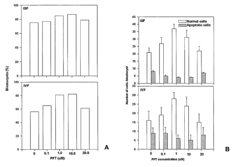
Fig. 3. The effect of short term inhibition of p53 in embryos by PFT-a on (A)
the proportion
of embryos developing to the blastocyst stage and (B) the number of cells per
blastocyst. Pifithrin-
a (PFT-a) was added as a media supplement to zygotes produced by either in
situ fertilisation
(ISF) or in vitro fertilisation (IVF).
ao Fig 4. The effect of short term inhibition of p53 in gametes by PFT-a on
(A) the proportion of
oocytes fertilized, (B) the proportion of embryos developing to the blastocyst
stage and (C) the
number of cells per blastocyst.
Fig. 5. Staining of blastocysts with anti-p53 antibody after treatment with
either p53-specific
siRNA (A) or non-specific scrambled (control) siRNA (B).
as Fig. 6. Effect of treatment of bovine sperm with PFT-a for one hour prior
to insemination on
the subsequent proportion of oocytes forming blastocysts (the number of
oocytes in each treatment
is shown in each bar).
Fig. 7. The response of embryos to the addition of exogenous PAF. Embryos were
either
collected fresh from the reproductive tract (fresh), fertilized in the
reproductive tract and cultured in
3o vitro (ISF) or fertilized and cultured in vitro (IVF). Response was
measured by the amplitude of
intracellular calcium concentration (A) and the proportion of 2-cell embryos
responding (B).
Fig. 8. The effect of co-treatment of AF and PFT-a on the development of in
vitro cultured
embryos as measured by the proportion of embryos progressing to blastocysts
(A) and the mean
number of cells per blastocyst (B). Embryos were treated with PAF alone, PFT-a
alone or both
ss PAF and PFT-a. Control embryos were cultured in the absence of PAF and PFT-
a.
CA 02536112 2006-02-16
WO 2005/019440 PCT/AU2004/001121
Fig. 9. The effect of p53 genotype on gamete and embryo viability. (A) The
effect of sperm
genotype on fertilization rate following ISF. (B) The effect of sperm genotype
on the genotype of
embryos resulting from IVF and ISF. (C) The effect of p53 genotype of a female
animal on the
number of eggs collected following superovulation.
s Best Mode of Performing the Invention
The p53 gene is known to play a crucial role as a tumour suppressor gene- it
is estimated
that loss of function mutations in p53 are found in more than half of all
human cancers. Other
functions of p53 and similar tumour suppressors in normal cells and tissues
are also increasingly
being identified, for example as cell cycle checkpoint regulators (Sherr,
2004, Cell 116, 235-246).
Accordingly, the inactivation of p53 has generally been viewed as undesirable.
p53 forms part of an interconnected network of pathways that allow cells to
sense and
respond to inappropriate stresses. Examples of tumour suppressors acting in
p53-associated
pathways include but are not limited to Rb, PTEN, P21, P27 ARF and INK.
The present invention is predicated, in part, on the inventor's surprising
finding that
~s expression of p53 is upregulated in embryos produced by assisted
reproductive technologies (ART)
such as in vifro fertilisation (IVF) and that this upregulation correlates
with poor embryo viability
following ART. Recognising that permanent inhibition of p53 in the developing
embryo is
undesirable, the inventor demonstrates herein that short term inhibition of
p53 during the culture of
gametes or embryos in vitro can increase the viability of embryos. For
example, the proportion of
ao embryos developing into morphologically normal blastocysts can be
increased, the number of cells
per blastocyst increased and the number of cells in the embryo undergoing
apoptosis decreased.
In one aspect, the present invention provides a method of enhancing embryo
viability by
administering to one or more of the embryo, oocytes, a female animal, sperm
and a male animal
an effective amount of at least one inhibitor of p53 or a p53-associated
pathway. The inhibitor may
as be an inhibitor of one or more of the following: p53, Rb, PTEN, P21, P27
ARF and INK. More than
one inhibitor or one or more of the above may be administered according to
methods of the
invention.
A large number of trophic factors have also been shown to have effects on the
growth,
development and survival of preimplantation mammalian embryos, many of which
are produced by
so the embryo itself (reviewed in Hardy and Spanos (2002) Journal of
Endocrinology 172, 221-236).
These include, but are not limited to, platelet activating factor (PAF),
insulin-like growth factors -I
(IGF-I) and -II (IGF-II), transforming growth factor-a (TGF-a), epidermal
growth factor (EGF),
leukemia inhibitory factor (LIF), colony stimulating factor-I (CSF-I), and
granulocyte-macrophage
colony stimulating factor (GM-CSF).
CA 02536112 2006-02-16
WO 2005/019440 PCT/AU2004/001121
12
Studies have demonstrated that ART embryos are deficient in several trophic
factors but that
provision of these factors exogenously has only a small beneficial effect in
terms of restoring
normal trophic factor functioning and increasing implantation rates andlor
embryo viability. As
described herein, studying PAF as an example, the present inventor has found
that poor response
s of ART embryos to exogenous trophic factor addition is due to a reduced
capacity of embryos
cultured in vitro to respond to trophic stimulation. This reduced capacity to
respond is manifested
as both a reduced amplitude of response and a decrease in the proportion of
embryos that
respond.
There is good evidence that the mechanism by which embryonic trophic factors
exert their
effects on the embryo are highly redundant of each other (Lu et al. (2003)
Journal of Cell Science
15, 1567-1576), involving among other mechanisms the phosphosphatidylinositol-
3-kinase
pathway, and further that the actions of several trophic factors are similar,
but not apparently
additive. It can therefore be reasonably argued and those skilled in the art
would readily
appreciate that the deficiencies observed in ART embryos in relation to
exogenous PAF
~s administration also occur for many of the embryonic trophic factors,
including IGF-I, IGF-II, TGF-a,
EGF, LIF, CSF-I and GM-CSF.
As disclosed herein, the inventor has now demonstrated that the upregulation
of p53
observed in embryos resulting from ART contributes to the limited benefit
achieved by the addition
of exogenous trophic factors to embryos in vitro. Treatment of in vifro
cultured embryos with both
zo an inhibitor of p53 and a trophic factor leads to a significantly greater
increase in embryo viability
than treatment of embryos with either the trophic factor alone or a p53
inhibitor alone.
Accordingly, in a further aspect, the present invention provides a method of
enhancing
embryo viability by administering to one or more of the embryo, oocytes, a
female animal sperm
and a male animal an effective amount of at least one inhibitor of p53 or a
p53-associated pathway
as and at least one growth promoting agent. In particular embodiments the
growth promoting agent is
a trophic factor, such as PAF, an analogue or derivative thereof or IGF-II, an
analogue or derivative
thereof.
p53 forms part of an interconnected network of pathways that allow cells,
including gametes,
and the embryo to sense and respond to inappropriate stresses. As disclosed
herein,
3o embodiments of the present invention demonstrate that disruption of key
components of this
network has the capacity to enhance the viability of gametes and embryos.
While the disclosure
following focuses largely on the inhibition of p53, those skilled in the art
will readily appreciate that
other components of p53-associated pathways, including for example, Rb, PTEN,
P21, P27 ARF
and INK may also be inhibited to achieve the desired effect. Accordingly,
embodiments of methods
CA 02536112 2006-02-16
WO 2005/019440 PCT/AU2004/001121
13
and compositions of the present invention contemplate the use of inhibitors or
p53 and p53-
associated pathways.
For the purposes of the present invention, embryo viability may be reflected
in a number of
indicators. For example increased embryo viability may result in increased
embryo implantation
s rates following in vifro fertilisation, decreased pre- and post-implantation
embryo lethality,
increased clinical pregnancy rates or increased birth rates. The present
invention therefore also
relates to methods of preventing apoptosis or retarded development in embryos
and to methods of
increasing pregnancy rates in animals.
The present invention is of particular benefit in increasirig embryo viability
following ART, and
in particular IVF. Other suitable ART techniques to which the present
invention is applicable
include, but are not limited to, gamete intrafallopian transfer (GIFT), zygote
intrafallopian transfer
(ZIFT), blastocyst transfer (BT), intracytoplasmic sperm injection (ICSI),
gamete, embryo and cell
cryopreservation, in vitro preparation of embryos such as in vitro oocyte
maturation, embryo biopsy
and other forms of embryo micromanipulation including formation of embryos by
nuclear transfer
~s and production transgenic lines and genetically modified lines. It is also
applicable to production of
embryonic stem cell lines.
Those of skill in the art will appreciate that the advantages offered by the
present invention
are not limited to ART-generated embryos. Rather the methods and compositions
of the present
invention are equally applicable as treatment to improve the viability of all
embryos, whether they
zo are produced in vitro via ART or in the reproductive tract of the animal.
The methods of the present
invention are therefore also applicable to improving embryo viability and
pregnancy rates in
otherwise unassisted pregnancies. Embodiments of the present invention also
provide for methods
of increasing ovulation rates in female animals and methods of increasing the
fertilizing capacity of
sperm in male animals.
zs The methods and compositions of the present invention are of use not only
for human
reproduction, but for a variety of species. For example, the methods and
compositions of the
present invention can be used to improve embryo viability and pregnancy rates
in animal
husbandry, for species of agricultural value, and in species bred for
conservation purposes. In
particular the present invention finds application in vertebrates, and more
particularly in mammals.
so For example, as disclosed herein, embodiments of the methods and
compositions of the invention
find application in bovine reproduction.
Further, the methods and compositions of the present invention are not only
applicable to
improving embryo viability for the purposes of increasing the success rate of
a pregnancy, but find
application in all circumstances in which it is beneficial to improve embryo
viability, for example in
ss the production of embryonic stem cells, the production of cloned embryos,
the formation of
CA 02536112 2006-02-16
WO 2005/019440 PCT/AU2004/001121
14
embryonic chimera, the production of transgenic or genetically modified cell
lines and organisms,
cryopreservation and all related techniques.
To achieve the desired result, agents and inhibitors according to embodiments
of the
invention may be administered directly to an embryo, for example as a
supplement to the medium
s in which the embryo is being cultured in vitro. Alternatively, either, or
both, of the male gametes or
the female gametes may be treated prior to fertilisation. Further, the present
invention also
contemplates the treatment of a female animal or a male animal directly with a
composition of the
invention.
Also disclosed herein are processes for the screening and identification of
agents for
increasing embryo viability, the processes comprising contacting a cell, cell
extract or embryo with
a candidate agent, determining whether the agent causes temporary inhibition
of the expression or
activity of p53 or a component of a p53-associated pathway, and thereby
determining whether the
agent is capable of increasing embryo viability. Typically the candidate
agents are compounds that
are not previously known to inhibit the expression or activity of the molecule
in question. The cell
~s may be, for example, a sperm cell, an oocyte or an embryonic stem cell.
Inhibition of p53 and p53-associated pathways
According to embodiments of the present invention, the inhibition of p53 or a
p53-associated
pathway suitable to achieve the desired outcomes is temporary inhibition. That
is, inhibition is short
zo term and reversible. For example, permanent inhibition of p53 is
undesirable due to the importance
of p53 activities. However as disclosed herein the short term inhibition of
p53 during the culture of
embryos in vitro can increase the viability of embryos. For example the
proportion of embryos
developing into morphologically normal blastocysts can be increased, the
number of cells per
blastocyst increased and the number of cells in the embryo undergoing
apoptosis decreased.
as For embryos and gametes in in vitro culture, treatment with at least one
suitable inhibitor is
preferably for the duration of the in vitro culture period of the embryos or
gametes, or for a portion
of this time. The appropriate duration of exposure to a suitable inhibitor can
be readily determined
by those skilled in the art by routine experimentation.
For embryos produced by ART, at least one inhibitor may be added as a
supplement to the
3o media in which the embryo is cultured, or in which gametes are cultured.
Alternatively or in addition
the female animal from which the oocytes are recovered andlor the male animal
from which the
sperm are collected may be treated with at least one inhibitor. It will be
appreciated that any
suitable concentration of inhibitor may be used, and the appropriate
concentration will likely depend
on a number of factors including but not limited to the nature, mode of action
and toxicity of the
3s inhibitor and the species of animal concerned. That is, for example, the
optimal concentration of
CA 02536112 2006-02-16
WO 2005/019440 PCT/AU2004/001121
inhibitor, and optimal time and mode of delivery may vary between species. One
skilled in the art
would be able to determine, by routine experimentation, the appropriate
parameters for use in any
given circumstance.
In all instances the appropriate concentration is one that is sufficient to
reduce or prevent the
s adverse effects of p53 or a component of a p53-associated pathway on embryo
survival. By way of
example, the inhibitor may be administered in a concentration of between about
0.01 ~,M and about
50 p,M, between about 0.1 ~.M and about 20 ~,M, between about 0.5 ~,M and
about 15 ~,M,
between about 1 ~,M and about 10 ~,M, or between about 2 ~,M and about 10 p,M.
The exact
concentration suitable for use in the methods of the invention will depend on
a number of factors
including, for example, the subject of the administration (i.e. embryo,
isolated cells or individual),
the species to be treated, the mode of administration and the inhibitor to be
administered. One
skilled in the art would be able to determine, by routine experimentation, the
appropriate
concentration to be used in any given circumstance.
The description of suitable inhibitors below is provided with particular
reference to inhibitors
~s of p53. However this description should not be construed as in any way
limiting the invention
thereto. Rather those skilled in the art will readily appreciate that p53-
associated pathways and
components or members of these pathways may also be inhibited using similar
mechanisms to
achieve the desired effect, and such inhibition is within the scope of the
present invention.
Inhibition of p53
ao A p53 inhibitor suitable for use in the methods of the present invention is
one that provides
reversible inhibition, provides inhibition for a time sufficient to prevent
p53-induced loss of viability
in the developing embryo and that has low toxicity. A p53 inhibitor suitable
for use according to the
present invention may be a small molecule inhibitor, a nucleic-acid based
inhibitor, a peptide-based
inhibitor or any combination thereof.
zs Such an inhibitor may act directly or indirectly on p53. For example the
inhibitor may act to
impair or prevent nuclear import and/or export of p53, may decrease the
stability of p53 or may
block any one or more of a number of other actions thereof, such as
transcriptional activation of
downstream acting genes. For example, p53 activates transcription of a number
of genes encoding
pro-apoptotic and cell-cycle arrest mediators, including, for example, bax,
p21/uvaf1, IGF-BP3 and
so PUMA (Agarwal et al., 1998, Journal of Biological Chemistry 273, 1-4). The
present invention
relates to the prevention of apoptosis or cell arrest in embryos which is
initiated by p53, and
accordingly, contemplates the inhibition of transcription or functioning of
apoptosis- or arrest-
inducing factors regulated by p53 such as those referred to above.
Additionally a suitable inhibitor may exert its inhibitory effect on p53
activity via its interaction
3s with or effect on a regulator of p53. For example, a key regulator of p53
activity is MDM-2
CA 02536112 2006-02-16
WO 2005/019440 PCT/AU2004/001121
16
(Momand ef al., 1992, Cell. 69:1237-45). MDM-2 causes nuclear export,
ubiquination and
degradation of p53 hence limiting its actions. A canonical regulator of MDM-2
activity is its
phosphorylation by the PI3K/Akt signalling pathway (Ogawara et al., 2002,
Journal of Biological
Chemistry. 277:21843-50).
s In one embodiment of the invention the p53 inhibitor is a small molecule
inhibitor. One
particularly suitable small molecule inhibitor for use in the methods of the
invention is pifithrin-a
(PFT-a; 2-(2-imino-4,5,6,7-tetrahydrobenzothiazol-3-yl)-1-p-tolylethanone)
(Komarov et al., 1999,
Science. 285:1733-1737) or variant or analogue thereof. The present inventor
has demonstrated
that the addition of between 0.1 and 20p,M of PFT-a to embryos in culture has
a significant effect
on increasing embryo survival.
It will be appreciated by those skilled in the art that temporary inhibition
is achievable by a
variety of means other than use of a chemical inhibitor such as PFT-a. For
example, nucleic acid-
based and peptide-based inhibitors are also contemplated, and a number of
alternative approaches
to achieving temporary p53 inhibition may be used in the methods of the
present invention.
~s For example embodiments of the invention may utilise antisense technology
to inhibit the
expression of a p53 gene by blocking translation of the protein. Antisense
technology takes
advantage of the fact that nucleic acids pair with complementary sequences.
Suitable p53-specific
antisense molecules can be manufactured by chemical synthesis or, in the case
of antisense RNA,
by transcription in vitro or in vivo when linked to a promoter, by methods
known to those skilled in
zo the art. A number of factors may operate to vary the level of inhibition
achieved using an antisense
construct according to the present invention, including, for example, the
design of the construct
(nucleotide sequence), dose of the construct used, dose of any transfection
agent used and
whether gametes, oocytes or individuals are treated.
For example, antisense oligonucleotides, typically of 18-30 nucleotides in
length, may be
as generated which are at least substantially complementary across their
length to a region of the
nucleotide sequence of the p53 gene. Binding of the antisense oligonucleotide
to their
complementary cellular nucleotide sequences may interfere with transcription,
RNA processing,
transport, translation and/or mRNA stability. Suitable antisense
oligonucleotides may be prepared
by methods well known to those of skill in the art and may be designed to
target and bind to
so regulatory regions of the nucleotide sequence or to coding (exon) or non-
coding (intron)
sequences. Typically antisense oligonucleotides will be synthesized on
automated synthesizers.
Suitable antisense oligonucleotides may include modifications designed to
improve their delivery
into cells, their stability once inside a cell, and/or their binding to the
appropriate target. For
example, the antisense oligonucleotide may be modified by the addition of one
or more
CA 02536112 2006-02-16
WO 2005/019440 PCT/AU2004/001121
17
phosphorothioate linkages, or the inclusion of one or morpholine rings into
the backbone (so-called
'morpholino' oligonucleotides).
By way of example only, a p53-specific antisense molecule may be administered
in a
concentration of between about 0.01 nM and about 200nM, between about 0.1 nM
and about
s 100nM, between about 0.5nM and about 50nM, between about 1nM and about 25nM,
or between
about 2nM and about 20nM. However those skilled in the art will readily
appreciate that for any
given antisense molecule the exact concentration to be used in any given
circumstance should be
determined empirically and will depend on a number of factors including, for
example, the molecule
to be administered, the method of transfection, the transfection agents) used,
if any, the subject of
the treatment (i.e. embryo, isolated cells, an individual) and the species to
be treated. One skilled
in the art would be able to determine, by routine experimentation, the
appropriate concentration to
be used in any given circumstance.
One suitable antisense technology, known as RNA interference (RNAi), may be
used,
according to known methods in the art (for example WO 99/49029 and WO
01170949, the
disclosures of which are incorporated herein by reference), to inhibit the
expression of p53
according to methods and compositions of the invention. RNAi refers to a means
of selective post-
transcriptional gene silencing by destruction of specific mRNA by small
interfering RNA molecules
(siRNA). The siRNA is generated by cleavage of double stranded RNA, where one
strand is
identical to the message to be inactivated. Double-stranded RNA molecules may
be synthesised in
zo which one strand is identical to a specific region of the p53 mRNA
transcript and introduced
directly. Alternatively corresponding dsDNA can be employed, which, once
presented
intracellularly is converted into dsRNA. Methods for the synthesis of suitable
molecule for use in
RNAi and for achieving post-transcriptional gene silencing are known to those
of skill in the art.
p53-specific siRNA molecules suitable for use according to embodiments of the
invention can be
zs readily designed and generated by those skilled in the art based on known
p53 nucleotide
sequences and using well known techniques. Suitable p53-specific siRNA
molecules are also
available commercially, for example from Santa Cruz Biotechnology, Inc.
A further means of inhibiting p53 gene expression may be achieved by
introducing catalytic
antisense nucleic acid constructs, such as ribozymes, which are capable of
cleaving mRNA
so transcripts and thereby preventing the production of wildtype protein.
Ribozymes are targeted to
and anneal with a particular sequence by virtue of two regions of sequence
complementarity to the
target flanking the ribozyme catalytic site. After binding the ribozyme
cleaves the target in a site-
specific manner. The design and testing of ribozymes which specifically
recognise and cleave p53
sequences can be achieved by techniques well known to those in the art (for
example Lieber and
CA 02536112 2006-02-16
WO 2005/019440 PCT/AU2004/001121
18
Strauss, 1995, Molecular and Cellular Biology, 15:540-551, the disclosure of
which is incorporated
herein by reference).
Any other inhibitor which is suitable for achieving temporary inhibition of
p53 or a p53-
associated pathway is also included within the scope of the present invention.
Additionally, it will
s be readily appreciated by those skilled in the art that the combination of
more than one means of
direct andlor indirect inhibition of p53 or its actions, or of a p53-
associated pathway may provide
the most benefit.
Trophic factors
Embodiments of the present invention provide for the use and administration of
growth-
promoting agents. Typically these grown-promoting agents are trophic factors.
Trophic factors suitable for use in the methods and compositions of the
invention may be
any trophic factors able to exert an effect on gametes or the pre- or post-
implantation embryo.
The trophic factors may be protein-based, polypeptide, peptide or lipid-based
or any combination
~s thereof.
The trophic factors may be natural compounds extracted from a suitable source,
be
synthetic or semi-synthetic compounds of the same structure and function, or
synthetic analogues
or mimetics of a natural trophic factor. In specific embodiments, the trophic
factor may be one or
more of the following: PAF, IGF-I, IGF-II, TGF-a, EGF, LIF, CSF-I or GM-CSF,
or an analogue or
ao derivative of one or more of the preceding factors. In a particular
embodiment, the growth-
promoting trophic factor is PAF or an analogue or derivative thereof, for
example a C2-carbamyl-
derivative of PAF. In another specific embodiment, the growth-promoting
trophic factor is IGF-II or
an analogue or derivative thereof.
Those skilled in the art will readily appreciate that for any given trophic
factor the exact
Zs concentration to be used in any given circumstance should be determined
empirically and will
depend on a number of factors including, for example, the trophic factor to be
administered, the
mode of administration, the subject of the treatment (i.e, embryo, isolated
cells, an individual) and
the species to be treated. One skilled in the art would be able to determine,
by routine
experimentation, the appropriate concentration to be used in any given
circumstance. By way of
3o example only, the trophic factor may be PAF. PAF may be administered in a
concentration of
between about 0.01 nM and about 50nM, between about 0.1 nM and about 20nM,
between about
0.5nM and about 15nM, between about 1nM and about 10nM, or between about 2nM
and about
10nM.
The present invention also contemplates the provision of trophic support by
artificial
3s stimulation of the down-stream signaling pathways of trophic factors. For
example in the case of
CA 02536112 2006-02-16
WO 2005/019440 PCT/AU2004/001121
19
PAF, transient increases in intracellular calcium concentration may be
stimulated by the transient
exposure of embryos to a calcium ionophore such as ionomycin. For example
exposure may be
for approximately 30 seconds, at a ionomycin concentration of between about
0.1 to 1 ~M.
Additionally, as a number of trophic factors act via a phosphotidylinositol-3-
kinase (P13K)
s dependent pathway (Lu et al. (2004) Journal of Cell Science 15, 1567-1576),
this pathway may be
activated in embodiments of the invention as a means of stimulating a trophic
factor signaling
pathway.
Administration of agents)
In embodiments of the present invention as they pertain to embryos produced by
ART in
vitro-cultured embryos are treated directly with at least one inhibitor of p53
or a p53-associated
pathway, optionally together with at least one growth promoting agent. The
inhibitors and/or agents
may be added as a supplement to the growth medium in which the embryos are
being cultured.
However, the present invention also includes within its scope embodiments in
which suitable
~s inhibitors and agents are added at other stages. For example, oocytes
recovered from a female
animal undergoing ART may be treated with the agents. Similarly sperm may be
so treated.
Additionally the suitable agents may be administered to a female and/or male
animal directly.
For example the animal may be attempting to conceive naturally or may be
undergoing ART
treatment, such as being treated on an IVF program. Alternatively treatment
may be administered
ao to a pregnant female.
For embodiments in which methods provide for the administration of at least
one growth-
promoting agent and at least one inhibitor of p53 or a p53-associated pathway,
the at least one
growth-promoting agent and at least one inhibitor may be administered
individually or alternatively
they may be components of a single composition. If administered individually,
the administration
zs may be sequential or simultaneous.
Compositions according to embodiments of the invention may be prepared
according to
methods which are known to those of ordinary skill in the art containing the
suitable agents. Such
compositions may include a pharmaceutically acceptable carrier, diluent and/or
adjuvant. The
carriers, diluents and adjuvants must be "acceptable" in terms of being
compatible with the other
3o ingredients of the composition, and not deleterious to the recipient
thereof. These compositions
can be administered by standard routes. In general, the compositions may be
administered by the
parenteral or oral route. More preferably administration is by the oral route.
Alternatively,
administration may be topical or vaginal, for example in the form of an
ointment, cream, lotion or
gel or by way of insertion of a vaginal pessary.
CA 02536112 2006-02-16
WO 2005/019440 PCT/AU2004/001121
It will be understood that the specific dose level for any particular
individual will depend upon
a variety of factors including, for example, the activity of the specific
agents employed, the age,
body weight, general health, diet, the time of administration, rate of
excretion, and combination with
any other treatment or therapy. Single or multiple administrations of the
agents or compositions
s can be carried out with dose levels and pattern being selected by the
treating physician.
Regardless, the agents or compositions used in the present invention should
provide a quantity of
the agent sufficient to enhance embryo viability.
Examples of pharmaceutically acceptable carriers or diluents are demineralised
or distilled
water; saline solution; vegetable based oils such as peanut oil, safflower
oil, olive oil, cottonseed
oil, maize oil, sesame oils such as peanut oil, safflower oil, olive oil,
cottonseed oil, maize oil,
sesame oil, arachis oil or coconut oil; silicone oils, including
polysiloxanes, such as methyl
polysiloxane, phenyl polysiloxane and methylphenyl polysolpoxane; volatile
silicones; mineral oils
such as liquid paraffin, soft paraffin or squalane; cellulose derivatives such
as methyl cellulose,
ethyl cellulose, carboxymethylcellulose, sodium carboxymethylcellulose or
~s hydroxypropylmethylcellulose; lower alkanols, for example ethanol or iso-
propanol; lower
aralkanols; lower polyalkylene glycols or lower alkylene glycols, for example
polyethylene glycol,
polypropylene glycol, ethylene glycol, propylene glycol, 1,3-butylene glycol
or glycerin; fatty acid
esters such as isopropyl palmitate, isopropyl myristate or ethyl oleate;
polyvinylpyrridone; agar;
carrageenan; gum tragacanth or gum acacia, and petroleum jelly. Typically, the
carrier or carriers
ao will form from 10% to 99.9% by weight of the compositions.
The compositions of the invention may be in a form suitable for parenteral
administration, or
in the form of a formulation suitable for oral ingestion (such as capsules,
tablets, caplets, elixirs, for
example).
For administration as an injectable solution or suspension, non-toxic
parenterally acceptable
zs diluents or carriers can include, Ringer's solution, isotonic saline,
phosphate buffered saline,
ethanol and 1,2 propylene glycol.
Some examples of suitable carriers, diluents, excipients and adjuvants for
oral use include
peanut oil, liquid paraffin, sodium carboxymethylcellulose, methylcellulose,
sodium alginate, gum
acacia, gum tragacanth, dextrose, sucrose, sorbitol, mannitol, gelatine and
lecithin. In addition
3o these oral formulations may contain suitable flavouring and colourings
agents. When used in
capsule form the capsules may be coated with compounds such as glyceryl
monostearate or
glyceryl distearate which delay disintegration.
Adjuvants typically include emollients, emulsifiers, thickening agents,
preservatives,
bactericides and buffering agents.
CA 02536112 2006-02-16
WO 2005/019440 PCT/AU2004/001121
21
Solid forms for oral administration may contain binders acceptable in human
and veterinary
pharmaceutical practice, sweeteners, disintegrating agents, diluents,
flavourings, coating agents,
preservatives, lubricants and/or time delay agents. Suitable binders include
gum acacia, gelatine,
corn starch, gum tragacanth, sodium alginate, carboxymethylcellulose or
polyethylene glycol.
s Suitable sweeteners include sucrose, lactose, glucose, aspartame or
saccharine. Suitable
disintegrating agents include corn starch, methylcellulose,
polyvinylpyrrolidone, guar gum, xanthan
gum, bentonite, alginic acid or agar. Suitable diluents include lactose,
sorbitol, mannitol, dextrose,
kaolin, cellulose, calcium carbonate, calcium silicate or dicalcium phosphate.
Suitable flavouring
agents include peppermint oil, oil of wintergreen, cherry, orange or raspberry
flavouring. Suitable
coating agents include polymers or copolymers of acrylic acid and/or
methacrylic acid and/or their
esters, waxes, fatty alcohols, zein, shellac or gluten. Suitable preservatives
include sodium
benzoate, vitamin E, alpha-tocopherol, ascorbic acid, methyl paraben, propyl
paraben or sodium
bisulphite. Suitable lubricants include magnesium stearate, stearic acid,
sodium oleate, sodium
chloride or talc. Suitable time delay agents include glyceryl monostearate or
glyceryl distearate.
~s Liquid forms for oral administration may contain, in addition to the above
agents, a liquid
carrier. Suitable liquid carriers include water, oils such as olive oil,
peanut oil, sesame oil,
sunflower oil, safflower oil, arachis oil, coconut oil, liquid paraffin,
ethylene glycol, propylene glycol,
polyethylene glycol, ethanol, propanol, isopropanol, glycerol, fatty alcohols,
triglycerides or
mixtures thereof.
zo Suspensions for oral administration may further comprise dispersing agents
andlor
suspending agents. Suitable suspending agents include sodium
carboxymethylcellulose,
methylcellulose, hydroxypropylmethyl-cellulose, poly-vinyl-pyrrolidone, sodium
alginate or acetyl
alcohol. Suitable dispersing agents include lecithin, polyoxyethylene esters
of fatty acids such as
stearic acid, polyoxyethylene sorbitol mono- or di-oleate, -stearate or -
laurate, polyoxyethylene
as sorbitan mono- or di-oleate, -stearate or -laurate and the like.
The emulsions for oral administration may further comprise one or more
emulsifying agents.
Suitable emulsifying agents include dispersing agents as exemplified above or
natural gums such
as guar gum, gum acacia or gum tragacanth.
Methods for preparing parenterally administrable compositions are apparent to
those skilled
3o in the art, and are described in more detail in, for example, Remington's
Pharmaceutical Science,
15th ed., Mack Publishing Company, Easton, Pa., hereby incorporated by
reference herein.
The composition may incorporate any suitable surfactant such as an anionic,
cationic or non-
ionic surfactant such as sorbitan esters or polyoxyethylene derivatives
thereof. Suspending agents
such as natural gums, cellulose derivatives or inorganic materials such as
silicaceous silicas, and
3s other ingredients such as lanolin, may also be included.
CA 02536112 2006-02-16
WO 2005/019440 PCT/AU2004/001121
22
Formulations suitable for topical or vaginal administration comprise active
ingredients
together with one or more acceptable carriers, and optionally any other
therapeutic ingredients.
Formulations suitable for topical or vaginal administration include liquid or
semi-liquid preparations
suitable for penetration through the skin to the site of where treatment is
required, such as lotions,
s creams, ointments, pastes or gels.
Creams, ointments or pastes according to the present invention are semi-solid
formulations
of the active ingredient for external application or for intra-vaginal
application. They may be made
by mixing the active ingredient in finely-divided or powdered form, alone or
in solution or
suspension in an aqueous or non-aqueous fluid, with a greasy or non-greasy
basis. The basis may
comprise hydrocarbons such as hard, soft or liquid paraffin, glycerol,
beeswax, a metallic soap; a
mucilage; an oil of natural origin such as almond, corn, arachis, castor or
olive oil; wool fat or its
derivatives, or a fatty acid such as stearic or oleic acid together with an
alcohol such as propylene
glycol or macrogols. The composition may incorporate any suitable surfactant
such as an anionic,
cationic or non-ionic surfactant such as sorbitan esters or polyoxyethylene
derivatives thereof.
~s Suspending agents such as natural gums, cellulose derivatives or inorganic
materials such as
silicaceous silicas, and other ingredients such as lanolin, may also be
included.
Gels are particularly suitable for vaginal administration. Gel compositions
may be designed
by means known to those skilled in the art to provide prolonged contact and
promote controlled and
sustained release of the active agent while minimising leakage.
zo The compositions may also be administered in the form of liposomes.
Liposomes are
generally derived from phospholipids or other lipid substances, and are formed
by mono- or multi-
lamellar hydrated liquid crystals that are dispersed in an aqueous medium. Any
non-toxic,
physiologically acceptable and metabolisable lipid capable of forming
liposomes can be used. The
compositions in liposome form may contain stabilisers, preservatives,
excipients and the like. The
zs preferred lipids are the phospholipids and the phosphatidyl cholines
(lecithins), both natural and
synthetic. Methods to form liposomes are known in the art, and in relation to
this specific reference
is made to: Prescott, Ed., Methods in Cell Biology, Volume XIV, Academic
Press, New York, N.Y.
(1976), p. 33 et seq., the contents of which are incorporated herein by
reference.
Also included within the scope of agents used in the present invention are
prodrugs.
3o Typically, prodrugs will be functional derivatives of the agents of the
present invention which are
readily converted in vivo to the required agents of the present invention as
described herein.
Typical procedures for the selection and preparation of prodrugs are known to
those of skill in the
art and are described, for instance, in H. Bundgaard (Ed), Design of Prodrugs,
Elsevier, 1985, the
disclosure of which is incorporated herein by reference.
CA 02536112 2006-02-16
WO 2005/019440 PCT/AU2004/001121
23
In vitro culture media
The present invention provides compositions for use as culture media in which
the culture
medium includes an effective amount of at least one inhibitor of p53 or a p53-
associated pathway,
s optionally also including at least one growth-promoting agent, in addition
to the necessary nutrients
and co-factors required for in vitro growth and development of gametes or
embryos.
For in vitro incubation and culture of gametes or embryos during ART
procedures, a range of
suitable media are available, the types and compositions of which are well
known to those of skill in
the art. Preferably the culture medium contains at least water, salts,
nutrients, essential amino
acids, vitamins and hormones, and may also include one or more growth factors.
A variety of
suitable culture media is commercially available, for example Earle's media,
Ham's F10 media and
human tubal fluid (HTF) media.
The present invention also contemplates the co-culture in vitro of embryos on
a layer of
'feeder cells' by methods known to the art. Appropriate 'feeder cells' for co-
culture may include, for
~s example, bovine oviductal cells or human tubal epithelial cells.
Loss-of-function mutations
The present inventor's results show that ART causes up-regulation of p53 and
that this is a
major cause of the embryopathy induced by ART. p53 causes the synthesis of a
number of
zo effectors and regulatory elements of apoptosis, including the Bax protein,
leading to the activation
of caspases and consequent cell death. Thus, the presence of loss-of-function
(LOF) mutations in
p53 or components of p53-associated pathways may favor the survival of ART
embryos.
Thus ART may provide a positive selection pressure for loss-of-function (LOF)
mutations in
the p53 gene, or genes encoding components of p53-associated pathways, even in
the
as hemizygous state. Selection that favours LOF mutations may result in an
over-representation of
tumour susceptibility in offspring produced by ART. Although the germ-line
frequency for such LOF
mutations is low, selection pressure of the scale observed in the mouse model
could result in
marked shifts in gene frequency over time. Applied to human ART practice, the
accumulation of
LOF mutations will have profound implications for the use of the technology.
3o Accordingly, the present invention provides methods of protecting embryos,
in particular
embryos produced by assisted reproductive technologies, from positive
selection pressures for
inherited, or acquired, defects or from the accumulation of LOF mutations in
the p53 gene or genes
encoding components of p53-associated pathways.
CA 02536112 2006-02-16
WO 2005/019440 PCT/AU2004/001121
24
The present invention will now be further described in greater detail by
reference to the
following specific examples, which should not be construed as in any way
limiting the scope of the
invention.
Examples
s General Methods
Mice: p53 knockout mice are from the 86.12952-T,rp53tm~ryi strain (p53 /)
backcrossed with
C57BLI6J. The mutant strain was developed in the laboratory of Dr. Tyler Jacks
at the Center for
Cancer Research at the Massachusetts Institute of Technology and is now bred,
under licence, at
the Gore Hill Research Laboratory, Royal North Shore Hospital. The strain is
maintained by mating
heterozygous females to homozygous males, and specific matings are set up to
produce p53 /,
p53+l and p53+/+ mice as required. Tail tissue is collected from all weanlings
and genotyped by
PCR using the following primers 5'- CTT ggg Tgg AgA ggC TAT TC-3' & 5'-Agg TgA
gAT gAC Agg
AgA TC-3' for the knock-out allele and 5'-ATA ggT Cgg Cgg TTC AT-3' & 5'-CCC
gAg TAT CTg
gAA gAC Ag-3' for the wild type allele.
~s Mouse IVF: Females superovulated by 5 IU pregnant mare serum gonadotrophin
followed 48
h later by 51U human chorionic gonadotrophin. Oocytes were collected 13-15 h
after hCG and
washed. Epididymal sperm collected from the epididymides of male mice (14-35
weeks old) of
proven fertility is immediately placed into 1 ml of medium. Sperm were allowed
to disperse for 10
min at 37°C and were then added to the oocytes at 0.1 X 1061m1.
Fertilisation was performed in a
zo final volume of 1 ml of medium. Oocytes and sperm were cultured together
for 5 h at 37°C in 5%
COz in air. The oocytes were then retrieved, washed in Hepes-buffered HTF and
their fertilisation
status assessed by microscopic detection of pronuclei and polar bodies.
Fertilised oocytes were
transferred to drops of modified HTF medium under mineral oil and their
development status
assessed each 24 h.
as Example 1
p53 expression in mouse embryos
Example1 (A) - mRNA expression
RT-PCR was used to compare the expression of p53 mRNA in fresh p53 +I+ mouse
embryos
(embryos fertilised and grown in the reproductive tract) and IVF mouse
embryos. For IVF embryos,
3o the model uses a graded range of stress induced by IVF and culture under
conditions of increasing
growth factor deprivation. Primers were designed (5' - GGA GTC TTC CAG TGT GAT
GAT, 3' -
GGG ACA GCC AAG TCT GTT ATG) that successfully produced an RT-PCR product of
the correct
size (429 bp) and sequence (SUPAMAC) for the p53 gene. Groups of 5 embryos,
oocytes or
sperm were pooled. mRNA was extracted and RT-PCR performed.
CA 02536112 2006-02-16
WO 2005/019440 PCT/AU2004/001121
p53 product was detected in oocytes, zygotes, 8-cell, morulae and blastocyst
stage mouse
embryos collected from the reproductive tract but not in epididymal sperm. In
2-cell embryos
detection was variable. Incubation of zygotes in the transcription inhibitor a-
amanitin had no effect
on qualitative expression of p53 RNA in zygotes, but incubation of 2-cell
embryos in a-amanitin
s gave embryos that were p53 mRNA negative after 24 h (normally the 8-cell
stage). This result
indicates that p53 mRNA in the zygote and early embryo was a product of the
gametes (probably
mainly oocytes) but that its continual expression past the 2-cell stage
required new transcription
from the zygotic genome. Quantitative RT-PCR of p53 was performed to compare
expression of
p53 mRNA in fresh and IVF embryos. Equivalent levels of RNA were found in the
oocyte, zygote,
morulae and blastocyst of fresh embryos; the levels were 50-70% lower in late
2-cell embryos than
in other development stages (data not shown). A similar pattern of mRNA
expression was
observed in fresh and IVF embryos.
Example 1(B) - Protein expression
F1 (C57BLI6j X CBAIJ), C57BI6j and p53-/- embryos or sperm were used to
examine p53
~s protein expression levels. T47D breast cell line was used as a positive
control for the detection of
p53. Four to eight week old females were superovulated by i.p. injection of 10
IU pregnant mare
serum gonadotrophin (Folligon, Intervet International, Boxmeer, The
Netherlands) followed 48h
later by 10 IU human chorionic gonadotrophin (hCG, Chorulon, Intervet).
Females were either left
unmated or paired with males of proven fertility. Day 1 of pregnancy was
confirmed by the
zo presence of a copulation plug the following morning.
Sample collection
Mice were killed by cervical dislocation. Cumulus masses or embryos were
flushed from the
reproductive tract with Hepes-buffered human tubal fluid medium with 3 mg
BSAImI (Hepes-HTF-
101.6 mM NaCI, 4.69 mM KCI, 0.2 mM MgS04, 0.37 mM KH2POa, 21.4 mM Na lactate,
2.78 mM
zs glucose, 2.04 mM CaClz, 5 mM NaHCOs, 0.33 mM Na pyruvate and 21 mM Hepes pH
7.4). All
components of the medium were tissue culture grade from Sigma (St. Louis, MO)
and contained 3
mg BSA Fraction Vlml unless otherwise stated (CSL Ltd, Melbourne, Vic,
Australia). Zygotes were
collected 20-21h after hCG and freed from their cumulus cells by brief
exposure to 300 IU
hyaluronidase (Sigma) in Hepes-HTF. Fresh 2-cell embryos, morulae and
blastocysts were
3o collected from the reproductive tract at 48h,72h and 96h, respectively.
Mouse sperm was collected from the epididymides of male mice (10-15 weeks old)
of proven
fertility and immediately placed into 1 ml of medium (HTF media). Sperm were
allowed to disperse
for 10 min at 37°C. Sperm concentration and motility was assessed by
haemocytometer and the
sperm suspension diluted to 0.5 X 1061m1 in HTF media containing 3 mg/ml
albumin. The sperm
ss was incubated for 1, 2, 3 or 4 hours and then collected by centrifugation.
CA 02536112 2006-02-16
WO 2005/019440 PCT/AU2004/001121
26
Western blot analysis
Embryos or sperm were collected and washed 3 times in cold PBS and transferred
in a
maximum volume of 1.5 p,l PBS into 1.5 p,l of 2X extraction buffer (2X PBS, 2%
Triton X-100, 24
mM deoxycholic acid, 0.2% sodium dodecyl sulfate, 20 mM NaF, 20 mM Na4P20~, 2
mM PMSF,
s 3.08 wM aprotinin, 42 p,M leupeptin and 2.91 p,M pepstatin A- all from
Sigma). Cells were lysed by
three cycles of freezing in liquid nitrogen and thawing (with vortexing).
Protein samples were
diluted with 1 pl of 5X Laemmli buffer (50 mM Tris-HCI, 5 mM EDTA pH 8.0,
12.5% Sodium
dodecyl sulfate, 0.05% bromophenol blue and 25% beta-mercaptoethanol),
incubated 10 min at
60°C and run on 20% homogenous SDS polyacrylamide gels (Pharmacia;
Sweden) using
PhastSystem apparatus separation and control unit (Pharmacia). Proteins were
blotted into PVDF
membranes (Hybond-P, Amersham Pharmacia) in a semi-dry blotting apparatus
overnight using
transfer buffer (12 mM Tris PH 7.0, 96 mM Glycine and 20% methanol).
Nonspecific binding was
blocked by 5% skim milk in PBS supplemented with 0.05% Tween-20 (PBST) at room
temperature
for 1 h. Membranes were probed with or 1:5000 diluted monoclonal anti-LIS-1
antibody overnight
~s at 4°C in 2.5% skim milk, and detected with horse radish peroxidase
(Jackson ImmunoResearch
Laboratories, West Grove, PA, USA) conjugated secondary antibody. Membranes
were developed
with Femto SuperSignal Chemiluminescent Substrate (Pierce, Rockford, IL, USA)
for 5 min at room
temperature.
Fig. 1A clearly illustrates that the expression of p53 increases with time as
sperm are
zo incubated capacitation media. Similarly, Fig. 1B demonstrates that p53
expression in the embryo
increases with development of the embryo, at least from a 1-cell embryo
through to blastocyst.
Further, p53 expression is higher in embryos cultured in vitro than in embryos
collected fresh from
the reproductive tract.
Figs. 1 C and 1 D confirm the specificity of the assays. In blastocysts from
p53 -I- embryos no
zs p53 expression was detected whereas p53 protein was readily detected in
blastocysts from p53 +I+
embryos (Fig. 1 C). Further, Fig. 1 D confirms that the increased p53
expression observed following
in vitro culture was not a consequence of an overall change in the pattern of
protein expression in
the embryos, as evidenced by the maintenance of expression of the
constitutively expressed
protein LIS-1.
so Example 2
Immunolocalisation of p53 in mouse embryos
The expression pattern of p53 protein in pre-implantation mouse embryos was
investigated
using immunofluorescence.
Embryos were washed 3-times (washing media: PBS with 0.1% BSA, 0.1% Tween-20)
and
ss fixed with fresh 2% paraformaldehyde (Sigma) in PBS PH 7.4 for 1 h at room
temperature.
CA 02536112 2006-02-16
WO 2005/019440 PCT/AU2004/001121
27
Embryos were permeabilised for 30 min at room temperature in PBS with 2% BSA,
0.2% Tween-20
and 0.2% Triton X-100 and blocked in 2% BSA and 30% blocking serum for 1-3h.
p53 antigen was
stained overnight at 4°C with 2 p,g anti-p53 sheep polyclonal antibody
Iml (Oncogene Research
Products (PC35) or rabbit polyclonal anti-Bax (Santa-Cruz Biotechnology, N-20)
in PBS with 2%
s BSA and then 5 ~,g rabbit anti-sheep FITC conjugated antibody/ml for 1h at
room temperature
(green channel). Controls were 2 ~.g non-immune IgG/ml instead of primary
antibody.
Immunofluorescence images were observed using a BioRad Radiance confocal
microscope
(Australian Key Centre for Microscopy and Microanalysis, University of Sydney)
using a Nikon Plan
Apo 60X11.40 oil immersion objective. Images were captured using LaserSharp
2000 Version 4.0
(build 365) software. Microscope and laser settings were adjusted such that no
fluorescence was
observed with non-immune control. All test specimens were observed with these
settings and
settings the same. Quantitative analysis of staining was performed using NIH
Image software. For
imaging the whole embryo 1 p,M sections were performed using the z-sectioning
facility of the
microscope.
~s The expression of immunodetectable p53 protein (Fig. 2) was low in
preimplantation mouse
embryos collected fresh from the reproductive tract (fresh embryos) (Fig. 2A
and 2D). Embryos
produced by IVF and cultured individually in 10 ~.I of media (Fig. 2C and 2F)
or those fertilised in
the reproductive tract and then cultured in vitro in groups of 10 in 10 ~,I of
media (Fig. 2B and 2E)
showed different patterns of p53 expression. There was little immunodetectable
p53 at the morula
ao through blastocyst stage in fresh embryos and no evidence of accumulation
of protein within the
nucleus of the embryos cells. In contrast, there was a substantial
accumulation of p53 in IVF
embryos with intense nuclear localisation. There was only modest difference in
p53 prior to the 8-
cell stage, but greatly increased expression at each stage thereafter. By the
8-cell stage onwards,
many IVF embryos showed nuclear staining as the predominant pattern of
staining with relatively
as less cytoplasmic staining. This pattern of protein expression correlates
with the pattern of
embryopathy after ART, with most embryo loss occurring after the 8-cell stage.
The regulation of protein expression (Example 1 B and 2), but not mRNA levels
(Example
1A), suggests that the regulation of p53 is at the post-transcriptional level.
Example 3
so Viability of embryos with p53 loss-of-function mutations
The correlation between high levels of p53 expression and poor embryo
viability following
ART (Examples 1 B and 2) argues that p53 is a major effector of the responses
of the embryo to cell
stress. As disclosed herein, this hypothesis is confirmed by observations of
improved viability of
embryos with loss-of-function (LOF) mutations to p53. Mating of p53+I- X p53+I-
or p53+I+ x
3s p53+I+ parents was performed by IVF and the resulting zygotes cultured for
96h. A greater
CA 02536112 2006-02-16
WO 2005/019440 PCT/AU2004/001121
28
proportion of embryos from the p53+I- X p53+I- cross reached the blastocyst
stage (75% v 57%)
and embryos had more cells (P<0.01 ) and fewer apoptotic cells (P < 0.001 )
compared to embryos
resulting from wild-type crosses. The p53+I- X p53+I- cross also resulted in a
reduction in the
number of embryos that were markedly retarded in development or showed a
degenerating of
s fragmented morphology (5% v 19%) and 3 times as many p53-I- embryos were
hatched from their
zona pellucida after 96h culture (36%) than was the case for p53+I+ embryos
(11 %).
Morphologically normal blastocysts were selected for transfer after zygotes
from p53+I- X
p53+I- crosses were cultured in vitro for 96h. There was a significantly
higher pregnancy rate and
higher proportion of viable foetuses than for embryos from p53+I+ X p53+/+
crosses. A foetal
viability rate of 4.7%; 9.9%; and 60.0% per embryo transferred was observed
for +/+; +/- and -/-
genotypes, respectively. This highly significant skewing of the genotype was
not a normal feature
of the mouse strain since natural mating of the same parental lines resulted
in normal Mendelian
segregation at birth. When +/+ embryos were collected fresh from the
reproductive tract and were
transferred immediately to pseudopregnant foster mothers, the foetal viability
rate was 57%. Thus
~s the absence of the p53 gene seemed to compensate for the adverse effects of
ART and
consequently resulted in an increased foetal viability rate.
Example 4
Inhibition of p53 and embryo viability
p53 has numerous important cellular functions and as a consequence its
permanent
Zo inhibition as a means of improving embryo viability is undesirable. The
results described above
indicate that the normal expression of p53 in the embryo within the
reproductive tract is very low
and thus it can be anticipated that providing the inhibition is short-term
there would not be adverse
effects and may improve embryo viability.
This hypothesis was tested using two different types of p53 inhibitor, a small
molecule
zs inhibitor (Example 4A) and a nucleic-acid based inhibitor, being a p53-
specific siRNA molecule
(Example 4B).
Example 4(A) - Small molecule inhibitor
A recently developed selective inhibitor of p53, pifithrin-a (PFTa [2-(2-imino-
4,5,6,7-
tetrahydrobenzothiazol-3-yl)-1-p-tolylethanone], Sigma-Aldrich), is a small
molecule that reversibly
so blocks p53-dependent transcriptional activation and apoptosis (Komarov et
al., 1999, Science 285,
1733-1737). PFT-a was prepared initially by solubilising to a concentration of
10mM in
dimethylsolfoxide and diluted to working concentrations.
(i) Treafment of embryos uvifh PFT a
Zygotes were produced by either fertilization in vitro (IVF) or fertilisation
in situ (ISF) and
3s cultured for 96 hours in modified HTFM medium individually in 10,1 drops.
PFT-a was added as a
CA 02536112 2006-02-16
WO 2005/019440 PCT/AU2004/001121
29
media supplement over the dose range 0,1 - 20 ~,M. The proportion of embryos
developing to the
blastocyst stage (Fig. 3A), the number of cells per normal blastocyst and the
proportion of cells
undergoing apoptosis (Fig, 3B), were assessed.
The results show that PFT-a caused a significant quadratic effect on the
proportion of IVF
s embryos that developed to the blastocysts stage and for both ISF and IVF
there was a significant
improvement in the number of cells per normal blastocyst and a significant
decrease in the number
of cells that were undergoing apoptosis. This marked improved survival of
embryos and cells is
clinically relevant.
(ii) Treatment of gametes with PFT a
Preparation of sperm in vitro for fertilisation leads to an increase in the
synthesis of p53 (see
Example 1B; Fig. 1A). Furthermore production of embryos by IVF leads to a
marked increase in
the synthesis of p53 in resulting embryos (Example 1 B; Fig. 1 B). The present
experiment was
designed to determine if treatment of sperm and oocytes during the process of
fertilisation in vitro
resulted in an improvement to their fertilisation rate and the subsequent
development of resulting
~s embryos.
PFT-a preparation, IVF and embryo culture was as previously described. The
study used F1
(C57BI6j x CBAj) males and females. PFT-a was added to sperm during its
capacitation and during
the process of fertilisation in vitro.
4-5 h after the addition of sperm to oocytes, the oocytes were washed free of
sperm and
ao PFT-a. They were examined for the presence of 2 pronuclei as evidence of
successful fertilisation.
Successfully fertilised oocytes were placed in culture in 10~.L of modified
HTF media, 1 zygote per
10~,L drop. They were culture for 120h and the proportion forming
morphologically normal
blastocysts were recorded. The blastocysts were fixed in 2% formaldehyde and
stained with 5 ~,G
. Hoechst stain/ml. The stain labels nuclei and was used to perform cell
counts on each blastocyst.
zs The effect of dose of PFT-a on the proportion of oocytes fertilised, the
proportion of zygotes
that developed to morphological blastocyst and the number of cells in each
blastocysts were
recorded and are shown in Figs. 4A, B and C respectively. The effect of PFT-a
dose was
assessed using the SPSS statistical package. The fertilisation rate and
development rate were
assessed using binary logistic regression analysis while cell numbers were
assessed by univariate
so analysis using the general linear model.
Figure 4A illustrates that there is a dose-dependent increase in fertilisation
rate in the
presence of PFT-a (P < 0.01). Of the oocytes that fertilised, there was a
further dose-dependent
increase in the proportion of zygotes that developed to the blastocyst stage
(Figure 4B) (P < 0.05).
Of embryos developing to the blastocyst stage there was a dose-dependent
significant increase in
ss the mean number of cells present in each embryo (P < 0.05, Figure 4C).
CA 02536112 2006-02-16
WO 2005/019440 PCT/AU2004/001121
The results demonstrate that treatments that decrease the expression of p53 in
gametes
prior to and during fertilisation result in an improvement in fertilisation,
and subsequent embryo
development.
Example 4(B): siRNA inhibition
s As an alternative to temporary inhibition of p53 using a small molecule
inhibitor, the ability of
transient treatment of embryos with p53-specific siRNA to inhibit p53
expression was investigated.
Oligonucleotides designed as siRNA against mouse p53 or control nonsense siRNA
sequences
were used, both of which were obtained from Santa Cruz Biotechnology, Inc. The
siRNA
oligonucleotides were prepared as 90nM with siMHTF media (102 mM NaCI, 4.6mM
KCI, 0.20 mM
MgSOa, 0.4 mM KHaP04, 21.4 mM Na lactate, 1 mM glutamine, 0.33 mM Na pyruvate,
2.78 mM
glucose, 2.0 mM CaCl2, 25 mM NaHCOs , pH 7.35; 285 mOsmll) containing 3mg
polyvinyl-
pyrrolidone /ml (PVP) (Sigma). All media were sterilized with a 0.1 ~m filter.
Cells were transfected
with the siRNA molecules in the presence of 25% (vlv) oligofectamine
(Invitrogen). Oligofectamine
was prepared in siMHTF, mixed gently and incubated for 5-10 minutes at room
temperature. 171
~s of siRNA was added to 3 p,l of 25% oligofectamine, mixed gently and
incubated for 15-20 minutes
at room temperature. It was then brought to 100 p,L with siMHTF, giving a
final oligonucleotide
concentration of 15nM.
Embryos were collected from F1 (C57BL6 x CBAIj) mice. Females were super
ovulated with
PMSG and HCG (5 LU.). Zygotes were collected ~10h after mating and cultured
for 96h in
ao modHTF media plus penicillin and phenol red, pH 7.35; 285 m0sm/l) at 37C in
5% C02.
Embryos were incubated with either p53 siRNA or control siRNA for 4 h at
37°C in 5% C02.
Embryos were then thoroughly washed and placed in conventional culture and the
development
and expression of p53 in resulting embryos analyzed.
Staining for p53 was performed by washing embryos three-times (washing media:
PBS with
as 0.1 % BSA, 0.1 % Tween-20 and 0.2% sodium azide) and fixing with fresh 2%
formaldehyde
(Sigma) in PBS PH 7.4 for 1 h at room temperature. Embryos were permeabilized
for 30 min at
room temperature in PBS with 2% BSA, 0.3% Tween-20 in the presence of 2%
formaldehyde and
after washing blocked in 2% BSA and 30% goat serum (Sigma) for 1-3h. p53
antigen was stained
overnight at 4°C with freshly prepared 1:50 sheep polyclonal antihuman
p53 antibody (Oncogene
3o Research Products) in PBS with 2% BSA and then anti-sheep FITC conjugated
antibody (Sigma)
for 1 h at room temperature. Staining was on a Nikon epifluoresecnce
microscope.
Figure 5 shows that even a single application of p53 siRNA to developing
embryos caused
marked down-regulation of p53 expression and a loss of p53 staining from the
nucleus, its primary
site of action.
CA 02536112 2006-02-16
WO 2005/019440 PCT/AU2004/001121
31
After treatment of zygotes with p53 siRNA or control siRNA for 4h followed by
culture in vitro
under standard conditions less p53 staining was observed in the resulting
blastocysts. Further, the
blastocysts resulting from treatment with p53 siRNA had significantly more
cells per embryos - an
average increase of 12.5% (P<0.05) (data not shown). Use of oligofectamine or
control siRNA had
s no adverse effect on embryo development to the blastocyst stage.
These results indicate that inhibition of p53 expression activity by means
other than PFT-a or
genetic deletion also has the capacity to improve embryo development.
Example 5
Treatment of bovine sperm with PFT-a on embryo viability
Example 4 demonstrates the beneficial effect of temporary inhibition of p53 on
mouse
embryo viability. As described below, experiments were then conducted to
investigate the effect of
treatment of bovine sperm with PFT-a on bovine embryo viability.
All embryo manipulations were performed at 39°C except for oocyte
harvesting and
activation which was performed at room temperature. All procedures were
performed in 4-well
~s Nunc dishes in Tissue Culture Medium 199 (TCM-199) supplemented with HEPES
(TCM-H TCM-
199; GibcoT"" BRLILife Technologies, Melbourne, Victoria, Australia) without
mineral oil overlay,
unless otherwise indicated.
Bovine ovaries collected from 2 different local slaughterhouse, transported at
30-35°C to the
laboratory and washed on 39°C physiologic saline (0.9% NaCI; Baxter
Healthcare Pty., Ltd., NSW,
ao Australia). Ovarian antral follicles (2-8 mm) were aspirated using an 18-
gauge needle and
collected into TCM-H with 30 IU ml-~ heparin (Pharmacia & Upjohn Pty., Ltd."
Perth, WA, Australia)
and 2% gamma radiated fetal bovine serum (FBS; JRH Biosciences Pty., Ltd.,
Melbourne, Victoria,
Australia). Cumulus oocyte complexes (COCs) showing an even cytoplasm and
surrounded by at
least three layers of compact cumulus cells were collected from the follicular
fluid. COCs were
as incubated and matured groups of 50 in 600 NL of TCM-199 (GibcoT"" BRLILife
Technologies,
Melbourne, Victoria, Australia) supplemented with 5 ~g ml-~ LH (Lutropin-V ~,
Bioniche Animal
Health, AIAsia Pty. Ltd., Armidale, Victoria, Australia), 1 ~g ml-~ (i-
Estradiol, 100 IU/100 ~g ml-~
penicillin/streptomycin and 10% FBS at 39°C in 5% C0~ in air for 18-22
hours without oil layer.
Frozen/thawed semen from an elite bull was placed on a Percoll gradient and
centrifuged at
30 600x g for 20 minutes. The top layers were removed and the sperm
concentration and motility of
the pellet determined. Preparations of PFT-a were added to the sperm
preparation at
concentrations of between 0.3 and 3 ~,M and incubated for 1 h. Sperm was then
added to COCs (1
x 106 sperm ml-~) in Bovine Vitro Fert (Cook~ Australia, Brisbane, QLD,
Australia) medium
supplemented with heparin (0.5 mg ml-~), hypotaurine (1.65 ~g ml-~),
epinephrine (0.27 ~g ml-1)
ss and penicillamine (4.5 ~g ml-~) for 24 hours at 39°C in 5% C02.
CA 02536112 2006-02-16
WO 2005/019440 PCT/AU2004/001121
32
Following 24 hours sperm-egg co-incubation, cumulus cells were removed from
presumptive
zygotes by vortexing 90 seconds in a 15 ml centrifuge tube (Becton-Dickinson
Labware, NJ, USA).
Denuded embryos were washed in TCM-H supplemented with 5% FBS before being
transferred
into 4-well Nunc plates (NuncT"", Roskilde, Denmark) containing Bovine Vitro
Cleave (Cook
s Australia) for 5 days in 5% 02; 5% C02 and 90% N2. On day 5 embryos were
transferred into
Bovine Vitro Blast (Cooks Australia) medium supplemented with 4% charcoal-
treated bovine serum,
4% supernatant obtained from embryonic carcinoma cells containing fibroblast
growth factor-4
(FGF-4 ) and 20 ~g ml-~ heparin.
In vifro maturation and fertilization was carried out at 2 different locations
using different
sources of oocytes and different operators.
The proportion of blastocysts were determined on Day 7 (Day 0= fertilization).
The results
are shown in Fig. 6. The results show that, compared to the control, there was
a significant positive
effect of PFT-a treatment of sperm on the proportion of resulting embryos that
developed to
morphological blastocysts. This effect occurred independently of the location
of the procedures or
~s the operators. These results are consistent with those obtained for
treatment of mouse embryos
with p53 inhibitors, thereby demonstrating the broad applicability of the
method and compositions of
the present invention.
Example 6
ART reduces the capacity of embryos to respond to PAF
ao Mammalian embryos express a receptor for PAF, and activation of this
receptor elicits a
transient increase in the embryo's intracellular calcium concentration
(Emerson et al., 2000, Journal
of Biological Chemistry 275, 21905-21913; Lu et al., 2003, Biology of
Reproduction 69,106-116).
Mouse embryos were incubated with exogenous PAF and the intracellular Ca2+
concentration
was determined as described by Emerson et al. (2000). Embryos tested were: (i)
those fertilized in
zs the reproductive tract and collected fresh from the reproductive tract
(fresh); (ii) embryos produced
by IVF and cultured in vitro (IVF); and (iii) embryos fertilized in the
reproductive tract and cultured in
vifro (ISF). Fig. 7 illustrates that the proportion of 2-cell IVF embryos that
responded to the addition
of exogenous PAF is substantially reduced in comparison to fresh and ISF
embryos. This reduced
signaling correlated with poor development of the embryos in vitro.
so Example 7
Co-treatment of embryos in vitro with PAF and PFT-a
To test the hypothesis that up-regulation of p53 may be a contributor to the
limited benefit
achieved by provided by exogenous trophic factors, the development of mouse
embryos cultured in
vitro was monitored following either: (i) inhibition of p53 by the synthetic
small molecule p53
CA 02536112 2006-02-16
WO 2005/019440 PCT/AU2004/001121
33
inhibitor, PFT-a; (ii) exposure of embryos to exogenous PAF; or (iii) a
combination of (i) and (ii).
PFT-a was prepared as described above. PAF (1-o-hexadecylloctadecyl-2-acetyl-
sn-glyceryl-3-
phosphocholine; an approximately equimolar mixture of hexadecyl and octadecyl
isoforms of PAF,
Sigma Chemical Company) was stored as a stock solution of 10 mglml in
chloroform at -20~ C.
s Aliquots were placed in sterile siliconised glass tubes and dried under N2.
The PAF was solubilised
by the addition of modified-HTF with 3 mg BSAImI, followed by vigorous
vortexing for 3 mins and
then allowed to stand for 1 hour at 37~C with gentle mixing. Desired
concentrations of PAF were
then achieved by serial dilution in modified-HTF.
Females were superovulated by 5 IU pregnant mare serum gonadotrophin followed
48 h later
by 51U human chorionic gonadotrophin. They were placed with mature males
overnight. Zygotes
were collected on day 1 of pregnancy at ~1300h. Zygotes were cultured for 96h
in modified HTFM
medium individually in 10w1 drops. The proportion of embryos developing to
morphologically
normal blastocysts, the number of cells in each blastocyst and the number of
apoptotic cells per
blastocyst were assessed in media containing 0, 10 nM PAF,10 ~,M PFT-a, or PAF
+ PFT-a.
~s As Fig. 8 shows, single treatments using PFT-a and PAF both resulted in an
increase in
embryo viability as assessed by the proportion of embryos that successfully
developed to the
blastocyst stage, while the combined treatment of PFT-a and PAF caused a
significant further
improvement ( P < 0.05) in development compared to either single treatment.
This effect was also
observed in the number of cells present within each embryo, with the combined
treatments having
zo significantly more cells than either treatment alone (P < 0.05).
Example 8
Effect of combined treatment of IGF-II and PFT-a on embryo development in
vitro
The normal development of the early embryo requires the action of autocrine
and paracrine
(and possibly endocrine) trophic factors. These factors act to induce the
normal survival and
zs proliferation of the embryo and reduce the incidence of cell cycle arrest
and apoptosis.
One example of an autocrine trophic factor is PAF and as described in Example
6 above,
the culture of embryos in vifro compromises the capacity of embryos to respond
to PAF. There
are many other potential trophic factors, examples of which include insulin-
like growth factors -I
(IGF-I) and -II (IGF-II), transforming growth factor-a (TGF-a), epidermal
growth factor (EGF),
so leukemia inhibitory factor (LIF), colony stimulating factor-I (CSF-I), and
granulocyte-macrophage
colony stimulating factor (GM-CSF). The present inventor has previously shown
that the
expression of both IGF-I and IGF-II by the early embryo is compromised by IVF
and culture
(Stojanov et al., 1999, Molecular Human Reproduction 5: 116-124; Stojanov and
0'Neil, 2001,
Biology of Reproduction 64: 696-705). Both IGF-I and IGF-II when applied to
embryo in vitro have
ss a beneficial effects on embryo development (0'Neil, 1997, Biology of
Reproduction 56: 229-237).
CA 02536112 2006-02-16
WO 2005/019440 PCT/AU2004/001121
34
As described in Example 7, when PAF treatment is combined with PFT-a there is
an
additive benefit to the developing embryo. Since many trophic factors act on
the embryo, the
inventor investigated whether an additive benefit between trophic factors and
p53 inhibition is a
general principle. Accordingly, the effects of IGF-II on embryo development
was analyzed with
s and without the simultaneous presence of PFT-a.
The IGF-II used was human recombinant insulin-like growth factor-II (expressed
in E. Colt'
(Sigma Chemical Co.), reconstituted with a minimal volume of 0.1 M acetic acid
and immediately
diluted with modified-HTF to a concentration of 50~,g/ml. Aliquots were frozen
at -70~C. Upon
thawing, aliquots were serially diluted with modified-HTF.
Zygotes produced by IVF from F1 crosses were prepared and cultured as
described above,
with 1 embryo per 10p,1 drop of media. Zygotes were cultured for 96h in
modified HTFM medium
individually in 10w1 drops. The proportion of embryos developing to
morphologically normal
blastocysts, the number of cells in each blastocyst and the number of
apoptotic cells per
blastocyst were assessed in media containing 10 nglml IGF-II, 1 ~,M PFT-oc, or
IGF-II + PFT-a,.
~s The results are shown in Table 1.
Table 1
Treatment Control IGF-II PFT IGF + PFT
blastocyst 31 42 44 52
Cells/blastocyst23 + 4 29 + 32 + 35 + 3
2 4
Significance 0.01 0.01 0.005
Example 9
ao The role of p53 genotype on gametes and embryo viability
The role of p53 in gamete and embryo viability was investigated by assessing
the fertility and
development of gametes and embryos collected from p53+/- mice.
Embryo, gamete and embryo collection was as described in previous Examples.
Females were mated with either wildtype (p53+/+) or heterozygous (p53+/-)
males. 12h after
zs mating the reproductive tract was flushed and the proportion of eggs that
were fertilized was
assessed. The results in Fig. 9A show that there was a significantly (P<0.001)
higher incidence of
fertilization following mating with p53+/- males than p53 +/+ males. The
experiment was repeated
4 times. A similar improvement in the fertilization rate was achieved
following fertilization by IVF
(data not shown).
3o The genotype of each of the resulting zygotes was analysed to determine if
the improvement
in fertility rate was due to a greater fertilizing capacity by p53 null sperm.
Fig. 9B shows that
CA 02536112 2006-02-16
WO 2005/019440 PCT/AU2004/001121
following fertilization by either IVF or ISF there was an unexpectedly greater
proportion of p53 +/+
zygotes than p53+I-, indicating that sperm produced by p53-deficient mice have
a higher fertilizing
capacity than sperm from wildtype animals. These results suggest that actions
and agents that
reduce p53 expression in males during important phases of spermatogenesis or
during sperm
s maturation enhance the fertilizing capacity of sperm.
The effect of p53 genotype on the number of oocytes released by females after
ovulation
induction was also studied. Fig. 9C shows that females with only one
functional p53 gene released
significantly more oocytes following ovulation induction. These results
suggest that actions or
agents that reduce the expression of p53 in females during follicular
development and ovulation
improve the ovulation rate.
To assess the viability and developmental potential of embryos with different
doses of the
p53 gene, p53+l- X p53+/- matings were performed and embryos were either
collected from the
reproductive tract at the zygote stage and their development assessed over 96h
in vifro, or
collected as blastocysts from the reproductive tract 4 days after mating. The
development stage
~s and morphology of each embryo was assessed and then individual embryos were
genotyped by
PCR. The results shown in Table 2 below demonstrate that there is a strong
association between
decreasing p53 gene dose and improved developmental potential for embryos
collected from the
zygote stage (A), but that no such association exists when embryos are
collected fresh from the
reproductive tract (B). These results illustrate that treatments that reduce
p53 expression or activity
zo in the early embryo will have the effect of increasing their development in
vitro.
Table 2 - Genotypes of embryos from p53+/ x p53+l cross. Embryos were
collected as (A)
zygotes on Day 1 and cultured in vitro from 96h or (B) as blastocysts on Day
4.
Genotype result
Development Sta a
after 96h % +/+
+/- -/-
A
Hatchin hlastoc sts***2 4 24 48 24 48
Blastoc st** 15 19.7 53 69.7 8 10.5
Morulae 14 21.5 38 58.5 13 20
Fra mented or de 20* 47.6 22* 52.4 0* 0
enerate***
Total* 51 * 21.9 137* 58.8 45* 19.5
B
Blastoc st 20 24.3 35 42.7 27 32.9
~ P < 0.05 ** P <0.01, *** P<0.001 when compared to expected Mendelian
segregation.
CA 02536112 2006-02-16
WO 2005/019440 PCT/AU2004/001121
36
~ Prior to genotyping embryos had their zona pellucida removed to ensure that
there were no adherent maternal
cells or sperm present. 98% of embryos were successfully genotyped.
To assess whether the reduced p53 expression also leads to improved fetal
viability of such
s embryos, embryos were produced by mating p53+/- X p53 +/- parents. The
embryos were cultured
either on the day of mating (zygotes) or 3 days after mating (morulae). The
embryos were collected
for either 96h or 24h to produce blastocysts. The resulting blastocysts were
transferred to the
uterus of day 3 pseudopregnant females. Nine days after embryo transfer the
foster mothers were
examined for pregnancy and the number and genotype of the viable fetuses was
assessed. The
results in Table 3 below show that prolonged culture of embryos in vitro
reduced their capacity to
form viable fetuses, but that there was a strong negative association between
the presence of the
p53 gene dose and the likelihood that embryos would form viable fetuses. This
association was not
present when embryos were cultured for only a short period.
Thus, these results indicate that the absence of p53 expression helps protect
embryos from
~s the adverse effects of culture in vifro, suggesting that actions or agents
that reduce p53 expression
within embryos during their culture or manipulation in vitro have a beneficial
effect on embryo
development and viability.
Table 3
Culture period 96h 24h
Implantation rate 1131195 1211196
(%) (58) (62)
Viable fetuses 261195 941196
(13) (48)
Genotype +/+ +/- -/- +/+ +/- -/-
Genotype of fetuses 3 (11.5)11 (42.3)12 (46.2)25 44 (40.8)25
(l of total) (26.6) (26)
Assumed genotype of 38 (19.7)136 20 (10.5)48 84 (42.7)64
embryos (69.7) (24.3) (32.9)
transferred (% of
total) from Table
3
Estimated Fetal viability7.9 8.1 60.0 52.1 52.4 39.1
(%)*
zo *Estimated viability is calculated by multiplying the total fetuses formed
by the relative assumed frequency of the
embryo genotype transferred.
Example 10
In vitro culture media
as In accordance with the best mode of performing the invention provided
herein, a specific
typical culture medium is outlined below. The following is to be construed as
merely an illustrative
CA 02536112 2006-02-16
WO 2005/019440 PCT/AU2004/001121
37
example of a suitable medium and not as a limitation of the scope of the
present invention in any
way.
A suitable culture medium is modified HTF medium comprising 101.6 mM NaCI,
4.69 mM
KCI, 0.2 mM MgS04, 0.37 mM KHZP04, 21.4 mM Na lactate, 2.78 mM glucose, 2.04
mM CaCl2, 25
s mM NaHCOs, 0.33 mM Na pyruvate, 0.11 mmoIIL EDTA and 1 mmoI/L glutamine, pH
7.4
supplemented with 3 mg serum albuminlml, supplemented with 10p,M of PFT-a, and
optionally
10nM PAF.
Example 11
Compositions
In accordance with the best mode of performing the invention provided herein,
specific typical
compositions are outlined below. The following are to be construed as merely
illustrative examples
of compositions and not as a limitation of the scope of the present invention
in any way.
Example 11 (A) - Capsule Composition
A composition in the form of a capsule for oral administration may be prepared
by filling a
~s standard two-piece hard gelatin capsule with 50 mg of a suitable inhibitor,
in powdered form,
optionally including 50 mg of trophic factor, 100 mg of lactose, 35 mg of talc
and 10 mg of
magnesium stearate.
Example 11(B) - Injectable Parenteral Composition
A composition suitable for administration by injection may be prepared by
mixing 1 % by
ao weight each of a suitable inhibitor, and optionally a suitable trophic
factor, in 10% by volume
propylene glycol and water. The solution is sterilised by filtration.
Example 11(C) - Composition for Parenteral Administration
A composition for intramuscular injection could be prepared to contain 1 ml
sterile buffered
water, and 1 mg each of a suitable inhibitor, and optionally a suitable
trophic factor.
zs Similarly, a composition for intravenous infusion may comprise 250 ml of
sterile Ringer's
solution, and 5 mg each of a suitable inhibitor, and optionally a suitable
trophic factor.
Example 11 (D) - Topical Cream Composition
A typical composition for delivery as a topical cream is outlined below:
Suitable inhibitor 1.0 g
30 (Suitable trophic factor 1.0 g)
Polawax GP 200 25.0 g
Lanolin Anhydrous 3.0 g
White Beeswax 4.5 g
Methyl hydroxybenzoate 0.1 g
ss Deionised & sterilised water to 100.0 g
CA 02536112 2006-02-16
WO 2005/019440 PCT/AU2004/001121
38
The polawax, beeswax and lanolin are heated together at 60°C, a
solution of methyl
hydroxybenzoate is added and homogenisation achieved using high speed
stirring. The
temperature is then allowed to fall to 50°C. The p53 inhibitor, and
optionally trophic factor, are
then added and dispersed throughout, and the composition is allowed to cool
with slow speed
s stirring.
Example 11 (E) - Gel Composition for Vaginal Administration
A typical composition for vaginal delivery as a gel includes mixing 1.Og of a
suitable inhibitor,
and optionally a suitable trophic factor, together with a water-soluble
bioadhesive polymer and pH
buffer in purified water. To the above solution low gelling temperature
agarose, is added to 100.Og
while gently stirring continuously until a uniform mixture is obtained.