Note: Descriptions are shown in the official language in which they were submitted.
CA 02536356 2005-10-25
WO 2004/097707 PCT/IB2004/000623
SYSTEM AND METHOD FOR FULLY AUTOMATED ROBOTIC=
ASSISTED IMAGE ANALYSIS FOR IN VITRO AND IN VIVO GENOTOXICITY
TESTING
REFERENCE TO COMPUTER PROGRAM LISTING APPENDIX
A Computer Program Listing Appendix to this document has been submitted to
the U.S. Patent and Trademark Office in accordance with 37 C.F.R. ~~ 1.52 and
1.96 on the
filing date of this document and is hereby incorporated herein by reference in
its entirety. The
Computer Program Listing Appendix is contained on one (1) CD-ROM, two copies
of which
have been filed with the U.S. Patent and Trademark Office and each of which
are labeled with
the name of the inventor of the present invention, the title of the invention,
the attorney's
docket number and the creation date of the CD-ROMs.
FIELD OF THE INVENTION
The present invention is directed to genotoxicity testing and, more
particularly,
to a method and system for utilizing, in conjunction, an automated robotic
slide feeder or
equivalent device, an electronically driven microscope, a microprocessor-based
computer and
additional components and software to facilitate high-throughput in vitro and
in vivo
genotoxicity testing.
BACKGROUND OF THE INVENTION
Toxicological testing is used in various technologies, industries and
disciplines
for assessing the effect of drugs and other chemical compounds on the nature
and properties of
biological matter. Genotoxicity testing is particularly useful for analyzing
the effect of certain
1
CA 02536356 2005-10-25
WO 2004/097707 PCT/IB2004/000623
chemicals on the DNA structure of the cells of humans, animals and other life
forms, including
the analysis of the potential for induction of hereditary diseases and
mutations. Genotoxicity
testing generally includes screening of either in vivo or in vitro biological
matter.
Well known in vitro test systems include, but are not limited to:
(1) the comet assay, which is used for detecting primary DNA damage, DNA to
DNA crosslinks, and DNA-protein interactions. A specific version of the comet
assay is the
Alkaline Comet Assay which is described in a publication titled "A simple
technique for
quantiation of low levels of DNA damage in individual cells," Singh et al.,
Experimental
Cellular Research, vol. 175, pp. 184-191 (1988). The Alkaline Comet Assay is
also described
in a publication titled "Modification of the Comet Assay for the detection of
DNA strand
breaks in extremely small tissue samples," Tebbs et al., Mutagenesis, vol. 14,
pp. 437 -438
( 1999) ;
(2) the micronucleus test in cell lines (V79 cells, Mouse Lymphome cells, TK6
cells) or human lymphocytes, which are all known to be useful in the early
screening of new
compounds in industrial toxicology; and
(3) the chromosome aberration test, which is required by certain regulatory
authorities, such as the Organization for Economic Co-Operation and
Development and the
United States Food and Drug Administration, for approval of new drugs. For
this in vitro test,
the assessment of chromosomal aberrations is done on the basis of metaphases
which must be
detected for analysis,
In,vivo genotoxicity test systems include, but are not limited to:
(1) the in vivo micronucleus test in bone marrow for clastogenic or aneugenic
potential of a test compound administered to rodents. This test is described
in a publication
titled "A Rapid in vivo test for chromosomal damage," Heddle, JA., Mutual
Res., vol. 18, pp.
187-90 (1973);
(2) the in vivo comet assay, which under certain circumstances may be accepted
as a regulatory assay in addition to the micronucleus test in vivo, to verify
in vitro test results.
The in vivo comet assay is described in a publication titled "Recommendations
for conducting
the in vivo alkaline Comet assay", Hartmann et al., Mutagenesis vol. 18, no.
1, pp. 45-51
(2003).
2
CA 02536356 2005-10-25
WO 2004/097707 PCT/IB2004/000623
Other in vivo and in vitro testing methods are also well known in the art.
Limited automated methods for facilitating genotoxicity screening ("screening"
being understood to refer to the analysis of biological material samples
previously treated with
the test compound) of both in vivo and in vitro materials have also been
attempted. As an
example, an automated in vivo micronucleus assay analysis of mouse bone marrow
used in the
pharmaceutical industry to test the genotoxicity potential of new compounds is
described in a
publication co-authored by the inventor of the present invention which is
titled "Technical
aspects of automatic micronucleus analysis in rodent bone," Cell Biology and
Toxicology, vol.
10, pp. 283-289 (1994). Automated forms of analysis for in vitro micronucleus
tests are also
known. The inventor of the present invention authored an article titled
"Automatic analysis of
the in vitro micronucleus test on V79 cells" in Mutation Research, vol. 413,
pp. 57-68 (1998),
describing an automated in vitro micronucleus test for V79 cells.
The techniques for automated genotoxicity screening for both in vivo and in
vitro biological material that were noted above utilize image analysis
software and techniques
that are individually designed for the specific type of test and the specific
type of material that
is being screened. Automation of genotoxicity testing that utilizes image
analysis simplifies the
process of compound screening, eliminates the tedium of manual scoring and
significantly
increases the overall number of genotoxicity screenings which can be performed
in any given
period of time. Generally, an automated electronically driven microscope with
image
capturing capabilities and a micro-processor based computer running microscope
control and
image analysis software, each specifically designed, calibrated and programmed
for the
particular screening being performed, is used to operate and facilitate the
image analysis-based
automated screening process.
To provide still further increases in the throughput of genotoxicity sample
screening, prior art devices are known to have incorporated robotic arm
assemblies and
equivalent devices to facilitate sample slide feeding, thus freeing the user
from the tedium of
manually loading slides for image analysis and further increasing screening
throughput rates.
Known prior art systems do not, however, allow for both in vivo and in vitro
genotoxicity screening using a single platform to perform automatically all
manners of in vitro
and in vivo genotoxicity testing such as the micronucleus test, the comet
assay and metaphase
3
CA 02536356 2005-10-25
WO 2004/097707 PCT/IB2004/000623
detection for chromosome analysis, nor do any known prior art system provide
for utilization
of a robotic slide feeder or equivalent device for all manner of in vitro and
in vivo testing
without the tedium of extensive user intervention.
Summary
An embodiment of a genotoxicity screening system of the present invention
includes : (1) one or more computers; (2) a frame grabber connected to the one
or more
computers; (3) a camera connected to the frame grabber; (4) a microscope
connected to the one
or more computers; (5) a slide feeder connected to the one or more computers;
and (6) a
program operating on the one or more computers. The program facilitates the
screening a
second batch of biological material using a second genotoxicity testing method
after screening
a first batch of biological material using a first genotoxicity testing
method. The genotoxicity
methods are performed substantially free of any manual manipulation of the
camera, the
microscope or the slide feeder.
In another embodiment of the present invention, software is provided that
controls the operation of a genotoxicity analysis system. The software
provides automatic
configuration of configurable components of the genotoxicity analysis system
and allows the
genotoxicity analysis system to perform a plurality of genotoxicity tests on
respective
pluralities of biological samples by way of the automatic configuration.
In another embodiment of the present invention, genotoxicity testing of
biological materials is performed using a genotoxicity analysis system. The
genotoxicity
system includes hardware components that are operated with software controls.
The
genotoxicity analysis system is capable of performing a multiplicity of
genotoxicity tests. Use
of the genotoxicity analysis system performs as follows: (1) preparing a first
batch of samples
of biological materials for processing using a first genotoxicity test; (2)
utilizing the
genotoxicity analysis system to perform a first genotoxicity test on the
samples of the first
batch of biological materials; (3) preparing a second batch of samples of
biological materials
4
CA 02536356 2005-10-25
WO 2004/097707 PCT/IB2004/000623
for processing using a second genotoxicity test; and (4) utilizing the
genotoxicity analysis ' J
system to perform a second genotoxicity test on the samples of the second
batch of biological
materials. The software controls manipulate the configuration of the hardware
components
during the time period between performance of the first and second
genotoxicity tests to allow
the first and second genotoxicity tests to be performed using the same
hardware components.
Yet another embodiment of the present invention includes a method for
performing various types of genotoxicity tests on respective batches of
biological samples using
a genotoxicity analysis system. The method including the steps of: (1)
receiving a command
from a user of the genotoxicity analysis system, the command specifying the
type of
genotoxicity test to be performed; (2) performing an automatic configuration
of the component
of the genotoxicity analysis system to thereby allow the genotoxicity analysis
system to
perform the genotoxicity test specified in step l; (3) performing the
specified genotoxicity test
on a batch of biological samples; (4) recording results of the genotoxicity
test; (5) repeating
steps 1 through 4.
In yet another embodiment of a method for performing genotoxicity screening in
accordance with the present invention, the following steps are performed: (1)
preparing a batch
of slides for genotoxicity screening; (2) selecting a genotoxicity test; (3)
automatically
retrieving the first of a plurality of slides containing biological samples
from a slide retaining
device; (4) automatically delivering the slide to an electronically driven
microscope; (5)
automatically focusing on the material contained on the slide; (6)
automatically recording a
visual representation of the focused image; (7) automatically delivering the
focused image to a
microprocessor-based computer; (8) automatically performing image analysis on
the recorded
image using image analysis software appropriate for the genotoxicity test
selected in step 2; (9)
automatically recording the data resulting from the analysis of the image;
(10) automatically
returning the slide retrieved in step 3 to the slide retaining device; (11)
automatically retrieving
the next slide for analysis; (12) automatically repeating steps 3 through 11
for successive slides
in the batch until all of the slides in the batch have been analyzed; and (13)
repeating steps 1
through 12 until all desired slides have been processed.
CA 02536356 2005-10-25
WO 2004/097707 PCT/IB2004/000623
Brief Description of the Drawings
The foregoing and other features of the present invention will be more readily
apparent from the following detailed description and drawings of illustrative
embodiments of
the invention in which:
Figure 1 illustrates, in block diagram form, an embodiment of the automated
genotoxicity analysis system;
Figure 2 illustrates, in logical block diagram form, an embodiment of
application software to control the operation of a genotoxicity analysis
system and files which
store the results of the genotoxicity analysis;
Figure 3 illustrates a flow chart describing the operation of an embodiment of
a
genotoxicity analysis system;
Figure 4 illustrates an embodiment of a user interface screen for entering
information relating to a slide that is to be analyzed using a genotoxicity
analysis system;
Figure 5 illustrates an embodiment of a user interface screen for entering
data
identifying particular slides to be processed using a genotoxicity analysis
system;
Figure 6 illustrates an embodiment of a user interface screen for adjusting
the
parameters for a particular slide to be analyzed using a genotoxitiy analysis
system;
Figure 7 illustrates an embodiment of a user interface screen that allows a
user
to adjust the threshold settings for the particular slide that is to be
analyzed using a
genotoxicity analysis system;
Figure 8 illustrates an embodiment of a user interface form for adjusting
microscope parameters for the a particular slide using a genotoxicity analysis
system;
Figure 8a illustrates an embodiment of a user interface form for use in
selecting
a genotoxicity test that to be processed using a genotoxicity analysis system;
Figure 9 illustrates an embodiment of a user interface screen for a user to
select
scanning options for a genotxicity analysis system;
Figure 10 illustrates an embodiment of a user interface screen for selecting
results of a genotoxicity test for review using a genotoxicity analysis
system;
6
CA 02536356 2005-10-25
WO 2004/097707 PCT/IB2004/000623
Figure 11 illustrates an embodiment of a user interface screen for specifying
a
particular study containing a slide desired to be reviewed by a user of a
genotoxicity analysis
system;
Figure 12 illustrates an embodiment of a user interface screen for identifying
the
particular slide for review in the study selected in the user interface screen
of Figure 11;
Figure 13 illustrates an embodiment of a user interface screen for displaying
the
results of screening of a particular slide using a particular genotoxicity
test;
Figure 14 illustrates an embodiment of a user interface screen that allows a
user
to retrieve objects that have been detected during an automatic scanning
process using an
automated genotoxicity testing system; and
Figures 15a through 15e present a listing of computer files for use in
creating an
embodiment of an automated genotoxicity testing system.
Detailed Description of an Embodiment of the Invention
Described herein is a single automated platform for genotoxicity screening
which can accommodate both in vivo and in vitro micronucleus testing, comet
assay screening
and in vitro metaphase finding, but requires minimal user monitoring and/or
user interaction.
As will be more fully described, the present invention is an automated system
and method for performing sample analysis for genotoxicity testing. An
embodiment of the
inventive system includes: (1) a robotic slide feeder, (2) an electronically
driven microscope,
(3) an image capturing apparatus, (4) a microprocessor-based computer running
program
control software, and (5) required communication cables and interface
apparatus for
interconnecting the various components. The invention is embodied in a system
and method as
exemplified in the embodiments described below, but is not limited to the
details of those
embodiments. One skilled in the art will readily appreciate that the invention
may include and
utilize equivalent components and processes that fall within the scope of the
invention, which
invention is defined solely by the claims that will accompany this disclosure.
Moreover, the
invention can comprise aspects of the foregoing components and their
interrelationship to one
another, including, without limitation, programmed control of such components.
7
CA 02536356 2005-10-25
WO 2004/097707 PCT/IB2004/000623
Using the inventive automated genotoxicity analysis system and method, a
laboratory technician or other user may optimally process, for analysis,
successive batches of
slides containing biological material using different tests and different
types of biological
material for each batch, without the need to manually adjust any hardware and
with only
minimal user interaction.
As will be more fully described below, the method of operation of the
automated genotoxicity analysis system of the present invention proceeds as
follows. A
laboratory technician or other user prepares a batch of slides for
genotoxicity screening. These
slides may include in vivo or in vitro biological materials and may be
prepared for screening
by any of the following (or additional) tests: (1) in vivo micronucleus test,
(2) in vitro
micronucleus test, (3) in vitro or in vivo comet assay and (4) in vitro
metaphase finding. Once
the slides are prepared for testing, the user selects the appropriate
genotoxicity test system
(from the described list of possibilities) from a menu or equivalent user
interface displayed on
the screen of the microprocessor based computer. The robotic slide feeder then
automatically
retrieves the first of the slides from the batch prepared by the user and
delivers the slide to the
electronically driven microscope, which then automatically and appropriately
focuses on the
material contained on the slide. Next, the image capturing apparatus records a
visual
representation of the,focused image and delivers it to the microprocessor-
based computer. The
microprocessor-based computer then performs image analysis on the recorded
image using the
appropriate image analysis software preloaded on the computer. The computer
then records
the data resulting from the analysis of the image until either the given
delimiting number of
cells have been counted or the maximum number of image fields to be analyzed
has been
reached for the slide currently under analysis. Once the analysis of the slide
is complete, the
robotic slide feeder returns the slide to the slide rack and retrieves the
next slide for analysis.
This process continues until all of the slides in the batch have been
analyzed. The user may
then prepare a new batch of slides of any type of in vivo or in vitro material
and initiate
automated screening of the material using any of the genotoxicity assays
described above
without the need to manually change or modify any of the system equipment.
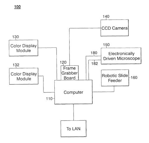
Figure 1 illustrates, in block diagram form, an embodiment of the automated
genotoxicity analysis system 100 of the present invention. Genotoxicity
analysis system 100
8
CA 02536356 2005-10-25
WO 2004/097707 PCT/IB2004/000623
includes a microprocessor-based computer 110 having a frame grabber board 120,
two color
display monitors 130 and 132, a charge coupled device (CCD) camera 140, an
electronically
driven microscope 150 and a robotic slide feeder 160.
Computer 110 of Figure 1 may be any of the many known IBM-compatible
personal or server computers running any known operating system for such
computers, e.g.,
Windows XP, Windows NT Server or UNIX. In the preferred embodiment, a Transtec
1300
IBM compatible PC, operating at 1.3 GHz., having at least 128 Mbyte of
internal RAM
memory and running the Windows NT 4, Service Pack 5 operating system or
Windows 2000 is
utilized. Computer 110 executes all operating, control and image processing
software, which
will be described more fully below, for genotoxicity analysis system 100 and
is connected to
and controls the operation of all other components of gentoxicity analysis
system 100.
Computer 110 is connected to electronic miscroscope 150 and robotic slide
feeder 160 via RS-
232 serial interfaces. Computer 110 includes a frame grabber board 120 which
is preferably a
Meteor-II frame grabber utilizing Matrox MIL 6.1 or later version driver
software available
from Matrox Imaging of Dorval, Quebec. Computer 110 stores all program
software and
generated data on a local harddrive. Alternately, computer 110 may be
connected to a local
area network (LAN 200) to support data on a networked data base (not
illustrated) or to allow
access, retrieval and storage of parameter data files and other program
software located on a
seperate networked computer server (not illustrated). In the preferred
embodiment, the
executable programs, compiled from the Visual Basic and C/C + + source code
and the
generated measurement data results files are stored on a networked database
and server while
the C-language DLLs and related files reside locally on the hard drive of
computer 110.
Robotic slide feeder 160 is preferably an ES-553S robot with an SRC-320 driver
available from Seiko Epson Corporation of Japan. Robotic slide feeder 160 is
controlled and
operated by electronic commands received from computer 110 via a serial cable
170. Robotic
slide feeder 160 functions primarily to remove a current slide from a slide
rack (not illustrated)
containing a multiplicitly of slides, then place the slide onto the stage of
electronically driven
microscope 150 and then return the slide to the slide rack after the analysis
of the slide is
complete. Under the embodiment of the invention described herein, the slide
rack may include
as many as 130 glass slides containing biological material or "samples."
9
CA 02536356 2005-10-25
WO 2004/097707 PCT/IB2004/000623
Electronically driven microscope 150 of genotoxicity analysis system 100 is
preferably a Leica DM RXA/2 electronic microscope running Leica SDK driver
software,
which is manufactured and sold by Leica Microsystems AG of Wetzlar, Germany.
Electronically driven microscope 150 preferably includes the following
modules: stage, focus
drive, illumination, objectives, fluorescence cubes, diaphragms for aperture
and field,
additional magnification changer and fluorescence shutter, all of which
components are
software driven and controlable. Electronically driven microcospe 150 is
controlled and
operated by electronic commands received from computer 110 via two serial
cables 180 and
182, one each for the stage controller and for the microscope stand of
electronically driven
microscope 150.
Camera 140 is preferably an XC-003 or DXC-390 CCD camera sold by Sony
Corporation of America. Camera 140 is mounted on electronically driven
microscope 150 in
the known manner using a C-mount adapter and is utilized to grab the current
image from
electronically driven microscope 150 and send the image in analog format to
frame grabber
board 120 via serial cable 190. Camera 140 is under operational control of
computer 110 via
frame grabber 120. The analog fomatted image received from camera 140 is
digitized by
computer 110.
Genotoxitiy analysis system 100 also includes color display modules 130 and
132 connected to computer 110. Preferably, color display module 130 provides
the user
interface to the user of the genotoxicity analysis system 100 while color
display module 132
displays the current image provided by electronically driven microscope 150
or, alternatively,
the result of the image processing analysis.
Computer 110 executes software which controls the operation of genotoxicitiy
analysis system 100.
Computer 110, and any networked server that may also be utilized to control
and operate genotoxicity analysis system 100, preferably runs Microsoft NT
version 4 or
Windows 2000 operating system software. The software executed by computer 110
to control
genotoxicity analysis system 110 is created using Microsoft Visual Basic
version 6 as well as
Microsoft Visual C/C++ version 6. Annotated source code that may be utilized
to create
executable code as well as additional software and data files are attached as
the Computer
CA 02536356 2005-10-25
WO 2004/097707 PCT/IB2004/000623
Program Listing Appendix for this documents and are described in greater
detail below. One
skilled in the art can implement the presently-described embodiment of the
claimed
genotoxicity analysis system, in part, by utilizing the software source code
and related files in
the Computer Program Listing Appendix and software available from third party
providers.
Figure 2 illustrates, in logical block diagram form, a preferred embodiment of
the application software 200 which resides in computer 110 and in a networked
control server,
to control the operation of genotoxicity analysis system 100 and also
illustrates the files which
store the results of the genotoxicity analysis. Application software 200
includes main
executable programs 210, library link and DLL files 220, parameter files 230
and data results
files 240, all of which will be described in greater detail below. Robot
control program 250 is
preferably control software provided by the manufacturer of robotic slide
feeder 160.
Main executable programs 210 include DataInput.exe 252, AutoScan.exe 254
and Relocation. exe 256. DataInput. exe 252 allows a user to enter information
particular to
each slide that is to be analyzed as shown, e.g., in Figure 5 which will be
explained below.
AutoScan.exe 254 is used to initiate and provide fully automated selected
genotoxicity
screening of the slides that are identified using DataInput.exe 252.
Relocation.exe 256 is a
utility which allows a user to retrieve and manually view slides that have
been processed using
AutoScan.exe 254 in order to allow the user to visually inspect features of
the biological
material contained on the slide if necessary.
Executable programs 210 are each preferably compiled and linked to library
link
and DLL files 220 using Microsoft Visual Basic version 6Ø The source code
for each of
executable programs 210 references a respective file named "Globals.bas," each
version of
which contains the respective "main" function for each of executable programs
210, and futher
includes other modules and necessary Visual Basic forms and code to create the
various user
interface windows. Also, as explained further below, executable programs 210
and the
modules and forms associated with executable programs 210 operate by calling
library link and
DLL files 220 during operation.
The Computer Program Listing Appendix for this document includes the source
code for creating each of executable programs 210 using Microsoft Visual Basic
version 6Ø
More particularly, the Computer Program Listing Appendix includes a folder
named "VB6"
11
CA 02536356 2005-10-25
WO 2004/097707 PCT/IB2004/000623
which contains various subfolders. The subfolders named "DATAINPUT,"
"AUTOMATICSCAN" and "RELOCATION" contain the source code for creating
DataInput.exe 252, AutoScan.exe 254 and Relocation.exe 256, respectively.
The remaining subfolders in the folder labeled "VB6" in the Computer Listing
Appendix contain source code for providing additional functionality for
genotoxicity analysis
system 100. These subfolders include "SUPERUSER" which stores source code for
creating
user interfaces that allow for manual adjustment of system parameters when
necessary,
"TOOLFORMS" which stores source code for user interface modules that may be
used by
executable programs 210, "PASSWORD," which stores source code for providing
password-
protected access to genotoxicity analysis system 100 and "MODULES," which
includes source
code for calling necessary library link and DLL files 220 during operation of
genotoxicity
analysis system 100.
In addition to the source code for creating executable programs 210, the
Computer Program Listing Appendix for this document also includes source code
for creating
library link and DLL files 220 using Microsoft Visual C/C+ + version 6Ø More
praticularly, the source code for generating library link and DLL files 220 is
found in the
subfolder fabled "VC6" on the Computer Program Listing Appendix.
The subfolder labeled "AUTOO" in the folder named "VC6" contains source
code for generating a C library called "auto0" 262 which provides
functionality for facilitating
the automatic functioning of genotoxicity analysis system 100, including
autofocus control and
automatic lamp adjustment, among others. The functionality provided by auto0
262 is based
on the related functionality provided by the "micro0" 264 and "improc0" 266
DLLs which are
described in greater detail below.
The subfolder labeled "COMET" in the folder named "VC6" contains source
code for generating a C library called "comet" 268 which provdes functionality
required for
performing image analysis on slides being analyzed for the comet assay.
The subfolder labeled "GENERALO" in the folder named "VC6" contains
source code for generating a C library called "general0" 270 which provides
functionality for
general purpose tools, including input and output functionality and graphic
display routines.
12
CA 02536356 2005-10-25
WO 2004/097707 PCT/IB2004/000623
The subfolder labeled "IMPROCO" contains source code for generating a C
library called "improc0" 266 which provides interface functionality for the
library of functions
associated with the Matrox driver software of frame grabber board 120. These
include
functions relating to general image processing.
The subfolder labeled "METFIN" contains source code for generating a C
library called "metfm" 272 which provides functionality required for
performing image
analysis on slides that are being analyzed for the metaphase finding
application.
The subfolder labeled "MICROO" contains source code for generating a C
library called "micro0" 264 which provides interface and control functionality
associated with
the Leica SDK driver software for electronically driven microscope 150.
The subfolder labeled "MNTINVIVO" contains source code for generating a C
library called "MNTinvivo" 274 which provides functionality required for
performing image
analysis on slides that are being analyzed for the micronucleus test in vivo
application.
The subfolder labeled "NNETO" contains source code for generating a C library
called "nnet0" 276 which provides functionality required for pattern
classification through
prediction using neural networks, e.g., the backpropagation algorithm, for the
micronucleus
test in vitro.
The subfolder labeled "RELOCO" contains source code for generating a C
library called "reloc0" 278 which provides functionality for object retrieval
within
Relocation.exe 254, e.g., data input and output functionality and retrieval of
analysis results.
The subfolder labeled "ROBOO" contains source code for generating a C library
called "robo0" 280 which provides functionality required for communicating
with robotic slide
feeder 160.
The subfolder labeled "SCANO" contains source code for generating a C library
called "scan0" 282 which provides functionality required for facilitating an
automatic scanning
process, e.g., handling scanning mode settings, triggering the sequential
analysis of the batch
of slides to be processed and interfacing to specific application DLLs.
Additional libraries may also be included with library link and DLL files 220,
including necessary library files provided by third party vendors for
controlling operation of
the electronically driven microscope 150 and frame grabber board 120.
13
CA 02536356 2005-10-25
WO 2004/097707 PCT/IB2004/000623
The source code for certain of the above-described library link and DLL files
220
define the algorithm and image analysis processing that is conducted for the
various
screenings .
The image analysis processing for the micronucleus test in vivo uses red and
blue
camera channel information and thresholding techniques for discrimination
between polychromatic and normochromatic erythrocytes. Thereafter, gradient
and watershed
transformation for segmentation of micronucleus candidates is utilized.
Individual analysis of
segmented objects uses supervised training of patterns on the basis of
morphometric features,
as well as structural features such as "periphery percentage," "focus
deviation" and "gray
deviation. " Reference may be made to the applicable source code described
above for further
detail.
Metaphase finding utilizes differences of spectral images as the gray image
basis and
thereafter utilizes a combination of watershed transformation and "top-hat"
segmentation for
nucleus candidate segmentation. That is followed by restriction of metaphase
range on non-
nuclear regions which is followed thereafter with another application of top-
hat and watershed
segmentation. Finally, feature base metaphase candidate classification,
involving individual
parameters for chromsomal structuring, is applied. Reference may be made to
the applicable
source code described above for further detail.
Comet assay analysis involves red channel uses of fluorescence image on a
first run to
detect valid nuclei, including classification on morphometric features.
Automatic relocation of
detected nuclei for tail moment measurement and use of a sequentially
degrading thresholding
technique which involves a gradient for the pixel sum change in the image is
also utilized.
Reference may be made to the applicable source code described above for
further detail.
The micronucleus test in vitro uses all three color channel images. The image
algorithms attempt segmentation of valid nuclei and cytoplasm range, and then
detect
micronucleus candidates using a combination of gradient, top-hat and
thresholding
segmentation. Final classification uses an off line trained backpropagational
neural network
for predicting the probability of a true micronucleus. Reference may be made
to the applicable
source code described above for further detail.
14
CA 02536356 2005-10-25
WO 2004/097707 PCT/IB2004/000623
Continuing with Figure 2, application software 200 further includes parameter
files 230 which store information about the proper settings for the operation
of electronically
driven microscope 150 and the image analysis software operating on
genotoxicity analysis
system 100, depending upon the particular analysis being conducted. Each of
the parameters
and adjustments varies depending on the genotoxicity test to be conducted and
is set
automatically by designating the particular test.
Parameter, files 230 include the following files:
- "cometpar.txt" 290 - contains parameters for the configuration of the
image analysis algorithms used for the Comet assay application;
- "metfmpar.txt" 292 - contains parameters for the configuration of the
image analysis algorithms used for the metaphase finding application;
- "mntinvivopar.txt" 294, contains parameters for the configuration of
the image analysis algorithms used for the micronucleus test in vivo
application; and
- "molymntpar.txt" 295 contains parameters for the configuration of the
image analysis algorithms used for the micronucleus test in vitro application.
Parameter files 230 further include a file called "focus std.txt" 284 which
contains parameter data that controls the automatic focus features of
electronically driven
microscope 150 in connection with the autofocus execution for Datainput.exe
252 and
AutoScan.exe 256. Parameter file 230 called "focus reloc.txt" 286 generally
contains the
same parameter definitions as "focus std.txt" 284, but is more refined to
allow for autofocus
performance that is better suited for operation under Relocation.exe 254.
Parameter file 230
labeled "scanref.txt" 288 contains parameter data that is used for the
configuration of
electronically operated microscope 150 depending on the selected application.
Such
configuration includes automatic adjustment of optical modules of the
microscope, and setting
general parameters referring to the scanning process of the application.
Also, the parameter file 230 called "roboplace.txt" 296 contains parameter
data
to control the initialization and placement of robotic slide feeder 160. These
parameters
include x,y positioning and speed.
Each of "focus std.txt" and "focus reloc.txt," are particularized for the
screening test being performed, i.e., there exists a "focus std.txt" and
"focus reloc.txt" for
CA 02536356 2005-10-25
WO 2004/097707 PCT/IB2004/000623
each of the in vivo micronucleus test, in vitro micronucleus test, comet assay
or in vitro
metaphase fording. Computer Program Listing Appendix stores the parameter
files 230 for
each screening type in respective file folders.
More particularly, Computer Program Listing Appendix includes a folder
named "Applications" which includes subfolders labeled "COMETASSAY" containing
the
above described parameter files 230 used for comet assay analysis. Similarly,
the subfolder
called "METFIN" contains the above described parameter files for metaphase
finding analysis.
The subfolder called "MNTINVIVO" contains the above described parameter files
for in vivo
micronucleus test analysis.
The subfolder called "MOLYMNT" contains the above described parameter
files for in vitro micronucleus test analysis. The "MOLMNT" subfolder further
includes a file
called "p21h9.net" and includes parameters for the neural network pattern
prediction and
classification utilized for the in vitro micronucleus test analysis.
In a preferred embodiment, "robiasaxt," which holds system specific
information for the application in general for genotoxiciy analysis system 100
and
"roboplace.txt" 296, which contains parameters for use by robotic slide feeder
160 during
initialization, reside locally on the hard drive of computer 110 while the
remaining programs
and files reside on a networked server connected to computer 110.
In addition to the above-described parameter data files, calibration files
containing "shadimages", including "shadref black" and "shadref whitbl", are
referenced by
the executable programs 210. One skilled in the art may generate these files
to provide
calibration for shading correction. Calibration files are particular to each
screening
application. The calibration files are preferably stored in a subdirectory
that is parallel to the
respective subdirectories containing the parameter data.
Application software 200 of Figure 2 further includes data results files 240
which are generated and modified by executable programs 210.
There are three types of data results files 240 having the following forms:
(1) < < path > > scanresults/ < study > / < experiment > / < slidename > .txt;
(2) < < path > > individualdata/ < study > / < experiment > / < slidename >
.txt;
and
16
CA 02536356 2005-10-25
WO 2004/097707 PCT/IB2004/000623
(3) < < path > > slidedata/slidedata < rackposition > .txt
In the above-listed file formats for data results files 240, < < path > >
indicates the
preliminary file path of the directory containing the file at issue. This part
of the path may
vary depending upon how the file structure of the overall operational software
is configured.
"scanresults," individualdata" and "slidedata" represent respective subfolder
names for the
files. < study > represents a placeholder for the study name coding the
toxicological testing of
a certain test compound and is correlated with a unique "study name" , <
experiment >
represents a placeholder for a particular experiment in the context of the
selected study.
Experiments belonging to a specific study can vary with respect to treatment
time or the
absence or presence of the metabolic activation of cells, or sampling time
after treatment of
animals. Generally, it specifies the "experimental" conditions for the same
test compound of
interest. < slidename > represents a placeholder for the identity of a
particular slide and
< rackposition > represents a placeholder for a particular position of a slide
in a rack.
The operation of genotoxicity analysis system 100 will now be described with
reference to the flow chart of Figure 3 and the exemplary screen interfaces of
genotoxicitiy
analysis system 100 illustrated in Figures 4 through 14.
At step 302 of the process of Figure 3, the user selects one of DataInput.exe,
AutoScan.exe and Relocation.exe for execution from the main display screen of
color display
monitor 130. Each application is preferably represented as an application or
shortcut icon on
the main display screen of the Windows NT platform. The user may select the
desired
program by double clicking the corresponding icon in the known manner.
If the user desires to enter information for each slide that is to be
analyzed, the
user selects the icon representing DataInput.exe for execution at step 302. As
a result, the
process proceeds to step 304 where the form illustrated in Figure 4 is
displayed to the user.
Using the form of Figure 4, the user specifies the current application, i.e.,
the analysis that is
to be performed, by selecting a unique path to which the slide data will be
written. Thus,
selection of the path also designates the analysis that will be performed,
i.e., comet assay,
micronucleus test in vivo, micronucleus test in vitro or metaphase finding.
The form of Figure
4 is created from the source code found in the file called "frmInit.frm" in
the
17
CA 02536356 2005-10-25
WO 2004/097707 PCT/IB2004/000623
VB6/TOOLFORMS subdirectory of the Computer Program Listing Appendix for this
document.
The process then moves to step 306 where the form illustrated in Figure 5 is
displayed to the user. Using this form, the user enters data for identifying
each slide that is to
be processed. The identification string for each slide consists of a study
name (col. 501),
followed by experiment name (col. 502) and a slide code (col. 503), each of
which may utilize
numerals or characters. It is noted that the exemplary slide codes 503
presented in figure 5 are
appended by "a" and "b. " In the presently disclosed embodiment of the present
invention, two
samples of biological material may be included on each slide, one designated
by "a", the other
by "b. " The precision provided by the components of genotoxicity analysis
system 100 in
combination with application software 200 allows for this efficient use of
slide space which
effectively doubles slide capacity for screening.
For slides sharing the same study and experiment code, a common folder for
resulting storage will be created. The form of Figure 5 is created from source
code found in
the file called "frmSlides.frm" in the VB6/DATAINPUT subdirectory of the
Computer
Program Listing Appendix for this document.
At step 308, the user accepts the settings entered at step 306 by pressing the
"Accept settings" button 506 of the form of Figure 5, at which point the
system ends operation
of DataInput.exe, creates all necessary folders (for studies and experiments)
and data files and
returns to step 302 of Figure 3.
Alternately, at step 310, the user may select any of the respective detail
buttons
(see column 504 of the form of Figure 5) for each slide to adjust specific
parameters relating to
each slide. Figure 6 represents the form presented to the user for adjusting
the parameters for
a particular slide. The form of Figure 6 is created from the source code found
in the file called
frmSlideparam.frm in the VB6/DATAINPUT subdirectory of the Computer Program
Listing
Appendix for this document.
Among the various parameters that the form of Figure 6 allows a user to
control
is threshold adjustment (button 602) and microscope adjustment (button 604).
The form for providing the user the ability to adjust the threshold settings
for
the particular slide is illustrated in Figure 7. This form is created from the
source code found
18
CA 02536356 2005-10-25
WO 2004/097707 PCT/IB2004/000623
in the file called "frmInterThresh.frm" in the VB6/TOOLFORMS subdirectory of
the
Computer Program Listing Appendix for this document. The form for providing
the user the
ability to adjust microscope parameters for the particular slide at issue is
illustrated in Fig. 8.
This form is created from source code found in the file called
"frmAdjustMicro. frm" in the
VB6/TOOLFORMS subdirectory of the Computer Program Listing Appendix for this
document.
Once the user is satisfied with the adjustments made to the particular slides,
the
user may select the "acc. Settings for ALL slides" button 606 of the form of
Figure 6 which
will set these parameters for all previously identified slides that have valid
slide code entries in
the form of Figure 5. Alternatively, the user may select the "acc. settings
for CURRENT
slide" (button 608) which saves parameter settings only for the currently
selected slide.
Control is then returned to the form of Figure 5 (step 306).
Returning now to step 302 of the process illustrated in Figure 3, if the user
selects the icon to initiate execution of AutoScan.exe, the process moves to
step 312 where the
form illustrated in Figure 8a is presented to the user. Here, the user selects
the genotoxicity
test that will be processed, i.e., one of the comet assay, micronucleus in
vivo, micronucleus in
vitro or metaphase finding analyis, by selecting the respective subdirectory
illustrated in
window 802 in the form of Figure 8a. The form of Figure 8a is created from
source code
found in the file called "frmInit.frm" in the VB6/TOOLFORMS subdirectory of
the Computer
Program Listing Appendix for this document.
The process then proceeds to step 314 where the form of Figure 9 is presented
to the user. The form of Figure 9 allows the user to select the scanning
options for the
genotxicity analysis to be performed as was specified' at step 312 using the
form of Figure 8.
The options presented by the form of Figure 9 include: (1) scanning the slides
without display
(button 902), meaning that no intermediate image display will be presented to
the user during
analysis of the slides; (2) scanning the slides with display (button 904),
meaning that the most
important intermediate image processing results will be displayed during
analysis without
requiring user interaction to continue analysis; (3) scanning the slides with
Testl level (button
906), meaning that several intermediate image processing steps are performed
and the process
is then halted until the user presses a key to continue automatic analysis;
and (4) scanning the
19
CA 02536356 2005-10-25
WO 2004/097707 PCT/IB2004/000623
slides with Test2 level (button 908), which results in operation similar to
that of button 906
except that detection results are not displayed. This last mode is utilized to
validate operation
of the application where a user performs manual analysis of a slide in
parallel with automated
analysis in the same image fields. Finally, the user may press button 910 for
scanning the
slides with autofocus test, which processes the slides while presenting a
graphical display of
the autofocus results, e.g., contrast curve, for each slide.
The user may also abort running the analysis by pressing the exit button 912.
If the user does not abort the automatic scanning, the process proceeds to
step
316 and the automatic scanning is executed by referencing the applicable
library link and DLL
files 220 and parameter files 230 of application software 200 for the specific
type of analysis
being performed. The form of Figure 9 is created from source code found in the
file called
"frmMain.frm" in the VB6/AUTOMATICSCAN subdirectory of the Computer Program
Listing Appendix for this document.
When the automatic scanning is complete and all results data has been written
and stored, the process returns to step 302 of Figure 3.
If at step 302, the user executes Relocations.exe, the process of Figure 3
proceeds to step 318 where the form of Figure 10 is presented to the user.
Using the form of
Figure 10, the user selects the genotoxicity test for which results are to be
reviewed, i.e., one
of the comet assay, micronucleus in vivo, micronucleus in vitro or metaphase
finding analyis,
by selecting the respective subdirectory illustrated in window 1002 in the
form of Figure 10.
The form of Figure 10 is created from the source code found in the file called
"frmInit.frm" in
the VB6/TOOLFORMS subdirectory of the Computer Program Listing Appendix for
this
document.
The process then proceeds to step 320 where the user is presented with forms
to
select a specific slide to be reviewed. More particularly, the user is
presented with the forms
illustrated in Figures 11 and 12. In the form of Figure 11, the user selects
the particular study
containing the slide by selecting the appropriate subdirectory labeled with
the appropriate study
and experiment name (see window 1102). Using the form of Figure 12, the user
identifies the
particular slide by selecting the file containing the slide data (see window
1202). The form of
Figure 11 is created from source code found in the file called "frmMain. frm"
in the
CA 02536356 2005-10-25
WO 2004/097707 PCT/IB2004/000623
VB6/RELOCATION subdirectory of the Computer Program Listing Appendix for this
document. The form of Figure 12 is created using a standard Visual Basic
CommonDialog
user interface object.
Next, at step 322, the user is presented with the form of Figure 13 which
includes a display (window 1302) of the scanning results associated with the
slide specified
using the form of Figure 12. The form of Figure 13 is similar to that of
Figure 11 except that it
now includes, in window 1302, the most relevant data that had been acquired
during automatic
slide analysis, such as the number of detected objects, number of scanned
fields, error codes,
and other application specific information for the slide under review.
The user may exit Relocation.exe by clicking button 1304 of the form of Figure
13 (step 324) at which point the process of Figure 3 returns to step 302.
Alternatively, the user may select button 1306 of the form of Figure 13
causing
the process of Figure 3 to move to step 326 at which point the user is
presented with the form
of Figure 14. The form of Figure 14 allows a user to retrieve the objects that
had been
detected during the automatic scanning process. For this purpose, one can move
from one
object to another (and then back again) using the arrow buttons 1402 and 1404.
Using the
additional controls presented in the form of Figure 14, each object's
coordinates, which had
been stored during scanning, and the current live image showing the object are
displayed on
color display screens 130 and 132 for visual inspection. The user may operate
the right or left
mouse button to flag an object under observation and discard an object as a
valid micronucleus
(by using the left mouse button) or accept an object as a valid micronucleus
(using the right
mouse button). By moving from the first detected object to the last for each
slide, the user can
assign the proper label (i.e., "accept" or "reject") to each object and,
therefore, adjust the
result of automatic scanning through supervised visual inspection. The
corrected result for the
cuirent slide, i.e. the number of micronuclei for micronucleus application, or
number of
metaphases for metaphase finding application, will be stored after exiting the
form of Figure
14. The other options present in the form in Figure 14 support the adjustment
of the current
image, e.g., microscope and focus, and support image analysis for other
objects of interest in
order to confirm proper performance of the the algorithms utilized for image
analysis.
21
CA 02536356 2005-10-25
WO 2004/097707 PCT/IB2004/000623
The form of Figure 14 is created from source code found in the file called
"frmRelocation.frm" in the VB6/RELOCATION subdirectory of the Computer Program
Listing Appendix for this document.
Thus, it is seen by the above, that by creating software code which can
facilitate
different types of genotoxicity screening and which references parameter data
files respectively
configured for each of various genotoxicity tests, the genotoxicity analysis
system of the
present invention provides a flexible and easy to use platform for performing
various
genotoxicity screenings with minimal user interaction. Depending upon the type
of screenings
being performed, no manual microscope module adaptation is necessary between
screening
runs for different analysis testing. In the case of comet assay screening, a
manual change to
incident illumination to support fluorescence staining in comet assay analysis
and then back to
transmitted light illumination for other genotoxicity screenings may be
necessary. Moreover,
as described above, the genotoxicity analysis system of the present invention
allows interactive
pattern control to permit a user to manually perform artifact rejection for
objects wrongly
classified during automatic scanning.
In accordance with 37 C.F.R. 1.52 (e), the name, respective creation date and
size (in bytes), of each file contained on the CD-ROM of the Computer Program
Listing
Appendix are listed in Figures 15a - 15. For ease of reference, the file names
are listed as they
appear in the directory structure of the Computer Program Listing Appendix.
22