Note: Descriptions are shown in the official language in which they were submitted.
CA 02536577 2006-02-21
WO 2005/021063 PCT/US2004/028078
DESCRIPTION
MEDICAL DEVICE FOR REDUCTION OF PRESSURE EFFECTS OF CARDIAC
TRICUSPID VALVE REGURGITATION
Field of the Inyention
The present invention relates generally to scented venous valves and, more
particularly, to stented valve bioprostheses with fixation means and methods
for reduction
of pressure effects of cardiac tricuspid valve regurgitation.
Background of the Invention
Among the quadruped heart valves in a human body, the tricuspid valve
separates
the right atrium (upper chamber) from the right ventricle (lower chamber), and
channels
the venous blood return to the heart on its way to the lungs. When the venous
blood is
impelled to the lung arteries, this tricuspid valve closes to block the blood
return from
backflowing to the atrium and thus provides efficiency to the ejection of
blood from the
right ventricle that directs the, flow towards the lung. In instances where
the tricuspid
valve is unable to close properly, the pumping pressure of the ventricle can
be transmitted
in reverse to the atrium and subsequently to the vena cause. Typically, the
superior vena
cava functions to bring blood to the heart from the head and the inferior vena
cava
functions to bring blood to the heart from the liver and other parts of the
body (kidneys,
gut, legs) that are located below the heart. This pressure can have
deleterious effects on
the work of the heart and circulatory system. The device herein described
provides means
of reduction or total nullification of the effects of pressure on the channels
of venous
return to the heart. '
The tricuspid heart valve has an area close to 10 square centimeters, and a
circumference approaching 12 centimeters. As the name implies it has three
cusps or
leaflets that separate to open the valve and allow the venous return from the
body to the
heart to enter the pumping chamber or right ventricle that redirects the flow
towards the
lung where venous blood is oxygenated and transformed into arterial blood to
supply all
tissues of the body. During the pumping action, the tricuspid valve closes to
impede
retrograde flow into the right atrium.
Acquired disease of the tricuspid valve is much less common than that of the
other
valves of the heart; this is a reflection of the lower pressures that are
experienced by the
CA 02536577 2006-02-21
WO 2005/021063 PCT/US2004/028078
2
right chambers of the heart, and thus, the valves of the right side of the
heart function
generally under less stresses than its left side counterparts. Disease can
affect the tricuspid
valve mostly in two forms, 1) as tricuspid valve stenosis, a restriction of
the opening of the
valve, most likely of rheumatic origin, and 2) as tricuspid valve
regurgitation or
incompetence, generally due to any disease process that causes alterations in
the tricuspid
valve apparatus that consists of leaflets, chords, tendinous material that
join the leaflet to
the muscle of the right side of the heart, or the annulus (the ring of tissue
where the leaflets
join the atrium). In the latter, the valve is unable to close completely thus
allowing
retrograde flow or regurgitation from the ventricle into the atrium.
A small degree of tricuspid regurgitation is found in normal hearts and the
prevalence increases with age. Physiologically, the regurgitation is seen as a
jet whose
velocity is proportional to the pressure differential between the right
ventricle and the right
atrium. Tricuspid regurgitation (TR) alone may be well tolerated. However,
patients
suffering from severe TR are troubled with swelling of the legs, pulsations of
the jugular
vein pulse at the neck due to reverse flow and pressure into the superior vena
cava. Other
problems associated with severe TR include liver congestion due to reverse
pressure to the
inferior vena cava and the liver veins, and fatigue and general malaise
because of
decreased pumping of blood through the heart (that is, decreased cardiac
output), that may
progress to cardiac cirrhosis and liver dysfunction with prolonged hepatic
congestion.
Furthermore, high venous pressure may contribute to renal dysfunction and
other
symptoms of abdominal bloating. All these findings are dependent on the
severity of
tricuspid regurgitation and pulmonary hypertension. Often the end effect is
right heart
failure.
Tricuspid regurgitation can be alleviated or eliminated by surgical means,
either by
replacement of the total valve apparatus with an artificially fabricated
replacement
tricuspid heart valve, or by constriction of the valve ring with means of an
annular
remodeling ring (annuloplasty ring). The tricuspid valve repair is not always
100%
effective in eliminating the TR, as it has been found in some instances that
patients (up to
about 15%) who have undergone tricuspid valve annuloplasty may leave the
hospital with
moderate to severe TR and the tricuspid dysfunction rate may steadily increase
to about
30-50%. If surgery is impossible to perform, i.e., if the patient is deemed
inoperable or
operable only at a too high surgical risk, an alternative possibility is to
treat the patient
with a stented valvular device and percutaneous means of device delivery for
protecting
the upper and/or lower body from high venous pressures.
CA 02536577 2006-02-21
WO 2005/021063 PCT/US2004/028078
.3
U.S. Pat. No. 6,503,272 issued on January 7, 2003, entire contents of which
are
incorporated herein by reference, discloses an artificial venous valve which
incorporates a
stent having one or more of the elements comprising its frame deformed
inwardly towards
its center and a biocompatible fabric attached to the one or more elements
utilized to
replace or supplement incompetent or damaged venous valves.
U.S. Pat. No. 5,855,601 issued on January 5, 1999, entire contents of which
are
incorporated herein by reference, discloses an artificial venous valve
comprising a tubular
valve segment containing venous valve means and at least one self expanding,
cylindrical
stmt member having a plurality of barbs extending from the outer surface of
the stmt
member to engage the natural tissue of the site to hold the valve in place
after
implantation.
U.S. Pat. No. 6,299,637 issued on October 9, 2001, entire contents of which
are
incorporated herein by reference, discloses a self expandable prosthetic
venous valve
comprising a tubular wire support, expandable from a first reduced diameter to
a second
enlarged diameter, and at least one leaflet pivotably positioned in the flow
path for
permitting flow in a forward direction and resisting flow in a reverse
direction.
U.S. Pat. No. 5,824,061 issued on October 20, 1998, entire contents of which
are
incorporated herein by reference, discloses an endovascular venous valve
prosthesis
comprising an endovascular stmt assembly including a stmt having a generally
cylindrical
body with a hollow bore extending longitudinally therethrough and first and
second
support struts formed on opposite sides of the outflow end of the cylindrical
body and
extending generally longitudinally therefrom; and a preserved segment of vein
having an
outer wall and a venous valve positioned therein, the valve having two
leaflets extending
generally longitudinally within the segment of vein with lateral edges
adjacent the outer
wall.
U.S. Pat. No. 5,607,465 issued on March 4, 1997, entire contents of which are
incorporated herein by reference, discloses a valve for use in a blood vessel
having a bent
flexible wire mesh with elasticity and plasticity so as to be collapsible and
implantable
remotely at a desired site and a monocusp sail-like valuing element mounted
onto it.
U.S. Pat. No. 5,997,573 issued on December 7, 1999, entire contents of which
are
incorporated herein by reference, discloses a dilation restrictor apparatus
for limiting the
extent to which a blood vessel may dilate adjacent to a point whereat a cut
end of the
blood vessel has been anastomosed to a venous valve implant, the dilation
restrictor
CA 02536577 2006-02-21
WO 2005/021063 PCT/US2004/028078
4
apparatus comprising an elongate tubular body having a hollow bore containing
a plurality
of apertures formed therein to permit passage of fluid therethrough.
U.S. Pat. No. 6,383,193 issued on May 7, 2002, entire contents of which are
incorporated herein by reference, discloses a delivery system fore the
percutaneous
insertion of a self expanding vena cave filter device being formed with a
length along a
longitudinal filter axis, the system comprising constraining the filter in a
compact
condition within an elongated, radially flexible and axially stiff tubular
member and a
displacement member attached to the tubular member for displacing the filter
from the
segment thereby to deploy the filter.
None of the above-referenced prior art discloses means for protecting the
upper
body and/or lower body of a patient from spiked or elevated venous pressure
resulting
from cardiac tricuspid valve regurgitation.
Co-pending patent application Ser. No. 10/418,677, filed on April 17, 2003,
entire
contents of which are incorporated herein by reference, discloses an elongate
valve stmt
comprising a first end, a middle section, and an opposite second end that is
connected to
the first end with at least one elongate connecting member, a first stent
member disposed
at and secured to the first end, the first stmt member comprising a first
support structure
and a first tissue valve, and a second stmt member disposed at and secured to
the second
end, the second stmt member comprising a second support structure and a second
tissue
valve.
Another co-pending patent application Ser. No. 10/418,663, filed on April 17,
2003, entire contents of which are incorporated herein by reference, discloses
a method of
protecting an upper body and a lower body of a patient from high venous
pressures
comprising implanting a first valve at a superior vena cave and a second valve
at an
inferior vena cave, wherein the first and second valves are configured to
permit blood flow
towards a right atrium of the patient and prevent blood flow in an opposite
direction.
However, means for anchoring the device has not been fully disclosed.
Therefore, it is one preferred object to provide a method of protecting an
upper
body and/or a lower body of a patient from high venous pressures comprising
implanting
an elongate valve stmt having a valued stmt member placed at a superior vena
cave
and/or at an inferior vena cave, wherein the stmt member is equipped with
anchoring
means for securely anchoring the device at an appropriate vena cave location.
It is another
preferred object to provide a valve stmt device with a venous filtering
capability.
CA 02536577 2006-02-21
WO 2005/021063 PCT/US2004/028078
Summary of the Invention
In general, it is one object of the present invention to provide a stented
valve
bioprosthesis and methods for reduction of pressure effects of cardiac
tricuspid valve
regurgitation.
5 In one aspect of the invention, it is provided an elongate valve stmt
comprising a
stmt member, the stmt member comprising a support structure that is
collapsible and
expandable and a tissue valve, wherein the tissue valve is configured to
permit fluid flow
in one direction and prevent fluid flow in an opposite direction and means for
anchoring
the stmt member onto surrounding tissue of a blood vessel.
In another aspect of the invention, it is provided an elongate valve stmt
comprising
a stmt member, the stmt member comprising a support structure and a tissue
valve,
wherein the tissue valve is configured to permit fluid flow in one direction
and prevent
fluid flow in an opposite direction, and means for filtering the fluid of a
blood vessel. In
one embodiment, the blood vessel is a vein, a superior vena cava or an
inferior vena cava.
In another embodiment, a filter member is mounted at an upstream side of the
stent
member.
In some aspect of the invention, it is provided a method of protecting an
upper or a
lower body of a patient from high venous pressures comprising: providing an
elongate
valve stent, wherein the stmt comprises a stmt member with a tissue valve
secured to a
support structure, wherein the support structure is collapsibly expandable,
and anchoring
means for anchoring the stmt member onto surrounding tissue of a vena cava;
passing the
elongate valve stmt through a blood vessel with the support structure in a
collapsed
position; deploying the stmt to an inferior vena cava or a superior vena cava
with the
support structure in an expanded shape; and securing the stmt by anchoring the
stmt
member onto the surrounding tissue of either the superior vena cava or the
inferior vena
cava with the anchoring means.
In a preferred aspect of the invention, at least a portion of the elongate
valve stent
is coated with a therapeutic agent, wherein the therapeutic agent is selected
from a group
consisting of anticoagulants, antithrombogenic agents, anti-proliferative
agents, anti-
inflammatory agents, antibiotics, stem cells, growth factors, angiogenesis
agents, anti-
angiogenesis agents, and statins.
CA 02536577 2006-02-21
WO 2005/021063 PCT/US2004/028078
6
Brief Description of the Drawings
Additional objects and features of the present invention will become more
apparent
and the invention itself will be best understood from the following Detailed
Description of
Exemplary Embodiments, when read with reference to the accompanying drawings.
FIG. 1 is a front view of a stmt member of an elongate valve stent according
to the
principles of the present invention.
FIG. 2 is a side view of the stented valve of FIG. 1.
FIG. 3 is a cross-sectional view of the stmt strut, section I-I, of the
stented valve in
FIG. 1.
FIG. 4 is a preferred embodiment of an elongate valve stmt with anchoring
means
in accordance with the principles of the present invention.
FIG. 5 is another preferred embodiment of an elongate valve stent with
filtering
means in accordance with the principles of the present invention.
FIG. 6 shows a delivery apparatus with an elongate valve stent at a collapsed
position during a delivery phase.
FIG. 7 shows a delivery apparatus with an elongate valve stmt at a partially
expanded position during a positioning phase.
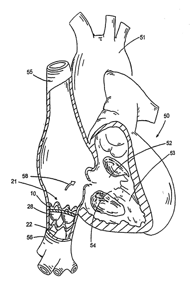
FIG. 8 is an illustrated procedure of implanting an elongate valve stmt having
anchoring means, wherein a stmt member with a tissue valve is placed at the
inferior vena
cava configured to permit blood flow towards the right atrium of a patient.
Detailed Description of the Exemplary Embodiments
The preferred embodiments of the present invention described below relate
particularly to.venous valve bioprostheses and methods for reduction of
pressure effects of
cardiac tricuspid valve regurgitation. While the description sets forth
various embodiment
specific details, it will be appreciated that the description is illustrative
only and should not
be construed in any way as limiting the invention. Furthermore, various
applications of
the invention, and modifications thereto, which may occur to those who are
skilled in the
art, are also encompassed by the general concepts described below.
CA 02536577 2006-02-21
WO 2005/021063 PCT/US2004/028078
7
A stented valve or valve stmt is a device to be placed inside a channel of the
body
that allows fluid flow in one direction and prevents fluid flow in an opposite
direction. In
a normal person, the superior vena cava functions to bring blood to the heart
from the head
and the inferior vena cava functions to bring blood to the heart from the
liver and other
parts of the body (kidneys, gut, legs) that are located below the heart.
In instances where the tricuspid valve (54 in FIG. 8) is unable to close
properly, the
pumping pressure of the ventricle 53 can be transmitted in reverse to the
atrium 52 and
subsequently to the vena cavae 55, 56. This pressure can have deleterious
effects on the
work of the heart and circulatory system. It is one aspect of the invention to
provide a
device and methods enabling reduction or total nullification of the effects of
elevated
pressure on the channels of venous return to the heart.
FIG. 1 shows a front view of a stmt member 10 of an elongate valve stmt while
FIG. 2 shows its side view according to the principles of the present
invention. Some
aspects of the invention relate to an elongate valve stmt (21 in FIG. 4)
comprising a stem
member 10, the stmt member comprising a support structure 26 and a tissue
valve 28,
wherein the tissue valve is configured to permit fluid flow in one direction
and prevent
fluid flow in an opposite direction, and means 29 for anchoring the stmt
member onto
surrounding tissue of a blood vessel, such as a vein or a vena cava.
The stmt member 10 comprises a tissue valve that is secured to a support
structure
26, wherein the support structure is collapsibly expandable (that is,
collapsible and
expandable). The tissue valve comprises at least one leaflet 13 securely
attached to an
annular base 12. The tissue valve is configured to permit fluid flow in a
first direction (as
shown by the arrow 18) and prevent fluid flow in an opposite direction. When
the fluid
flows in the first direction, the leaflet 13 is open having a flow-through
opening 14.
In one embodiment, the support structure 26 of the stmt member 10 is self
expandable out of a delivery apparatus 31. In one embodiment of operations,
the stmt is
compressed radially to be held within the lumen of the delivery apparatus,
sheath, catheter,
applicator, or cannula. Upon delivery out of the apparatus 31, the stmt self
expands to its
pre-compressed state. The stmt is typically made of a material selected from a
group
consisting of stainless steel, Nitinol, plastics or the like, particularly the
shape-member
material with flexibility and strength. In another embodiment, the stmt member
10 of the
valve stmt 21 is expandable by an inflatable balloon, which is well known to
an ordinary
artisan who is skilled in the art.
CA 02536577 2006-02-21
WO 2005/021063 PCT/US2004/028078
8
In still another embodiment the support structure 26 is made of a shape-memory
material having a first shape transition temperature of between about
30°C and 45°C and a
second shape transition temperature of between about 25°C and -
20°C, preferably between
about 5°C and -10°C. In operations, the stmt is collapsibly
deformed to a small diameter
and held at about or below 5°C, preferably between about 5°C and
-10°C. The deformed
stmt is then inserted within a delivery apparatus 31. During a delivery phase,
the stmt is
maintained at below the second shape transition temperature by flushing or
contacting
with super-cooled saline. At a desired location, the stmt is pushed out of the
sheath of the
delivery apparatus. Upon reaching the first shape transition temperature, the
stent expands
to lock itself in position.
The use of shape memory alloys or intermetallics and, specifically, Nitinol in
the
construction of medical devices is well known. U.S. Pat. No. 6,451,025 issued
on
September 17, 2002, entire contents of which are incorporated herein by
reference,
discloses hysteresis behavior of Nitinol to generate shape change or force at
or around
constant body temperature by forming the device to the final shape desired,
straining the
device in a direction which tends to facilitate placement into the body,
restraining the
device in this strained shape during insertion into or placement near the
body, then
releasing all or part of the device such that it returns or tends to return to
the desired shape
with temperature activation.
In one aspect, the first valve stmt 21 is delivered to the superior vena cava
55
endoluminally from a subclavian or femoral vein. In another aspect, the second
valve
stmt is delivered from a femoral vein or jugular vein to the inferior vena
cava 56.
The step of delivering the elongate valve stmt endoluminally is through an
incision
at a blood vessel selected from a group consisting of a jugular vein, a
femoral vein, a
subclavian vein or other veins. The stmt member is expanded from a collapsible
position
when the stem member reaches an appropriate site. In a further aspect, the
valve stmt 21
further comprises anchoring means 29 for anchoring the stent onto surrounding
tissue of
either the superior vena cava or the inferior vena cava for example, hooks,
barbs, needles,
protrusion, or the like. By way of example, U.S. Pat. No. 6,610,085, entire
contents of
which are incorporated herein by reference, discloses anchoring means that is
well known
to one who is skilled in the art.
In an alternate embodiment, the venous valve to be placed at either the
superior
vena cava or the inferior vena cava is a stentless valve. In still another
embodiment, the
venous valves are to be implanted by an open chest procedure at the superior
vena cava
CA 02536577 2006-02-21
WO 2005/021063 PCT/US2004/028078
9
and/or the inferior vena cava, wherein the valves can be either a stented
valve or a
stentless valve.
In a prefeiTed embodiment, the valve stmt 21 would deploy in. the superior
vena
cava 55 just above the right atrial junction but below the azygos vein.
Alternately, the
valve stmt would deploy in the inferior vena cava 56 just below the right
atrium 52 but
above the hepatic veins. In effect, the physiologic changes from the therapy
disclosed
herein would be to protect the upper andlor lower body from high or elevated
venous
pressures. Patients with severe tricuspid regurgitation are troubled by
ascites, peripheral
edema frequently with stasis changes in the legs, hepatic congestion, which
may progress
to cardiac cirrhosis and liver dysfunction with prolonged hepatic congestion.
Furthermore,
high venous pressure may contribute to renal dysfunction and other symptoms of
abdominal bloating. The neck vein and upper body congestion is sometimes quite
visible
in patients including the pulsatile neck veins. By placing the valve stems of
the invention,
it should protect the patient from ascites, hepatic congestion, edema and the
eventual
development of cardiac cirrhosis.
To enhance the biocompatibility of the device or improved therapy to the
surrounding tissue, it is provided that at least a portion of the stmt member
10 of the
elongate valve stmt 21 is coated with a therapeutic agent, wherein the
therapeutic agent is
selected from a group consisting of anticoagulants, antithrombogenic agents,
antiproliferative agents, anti-inflammatory agents, antibiotics, stem cells,
growth factors,
angiogenesis agents, anti-angiogenesis agents, and statins. The therapeutic
agent is to
slowly release to the tissue or blood stream at an effective amount over time.
For illustration purposes, FIG. 3 shows a cross-sectional view of the stmt
strut 17
of the support structure 26, section I-I, of the stmt member 10 in FIG. 1,
wherein a
polymer layer 16 is coated onto the periphery surface of the stmt strut 17 and
the polymer
layer 16 is loaded with the desired therapeutic agent 1 S for slow release at
an effective
amount over time to the surrounding tissue.
Many medical materials used in the treatment of cardiovascular diseases are
required to possess biocompatible and hemo-compatible properties with reduced
antigenicit. One method to treat tissue so as to render the tissue more
suitable as a
biomaterial is a process called chemical treatment. Several chemical treatment
agent and
methods have been disclosed. Among them, aldehydes (glutaraldehyde,
formaldehyde,
dialdehyde starch and the like), epoxy compounds, genipin, and their analog or
derivatives
thereof are all applicable in treating a tissue. Chemical treatment conditions
and
CA 02536577 2006-02-21
WO 2005/021063 PCT/US2004/028078
procedures to render the tissue suitable as a biomaterial depend on the
property of each
tissue and intended medical applications, wherein the conditions/procedures
are well
documented in published literature and well known to one who is skilled in the
art.
The tissue valve 28 of the stmt member 10 has at least one valve leaflet 13.
5 Sometimes, the tissue valve may have two, three or more leaflets. In some
aspect of the
present invention, the leaflet 13 is made from a pericardium, the pericardium
being
selected from a group consisting of a bovine pericardium, an equine
pericardium, a
porcine pericardium, an ovine pericardium and the like. Further, the tissue
valve is
chemically treated with a chemical treating agent selected from a group
consisting of
10 glutaraldehyde, formaldehyde, dialdehyde starch, epoxy compounds, genipin,
and mixture
thereof. In one embodiment, the tissue valve is a venous valve selected or
procured from a
group consisting of a bovine jugular vein, an equine jugular vein, a porcine
jugular vein,
and an ovine jugular vein. In another embodiment, the tissue valve is a
porcine valve.
U.S. Pat. No. 4,806,595 issued on February 21, 1989, entire contents of which
are
incorporated herein by reference, discloses a novel method for preparing
medical materials
by using epoxy compounds as chemical treatment agent for tissue, wherein the
"epoxy
compounds" include glycol diglycidyl ether, polyol polyglycidyl ether,
dicarboxylic acid
diglycidylester, the analog, and derivatives thereof.
U.S. Pat. No. 6,608,040 issued on August 19, 2003, entire contents of which
are
incorporated herein by reference, discloses a novel method for preparing
medical materials
by using genipin as chemical treatment agent for tissue.
FIG. 4 shows a preferred embodiment of an elongate valve stmt with anchoring
means 29 in accordance with the principles of the present invention. In some
aspect, it is
provided an elongate valve stmt 21 comprising a stmt member 10, the stmt
member
comprising a support structure 26 and a tissue valve 28, wherein the tissue
valve is
configured to permit fluid flow in one direction and prevent fluid flow in an
opposite
direction. The anchoring means 29 for anchoring the stmt member 10 onto
surrounding
tissue of a blood vessel comprises at least one anchoring member 22, wherein
each
anchoring member 21 comprises a proximal end 24 connected to one end of the
stmt
member 10 and a distal end with a needle or hook 23 for penetrating and
hooking into
tissue. In one preferred embodiment, the tissue valve 28 has at least one
valve leaflet 13
sized and configured to permit fluid flow in one direction (shown by an arrow
58) and
prevent fluid flow in an opposite direction.
CA 02536577 2006-02-21
WO 2005/021063 PCT/US2004/028078
11
FIG. 5 shows another preferred embodiment of an elongate valve stmt 21 with
filtering means 27 in accordance with the principles of the present invention.
Some
aspects of the invention relate to an elongate valve stent 21 comprising a
stent member 10,
the stmt member comprising a support structure 26 and a tissue valve 28,
wherein the
tissue valve is configured to permit fluid flow in one direction and prevent
fluid flow in an
opposite direction, and means 27 for filtering the fluid of a blood vessel,
wherein the blood
vessel is a superior vena cave or an inferior vena cave. In one embodiment,
the filtering
means 27 for filtering the fluid of the blood vessel comprises a filter member
mounted at
an upstream side of the stent member 10. By way of example, a filter member is
attached
at a proper attaching point on the anchoring member, for example at the
attaching points
25A, 25B, 25C, 25D, and 25E on the anchoring members 22A, 22B, 22C, 22D, and
22E,
respectively. Other types of venous filtering means are also applicable, for
example,
stainless steel Greenfield filters by Boston Scientific Corporation (Natick,
MA), bird's nest
filters by Cook, Inc. (Bloomington, IN), LGM Vena-Tech filters by B. Braun
(Evanston,
IL), and Simon nitinol filters by Medical Technologies (Woburn, MA).
The support structure 26 of the elongate valve stmt 21 is configured
collapsibly
expandable from a first collapsed position to a second expanded position,
wherein the
stent is delivered through a blood vessel with the support structure in the
collapsed
position within a delivery apparatus and the stmt is secured to a desired
valve location at
the superior and inferior vena cave with the support structure in the expanded
shape and
the anchoring means 29 is deployed. In an alternate embodiment, the elongate
valve stmt
21 with its anchoring means 29 and/or filtering means 27 can be implanted by
an open
chest procedure at the superior vena cave and the inferior vena cave.
The support structure 26 may be self expandable, expandable by an inflatable
balloon, or by other expanding means. Further, the support structure. of the
stmt member
10 is made of a shape-memory material. One preferred shape-memory material has
a first
shape transition temperature of between about 30°C and 45°C and
a second shape
transition temperature of between about 25°C and -20°C,
preferably between about S°C
and -10°C. In operations, the support structure is collapsibly deformed
to a small diameter
and held at about or below 5°C, preferably between about 5°C and
-10°C. The deformed
support structure is then inserted within a delivery apparatus. During a
delivery phase, the
support structure 26 with its mounted tissue valve 28 is maintained at below
the second
shape transition temperature by flushing or contacting with super-cooled
saline. At a
desired location, the elongate valve stmt 21 is pushed out of the lumen of the
apparatus.
CA 02536577 2006-02-21
WO 2005/021063 PCT/US2004/028078
12
Upon reaching the first shape transition temperature, the support structure 26
expands to
lock itself in position.
The support structure 26 is made of shape memory Nitinol with at least one
shape
transition temperature. In one embodiment, the stmt or the support structure
is sized and
configured to be reversibly collapsed by lowering the Nitinol temperature
below its second
shape transition temperature (for example, about 5°C and -10°C
in one case) enabling
removing the stent or the support structure from a patient percutaneously when
needed.
This is usually carried out by a retrieval apparatus by grasping the radially
deformed
device endoluminally.
FIG. 6 shows a delivery apparatus 31 with an elongate valve stmt 21 at a
collapsed
position during a delivery phase. In one embodiment, the delivery apparatus 31
is a
catheter with a catheter sheath 32 and a lumen 36, wherein a plunger 34 with
its pushing
rod 33 is used to deploy the valve stmt 21 out of the catheter distal end 35.
FIG. 7 shows a delivery apparatus 31 with an elongate valve stmt 21 at a
partially
expanded position during a positioning phase. In one embodiment as shown in
FIG. 7, the
stent member 10 of the valve stmt 21 is out of the catheter distal end 35
while a distal
hook portion of the anchoring members 22 is still within the lumen 36 of the
delivery
apparatus 31. When a practitioner continues to advance the plunger 34, the
distal hook
portion of the anchoring member 22 is deployed out of the catheter distal end
35. When
the' compressing constraint is removed from the anchoring members 22, the
anchoring
means 29 tends to recover its resilient preshape and spring outwardly enabling
the at least
one hook 23 to penetrate and hook into the surrounding tissue.
FIG. 8 shows a preferred embodiment of procedures of protecting a lower body
of
a patient from high venous pressures, the method comprising implanting an
elongate valve
stent 21 having a valued stmt member 10 suitably placed at an inferior vena
cava 56
location, wherein the stmt member 10 with a tissue valve 28 is configured to
permit blood
flow (as indicated by an arrow 58) towards a right atrium 52 of the heart 50
and prevent
blood flow in an opposite direction. In a normal patient, the oxygenated blood
is pumped
from the heart 50 through aorta 51 to the body. Similarly, an elongate valve
stent can be
implanted at a superior vena cava SS location for protecting an upper body of
a patient
from high venous pressure.
Some aspects of the invention relate to a method of protecting an upper or a
lower
body of a patient from high venous pressures comprising: (a) providing an
elongate valve
CA 02536577 2006-02-21
WO 2005/021063 PCT/US2004/028078
13
stmt, wherein the stmt comprises a stmt member with a tissue valve secured to
a support
structure, wherein the support structure is collapsibly expandable, and
anchoring means
for anchoring the stmt member onto surrounding tissue of a vena cave; (b)
passing the
elongate valve stmt through a blood vessel with the support structure in a
collapsed
position; (c) deploying the stmt to an inferior vena cave or a superior vena
cave with the
support structure in an expanded shape; and (d) securing the stmt by anchoring
the stmt
member onto the surrounding tissue of either the superior vena cave or the
inferior vena
cave with the anchoring means.
The medical device of the invention is for reduction of pressure effects of
cardiac
tricuspid valve regurgitation. The device does not treat tricuspid valve
regurgitation but
rather slows down or attempts to block the decay due to the sequels or effects
of tricuspid
valve regurgitation on the body, namely hepatic dysfunction and renal
dysfunction or
failure and the build up of fluid in the abdominal cavity and the lower body,
legs etc.
Although preferred embodiments of the invention have been described in detail,
certain variations and modifications will be apparent to those skilled in the
art, including
embodiments that do not provide all of the features and benefits described
herein.
Accordingly, the scope of the present invention is not to be limited by the
illustrations or
the foregoing descriptions thereof, but rather solely by reference to the
appended claims.