Note: Descriptions are shown in the official language in which they were submitted.
CA 02536909 2006-02-24
WO 2005/021738 PCT/US2004/028231
KIDNEY DERIVED STEM CELLS AND METHODS FOR
THEIR ISOLATION, DIFFERENTIATION AND USE
Priority of Invention
This application claims the benefit of priority under 35 U.S.C. ~ 119(e)
to U.S. Provisional Patent Application Serial No. 60/499,127, which is hereby
incorporated by reference for all purposes.
Field of the Invention
The invention relates generally to methods for isolation of kidney stem
cells, cells isolated by the methods, and therapeutic uses for those cells.
More
specifically, the invention relates to isolated kidney-derived progenitor
cells that
have the potential to differentiate to form cells of any one or all three gene
cell
layers (endoderm, mesoderm, ectoderm), as well as methods for isolating the
cells and for inducing specific differentiation of the cells isolated by the
method,
and specific markers that are present in these cells such as proteins and
transcription factors.
Back~,round of the Invention
Nephrotoxic and ischemic insults to the kidney lead to acute renal failure
that most often manifests as acute tubular necrosis (ATN). Following injury,
the
kidney undergoes a regenerative response leading to recovery of renal
function.
The cell source for regenerating tubules is poorly understood. Three possible
sources of new tubular cells are: (1) adjacent less damaged tubular cells; (2)
extra-renal cells, presumably of bone marrow origin, that home to the injured
kidney; or (3) resident renal stem cells. There is evidence to support a role
for
less damaged tubular cells. Recapitulating developmental paradigms, these
cells
dedifferentiate, proliferate, and eventually reline denuded tubules, restoring
the
structural and functional integrity of the lcidney [1-5]. Molecular events
defining
this renal regeneration have been characterized and strategies to accelerate
the
repair process tested in both experimental models and in humans [1-6].
CA 02536909 2006-02-24
WO 2005/021738 PCT/US2004/028231
The discovery of bone marrow derived stem cells that possess the ability
to differentiate into different cell lineages has led to a reexamination of
the
cellular source and processes involved in recovery from organ injury [7-14].
Bone marrow derived cells can migrate to the kidney and form tubular
epithelial
cells [15-17]. However, the contribution of extra-renal cells to the
regenerative
renal response is small. Bone marrow cells can also contribute cells to the
glomerulus in aumal models of glomerulonephritis and to the endothelium and
interstitium following kidney transplantation [18-26].
Stem cells have been found in many organs including bone marrow,
gastrointestinal mucosa, liver, brain, pancreas, prostate, and skin [27-31].
These
cells participate in the normal cell turnover of these organs and are a source
of
cells following organ injury. Clonal analysis has demonstrated that individual
cells in the adult kidney have the ability for kidney tubulogenesis, although
the
cells have not been characterized in much detail [32]. Elegant studies of
renal
development have demonstrated that single metanephric mesenchyrnal cells can
form epithelial cells of all parts of the nephron, other than the collecting
duct that
is formed from ureteric bud cells [33]. Lineage restriction of metanephric
mesenchyme occurs at later stages of development [34].
Summary of the Invention
The present invention provides an isolated multipotent renal progenitor
cell (MRPC) that is cell marlcer positive for vimentin, Oct-4, CD90 and CD44,
and negative for zona occludens, cytokeratin, SSEA-1, NCAM, CD 1 1b, CD45,
CD31, CD106 and MHC class I and II molecules. The present invention
provides an isolated MRPC that is non-embryonic and/or a non-germ cell. The
cells of the present invention described above may have the capacity to be
induced to differentiate, in vitro, ex vivo or ifz vivo, to form at least one
differentiated cell type of mesodermal, ectodermal and endodermal origin. The
cells of the present invention may have the capacity to be induced to
differentiate
into two differentiated cell types, or into all three differentiated cell
types. For
example, the cells may have the capacity to be induced to differentiate to
form
cells of at least kidney, endothelium, neuron, and liver cell type ("cells of
a
specified type" refers to all cells that make up the organ, or participate in
the
2
CA 02536909 2006-02-24
WO 2005/021738 PCT/US2004/028231
function of the organ, of interest (e.g., mesangial cells and renal tubule
cells, to
name a few, are cells of the kidney cell type). The cell may be a human cell,
rat
cell or a mouse cell. The cell may be from a fetus, newborn, child, or adult.
The
cell may also express high levels of telomerase and maintain long telomeres,
for
example, telomeres of about 12 Kb, about 16 Kb or about 23 Kb in length, after
extended in vitYO culture (for example, cells that been cultured for over 4
months
or have under undergone at least about 90 to about 160 population doublings).
The present invention also provides a composition of a population of
MRCPs described above and a culture medium that expands the MRCPs. The
culture medium may include platelet derived growth factor (PDGF-BB),
epidermal growth factor (EGF), and leukemia inhibitory factor (LIF). The cells
of the composition may also have the capacity to be differentiated to form at
least one differentiated cell type of mesodermal, ectodermal and endodermal
origin.
The present invention further provides differentiated cells obtained from
the MRPC described above, wherein the progeny cell may be a kidney, liver,
neuronal, or endothelial cell. The kidney cell may be a tubule cell.
The present invention provides an isolated transgenic MRPC, wherein
the genome of the MRPC has been altered by insertion of preselected isolated
DNA, by substitution of a segment of the cellular genome with preselected
isolated DNA, or by deletion of or inactivation of at least a portion of the
cellular
genome. This alteration may be by viral transduction, such as by insertion of
DNA by viral vector integration, or by using a DNA virus, RNA virus or
retroviral vector. Alternatively, a portion of the cellular genome of the
isolated
transgenic cell may be inactivated using an antisense nucleic acid molecule
whose sequence is complementary to the sequence of the portion of the cellular
genome to be inactivated. Further, a portion of the cellular genome may be
inactivated using a ribozyme sequence directed to the sequence of the portion
of
the cellular genome to be inactivated. Also, a portion of the cellular genome
may be inactivated using a small interfering RNA (siRNA) sequence directed to
the sequence of the portion of the cellular genome to be inactivated. The
altered
genome may contain the genetic sequence of a selectable or screenable marker
gene that is expressed so that the progenitor cell with an altered genome, or
its
3
CA 02536909 2006-02-24
WO 2005/021738 PCT/US2004/028231
progeny, can be differentiated from progenitor cells having an unaltered
genome.
For example, the marker may be a green, red, or yellow fluorescent protein,
Beta-gal, Neo, DHFRm, or hygromycin. The transgenic cell may express a gene
that can be regulated by an inducible promoter or other control mechanism to
regulate the expression of a protein, enzyme or other cell product.
The present invention provides a method for isolating MRPCs by
culturing renal cells in a medium consisting essentially of DMEM-LG, MCDB
201, insulin-transferrin-selenium (ITS), dexamethasone, ascorbic acid 2-
phosphate, penicillin, streptomycin and fetal calf serim (FCS), and with
epidermal growth factor (EGF), platelet derived growth factor (PDGF-BB) and
leukemia iWibitory factor (LIF) for about four weeks. The cells may be
cultured
for about four to six weeks, or even longer, or when most of the cell types
have
died out and the culture becomes monomorphic with spindle shaped cells. The
cells may be cultured on fibronectin, and may be maintained at a concentration
of between about 2 and 5 x 10² cells/cm2. The method may further involve
culturing the plated cells in media supplemented with growth factors. The
growth factors used may be chosen from PDGF-BB, EGF, insulin-like growth
factor (IGF), and L1F.
The present invention provides a cell differentiation solution comprising
factors that promote continued growth or differentiation of undifferentiated
MRPCs. Particularly, the invention provides the culture method and media
whereby MRPCs are derived directly from kidney tissue using a media that
supports the selective growth of these cells. For example, the medium may
consist of 60% DMEM-LG (Gibco-BRL, Grand Island, NY), 40% MCDB-201
(Sigma Chemical Co, St. Louis, MO), with 1X insulin-transferrin-selenium
(ITS), 10-9M dexamethasone (Sigma) and 10~M ascorbic acid 2-phosphate
(Sigma), 100U penicillin and 1000U streptomycin (Gibco) with 2% fetal calf
serum (FCS) (Hyclone Laboratories, Logan, UT) and with epidermal growth
factor (EGF) 10 ng/ml, platelet derived growth factor (PDGF)-BB 10 ng/m and
leukemia inhibitory factor (LIF) 10 ng/ml (all from RED Systems, Minneapolis,
MN). The cells may be grown on fibronectin (FN) (Sigma). The cells may be
maintained at a concentration of between 2 and 5 x 10² cells/cm2.
4
CA 02536909 2006-02-24
WO 2005/021738 PCT/US2004/028231
The present invention further provides a renal cell and a cultured clonal
population of manunalian MRPCs isolated according to the above-described
method.
The present invention provides a method to reconstitute the kidney of a
mammal by administering to the mammal fully allogenic MRPCs to induce
tolerance in the mammal for subsequent MRPC-derived tissue transplants or
other organ transplants.
The present invention provides a method of expanding undifferentiated
MRPCs into differentiated cells ex vivo by administering appropriate growth
factors, and growing the cells. Such growth factors may include FGF2, TGF,
LIF, VEGF, bFGF, FGF-4, hepatocyte growth factor, or a combination thereof.
The present invention also provides a differentiated cell obtained by such a
method. This differentiated cell may be an ectoderm, mesoderm or endoderm
cell. The differentiated cell may also be of the kidney, endothelium, neuron,
or
liver cell type. Additionally, the differentiated kidney cell may be a kidney
tubule cell.
The present invention provides numerous uses for the above-described
cells. For example, the invention provides a method for differentiating MRCPs
ifZ vivo by isolating a multipotent renal progenitor cell by the methods
described
above and administering the an expanded cell population to a subject resulting
in
the cell population becoming engrafted and differentiated in vivo into tissue
specific cells, such that the function of a cell or organ, defective due to
injury or
disease, is augmented, reconstituted or provided for the first time. The
tissue
specific cells may be of the kidney, endothelium, neuron or liver cell type.
Also
provided a differentiated cell obtained by this method.
The invention also provides a method of treating a subject in need thereof
by administering a therapeutically effective amount of the cells described
above
or their progeny. The MRCPs or their progeny may home to one or more organs
in the subject and engraft therein and/or thereon such that the function of
the cell
or organ, defective due to injury or disease, is augmented, reconstituted, or
provided for the first time. The progeny may have the capacity to fuuther
differentiate or they may be terminally differentiated.
5
CA 02536909 2006-02-24
WO 2005/021738 PCT/US2004/028231
The invention provides a method of using the isolated cells by
performing an ifi utero transplantation of a population of the cells to form
chimerism of cells or tissues, thereby producing human cells in prenatal or
post-
natal humans or animals following transplantation, wherein the cells produce
therapeutic enzymes, proteins, or other products in the human or animal so
that
genetic defects are corrected. The present invention also provides a method of
using the cells for gene therapy in a subject in need of therapeutic
treatment,
involving genetically altering the cells by introducing into the cell an
isolated
pre-selected DNA encoding a desired gene product, expanding the cells in
culture, and adminstering the cells to the subj ect to produce the desired
gene
product.
The present invention also provides a method of repairing damaged
tissue in a subject in need of such repair by expanding the isolated MRPCs in
culture, and administering an effective amount of the expanded cells to the
subj ect with the damaged tissue. Additionally, the invention also provides a
method of repairing damaged tissue in a subject in need of such repair by
administrating exogenous molecules to the subject to stimulate endogenous
MRPCs to proliferate and differentiate into different cell lineages of the
kidney.
For example, the present invention provides a method to induce endogenous
MRPC cells present in the kidney to proliferate and differentiate into
different
cell lineages of the kidney when stimulated by the administration of molecules
such as LIF, colony stimulating factor, or insulin-like growth factor. These
stimulated MRPCs can then contribute to the regeneration of the kidney in
diseases such as acute tubular necrosis, and non-kidney tissue in diseases
such as
cirrhosis of the liver.
The present invention provides a method of using MRPCs for inducing
an immune response to an infectious agent involving genetically altering an
expanded clonal population of multipotent renal progenitor cells in culture to
express one or more pre-selected antigenic molecules that elicit a protective
immune response against an infectious agent and adminstering to the subject an
amount of the genetically altered cells effective to induce the immune
response.
The present invention provides a method of using MRPCs to identify
genetic polymorphisms associated with physiologic abnormalities, involving
6
CA 02536909 2006-02-24
WO 2005/021738 PCT/US2004/028231
isolating the MRPCs from a statistically significant population of individuals
from whom phenotypic data can be obtained, culture expanding the MRPCs
from the statistically significant population of individuals to establish MRPC
cultures, identifying at least one genetic polymorphism in the cultured MRPCs,
inducing the cultured MRPCs to differentiate, and characterizing aberrant
metabolic processes associated with said at least one genetic polymorphism by
comparing the differentiation pattern exhibited by an MRl'C having a normal
genotype with the differentiation pattern exhibited by an MRPC having an
identified genetic polymorphism.
The present invention further provides a method for treating cancer in a
subject involving genetically altering MRPCs to express a tumoricidal protein,
an anti-angiogenic protein, or a protein that is expressed on the surface of a
tumor cell in conjunction with a protein associated with stimulation of an
immune response to antigen, and administering an effective anti-cancer amount
of the genetically altered MRPCs to the subject.
The present invention provides a method of using MRPCs to characterize
cellular responses to biologic or pharmacologic agents involving isolating
MRPCs from a statistically significant population of individuals, culture
expanding the MRPCs from the statistically significant population of
individuals
to establish a plurality of MRPC cultures, contacting the MRPC cultures with
one or more biologic or phannacologic agents, identifying one or more cellular
responses to the one or more biologic or pharmacologic agents, and comparing
the one or more cellular responses of the MRPC cultures from individuals in
the
statistically significant population.
The present invention also provides a method of using specifically
differentiated cells for therapy comprising administering the specifically
differentiated cells to a patient in need thereof. It fixrther provides for
the use of
genetically engineered MRPCs to selectively express an endogenous gene or a
transgene, and for the use of MRPCs grown ifz vivo for
transplantation/administration into an animal to treat a disease. For example,
differentiated cells derived from MRPCs can be used to treat disorders
involving
tubular, vascular, interstitial, or glomerular structures of the kidney. For
example
cells can be used to treat diseases of the glomerular basement membrane such
as
7
CA 02536909 2006-02-24
WO 2005/021738 PCT/US2004/028231
Alpons Syndrome; tubular transport disorders such as Banter syndrome,
cystinuria or nephrogenic diabetes insipidus; progressive kidney diseases of
varied etiologies such as diabetic nephropathy or glomerulonephritis; Fabry
disease, hyperoxaluria, and to accelerate recovery from acute tubular
necrosis.
The cells can be used to engraft a cell into a mammal comprising administering
autologous, allogenic or xenogenic cells, to restore or correct tissue
specific
metabolic, enzymatic, structural or other function to the mammal. The cells
can
be used to engraft a cell into a mammal, causing the differentiation iya vivo
of cell
types, and for administering the differentiated kidney progenitor cells into
the
mammal. The cells, or their ifz vitro or ifs vivo differentiated progeny, can
be
used to correct a genetic disease, degenerative disease, or cancer disease
process.
They can be used as a therapeutic to aid for example in the recovery of a
patient
from chemotherapy or radiation therapy in the treatment of cancer, in the
treatment of autoimmune disease, or to induce tolerance in the recipient.
The present invention further provides a method of gene profiling of a
MRPCs as described above, and the use of this gene profiling in a data bank.
It
also provides for the use of gene profiled MRPCs as described above in data
bases to aid in drug discovery.
The present invention further provides using MRPCs or cells that were
differentiated from MRPCs in conjunction with a Garner device to form an
artificial kidney. Suitable carrier devices are well-known in the art. For
example, the carrier device may be a hollow, fiber based device. The
differentiated MRCPs used in with the device may be a kidney cells. The
invention further provides a method for removing toxins from the blood of a
subject by contacting the blood ex vivo with isolated MRPCs which line a
hollow find, based device.
Additionally, in the methods described above, the cells may be
administered in conjunction with an acceptable matrix, e.g., a
pharmaceutically
acceptable matrix. The matrix may be biodegradable. The matrix may also
provide additional genetic material, cytokines, growth factors, or other
factors to
promote growth and differentiation of the cells. The cells may also be
encapsulated prior to administration. The encapsulated cells may be contained
within a polymer capsule.
8
CA 02536909 2006-02-24
WO 2005/021738 PCT/US2004/028231
The cells of the present invention may also be administered to a subject
by a variety of administration methods, including, localized injection,
systemic
injection, parenteral administration, oral administration, or intrauterine
injection
into an embryo. The subj ect of the methods described above may be a mammal.
The mammal may be a human.
The present invention also provides a method to identify pharmaceutical,
including biological, agents that facilitate kidney regeneration including
transfecting MRPCs with a promoter region of a gene that is activated during
the
process of nephron formation, wherein the promoter region is operably linked
to
a reporter gene, contacting the transfected cells of with a pharmaceutical
agent,
and detecting an expressed protein coded by the marker gene, wherein detection
of the protein identifies a pharmaceutical agent as one that facilitates
kidney
regeneration. The marker gene may be green, red, or yellow fluorescent
protein,
Beta-gal, Neo, DHFRm, or hygromycin.
Brief Description of the Figures
Figures lA-C. Phase contrast microscopy of (A) mouse MAPCs derived
from adult bone marrow; (B) mouse multipotent renal progenitor cells; and (C)
rat multipotent renal progenitor cells. All three cells have similar spindle
shaped
morphology.
Figures 2A-B. Phase contrast (A) and scanning electron microscopy (B)
of mouse MRPCs demonstrating condensation of cells into primitive globules.
Figures 3A-B. Immunohistochemistry of mouse MRPCs stained with
(A) FITC-labeled anti-cytokeratin antibody demonstrating cytoplasmic staining
for cytolceratin; and (B) Texas red labeled anti-ZO-1 antibody demonstrating
characteristic spickled staining along cell borders.
Figures 4A-D. Phase contrast (A and C) and same image fluorescence
microscopy (B and D) of mouse MRPCs incubated with control media (A and B)
or media containing a nephrogenic cocktail (C and D). In the presence of the
cocktail, cells aggregated and became positive for eGFP consistent with Pax-2
expression.
Figures SA-F. Rat MRPCs (A) could be induced to differentiate into
endothelium (B), neurons (C), and liver cells (D). Characteristic phase
contrast
9
CA 02536909 2006-02-24
WO 2005/021738 PCT/US2004/028231
morphology and immunohistochemistry for markers is shown as labeled (E and
F).
Figures 6A-B. Kidney from Oct-4 (3-Geo transgenic rats stained for (A)
(3-galactosidase activity (blue cells indicative of positive staining); (B) (3-
galactosidase enzyme by immunohistochemistry (brown staining indicative of
positive cells). Arrows indicate positive staining cells in the interstitial
space.
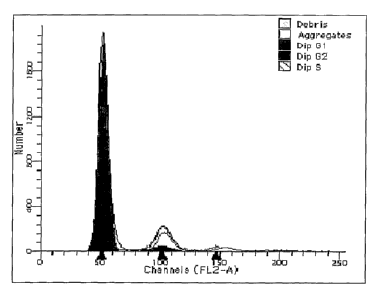
Figure 7. FACS analysis of MRPCS at 200 population doublings
demonstrating 100% diploid cells.
Figure ~. Southern blot analysis demonstrating that telomere length was
maintained after 90 and 160 population doublings.
Figure 9. Transfection and ira vitr~ differentiation of rat MRPCs. Rat
MRPCs were transfected with MSCV-eGFP retrovirus and cells with high levels
of GFP expression were selected by FAGS. These cells are referred to as
eMRCPs. As depicted in Figure 9, eGFP could be easily detected by both direct
fluorescence and with an anti-GFP antibody. eGFP transfected cells could still
be differentiated into other cell types using the appropriate selection media.
Examples of the morphology of eMRPCs differentiated into endothelial cells and
neurons are shovcnl.
Figures 10A-B. Ih. vivo differentiation following subcapsular injection.
eMRCPs were injected under the renal capsule of Fisher rats. Three weeks
later,
the kidneys were harvested and examined by confocal microscopy. Figure 10A
depicts GFP positive cellular nodules formed under the capsule at the site of
injection and included cystic like structures. Figure lOB demonstrates that
some
GFP-positive cells have been incorporated into tubules.
Figures 11A-F. Ira vivo differentiation of MRPCs following renal
ischemia/ reperfusion (regenerating kichzey following ischemia/reperfusion).
A)
Tubular cast of MRPCs; B) MRPCs lodged in glomerulus; C) Several MRPCs
present in regenerating tubule (arrow); D) A grouping of MRPC positive
tubules; E) A tubule with many MRPCs; F) Several positive cells in this
tubule,
including a cluster of cells that may be derived from an interstitial MRPC
cell.
Figure 12. PCNA Staining: Intra-aortic injection in ARF model. A
frozen section of kidney from a Fisher Rat was harvested 2 weeks following
Ischemia-Reperfusion injury and MRPC injection. Cells of the section stained
CA 02536909 2006-02-24
WO 2005/021738 PCT/US2004/028231
positive for Proliferative Cell Nuclear Antigen (PCNA, pink), Nucleus
TOPR03, blue) and eGFP expressing MRPCs (green). MRPCs incorporated
into the renal tubules are positive for PNCA.
Figure 13. Z0-1 Staining. A frozen section of kidney from a Fisher Rat
S was harvested 2 weeks following Ischemia-Reperfusion injury and MRPC
injection. Cells of the section stained positive for tight junction protein
Zona
Occludens-1 (Z0-1, red), Nucleus ( TOPR03, blue) and eGFP expressing
MRPCs (green). MRPCs axe thus expressing ZO-1 following their incorporation
into the renal tubules.
Figure 14. Vimentin Staining. A frozen section of kidney from a Fisher
Rat was harvested 2 weeks following Ischemia-Reperfusion injury and MRPC
injection. Cells of the section stained positive for vimentin (red) in the
interstitium, Nucleus ( TOPR03, blue) and eGFP expressing MRPCs (green).
Thus, MRPCs following incorporation into the renal tubules have lost vimentin
expression.
Figure 15. PHE-A (proximal tubule marker) Staining. A frozen section
of kidney from a Fisher Rat was harvested 2 weelcs following Ischemia-
Reperfusion injury and MRPC injection. Cells of the section stained positive
for
proximal tubular marker PHE-A (red), Nucleus ( TOPRO3, blue) and eGFP
expressing MRPCs (green). Therefore, MRPCs incorporated into the renal
tubules stain positive for PHE-A.
Figure 16. PNA (distal tubule marker) Staining. A frozen section of
kidney from a Fisher Rat was harvested 2 weeks following Ischemia-
Reperfusion injury and 1VIRPC injection. Cells of the section stained positive
for
distal tubular marker Peanut Aglutinin (PNA, red), Nucleus (TOPRO3, blue) and
eGFP expressing MRPCs (green). MRPCs incorporated into the renal tubules
stain positive for PNA.
Figure 17. THP (Loop of Henle marker) Staining. A frozen section of
kidney from a Fisher Rat was harvested 2 weeks following Ischemia-
Reperfusion injury and MRPC injection. Cells of the section stained positive
for loop of Henle marker Tamm Horsfall Protein (THP, red), Nucleus
(TOPR03, blue) and eGFP expressing MRPCs (green). MRPCs incorporated
into the renal tubules stain weakly for THP.
11
CA 02536909 2006-02-24
WO 2005/021738 PCT/US2004/028231
Figure 18. Model for Rapid Drug Discovery: Directing Cell Fate.
Detailed Description of the Invention
Recovery of renal function following acute renal failure is dependent on
the replacement of necrotic tubular cells with functioning renal epithelium.
The
source of these new tubular cells is thought to be adjacent, less damaged
tubular
cells, although extra-renal cells contribute to some degree.
The present inventors have isolated and characterized stem cells present
in the kidney that can differentiate into different cell lineages. These stem
cells
derived from kidneys are referred to herein as multipotent renal progenitor
cells
(MRPCs). The source for MRPCs include kidneys from adults, newborns,
children, or fetuses. The MRPCs can be from normal and/or transgenic animals.
The MRPCs may be from injured or uninjured, healthy or diseased kidneys.
MRPCs can differentiate to form any or all three germ cell layers (endoderm,
mesoderm, ectoderm). The multipotent adult stem cells described herein were
isolated by the method developed by the inventors, who identified a number of
specific cell markers that characterize the MRPCs.
The method of the present invention can be used to isolate MRPCs from
any adult, child, or fetus, of human, rat, marine and other species origin. It
is
therefore now possible for one of skill in the art to obtain kidney biopsies
and
isolate the cells using positive or negative selection techniques known to
those of
skill in the art, relying upon the markers expressed on or in these cells, as
identified by the inventors, without undue experimentation, to isolate MRPCs.
The present inventors have generated important data on the isolation and
characterization of adult kidney derived stem cells. The existence of such
cells
has important implications for the understanding of the repair responses of
the
injured kidney and changes the current paradigm of renal regeneration. The
present ih uit~o model system of MRPC differentiation allows for testing of
specific factors responsible for renal cell lineage progression (e.g., the
progression of undifferentiated stem cells to differentiated renal cells,
including
tubule cells of the kidney). MRPCs, either in the uninduced state or following
different degrees of differentiation, provide an important therapeutic tool
for
cellular therapy of kidney disease or as a vehicle for delivering therapeutic
genes
12
CA 02536909 2006-02-24
WO 2005/021738 PCT/US2004/028231
or agents to the damaged kidney. The existence of an adult renal derived stem
cell also has important implications for the study of injury and repair in
other
organ systems.
Verfaillie et al. isolated mesenchymal stem cells derived from adult bone
marrow termed multipotent adult progenitor cells or MAPCs that have the
ability
to differentiate into mesenchymal cells, as well as cells with visceral
mesoderm,
neuroectoderm and endoderm characteristics in vitf°o [35]. The present
inventors
applied similar culture conditions to the adult kidney to determine if kidney
stem
cells were present in adult kidneys. They were successful in deriving a
population of cells that are renal stem cells.
Isolation of kidney progenitor cells (MRPC)
Kidney progenitor (i.e., stem) cells were isolated from mouse and rat
kidneys using culture conditions similar to those used for culture of MAPCs
[35]. In particular, the cells were plated in low-serum medium. For example,
the medium may contain the following: 50-60% DMEM-LG (Gibco-BRL,
Grand Island, NY), 30-40% MCDB-201 (Sigma Chemical Co, St. Louis, MO),
with 1X insulin-transferrin-selenium (ITS), 10-8M to 10-9M dexamethasone
(Sigma) and 10-3M to 10~M ascorbic acid 2-phosphate (Sigma), 100U penicillin
and 1000U streptomycin (Gibco) on fibronectin (FN) (Sigma) with 1-3% fetal
calf serum (FCS) (Hyclone Laboratories, Logan, UT) and with 5-20 ng/ml
epidermal growth factor (EGF), 5-20 ng/ml platelet derived growth factor
(PDGF)-BB and 5-20 ng/ml leulcemia inhibitory factor (LIF) (all from R&D
Systems, Minneapolis, MIA. In one embodiment, the medium contains 60%
DMEM-LG, 40% MCDB-201, with 1X ITS, 10-9M dexamethasone and 10~M
ascorbic acid 2-phosphate, 100U penicillin and 1000U streptomycin on
fibronectin with 2% fetal calf serum and with 10 ng/ml EGF, 10 ng/ml PDGF-
BB and 10 ng/ml LIF. This medium is used to maintain and expand the cells in
the undifferentiated state. Cells were maintained between 2 and Sx102
cells/cm2.
The isolated cells are cell-marker positive for vimentin and Oct-4, and
negative
for zona occludens, cytolceratin, and MHC class I and II molecules. The cells
are also antigen positive for CD90 and CD44 and antigen negative for SSEA-1,
NCAM, CD 11b, CD45, CD31 and CD106.
13
CA 02536909 2006-02-24
WO 2005/021738 PCT/US2004/028231
Once established in culture, cells can be frozen and stored as frozen
stocks, using DMEM with 40% FCS and 10% DMSO. Other methods for
preparing frozen stocks for cultured cells are also known to those of skill in
the
art.
ha vitro differentiation of kidney progenitor cells
Using appropriate growth factors, chemokines, and cytokines, MRPCs of
the present invention can be induced to differentiate to form a number of cell
lineages, including, for example, a variety of cells of ectodermal, mesodermal
or
endodermal origin.
In one example,.the cells isolated as described above could be induced to
differentiate. MRFCs were incubated with a "nephrogenic cocktail" containing
FGF2, TGF-[3, and LIF. In addition to changing morphology, the cells expressed
epithelial cell markers including cytokeratin and zone occludens-1 (ZO-1).
These cells are a source of regenerating cells following acute renal failure.
Approaches for transplantation to prevent immune rejection
Universal donor cells: MRPCs can be manipulated to serve as universal
donor cells and for gene therapy to remedy genetic or other diseases and to
replace enzymes. Although undifferentiated MRPC express no HLA-type I or
HLA-type II antigens, some differentiated progeny express at least type I HLA-
antigens. MRPCs can be modified to serve as universal donor cells by
eliminating HLA-type I and HLA-type II antigens, and potentially introducing
the HLA-antigens from the prospective recipient so that the cells do not
become
easy targets for NK-mediated killing, or become susceptible to unlimited viral
replication and/or malignant transformation. Elimination of HLA-antigens can
be accomplished by homologous recombination or via introduction of point-
mutations in the promoter region or by introduction of a point mutation in the
initial exon of the antigen to introduce a stop-codon, such as with
chimeroplasts.
Transfer of the host HLA-antigen can be achieved by retroviral, lentiviral,
adeno
associated virus or other viral transduction or by transfection of the target
cells
with the HLA-antigen cDNAs.
14
CA 02536909 2006-02-24
WO 2005/021738 PCT/US2004/028231
Intrauterine transplant to circumvent immune recognition: MRPC can be
used in intrauterine transplantation setting to correct genetic abnormalities,
or to
introduce cells that will be tolerated by the host prior to immune system
development. This can be a way to make human cells in large quantities, in
animals or it could be used as a way to correct human embryo genetic defects
by
transplanting cells that make the correct protein or enzyme.
Gene therapy
MRPCs of the present invention can be extracted and isolated from the
body, grown in culture in the undifferentiated state or induced to
differentiate in
culture, and genetically altered using a variety of techniques, especially
viral
transduction. Uptake and expression of genetic material is demonstrable, and
expression of foreign DNA is stable throughout development. Retroviral and
other vectors for inserting foreign DNA into stem cells are known to those of
skill in the art. Once transduced using a retroviral vector, enhanced green
fluorescent protein (eGFP) expression persists in terminally differentiated
cells,
demonstrating that expression of retroviral vectors introduced into MRPC
persists throughout differentiation.
Candidate genes for gene therapy include, for example, genes encoding
the alpha 5 chain of type IV collagen (COL4A5) , polycystin, alpha-
galactosidase A, thiazide-sensitive sodium chloride cotransporter (NCCT),
nephrin, actinin, or aquaporin 2.
These genes can be driven by an inducible promoter so that levels of
enzyme can be regulated. These inducible promoter systems may include a
mutated ligand binding domain of the human estrogen receptor (ER) attached to
the protein to be produced. This would require that the individual ingest
tamoxifen to allow expression of the protein. Alternatives are tetracyclin on
or
off systems, RU486, and a rapamycin inducible system. An additional method
to obtain relatively selective expression is to use tissue specific promoters.
For
instance, one could introduce a transgene driven by the KSP-cadherin, nephrin
or
uromodulin-specific promoter.
Genetically altered MRPCs can be introduced locally or infused
systemically. They can migrate to the kidney, where cytokines, growth factors,
CA 02536909 2006-02-24
WO 2005/021738 PCT/US2004/028231
and other factors induce differentiation of the cell. The differentiated cell,
now a
part of the surrounding tissue, retains its ability to produce the protein
product of
the introduced gene.
Genetically altered MRPCs can also be encapsulated in an inert carrier to
allow the cells to be protected from the host immune system while producing
the
secreted protein. TechW ques for microencapsulation of cells are known to
those
of skill in the art (see, for example, Chang, P., et al. [45]). Materials for
microencapsulation of cells include, for example, polymer capsules, alginate
poly-L-lysine-alginate microcapsules, barium poly-L-lysine alginate capsules,
barium alginate capsules, polyacrylonitrile/polyvinylchloride (PAN/PVC)
hollow fibers, and polyethersulfone (PES) hollow fibers. U. S. Patent No.
5,639,275 (Baetge, E., et al.) [46], for example, describes improved devices
and
methods for long-term, stable expression of a biologically active molecule
using
a biocompatible capsule containing genetically engineered cells. Such
biocompatible immunoisolatory capsules, in combination with the MRPCs of the
present invention, provide a method for treating a number of physiologic
disorders.
Another advantage of microencapsulation of cells of the present
invention is the opportunity to incorporate into the microcapsule a variety of
cells, each producing a biologically therapeutic molecule. MRPCs of the
present
invention can be induced to differentiate into multiple distinct lineages,
each of
which can be genetically altered to produce therapeutically effective levels
of
biologically active molecules. MRPCs carrying different genetic elements can
be encapsulated together to produce a variety of biologically active
molecules.
MRPCs of the present invention can be genetically altered ex vivo,
eliminating one of the most significant barriers for gene therapy. For
example, a
subject's kidney biopsy is obtained, and from the biopsy MRPCs are isolated.
The MRPCs are then genetically altered to express one or more desired gene
products. The MRPCs can then be screened or selected ex vivo to identify those
cells which have been successfully altered, and these cells can be
reintroduced
into the subject, either locally or systemically. Alternately, MRPCs can be
genetically altered and cultured to induce differentiation to form a specific
cell
lineage for transplant. In either case, the transplanted MRPCs provide a
stably-
16
CA 02536909 2006-02-24
WO 2005/021738 PCT/US2004/028231
transfected source of cells that can express a desired gene product. The
method
can be used for treatment of Alpons Syndrome, Banter syndrome, cystinuria
nephrogenic diabetes insipidus, renal tubular acidosis, Fanconi syndrome,
Fabry
disease, polycystic kidney disease, to name only a few examples. Cells of the
present invention can be stably transfected or transduced, and can therefore
provide a more permanent source of a targeted gene product.
Methods for Genetically Altering MRPCs
Cells isolated by the method described herein can be genetically
modified by introducing DNA or RNA into the cell by a variety of methods
known to those of skill in the an. These methods are generally grouped into
four
major categories: (1) viral transfer, including the use of DNA or RNA viral
vectors, such as retroviruses (including lentiviruses), Simian virus 40
(SV40),
adenovirus, Sindbis virus, and bovine papillomavirus for example; (2) chemical
transfer, including calcium phosphate transfection and DEAF dextran
transfection methods; (3) membrane fusion transfer, using DNA-loaded
membrane vesicles such as liposomes, red blood cell ghosts, and protoplasts,
for
example; and (4) physical transfer techniques, such as microinjection,
electroporation, or direct "naked" DNA transfer. MRPCs can be genetically
altered by insertion of pre-selected isolated DNA, by substitution of a
segment of
the cellular genome with pre-selected isolated DNA, or by deletion of or
inactivation of at least a portion of the cellular genome of the cell.
Deletion or
inactivation of at least a portion of the cellular genome can be accomplished
by a
variety of means, including but not limited to genetic recombination, by
antisense technology (which can include the use of peptide nucleic acids, or
PNAs), or by ribozyme technology, for example. Insertion of one or more pre-
selected DNA sequences can be accomplished by homologous recombination or
by viral integration into the host cell genome. The desired gene sequence can
also be incorporated into the cell, particularly into its nucleus, using a
plasmid
expression vector and a nuclear localization sequence. Methods for directing
polynucleotides to the nucleus have been described in the art. The genetic
material can be introduced using promoters that will allow for the gene of
interest to be positively or negatively induced using certain chemicals/drugs,
to
17
CA 02536909 2006-02-24
WO 2005/021738 PCT/US2004/028231
be eliminated following administration of a given drug/chemical, or can be
tagged to allow induction by chemicals (including but not limited to the
tamoxifen responsive mutated estrogen receptor) for expression in specific
cell
compartments (including but not limited to the cell membrane).
Calcium phosphate transfection, which relies on precipitates of plasmid
DNAlcalcium ions, can be used to introduce plasmid DNA containing a target
gene or polynucleotide into isolated or cultured MRPCs. Briefly, plasmid DNA
is mixed into a solution of calcium chloride, then added to a solution which
has
been phosphate-buffered. Once a precipitate has formed, the solution is added
directly to cultured cells. Treatment with DMSO or glycerol can be used to
improve transfection efficiency, and levels of stable transfectants can be
improved using bis-hydroxyethylamino ethanesulfonate (BES). Calcium
phosphate transfection systems are commercially available (e.g., ProFection~
from Promega Corp., Madison, WI).
DEAF-dextran transfection, which is also known to those of skill in the
art, may be preferred over calcium phosphate transfection where transient
transfection is desired, as it is often more efficient.
Since the cells of the present invention are isolated cells, microinjection
can be particularly effective for transfernng genetic material into the cells.
Briefly, cells are placed onto the stage of a light microscope. With the aid
of the
magnification provided by the microscope, a glass micropipette is guided into
the nucleus to inject DNA or RNA. This method is advantageous because it
provides delivery of the desired genetic material directly to the nucleus,
avoiding
both cytoplasmic and lysosomal degradation of the injected polynucleotide.
This
technique has been used effectively to accomplish germline modification in
transgenic animals.
Cells of the present invention can also be genetically modified using
electroporation. The target DNA or RNA is added to a suspension of cultured
cells. The DNA/RNA-cell suspension is placed between two electrodes and
subjected to an electrical pulse, causing a transient permeability in the
cell's
outer membrane that is manifested by the appearance of pores across the
a
membrane. The target polynucleotide enters the cell through the open pores in
18
CA 02536909 2006-02-24
WO 2005/021738 PCT/US2004/028231
the membrane, and when the electric field is discontinued, the pores close in
approximately one to 30 minutes.
Liposomal delivery of DNA or RNA to genetically modify the cells can
be performed using cationic liposomes, which form a stable complex with the
polynucleotide. For stabilization of the liposome complex, dioleoyl
phosphatidylethanolamine (DOPE) or dioleoyl phosphatidylcholine (DOPC) can
be added. A recommended reagent for liposomal transfer is Lipofectin~ (Life
Technologies, Inc.), which is commercially available. Lipofectin~, for
example,
is a mixture of the cationic lipid N-[1-(2,3-dioleyloyx)propyl]-N-N-N-
trimethyl
ammonia chloride and DOPE. Delivery of linear DNA, plasmid DNA, or RNA
can be accomplished either ira vitf°o or in vivo using liposomal
delivery, which
may be a preferred method due to the fact that liposomes can carry larger
pieces
of DNA, can generally protect the polynucleotide from degradation, and can be
taxgeted to specific cells or tissues. A number of other delivery systems
relying
on liposomal technologies are also commercially available, including
EffecteneTM (Qiagen), DOTAP (Ruche Molecular Biochemicals), FuGene 6TM
(Ruche Molecular Biochemicals), and Transfectam~ (Promega). Cationic lipid-
mediated gene transfer efficiency can be enhanced by incorporating purified
viral or cellular envelope components, such as the purified G glycoprotein of
the
vesicular stomatitis virus envelope (VSV-G), in the method of Abe, A., et al.
[47] .
Gene transfer techniques which have been shown effective for delivery
of DNA into primary and established mammalian cell lines using lipopolyamine-
coated DNA can be used to introduce target DNA into MRPCs. This technique
is generally described by Loeffler, J. and Behr, J. [4~].
Naked plasmid DNA can be injected directly into a tissue mass formed of
differentiated cells from the isolated MRPCs. This technique has been shown to
be effective in transferring plasmid DNA to skeletal muscle tissue, where
expression in mouse skeletal muscle has been observed for more than 19 months
following a single intramuscular injection. More rapidly dividing cells take
up
naked plasmid DNA more efficiently. Therefore, it is advantageous to stimulate
cell division prior to treatment with plasmid DNA.
19
CA 02536909 2006-02-24
WO 2005/021738 PCT/US2004/028231
Microprojectile gene transfer can also be used to transfer genes into
MRPCs either in vitr~ or ih vivo. The basic procedure for microprojectile gene
transfer was described by J. Wolff [49]. Briefly, plasmid DNA encoding a
target
gene is coated onto microbeads, usually 1-3 micron sized gold or tungsten
particles. The coated particles are placed onto a carrier sheet inserted above
a
discharge chamber. Once discharged, the Garner sheet is accelerated toward a
retaining screen. The retaining screen forms a barrier which stops further
movement of the carrier sheet while allowing the polynucleotide-coated
particles
to be propelled, usually by a helium stream, toward a target surface, such as
a
tissue mass formed of differentiated MRPCs. Microparticle injection techniques
have been described previously, and methods are known to those of skill in the
art (see [50-52]).
Signal peptides can be attached to plasmid DNA [53] to direct the DNA
to the nucleus for more efficient expression.
Viral vectors can be used to genetically alter MRPCs of the present
invention and their progeny. Viral vectors are used, as are the physical
methods
previously described, to deliver one or more target genes, polynucleotides,
antisense molecules, or ribozyme sequences, for example, into the cells. Viral
vectors and methods for using them to deliver DNA to cells are well known to
those of skill in the art. Examples of viral vectors which can be used to
genetically alter the cells of the present invention include, but are not
limited to,
adenoviral vectors, adeno-associated viral vectors, retroviral vectors
(including
lentiviral vectors), alphaviral vectors (e.g., Sindbis vectors), and herpes
virus
vectors.
Retroviral vectors are effective for transducing rapidly-dividing cells,
although a number of retroviral vectors have been developed to effectively
transfer DNA into non-dividing cells as well [54]. Packaging cell lines for
retroviral vectors are known to those of skill in the art. Packaging cell
lines
provide the viral proteins needed for capsid production and virion maturation
of
the viral vector. Generally, these include the gag, pol, and env retroviral
genes.
An appropriate packaging cell line is chosen from among the known cell lines
to
produce a retroviral vector which is ecotropic, xenotropic, or amphotropic,
providing a degree of specificity for retroviral vector systems.
CA 02536909 2006-02-24
WO 2005/021738 PCT/US2004/028231
A retroviral DNA vector is generally used with the packaging cell line to
produce the desired target sequence/vector combination within the cells.
Briefly,
a retroviral DNA vector is a plasmid DNA which contains two retroviral LTRs
positioned about a multicloning site and SV40 promoter so that a first LTR is
located 5' to the SV40 promoter, which is operationally linked to the target
gene
sequence cloned into the multicloning site, followed by a 3' second LTR. Once
formed, the retroviral DNA vector can be transferred into the packaging cell
line
using calcium phosphate-mediated transfection, as previously described.
Following approximately 48 hours of virus production, the viral vector, now
containing the target gene sequence, is harvested.
Targeting of retroviral vectors to specific cell types was demonstrated by
Martin, F., et al. [55], who used single-chain variable fragment antibody
directed
against the surface glycoprotein high-molecular-weight melanoma-associated
antigen fused to the amphotropic murine leukemia virus envelope to target the
vector to delivery the target gene to melanoma cells. Where targeted delivery
is
desired, as, for example, when differentiated cells are the desired objects
for
genetic alteration, retroviral vectors fused to antibody fragments directed to
the
specific markers expressed by each cell lineage differentiated from the MRPCs
of the present invention can be used to target delivery to those cells.
Lentiviral vectors are also used to genetically alter cells of the invention.
Many such vectors have been described in the literature and are known to those
of skill in the art [56]. These vectors have been effective for genetically
altering
human hematopoietic stem cells [57]. Paclcaging cell lines have been described
'
for lentivirus vectors [58-59].
Recombinant herpes viruses, such as herpes simplex virus type I (HSV-
1) have been used successfully to target DNA delivery to cells expressing the
erythropoietin receptor [60]. These vectors can also be used to genetically
alter
the cells of the present invention, which the inventors have demonstrated to
be
stably transduced by a viral vector.
Adenoviral vectors have high transduction efficiency, can incorporate
DNA inserts up to 8 I~b, and can infect both replicating and differentiated
cells.
A number of adenoviral vectors have been described in the literature and are
known to those of slcill in the art [61-62]. Methods for inserting target DNA
into
21
CA 02536909 2006-02-24
WO 2005/021738 PCT/US2004/028231
an adenovirus vector are known to those of skill in the art of gene therapy,
as are
methods for using recombinant adenoviral vectors to introduce target DNA into
specific cell types [63]. Binding affinity for certain cell types has been
demonstrated by modification of the viral vector fiber sequence. Adenovirus
vector systems have been described which permit regulated protein expression
in
gene transfer [64]. A system has also been described for propagating
adenoviral
vectors with genetically modified receptor specificities to provide
transductional
targeting to specific cell types [65]. Recently described ovine adenovirus
vectors even address the potential for interference with successful gene
transfer
by preexisting humoral immunity [66].
Adenovirus vectors are also available that provide targeted gene transfer
and stable gene expression using molecular conjugate vectors, constructed by
condensing plasmid DNA containing the target gene with polylysine, with the
polylysine linked to a replication-incompetent adenovirus. [67]
Alphavirus vectors, particularly the Sindbis virus vectors, are also
available for transducing the cells of the present invention. These vectors
are
commercially available (Invitrogen, Carlsbad, CA) and have been described in,
for example, U.S. Patent No. 5,843,723 [68], as well as by Xiong, C., et al.
[69],
Bredenbeek, P.J., et al. [70], and Frolov, L, et al. [71 ].
Successful transfection or transduction of target cells can be
demonstrated using genetic markers, in a technique that is known to those of
skill in the art. The green fluorescent protein of Aequor~ea victo~ia, for
example,
has been shov~nn to be an effective marlcer for identifying and tracking
genetically
modified hematopoietic cells [72]. Alternative selectable markers include the
~-
Gal gene, the truncated nerve growth factor receptor, drug selectable markers
(including but not limited to NEO, MTX, hygromycin)
MRPCs Are Useful For Tissue Repair
The stem cells of the present invention can also be used for tissue repair.
The inventors have demonstrated that MRPCs of the present invention
differentiate to form all three germ cell layers. For example, MRPCs induced
to
differentiate into hepatocytes, endothelial cells, and neurons, by the method
previously described herein, or can be implanted into the l~idney to enhance
22
CA 02536909 2006-02-24
WO 2005/021738 PCT/US2004/028231
recovery from disorders of tubular epithelial cells, such as transport
disorders or
acute tubular necrosis; glomerular diseases, such as Alports syndrome; tubulo-
interstitial disease; and disorders of the renal vasculature such as HCJS/TTP.
Matrices are also used to deliver cells of the present invention to specific
anatomic sites, where particular growth factors incorporated into the matrix,
or
encoded on plasmids incorporated into the matrix for uptake by the cells, can
be
used to direct the growth of the initial cell population. DNA can be
incorporated
within pores of the matrix, for example, during the foaming process used in
the -
formation of certain polymer matrices. As the polymer used in the foaming
process expands, it entraps the DNA within the pores, allowing controlled and
sustained release of plasmid DNA. Such a method of matrix preparation is
described by Shea, et al. [73].
Plasmid DNA encoding cytokines, growth factors, or hormones can be
trapped within a polymer gene-activated matrix carrier, as described by
Bonadio,
J., et al. [74]. The biodegradable polymer is then implanted near the kidney,
where MRPCs are implanted and take up the DNA, which causes the MRPCs to
produce a high local concentration of the cytokine, growth factor, or hormone,
accelerating healing of the damaged tissue.
Cells provided by the present invention, or MRPCs isolated by the
method of the present invention, can be used to produce tissues or organs for
transplantation. Oberpenning, et al. [75] reported the formation of a working
bladder by culturing muscle cells from the exterior canine bladder and lining
cells from the interior of the canine bladder, preparing sheets of tissue from
these
cultures, and coating a small polymer sphere with muscle cells on the outside
and lining cells on the inside. The sphere was then inserted into a dog's
urinary
system, where it began to function as a bladder. Niclclason, et al. [76]
reported
the production of lengths of vascular graft material from cultured smooth
muscle
and endothelial cells. Other methods for forming tissue layers from cultured
cells are known to those of skill in the art (see, for example, Vacanti, et
al., U. S.
Patent No. 5,55,610 [77]). These methods can be especially effective when
used in combination with cells of the present invention.
For the purposes described herein, either autologous or allogeneic
MRPCs of the present invention can be administered to a patient, either in
23
CA 02536909 2006-02-24
WO 2005/021738 PCT/US2004/028231
differentiated or undifferentiated form, genetically altered or unaltered, by
direct
injection to a kidney site, systemically, on or around the surface of an
acceptable
matrix, or in combination with a pharmaceutically acceptable carrier.
MRPCs Provide a Model System for Studying Differentiation Pathways
Cells of the present invention are useful for further research into
developmental processes, as well. Ruley, et al. (WO 98/40468) [78], for
example, have described vectors and methods for inhibiting expression of
specific genes, as well as obtaining the DNA sequences of those inhibited
genes.
Cells of the present invention can be treated with the vectors such as those
described by Ruley, which inhibit the expression of genes that can be
identified
by DNA sequence analysis. The cells can then be induced to differentiate and
the effects of the altered genotype/phenotype can be characterized.
Haln~, et al. [79] demonstrated, for example, that normal human
epithelial fibroblast cells can be induced to undergo tumorigenic conversion
when a combination of genes, previously correlated with cancer, were
introduced into the cells.
Control of gene expression using vectors containing inducible expression
elements provides a method for studying the effects of certain gene products
upon cell differentiation. Inducible expression systems are known to those of
skill in the art. One such system is the ecdysone-inducible system described
by
No, D., et al. [80].
MRPCs can be used to study the effects of specific genetic alterations,
toxic substances, chemotherapeutic agents, or other agents on the
developmental
pathways. Tissue culture techniques lcnown to those of skill in the art allow
mass culture of hundreds of thousands of cell samples from different
individuals,
providing an opportunity to perform rapid screening of compounds suspected to
be, for example, teratogenic or mutagenic.
For studying developmental pathways, MRPCs can be treated with
specific growth factors, cytokines, or other agents, including suspected
teratogenic chemicals. MRPCs can also be genetically modified using methods
and vectors previously described. Furthermore, MRPCs can be altered using
antisense technology or treatment with proteins introduced into the cell to
alter
24
CA 02536909 2006-02-24
WO 2005/021738 PCT/US2004/028231
expression of native gene sequences. Signal peptide sequences, for example,
can
be used to introduce desired peptides or polypeptides into the cells. A
particularly effective technique for introducing polypeptides and proteins
into
the cell has been described by Rojas, et al. [81]. This method produces a
polypeptide or protein product that can be introduced into the culture media
and
translocated across the cell membrane to the interior of the cell. Any number
of
proteins can be used in this manner to determine the effect of the target
protein
upon the differentiation of the cell. Alternately, the technique described by
Phelan et al. [82] can be used to link the herpes virus protein VP22 to a
functional protein for import into the cell.
Cells of the present invention can also be genetically engineered, by the
introduction of foreign DNA or by silencing or excising genomic DNA, to
produce differentiated cells with a defective phenotype in order to test the
effectiveness of potential chemotherapeutic agents or gene therapy vectors.
MRPCs Provide a Variety of Differentiated and Undifferentiated Cultured
Cell Types for High-Throughput Screening
MRPCs of the present invention can be cultured in, for example, 96-well
or other mufti-well culture plates to provide a system for high-throughput
screening of, for example, target cytokines, chemokines, growth factors, or
pharmaceutical compositions in phannacogenomics or phannacogenetics. The
MRPCs of the present invention provide a unique system in which cells can be
differentiated to form specific cell lineages from the same individual. Unlike
most primary cultures, these cells can be maintained in culture and can be
studied over time. Multiple cultures of cells from the same individual and
from
different individuals can be treated with the factor of interest to determine
whether differences exist in the effect of the cellular factor on certain
types of
differentiated cells with the same genetic makeup or on similar types of cells
from genetically different individuals. Cytolcines, chemokines, pharmaceutical
compositions and growth factors, for example, can therefore be screened in a
timely and cost-effective manner to more clearly elucidate their effects.
Cells
isolated from a large population of individuals and characterized in terms of
presence or absence of genetic polymorphisms, particularly single nucleotide
CA 02536909 2006-02-24
WO 2005/021738 PCT/US2004/028231
polymorphisms, can be stored in cell culture banks for use in a variety of
screening techniques. For example, multipotent adult stem cells from a
statistically significant population of individuals, which can be determined
according to methods known to those of skill in the art, provide an ideal
system
for high-throughput screening to identify polymorphisms associated with
increased positive or negative response to a range of substances such as, for
example, pharmaceutical compositions, vaccine preparations, cytotoxic
chemicals, mutagens, cytokines, chemokines, growth factors, hormones,
inhibitory compounds, chemotherapeutic agents, and a host of other compounds
or factors. Information obtained from such studies has broad implication for
the
treatment of infectious disease, cancer, and a number of metabolic diseases.
W the method of using MRPCs to characterize cellular responses to
biologic or pharmacologic agents, or combinatorial libraries of such agents,
MRPCs are isolated from a statistically significant population of individuals,
culture expanded, and contacted with one or more biologic or pharmacologic
agents. MRFCs can be induced to differentiate, where differentiated cells are
the
desired target for a certain biologic or pharmacologic agent, either prior to
or
after culture expansion. By comparing the one or more cellular responses of
the
MRPC cultures from individuals in the statistically significant population,
the
effects of the biologic or pharmacologic agent can be determined. Alternately,
genetically identical MRPCs, or cells differentiated therefrom, can be used to
screen separate compounds, such as compounds of a combinatorial library.
Gene expression systems for use in combination with cell-based lugh-throughput
screening have been described [83]. A high volume screening technique used to
identify inhibitors of endothelial cell activation has been described by Rice,
et
al., which utilizes a cell culture system for primary human umbilical vein
endothelial cells [84]. The cells of the present invention provide a variety
of cell
types, both terminally differentiated and undifferentiated, for high-
throughput
screening techniques used to identify a multitude of target biologic or
pharmacologic agents. Most important, the cells of the present invention
provide
a source of cultured cells from a variety of genetically diverse individuals
who
may respond differently to biologic and pharmacologic agents.
26
CA 02536909 2006-02-24
WO 2005/021738 PCT/US2004/028231
MRPCs can be provided as frozen stocks, alone or in combination with
prepackaged medium and supplements for their culture, and can be additionally
provided in combination with separately packaged effective concentrations of
appropriate factors to induce differentiation to specific cell types.
Alternately,
MRPCs can be provided as frozen stocks, prepared by methods known to those
of skill in the art, containing cells induced to differentiate by the methods
described hereinabove.
MRPCs and Genetic Profiling
Genetic variation can have indirect and direct effects on disease
susceptibility. In a direct case, even a single nucleotide change, resulting
in a
single nucleotide polymorphism (SNP), can alter the amino acid sequence of a
protein and directly contribute to disease or disease susceptibility.
Functional
alteration in the resulting protein can often be detected in vitro. For
example,
certain APO-lipoprotein E genotypes have been associated with onset and
progression of Alzheimer's disease in some individuals.
DNA sequence anomalies can be detected by dynamic-allele specific
hybridization, DNA chip technologies, and other techniques known to those of
skill in the art. Protein coding regions have been estimated to represent only
about 3% of the human genome, and it has been estimated that there are perhaps
200,000 to 400,000 common SNPs located in coding regions.
Previous investigational designs using SNP-associated genetic analysis
have involved obtaining samples for genetic analysis from a large number of
individuals for whom phenotypic characterization can be performed.
Unfortunately, genetic correlations obtained in this manner are limited to
identification of specific polymorphisms associated with readily identifiable
phenotypes, and do not provide further information into the underlying cause
of
the disease.
MRPCs of the present invention provide the necessary element to bridge
the gap between identification of a genetic element associated with a disease
and
the ultimate phenotypic expression noted in a person suffering from the
disease.
Briefly, MRPCs are isolated from a statistically siguficant population of
individuals from whom phenotypic data can be obtained [~5]. These MRPC
27
CA 02536909 2006-02-24
WO 2005/021738 PCT/US2004/028231
samples are then cultured expanded and subcultures of the cells are stored as
frozen stocks, which can be used to provide cultures for subsequent
developmental studies. From the expanded population of cells, multiple genetic
analyses can be performed to identify genetic polymorphisms. For example,
single nucleotide pohymorphisms can be identified in a large sample population
in a relatively short period of time using current techniques, such as DNA
chip
technology, known to those of skill in the art [86-90]. Techniques for SNP
analysis have also been described by those of skill in the art [91-97].
When certain polymorphisms are associated with a particular disease
phenotype, cells from individuals identified as carriers of the pohymorphism
can
be studied for developmental anomalies, using cells from non-carriers as a
control. MRPCs of the present invention provide an experimental system for
studying developmental anomalies associated with particular genetic disease
presentations, particularly, since they can be induced to differentiate, using
certain methods described herein and certain other methods known to those of
skill in the art, to form particular cell types. For example, where a specific
SNP
is associated with a renal disorder, both undifferentiated MRPCs and MRPCs
differentiated to form renal precursors, or other cells of renal origin, can
be used
to characterize the cellular effects of the polymorphism. Cells exhibiting
certain
polymorphisms can be followed during the differentiation process to identify
genetic elements which affect drug sensitivity, chemokine and cytokine
response, response to growth factors, hormones, and inhibitors, as well as
responses to changes in receptor expression and/or function. This information
can be invaluable in designing treatment methodologies for diseases of genetic
origin or for which there is a genetic predisposition.
In the present method of using MRPCs to identify genetic
polymorphisms associated with physiologic abnormalities, MRPCs are isolated
from a statistically significant population of individuals from whom
phenotypic
data can be obtained (a statistically significant population being defined by
those
of skilh in the art as a population size sufficient to include members with at
least
one genetic polymorphism) and culture expanded to establish MRPC cultures.
DNA from the cultured cells is then used to identify genetic polymorphisms in
the cultured MRPCs from the population, and the cells are induced to
28
CA 02536909 2006-02-24
WO 2005/021738 PCT/US2004/028231
differentiate. Aberrant metabolic processes associated with particular genetic
polymorphisms are identified and characterized by comparing the
differentiation
patterns exhibited by MRPCs having a normal genotype with differentiation
patterns exhibited by MRPCs having an identified genetic polymorphism or
response to putative drugs.
MRPCs and Vaccine Delivery
MRPCs of the present invention can also be used as antigen-presenting
cells when genetically altered to produce an antigenic protein. Using
multiple,
altered autologous or allogeneic progenitor cells, for example, and providing
the
progenitor cells of the present invention in combination with plasmids
embedded
in a biodegradable matrix for extended release to transfect the accompanying
cells, an immune response can be elicited to one or multiple antigens,
potentially
improving the ultimate effect of the immune response by sequential release of
antigen-presenting cells. It is known in the art that multiple administrations
of
some antigens over an extended period of time produce a heightened immune
response upon ultimate antigenic challenge.
Differentiated or undifferentiated MRPC vaccine vectors of heterologous
origin provide the added advantage of stimulating the immune system through
foreign cell-surface markers. Vaccine design experiments have shown that
stimulation of the immune response using multiple antigens can elicit a
heightened immune response to certain individual antigens within the vaccine
preparation.
Immunologically effective antigens have been identified for hepatitis A,
hepatitis B, varicella (chickenpox), polio, diphtheria, pertussis, tetanus,
Lyme
disease, measles, mumps, rubella, Haemophilus influenzae type B (Hib), BCG,
Japanese encephalitis, yellow fever, and rotavirus, for example.
The method for inducing an immune response to an infectious agent in a
subject, e.g., a human, using MRPCs of the present invention can be performed
by expanding a clonal population of multipotent renal progenitor cells in
culture,
genetically altering the expanded cells to express one or more pre-selected
antigenic molecules to elicit a protective immune response against an
infectious
agent, and introducing into the subject an amount of genetically altered cells
29
CA 02536909 2006-02-24
WO 2005/021738 PCT/US2004/028231
effective to induce the immune response. Methods for administering genetically
altered cells are known to those of skill in the art. An amount of genetically
altered cells effective to induce an immune response is an amount of cells
which
produces sufficient expression of the desired antigen to produce a measurable
antibody response, as determined by methods known to those of skill in the
art.
Preferably, the antibody response is a protective antibody response that can
be
detected by resistance to disease upon challenge with the appropriate
infectious
agent.
MRPCs and Cancer Therapy
MRPCs of the present invention provide a novel vehicle for cancer
therapies. For example, MRPCs can be induced to differentiate to form cells
that
will home to renal tissue when delivered either locally or systemically. By
genetically engineering these cells to undergo apoptosis upon stimulation with
an externally-delivered element, the newly-formed blood vessels can be
disrupted and blood flow to the tumor can be eliminated. An example of an
externally-delivered element would be the antibiotic tetracycline, where the
cells
have been transfected or transduced with a gene which promotes apoptosis, such
as Caspase or BAD, under the control of a tetracycline response element.
Tetracycline responsive elements have been described in the literature [9~],
provide in vivo transgene expression control in endothelial cells [99], and
are
cormnercially available (CLONETECH Laboratories, Palo Alto, CA).
Alternately, undifferentiated MRPCs or MRPCs differentiated to form
specific cell lineages can be genetically altered to produce a product, for
export
into the extracellular environment, which is toxic to tumor cells or which
disrupts angiogenesis (such as pigment epithelium-derived factor (PEDF)
[100]).
For example, Koivunen, et al. [101], describe cyclic peptides containing an
amino acid sequence which selectively inhibits MMP-2 and MMP-9 (matrix
metalloproteinases associated with tumorigenesis), preventing tumor growth and
invasion in animal models and specifically targeting angiogenic blood vessels
in
vivo. Where it is desired that cells be delivered to the tumor site, produce a
tumor-inhibitory product, and then be destroyed, cells can be further
genetically
CA 02536909 2006-02-24
WO 2005/021738 PCT/US2004/028231
altered to incorporate an apoptosis-promoting protein under the control of an
inducible promoter.
MRPCs also provide a vector for delivery of cancer vaccines, since they
can be isolated from the patient, cultured ex vivo, genetically altered ex
vivo to
express the appropriate antigens, particularly in combination with receptors
associated with increased immune response to antigen, and reintroduced into
the
subject to invoke an immune response to the protein expressed on tumor cells.
Kits Containing MRPCs or MRPC Isolation and Culture Components
MRPCs of the present invention can be provided in kits, with appropriate
packaging material. For example, MRPCs can be provided as frozen stocks,
accompanied by separately packaged appropriate factors and media, as
previously described herein, for culture in the undifferentiated state.
Additionally, separately packaged factors for induction of differentiation, as
previously described, can also be provided.
Kits containing effective amounts of appropriate factors for isolation and
culture of a patient's cells are also provided by the present invention. Upon
obtaiung a renal biopsy from the patient, the clinical technician only need
select
the MRPCs, using the method described herein, with the stimulating factors
provided in the lcit, then culture the cells as described by the method of the
present invention, using culture medium supplied as a kit component. The
composition of the basic culture medium has been previously described herein.
One aspect of the invention is the preparation of a kit for isolation of
MRPCs from a human subject in a clinical setting. Using kit components
packaged together, MRPCs can be isolated from a renal biopsy. Using
additional kit components including differentiation factors, culture media,
and
instructions for isolating and/or inducing differentiation of MRPCs in
culture, a
clinical technician can produce a population of undifferentiated or
differentiated
cells from the patient's own renal tissue sample. Additional materials in the
kit
can provide vectors for delivery of polynucleotides encoding desired proteins
for
expression by the cells. Such vectors can be introduced into the cultured
cells
using, for example, calcium phosphate transfection materials, and directions
for
31
CA 02536909 2006-02-24
WO 2005/021738 PCT/US2004/028231
use, supplied with the kit. Additional materials can be supplied for injection
of
genetically-altered MRPCs back into the patient.
The invention will be further described by reference to the following
detailed examples.
Example 1. Isolation of kidney progenitor cells (MRPC)
The source for the mouse kidney cells included 2-4 month old C57B1/6
ROSA26 mice transgenic for the (3-galactosidase gene. In addition cells were
isolated from the kidneys of FVB mice containing a transgene consisting of the
Pax-2 promoter controlling eGFP protein expression (gift from Dr. Michael
Bendel-Stenzel, U. of Minnesota). The source for the rat kidneys included 2-4
month old Fisher rats including Oct-4 (3-Geo transgenic rats that contain a
transgene that combines a neomycin-resistance gene with a lacZ reporter under
the control of 3.6 kb of the mouse Oct-4 upstream sequence including both
proximal and distal enhancers (gift from Dr. Austin Smith, U. of Edinburgh)
[36]. Tlus strategy allowed for direct selection of Oct-4 expressing cells by
including 6418 in the culture medium. Oct-4 is associated with pluripotency.
Kidneys were harvested immediately following euthanasia, partially
digested and the cell suspension plated in the medium described above, which
is
low in serum and devoid of growth factors needed to support growth of known
primary kidney cell lines but containing growth factors known to support
growth
of MAPCs. The cell density was kept low to avoid cell-cell contact. After 4-6
weeks most of the cell types died out and the cultures became monomorphic
with spindle shaped cells (Figures 1A-1C). These cells had apopulation
doubling time of 24-36 hours and have been cultured for 90 population
doublings without evidence for senescence. These cells have normal lcaryotype
and DNA content by FACS analysis, malting them unlikely to be cancerous
cells. MRPCs expressed Oct-4 and vimentin but not cytokeratin or MHC class I
or II molecules consistent with a "stem cell" phenotype.
Example 2. FRCS analysis for surface markers
Cell surface markers present on the MRPCs was analyzed via FACS.
The cytometric analysis was performed on a FACSAria flow cytometer (Beckton
32
CA 02536909 2006-02-24
WO 2005/021738 PCT/US2004/028231
Dickinson, San Diego, USA). Dead cells were excluded with 7AAD, doublets
were excluded based on 3 hierarchical gates (forward/side scatter (FSC/SSC)
area, FSC height/width and SSC height/width). Unstained cells and
corresponding isotype-antibodies were used as negative controls. For each
reaction 5,000 events were counted. The antibodies used included: mouse anti-
rat CD90-PerCP, CDllb-FITC, CD45-PE, CD106-PE, CD44H-FITC, RT1B-
biotin, RT1A-biotin, CD31-biotin (all from Beclcton Diclcinson, San Diego,
USA), and purified anti-mouse SSEA-1 (MAB4301 from Chemicon, Temecula,
USA). Mouse ES cells were used as a positive control for SSEA-1 and fresh rat
bone manow cells were used for other markers. The results of the cell surface
marker analysis are depicted below in Table 1.
Table 1
CD90 OSITIVE
CD44 QSITIVE/LOW
C I GATIVE
HC II GAT1VE
S SEA-1 GATIVE
CAM GATIVE
CD llb GATIVE
CD45 GATIVE
CD31 NEGATIVE
CD 106 GATIVE
As demonstrated in Table 1 above, the MRPC cells are positive for CD90
and CD44, differentiating them bone marrow derived MAPCs. The absence of
MHC Class I and II molecules further supports that these cells are primitive
undifferentiated cells.
Example 3. DNA analysis and cyto~enetics of rat MRPCs
Rat MRPCS were cultured for over 200 population doublings while
maintaining their original phenotype and appearance. DNA analysis by FACS
confirms that the MRPCs at 200 population doublings are 100% diploid without
evidence for polyploidy (Figure 7) and cytogenetic abnormalities.
33
CA 02536909 2006-02-24
WO 2005/021738 PCT/US2004/028231
Additionally, telomere length and telomerase activity were investigated
at 90 and 160 population doublings (Figure 8). To investigate telomere length,
DNA was prepared from cells by standard methods. 2 ~.g of DNA was digested
overnight with HinfIII and RsaI. The resulting fragments were run on a 0.6%
agarose gel and vacuum blotted onto a (+) nylon membrane. The blot was then
probed overnight with a digoxigenin (DIG)-labeled hexamer (TTAGGG). Next,
after washing, the blot was incubated with anti-DIG-alkaline phosphatase for
30
minutes. Telomere fragments were then detected by chemiluminescence. No
telomere shortening was observed.
To investigate telomerase activity, equal numbers of cells were lysed in
1X CHAPS buffer for 10 minutes on ice. Debris was pelleted at 13,OOOOxg for
10 minutes. Protein was quantitated by the Bradford method. 1-2 ~.g of protein
was used in the telomere repeat amplification protocol (TRAP). The TRAP
protocol adapted by Roche was followed according to the manufacturers
instructions. This protocol uses an ELISA based detection system to determine
telomerase activity. The enzyme data show that telomerase activity was
maintained. The data also demonstrate a 30.3 fold and a 15.4 fold acquisition
in
telomerase activity from the earlier to the later time course. This may be due
to
selection of stem cells from a heterogeneous population.
Thus, despite 200 population doublings, no malignant transformation of
the cells has occurred and there is no evidence for cell senescence.
Additionally,
the cells have retained their capability to differentiate into kidney cells,
as well
as cells of all three germ cell lineages.
Examule 4. Ih vitro differentiation of kidney bro~enitor cells
The cells isolated as described above could be induced to differentiate.
MRPCs were incubated with a "nephrogenic cocktail" containing 50 ng/ml
FGF2, 4 ng/ml TGF-~3, and 20 ng/ml LIF. After 14 days the phenotype of the
cells changed from single spindle shaped cells to cell aggregates (Figures 2A
and
2B). In the absence of the nephrogenic cocktail no change in cell morphology
was seen. In addition to changing morphology, the cells expressed epithelial
cell
markers including cytokeratin and zona occludens-1 (Z0-1) (Figures 3A and
3B). Pax-2 is a developmentally regulated gene expressed only during defined
34
CA 02536909 2006-02-24
WO 2005/021738 PCT/US2004/028231
phases of nephron development with near absent expression in the adult nephron
[37]. When MRPCs derived from the Pax-2-eGFP mouse were grown in culture
no Pax-2 expression was seen. When these cells were incubated with the
nephrogenic cocktail the cells aggregated and expressed eGFP consistent wth
Pax-2 expression (Figures 4A-4D). It is important to note that MAPCs derived
from adult bone marrow did not change morphology or express epithelial cell
markers in response to nephrogenic growth factors making it unlikely MAPCs
and MRPCs are the same cell.
Rat MRPCs express Oct-4, a marker of pluripotency. To detrmine
whether rat MRPCs were able to differentiate into other cell lineages, MRPCs
were incubated under culture conditions that promote differentiation into
cells of
all three germ layers namely mesoderm (endothelium), ectoderm (neurons), and
endoderm (liver) (Figure 5). Endothelial (mesoderm) differentiation was
induced
by growing MRPCs on fibronectin (FN) coated wells with 10 ng/ml vascular
endothelial growth factor (VEGF). Neuronal (ectoderm) differentiation was
induced by growing MRPC's on FN coated wells with 100 ng/ml bFGF in the
absence of PDGF-BB and EGF. Hepatocyte (endoderm) differentiation can be
induced by growing MRPC's on MatrigelTM with 10 ng/ml FGF-4 and 20ng/ml
hepatocyte growth factor. Thus, the present inventors have isolated and
characterized multipotent progenitor cells from adult kidneys. These cells are
a
source of regenerating cells following acute renal failure.
Example 5 Transfection and i~z vitro differentiation of rat MRPCs
Rat MRPCs were transfected with MSCV-eGFP retrovirus and cells with
high levels of GFP expression were selected by FAGS. These cells are referred
to as eMRCPs. As depicted in Figure 9, eGFP was easily detected by both direct
fluorescence and with an anti-GFP antibody. eGFP transfected cells could still
be differentiated into other cell types using the selection media described
herein.
Fox example, Figure 9 depicts the morphology of eMRPCs which where
differentiated into endothelial and neuronal cells. Therefore, MRCPs can be
efficiently transfected and still maintain the ability to differentiate into
different
cell lineages following transfection.
CA 02536909 2006-02-24
WO 2005/021738 PCT/US2004/028231
Example 6 In vivo localization of kidney progenitor cells
Kidneys from Oct-4 (3-Geo transgenic rats were harvested and examined
by immunohistochemistry and ifz situ [3-galactosidase activity to determine if
Oct-4 expressing cells were present in the adult kidney. Since Oct-4 is a
marker
of pluripotent stem cells, finding cells expressing Oct-4 in the kidney would
provide supporting evidence for the cell isolation studies that MRPCs exist in
the
kidney. In this transgenic rat, promoter and enhancer elements form the Oct-4
gene drive the expression of the lacZ reporter. Tissue sections were stained
for
[3-galactosidase activity with the (3-gal staining kit from Invitrogen at pH
7.4.
Cells in the interstitium stained blue indicating (3-galactosidase activity
(Figure
6A). Similar localization was seen by immunohistochemistry using an HRP-
labeled anti- (3-galactosidase antibody developed with DAB (Figure 6B).
Control kidneys from non-transgenic rats were negative.
Thus, a unique renal cell (MRPC) that behaves in a manner consistent
with it being a renal stem cell was isolated. MRPCs have morphologic features
and markers similar to bone marrow derived MAPCs but, as described above,
respond differently to nephrogenic growth factors. These cells can be induced
to
an epithelial phenotype and to cells of all three germ cell layers.
Example 7. Gene expression patterns of uninduced and induced MRPCs
Additional studies are performed to characterize the mouse and rat
MRPCs, focusing on patterns of gene expression of the cells under uninduced
and induced conditions, and also between MRPCs and of bone marrow derived
MAPCs. The main goal of these studies is to determine what genes are
expressed in uninduced and induced MRPCs in order to further characterize the
cells and to compare them with other stem cells, particularly MAPCs.
Microarray gene analysis is performed on isolated rat and mouse MRPCs
under uninduced conditions and following 7 days of incubation with a
"nephrogenic cocktail" that contains FGF-2 (50 ng/ml), TGF-(3 (0.67 ng/ml),
and
LIF (20 ng/ml). This combination of factors has been demonstrated to cause
tubulogenesis in metanephric mesenchyme [3~-43]. As described above, this
combination of factors induced phenotypic changes in MRPCs including
condensation, expression of cytolceratin and ZO-1, and expression of Pax-2.
36
CA 02536909 2006-02-24
WO 2005/021738 PCT/US2004/028231
RNA is isolated from uninduced and induced mouse and rat MRPCs from three
separate experiments and subjected to expression analysis on Affymetrix Mouse
U74Av2 GeneChips or for rat cells on Affymetrix GeneChip Rat Expression Set
230. RNA sample quality is assessed via the determination of the 28S:18S ratio
>2.0 using an Agilent Bioanalyzer 2100 LabOnChip system. Probes for
microarray analysis are generated using the Affymetrix protocol. Arrays are
graded for overall signal intensity, background signal, internal standard
performance, and lack of surface defects. Resulting chip images are analyzed
using Affymetrix MicroArraySuite 5.0 using All Probe Sets scaling to a target
intensity of 1500. Data is analyzed in GeneSpring v4.2.1 from Silicon
Genetics.
Example 8. Factors needed to differentiate MRPC into different linea~es
of the adult kidney
Studies are also performed to determine what are the necessary factors
needed to induce cell lineage changes in MRPCs. The present inventors have
demonstrated that a combination of FGF-2, TGF-(3, and LIF leads to an
epithelial cell phenotype. Different candidate molecules are tested in
different
sequences and concentrations for their ability to induce phenotypic changes in
MRPCs focusing on the ability of factors to induce tubulogenesis or the
formation of specific tubule cells.
Rat and mouse MRPCs are incubated with different candidate molecules
such as FGF-2, TGF-(3, and LIF, HGF, Wnt-4, TIMP-2; or with conditioned
media from a rat ureteric bud cell line (RUB-1) that has been demonstrated to
induce nephron formation in kidney metanephric mesenchyme [40]; or co-
cultured with RUB-1 cells, metanephric mesenchyme, or transgenic cells
expressing different wnt proteins, with the read out being morphologic changes
and expression of specific tubular cell markers. The different molecule
candidates are added at different times in order to optimize the outcome
differentiation. For example, TGF-(3 may be added at time 0 or 24h, 48h, or
72h
after addition of other growth factors. The additional components of the
"differentiation cocktail" may vary, e.g., a combination of HGF, EGF, and TGF-
alpha to induce tubulogenesis. Also, the extracellulax matrix may be varied
including culturing cells on fibronection, type IV collagen, matrigel, or type
I
37
CA 02536909 2006-02-24
WO 2005/021738 PCT/US2004/028231
collagen to induce tubulogenesis or other desired differentiation. Also,
conditioned media may be used, such as conditioned media from the uretic bud
cell line RUB1, which has been demonstrated to induce tubule formation in
metanephric mesenchyme [40].
Example 9 MRPCs exist in the adult kidney and can differentiate into
different cell linea~es following acute renal failure
As described above, the inventors have demonstrated that they can isolate
MRPCs from the adult mouse and rat kidney. In the Oct-4 (3-Geo transgenic
rats, cells were detected in the interstitium that demonstrate [3-
galactosidase
immunoreactivity and enzyme activity indicating that these cells express Oct-4
and that they are pluripotent progenitor cells existing in the adult kidney.
These
cells are responsible for regeneration of damaged tubules following ATN.
The following studies are performed in the uninjured mouse and rat
kidney. For the studies in the rat, Oct-4 expression is examined by several
methods in frozen sections of kidneys derived from the Oct-4 (3-Geo transgenic
rat. Since the Oct-4 promoter drives expression of the (3-galactosidase
reporter
gene, the same or serial sections is examined for (3-galactosidase
immunoreactivity using a FITC or Texas Red labeled rabbit polyclonal antibody
against (3-galactosidase (Rockland); (3-galactosidase activity is examined
with
the (3-gal staining kit from Invitrogen at pH 7.4. In addition in situ
hybribization
is performed for (3-galactosidase mRNA using a GreenStaxTM FITC labeled
oligonucleotide probe according to the manufacturer's protocol (GeneDetect,
Aukland, New Zealand). As additional proof of Oct-4 expression,
irnmunohistochemistry is performed using an anti-Oct-4 antibody (Active
Motif). Finally, in situ hybridization is performed using digoxigenin-labeled
antisense riboprobes s~nlthesized on templates of mouse cDNA sequences.
Specifically, the protocol described by Buehr et al. is used using a Stul
fragment
corresponding to nucleotides 951-489 of GenBanlc accession number X52437
[36]. Oct-4 expressing cells in mouse kidneys derived from Oct40PE:GFP mice
in which green fluorescent protein is expressed under the control of a
truncated
Oct-4 promoter are examined [44]. GFP expression is examined by fluorescent
microscopy (450 nm) and irnmunohistochemistry using an anti-eGFP antibody
38
CA 02536909 2006-02-24
WO 2005/021738 PCT/US2004/028231
(Rockland). Confirmatory studies include immunohistochemistry ~d in situ
hybridization for Oct-4 as described above.
The expression of Oct-4 in the Oct40PE: GFP mouse kidney is then
examined following induction of acute renal failure. Two models are studied.
1)
Ischemia/reperfusion in which both renal arteries are clamped for 30 minutes
and
then the kidneys harvested 6, 18, 24, and 48 hours later (n=3 each time
point).
Controls are sham operated mice. 2) The second model is folic acid nephropathy
induced by intraperitoneal injection of folic acid (125 mglkg) with kidneys
being
harvested at 6, 18, 24, and 48 hours later (n=3 each time point). Controls are
mice inj ected with NaHC03 vehicle. It is determined if Oct-4 expression is
upregulated by the techniques described above. In addition, the cell lineages
derived from Oct-4 expressing cells are followed by examining eGFP expression
because eGFP is expressed in offspring cells derived from Oct-4 expressing
cells
and persists in cells for several weeks. To define the nephron segments
derived
from Oct-4 cells a series of tubulax cell markers as described in Table 2
below is
used. In all studies acute renal failure is confirmed by measuring serial
serum
creatinine levels.
Table 2
Proximal Tubule Distal Tubule Collecting Duct
Teragonolobus purpureasTamm-Horsfall Sodium-potassium
ATPase
Phaseolus vulgaris Peanut agglutinin Band-3 anion exchanger
erythroagglutinin
Lotus tetraggonolobusJacalin (also someAquaporin 2
(also recognizes collecting duct
cells)
collecting duct)
Alkaline phosphatase Dolichos biflorus
Aquaporin 1
Oct-4 expressing cells are seen in the adult kidney, indicating that a
pluripotent progenitor cell exists in the adult kidney. Upregulation of these
cells
occurs following acute renal failure and cells derived from Oct-4 expressing
39
CA 02536909 2006-02-24
WO 2005/021738 PCT/US2004/028231
cells (MRPCs) give rise to different tubular cell lineages as part of the
regenerative response of the injured kidney.
Example 10 Ifz vivo differentiation of rat MRPCs following subcausular
inf ection.
eMRPCs (MRPCs transfected with MSCV-eGFP) were injected into
Fisher rats in two different models. In the first model, eMRPCS were injected
under the renal capsule. Three weeks later, the kidneys were harvested and
examined by confocal microscopy. As depicted in Figure 10A, GFP positive
cellular nodules formed under the capsule at the site of injection and
included
cystic like structures. In addition, Figure l OB demonstrates that some GFP-
positive cells became incorporated into tubules. Thus, MRPCs incorporate into
renal tubules following injection under the renal capsule, suggesting that
these
cells can migrate to more distant sites and participate in the normal turnover
of
tubular cells.
Example 11. Infected MRPCs uarticipate in renal repair following acute
renal failure.
These studies show that injection of MRPCs following acute renal failure
leads to homing of these cells to the kidney and show that these cells
participate
in the renal repair response. Studies from the inventors' laboratory and other
laboratories have demonstrated extra-renal cells can contribute to tubular
regeneration following ATN. Two established models of ATN
(ischemia/reperfusion and folic acid nephropathy) are studied to obtain
information about injury specific responses. Multiple methods of identifying
injected cells are utilized to reduce false positive results.
ATN is induced either by intraperitoneal injection of folic acid (125
mg/kg), or by bilateral renal artery clamping for 30 minutes. Stem cells are
injected as described below. Serial measurements of serum creatinine are
performed to confirm ATN. Rats are euthanized 6, 24 and 48 hours following
injury and lcidneys harvested and examined for the presence of MRPCs and the
cell lineages derived from them. ATN is induced in female Fisher rats to avoid
CA 02536909 2006-02-24
WO 2005/021738 PCT/US2004/028231
histocompatibility issues related to the injected cells. Female rats were
selected
for easy identification of the injected Y chromosome positive MRPCs.
MRPCs derived from male Oct-4 (3-Geo transgenic rats are isolated as
described above and injected either via tail vein or directly into the renal
artery.
In rats receiving tail vein injection, 106 cells are administered 6 hours
after
inducing ATN, or 6, 24, and 4~ hours after inducing ATN. For the renal artery
injection, 106 cells are given 6 hours post injury. The number of cells is
based on
the preliminary dose-response curves.
MRPCs in the regenerating kidney are identified by several methods
including FISH for the Y chromosome; FISH for the (3-galactosidase gene;
quantitative-PCR for the (3-galactosidase and neomycin genes.
Irnmuriohistochemical staining for pan-cytokeratin identifies epithelial
cells,
while specific tubular segments are detected by the markers described above.
The presence of markers of MRPCs in regenerating tubules proves that
MRPCs repopulate the regenerating kidney.
Example 12 hz vivo differentiation of rat MRPCs following renal
iscliemia/reuerfusion.
Fisher rats underwent 40 minutes of ischemia induced by bilateral renal
artery clamps. At the end of 40 minutes the clamps were released and 1x10⁶
eMRPCs (MRPCs transfected with MSCV-eGFP) were injected into the
suprarenal aorta with temporary clamping of the distal aorta to ensure
delivery of
cells to the kidneys. Ten days following ischemia the leidneys were harvested
and were examined by confocal microscopy. Renal injury and recovery was
confirmed by measuring serum creatinine. As can be seen in Figure 1 1A and B,
some GFP-positive (MRCPs) were found as cellular casts and some cells were
lodged in the glomerulus. Evidence for the incorporation of injected MRPCs
into renal tubules was seen in many areas of the kidney and examples are shown
in Figure 11 C-F. In some areas all cells in the tubule were GFP positive,
while
in other areas only some cells were positive.
These cells stained positive for proliferative cell nuclear antigen (PCNA)
(Figure 12). The cells also stained for the tight junction protein Zona
Occludens-1 (Z0-1) which is a marker of differentiation (Figure 13). Green
41
CA 02536909 2006-02-24
WO 2005/021738 PCT/US2004/028231
staining cells in the interstitium were positive for vimentin, a marker of
mesenchymal cells (Figure 14). The MR.PCs lost vimentin expression following
incorporation into renal tubules providing evidence for epithelial
differentiation
(Figure 14). Incorporated cells stained for the proximal tubular marker PHE-A
(Figure 15) and in some cases the distal tubular marker agglutinin (PNA)
(Figure
16) and THP (Figure 17) providing evidence of further differentiation of
injected
cells.
Thus, following ischemiaJreperfusion extensive incorporation and
differentiation of MRPCs occurs, demonstrating that MRPCs can participate in
the regenerative response following renal injury. This provides support for
the
use of MRPCs in the cellular therapy of kidney disease.
Examule 13 The use of kidney derived stem cells in drug discovery.
Kidney derived stem cells are used to screen pharmaceutical agents for
their ability to facilitate regeneration of the injured kidney. It is believed
that
kidney derived stem cells exist in the kidney and become mobilized at the time
of injury or when the need for cell turnover exists. The undifferentiated stem
cells then differentiate into the different cell lineages of the kidney. The
ability
of these stem cells to differentiate into renal tubular cells can be used for
drug
discovery. A model for such rapid drug discovery is presented in Figure 18.
In this model, MRPCs are transfected with the promoter region of
different genes chosen for their sequential activation during the process of
nephron formation. Each promoter drives the expression of different color
reporter genes including GFP (green), YFP (yellow), and RFP (red). Cells are
plated at the appropriate density on 96 well plates. Different pharmaceutical
agents 'are added to the cells either individually, in combination or
sequentially
and are incubated for various time periods ranging from about 3 hours to about
24 hours. If the promoter is activated by the pharmaceutical agent then the
color
of the respective gene will be induced and detected using a fluorescent
microplate reader. This system allows for high throughput screening of
multiple
agents taking' advantage of the ability of MRPCs to differentiate into renal
tubules. A reverse strategy is also used starting with differentiated renal
tubular
42
CA 02536909 2006-02-24
WO 2005/021738 PCT/US2004/028231
cells and examining the ability of these cells to dedifferentiate into a more
primitive cell.
Thus, use of this screening tool will result in the identification of
pharmaceutical compounds that will mobilize or facilitate differentiation of
resident stem cells in the kidney or facilitate the dedifferentiation of
mature cells
which can then go on to proliferate and redifferentiate into multiple tubular
cells.
The invention is described with reference to various specific and
preferred embodiments and techniques. However, it should be understood that
many variations and modifications may be made while remaining within its
scope. All referenced publications, patents and patent documents are intended
to
be incorporated by reference, as though individually incorporated by
reference.
Citations
1. Safirstein R, Price PM, Saggi SJ, et al.: Changes in gene expression after
temporary renal ischemia. Kidney Int 37: 1515-1521, 1990
2. Safirstein R: Gene expression in nephrotoxic and ischemic acute renal
failure. JAm Soc Nephrol 4: 1387-1395, 1994
3. Bacallao R, Fine LG: Molecular events in the organization of renal
tubular epithelium: from nephrogenesis to regeneration. Am .I Physiol
257: F913-F924, 1989
4. Witzgall R, Brown D, Schwartz C, et al.: Localization of proliferating
cell nuclear antigen, vimentin, c-Fos, and clusterin in the postischemic
kidney. Evidence for a heterogenous genetic response among nephron
segments, and a large pool of mitotically active and dedifferentiated
cells. J Clizz Invest 93: 2175-2188, 1994
5. Safirstein R: Renal regeneration: reiterating a developmental paradigm.
Kidney Izzt 56: 1599, 1999
6. Imgrund M, Grone E, Grone HJ, et al.: Re-expression of the
developmental gene Pax-2 during experimental acute tubular necrosis in
mice. Kidney Intez°national 56: 1423-1431, 1999
7. Petersen BE, Bowen WC, Patrene IUD, et al.: Bone marrow as a potential
source of hepatic oval cells. Science 284: 1168-1170, 1999
43
CA 02536909 2006-02-24
WO 2005/021738 PCT/US2004/028231
8. Theise ND, Badve S, Saxena R, et al.: Derivation of hepatocytes from
bone marrow cells in mice after radiation-induced myeloablation.
Hepatology 31: 235-240, 2000
9. Theise ND, Nimmakayalu M, Gardner R, et al.: Liver from bone marrow
in humans. Hepatology 32: 11-16, 2000
10. Lagasse E, Coimors H, Al-Dhalimy M, et al.: Purified hematopoietic
stem cells can differentiate into hepatocytes in vivo. Nat Med 6: 1229-
1234, 2000
11. Ferrari G, Cusella-De Angelis G, Coletta M, et al.: Muscle regeneration
by bone marrow-derived myogenic progenitors. Seience 279: 1528-1530,
1998
12. Gussoni E, Soneoka Y, Strickland CD, et al.: Dystrophin expression in
the mdx mouse resored by stem cell transplantation. Nature 401: 390-
394, 1999
13. Eglitis MA, Mezey E: Hematopoietic cells differentiate into both
microglia and macroglia in the brains of adult mice. Proc Natl Acad Sci
USA 94: 4080-4085, 1997
14. Kopen GC, Prockop DJ, Phinney DG: Marrow stromal cells migrate
throughout forebrain and cerebellum, and they differentiate into
astrocytes after inj ection into neonatal mouse brains. P~oc Natl Acad Sci
USA 96: 10711-10716, 1999
15. Poulsom R, Forties SJ, Hodivala-Dilke K, et al.: Bone marrow
contributes to renal parenchyma) turnover and regeneration. JPathol
195: 229-235, 2001
16. Gupta S, Verfaille C, Chmielewski D, et al.: A role for extrarenal cells
in
the regeneration following acute renal failure. Kidney Int 62: 1285-1290,
2002
17. Lin F, Cordes K, Li L, et al.: Hematopoietic cells contribute to the
regeneration of renal tubules after ischemia-reperfusion injury in mice. J
Arn Soc Nephr~ol 14: 1188-1199, 2003
18. Sinclair R: Origin of endothelium in human renal allografts. Br Med J 4:
15-16, 1972
44
CA 02536909 2006-02-24
WO 2005/021738 PCT/US2004/028231
19. Lagaaij E, Cramer-Knijnenburg G, van Kemenade F, et al.: Endothelial
cell chimerism after renal transplantation and vascular rejection. Lancet
357: 33-37, 2001
20. Cornacchia F, Fornoni A, Plati AR, et al.: Glomerulosclerosis is
transmitted by bone marrow-derived mesangial cell progenitors. J ClifZ
Invest 108: 1649-1656, 2001
21. Ito T, Suzuki A, Imai E, et al.: Bone marrow is a reservoir of
repopulating mesangial cells during glomerular remodeling. JAm Soc
Nephr~ol 12: 2625-2635, 2001
22. Grirmn PC, Nickerson P, Jeffrey J, et al.: Neointimal and
tubulointerstitial infiltration by recipient mesenchymal cells in chronic
renal-allograft rejection. NEhgl JMed 345: 93-97, 2001
23. Poulsom R: Does bone marrow contain renal precursor cells? Nephron
Exp Neplzf°ol 93: e53, 2003
24. Poulsom R, Alison MR, Cook T, et al.: Bone marrow stem cells
contribute to healing of the kidney. JAyfz S~c Neplz~ol 14: S48-54, 2003
25. Imai E, Ito T: Can bone marrow differentiate into renal cells?
Pediatf°
Neph~ol 17: 790-794, 2002
26. Ito T: Stem cells of the adult kidney: where are you from? Nephrol Dial
Trafasplayit 18: 641-644, 2003
27. Forbes SJ, Vig P, Poulsom R, et al.: Adult stem cell plasticity: new
pathways of tissue regeneration become visible. Clifa Sci (Loud) 103:
355-369, 2002
28. Morrison SJ, White PM, Zoclc C, et al.: Prospective identification,
isolation by flow cytometry and ih vivo self renewal of mutipotent
mammalian neural crest stem cells. Cell 96: 737-749, 1999
29. Wright NA: Epithelial cell repertoire in the gut: clues to the origin of
cell
lineages, proliferative units, and cancer. hr.t JExp Pathol 81: 117-143,
2000
30. Alison M, Poulsom R, Forbes S: Update on hepatic stem cells. Liver' 21:
367-373, 2001
31. Bernard-Kargar C: Endocrine pancreas plasticity under physiological and
pathological conditions. Diabetes 50 Suppl 1: S30-535, 2001
CA 02536909 2006-02-24
WO 2005/021738 PCT/US2004/028231
32. Hmnes HD, Krauss JC, Cieslinski DA, et al.: Tubulogenesis from
isolated single cells of adult mammalian kidney: clonal analysis with a
recombinant retrovirus. Am JPhysiol 271: F42-49, 1996
33. Herzlinger D, Koseki C, Mikawa T, et al.: Metanephric mesenchyme
contains multipotent stem cells whose fate is restricted after induction.
Development 114: 565-572, 1992
34. Al-Awqati Q, Oliver JA: Stem cells in the kidney. Kidney Int 61: 387-
395, 2002
35. Jiang Y, Jahagirdar BN, Reinhardt RL, et al.: Pluripotency of
mesenchyrnal stem cells derived from adult marrow. Nature 418: 41-49,
2002
36. Buehr M, Nichols J, Stenhouse F, et al.: Rapid loss of Oct-4 and
pluripotency in cultured rodent blastocysts and derivative cell lines. Biol
Rep~od 68: 222-229, 2003
37. Torres M, Gomez-Pardo E, Dressier GR, et al.: Pax-2 controls multiple
steps of urogenital development. Development 121: 4057-4065, 1995
38. Perantoni AO, Dove LF, Karavanova I: Basic fibroblast growth factor
can mediate the early inductive events in renal development. Ps°oc Natl
Acad Sci U S A 92: 4696-4700., 1995
39. Karavanov AA, Karavanova I, Perantoni A, et al.: Expression pattern of
the rat Lim-1 homeobox gene suggests a dual role during kidney
development. Int JDev Biol 42: 61-66, 1998
40. Karavanova ID, Dove LF, Resau JH, et al.: Conditioned medium from a
rat ureteric bud cell line in combination with bFGF induces complete
differentiation of isolated metanephric mesenchyme. Development 122:
4159-4167, 1996
41. Yoshino K, Rubin JS, Higinbotham KG, et al.: Secreted Frizzled-related
proteins can regulate metanephric development. Mecla Dev 102: 45-55,
2001
42. Barasch J, Qiao J, McWilliams G, et al.: Ureteric bud cells secrete
multiple factors, including bFGF, which rescue renal progenitors from
apoptosis. Am JPIZysiol 273: F757-767., 1997
46
CA 02536909 2006-02-24
WO 2005/021738 PCT/US2004/028231
43. Barasch J, Yang J, Ware CB, et al.: Mesenchymal to epithelial
conversion in rat metanephros is induced by LIF. Cell 99: 377-386., 1999
44. Anderson R, Fassler R, Georges-Labouesse E, et al.: Mouse primordial
germ cells lacking [31 integrins enter the germline but fail to migrate
normally to the gonads. Development 126: 1655-1664, 1999
45. Chang, P., et al., Trends in Biotech. (1999) 17(2): 78-83
46. U. S. Patent No. 5,639,275 (Baetge, E., et al.)
47. Abe, A., et al. (J. Virol. (1998) 72: 6159-6163)
48. Loeffler, J. and Behr, J., Methods in Enzymology (1993) 217: 599-618
49. J. Wolff in Gene Thera ep utics (1994) at page 195
50. Johnston, S.A., et al., Genet. En~~NYI (1993) 15: 225-236
51. Williams, R.S., et al., Proc. Natl. Acad. Sci. USA (1991) 88: 2726-2730
52. Yang, N.S., et al., Proc. Natl. Acad. Sci. USA (1990) 87: 9568-9572
53. Sebestyen, et al. Nature Biotech. (1998) 16: 80-85
54. Mochizuki, H., et al., J. Virol. (1998) 72: 8873-8883
55. Martin, F., et al., J. Virol. (1999) 73: 6923-6929
56. Salmons, B. and Gunzburg, W.H., "Targeting of Retroviral Vectors for
Gene Therapy," Hum. Gene Therapy (1993) 4: 129-141
57. Sutton, R., et al., J. Virol. (1998) 72: 5781-5788
58. Kafri, T., et al., J. Virol. (1999) 73: 576-584
' S9. Dull, T., et al., J. Virol. (1998) 72: 8463-8471
60. Laquerre, S., et al., J. Virol. (1998) 72: 9683-9697
61. Davidson, B.L., et al., Nature Genetics (1993) 3: 219-223
62. Wagner, E., et al., Proc. Natl. Acad. Sci. USA (1992)89: 6099-6103
63. Wold, W., Adenovirus Methods and Protocols, Humana Methods in
Molecular Medicine (1998), Blackwell Science, Ltd.
64. Molin, M., et al., J. Virol. (1998) 72: 8358-8361
65. Douglas, J., et al., Nature Biotech. (1999) 17: 470-475
66. Hofinann, C., et al., J. Virol. (1999) 73: 6930-6936
67. Schwarzenberger, P., et al., J. Virol. (1997) 71: 8563-8571
68. U.S. Patent No. 5,843,723
69. Xiong, C., et al., Science (1989) 243: 1188-1191
70. Bredenbeek, P.J., et al., J. Virol. (1993) 67: 6439-6446
47
CA 02536909 2006-02-24
WO 2005/021738 PCT/US2004/028231
71. Frolov, L, et al., Proc. Natl. Acad. Sci. USA (1996)
93: 11371-11377
72. Persons, D., et al., Nature Medicine (1998) 4:
1201-1205
73. Shea, et al., in Nature Biotechnolo~y (1999) 17:
551-554
74. Bonadio, J., et al., Nature Medicine (1999) 5:
753-759
75. Oberpenning, et al. Nature Biotechnolo~y (1999)
17: 149-155
76. Nicklason, et al., Science (1999) 284: 489-493
77. Vacanti, et al., U. S. Patent No. 5,855,610
78. Ruley, et al. WO 98/40468
79. Hahn, et al. Nature (1999) 400: 464-468
1080. No, D., et al. Proc. Natl. Acad. Sci. USA (1996)
93: 3346-3351
81. Rojas, et al., in Nature Biotechnolo~y (1998) 16:
370-375
82. Phelan et al. Nature Biotech. (1998) 16: 440-443
83. Jayawickreme, C. and Kost, T., Curr. OPin. Biotechnol.
(1997) 8: 629-
634
1584. Rice, et al., Anal. Biochem. (1996) 241: 254-259
85. Collins, et al., Genome Research (1998) 8: 1229-1231
86. Wang, D., et al., Science (1998) 280: 1077-1082
87. Chee, M., et al., Science (1996) 274: 610-614
88. Cargill, M., et al., Nature Genetics (1999) 22:
231-238
2089. Gilles, P., et al., Nature Biotechnolo~y (1999)
17: 365-370
90. Zhao, L.P., et al., Am. J. Human Genet. (1998)
63: 225-240)
91. Syvanen, A., Hum. Mut. (1999) 13: 1-10
92. Xiong, M. and L. Jin, Am. J. Hum. Genet. (1999)
64: 629-640
93. Gu, Z., et al., Human Mutation (1998) 12: 221-225
2594. Collins, F., et al., Science (1997) 278: 1580-1581
95. Howell, W., et al., Nature Biotechnolo~y (1999)
17: 87-88
96. Buetow, I~., et al., Nature Genetics (1999) 21:
323-325
97. Hoogendoorn, B., et al., Hum. Genet. (1999) 104:
89-93
98. Gossen, M. & Bujard, H., Proc. Natl. Acad. Sci.
USA (1992) 89: 5547-
30 5551
99. Sarao, R. ~ Dumont, D., Transgenic Res. (1998) 7: 421-427
100. Dawson, et al., Science (1999) 285: 245-248
101. I~oivunen, E., Nat. Biotech. (1999) 17: 768-774
48