Note: Descriptions are shown in the official language in which they were submitted.
CA 02537263 2006-10-24
TECHNIQUES AND COMPOSITIONS FOR THE DIAGNOSIS AND TREATMENT OF
CANCER(MUC1)
Field of the Invention
The invention relates to drug screening assays, products for cancer diagnosis
and for the
evaluation of cancer treatment and using the portion of the receptor that
remains on the cell as a
molecular target for cancer therapeutics, to binding peptides, such as
antibodies or antigen-
binding fragments thereof to such receptor cleavage products, polypeptides
comprising the
receptor cleavage products, and nucleic acid molecules for encoding the same.
Background of the Invention
The molecular basis of cell growth and programmed cell death, termed
apoptosis, is of
great interest to pharmaceutical companies and cancer researchers, in general.
It appears that in
cancers one or both of these processes has gone awry. Drug discovery for
cancers is increasingly
focused on the development of therapeutics that interfere with critical steps
in the processes of
cell growth and programmed cell death. Of particular interest are agents that
interfere with
growth factor receptors. Typically, growth factor receptors have extracellular
domains that
interact in a highly specific way with cognate ligands to transmit a
proliferation signal to the
inside of the cell. Interactions and signaling pathways inside the cell tend
to be conserved and are
not cell-specific. Specificity is usually achieved via extracellular
interactions. Agents that
interfere with intracellular processes may be undesirable as therapeutics
because they may have
widespread effects in healthy as well as diseased cells. In contrast,
therapeutics that target
extracellular portions of growth factor receptors, especially if those
portions are in some way
altered in cancer cells, are highly desirable as they would specifically
target cancer cells.
Accordingly, cell surface receptors, that have been linked to cancer, make up
an
important class of therapeutic targets. Many pharmaceutical companies are
actively
CA 02537263 2011-06-20
- 2 --
involved in screening drug libraries for compounds that bind to and block
these cell surface
receptors. For example, an important drug used to treat breast cancer is
Herceptin TM (Pep=
M, Lipton A, Hayes D, Webber B, Baselga I, Tripathy D, Baly D, Baughman S.
Twaddell
T, Glaspy J, Slamon D: Phase 11 study of receptor-enhanced themosensitivity
using
s recombinant humanized anti-p185 Het2/neu monoclonal antibody plus
cisplatin, in Patients
with 14er2/neu-overexpressing metastatic breast cancer refractory to
chemotherapy
treatment, J Clin Oncol, 1998, 16(8): 2659-2671). This drug binds to and
blocks HERWriell
(Ross I, Fletcher J: review, The Her2/nen oncogene in breast cancer:
prognostic factor,
predictive factor, and target for therapy. Stem Cells, 1998, 16(6): 413-428)
which is a cell
surface receptor that is over-expressed on 30% of breast tumors.
Another cell surface receptor is called MUCI (Treon S, Iviollick I, Urashima
M,
Teoh 0, Chauhan D, Ogata A, Raje N, kfilgers J, Nadler L, Belch A, Pilarski L
and
Anderson K: MTJC1 core protein is expressed on multiple myeIoma cells and is
induced by
dexamethasone. Blood, 1999, 93(4): 12874298), The MUC1 receptor is a Type I
ts transmembrane glycoprotein from the mucin family that has been
implicated in many
human cancers. It is estimated that approximately 75% of all solid tumors
aberrcm*
express- the MUC1 receptor. The group of MUC1 cancers includes more than 90%
of
breast carcinomas, 47% of prostate tumors and a high percentage of ovarian,
colorectal,
lung, and pancreatic cancers. MUC1 is normally expressed on glandular
secretory epithelial
zo cells is well as on epithelium that line the airways. There is some
evidence that among the
normal functions of the MUC1 receptor are roles in cell adhesion, fertility
and immune
response, The role of the MTJC1 receptor in cancers has not yet been
established in the
literature. However, major differences in cell surface expression and receptor
patterning in
cancers have been well documented. The most striking difference between MUC1
25 expression on a healthy cell and expression in a cancer cell is that on
a healthy cell, the
receptor is clustered at the apical border, while on cancer cells the receptor
is uniformly
distributed over the entire surface of the cell. Additionally, there is some
evidence that the
receptor is overexpressed on tumor cells in addition to the aberrant
patterning.
The normal function of MUCI as well as its link to cancer has not yet been
30 definitively determined. What is known is that a portion of the
extra.cellular domain of
IVIUC1 is shed or cleaved and can be detected in the serum of breast cancer
patients. In
breast cancer patients, levels of shed MUC1 in the serum are sometimes
measured to
CA 02537263 2006-02-27
WO 2005/019269
PCT/US2004/027954
- 3 -
monitor the patient's response to treatment. The cytoplasmic tail of MUC1 is
rich in motifs
for a variety of signal transduction proteins. It has been reported in the
literature that Grb2
and SOS, which are common signaling proteins, associate with MUCl's
cytoplasmic tail. It
is noted in the scientific literature that in cancer cells, the extracellular
domain is
underglycosylated. Although the MUC1 receptor was cloned in 1990, its link to
cancer has
remained elusive.
The present invention describes discoveries that elucidate critical aspects of
the
mechanism by which MUC1 triggers cell proliferation and tumorigenesis. These
discoveries provide novel molecular targets for drug screening assays which
the inventors
have used to identify compounds and binding peptides that inhibit the MUC1-
dependent
tumorigenesis. These discoveries also enable an early diagnostic assay and an
accurate
method for tracking the progress of cancer patients undergoing treatment.
Summary of the Invention
The inventors present evidence herein supporting a mechanism whereby that a
portion of the MUC1 receptor (proximal, i.e. external, to the cell surface),
functions as a
growth factor receptor. The addition of compounds that bind to the PSMGFR
portion of the
MUC1 receptor is shown herein to inhibit cell growth, presumably by preventing
the
dimerization of the MGFR portion of the receptor. The inventors also
demonstrate herein
that monovalent antibodies raised against the MGFR portion of the MUC1
receptor also
inhibit cell growth by binding to and blocking the association of the MGFR
portion of the
receptor with cognate ligands.
The present invention, in certain aspects, describes that a shorter form of
the MUC1
receptor, either a proteolyzed fragment that is comprised essentially of the
natural sequence
of the PSMGFRTC (i.e. nat-PSMGFRTC - SEQ ID NO: 37 in Table 1 below) or an
alternative splice isoform such as the MUC1-Y (SEQ ID NO: 40 ¨ Table 1),
functions as a
growth factor receptor. Herein, evidence is provided that supports the
hypothesis that
dimerization of a shorter (i.e. truncated) form of the MUC1 receptor,
comprised essentially
of the nat-PSMGFRTC (SEQ ID NO: 37), transmits a signal to the inside of the
cell, which
then activates a cell growth signaling cascade. The present invention also
describes a
monovalent fragment of an antibody and monovalent, single-chain antibodies
that target a
portion of the MUC1 receptor proximal to the cell surface (e.g. MGFR) that
inhibits
CA 02537263 2006-02-27
WO 2005/019269
PCT/US2004/027954
- 4 -
receptor dimerization and thus can be used as a cancer therapeutic for MUC1+
cancers. A
cell line that mimics MUC1+ cancer cells for use as a research tool for drug
discovery is
described. The present invention also provides experimental evidence that the
dominant
MUC1 species in breast tumors is a cleavage product that is comprised
essentially of nat-
PSMGFRTC (SEQ lD NO: 37). Also provided are methods for utilizing labeled anti-
PSMGFR abtibodies, or antigen binding fragments thereof, for cancer
diagnostics and
imaging purposes. In one such embodiment, such a labeled antibody that can be
visualized
by a surgeon during an operation to remove a MUC1+ cancer, is used to during
an operation
to selectively stain cancerous tissue so that the surgeon my be better able to
ascertain when
all such cancerous tissue has been excised from the patient.
The present invention provides a variety of kits, methods, compositions,
peptide
species, antibodies or fragments thereof specifically binding to the peptide
species, nucleic
acid molecules encoding such peptide species, and articles associated with
cell proliferation,
specifically cancer. The invention involves primarily techniques and
components for the
diagnosis and treatment of cancer.
In one aspect, the invention provides a series of kits.
One kit comprises an antibody or antigen-binding fragment thereof provided by
the
invention.
One kit includes a first article having a surface, and a peptide sequence
immobilized
relative to or adapted to be immobilized relative to the surface. The peptide
sequence
includes a portion of a cell surface receptor that interacts with an
activating ligand, such as a
growth factor or a modifying enzyme, to promote cell proliferation. Also
included in the kit
is a candidate drug for affecting the ability of the peptide sequence to bind
directly or
indirectly to other identical peptide sequences in the presence of the
activating ligand. The
portion includes enough of the cell surface receptor to interact with the
activating ligand.
Another kit of the invention comprises a species able to become immobilized
relative to a shed cell surface receptor interchain binding region, and a
signaling entity
immobilized relative to or adapted to be immobilized relative to the species.
Another kit of the invention comprises a species able to bind to a portion of
a cell
surface receptor that remains attached to the cell surface after shedding of a
cell surface
receptor interchain binding region, and a signaling entity immobilized
relative to or adapted
to be immobilized relative to the species.
CA 02537263 2006-02-27
WO 2005/019269
PCT/US2004/027954
- 5 -
Another kit of the invention comprises a species able to bind to a portion of
a cell
surface receptor that includes the interchain binding region, and a signaling
entity
immobilized relative to or adapted to be immobilized relative to the species.
Another kit of the invention comprises an article (which can be a particle),
and at
least a fragment of the sequence that corresponds to that portion of a cell
surface receptor
that interacts with an activating ligand, such as a growth factor or modifying
enzyme, to
promote cell proliferation, the fragment being detached from any cell,
fastened to or adapted
to be fastened to the article.
In another aspect, the invention provides a series of methods.,
One method comprises providing a peptide including a portion of a cell surface
receptor that interacts with an activating ligand such as a growth factor to
promote cell
proliferation, the portion including enough of the cell surface receptor to
interact with the
activating ligand and the portion; and generating a antibody or antigen-
binding fragment
thereof that specifically binds to the peptide. An antibody or antigen binding
fragment
thereof produced by the above method is also disclosed.
In another embodiment, a method for treating a subject having a cancer
characterized by the aberrant expression of WIC I, comprising administering to
the subject
an antibody or antigen-binding fragment thereof in an amount effective to
ameliorate the
cancer is disclosed.
In yet another embodiment, a method of treating a subject having cancer or at
risk
for developing cancer comprising administering to the subject an antibody or
antigen-
binding fragment thereof that specifically binds to a peptide including a
portion of a cell
surface receptor that interacts with an activating ligand such as a growth
factor to promote
cell proliferation, the portion including enough of the cell surface receptor
to interact with
the activating ligand is disclosed.
In another embodiment, a method of determining the aggressiveness and/or
metastatic potential of a cancer comprising contacting a sample obtained from
a subject
having or suspected of having the cancer with an antibody, antigen-binding
fragment
thereof, or similar recognition entity that specifically binds to a peptide
expressed on a cell
surface; and determining an amount of the antibody, antigen-binding fragment
thereof or
cognate ligand that specifically binds to the sample is disclosed.
CA 02537263 2006-02-27
WO 2005/019269
PCT/US2004/027954
- 6
In yet another embodiment, a method is disclosed comprising transfecting or
transforming a host cell with an expression vector encoding an amino acid
sequence
comprising a cell surface peptide including a portion of a cell surface
receptor, the portion
including enough of the cell surface receptor both to interact with an
activating ligand, such
as a growth factor or modifying enzyme, and to promote cell proliferation and
being free of
an interchain binding region of the cell surface receptor to the extent
necessary to prevent
spontaneous binding between portions; and facilitating expression of the
peptide by the cell
so that the cell presents the peptide on its surface.
In another embodiment, a method is disclosed comprising providing a peptide
including a portion of a cell surface receptor, the portion including enough
of the cell
surface receptor both to interact with an activating ligand, such as a growth
factor or
modifying enzyme, and to promote cell proliferation and being free of an
interchain binding
region of the cell surface receptor to the extent necessary to prevent
spontaneous binding
between portions; and developing an expression vector comprising a nucleic
acid molecule
that encodes the peptide. An expression vector produced by the method
described above is
also disclosed.
In yet another embodiment, a method is disclosed comprising providing a cell
expressing on its surface a peptide including a portion of a cell surface
receptor, the portion
including enough of the cell surface receptor both to interact with an
activating ligand such
as a growth factor and to promote cell proliferation and being free of an
interchain binding
region of the cell surface receptor to the extent necessary to prevent
spontaneous binding
between portions; contacting the cell with a candidate drug for affecting the
ability of the
activating ligand to interact with the peptide, and to the activating ligand;
and
determining whether an intracellular protein that becomes phosphorylated upon
interaction of the activating ligand with the peptide is phosphorylated.
In another embodiment, a method is disclosed comprising providing a cell
expressing on its surface a peptide comprising MGFR; contacting the cell with
a candidate
drug for affecting the ability of an activating ligand to interact with MGFR,
and to the
activating ligand; and determining whether an ERK-2 protein within the cell is
phosphorylated.
In yet another embodiment, a method is disclosed comprising simultaneously
determining whether a drug candidate suspected of having the ability to
interfere with the
CA 02537263 2006-02-27
WO 2005/019269
PCT/US2004/027954
- 7 -
binding of an activating ligand to a cell surface receptor interferes with the
binding of the
activating ligand to the cell surface receptor and whether the drug candidate
interacts with
the cell surface receptor or the ligand.
In another embodiment, a method for determining the modification state of a
biological molecule is disclosed, comprising providing a colloid particle,
which is
configured to become immobilized with respect to the biological molecule when
the
biological molecule is in a first modification state to a different extent
than when the
biological molecule is in a second modification state, in proximity with the
biological
molecule; and detecting immobilization of the colloid particle relative to the
biological
molecule.
Another method of the invention involves treating a subject having cancer or
being
at risk for developing cancer, the method comprises administering to the
subject an agent
that reduces cleavage of a cell surface receptor.
Another method of the invention for treating a subject having cancer or at
risk for
developing cancer comprises administering to the subject an agent that reduces
cleavage of
a cell surface receptor interchain binding region from the cell surface.
Another method of the invention comprises determining an amount of cleavage of
a
cell surface receptor interchain binding region from a cell surface, and
evaluating indication
of cancer or potential for cancer based upon the determining step.
Another method of the invention comprises determining a site of cleavage of a
cell
surface receptor in a sample from a subject, and evaluating an indication of
cancer or
potential for cancer based upon the determining step.
Another method of the invention involves determining a cleavage site of a cell
surface. The method comprises contacting a cell with an agent that binds
specifically to one
potential cell surface receptor cleavage site and another agent that binds
specifically to
another potential cell surface receptor cleavage site. The ratio of binding of
the two agents
to the cell surface is compared in the method.
Another method of the invention comprises determining a first amount of
cleavage
of a cell surface receptor interchain binding region from a cell surface of a
sample from a
subject. A second amount of cleavage of cell surface receptor interchain
binding region
from a cell surface of a sample from the subject is also determined, and the
first amount is
compared to the second amount.
CA 02537263 2006-02-27
WO 2005/019269 PCT/US2004/027954
- 8 -
Another composition comprises an antibody or antigen-binding fragment thereof
provided according to the invention.
Another composition comprises an antibody or antigen-binding fragment thereof
that specifically binds to MGFR.
The invention also provides peptide species. One peptide species of the
invention
comprises at least a fragment of a sequence that corresponds to that portion
of a cell surface
receptor that interacts with an activating ligand such as a growth factor to
promote cell
proliferation, the portion being detached from any cell, and an affinity tag.
In another embodiment, an antibody or antigen-binding fragment thereof that
specifically binds to MGFR is disclosed.
In yet another embodiment, an isolated protein or peptide comprising PSMGFR at
its N-terminus, wherein the isolated protein or peptide does not comprise any
of the amino
acid sequences set forth in SEQ ID NOs: 1, 2, 3, 6, or 7 is disclosed.
In another embodiment, an isolated protein or peptide comprising the amino
acid
sequences set forth in SEQ ID NO: 7 at its N-terminus is disclosed. ,
In another embodiment, an isolated protein or peptide comprising the amino
acid
sequences set forth in SEQ ID NO: 64 at its N-terminus is disclosed.
In another embodiment, an isolated protein or peptide comprising the amino
acid
sequences set forth in SEQ ID NO: 2 is disclosed.
In another embodiment, an isolated protein or peptide comprising the amino
acid
sequences set forth in SEQ ID NO: 60 is disclosed.
In another embodiment, an isolated protein or peptide comprising the amino
acid
sequences set forth in SEQ ID NO: 7 is disclosed.
In another embodiment, an isolated protein or peptide comprising the amino
acid
sequences set forth in SEQ ID NO: 64 is disclosed.
In another embodiment, an antibody or antigen binding fragment thereof that
specifically binds to the amino acid sequence set forth in SEQ ID NO: 8 is
disclosed.
In another embodiment, an antibody or antigen binding fragment thereof that
specifically binds to the amino acid sequence set forth in SEQ ID NO: 65 is
disclosed.
In another embodiment, an antibody or antigen binding fragment thereof that
specifically binds to the unique region of the amino acid sequence set forth
in SEQ ID NO:
39 is disclosed.
CA 02537263 2006-02-27
WO 2005/019269
PCT/US2004/027954
- 9 -
In another embodiment, an antibody or antigen binding fragment thereof that
specifically binds to a region spanning the N-terminus and amino acid no. 104
of the amino
acid sequence set forth in SEQ lD NO: 39 is disclosed.
In another series of embodiments, a method comprising acts of applying an
antibody
or antigen-binding fragment thereof as disclosed herein to a sample; observing
an
interaction of the antigen-binding fragment thereof with the sample; and
making a diagnosis
of the presence or absence of cancer or the agressiveness of a cancer based at
least in part
on information observed in the observing act.
In another embodiment, an isolated protein or peptide comprising His-PSMGFR,
wherein the isolated protein or peptide does not comprise any of the amino
acid sequences
set forth in SEQ ID NOs: 1, 2, or 3 is disclosed.
In yet another embodiment, An isolated protein or peptide comprising the amino
acid sequence set forth in SEQ ID NO: 7 is disclosed.
The invention also provides a series of isolated nucleic molecules, expression
vectors comprising the nucleic acid molecules, and cells transfected with the
expression
vectors or the nucleic acid molecules. In one embodiment, an isolated nucleic
acid molecule
that encodes PSMGFRTC and functional variants and fragments thereof is
disclosed.
In another embodiment, an isolated nucleic acid molecule that encodes the
amino
acid sequence set forth in SEQ JD NO: 37 and functional variants and fragments
thereof is
disclosed.
In yet another embodiment, an expression vector comprising either of the above-
mentioned isolated nucleic acid molecules operably linked to a promoter is
disclosed.
In another embodiment, a host cell transfected or transformed with an
expression
vector comprising either of the above-mentioned isolated nucleic acid
molecules is
disclosed.
In yet another embodiment, an isolated nucleic acid molecule that hybridizes
to the
nucleic acid sequence set forth in SEQ ID NO: 37 under high stringency
conditions, and
complements thereof is disclosed.
In another embodiment, an expression vector comprising the above-identified
isolated nucleic acid molecule or complement thereof operably linked to a
promoteris
disclosed.
CA 02537263 2016-06-03
=
In yet another embodiment, a host cell transfected or transformed with
an expression vector comprising the above-identified isolated nucleic acid
molecule or complement thereof is disclosed.
According to one aspect of the present invention, there is provided a
5 method comprising:
transfecting or transforming a host cell in vitro with an expression
vector encoding an amino acid sequence of SEQ ID NO.37 or a functional
variant or fragment thereof having amino acid substitutions up to 15 or any
amino acid additions or deletions up to 15 at its N-terminus or 0-terminus,
10 being free of an interchain binding region of the cell surface receptor
to the
extent necessary to prevent spontaneous binding between them; and
facilitating expression of the peptide by the cell so that the cell
expresses the peptide as a transmembrane peptide
According to a further aspect of the present invention, there is provided
a method comprising:
injecting a host non-human animal with an expression vector encoding
an amino acid sequence of SEQ ID NO.37 or a functional variant or fragment
thereof having amino acid substitutions up to 15 or any amino acid additions
or deletions up to 15 at its N-terminus or C-terminus; and
facilitating expression of the peptide by the cell so that the cell
expresses the peptide as a transmembrane peptide.
According to a further aspect of the present invention, there is provided
a method comprising: transfecting or transforming a host cell in vitro with an
expression vector encoding an amino acid sequence of SEQ ID NO.37 or a
variant thereof having amino acid additions or deletions up to 15 at its N-
= terminus or C-terminus, being free of an interchain binding region of the
cell
surface receptor to the extent necessary to prevent spontaneous binding
between the peptide and the cell surface receptor; and facilitating expression
of the peptide by the cell so that the cell expresses the peptide as a
transmembrane peptide on the cell surface.
According to a further aspect of the present invention, there is provided
a use of an expression vector encoding an amino acid sequence of SEQ ID
CA 02537263 2016-06-03
10a
NO.37 or a variant thereof having amino acid additions or deletions up to 15
at its N-terminus or C-terminus for facilitating the expression of a peptide
by
the cells of a host non-human animal so that the cells express the peptide as
a transmembrane peptide on the cell surface.
According to a further aspect Of the present invention, there is provided
a use of the host non-human animal as described above for testing the
biochemical or physiological effects of diagnostics or therapeutics on the
cells.
Brief Description of the Drawings
Fig..' is a schematic illustration of the MUC1 receptor (top) and the
various truncated MUCI receptor isoforms produced according to the
invention;
Fig. 2 is a graph of percent cell proliferation that shows that an
inventive antibody against an epitope of the MUC 1 receptor which is proximal
to the cell surface, Le. extracellular, and that dimerizes the receptor,
enhances cell proliferation in a manner typical of a growth factor/receptor..
antibody interaction;
Fig. 3 is a graph of percent cell proliferation that shows that an
inventive antibody against an epitope of the MUC1 receptor which is proximal
to the cell surface, and that dimerizes the receptor, dramatically enhances
cell
proliferation;
Fig. 4 is a silver-stained gel showing ligands that were fished out of cell
lysates using a particular PSMGFR peptide, in the presence of the protease
inhibitor PMSF;
Fig. 5 is a silver-stained gel showing ligands that were fished out of cell
lysates using the PSMGFR peptide of Fig. 4, in the absence of the protease
inhibitor PMSF;
Fig. 6 is a graph showing that bivalent anti-PSMGFR antibody
stimulates cell growth in MUC1+ breast tumor cell line 1504;
Fig. 7 is a graph showing that bivalent anti-PSMGFR antibody
stimulates cell growth in MUCH- breast tumor cell line 1500;
CA 02537263 2016-06-03
10b
Fig. 8 is another graph showing that bivalent anti-PSMGFR antibody
stimulates cell growth in MUC1+ breast tumor cell line 1500;
Fig. 9 is a graph showing that bivalent anti-PSMGFR antibody
stimulates cell growth in MUC1+ breast tumor cell line T47D;
Fig. 10 is a graph showing that bivalent anti-PSIVIGFR antibody
stimulates cell growth in MUC1+ breast tumor cell line BT-474;
Fig. Ills a graph showing that monovalent anti-PSMGFR inhibits cell
growth in MUC1+ breast tumor cell line 1504;
Fig. 12 is a graph showing that monovalent anti-PSMGFR inhibits cell
growth in MUC1+ breast tumor cell line 1500;
CA 02537263 2011-06-20
-11
Fig. 13 is a histogtam showing that monovalent anti-PSMOF8, competes with
bivalent anti-
PSMCIFR and blocks .00101 Ow:TA nanqpardcle assay;
Wig, 14 are *cetera blots shale* the breast tumor cells produce MCI clevage
produets
of apparent molecular weight ZO-30 kDal
Fig. /5 in a %.!nstato Net shearing that bivalent anti-PSMGFR .4iLUSCIZ48 MUC1
in T471)
cells and ac tea intracelbalar MAP' Linage cell proliferation pathway;
Fig. 16 is a western blot showing thatbivalcut anti-28MGPR. activates
intracellular lviAP
iaeceU proli.firation pathway in 1304 breast tumor cell%
Fig. Ilia a western Met showing thathivalent taiti-PSIAGER activates
intracellular MAP
'Mune cell proliferation pathway in 1:500:breast tanserca,;
Fig. 18 is a western blot showing that drag compound a compete with bivalent
anti-,
PSMOER and block activation of intracellular MAP kinase cell proliferation
pathwar.
Fig. 19 is a western, blot showing that monovalent antRSMOPil. competes with
bivalent
anti-PSIvit3PR. and block activation .O intracellular MAP linage cell
proliferation pathway;
Fig. 20 is a westerohlotshowing that breast tumor cells present .full-length
as .weli as
cleaved Mud.;
Fig. 21 is a western blot showing that MIMI cleavage products are N-
glycosylatad;
Fig. 22 is a schematic illustration of the MC I reC'eptor variants-transfected
into ,usK cells;
Fig.. 23 is a western blot showing a MOO tunitn,speettii cleavage product ions
as an
approximately -29 HU 'band.;
Fig, 24 is a his' togram showing that .monovalent anti-PSMM inhibits cell
growth isollat'
PSblefRIC transfectant%
Fig. 25 isa western blot showing &bivalent anti:P8M0Flt, antibody induces
ERK.2
pliosPhorYlarion in liF4C: cella nansfected with mit-Prakit3FRIC isofonn;
Fig. 26 iS t western blot showing a in flat-PE:MGM= transfectants, bivalent
anti-
PSMOrR aatibody induces ER= phttapliorylation and monovalent anti-PSIVIGFIt.
antibody
inhibits EXIC.2 phosphorylatias;
= AI& 27 is a western, blot showing togeptpt .devAge products forMUC1+
tumor tells and
imitsfectara% and
Fit. 28 is a western blot showingthat breast tumor cells may produce two MiX1
clevage
= prodects.
=
CA 02537263 2006-02-27
WO 2005/019269
PCT/US2004/027954
- 12 -
Detailed Description of the Invention
Definitions:
The term "MUC1 Growth Factor Receptor" (MGFR) is a functional definition
meaning that portion of the MUC1 receptor that interacts with an activating
ligand, such as
a growth factor or a modifying enzyme such as a cleavage enzyme, to promote
cell
proliferation. The MGFR region of MUC1 is that extracellular portion that is
closest to the
cell surface and is defined by most or all of the PSMGFR, as defined below.
The MGFR is
inclusive of both unmodified peptides and peptides that have undergone enzyme
modifications, such as, for example, phosphorylation, glycosylation, etc.
Results of the
invention are consistent with a mechanism in which this portion is made
accessible to the
ligand upon MUC1 cleavage at a site associated with tumorigenesis that causes
release of
the some or all of the IBR from the cell.
The term "Interchain Binding Region" (IBR) is a functional definition meaning
that
portion of the MUC1 receptor that binds strongly to identical regions of other
MUC1
molecules giving MUC1 the ability to aggregate (i.e. self-aggregate) with
other MUC1
receptors via the D3Rs of the respective receptors. This self-aggregation may
contribute to
MUC1 receptor clustering, observed in healthy cells.
In a preferred embodiment, the IBR may be approximately defined as a stretch
of at
least 12 to 18 amino acid sequence within the region of the full-length human
MUC1
receptor defined as comprising amino acids 507 to 549 of the extracellular
sequence of the
MUC1 receptor (SEQ ED NO: 10), with amino acids 525 through 540 and 525
through 549
especially preferred (numbers refer to Andrew Spicer et al., J. Biol. Chem Vol
266 No. 23,
1991 pgs. 15099-15109; these amino acid numbers correspond to numbers 1067,
1109,
1085, 1100, 1085, 1109 of Genbank accession number P15941; HD G547937, SEQ ID
NO:
10) or fragments, functional variants or conservative substitutions thereof,
as defined in
more detail below.
The term "cleaved IBR" means the IBR (or a portion thereof) that has been
released
from the receptor molecule segment which remains attached to the cell surface.
The release
may be due to enzymatic or other cleavage of the MR. As used herein, when the
IBR is "at
the surface of a cell", it means the IBR is attached to the portion of the
cell surface receptor
that has not been shed, or cleaved. The cleaved IBR of interest is a "disease-
associated
cleavage", i.e. that type of cleavage that can result in cancer.
CA 02537263 2006-02-27
WO 2005/019269
PCT/US2004/027954
- 13 -
The term "Constant Region" (CR) is any non-repeating sequence of MUC1 that
exists in a 1:1 ratio with the IBR and forms part of the portion of MUC1 that
is shed upon
cleavage in healthy and tumorigenesic cells.
The term "Repeats" is given its normal meaning in the art.
The term "PrimarY Sequence of the MUC1 Growth Factor Receptor" (PSMGFR) is
a peptide sequence that defines most or all of the MGFR in some cases, and
functional
variants and fragments of the peptide sequence, as defined below. The PSMGFR
is defined
as SEQ ID NO: 36 listed below in Table 1, and all functional variants and
fragments thereof
having any integer value of amino acid substitutions up to 20 (i.e. 1, 2, 3,
4, 5, 6, 7, 8, 9, 10,
11, 12, 13, 14, 15, 16, 17, 18, 19, or 20) and/or any integer value of amino
acid additions or
deletions up to 20 at its N-terminus and/or C-terminus. A "functional variant
or fragment"
in the above context refers to such variant or fragment having the ability to
specifically bind
to, or otherways specifically interact with, ligands that specifically bind
to, or otherwise
specifically interact with, the peptide of SEQ ID NO: 36, while not binding
strongly to
identical regions of other peptide molecules identical to themselves, such
that the peptide
molecules would have the ability to aggregate (i.e. self-aggregate) with other
identical
peptide molecules. One example of a PSMGFR that is a functional variant of the
PSMGFR
peptide of SEQ NO: 36 (referred to as nat-PSMGFR ¨ for "native") is SEQ NO: 7
(referred
to as var-PSMGFR, which differs from nat-PSMGFR by including an ¨SPY- sequence
instead of the native ¨SRY- (see bold text in sequence listings)). Var-PSMGFR
may have
enhanced conformational stability, when compared to the native form, which may
be
important for certain applications such as for antibody production. The PSMGFR
is
inclusive of both unmodified peptides and peptides that have undergone enzyme
modifications, such as, for example, phosphorylation, glycosylation, etc. A
histidine-tagged
PSMGFR (e.g. See Table 1 ¨ SEQ ID NO: 2) is abbreviated herein as His-PSMGFR.
His-
tagged peptide sequences are typically tagged at their C-terminus. In certain
embodiments,
the invention provides an isolated protein or peptide comprising a PSMGFR, for
example at
the N-terminus of the protein or peptide, or consisting of a PSMGFR, wherein
the isolated
protein or peptide does not comprise any of the amino acid sequences set forth
in SEQ IDs:
1, 2, 3, 6, or 7 listed below. In certain embodiments, the invention provides
an isolated
protein or peptide comprising His- PSMGFR, for example at the N-terminus of
the protein
CA 02537263 2006-02-27
WO 2005/019269
PCT/US2004/027954
- 14 -
or peptide, or consisting of His- PSMGFR, wherein the isolated protein or
peptide does not
comprise any of the amino acid sequences set forth in SEQ IDs: 1, 2, or 3
listed below.
The term "Extended Sequence of the MUC1 Growth Factor Receptor" (ESMGFR) is
a peptide sequence, defined below (See Table 1 - SEQ ID NO: 3), that defines
all of His-
var-PSMGFR plus 9 amino acids of the proximal end of PSIBR.
The term "Tumor-Specific Extended Sequence of the MUC1 Growth Factor
Receptor" (TSESMGFR) is a peptide sequence (See, as an example, Table 1 - SEQ
ID NO:
66) that defines a MUC1 cleavage product found in tumor cells that remains
attached to the
cell surface and is able to interact with activating ligands in a manner
similar to the
PSMGFR.
PSB3R is a peptide sequence, defined below (See Table 1 - SEQ ID NO: 8), that
defines most or all of the B3R.
"Truncated Interchain Binding Region" (TPSIBR) is a peptide sequence defined
below (See Table 1 - SEQ ID NO: 65), that defines a smaller portion of the IBR
that is
released from the cell surface after receptor cleavage in some tumor cells.
PSMGFRTC is a truncated MUC1 receptor isoform comprising PSMGFR and a at
or within about up to 30 (i.e. within 1, 2, 3, 4, 5, 6, 7, 8, 9, 10, 11, 12,
13, 14, 15, 16, 17, 18,
19, 20, 21, 22, 23, 24, 25, 26, 27, 28, 29, or 30) amino acids of its N-
terminus and
comprising the transmembrane and cytoplasmic sequences of full-length MUC1
receptor.
As used herein, The phrase "at its N-terminus" referring to the location of a
recited
sequence within a larger molecule, such as a polypeptide or receptor, refers
to such a
sequence being no more than 30 amino acids from the N-terminal amino acid of
the
molecule. Optionally the PSMGFRTC, as well as the other truncated MUC1
receptor
isoforms discussed below, can include a MUC1 N-terminal signaling sequence
(Table 1-
SEQ ID NO: 47, 58, or 59), typically between 20 and 30 amino acids in length,
or a
functional fragment or variant thereof. Such a sequence is typically encoded
by the nucleic
acid constructs encoding the truncated MUC1 receptor isoform and is translated
but is
typically cleaved prior to or upon insertion of the receptor in the membrane
of the cell.
Such a PSMGFRTC, i.e. including the optional signal sequence, would still be a
peptide or
protein "having a PSMGFR" sequence "at its N-terminus" by the above
definition. An
example is nat-PSMGFRTC (SEQ ID NO: 37, with or without the signal peptide of
SEQ ID
CA 02537263 2006-02-27
WO 2005/019269
PCT/US2004/027954
- 15 -
NO: 47, 58, or 59 at the extreme N-terminus) having nat-PSMGFR (SEQ NO: 36) at
its N-
terminus (i.e. at the extreme N-terminal end or within 30 amino acids
thereof).
The term "separation" means physical separation from a cell, i.e. a situation
in which
a portion of MUC 1 that was immobilized with respect to a cell is no longer
immobilized
with respect to that cell. E.g. in the case of cleavage of a portion of MUC 1,
the portion that
is cleaved is "separated" if it is free to migrate away from the cell and
thereafter may be
detected in a bodily fluid, or immobilized at a location remote from the cell
from which it
was cleaved such as another cell, a lymph node, etc.
The term "binding" refers to the interaction between a corresponding pair of
molecules that exhibit mutual affinity or binding capacity, typically specific
or non-specific
binding or interaction, including biochemical, physiological, and/or
pharmaceutical
interactions. Biological binding defines a type of interaction that occurs
between pairs of
molecules including proteins, nucleic acids, glycoproteins, carbohydrates,
hormones and the
like. Specific examples include antibody/antigen, antibody/hapten,
enzyme/substrate,
enzyme/inhibitor, enzyme/cofactor, binding protein/substrate, carrier
protein/substrate,
lectin/carbohydrate, receptor/hormone, receptor/effector, complementary
strands of nucleic
acid, protein/nucleic acid repressor/inducer, ligand/cell surface receptor,
virus/ligand, etc.
The term "binding partner" refers to a molecule that can undergo binding with
a
particular molecule. Biological binding partners are examples. For example,
Protein A is a
binding partner of the biological molecule IgG, and vice versa.
The term "aggregate" (noun) means a plurality of cell surface receptors or
fragments
thereof (e.g. MUC 1) immobilized with respect to each other with or without an
intermediate auxiliary to the host system. This includes self-aggregation of
healthy
receptors at a cell surface; self-aggregation of cleaved receptors or
fragments bound to each
other; cleaved receptors or fragments bound to receptors or fragments attached
to a cell
surface; receptors or fragments, whether attached to a cell or cleaved,
immobilized with
respect to each other via an intermediate auxiliary to the host. "Intermediate
auxiliary to the
host system" includes a synthetic species such as a polymer, dendrimer, etc.,
or a naturally-
occurring species, for example an IgM antibody, which is not simply naturally
present in the
host system but is added to the host system from a source external to the host
system. This
excludes aggregation that is the result of an intermediate naturally present
in the host system
such as a growth factor that can cause disease-associated aggregation
("Inductive
CA 02537263 2006-02-27
WO 2005/019269
PCT/US2004/027954
- 16 -
multimerization"). "Aggregate" (verb) or "aggregation" means the process of
forming an
aggregate (noun).
"Inductive multimerization" refers to aggregation wherein the aggregate formed
can
act to induce the cells to grow or proliferate. Inductive multimerization
typically involves
dimerization or tetramerization of cell surface receptors, for example by a
growth factor or
other activating ligand, but can also involve higher order multimerization, so
long as the
degree of multimerization is not so great as to mimic natural receptor
clustering, in a
particular cell type, which prevents receptors from signaling the cell to grow
or proliferate.
"Preventative clustering" refers to multimerization of receptors to form an
aggregate
involving a sufficient number of receptors to mimic natural receptor
clustering, in a
particular cell type, which prevents receptors from signaling the cell to grow
or proliferate,
for example with an intermediate auxiliary to the host system.
A "ligand" to a cell surface receptor, refers to any substance that can
interact with
the receptor to temporarily or permanently alter its structure and/or
function. Examples
include, but are not limited to binding partners of the receptor, (e.g.
antibodies or antigen-
binding fragments thereof), and agents able to alter the chemical structure of
the receptor
(e.g. modifying enzymes).
An "activating ligand" refers to a ligand able interact with a receptor to
transduce a
signal to the cell. Activating ligands can include, but are not limited to,
species that effect
inductive multimerization of cell surface receptors such as a single molecular
species with
greater than one active site able to bind to a receptor; a dimer, a tetramer,
a higher multimer,
a bivalent antibody or bivalent antigen-binding fragment thereof, or a complex
comprising a
plurality of molecular species. Activating ligands can also include species
that modify the
receptor such that the receptor then transmits a signal. Enzymes can also be
activating
ligands when they modify a receptor to make it a new recognition site for
other activating
ligands, e.g. glycosylases are activating ligands when the addition of
carbohydrates
enhances the affinity of a ligand for the receptor. Cleavage enzymes are
activating ligands
when the cleavage product is the more active form of the receptor, e.g. by
making a
recognition site for a ligand more accessible. In the context of MUC1 tumor
cells, an
activating ligand can be a species that cleaves MUC1, chemically modifies the
receptor, or
species that interact with the MGFRs on the surface of the MUC1 tumor cells to
transduce a
CA 02537263 2006-02-27
WO 2005/019269
PCT/US2004/027954
- 17 -
signal to the cell that stimulates proliferation, e.g. a species that effects
inductive
multimerization.
A "growth factor" refers to a species that may or may not fall into a class of
previously-identified growth factors, but which acts as a growth factor in
that it acts as an
activating ligand.
A "MUC1 presenting cell" refers to both non-cancerous and cancerous cells
expressing MUC1 and/or MGFRs on the surface. A "MUC1 tumor cell" or "MUC1
cancer
cell" or "cancerous MUC1 cell" refers to a cancerous tumor cell that
aberrantly expresses
MUC1 and/or MGFR on its surface.
"Colloids", as used herein, means nanoparticles, i.e. very small, self-
suspendable or
fluid-suspendable particles including those made of material that is, e.g.,
inorganic or
organic, polymeric, ceramic, semiconductor, metallic (e.g. gold), non-
metallic, crystalline,
amorphous, or a combination. Typically, colloid particles used in accordance
with the
invention are of less than 250 nm cross section in any dimension, more
typically less than
100 nm cross section in any dimension, and in most cases are of about 2-30 nm
cross
section. One class of colloids suitable for use in the invention is 10-30 nm
in cross section,
and another about 2-10 nm in cross section. As used herein this term includes
the definition
commonly used in the field of biochemistry.
As used herein, a component that is "immobilized relative to" another
component
either is fastened to the other component or is indirectly fastened to the
other component,
e.g., by being fastened to a third component to which the other component also
is fastened,
or otherwise is transitionally associated with the other component. For
example, a signaling
entity is immobilized with respect to a binding species if the signaling
entity is fastened to
the binding species, is fastened to a colloid particle to which the binding
species is fastened,
is fastened to a dendrimer or polymer to which the binding species is
fastened, etc. A
colloid particle is immobilized relative to another colloid particle if a
species fastened to the
surface of the first colloid particle attaches to an entity, and a species on
the surface of the
second colloid particle attaches to the same entity, where the entity can be a
single entity, a
complex entity of multiple species, a cell, another particle, etc.
"Signaling entity" means an entity that is capable of indicating its existence
in a
particular sample or at a particular location. Signaling entities of the
invention can be those
that are identifiable by the unaided human eye, those that may be invisible in
isolation but
CA 02537263 2006-02-27
WO 2005/019269
PCT/US2004/027954
- 18 -
may be detectable by the unaided human eye if in sufficient quantity (e.g.,
colloid particles),
entities that absorb or emit electromagnetic radiation at a level or within a
wavelength range
such that they can be readily detected visibly (unaided or with a microscope
including an
electron microscope or the like), or spectroscopically, entities that can be
detected
electronically or electrochemically, such as redox-active molecules exhibiting
a
characteristic oxidation/reduction pattern upon exposure to appropriate
activation energy
("electronic signaling entities"), or the like. Examples include dyes,
pigments, electroactive
molecules such as redox-active molecules, fluorescent moieties (including, by
definition,
phosphorescent moieties), up-regulating phosphors, chemiluminescent entities,
to electrochemiluminescent entities, or enzyme-linked signaling moieties
including
horseradish peroxidase and alkaline phosphatase. "Precursors of signaling
entities" are
entities that by themselves may not have signaling capability but, upon
chemical,
electrochemical, electrical, magnetic, or physical interaction with another
species, become
signaling entities. An example includes a chromophore having the ability to
emit radiation
within a particular, detectable wavelength only upon chemical interaction with
another
molecule. Precursors of signaling entities are distinguishable from, but are
included within
the definition of, "signaling entities" as used herein.
As used herein, "fastened to or adapted to be fastened", in the context of a
species
relative to another species or to a surface of an article, means that the
species is chemically
or biochemically linked via covalent attachment, attachment via specific
biological binding
(e.g., biotin/streptavidin), coordinative bonding such as chelate/metal
binding, or the like.
For example, "fastened" in this context includes multiple chemical linkages,
multiple
chemical/biological linkages, etc., including, but not limited to, a binding
species such as a
peptide synthesized on a polystyrene bead, a binding species specifically
biologically
coupled to an antibody which is bound to a protein such as protein A, which is
attached to a
bead, a binding species that forms a part (via genetic engineering) of a
molecule such as
GST or Phage, which in turn is specifically biologically bound to a binding
partner
covalently fastened to a surface (e.g., glutathione in the case of GST), etc.
As another
example, a moiety covalently linked to a thiol is adapted to be fastened to a
gold surface
since thiols bind gold covalently. Similarly, a species carrying a metal
binding tag is
adapted to be fastened to a surface that carries a molecule covalently
attached to the surface
(such as thiol/gold binding) which molecule also presents a chelate
coordinating a metal. A
CA 02537263 2006-02-27
WO 2005/019269
PCT/US2004/027954
- 19 -
species also is adapted to be fastened to a surface if a surface carries a
particular nucleotide
sequence, and the species includes a complementary nucleotide sequence.
"Covalently fastened" means fastened via nothing other than one or more
covalent
bonds. E.g. a species that is covalently coupled, via EDC/NHS chemistry, to a
carboxylate-
presenting alkyl thiol which is in turn fastened to a gold surface, is
covalently fastened to
that surface.
"Specifically fastened" or "adapted to be specifically fastened" means a
species is
chemically or biochemically linked to another specimen or to a surface as
described above
with respect to the definition of "fastened to or adapted to be fastened", but
excluding all
to non-specific binding.
Certain embodiments of the invention make use of self-assembled monolayers
(SAMs) on surfaces, such as surfaces of colloid particles, and articles such
as colloid
particles having surfaces coated with SAMs. In one set of preferred
embodiments, SAMs
formed completely of synthetic molecules completely cover a surface or a
region of a
surface, e.g. completely cover the surface of a colloid particle. "Synthetic
molecule", in this
context, means a molecule that is not naturally occurring, rather, one
synthesized under the
direction of human or human-created or human-directed control. "Completely
cover" in
this context, means that there is no portion of the surface or region that
directly contacts a
protein, antibody, or other species that prevents complete, direct coverage
with the SAM.
I.e. in preferred embodiments the surface or region includes, across its
entirety, a SAM
consisting completely of non-naturally-occurring molecules (i.e. synthetic
molecules). The
SAM can be made up completely of SAM-forming species that form close-packed
SAMs at
surfaces, or these species in combination with molecular wires or other
species able to
promote electronic communication through the SAM (including defect-promoting
species
able to participate in a SAM), or other species able to participate in a SAM,
and any
combination of these. Preferably, all of the species that participate in the
SAM include a
functionality that binds, optionally covalently, to the surface, such as a
thiol which will bind
to a gold surface covalently. A self-assembled monolayer on a surface, in
accordance with
the invention, can be comprised of a mixture of species (e.g. thiol species
when gold is the
surface) that can present (expose) essentially any chemical or biological
functionality. For
example,, they can include tri-ethylene glycol-terminated species (e.g. tri-
ethylene glycol-
terminated thiols) to resist non-specific adsorption, and other species (e.g.
thiols)
CA 02537263 2006-02-27
WO 2005/019269
PCT/US2004/027954
- 20 -
terminating in a binding partner of an affinity tag, e.g. terminating in a
chelate that can
coordinate a metal such as nitrilotriacetic acid which, when in complex with
nickel atoms,
captures a metal binding tagged-species such as a histidine-tagged binding
species. The
present invention provides a method for rigorously controlling the
concentration of
essentially any chemical or biological species presented on a colloid surface
or any other
surface. Without this rigorous control over peptide density on each colloid
particle, co-
immobilized peptides would readily aggregate with each other to form micro-
hydrophobic-
domains that would catalyze colloid-colloid aggregation in the absence of
aggregate-
forming species present in a sample. This is an advantage of the present
invention, over
existing colloid agglutination assays. In many embodiments of the invention
the self-
assembled monolayer is formed on gold colloid particles.
The kits described herein, contain one or more containers, which can contain
compounds such as the species, signaling entities, biomoleculcs, and/or
particles as
described. The kits also may contain instructions for mixing, diluting, and/or
administrating
the compounds. The kits also can include other containers with one or more
solvents,
surfactants, preservative and/or diluents (e.g. normal saline (0.9% NaC1, or
5% dextrose) as
well as containers for mixing, diluting or administering the components to the
sample or to
the patient in need of such treatment.
The compounds in the kit may be provided as liquid solutions or as dried
powders.
When the compound provided is a dry powder, the powder may be reconstituted by
the
addition of a suitable solvent, which also may be provided. Liquid forms of
the compounds
may be concentrated or ready to use. The solvent will depend on the compound
and the
mode of use or administration. Suitable solvents for are well known for drug
compounds
and are available in the literature.
The term "cancer", as used herein, may include but is not limited to: biliary
tract
cancer; bladder cancer; brain cancer including glioblastomas and
medulloblastomas; breast
cancer; cervical cancer; choriocarcinoma; colon cancer; endometrial cancer;
esophageal
cancer; gastric cancer; hematological neoplasms including acute lymphocytic
and
myelogenous leukemia; multiple myeloma; AIDS-associated leukemias and adult T-
cell
leukemia lymphoma; intraepithelial neoplasms including Bowen's disease and
Paget's
disease; liver cancer; lung cancer; lymphomas including Hodgkin's disease and
lymphocytic lymphomas; neuroblastomas; oral cancer including squamous cell
carcinoma;
CA 02537263 2006-02-27
WO 2005/019269
PCT/US2004/027954
- 21 -
ovarian cancer including those arising from epithelial cells, stromal cells,
germ cells and
mesenchymal cells; pancreatic cancer; prostate cancer; rectal cancer; sarcomas
including
leiomyosarcoma, rhabdomyosarcoma, liposarcoma, fibrosarcoma, and osteosarcoma;
skin
cancer including melanoma, Kaposi's sarcoma, basocellular cancer, and squamous
cell
cancer; testicular cancer including germinal tumors such as seminoma, non-
seminoma
(teratomas, choriocarcinomas), stromal tumors, and germ cell tumors; thyroid
cancer
including thyroid adenocarcinoma and medullar carcinoma; and renal cancer
including
adenocarcinoma and Wilms tumor. Preferred cancers are; breast, prostate, lung,
ovarian,
colorectal, and brain cancer.
The term "cancer treatment" as described herein, may include but is not
limited to:
chemotherapy, radiotherapy, adjuvant therapy, or any combination of the
aforementioned
methods. Aspects of treatment that may vary include, but are not limited to:
dosages,
timing of administration, or duration or therapy; and may or may not be
combined with
other treatments, which may also vary in dosage, timing, or duration. Another
treatment for
cancer is surgery, which can be utilized either alone or in combination with
any of the
aforementioned treatment methods. One of ordinary skill in the medical arts
may determine
an appropriate treatment.
An "agent for prevention of cancer or tumorigenesis" means any agent that
counteracts any process associated with cancer or tumorigenesis described
herein. For
example, an agent that interacts with (e.g. binds to) to MGFR thereby reducing
or
preventing interaction, with MGFR, of an agent that promotes tumorigenesis by
its
interaction with MGFR.
An "agent that reduces cleavage of a cell surface receptor interchain binding
region"
as used herein is any composition that prevents or reduces cleavage of the
MUC1 receptor
between the MGFR and the N-terminus of the IBR that would otherwise occur in
the
absence of the agent. Cleavage of the receptor between the MGFR and the N-
terminus of
the 1BR can be caused by activity of enzymes that are membrane-associated or
soluble, e.g.
matrix metalloproteases (MMTs and MT-IVIMPs). Some of these enzymes are
directly
responsible for cleavage. Other enzymes can affect cleavage, (e.g. prevent
cleavage at a
particular location) by modifying MUC1 with sugar groups or phosphates that
mask a
recognition epitope associated with cleavage. Other enzymes can promote
cleavage at a
particular location by modifying MUC1 with sugar groups or phosphates that
create a
CA 02537263 2006-02-27
WO 2005/019269
PCT/US2004/027954
- 22 -
recognition motif for cleavage at that location. Other enzymes can promote
cleavage of
receptors by activating other cleavage enzymes. One way to select agents that
reduce
cleavage of a cell surface receptor fl3R is to first identify enzymes that
affect cleavage as
described above, and screen agents, and their analogs, for their ability to
alter the activity of
those enzymes. Another way is to test agents that are known to affect the
activity of similar
enzymes (e.g. from the same family) for their ability to alter the site of
cleavage of MUC1,
and to similarly test analogs of these agents. Alternatively, agents are
screened in a cell-
free assay containing the enzyme and MUC1 receptors, and the rate or position
of cleavage
measured by antibody probing, Polymerase Chain Reaction (PCR), or the like.
Alternatively, without first identifying enzymes that affect MUC1, agents are
screened
against cells that present MUC1 for the agents' ability to alter cleavage site
or the rate of
cleavage of MUCl. For example, agents can be screened in an assay containing
whole cells
that present MUC1 and aggregation potential of the cell supernatant can be
measured, an
indication of the amount of lBR that remains attached to the cleaved portion
of MUC1, i.e.
the degree of cleavage between MGFR and lBR. In another technique, agents can
be
screened in an assay containing whole cells that present MUC1, the supernatant
removed,
and the cell remain tested for accessibility of the MGFR portion, e.g. using a
labeled
antibody to the MGFR. Agents can be identified from commercially available
sources such
as molecular libraries, or rationally designed based on known agents having
the same
functional capacity and tested for activity using the screening assays.
An "agent that reduces cleavage of the MUC1 receptor" is any composition that
prevents or reduces cleavage of the MUC1 receptor at any location. Such an
agent can be
used to treat a subject having cancer or at risk for developing cancer because
if cleavage is
prevented, then the accessibility of the MGFR, a functional receptor
associated with cancer,
is reduced or prevented. Such agents can be selected by exposing cells to a
candidate agent
and determine, in the supernatant, the amount of cleaved MUC1 receptor,
relative to a
control.
A subject, as used herein, refers to any mammal (preferably, a human), and
preferably a mammal that may be susceptible to tumorigenesis or cancer
associated with the
abherrant expression of MUCl. Examples include a human, non-human primate,
cow,
horse, pig, sheep, goat, dog, or cat. Generally, the invention is directed
toward use with
humans.
CA 02537263 2013-04-29
-23-
The samples used herein are any body tissue or body fluid sample obtained from
a
subject Preferred are body fluids, for example lymph, saliva, blood, urine,
milk and breast
secretions, and the like. Blood is most preferred_ Samples of tissue and/or
cells for use in the
various methods described herein can be obtained through standard methods
including, but
not limited to: tissue biopsy, including punch biopsy and cell scraping,
needle biopsy, and
collection of blood or other bodily fluids by aspiration or other methods.
The following patent applications and publications disclose or may disclose
compositions, articles, and methods useful for practicing the present
invention: U.S. Patent
Application Publication No. 2003/0036199; International Publication No.
02/056022 A2;
International patent application serial no. PCT/US00/01997, filed 01/25/00,
entitled "Rapid
and Sensitive Detection of Aberrant Protein Aggregation in Neurodegenerative
Diseases",
published as no. WO 00/43791, international patent application serial no.
PCT/US00/01504,
filed 01/21/00, entitled "Assays involving Colloids and Non-Colloidal
Structures", published
07/27/00 as international patent publication no. WO 00/34783, WO 02/01230
filed 06/25/01,
entitled "Rapid and Sensitive Detection of Protein Aggregation", and U.S.
Patent Publication
2002 0156112 by Bamdad, et al., entitled "Endostatin-Like Angiogenesis
Inhibition," filed
11/15/2001_
The present invention involves, in certain aspects, novel molecular targets
for drug
screening, therapeutics and diagnostics related to cancers that are
characterized by the
aberrant expression of a class of cell surface receptors characterized by
interchain binding
regions. One such set of cancers are those characterized by the aberrant
expression of
MUCl. Much of the description of the invention herein involves cells that
aberrantly express
MUC1. It is to be understood that in these instances the description is to be
considered
exemplary, and that the principles of the invention apply to other cell
surface receptors that
function by a similar mechanism. With the disclosure herein, those of ordinary
skill in the art
will readily be able to identify other cell surface receptors that function by
this or a similar
mechanism, and to apply the invention to those cancers characterized by
aberrant expression
of receptors. The invention is based on a novel mechanism involving cell
surface receptors
that have regions that self-aggregate,
CA 02537263 2006-02-27
WO 2005/019269
PCT/US2004/027954
- 24 -
exemplified by MUC1, which was elucidated by the inventors. MUC1 comprises
several
regions termed herein as follows, recited in an order starting from the region
closest to the
cell surface and progressing away from the cell. In U.S. Patent Application
Publication No.
2003/0036199; International Publication No. 02/056022 A2; ("earlier
application(s)") filed
by the same inventors certain region of MUC 1 was defined differently. It is
to be
understood that the present definition supercedes. In the earlier, above-
identified
applications, the term "PSMGFR" was with reference to the exempleary peptide
sequence
of SEQ ID NO: 7 (currently referred to as "var-PSMGFR"). The expanded
definition of
PSMGFR given above is intended to apply in the present application. The basic
structure of
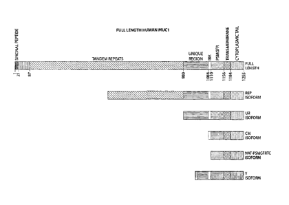
the MUC1 receptor is illustrated in FIG.1. The receptor, as illustrated
comprises: 1)
cytoplasmic tail; 2) transmembrane section; 3) MGFR; 4) IBR, 5) Unique Region,
6)
repeats, and N-terminus region comprising a signal peptide.
In healthy cells, MUC1 receptors are clustered at one portion of the cell
surface. In
contrast, MUC1-positive tumor cells are characterized by a loss of this
"healthy" clustering.
The invention anticipates uses for detecting and treating aberrant expression
of the MUC1
receptor in conditions other than cancer. For example, the MUC1 receptor is a
key element
in immune response and fertility. In the case of fertility, it may be
beneficial for portions of
the extracellular domain to be cleaved to induce embryo implantation. Methods
of the
invention may be used for non-cancerous conditions to promote or inhibit
receptor cleavage.
Additionally, method of the invention may be used to diagnose conditions of
infertility. In
tumor cells, the MUC1 receptors are no longer clustered but instead are
typically distributed
over the entire cell surface or in some cancer types, the receptors form a
series of clustered
islands that are expressed over a considerable portion of the cell surface.
This loss of
clustering of the MUC1 receptor has been correlated to tumor aggressiveness,
metastaic
potential and eventual outcome for the patient. The inventors have shown that
a cleavage
product of the MUC1 receptor, that remains attached to the cell surface,
referred to herein as
the MGFR, functions as a growth factor receptor. When this portion of the
receptor is
available to activating ligands, cell proliferation is stimulated. The MGFR
portion of the
receptor can become accessible to activating ligands by a variety of methods.
For example,
cleavage of the receptor that releases some or all of the IBR makes the MGFR
more
accessible to activating ligands. Agents that reduced the cell surface
expression of the
entire MUC1 receptor keeps the receptors too far apart to cluster and thus
increases
CA 02537263 2006-02-27
WO 2005/019269
PCT/US2004/027954
- 25 -
availability of the PSMGFR and ESMGFR to activating ligands that transduce the
signal to
the cell to proliferate. Agents that essentially completely inhibit the
expression of the
MUC1 receptor are good therapeutic candidates are provided according to one
aspect of the
invention. Examples of such inhibitory agents include but are not limited to
anti-sense
oligos and RNAis, or inhibitory RNAs.
In some cases, the MUC1 receptor may be cleaved to release the IBR or the
TPSIBR, from the cell surface. Alternatively, cleavage can result in a release
of a sufficient
portion of the IBR that causes the MUC1 receptor to lose the ability to self-
aggregate. Loss
of aggregation of MUC1 may have several ramifications. Release of the IBR or
sufficient
portion of the IBR from the cell surface allows the receptors to evenly
distribute on the cell
surface, leaving the cytoplasmic tails free to associate with intracellular
signaling proteins.
External agents, such as modifying enzymes and/or activating ligands, are then
able to bind
to the remaining extracellular portion of the receptor and induce disease-
associated signals,
either via a change in the multimerization state, i.e., inductive
multimerization, or as an
induced conformational change. As is appreciated by those of ordinary skill in
the art,
ligands such as growth factors and hormones often induce receptor dimerization
which
triggers, in turn, an intracellular signaling cascade. Additional support for
this mechanism
is presented below in data showing that in MUC1+ tumor cell lines and
transfected cells
expressing truncated isoforms of the MUC1 receptor lacking an IBR, bivalent
ligands, such
as a bivalent antibody, directed against MGFR trigger signaling, and resulting
cell
proliferation, through the well-known MAP (mitogen activated protein) kinase
signaling
cascade, as indicated by detection of phosphorylation of ERK2 kinase.
Significantly, such
phosphorylation and proliferation was absent or less evident in similar cells
treated with
monovalent ligands to MGFR, such as a single chain antibody or a monovalent
antigen-
binding fragment of antibody.
Cell proliferation may result from accessibility of the MGFR portion to an
activating
ligand which can interact with the MGFR portion. For example, the self-
aggregating IBR
of the MUC1 receptor may form a dense reticulum which sterically prevents a
ligand such
as a growth factor from interacting with the MGFR portion of the receptor,
which is
proximal to the cell relative to the IBR. In a cancerous or tumor cell, this
reticulum may be
lost, allowing ligand interaction with the MGFR.
CA 02537263 2011-06-20
- 26
The *We mechanistic model is oraidateutwith a mechanism whereby the pc=rtIon
oftheMUC1 damper, ihatrectains Vatted to theca =ace after shedding of the 1DR
region or the TESIBR, Le_the man, fitnctio'ns as a receptor kr Haw& that
hiliaer
praferatien. Evidence is also presented hemin that.dentonstrabs that (a) an
listereetion
s between a ligand and a poratm:ofate MCI receptor (191(3FR),
w$ttdhaerl7rsth
receptor, triggenteell prorifetationzicid(b) blocking the intentation of thie
portion of the
lara lremPotor ORM with its ligand(s), 'blocksccli prolithrudon. When tuner
cell lines,
hswhith the MUCI **got isboulogoneea* opressed accessate entity cell surface,
are
toited with aninuentivaIgG noUdy raised agahot tin aftait portion ofthe mum
receptor (e.g, PiThAtenthe Wee cellptulifecationisgteadianhencet SIM* intact
1SG
antibodies are bivalent Le. Mie anhlody simultaneously *WSW two at aceut bIGFR
portions on the cell.odom thasetesults .demonstrate that:the antibody acts as
an activating
Heat mimichig the eilhet ofa growth factor, which dimerines MGM pottions, and
thus
= triggers a cell prolithration *tang cascade which is:consistent
withsignalin' g via the
cYtolgesefi eleas Of thc rompers. 'Mists .thrther supported by the
experimentadiacussed
helowshowittgamtenedrsdietkoftwontgacentiVICFR portions estate cell surface
induces.
ERX-2phosphotylation indicative 0114MM:ease ccaproliferatitutlignaling (See
44.,Fig.
Is ). 'This ftnding leads to trio conehedons. First, an activating ligand(s)
that binds to the
MOM portion of the MIMI receptermsec inductivenudlimmitationof the receptor.
Secondly, an effective therapentiostrategy istiie&efireb hie& the MGM. pottion
QUI=
receptOr with a ntenomeric composition, thus croventing ifidectbe
multhnerizatio' n and
subsequent sigettiog =modes. 'Fee example, a single chain, or monovalent,
antibody, or a
monevalent fragmont of en nowt, liAdentmdbody, See 4130053i011 Of antibodies
and
linfilleil-bieding fragments dieteofbelox
raised whist thele3PRpestion ofthe MUCI
23 receptor (e.g. rats' ed against PRAGER ar against any peptide comprising
a PS:WM
sequence at Wig-terminus) woilidlunclion as an .atbetivencid-caneorthampentio.
Data and
Comptes .supproding this Wendell :are pumentedbelow. Another-therapeutic
strategy is 'to
block the activity (*enzymes thatmodify the receptor, which/nay be requited
for some
BMA binding
The *Vectors present evidence that dimerizodion of the MUC1 receptor triggered
cell growth in Tfilahreast tumor cells. trronsi2atieu was achieved in
byraising an JO
Maio* tO MGR, .l igeautibocrms asc blvalentand Therefore can cOmerize, a
CA 02537263 2011-06-20
- 27
portion of the Wel retvtor thga is pnurimal to the cull surface. The pudica of
the MUCI
recepiror that was used was theMGFRI..and the sequence afire pepede used for
generatiag
the autthodywasSEQ JD NO: 7 (teas 1¨ vargth4OIllt.)õ "Teeth additional
experimental
results are presented that suppartihe premise tbatifmtwiaation date Wei
'walla:1r
s triggers cell proliferation In a. 'amber ofhltlel+ tumor cell lines,
while baiting virtually no
effect nails *It do atattaPree$ ornzinintally coitus the MUM receptor.
Ivitler breast
turner cella. T471), 1500, UK and BT-474 wereobtabled flunk the ATCC, as
described
below in Example 5. As:controls, Mt ler breast tumor cells 143,1=11B 453,
Iff/X (human
renbilicallddney) cell line K293 and WA were also obtained DOM the ATCCõ RS
&soil* belowinErtimple.5. 'Western blotanalysis, performed as &Mita below in
&le 5, conlioned that T470õ. 1$00, and SO41 cells all etspreased high
levels of cleaved
as well as =cleaved MUM and thatthecontrOlvells did not. Our analyals showed
that cell
line ST-474 expreetedno detectable levels ofuncleavedMUC1, Wirral express an
interatedistc amount of cleaved ltUC I,
' Rabbit
polyclonalmtlodir: s vitae raised against &synthetic peptide.* sequence of
width was dcrivedfr tAIM (vat6PSUGFR¨ SEQ ID NO: 7 of Table 1).by a
conunercial antibody service company gyared, CA), as described below in
Example IL
The resultant antibody was purified by affinilythromatography over a wham
derivate' ed
with the same peptide audio immuel* the. rabbits. TO confirm that the
resultant antibOdY
recognizedihOlKilflt. Of tbe IVILIC1 receptor, the: antlltodY was =ad as the
cognate probe in
vreirem blot; see Saarnp1e5, wbereinsatuples fee houtealaingpectide and
lawein
preparations from theAl(iCt positive:as well as MUM negative ea lines were MR
on A
15,6 oolyanrylinrida gel. The bivaient,(able to dinicrize) antibody was added
to the panel of
hreatatueror cell lines that express the MUM reeepttc along with connel cell
lines that did
not. TheaddiLlonoffi.anI1b04ysljnniJa1edilj1jdiratirai.oidyjii cells that
=missed
MGM =VOL Tha 1564 breast haaor cells that were treated foreither 5 or 6 days
with the bwaltenb..SMGF . antibody tapioca/eat 400% 000% cabs we of cell
prnwth, seer% Anal 13anatple 11. ssfertingto
Fig. 6, control cells K293 and Rola
were unairectedbythesanie dosegeofthe sante antibody, auti-rShalflt- The shape
of the
proliferation enhancementatrves argue that. the antibodies dirnerize
thereeeptnr; at very
high min-body concentrations thereto of Cell growth &Creases, as each receptor
Is bound to
a siallie aalbody rather than onobivaleatantlhody bound to two tempi= Fig... 7
'idiom
CA 02537263 2011-06-20
-
that cellsfrom the bawd Minor cell Rae 110Ounderwent a 200% enhancement of
cell
pratiferation after treatment withthebivekettand-PSNRWRIer3 days. When
bivalent
rmti,,PSIVIGUR treattrentwasentaided.fit a fcardi day, thermal& cnbaneement of
cell
growth imeased 300314 see lin: g- Eneast Maar irclls final **MD ceR tire were
s also tested for the ability of the b1valcatnati-P$M0ft. antibody Wilmer
cell proliferation.
.Fig, 9., alums ihetthcoe ,cellaelso urcierwrotanappectitn' ately 12.5%
enhancement often
gam* amiss with 150004 Mit emit times, theresponst was dependent on the
concentration of the enftody. Breastiunoor veil lbw Iff474 &Owed
similaratlandetioa
oftell gang,* (I-50 ¨20086) laleaPallee to bivalent anti7PSUGPR, see 10.
iducrcell
le W4-M3-4S3 was not affected byths addition of anti-PSMOFIt. at any
cementation
Wetting sigma/
Monovalent forms awl-MOM .fiagments...block cell. growth in IKUC1 positive
1Ottnr Aspieviouslydasoaed" MUM+ lemur calls, am induced to polar= "Axe
the MUC1 receptcria fraterited. Specifically*the signal to l*tlI cisgutvd when
a
t.s portion ~AM reereptorpmramti to lite celisurfiree is elhnorized. As
deserled
shove,- newsy lawhich tbexeceptcra can he eihncrisedis flik a billilent
ani&OCIY dire*C1
806.1; ihe AIM receptor: Inapreterred embodiment; the antlody is directed
against the
MOFR. and in yeta more preihrred ernhocroment it is &meted against at hosts
partion fthe
PSMOFR. As described sham and *ether helow; agents that hind to the Mt3FR.
POrtion of
ze **MIMI reacPtO1* in amanamerittagacr than rfrmetio faabhott cao block
the elimetization
ofthe receptor radian, doing Whit proliferation- The tlincusaion below
deserThog
several, chemical compounds that bilnlitthe growth othoWC1+ tumor cells by
binding to
the MGM:part:Ian ofthe MUM receptor
-
As mer;loneel above, yet another method of providing an agent that prevents
25 trunerizeticetafthelArl receptor le generating amosovakatt antaiwsly cc
a manovateat
an 'binding fragment atm antibody directedmatt' st the MUCI receptor.
Monovalent
andhatesiftegments raised eiptinstibthilICI receptor woukl be
escellentthampentic
netts for MUCI potilive cancers. .lifenovalentantiboorresicregment that target
par-dims of
**receptor preaches/to, the cc11 sorter:0 arc pre:titre& Fatircially preferred
are monovalent
30 autalaiiestfralaentsthat lobate portions ofihe MUM receptor that .are
terminal to the
toeglanitg of** repeat* section: Still more preferred am mccoveket
antibodiestfragtecota.
rhattatyptpordonaofthe receptor Creeachal to the ttnictueregion of the MUM
receptor
CA 02537263 2006-02-27
WO 2005/019269
PCT/US2004/027954
- 29 -
and yet more preferred are monovalent antibodies/fragments that are directed
against the
PSMGFR sequence.
Peptides used for antibody production may or may not be glycosylated prior
immunizing animals. The sequence of these peptides need not exactly reflect
the sequence
of MUC1 receptor as it exists in the general population. For example, the
inventors
observed that antibodies raised against the the PSMGFR peptide variant var-
PSMGFR
(SEQ ID NO: 7), having an "-SPY-" motif have a higher affinity and greater
specificity for
the MUC1 protein than antibodies raised against the actual native sequence
(i.e. nat-
PSMGFR, SEQ ID NO: 36), having an "-SRY-" motif. One may also, in certain
embodiments, introduce mutations into the PSMGFR peptide sequence to produce a
more
rigid peptide that may enhance antibody production. For example the R to P
mutation in the
var-PFMGFR sequence of SEQ ID NO: 7 may actually have provided a more rigid
peptide
and was thus more immunogenic. Another method for producing antibodies against
regions
of peptides that are not particularly immunogenic, such as the IBR or TPSIBR
is to tag the
specific peptide sequence with an irrelevant sequence in which the amino acids
are of the D-
form and thus act to stimulate the immune response of the host animal. Peptide
sequences
that are used to immunize animals for antibody production may also be
glycosylated. The
MUC1 peptide sequences that were used herein for drug screening and to
generate cognate
antibodies were derived from the human species Of MUCl. Since there is
considerable
conservation across species for the PSMGFR and IBR and some portions of the
UR, it is
anticipated that MUC1 peptides whose sequences are derived from other species
can also be
used in drug screens and to generate antibodies for these same purposes. The
invention
also involves, in certain embodiments the generation of bi-specific antibodies
and bi-
specific antibodies formed thereby. Those skilled in the art are familiar with
methods to
generate antibodies wherein each recognition fragment of a bivalent antibody
binds to
different but essentially adjacent sites on the same antigen.
As described in greater detail below, MLTC1 monovalent antibodies/fragments
described above for use as cancer therapeutics may be polyclonal or monoclonal
and may
be obtained by immunizing a number of different animal species, i.e. rabbit,
goat and the
like. Additionally, techniques are known to those skilled in the art for
generating
hybridoma cells, which then are grown and harvested to yield a supply of
antibody without
the need for repeated animal immunization. Alternatively, humanized monovalent
CA 02537263 2011-06-20
- 30 -
antzlodieslfragments, also described in famodettil below, thatiarget these
portions of the
1Ville1 =captor maybe osedas effective 3116-' 00011ef agents at ant less Rely
to awoke
karma responses hi 'Impudent. Methods of the inverifionalso encompass
recombinant
methods br antibody and Fah prodnethin tag& not include animal finmtmizstion.
Itserplainedbelow, metittalsof generating monovalent antrixrdies and
monovalent
autigerrhinding &warfare ant&odes are known UltilLOSC skilled in the art_ A
standard
method is, the controlled proteolySis of a bivalent antibody. The inventors
generated a
inonvtdentPitMetk-specille anhlody liagmeat by plotectoting their invenfive
bivalent
anti-FSIAGEIR. Monovalent. anti.PSWin. competes. with-the bivalent anIMMGFR
to antibody for 0*mm:binding silo Within the MGFR fordo ofthe MUdI
receptor.
The 'Present invention, in cenain embodiments, details. how monovalent
nalliodinsIfiaginests,whicAistaSttentad against a portion of the MUC1
receptorthat is
proximal tothe eell-surthee, Inhibk.the grovotherMUCI+ twat tells. Monovalent
autibodlesgragosentsihat targeted the P8MGFIt. portka ofthe WWI receptor were
ts produced by pnatroining the tivalentanti4SMCIFE: antibody, whiCh has
beim herein to
, induce cell. grow* presumably by4imerizing theMLIC I receptors. Thus,
itfollows. that
themonovalent form of this very antibody would *eke& prolithralion by binding
to the
MOPS. prnibm sod thusprovent the biodng (Wet:theta Morn& andlor crunerizahom¨
Ben* we provide eaperimentalresultithetdeffionstrate that 10000vsiont anithedT
20 fragments thatterget lire PSIkftin. do in fnetiolnlit thegtowth of MUCI
pkritivehmtor
cells and have virtually no edfecton control cell lines. Warring' now to Fig.
6..:;ecall that
the edilljor? of bivalent anti-PSMGFR &rimed 8.600% enhancement Of,c011.
growth. Fig.
ii :themethod by whichthe resuhrwere generated beingdescribedin F.. 11, shows
that
the efitlitimafthentonovident form ofthe santelinti-rdMGFR tO a IWO. positive
*east
25 tomer Cell ihre. 1504, bad the opposite effect in that cell growth
Inhbfted by about I50%,
which intftcates indocedoell death. The additioa. of thenioaova1esnti-PSMGFR
itad a
similar cab:ton boast tumor gall line Mk which is also MUM; see Fig. 12.
Monovalentmai-. FSMIZIFIL va0cLwart.the in vino drug sem% it inhibits the
color
change` of the FSMGPVinunehOized tuntopartieles ,caused by the addition of
tumor cell
30 bytes to the oanapatticlek aadoscrbed inD1010 data below. Itshould be
noted that the
mouvIdentadibodyffrogrocart aIat ean *titbit thediwerization tithe PSMaFit
peptide in
vitro. liancvadinta-based drag samba assays to idantgy sompotmds that *Whit
CA 02537263 2011-06-20
-31.
dimmization ofthe.MGER portion of the1417C1 receptor aredadedbedhereht and in
commnalyinemed IL8.,patratupplicationpublicadonno..2003/0036199;
amfinternattomd
PnblimitionNo.02,036022 A2. ht certain oftheseassays,histidine-tegged P$MGITIt
PsPlhies, (44 67SQ VO.t. 4.werekuobilizeirlOnNVOTO44A116caatect gold
3 14110PartiCk& Lanates and sarranalentatomMiltl posidvetuntorvells, which
possuntably contain the cognate ligands of Mitt receptor, werc added to the
nanopiXticies. tipoh. addition ofthelysatehtupematmt mkt)" *scalar
ofthenanopetfiele
sOlutionturns *mix ha characteristic pink tó bhte, presentably when the
cognate figands
dimerine.MUCI mover Peptides on two didiarentnanoparticles. The.additioa of
bivalent
30 ard143.,3143Fltentiho4y, in placenfthe lysateisapetnarantsolution, also
maws the
tuteopetticle solutionto Mat fromphikto blue, as the bivalent antibody also
dimerises two
P$MCIFR peptIdee en Afferent nanoparticlea. However, the addition of
monovtdent anti-
,
PSWIGFR to:the:drug screening, assay, to Whith the lyntersepematant has also
heenedded,
inhthiis the color change,. ptesumahte by cOmpeari' glvhhttehnei, cognate
1iga0d0 for
0 thiaring to the PShIGER, peptide. Pig. 13 kiwis that the characteristic
reamPerdele cater
_
*464044041utt *cm tVentheaddenothivadurt enti4"alt3PR was inhibited Rpm.
addition
ef the monovelmt anti-M.03W
The puma tenths also suggest tattle WU receptor is involved in spoptosis.
The adEtign' t of thetwahart setP&Mantitot ont.y inhibited cell growth. but
aim
20 .cell .d This laditzttes lizettho Wel receptor also mediates
sigtuding pathways
involved in the process. of programmed cell death known as ig3eptosis.
Present caner research lite/atm" premise conking picture este-whether cc net
the overall. exammtorMlIel .receptor pro.duced by the cell out be correlated
to meradatic
potential' wanner agates:simian The lentils deserib3d herein suppostthe idea
the a key
25 mechanism of cell ;growth InMUC1 positivecancelo may *paid more on the
amount of
MC deivagoilial corms tailartbanthetwarall runountorbillIC1 never that is
expatmed. Low.inoleculer weight species thatmiginte OA eariallimaide gel-with
an
apparent atoleculativelebt of arotmd.20-30.1M (some glyeosyleted) exist in NMI
positive
tumor cabs but **not exist in salrldentittunhers to be detectable innon-tumor
hirLIC1
30 MOS- The lavvair0. 14010,ffied two cleavage silts ofthe MUM
receptor in hanoreells. The
instole!age &concurs lflthet,iMle ofthe falt. spathe. sewed cleavage site, -
which our
wider= lirdlgatet lathe inorolutentigenta than, occurs nthe C-tetmibl end
ofthe MR: the
CA 02537263 2011-06-20
-32 -
first cleavage site being loaded* the N-terminus of1PSB31 (SEQ IDNO: it) and
the
secendeleavageafte beinglocatedottheN-terminosef the nat-PSNICIER havbrg SBQ
ID
* NO: 60. When cleavage wars atthe fostaite, the pectic' in ofthe receptor
that remains
attached to the:ceII. thesimiler to ISIISMGFR(See Table 1, SBQ ID NO. 66,
but
- with the native WY sequence). When cleaved at the second site,thopotikur
that remaining
pottiest b aPS/iWO. as shown hi Table l EQ:IDNO.63. Thia 1)1W Malawi* weight
species that ;sterner speciftneonalsts eisthaftellyofthe native PSIMPII.
sequence and in
same easesthe TSBSMOPRecrotence sod is available to cognate Uganda, Le. not
self-
aggregated, than certhnoverall amountof WIC [receptor awes:led by the cell
to Supporting this
conehadon, suseepttlfthyofturnor cellstoproftthrete was found, within the
:coated -ores meant invection,th be a fundien of the emountofthe shorter term
oftba
MUCI. :tecephu.
Comparison of the present results generated by western blot analyses, which
quantitated the atnotirtt of low molecular weight Wel opecieff produced by
each cell type
35' tested, with the above presented cell proliferation data shows that the
susceptibility of the
breast tumor cells to antibody-induced .cell growth is proportional to the
amount of the low
molecular weight WC]. species (25-30 Itd .glycosylated; 19-201(4
unglycosylated) that the
cell produces. Referring now to Fig 14, breast tumor tell lines 1.500 and 1504
produce a
considerably greater amount of the MIMI eleavage product that runs at 19-20
1(d than the
20. BT474 BT cells or the Ocnitrol K.295 and, ReLa cells. Cortesponcliegy, the
anti-PS1VIGER7
induced inerease in the proliferation of cell lines 1500 and 1504 was up to
about 400% (Fig.
8) and up to about 600% (Fig. 6) respectively, while there was no detectable
increase in the
rate of cell. growth for cnistrO1 colts (Fig:6) andthe growth of BT-474 cells
increased by
only up to about 200%, see Fig. 10.
25 Intinther support
oftheeanehelleer that cleavage prodectsof the AttUCI recepaar
.ftmation. at growth Suter tecoptarainbimor cells, flliK cells were-
traosfectix11401 WitiC1
vadants that were either .terudnated after the PSMLIIIM (see Tebbe 1, SBQ ID
NO: 37) or
after the etttire :thterchsin binding neon (PSISR) Mg ID 140-3.10. Cells
transfected with
the taceptor that *eluded the PaBR grew at 114444 6nreardower than cells
tranaseted
30 with the.MITC1
rilisitta that Were tannicated after the paten (e.g. SBQ /40: 37).
These results support die eonclasio. n that the pordan oftheMUCI receptor that
acts as a
growth factor receptor Is aclerwage polite in vihich much or all of the IBR is
released
CA 02537263 2006-02-27
WO 2005/019269
PCT/US2004/027954
- 33 -
from the cell surface. Further, these results support the conclusion that
tumors in which a
good percentage of the MUC1 receptors have been cleaved to release the TPSIBR
(SEQ ID
NO: 65) are especially aggressive cancers and those that are cleaved to
release the entire
IBR, leaving PSMGFR (SEQ ID NO: 63) attached to the cell surface are even more
aggressive. Therefore, antibodies that are raised against the TPSIBR (SEQ ID
NO: 65)
portion of the MUC1 receptor can be used to assess the aggressiveness of
cancers that are
MUCl-positive.
Consistent with these findings, the amount of MGFR that is accessible on cells
(tissues) predicts tumor aggressiveness and metastatic potential. Therefore,
antibodies that
recognize the MGFR portion of the receptor can be used to diagnose cancer or
the
propensity to develop cancer, to predict cancer aggressiveness and metastatic
potential, to
suggest therapeutic protocols and to track the progress of the therapeutic
protocols.
Consequently, the aggressiveness or metastatic potential of tumor cells can be
assessed by determining the amount of lower molecular weight MUC1 species that
the cells
produce. This can be determined, for example by SDS-PAGE analysis or western
blot
analysis using antibodies or antigen-binding fragments thereof raised against
the MGFR
portion of the receptor. In certain embodiments, the inventive colloid assay
techniques
described herein could be utilized in which the antibodies/fragments are
attached to a carrier
that can be a nanoparticle or colloid. In a preferred embodiment, a patient's
cells are probed
with antibodies or antigen-binding fragments thereof directed toward the MGFR
as this
method reveals the amount of MGFR-containing MUC1 that remains attached to the
cell
surface and, importantly, whether or not it is accessible to cognate ligands.
In a yet more
preferred embodiment, a patient's cells are probed with antibodies or antigen-
binding
fragments thereof directed toward the PSMGFR. In practice, cells that display
a high
degree of MUC1 receptor that reacts with antibodies or antigen-binding
fragments thereof
that recognize the PSMGFR, or a portion thereof, is an indication that the
MUC1 receptors
present on these cells have undergone of a greater degree of cleavage, leaving
the PSMGFR
portion accessible to cognate ligands that activate a cell growth pathway.
Tumors
comprised of cells thusly characterized are of higher metastatic potential
and/or are more
aggressive. Therefore, using antibodies or antigen-binding fragments thereof
that recognize
the PSMGFR, or portions thereof, can be used to diagnose the metastatic
potential or
aggressiveness of a patient's tumor.
CA 02537263 2006-02-27
WO 2005/019269
PCT/US2004/027954
- 34 -
In certain aspects, the invention provides antibodies or antigen-binding
fragments
thereof. In one embodiment, the invention provides an antibody or antigen-
binding
fragment that specifically binds to MGFR. In certain embodiments, such an
antibody or
antigen-binding fragment thereof is bivalent, while in other embodiments it is
monovalent.
In certain embodiments, the above-mentioned antibodies or antigen-binding
fragments
thereof specifically bind to PSMGFR. In certain such embodiments, the
antibodies or
antigen-binding fragments thereof can specifically bind to the amino acid
sequence set forth
in SEQ. ID. NO.: 36 or a functional variant or fragment thereof comprising up
to 15 amino
acid additions or deletions at its N-terminus or comprising up to 20 amino
acid
substitutions; in other embodiments, it specifically binds to the amino acids
set forth in
SEQ. ID. NO.: 36 or a functional variant or fragment thereof comprising up to
10 amino
acid substitutions; in other embodiments, the antibodies or antigen-binding
fragments
thereof specifically bind to the amino acid set forth in SEQ. ID. NO.: 36 or a
functional
variant or fragment thereof comprising up to 5 amino acid substitutions; and
in yet another
embodiments the antibodies or antigen-binding fragments thereof specifically
bind to the
amino acid sequence set forth in SEQ. ID. NO.: 36. In certain embodiments, the
antibody
or antigen-binding fragment of the invention is a human, humanized, xenogenic
or a
chimeric human-non-human antibody or antigen-binding fragment thereof. In
certain
embodiments, the antibodies or antigen-binding fragments thereof of the
invention comprise
an intact antibody or an intact single-chain antibody. For antibodies or
antigen-binding
fragments that are monovalent, in certain embodiments, they may comprise a
single-chain
Fv fragment, a Fab' fragment, a Fab fragment, or a Fd fragment. For antibodies
or antigen-
binding fragments of the invention that are bivalent, certain embodiments
comprise an
antigen-binding fragment that is a F(ab')2.
The present invention also provides, in certain embodiments, compositions
comprising the antibody or antigen-binding fragments of the invention as an
ingredient. In
certain embodiments, such compositions comprise pharmaceutical compositions
and further
comprise a pharmaceutically-acceptable carrier. In certain such compositions,
the antibody
or antigen-binding fragment thereof can be polyclonal, while in other
embodiments it can be
monoclonal.
The invention also provides, in certain embodiments, a variety of kits, in
certain
embodiments including any of the above-mentioned antibodies or antigen-binding
CA 02537263 2006-02-27
WO 2005/019269
PCT/US2004/027954
- 35 -
fragments thereof of the invention. In certain embodiments, such kit may also
provide an
article having a surface. In certain such embodiments, the antibody or antigen-
binding
fragment thereof can be fastened or adapted to be fastened to the surface of
the article. In
certain embodiments, the article comprises a particle. In such embodiment, the
kit further
includes a second particle and a peptide sequence comprising a portion of a
cell surface
receptor that remains attached to the cell surface after shedding of the cell
surface receptor
inter-chain binding region, the peptide sequence being detached from any cell,
and fastened
to or adapted to be fastened to the second particle. In some embodiments, the
kit may
further include a candidate drug for affecting the ability of the peptide
sequence to bind to
other identical peptide sequences, and/or to the antibody or antigen-binding
fragment
thereof, in the presence of the antibody or antigen-binding fragment thereof.
The peptide
sequence provided can comprise, in certain embodiments, MGFR. In some of the
above-
described kits including a particle, the kit may further comprise a peptide
sequence
comprising a portion of a cell surface receptor that remains attached to the
cell surface after
shedding of the cell surface receptor interchain binding region, such peptide
sequence being
detached from any cell, and fastened to or adapted to be fastened to the
particle. The above
kit may, in certain embodiments, further comprise a second particle and have
the peptide
sequence mentioned above fastened to or adapted to be fastened to the second
particle. The
above-mentioned kits may be useful, in the context of the present invention,
for performing
various diagnostic, drug screening and other assays, which can involve colloid-
colloid
interactions and/or aggregation, as described in detail herein.
The invention also involves, in certain embodiments, methods for producing or
generating antibodies or antigen-binding fragments thereof that specifically
bind to certain
peptides, for example certain inventive peptides disclosed herein. One
particular
embodiment, an antibody or antigen-binding fragment is raised against a
peptide including a
portion of a cell surface receptor that interacts with an activating ligand
such as a growth
factor to promote cell proliferation, such portion including enough of the
cell surface
receptor to interact with the activating ligand and being free of an
interchain binding region
to the extent necessary to prevent spontaneous binding between such portions.
In certain
such methods, the cell surface receptor comprises MUC1, in other embodiments
MGFR,
and in yet other embodiments a peptide comprising PSMGFR at it N-terminus; in
yet other
embodiments, the peptide comprises at its N-terminus the amino acid set forth
in SEQ. ID.
CA 02537263 2006-02-27
WO 2005/019269
PCT/US2004/027954
- 36 -
NO.: 36 or a functional variant or fragment thereof comprising up to 15 amino
acid
additions or deletions at its N-terminus and comprising up to 20 amino acid
substitutions.
In certain embodiments of the inventive methods for producing antibodies or
antigen-
binding fragments thereof, an antibody or antigen-binding fragment is raised
against
PSMGFR. In certain such embodiments, such peptides used to generate the
antibody or
antigen-binding fragment thereof can consists of the amino acid sequence set
forth in SEQ.
ID. NOS.: 36 or 37 or a functional variant or fragment thereof that comprises
up to 15
amino acid additions or deletions at its N-terminus and up to 20 amino acid
substitutions.
In yet other embodiments, the invention provides methods for treating a
subject
having a cancer or other condition requiring treatment with one or more of the
antibodies or
antigen-binding fragments thereof of the invention. In one such embodiment,
the invention
provides a method for treating a subject having a cancer characterized by the
aberrant
expression of MUCl. The method involves administering to the subject an
antibody or
antigen-binding fragment thereof in an amount effective to ameliorate the
cancer. In certain
such embodiments, the antibody or antigen-binding fragment thereof is
administered in an
amount effective to reduce tumor growth. In certain embodiments, any of the
above-
mentioned antibodies or antigen-binding fragments thereof, especially those
which
specifically bind to MGFR, PSMGFR, etc. can be used. In certain preferred
embodiments,
the antibody or antigen-binding fragment thereof is administered in an amount
effective to
block the interaction of a natural ligand with a portion of a MUC1 receptor,
for example,
MGFR, that remains attached to a cell after shedding of a interchain binding
region of the
MUC1 receptor. In other embodiments, the method involves administering an
antibody or
antigen-binding fragment thereof that is effective to reduce shedding of an
interchain
binding region of a MUC1 receptor. In many such embodiments of the method,
particularly
those in which the antibody or antigen-binding fragment thereof specifically
binds to
MGFR, such a treatment method can involve administering to the subject the
antibody or
antigen-binding fragment thereof in an amount effective to prevent inductive
dimerization
of a cancer-associated growth factor receptor, such as aberrantly cleaved
MUCl.
In yet another method provided by the invention, the above-described
antibodies or
antigen-binding fragments thereof can be utilized in a method for determining
the
aggressiveness and/or metastatic potential of a cancer. In one such method, a
sample
obtained from a subject having or suspected of having a cancer is contacted
with an
CA 02537263 2006-02-27
WO 2005/019269
PCT/US2004/027954
- 37 -
antibody or antigen-binding fragment thereof of the invention that
specifically binds to a
peptide associated with the cancer that is expressed on the cell surface. The
method
involves determining an amount of the antibody or antigen-binding fragment
that
specifically binds to the sample, such amount being indicative of the
aggressiveness and/or
metastatic potential of the cancer. Certain such embodiments, the sample
utilized comprises
cells of the subject and/or a solublized lysate thereof. In certain such
embodiments, the
peptide expressed on the cell surface can include a portion of a cell surface
receptor that
interacts with an activating ligand such as a growth factor to promote cell
proliferation,
wherein the portion includes enough of the cell surface receptor to interact
with the
activating ligand while being free of any interchain binding region to the
extent necessary to
prevent spontaneous binding between portions. In certain such embodiments, the
cell
surface receptor is MUC1 and the peptide comprises MGFR or a peptide
comprising
PSMGFR at its N-terminus. In certain preferred embodiments of such methods,
the
antibody or antigen-binding fragment thereof can be immobilized relative to or
adapted to
be mobilized relative to a signaling entity, such as any of the signaling
entities described
previously. In certain such embodiments, the signaling entity can comprise one
or more
particles, such as colloid particles.
The invention, therefore, embraces peptide binding agents which, for example,
can
be antibodies or fragments of antibodies having the ability to selectively
bind to PSMGFR
and/or MGFR. Antibodies include polyclonal and monoclonal antibodies, prepared
according to conventional methodology.
Significantly, as is well-known in the art, only a small portion of an
antibody
molecule, the paratope, is involved in the binding of the antibody to its
epitope (see, in
general, Clark, W.R. (1986) The Experimental Foundations of Modern Immunology
Wiley
& Sons, Inc., New York; Roitt, I. (1991) Essential Immunology, 7th Ed.,
Blackwell
Scientific Publications, Oxford). The pFc' and Fc regions, for example, are
effectors of the
complement cascade but are not involved in antigen binding. An antibody from
which the
pFc' region has been enzymatically cleaved, or which has been produced without
the pFc'
region, designated an F(abp2 fragment, retains both of the antigen binding
sites of an intact
antibody. Similarly, an antibody from which the Fe region has been
enzymatically cleaved,
or which has been produced without the Fe region, designated an Fab fragment,
retains one
of the antigen binding sites of an intact antibody molecule and comprises one
type of
CA 02537263 2011-06-20
- 38 -
monovalent antibody fragment according to the invention, Proceeding further,
Fab
fragments consist of a covalently bound antibody light chain and a portion of
the antibody
heavy chain denoted Fd. The Fd fragments are the major determinant of antibody
specificity (a single Fd fragment may be associated with up to ten different
light chains
without altering antibody specificity) and Fd fragments retain epitope-binding
ability in
isolation. Accordingly, a monovalent antibody fragment according to certain
embodiments
of the invention may be an Fd fragment,
Within the antigen-binding portion of an antibody, as is well-known in the
art, there
are complementarity determining regions (CDRs), which directly interact with
the epitope
of the antigen, and framework regions (FRs), which maintain the tertiary
structure of the
paratope (see, in general, Clark, 1986; Rohl, 1991). In both the heavy chain
Fd fragment
and the light chain of IgG immunoglobulins, there are four framework regions
(FR1 through
FR4) separated respectively by three complementarity determining regions (CDRI
through
CDR3). The CDRs, and in particular the CDR3 regions, and more particularly the
heavy
is chain CDR3, are largely responsible for antibody specificity.
As is now well known in the art, the non-CDR regions of a mammalian antibody
may be replaced with similar regions of conspecific or heterospecific
antibodies while
retaining the epitopie specificity of the original antibody. This is most
clearly manifested in
the development and use of "humanized" antibodies in which non-human CDRs are
zo covalently joined to human FR and/or Fc/pFc' regions to produce a
functional antibody.
See, e.g., U.S. patents 4,816,567, 5,225,539, 5,585,089, 5,693,762 and
5,859,205. Such
antibodies, or fragments thereof are within the scope of the present
invention.
In certain embodiments, fully human monoclonal antibodies also can be prepared
by
immunizing mice transgenic for large portions of human itnmunoglobulin heavy
and light
2.5 chain loci. rollowing immunization of these mice (e.g., Xenolviouse TM
(Abgenix), HuMAb TN
mice (Medarex/GenPharrn)), monoclonal antibodies can be prepared awarding to
standard
hybridorna technology. These monoclonal antibodies will have human
immunoglobulin
amino acid sequences and therefore will not provoke human anti-mouse antibody
(HAIVIA)
responses when administered to humans.
30 In certain embodiments the present invention comprises methods for
producing the
inventive antibodies, or antigen-binding fragments thereof, that include any
one of the
step(s) of producing a chimeric antibody, humanized antibody, single-chain
antibody, Fab-
CA 02537263 2006-02-27
WO 2005/019269
PCT/US2004/027954
- 39 -
fragment, F(ab')2 fragment, bi-specific antibody, fusion antibody, labeled
antibody or an
analog of any one of those. Corresponding methods are known to the person
skilled in the
art and are described, e.g., in Harlow and Lane "Antibodies, A Laboratory
Manual", CSH
Press, Cold Spring Harbor, 1988. The production of chimeric antibodies is
described, for
example, in W089/09622. Methods for the production of humanized antibodies are
described in, e.g., EP-Al 0 239 400 and W090/07861. A further source of
antibodies to be
utilized in accordance with the present invention are so-called xenogeneic
antibodies. The
general principle for the production of xenogeneic antibodies such as human
antibodies in
mice is described in, e.g., WO 91/10741, WO 94/02602, WO 96/34096 and WO
96/33735.
As discussed below, the antibodies, of the invention may exist in a variety of
forms (besides
intact antibodies; including, for example, antigen binding fragments thereof,
such as Fv,
Fab and F(ab')2, as well as in single chains (i.e. as single chain
antibodies); see e.g.,
W088/09344.
Thus, as will be apparent to one of ordinary skill in the art, the present
invention also
provides, in certain embodiments, for F(ab')2, Fab, Fv and Fd fragments;
chimeric
antibodies in which the Fc and/or FR and/or CDR1 and/or CDR2 and/or light
chain CDR3
regions have been replaced by homologous human or non-human sequences;
chimeric
F(a13)2 fragment antibodies in which the FR and/or CDR1 and/or CDR2 and/or
light chain
CDR3 regions have been replaced by homologous human or non-human sequences;
chimeric Fab fragment antibodies in which the FR and/or CDR1 and/or CDR2
and/or light
chain CDR3 regions have been replaced by homologous human or non-human
sequences;
and chimeric Fd fragment antibodies in which the FR and/or CDR1 and/or CDR2
regions
have been replaced by homologous human or non-human sequences. The present
invention
also includes so-called single chain antibodies.
Moreover, the present invention, in certain embodiments, relates to
compositions
comprising the aforementioned antibodies or antigen-binding fragments of the
invention or
chemical derivatives thereof. The composition of the present invention may
further
comprise a pharmaceutically acceptable carrier. The term "chemical derivative"
describes a
molecule that contains additional chemical moieties that are not normally a
part of the base
molecule. Such moieties may improve the solubility, half-life, absorption,
etc. of the base
molecule. Alternatively the moieties may attenuate undesirable side effects of
the base
molecule or decrease the toxicity of the base molecule. Examples of such
moieties are
CA 02537263 2011-06-20
described in a variety of texts, such as Remingtoe's Pharmaceutieg Selene,
18th Edition,
1990, Mark Publishing Co., Easton, P.A. Examples of
suitable pharmaceutical carriers are well known in the art arid include
phosphate buffered
saline solutions, water, emulsions, such as oil/water emulsions, various types
of wetting
agents, sterile solutions etc. Compositions comprising such carriers can be
formulated by
well known conventional methods. These pharmaceutical compositions can be
administered to the subject at a suitable dose. Administration of the suitable
compositions
may be effected by different ways, e.g., by intravenous, intraperitoneal,
subcutaneous,
intramuscular, topical or infradermal administration. Aerosol formulations
such as nasal
spray formulations include purified aqueous or other solutions of the active
agent with
preservative agents and isotonic agents. Such formulations are preferably
adjusted to a pH
and isotonic state compatible with the nasal mucous membranes, e.g., for
intranasal
administration. Formulations for rectal or vaginal administration may be
presented as a
suppository with a suitable carrier.
A therapeutically effective dose refers to that amount of antibodies and/or
antigen-
binding fragments of the invention ameliorate the symptoms or conditions of
the cancer or
other disease being treated. Therapeutic efficacy and toxicity of such
compositions can be
determined by standard pharmaceutical procedures in cell cultures or
experimental animals,
e.g., EDS (the dose therapeutically effective in 50% of the population) and
LD50 (the dose
lethal to SO% of the population). The dose ratio between therapeutic and toxic
effects is the
therapeutic index, and it can be expressed as the ratio, LD50/ED50.
The biological activity of the antibodies and/or antigen binding fragments
thereof; of
the invention indicates that they may have sufficient affinity to make them
candidates for
drug localization to cells expressing the appropriate surface structures, e.g.
MGFR. Thus,
targeting and binding to cells of the antibodies and/or antigen binding
fragments thereof; of
the invention could be useful for the delivery of therapeutically or
diagnostically active
agents (including targeting drugs. DNA sequences, RNA sequences, lipids,
proteins and
gene therapy/me delivery. Thus, the antibody and/or antigen binding fragments
thereof of
the invention can be labeled (e.g., fluorescent, radioactive, enzyme, nuclear
magnetic,
colloid, other signaling entity, etc.) and used to detect specific targets in
vivo or in vitro
including "immunochemistry" like assays in vitro. In vivo they could be used
in a manner
similar to nuclear medicine imaging techniques to detect tissues, cells, or
other material
expressing 1VIGFR. Another method involves delivering a therapeutically active
agent to a
CA 02537263 2006-02-27
WO 2005/019269
PCT/US2004/027954
- 41 -
patient. The method includes administering at least one antibody or an antigen-
binding
fragment thereof and the therapeutically active agent to a patient.
Preferably, the
therapeutically active agent is selected from drugs, DNA sequences, RNA
sequences,
proteins, lipids, and combinations thereof.
In certain embodiments, the present invention also provides inventive drug
screening
assays, treatment protocol screening assays, diagnostic assays, etc. that
involve determining
whether or not an intracellular protein has become chemically modified in a
manner
indicative of its participating in an intracellular signaling pathway. One
such method
involves providing a cell expressing on its surface a peptide that can act as
a growth factor
receptor, such as MUC1. The assay involves contacting such a cell with a
candidate drug
for affecting the ability of an activating ligand of the cell surface peptide
to interact with the
peptide, in the presence of the activating ligand, and determining whether an
intracellular
protein that becomes phosphorylated if the activating ligand interacts with
the cell surface
peptide, in fact, becomes phosphorylated. Such method is especially useful, in
the context
of the present invention, for cells expressing MGFR, or a peptide comprising
PSMGFR at
its N-terminus, such as PSMGFRTC.
As described below, for embodiments involving MUC1-expressing cells,
intracellular cell proliferation signaling occurs via the MAP kinase pathway
and interaction
of the cell surface receptor with its ligand, and associated inductive
multimerization,
involves phosphorylation of an intracellular protein comprising ERK-2. In
certain such
cases, the inventive screening method utilizes a sample comprising a plurality
of cells,
which may, in certain embodiments, be lysed or permeablized, after having
being exposed
to the candidate drug and activating ligand. In certain embodiments, after
exposure to the
drug candidate and activating ligand and following cell lysis or
permeabilization, the
method involves separating proteins contained in intracellular contents of the
cells on a gel,
for example using Western blot techniques, and visualizing or otherwise
detecting the
separated proteins. Such detection can be effected, as well understood by
those of ordinary
skill in the art utilizing antibodies or other molecules that specifically
bind to the
intracellular proteins to be detected. In certain embodiments, such molecules
can be
antibodies or antigen-binding fragments thereof, preferably including an
auxiliary signaling
entity permitting detection or visualization, such as one of those described
previously. In
certain embodiments, detecting phosphorylation of the intracellular protein
after gel
CA 02537263 2006-02-27
WO 2005/019269
PCT/US2004/027954
- 42 -
separation involves contacting the gel-separated proteins with a biological
molecule, such as
the above-mentioned antibodies, etc. that specifically bind to a
phosphorylated form of the
intracellular protein (e.g., phps-ERK-2, but not to the intracellular protein
when it is not
phosphorylated). Western blot techniques useful for performing the above-
described
method are well known to those skilled in the art and are described in more
detail below in
Example 5.
In certain embodiments, instead of using the above-described gel-based method
for
determining phosphorylation of the intracellular protein within the context of
the present
assays, a colloid-based aggregation assay, similar to those described
elsewhere and herein,
can be utilized for detecting the presence of the phosphorylated form of the
intracellular
protein. In such embodiments, after the above-described step of lysing or
permeabilizing
the cells exposed to the activating ligand and candidate drug, the
intracellular contents are
contacted with a plurality of colloid particles. The plurality of colloid
particles preferably
includes a first subset thereof that are immobilized relative to a first
biological molecule,
such as an antibody or antigen-binding fragment thereof, that specifically
binds to a
phosphorylated form of the intracellular protein but not to the intracellular
protein when it is
not phosphorylated, and to a second subset of colloid particles that are
immobilized relative
to another biological molecule, e.g., antibody or antigen-binding fragment
thereof, that
specifically binds to the intracellular protein at an epitope that is
different from that to
which the first biological molecule specifically binds. If the sample includes
a
phosphorylated form of the intracellular protein, the colloids will aggregate
and a color
change will be observed. However, if the intracellular protein is not
phosphorylated, no
aggregation indicative of cross-linking of he colloid particles to each other
will be
observed.
Such methods as described above can enable these methods to facilitate
simultaneously determining whether a drug candidate suspected of having the
ability to
interfere with the binding of an activating ligand to a cell surface receptor
interferes with the
binding of the activating ligand to the cell surface receptor, and whether the
drug candidate
acts by interacting with the cell surface receptor or with the ligand. In
short, most
advantageously when the present method is performed under conditions of excess
ligand
concentration (as compared to drug candidate concentration), the above-
described methods
for drug screening based on detection of phosphorylation, or other
modification, of
CA 02537263 2006-02-27
WO 2005/019269
PCT/US2004/027954
- 43 -
intracellular proteins will tend to show a positive test result only when the
candidate drug
acts by interacting directly with, e.g. by becoming immobilized relative to,
the cell surface
receptor, as opposed its acting via a mode of action wherein the candidate
drug binds to or
otherwise interacts with the ligand. This one-step behavior will be best
observed whenever
the screening assay is performed utilizing the cell surface receptor ligand in
sufficient
excess such that any binding to the ligand by the candidate drug, which may
prevent
binding of the ligand to the receptor, would not reduce the ligand available
for receptor
binding and inductive multimerization within the assay system.
Moreover, such a colloid-based assay as described immediately above is not
limited
to assays and systems for detecting intracellular signaling via
phosphorylation of
intracellular proteins. The above-described colloid-based assays are more
generally
applicable. For example, such an assay method can involve a screening test for
determining
the modification state of essentially any biological molecule. Such a
screening method can
involve, in certain embodiments, assays involving detection of immobilization
of a colloid
particle relative to a biological molecule, wherein the colloid particle is
configured such that
it becomes immobilized with respect to the biological molecule when the
biological
molecule is in a first modification state to a different extent than when the
biological
molecule is in a different modification state.
Modification states that may be determined using such methods include whether
or
not a particular biological molecule is phosphorylated, glycosylated,
acetylated, etc.
Moreover, while, in preferred embodiments, such colloid-based assays involve
colloid-
colloid aggregation for detection, in other embodiments, the colloid-utilized
may simply be
used as a signaling entity for detecting whether or not the biological
molecule under
evaluation is modified or not by using Western blotting or another gel-based
assay, etc. For
example, in one such assay, biological molecules whose modification state is
to be tested
can be contacted with an agent, such an antibody, that specifically binds to
the biological
molecule when it is in a first state of modification but not when it is in a
second state of
modification. Such an agent could, in certain embodiments, be immobilized
relative to a
colloid particle, which could provide a signaling entity able to be detected,
for example, in a
gel; or, alternatively, the colloid could be immobilized with respect to
another binding
entity, such as a secondary antibody, having specificity for the agent binding
to the
biological molecule whose modification state is being determined.
CA 02537263 2006-02-27
WO 2005/019269
PCT/US2004/027954
- 44 -
In certain embodiments of such a method - utilizing a colloid-aggregation
detection
assay for determining the modification state of the biological molecule - a
sample
containing the biological molecule is contacted with a plurality of colloid
particles of at
least a first and a second type. A first subset (type) of the colloid
particles is immobilized
relative to a first agent that specifically binds to the biological molecule
when it is in a first
state of modification but not to the biological molecule when it is in the
second state of
modification, and a second set (type) of the colloid particles is immobilized
relative to a
second agent that specifically binds to the biological molecule at an epitope
that is different
from the epitope at which the first agent specifically binds. As described
above, in such a
test, if the biological molecules, or a subset thereof, are in an first state
of modification, the
plurality of colloid particles, as described above, will tend to aggregate, in
proportion of the
concentration of the biological molecule in the first state of modification,
thereby causing a
color change in the assay sample. However, if the biological molecule is
present only in the
second state of activation, the first type of colloid will not become bound to
it and no cross-
linking or aggregation of the colloids, or color change resulting therefrom,
will occur. In
certain embodiments of such an assay as described above, the first agent on
the first subset
of colloid particles would be an antibody or other binding entity that
specifically binds to
the biological molecule only when it is in the first state of modification,
and the second
agent on the second subset of colloid particles could be an agent that binds
to the biological
molecule in either the first state of modification or the second state of
modification.
Such a colloid aggregation assay as described immediately above can be
advantageously employed for determining, for example, which of a plurality of
intracellular
signaling pathways is activated upon binding of an activating ligand to a cell
surface
receptor. In such assays, a plurality of different types of colloid particles
could be
employed including a plurality of different first and second colloid types
having agents
immobilized thereon able to detect a plurality of different modification
states of a plurality
of different intracellular signaling proteins enabling determination of the
activity of various
cell signaling pathways within a cell to be determined, in certain embodiments
in a single
assay.
As discussed above, one aspect of the invention involves the discovery that
dimerization of the extracellular portion of the MUC1 receptor activates the
MAP kinase
pathway within the cell, which is a known signaling cascade that induces cell
proliferation.
CA 02537263 2011-06-20
-.45 -
The present inversion aisle, in attain enthotrunentaõ involves wades
diagneufteassaya,
dfttg andtreammut screening pram*, est&õ related to the discovery by the load=
dug
dinurriration recepbor triggem poet:radon viathe MAP
(mitogen
activated promitr) Haase cell ,proliferatiou __ sivuttiot pathway. mere
specifically,
diMeltaltiOZIofthe pretirm ofthe receptur utteessmy and
Saffiejellitaitedvate the
MAP kin pathway and induce cell ptolithration: The MAP kinase signaling
cascade is
onent.the intragellublr.s%nal*Fathwaysthatiir fairly's/nal understood. To
tuunnadze, a
milogen binds Mite eactraceihdar portion Of Etansrunagattne terePt9r and
alters its
coakenlati011 blanche manner that a slgastrathentinuducedto the cell interior.
As
described Wpm, a:downstream step in this cascade is the phosphorylation of K2.
It is
known in the attfilitt once 'MP lies been phosphoolated, cell preftration
proceeds, The
invaders. demonstrate &stifle addition of a bivalent aodbody, whichr minims
the NIGER,
dim ..*á the MCI nogg= aml insome vuty generates or length; binding slim tor
signaling prottinghat bind to the cyloplasmittaiLt ofthe.hATC1 receptor. Figs.
1547,
Is : tesPeoliv0139. .0b01µ *Min WA *504esd t50OinLWmcefls.
dimedzaliottorthe
- :MUM teeereorMapivelent aDAIMMIR results %BM phesphoniation. =Si. 1Z.
The 'Moth doserdependeet and time-dtpcodent. Further, synthetic compounds,
which the
invaders previously showed bind to the IviGFIt.pootion of the MCI receptor,
compete
with thebiva tdP8MGFfor Wolfing to liristegion ofthe MLIC1 receptor- In a
comPetitiVe Whitt= US% the mmplzundeditedvely preveg (also la adosedepcedant
way)thebiaingoffls bivalent arxtibody lc, the MOM, requg braless of
dbneriustion
43fthe nmelotor a114.01eds.cifMtirl pbospbcrytatics, sec 1ig. 18 ndEL 12
Addona1ly. the monovalent item of the anti-PS1v1Cila competed with the
bivalent
antibody and effectively Ululated BR= phosphotylation. When =CCM monovalent
anti-
.
~an was added to breast:tumor cells along with the =IOW ofbivalent
antieSMGPII.
that Was show* be sufficientto ad/notate WW2 phosphoryletion, that
pbosphorylation
was blocked, presumably because themonovalent ardrimtly blocked the
climmizalion of the
=
*MCI receptor.. Fit) 9 Acme that monovakatantl.PSMOBR, competes. with the
bivalent
antibody and .bloelm the phosphoryhdion of PAU in cell. line ISM
Actording130110catalkernhodImentslatthe invention, the phosphorylation state
of
illtlUt can be monimurd.as a method for Identifying therapeutics for MX 1
positive
cantata was &scared abovethattnonovakuteempounds and monovalent anti-
PSIvkliFit
CA 02537263 2006-02-27
WO 2005/019269
PCT/US2004/027954
- 46 -
that bound to the MGFR competed with the bivalent antibody for binding to the
site and in
so doing inhibited the activation of the MAP kinase signaling pathway; ERK2
phosphorylation did not occur. This suggests a drug screen that will identify
agents that
affect signaling through the MUC1 receptor. In this drug screen, bivalent anti-
PSMGFR is
added to MUC1 positive tumor cells. Drug candidates are also added and the
phosphorylation state of ERK2 is measured, as described previously. Cells in
which ERK2
phosphorylation is inhibited indicates that that drug candidate successfully
competed with
the bivalent antibody for binding to the MGFR and in so doing inhibited its
dimerization
and subsequent activation of the MAP kinase signaling pathway. This drug
screen also can
identify compounds that act on intracellular proteins that affect the ERK2 arm
of the MAP
kinase signaling pathway.
Monitoring levels of ERK2 phosphorylation, induced by adding the bivalent anti-
PSMGFR, also provides a method for determining which compounds identified as
being
able to inhibit the MUC1 cell proliferation pathway do so by directly binding
to the iMGFR
rather than to an associated factor such as the ligand.
In addition, a more efficacious M1JC1+ cancer treatment protocol can result
from
simultaneously treating the patient with drugs that target: the MUC1 receptor,
signaling
elements within the MAPkinase/ERK2 pathway, and/or drugs that target the
ligands to
MGFR
Another aspect of the invention provides an agent that binds together MGFR
portions of MUC1 following disease-associated cleavage to effect preventative
clustering of
the receptors. The agent can be any species that includes multiple sites each
able to bind to
a MFGR portion, and immobilized with respect to each other. E.g. a polymer or
dendrimer
or other continuous entity can include multiple sites each able to bind to a
MGFR portion,
causing clustering of these portions or other structural constraint that
inhibits their
association with factors that promote cell proliferation. Alternatively, IgM-
type
monoclonal or polyclonal antibodies raised against the MGFR or PSMGFR could be
utilizied. Each anti-MGFR IgM antibody could be able to aggregate ten MGFRs on
the cell
surface to form preventative clusters.
In addition, some or all of the above-identified antibodies, or antigen-
binding
fragments thereof directed that were specifically bind to the MGFR portion of
the MUC1
receptor can be modified to allow the antibodies, or antigen-binding fragments
to act as a
CA 02537263 2006-02-27
WO 2005/019269
PCT/US2004/027954
- 47 -
targeted delivery agent by attaching a cytotoxic drug or other agent (e.g. a
radioactive
substance) able to selectively kill cells to which the ligands become
immobilized. In this
way, such a therapeutic can be directed to the tumor cells. For example, an
agent that binds
to the MGFR region of the MUC1 receptor can be modified with a radioactive
substance to
destroy tumor cells that aberrantly express the MUC1 receptor. Other toxic
substances,
such as ricin, as well as other therapeutics, can be attached to agents that
bind the MGFR.
Alternatively, antibodies, or antigen-binding fragments that bind to the MGFR
could be
modified to present a imaging agent for use in diagnostic imaging of MUC 1+
tumors and
metastases. Such antibodies, or antigen-binding fragments can also,
alternatively, be
modified to act as drugs that can be useful for prevention and/or treatment of
cancer. In one
embodiment, an antibodies, or antigen-binding fragments, which in its
unmodified form
binds to multiple MGFRs causing inductive multimerization, is modified to
remove or de-
activate all but one of its active binding sites for MGFR, such that each
modified antibody
or antigen-binding fragment is able to bind to only a single receptor. A
specific example of
this would be the production of monovalent fragments of an anti-MGFR IgG via,
for
example, enzymatic or other cleavage methods. In another embodiment,
individual
antibody or antigen-binding fragment are modified such that they are
immobilized with
respect to additional ligand molecules/peptides also able to bind MGFR, e.g.
through
covalent coupling, non-covalent coupling, co-immobilization with respect to a
substrate,
etc., such that the modified, multi-unit ligand is able th effect preventative
clustering of the
receptors to which it binds.
Identification of ligand(s) for the portion of MUC1 that remains bound to the
cell
after cleavage can allow for development of powerful assays to screen for
drugs that disrupt
this interaction. Interaction of potential binding partners with the
extracellular portion of
MUC 1 that remains after cleavage can be studied both by conventional
techniques (western
blotting, ELISA, MALDI, etc.) and using our colloid-colloid color change assay
or colloid-
bead coloration assay. The peptide sequence of the remaining extracellular
portion of
MUC1 can be attached to beads or colloids via a histidine tag. Potential
binding partners
can be histidine-tagged and attached to a second set of colloids (or beads)
and assayed for
binding to the colloid-immobilized portion of MUCL Alternatively, potential
binding
partners can be attached to beads or colloids by EDC/NHS coupling or can be
nonspecifically adsorbed to beads for the assay. An interaction between the
MUC1 peptide
CA 02537263 2011-06-20
- 48 -
and the potential binding partner can be detected by either a change in
solution color (for,
the colloid-colloid assay) or by agglomeration of the colloids onto the bead,
causing the
bead to appear red (for the colloid-bead assay). An entire cDNA library can be
screened
using this technique in a short period of time to identify the natural ligand
of the remaining
extracellular IvIUCl. (see PCT/US00/01997, WO 00/34783, WO 02/01230 and U.S.
patent
publication No. 20050064446, entitled "Detection of Binding Species with
Colloidal and
Non-Colloidal Structures", filed 01/23/04.
In certain embodiments of the invention, biopsy specemins can be studied, or
tissue
can be studied interoperatively (e.g. tissue at a surgical site can be studied
without removal
of the tissue from the subject) to determine tumorigenesis or potential for
tumorigenesis. In
either of these studies, a primary indicator of tumorigenesis or potential for
tumorigenesis is
the amount of MGFR at a cell surface accessible to interaction with external
agents such as
growth factors, etc. This determination can be made, for example, by
determining the
amount of an antibody to the MGFR region that binds to the sample, either
using standard
antibody binding study techniques, or by exposing the sample to colloids to
which
antibodies specific to the MGFR. region have been immobilized and determining
binding of
the colloids to the samples raising techniques described in International
patent publication
numbers WO 00/34783 and WO 00/43791, referenced above. In another technique
(perhaps more suited for an excised sample), antibodies to the MGFR region and
to the IBR
can be exposed to the sample and a determination made of the ratio of binding
of each to the
sample. A healthy sample will exhibit little or no antibody binding to the
MGFR region. A
sample indirpting tumeriganesiS or potential for tumorigenesis will show a non-
zero ratio of
MGFR antibody binding to D3R antibody binding.
One aspect of the invention is the identification of antibodies or antigen-
binding
fragments thereof that directly bind to the MGFR portion of the MUC1 receptor.
Therefore,
a sensitive method for diagnosing early tumors is to administer to the
patient, antibodies or
antigen-binding fragments thereof that bind to a PSMGFR that have also been
derivatized
with contrast or imaging agents. These antibodies or antigen-binding fragments
thereof will
agglomerate onto tumors wherein this portion of the MUCI receptor is
accessible.
Antibodies or antigen-binding fragments thereof described herein that bind to
the MGFR
region as well as other compounds that can be identified using methods of the
invention can
be readily modified to carry imaging agents. Such imaging agents may include
but are not
CA 02537263 2006-02-27
WO 2005/019269
PCT/US2004/027954
- 49 -
limited to, technetium, rhenium, 1231, and other contrast agents or
radioactive entities
commonly used in imaging techniques. Imaging techniques include but are not
limited to
single photon computed tomography (SPECT), MR1, microscopy and the like. In
some
applications, an attached colloid can act as an imaging agent. Since the
carrier for the
imaging agent can also be a therapeutic, this technique can combine an early
diagnostic with
a directed therapeutic.
According to another aspect of the invention, a series of isolated proteins or
peptides
is provided. Inventive peptides may include, but are not limited to, those
defined above as
PSMGFR and PSMGFRTC, and those listed as SEQ ID NOs: 1, 2, 3, 4, 5, 6, 36, 7,
8, 9, 37,
38, 39, 40, 41, 47, 60-66 and 14-35. Additionally, the invention encompasses
any protein or
peptide, not specifically mentioned above that is encoded by any of the
isolated nucleic acid
molecules of the invention discussed below. The invention also encompasses
unique
fragments of the above-mentioned proteins or peptides.
Proteins can be isolated from biological samples including tissue or cell
homogenates, and can also be expressed recombinantly in a variety of
prokaryotic and
eukaryotic expression systems by constructing an expression vector appropriate
to the
expression system, introducing the expression vector into the expression
system, and
isolating the recombinantly expressed protein. Short polypeptides, including
antigenic
peptides (such as are presented by MHC molecules on the surface of a cell for
immune
recognition) also can be synthesized chemically using well-established methods
of peptide
synthesis.
Thus, as used herein with respect to proteins, "isolated" means separated from
its
native environment and present in sufficient quantity to permit its
identification or use.
Isolated, when referring to a protein or polypeptide, means, for example: (i)
selectively
produced by expression of a recombinant nucleic acid or (ii) purified as by
chromatography
or electrophoresis. Isolated proteins or polypeptides may, but need not be,
substantially
pure. The term "substantially pure" means that the proteins or polypeptides
are essentially
free of other substances with which they may be found in nature or in vivo
systems to an
extent practical and appropriate for their intended use. Substantially pure
proteins may be
produced by techniques well known in the art. Because an isolated protein may
be admixed
with a pharmaceutically acceptable carrier in a pharmaceutical preparation,
the protein may
comprise only a small percentage by weight of the preparation. The protein is
nonetheless
CA 02537263 2006-02-27
WO 2005/019269
PCT/US2004/027954
- 50 -
isolated in that it has been separated from the substances with which it may
be associated in
living systems, e.g. isolated from other proteins.
The invention also encompasses unique fragments of the inventive proteins or
peptides. A fragment of any one of the inventive proteins or peptides, for
example,
generally has the features and characteristics of fragments including unique
fragments as
discussed herein in connection with nucleic acid molecules. As will be
recognized by those
skilled in the art, the size of a fragment which is unique will depend upon
factors such as
whether the fragment constitutes a portion of a conserved protein domain.
Thus, some
regions of the inventive proteins or peptides will require longer segments to
be unique while
to others will require only short segments, typically between 5 and 12
amino acids (e.g. 5, 6, 7,
8, 9, 10, 11, and 12 amino acids long).
Unique fragments of a protein preferably are those fragments which retain a
distinct
functional capability of the protein. Functional capabilities which can be
retained in a
fragment of a protein include interaction with antibodies, interaction with
other proteins or
fragments thereof, selective binding of nucleic acid molecules, and enzymatic
activity. One
important activity is the abi1i6T to act as a signature for identifying the
polypeptide.
Those skilled in the art are well versed in methods for selecting unique amino
acid
sequences, typically on the basis of the ability of the fragment to
selectively distinguish the
sequence of interest from non-family members. A comparison of the sequence of
the
fragment to those on known data bases typically is all that is necessary.
The invention embraces variants of the inventive proteins or peptides
described
herein. As used herein, a "variant" of a protein is a protein which contains
one or more
modifications to the primary amino acid sequence of such protein.
Modifications which
create a protein variant can be made to such protein 1) to produce, increase,
reduce, or
eliminate-an activity of the protein; 2) to enhance a property of the protein,
such as protein
stability in an expression system or the stability of protein-protein binding;
3) to provide a
novel activity or property to a protein, such as addition of an antigenic
epitope or addition of
a detectable moiety; and/or 4) to provide equivalent or better binding to a
ligand molecule.
Modifications to a protein can be made via modifications to the nucleic acid
molecule
which encodes the protein, and can include deletions, point mutations,
truncations, amino
acid substitutions and additions of amino acids or non-amino acid moieties.
Alternatively,
modifications can be made directly to the protein, such as by cleavage,
substitution of one
CA 02537263 2006-02-27
WO 2005/019269
PCT/US2004/027954
- 51 -
or more amino acids during chemical systhesis, addition of a linker molecule,
addition of a
detectable moiety, such as biotin, addition of a fatty acid, etc.
Modifications also embrace
fusion proteins comprising all or part of an amino acid sequence of the
invention. One of
skill in the art will be familiar with methods for predicting the effect on
protein
conformation of a change in amino acid sequence, and can thus "design" a
variant
polypeptide according to known methods. One example of such a method is
described by
Dahiyat and Mayo in Science 278:82-87, 1997, whereby proteins can be designed
de novo.
The method can be applied to a known protein to vary only a portion of the
protein
sequence. By applying the computational methods of Dahiyat and Mayo, specific
variants
of a DOS protein can be proposed and tested to determine whether the variant
retains a
desired conformation.
In certain embodiments,, variants include proteins which are modified
specifically to
alter a feature of the protein unrelated to its desired physiological
activity. For example,
cysteine residues can be substituted or deleted to prevent unwanted disulfide
linkages.
Similarly, certain amino acids can be changed to enhance expression of a
protein by
eliminating proteolysis by proteases in an expression system (e.g., dibasic
amino acid
residues in yeast expression systems in which KEX2 protease activity is
present).
Mutations of a nucleic acid molecule which encode a protein or peptide of the
invention preferably preserve the amino acid reading frame of the coding
sequence, and
preferably do not create regions in the nucleic acid which are likely to
hybridize to form
secondary structures, such a hairpins or loops, which can be deleterious to
expression of the
variant protein.
Mutations can be made by selecting an amino acid substitution, or by random
mutagenesis of a selected site in a nucleic acid which encodes the protein.
Variant proteins
are then expressed and tested for one or more activities to determine which
mutation
provides a variant protein with the desired properties. Further mutations can
be made to
variants (or to the non-variant proteins) which are silent as to the amino
acid sequence of
the protein, but which provide preferred codons for translation in a
particular host, as well
known to those of ordinary skill in the art. Still other mutations can be made
to the non-
coding sequences of a gene expressing the protein or cDNA clone to enhance
expression of
the protein. The activity of variants of particular proteins can be tested by
cloning the gene
encoding the variant protein into a bacterial or mammalian expression vector,
introducing
CA 02537263 2006-02-27
WO 2005/019269
PCT/US2004/027954
- 52 -
the vector into an appropriate host cell, expressing the variant protein, and
testing for a
functional capability of the protein, for example its ability to bind to or
interact with a
particular ligand or its ability to act as ligand to a particular biomolecule,
such as a receptor.
Preparation of other variant proteins may favor testing of other activities,
as will be known
to one of ordinary skill in the art.
The skilled artisan will also realize that certain amino acid substitutions,
such as for
example conservative amino acid substitutions, may be made in the inventive
proteins or
peptides to provide "functional variants" of the foregoing proteins or
peptides, i.e, variants
which possess functional capabilities of the corresponding inventive proteins
or peptides.
As used herein, a "conservative amino acid substitution" refers to an amino
acid substitution
which does not alter the relative charge or size characteristics of the
protein in which the
amino acid substitution is made. Conservative substitutions of amino acids
include
substitutions made amongst amino acids within the following groups: (a) M, I,
L, V; (b) F,
Y, W; (c) K, R, H; (d) A, G; (e) S, T; (f) Q, N; and (g) E, D.
For example, in one embodiment, one can make amino acid substitutions, e.g.
conservative amino acid substitutions, to the amino acid sequence of a protein
or peptide of
the invention. The substituted peptides can then be tested for one or more of
the desired
functions of the non-substituted peptide, in vivo and/or in vitro. These
variants can be tested
for, for example, improved stability or other desirable properties and, which
could, for
example, render them more useful, inter alia, in pharmaceutical compositions.
Functional variants of the inventive proteins or peptides, i.e., variants of
proteins or
peptides which retain functionality of the original proteins or peptides, can
be prepared
according to methods for altering polypeptide sequence known to one of
ordinary skill in
the art such as are found in references which compile such methods, e.g.
Molecular
Cloning: A Laboratory Manual, J. Sambrook, et al., eds., Second Edition, Cold
Spring
Harbor Laboratory Press, Cold Spring Harbor, New York, 1989, or Current
Protocols in
Molecular Biology, F.M. Ausubel, et al., eds., John Wiley & Sons, Inc., New
York.
Conservative amino-acid substitutions typically are made by alteration of the
nucleic acid
molecule encoding a protein or peptide. Such substitutions can be made by a
variety of
methods known to one of ordinary skill in the art. For example, amino acid
substitutions
may be made by PCR-directed mutation, site-directed mutagenesis according to
the method
of Kunkel (Kunkel, Proc. Nat. Acad. Sci. U.S.A. 82: 488-492, 1985), or by
chemical
CA 02537263 2006-02-27
WO 2005/019269
PCT/US2004/027954
- 53 -
synthesis of a gene encoding a protein. Where amino acid substitutions are
made to a small
unique fragment of a protein or peptide of the invention, the substitutions
can be made by
directly synthesizing the peptide. The activity of functional variants or
fragments of the
inventive protein or peptides can be tested by cloning the gene encoding the
altered protein
into a bacterial or mammalian expression vector, introducing the vector into
an appropriate
host cell, expressing the altered protein, and testing for a functional
capability of the
proteins as disclosed herein.
The foregoing methods can be performed, e.g. by sequential repetition, to
yield functional
variants having up to 1,2, 3,4, 5, 6, 7, 8, 9, 10, 11, 12, 13, 14, 15, 16, 17,
18, 19, 20, or
more amino acid substitutions. Similarly, the above or other functional
variants can be
prepared having, or also having, up to 1, 2, 3, 4, 5, 6, 7, 8, 9, 10, 11, 12,
13, 14, 15, 16, 17,
18, 19, 20, 21, 22, 23, 24, 25, 26, 27, 28, 29, 30, or more amino acid
additions or deletions
at their C- and/or N-terminus. Variants of the proteins or peptides prepared
by the foregoing
methods can be sequenced, if desired, to determine the amino acid sequence and
thus
deduce the nucleotide sequence which encodes such variants. The present
invention in another aspect provides nucleic acid sequences encoding a variety
of truncated
MUC1 receptor proteins, or functional variants or fragments thereof, and other
nucleic acid
sequences that hybridize to the above nucleic acid sequences under high
stringency
conditions. The sequence of certain of the nucleic acid molecules of of the
present
invention are presented in Table 2 below as SEQ ID NOs : 42-46, and the
predicted amino
acid sequences of these genes' protein products, each comprising an isoform of
a truncated
MUC1 receptor protein, are presented in Table 1 below. The invention thus
involves in one
aspect peptide sequences representing truncated isoforms of the MUC1 receptor,
genes
encoding those peptide sequences and functional modifications and variants of
the
foregoing, useful fragments of the foregoing, as well as therapeutic and
diagnostic products
and methods relating thereto. The peptides referred to herein as truncated
MUC1 receptor
proteins include fragments of the full length MUC1 receptor but do not include
the full
length MUC1 receptor protein (i.e. SEQ lD NO: 10). Likewise, nucleic acid
molecules that
encode the various truncated isoforms of the MUC1 receptor described herein
can include
fragments of the MUC1 gene coding region, but do not include the full length
MUC1
coding region.
CA 02537263 2006-02-27
WO 2005/019269
PCT/US2004/027954
- 54 -
According to one embodiment of the invention, an isolated nucleic acid
molecule is
provided. The isolated nucleic acid molecule is selected from the group
consisting of:
(a) nucleic acid molecules which encode the MUC1 truncated receptor isoform
peptides listed as SEQ ID NOs. 37, 38, 39, 40, and 41 in Table 1), or
functional variants or
fragments thereof, including, for example, the nucleotide sequences: SEQ ID
NOs: 42, 43,
44, 45, and 46, respectively, and
(b) nucleic acid molecules which hybridize under highly stringent conditions
to the
nucleic acid molecules of (a),
(c) deletions, additions and substitutions of the nucleic acid molecules of
(a) or (b),
(d) nucleic acid molecules that differ from the nucleic acid molecules of (a),
(b) or
(c) in codon sequence due to the degeneracy of the genetic code, and
(e) complements of (a), (b), (c), or (d).
Certain isolated nucleic acids of the invention are nucleic acid molecules
which
encode a truncated isoform of the MUC1 receptor, or a functional fragment or
varient
thereof, or a functional equivalent thereof (e.g., a nucleic acid sequence
encoding the same
protein as encoded by one of the nucleic acid sequences, e.g. SEQ ID NO. 42,
listed below
in Table 2), provided that the functional fragment or equivalent encodes a
protein which
exhibits the functional activity of a truncated isoform of the MUC1 receptor
encoded by
such a listed sequence. As used herein, the functional activity of the
truncated isoforms of
the MUC1 receptor refers to the ability of the truncated isoforms of the MUC1
receptor
peptide sequence to specifically interact with ligands for MGFR and to
modulate cell
growth or cell proliferation in response to such interaction. In certain
embodiments, the
isolated nucleic acid molecule is SEQ ID NO: 42.
The invention provides nucleic acid molecules which hybridize under high
stringency conditions to a nucleic acid molecule consisting of the nucleotide
sequences set
forth in SEQ ID NOs: 42-46. Such nucleic acids may be DNA, RNA, composed of
mixed
deoxyribonucleotides and ribonucleotides, or may also incorporate synthetic
non-natural
nucleotides. Various methods for determining the expression of a nucleic acid
and/or a
polypeptide in normal and tumor cells are known to those of skill in the art.
The term "highly stringent conditions" or "high stringency conditions"as used
herein
refers to parameters with which those skilled in the art are familiar. Nucleic
acid
hybridization parameters may be found in references which compile such
methods, e.g.
CA 02537263 2011-06-20
-55 -
Molecular Cloning: 4 Laboratory Manual, 5. Sambrook, at al., eds., Second
Edition, Cold
Spring Harbor Laboratory Press, Cold Spring Harbor, New York, 1989, or Current
Protocols in Molecular biology, P.M. Ausubel, at al., eds., John Wiley & Sons,
Inc., New
York. More specifically, stringent conditions, as used herein, refers, for
example, to
hybridization at 650C in hybridization buffer (3.5 x SSC, 0.02% Pico11,111,
0.02% polyvinyl
PYrrotidone, 0.02% Bovine Serum Albumin, 2.5mM Nalf2PO4 (pH 7), 0.5% SI)S, 2mM
EDTA). SSC is 0.I5M sodium chloride/O.I5M sodium citrate, pH 7; SDS is sodium
dodecyl sulphate; and EDTA is ethylenediaminetetracetic acid. After
hybridization, the
membrane upon which the DistA is transferred is washed at 2 x SSC at room
temperature
to and then at 0.1 x SSC/0.1 x SDS at temperatures up to 68 C.
The foregoing set of hybridization conditions is but one example of highly
stringent
hybridization conditions known to one of ordinary skill in the art. There are
other
conditions, reagents, and so forth which can be used, which result in a highly
stringent
hybridization. The skilled artisan will be familiar with such conditions, and
thus they are not
given here. It will be understood, however, that the skilled artisan will be
able to
manipulate the conditions in a manner to permit the clear identification of
homologs and
alleles of the nucleic acid molecules of the invention. The skilled artisan
also is familiar
with the methodology for screening cells and libraries for expression of such
molecules
which then are routinely isolated, followed by isolation of the pertinent
nucleic acid
molecule and sequencing.
In general homologs and alleles of a specific SEQ ID NO. enumerated herein
(see
Table 2) typically will share at least 40% nucleotide identity and/or at least
50% amino acid
identity to such a nucleotide sequence or amino acid sequence, respectively,
in some
instances will share at least 50% nucleotide identity and/or at least 65%
amino acid identity
and in still other instances will share at least 60% nucleotide identity
and/or at least 75%
amino acid identity. Preferred homologs and alleles share nucleotide and amino
acid
identities with SEQ ID NO: 42 and SEQ ID NO: 37, respectively; or SEQ ID NO:
43 and
SEQ ID NO: 38, respectively; or SEQ ID NO: 44 and SEQ ID NO: 39, respectively;
or SEQ
133 NO: 45 and SEQ ID NO: 40, respectively; or SEQ ID NO: 46 and SEQ ID NO: 4L
respectively; and encode polypeptides of greater than 800/0, more preferably
greater than
90%, still more preferably greater than 95% and most preferably greater than
99% identity.
The percent identity can be calculated using various, publicly available
software tools
CA 02537263 2011-06-20
- 56 -
developed by 1.1C.BI (Bethesda, Maryland).
Exemplary tools include the BLAST system which uses algorithms developed by
Altschul et al.
(Nucleic Acids Res. 25:3389-3402, 1997). Pairwise and ClustalW alignments
(BLOST.11vf30 matrix
setting) as well as Kyte-Doolittle hydropathic analysis can be obtained using
the MacVector
sequence analysis software (Oxford Molecular Group). Watson-Crick complements
of the
foregoing nucleic acid molecules also are embraced by the invention.
The invention also includes degenerate nucleic acid molecules which include
alternative codons to those present in the native materials. For example,
serine residues are
encoded by the codons TCA, AGT, TCC, TCG, Ter and AGC. Each of the six codons
is
equivalent for the purposes of encoding a serine residue. Thus, it will be
apparent to one of
ordinary skill in the art that any of the serine-encoding nucleotide triplets
may be employed
to direct the protein synthesis apparatus, in vitro or in vivo, to incorporate
a serine residue
into an elongating peptide sequence of the invention. Similarly, nucleotide
sequence triplets
which encode other amino acid residues include, but are not limited to: CCA,
CCC, CCG
and CCT (proline codons); CGA, CGC, CGG, CGT, AGA and AGG (arginirte codons);
ACA, ACC, ACG and ACT (threonine codons); AAC and AAT (asparagine codons); and
ATA. ATC and ATT (isoleucine codons). Other amino acid residues may be encoded
similarly by multiple nucleotide sequences. Thus, the invention embraces
degenerate
nucleic acids that differ from the biologically isolated nucleic acids in
codon sequence due
to the degeneracy of the genetic code.
The invention also provides isolated unique fragments of SEQ ID Ms: 42-46
and/or
complements of SEQ ID NOs: 42-46. A unique fragment is one that is a
'signature' for the
larger nucleic acid. It, for example, is long enough to assure that its
precise sequence is not
found in molecules outside of the inventive nucleic acid molecules defined
above. Those of
ordinary skill in the art may apply no more than routine procedures to
determine if a
fragment is unique within the human or mouse genome.
Unique fragments can be used as probes in Southern blot assays to identify
such
nucleic acid molecules, or can be used in amplification assays such as those
employing
1"Clt. 'Unique fragments also can be used to produce fusion proteins for
generating
antibodies or determining binding of the polypeptide fragments, or for
generating
immunoassay components. Likewise, unique fragments can be employed to produce
CA 02537263 2006-02-27
WO 2005/019269
PCT/US2004/027954
- 57 -
nonfused fragments of certain polypeptides of the invention useful, for
example, in the
preparation of antibodies, in immunoassays. Unique fragments further can be
used as
antisense oligonucleotides to inhibit the expression of nucleic acids and
polypeptides,
particularly for therapeutic purposes. The invention also encompasses
antisense
oligonucleotides of the above-described nucleic acid molecules of the
invention.
Generally, as used herein, the term "antisense oligonucleotide" or "antisense"
describes an oligonucleotide that is an oligoribonucleotide,
oligodeoxyribonucleotide,
modified oligoribonucleotide, or modified oligodeoxyribonucleotide which
hybridizes
under physiological conditions to DNA comprising a particular gene or to an
mRNA
transcript of that gene and, thereby, inhibits the transcription of that gene
and/or the
translation of that mRNA. Those skilled in the art will recognize that the
exact length of the
antisense oligonucleotide and its degree of complementarity with its target
will depend upon
the specific target selected, including the sequence of the target and the
particular bases
which comprise that sequence. It is preferred that the antisense
oligonucleotide be
constructed and arranged so as to bind selectively with the target under
physiological
conditions, i.e., to hybridize substantially more to the target sequence than
to any other
sequence in the target cell under physiological conditions. One of skill in
the art can easily
choose and synthesize any of a number of appropriate antisense molecules for
use in
accordance with the present invention. In order to be sufficiently selective
and potent for
inhibition, such antisense oligonucleotides should comprise at least 10 and,
more preferably,
at least 15 consecutive bases which are complementary to the target, although
in certain
cases modified oligonucleotides as short as 7 bases in length have been used
successfully as
antisense oligonucleotides (Wagner et al., Nature Biotechnology 14: 840-844,
1996). In
certain embodiments, the antisense oligonucleotides of the invention also may
include
"modified" oligonucleotides. That is, the oligonucleotides may be modified in
a number of
ways known in the art, which do not prevent them from hybridizing to their
target but which
enhance their stability or targeting, or which otherwise enhance their
therapeutic
effectiveness. Antisense oligonucleotides may be administered as part of a
pharmaceutical
composition. Such a pharmaceutical composition may include the antisense
oligonucleotides in combination with any standard physiologically and/or
pharmaceutically
acceptable carriers which are known in the art. The compositions should be
sterile and
CA 02537263 2006-02-27
WO 2005/019269
PCT/US2004/027954
- 58 -
contain a therapeutically effective amount of the antisense oligonucleotides
in a unit of
weight or volume suitable for administratiori to a patient.
As will be recognized by those skilled in the art, the size of the above-
mentioned
unique fragment will depend upon its conservancy in the genetic code. Thus,
some regions
of SEQ ID NOs : 42-46 and their complements will require longer segments to be
unique
while others will require only short segments, typically between 12 and 32
nucleotides or
more in length (e.g. 12, 13, 14, 15, 16, 17, 18, 19, 20, 21, 22, 23, 24, 25,
26, 27, 28, 29, 30,
31, 32, 33, 34, 35, 36, 37, 38, 39, 40, 41, 42, 43, 44, 45, 46, 47, 48, 49,
50, 51, 52, 53, 54,
55, 56, 57, 58, 59, 60, 61, 62, 63 ,64, 65, 66, 67, 68, 69, 70, 71, 72, 73,
74, 75, 76, 77, 78,
79, 80, 81, 82, 83, 84, 85, 86, 87, 88, 89, 90, 91, 92, 93, 94, 95, 96, 97,
98, 99, 100, 101,
102, 103, 104, 105, 106, 107, 108, 109, 110, 111, 112, 113, 114, 115 or more),
up to the
entire length of the disclosed sequence. Many segments of the polynucleotide
coding
region or complements thereof that are 18 or more nucleotides in length will
be unique.
Those skilled in the art are well versed in methods for selecting such
sequences, typically on
the basis of the ability of the unique fragment to selectively distinguish the
sequence of
interest from other, unrelated nucleic acid molecules. A comparison of the
sequence of the
fragment to those on known data bases typically is all that is necessary,
although in vitro
confirmatory hybridization and sequencing analysis may be performed.
A unique fragment can be a functional fragment. A functional fragment of a
nucleic
acid molecule of the invention is a fragment which retains some functional
property of the
larger nucleic acid molecule, such as coding for a functional polypeptide,
binding to
proteins, regulating transcription of operably linked nucleic acid molecules,
and the like.
One of ordinary skill in the art can readily determine using the assays
described herein and
those well known in the art to determine whether a fragment is a functional
fragment of a
nucleic acid molecule using no more than routine experimentation.
As used herein with respect to nucleic acid molecules, the term "isolated"
means: (i)
amplified in vitro by, for example, polymerase chain reaction (PCR); (ii)
recombinantly
produced by cloning; (iii) purified, as by cleavage and gel separation; or
(iv) synthesized
by, for example, chemical synthesis. An isolated nucleic acid molecule is one
which is
readily manipulable by recombinant DNA techniques well known in the art. Thus,
a
nucleotide sequence contained in a vector in which 5' and 3' restriction sites
are known or
for which polymerase chain reaction (PCR) primer sequences have been disclosed
is
CA 02537263 2006-02-27
WO 2005/019269
PCT/US2004/027954
- 59 -
considered isolated but a nucleic acid sequence existing in its native state
in its natural host
is not. An isolated nucleic acid molecule may be substantially purified, but
need not be.
For example, a nucleic acid molecule that is isolated within a cloning or
expression vector is
not pure in that it may comprise only a tiny percentage of the material in the
cell in which it
resides: Such a nucleic acid molecule is isolated, however, as the term is
used herein
because it is readily manipulable by standard techniques known to those of
ordinary skill in
the art. An isolated nucleic acid molecule as used herein is not a naturally
occurring
chromosome.
According to yet another aspect of the invention, the invention embraces the
use of
sequences, such as those discussed immediately above, that encode a peptide or
fragment or
variant thereof of the invention, in expression vectors, as well their use to
transfect host
cells and cell lines, be,these prokaryotic (e.g., E. coli), or eukaryotic
(e.g., CHO cells, COS
cells, yeast expression systems and recombinant baculovirus expression in
insect cells).
Especially useful are mammalian cells such as hunian, mouse, hamster, pig,
goat, primate,
etc. They may be of a wide variety of tissue types, and they may be primary
cells or cell
lines. The expression vectors include the pertinent sequence, i.e., those
inventive nucleic
acids encoding the peptide sequences of the invention, described above,
operably linked to a
promoter.
In certain embodiments, expression vectors comprising any of the isolated
nucleic
acid molecules of the invention, preferably operably linked to a promoter, are
provided. In
a related aspect, host cells transformed or transfected with such expression
vectors also are
provided. Expression vectors containing all the necessary elements for
expression are
commercially available and known to those skilled in the art. See, e.g.,
Sambrook et al.,
Molecular Cloning: A Laboratory Manual, econd Edition, Cold Spring Harbor
Laboratory
Press, 1989. Cells are genetically engineered by the introduction into the
cells of
heterologous DNA (RNA) encoding a protein of the invention, fragment, or
variant thereof.
The heterologous DNA (RNA) is placed under operable control of transcriptional
elements
to permit the expression of the heterologous DNA in the host cell.
As used herein, a "vector" may be any of a number of nucleic acid molecules
into
which a desired sequence may be inserted by restriction and ligation for
transport between
different genetic environments or for expression in a host cell. Vectors are
typically
composed of DNA although RNA vectors are also available. Vectors include, but
are not
CA 02537263 2006-02-27
WO 2005/019269
PCT/US2004/027954
- 60 -
limited to, plasmids, phagemids and virus genomes. A cloning vector is one
which is able
to replicate in a host cell, and which is further characterized by one or more
endonuclease
restriction sites at which the vector may be cut in a determinable fashion and
into which a
desired DNA sequence may be ligated such that the new recombinant vector
retains its
ability to replicate in the host cell. In the case of plasmids, replication of
the desired
sequence may occur many times as the plasmid increases in copy number within
the host
bacterium or just a single time per host before the host reproduces by
mitosis. In the case of
phage, replication may occur actively during a lytic phase or passively during
a lysogenic
phase.
An "expression vector" is one into which a desired DNA sequence may be
inserted
by restriction and ligation such that it is operably joined to regulatory
sequences and may be
expressed as an RNA transcript. Vectors may further contain one or more marker
sequences suitable for use in the identification of cells that have or have
not been
transformed or transfected with the vector. Markers include, for example,
genes encoding
proteins that increase or decrease either resistance \or sensitivity to
antibiotics or other
compounds, genes that encode enzymes whose activities are detectable by
standard assays
known in the art (e.g., 0-galactosidase or alkaline phosphatase), and genes
that visibly affect
the phenotype of transformed or transfected cells, hosts, colonies or plaques
(e.g., green
fluorescent protein). Preferred vectors are those capable of autonomous
replication and
expression of the structural gene products present in the DNA segments to
which they are
operably joined.
As used herein, a coding sequence and regulatory sequences are said to be
"operably" joined when they are covalently linked in such a way as to place
the expression
or transcription of the coding sequence under the influence or control of the
regulatory
sequences. If it is desired that the coding sequences be translated into a
functional protein,
two DNA sequences are said to be operably joined if induction of a promoter in
the 5'
regulatory sequences results in the transcription of the coding sequence and
if the nature of
the linkage between the two DNA sequences does not (1) result in the
introduction of a
frame-shift mutation, (2) interfere with the ability of the promoter region to
direct the
transcription of the coding sequences, or (3) interfere with the ability of
the corresponding
RNA transcript to be translated into a protein. Thus, a promoter region would
be operably
joined to a coding sequence if the promoter region were capable of effecting
transcription of
CA 02537263 2006-02-27
WO 2005/019269
PCT/US2004/027954
- 61 -
that DNA sequence such that the resulting transcript might be translated into
the desired
protein or polypeptide.
The precise nature of the regulatory sequences needed for gene expression may
vary
between species or cell types, but shall in general include, when necessary,
5' non-
transcribed and 5' non-translated sequences involved with the initiation of
transcription and
translation respectively, such as a TATA box, capping sequence, CAAT sequence,
and the
like. Especially, such 5' non-transcribed regulatory sequences will include a
promoter
region that includes a promoter sequence for transcriptional control of the
operably joined
gene. Regulatory sequences may also include enhancer sequences or upstream
activator
sequences as desired. The vectors of the invention may optionally include 5'
leader or
signal sequences. The choice and design of an appropriate vector is within the
ability and
discretion of one of ordinary skill in the art.
Possible regulatory elements permitting expression in prokaryotic host cells
comprise, e.g., the PL, lac, trp or tac promoter in E. coli, and examples of
regulatory
elements permitting expression in eukaryotic host cells are the A0X1 or GAL1
promoter in
yeast or the CMV-promoter, SV40-promoter, RSV-promoter (Rous sarcoma virus),
CMV-
enhancer, SV40-enhancer or a globin intron in mammalian and other animal
cells.
Beside elements which are responsible for the initiation of transcription such
regulatory elements may also comprise transcription termination signals, such
as the SV40-
poly-A site or the tk-poly-A site, downstream of the polynucleotide.
Furthermore,
depending on the expression system used, leader sequences capable of directing
the
polypeptide to a cellular compartment, e.g. the cell cytoplasmic membrane, or
secreting it
into the medium may be added to the coding sequence of the polynucleotides of
the
invention and are well known in the art. The leader sequence(s) is (are)
assembled in
appropriate phase with translation, initiation and termination sequences, and
in certain
embodiments, a leader sequence capable of directing the polypeptide to a
cellular
compartment or directing secretion of translated protein, or a portion
thereof, into the
periplasmic space or extracellular medium. In this context, suitable
expression vectors are
known in the art such as Okayama-Berg cDNA expression vector pcDV1
(Pharmacia),
pCDM8, pRc/CMV, pcDNA1, pcDNA3 (Invitrogen), or pSPORT1 (GIBCO BRL).
The present invention furthermore relates to host cells transformed with a
polynucleotide or vector of the invention. Such host cell may be a prokaryotic
or eukaryotic
CA 02537263 2011-06-20
- 62 -
cell. The polynucleotide or vector of the invention which is present in the
host cell may
either be integrated into the genotne of the host cell or it may be maintained
extrachromosomally. The host cell can be any prokaryotic or eukaryotic cell,
such as a
bacterial, insect, fungal, plant, animal or human cell. Certain fungal cells
are, for example,
those of the genus Saccharomyces, in particular those of the species S.
cerevisiae. The term
"prokaryotic" is meant to include all bacteria which can be transformed or
transfected with
DNA or RNA molecules for the expression of a peptide sequence of the
invention.
Prokaryotic hosts may include grain negative as well as gram positive bacteria
such as, for
example, E. coil, S. typhimurium, Sandia marcescens and Bacillus subtilis. The
term
to "eukaryotic" is meant to include yeast, higher plant, insect and
preferably mammalian cells,
for example MO and CHO cells. Depending upon the host employed in a
recombinant
production procedure, the peptide sequences encoded by the polynucleotides of
the present
invention may be g,lycosylated or may be non-glycosylated. A polynucleotide of
the
invention can be used to transform or transfect the host using any of the
techniques
commonly known to those of ordinary skill in the art. Furthermore, methods for
preparing
fused, operably linked genes and expressing them in, e.g., mammalian cells and
bacteria are
well-known in the art (Sambrook, Molecular Cloning: A Laboratory Manual, Cold
Spring
Harbor Laboratory, Cold Spring Harbor, NY, 1989). The genetic constructs and
methods
described therein, or straightforward modifications thereof can be utilized
for expression of
the polypeptides of the invention in eukaryotic or prokaryotic hosts. In
certain
embodiments, expression vectors containing promoter sequences which facilitate
the
efficient transcription of the inserted polynucleotide are used in connection
with the host.
The expression vector typically contains an origin of replication, a promoter,
and a
terminator, as well as specific genes which are capable of providing
phenotypic selection of
the transformed cells. Suitable source cells for the DNA sequences and host
cells for
polypeptide expression can be obtained from a number of sources, such as the
American
Type Culture Collection ("Catalogue of Cell Lines and Hybridomas," Fifth
edition (1985)
Rockville, Maryland, U.S.A. ).
In some embodiments, a virus vector for delivering a nucleic acid molecule
encoding a peptide sequence of the invention is selected from the group
consisting of
adenoviruses, adetko-associated viruses, poxvinnes including vaccinia viruses
and
attenuated poxvinises, Semliki Forest virus, Venezuelan equine encephalitis
virus,
CA 02537263 2006-02-27
WO 2005/019269
PCT/US2004/027954
- 63 -
retroviruses, Sindbis virus, and Ty virus-like particle. Examples of viruses
and virus-like
particles which have been used to deliver exogenous nucleic acids include:
replication-
defective adenoviruses (e.g., Xiang et al., Virology 219:220-227, 1996; Eloit
et al., J. ViroL
7:5375-5381, 1997; Chengalvala et al., Vaccine 15:335-339, 1997), a modified
retrovirus
(Townsend et al., J. ViroL 71:3365-3374, 1997), a nonreplicating retrovirus
(Irwin et al., J.
ViroL 68:5036-5044, 1994), a replication defective Semliki Forest virus (Zhao
et al., Proc.
Natl. Acad. Sc!. USA 92:3009-3013, 1995), canarypox virus and highly
attenuated vaccinia
virus derivative (Paoletti, Proc. Natl. Acad. Sc!. USA 93:11349-11353, 1996),
non-
replicative vaccinia virus (Moss, Proc. Natl. Acad. Sc!. USA 93:11341-11348,
1996),
replicative vaccinia virus (Moss, Dev. Biol. Stand. 82:55-63, 1994), Venzuelan
equine
encephalitis virus (Davis et al., J. ViroL 70:3781-3787, 1996), Sindbis virus
(Pugachev et
al., Virology 212:587-594, 1995), and Ty virus-like particle (Allsopp et al.,
Eur. Immunol
26:1951-1959, 1996). In certain embodiments, the virus vector is an
adenovirus.
Another virus, which can potentially be used for certain applications, is the
adeno-
associated virus, a double-stranded DNA virus. The adeno-associated virus is
capable of
infecting a wide range of cell types and species and can be engineered to be
replication-
deficient. It further has advantages, such as heat and lipid solvent
stability, high
transduction frequencies in cells of diverse lineages, including hematopoietic
cells, and lack
of superinfection inhibition thus allowing multiple series of transductions.
The adeno-
associated virus can integrate into human cellular DNA in a site-specific
manner, thereby
minimizing the possibility of insertional mutagenesis and variability of
inserted gene
expression. In addition, wild-type adeno-associated virus infections have been
followed in
tissue culture for greater than 100 passages in the absence of selective
pressure, implying
that the adeno-associated virus genomic integration is a relatively stable
event. The adeno-
associated virus can also function in an extrachromosomal fashion.
Other viral vectors are based on non-cytopathic eukaryotic viruses in which
non-
essential genes have been replaced with the gene of interest. Non-cytopathic
viruses
include retroviruses, the life cycle of which involves reverse transcription
of genomic viral
RNA into DNA with subsequent proviral integration into host cellular DNA.
Adenoviruses
and retroviruses have been approved for human gene therapy trials. In general,
the
retroviruses are replication-deficient (i.e., capable of directing synthesis
of the desired
proteins, but incapable of manufacturing an infectious particle). Such
genetically altered
CA 02537263 2006-02-27
WO 2005/019269
PCT/US2004/027954
- 64 -
retroviral expression vectors can have general utility for the high-efficiency
transduction of
genes in vivo. Standard protocols for producing replication-deficient
retroviruses (including
the steps of incorporation of exogenous genetic material into a plasmid,
transfection of a
packaging cell lined with plasmid, production of recombinant retroviruses by
the packaging
cell line, collection of viral particles from tissue culture media, and
infection of the target
cells with viral particles) are provided in Kriegler, M., Gene Transfer and
Expression, A
Laboratoty Manual, W.H. Freeman Co., New York (1990) and Murry, E.J. Ed.
"Methods in
Molecular Biology," vol. 7, Humana Press, Inc., Cliffton, New Jersey (1991).
In certain embodiments, the foregoing nucleic acid delivery vectors: (1)
contain
to exogenous genetic material that can be transcribed and translated in a
mammalian cell and
that can produce a peptide sequence of the invention that is localized within,
and oriented
with respect to, the cytoplasmic membrane of the cell, such that an
extracellular receptor
portion of the peptide sequence (e.g. PSMGFR) is expressed on the external
surface of the
cell cytoplasmic membrane, and (2) contain on a surface a ligand that
selectively binds to a
receptor on the surface of a target cell, such as a mammalian cell, and
thereby gains entry to
the target cell.
Various techniques may be employed for introducing nucleic acid molecules of
the
invention into cells, depending on whether the nucleic acid molecules are
introduced in
vitro or in vivo in a host. Such techniques include transfection of nucleic
acid molecule-
calcium phosphate precipitates, transfection of nucleic acid molecules
associated with
DEAE, transfection or infection with the foregoing viruses including the
nucleic acid
molecule of interest, liposome-mediated transfection, and the like.
For certain uses, it is preferred to target the nucleic acid molecule to
particular cells.
In such instances, a vehicle used for delivering a nucleic acid molecule of
the invention into
a cell (e.g., a retrovirus, or other virus; a liposome) can have a targeting
molecule attached
thereto. For example, a molecule such as an antibody specific for a surface
membrane
protein on the target cell or a ligand for a receptor on the target cell can
be bound to or
incorporated within the nucleic acid molecule delivery vehicle. Especially
preferred are
monoclonal antibodies. Where liposomes are employed to deliver the nucleic
acid
molecules of the invention, proteins that bind to a surface membrane protein
associated with
endocytosis may be incorporated into the liposome formulation for targeting
and/or to
facilitate uptake. Such proteins include capsid proteins or fragments thereof
tropic for a
CA 02537263 2006-02-27
WO 2005/019269
PCT/US2004/027954
- 65 -
particular cell type, antibodies for proteins which undergo internalization in
cycling,
proteins that target intracellular localization and enhance intracellular half
life, and the like.
Polymeric delivery systems also have been used successfully to deliver nucleic
acid
molecules into cells, as is known by those skilled in the art. Such systems
even permit oral
delivery of nucleic acid molecules.
In addition to delivery through the use of vectors, nucleic acids of the
invention may
be delivered to cells without vectors, e.g. as "naked" nucleic acid delivery
using methods
known to those of skill in the art.
According to another aspect of the invention, a transgenic non-human animal
comprising an expression vector of the invention is provided. As used herein,
"transgenic
non-human animals" includes non-human animals having one or more exogenous
nucleic
acid molecules incorporated in germ line cells and/or somatic cells. Thus the
transgenic
animal include animals having episomal or chromosomally incorporated
expression vectors,
etc. In general, such expression vectors can use a variety of promoters which
confer the
desired gene expression pattern (e.g., temporal or spatial). Conditional
promoters also can
be operably linksd to nucleic acid molecules of the invention to increase or
decrease
expression of the encoded polypeptide molecule in a regulated or conditional
manner.
Trans-acting negative or positive regulators of polypeptide activity or
expression also can
be operably linked to a conditional promoter as described above. Such trans-
acting
regulators include antisense nucleic acid molecules, nucleic acid molecules
that encode
dominant negative molecules, transcription factors, ribozyme molecules
specific for nucleic
acid molecules, and the like. The transgenic non-human animals are useful in
experiments
directed toward testing biochemical or physiological effects of diagnostics or
therapeutics,
for example for cancers characterized by aberrant expression of MUCl. Other
uses will be
apparent to one of ordinary skill in the art.
The invention also embraces so-called expression kits, which allow the artisan
to
prepare a desired expression vector or vectors. Such expression kits include
at least one of
the previously discussed inventive nucleotide sequences encoding a peptide
sequence of the
invention. Other components may be added, as desired, as long as the above-
mentioned
sequences are included.
The results presented herein, within the context of the invention, demonstrate
that
MUC1 transfectants (i.e. cells transfected with nucleic acid molecules
encoding isoforms of
CA 02537263 2011-06-20
- 66 -
the MUM receptor onthecell surface) behave sanilarlyto native MUC1-1- breast
teeter
cells _
The results lamented within *event/sat ethic presort invention also suggest
that the
MAGER is the fturetionaftynecessary and sufficient portion ofthe MUd receptor
that
mediates cell growth. EVidence presented herein suggests that the hiliC1
receptor* which is
aberrantly moressed in. about 75% fainter= solidiumars, is a ke3rreoeptor
that mediates
the growth Ammer cells. Resta; preseuted hereinand discussed above and in the
enamPles
provide evidence that &portion of the MUC1 receptor, which remains attaehedto
the cell
magus) after cleavage, is the partoftheloiDel extracellular &stain that
iszufffeltztt end
to necessary for MUC1-depeodent call prolDbration, As *awn and discussed
previously,
dimetizationof the MGM portionofthe MUCI. re.cepeor induced cell
prolifttradon.
Although the exact site of receptor ale:Wege baseot' yet been determined, the
present
Invention presents experianenralresults that suggest that the necessary and
sufficient ration
afire MUC1 receptor that Is requhed to stimulate cell proliferation is the
Portion of the
receptor that includes essentielly all Abe indiv6k$14017R (naPfSMGER SEQ
li).110: 36)
MUCrbreast tumor cells, T47D,.1500} 1504, actiff,474 wme obtainelltbm the
A1CC(SeeEXemp1e5). Ala controls, Poltlerhreast tmnor cells biDAAR3-453,11RK
*man embilicalkidney) cell litieS293õ and Bettye& were also obiained from the
Atcc (SeeExample.5). Western blot atudysis was performed (See Example 5) 03
determine the expressed levels-004DM in each cell type_ .Irigh percentage
(15%) as well
as low percentage (6%) polyartylitnide gels were run in order to visualize
=cleaved as well
as eleasedMUC1 teeeptor, Lower percentage gels, able to visualize proteins of
molecular
weights ranging-from 50 kDa to 350 Ws, were probed withan antibody, Vt.14115
(Santa
thati001711010gya Salta CSUZ, (A). thatiecognized the Aurint sequence that is
prenon
in the terminal cepeatverden of the extracellular domain. Higherpercentage
gels that were
used to visualize proteins between 65 ¨50k)a were prolled with a the rabbit
polyclomil
antibedythat targets theMbAGER, desczibed above- Pig: 26, the method for
producing
which is described in Ex. 5, hie westemblot that shows **veil line 15,04
expressed the
highest levels of tumleaved MUC1, followed by TOD, then 1500 cells, with
RT474,1C293,
and 14eIn cells showing no detectable amount of endowed MCI. Pig, 14, the
method for
producing which is described in Ex. 5, is another western blot that shows that
three breast
tentor cell lines 1504, 1500 and T47D allettlft90001.0irliilik quantities of a
proteolyzed
CA 02537263 2013-04-29
- 67 -
MUC1 that ran with an apparent molecular weight of 20 - 30 kDa. It is noted
that
WO 02/078598 to Wreschner at. al published data that showed that this MUC1
cleavage product is not expressed in normal, healthy breast tissue. BT-474
expressed a considerably lower level of cleaved MUCl. In addition, the protein
bands of BT-474 are concentrated at 20 kDa with a low intensity presence at
around
kDa (See Fig. 14). K293 cells showed no MUC1 expression as expected and
HeLa cells showed minimal MUC1 between 20 - 30 kDa as previously reported in
the
literature. MDA-MB-453 also did not express detectable levels of cleaved or
uncleaved MUC1 (data not shown).
10 Cellular proteins often undergo post-translational modifications such as
cleavage, glycosylation and phosphorylation. In particular, it is known that,
under
some circumstances, the extracellular domain of the MUC1 receptor is cleaved
(at
an unknown location) and shed into the bloodstream. The extracellular portion
of the
receptor can also be glycosylated, although it has been reported that in tumor
cells, it
15 is often underglycosylated. The fact that the MUC1 receptor undergoes
these
indeterminate modifications makes it difficult to characterize the portion of
the
receptor that remains on the cell surface after cleavage in terms of length.
Note that
the degree of glycosylation alters the molecular weight of the receptor when
analyzed by western blot, for example. Therefore, in order to compare
expression
levels of MUC1 among various cells types and to get a better determination of
their
true molecular weights, it is advantageous to deglycosylate protein samples
prior to
western gel analysis_ Fig. 21, the method for producing which is described in
Ex. 5,
shows that after deglycosylation, the MUC1 protein bands converged to form a
prominent band with an apparent molecular weight of about 20 kDa. These
results
suggest that all the breast tumor cell lines tested produced MUC1 cleavage
products
of approximately the same length but that had differential glycosylation. Note
that
non-deglycosylated 1504, 1500 and T47D samples showed three clear MUC1
protein bands. The IDSMGER sequence contains three Arginine residues that can
be
glycosylated. Further analysis, refer to see Fig. 21 (lanes 3 versus 4),
suggested that
the MUC1 proteolysis product was in fact N- and not 0-glycosylated.
To determine which amino acids were being glycosylated, the 1500 cell line
was deglycosylated using enzymes that specifically remove 0- or N-lined glycol
units_ Fig. 21 shows that the shift in molecular weight only occurs after
treatment
with the N-specific deglycosylase.
CA 02537263 2011-06-20
68 -
deglyrosylese. This rolggestrthat the PSEIGFR region of the MUC1 receptor Is
ortlY N-
glycosylated.
In /111011= set of embodiments, NMI isothrm constructs of vrayiag length were
transfected into BM eels andannlyzed by western Nut tisk g anti- = * to
deknninC
cleaVage parteinS of the various MCI cons!tucts. To investigate which portion
of the
extracellabr domains:4'ft MUCI receptor isnecassary and sufficient fat or
Involved in the
growth frolnr-lAre function describeditereta and to investigatethe sequence ef
the portion
of the receptor bat remains cm theca surface alter cleavage in tumor cells,
ElliK ORIMSIn
=elliblytonic kidney) cab; were trausibcted with plasma' designed t3 generate
MUM
receptor variants of &Med lengths. Pia, p is a cartoon that depicts theMUCI
constructs
that vim generated, see ass. In summary, consiluats were generadzd to
proilece the
entire MUC1 receptor (Mg /DM: 10 -Table I), a MUCI orceptor variant with only
13
kilobases of the rgierds section (''Rep bargain.- SEQIpNO: 41 -Table 1), a MUC
I
receptor variant drat terminates after the MR (interchain binding &malaria"!
benne -,
SEQ ID Isl0238.- Table I), a MUC1 Templer variartgrat ternitadnsalier the
P8M6FR
egnar-PalpFRIC isahrm"- SligiD NO: $7-Table 1).and the entire,. MUC1-1)-
alternative splice variant (KY isoform" -SflQiD NO; 40 Table I) Fig23 shows
that
eminently all ate IdUCI varinifis undergo cleavage, exceptibenat-PSVIGIRTC
isannn
and the CM /soft= constructs. Thore is evidence in the literature
that.sequences C-
2o terminal to the end of the Mare required for Cl2Vme deaVage athe
tneePtor.
It should he noted that it 'Wee observed that the cells traosfected with the
nal:,
P8MORITC both= gruel faster than the parent cell line and considerably faster
(apraterimatebi Nitner) theatre full length or repeats (ltep
isoform)11OngrOC18., Further,
the inventors lam observed lber MK cells trans&cted with the nat-PSMGFRTC
isotheci
are capable of anchorage-indepensient cell growth. Note that
gateheraP44epeadeat cell growth Is a phenomenon that is not yet understood
hut is a halinuak characteristic' atom tumor cells. It was also observed that
attempts to
1=1101=4 handed cells with-the thil length or.repeals (Rep Worm) consttucts
were
difficult and frequently raged. lu sharp contrast, the net-Ptah/1OP= hokum
construct
trausfected easily each and every time it eras tried. These results support
the prtardse that it
is the MAWR portion of receptor that eels as a grol.vlb factor receptor.
=
CA 02537263 2011-06-20
-69-
TodetLñine whether la OW cells the MIJCI truneated.receptcas WwWbehave as
theY.4 in tumor teak a sedesofeell growthsllandation and 11).10)160n were
parfonned. In
mikado, bivalentantlhodies and monovalent antibody binding Ikagineuts WM.
added to
tbea,s. cells to determine their effect oaten growth
andtheph9spherylationntateofERK2.
s Pie. 34 lone that the addition ofblvalent antip/1103ER (grey Ws) -rVi to
the .nat-
PSMGFOCisollam. coast:met tismafected cells calmed en BO% ontaneemcsstof cell
prorsfixellon, vdnlidte additionotmetiosalent ran-- Progint lolulthed native
cell growth
by 50%. Mortgage also sbowathatthomonovalent antibody ccenjeted with.the
bivalent
notibmiy to rThnintsh theestent edit enhanced cell growth. Pis. 2$ shows
entitle addition
ofthe bivaket. antibody ttiggeto Eltra phosphoryietkai while.F40 26 Vows &et
the
monaplat antibody competes whittle bbadeetto block Ian phospliotyladon. nat-
PSM0FR1C isolltrimsfected cell lines th ccc1ftraau
excelicatrcseareh tool for
drediscovery for MIME' ixemesa, SitiCO they behayesimilarlyto minor cells and
this form
of the seeePeutliPPeata beconalitufwely active-
.15 rainnnartne, the gOraCellaiax deeineheithat were rqueseeledin ibe
transfected
= - cons lienennedmitbfittlattattentofthe presentimental included: lithe
?SIKER 'alone
(nst-P811(11RTC both= ¨SEQ 113 NO: 37 Table I); 2) the MOM andthe 141BR
(CM *form ¨ SEQ. ID NO: 38 ¨Table 1); 3) thoPSM(WR, and a unique
sequence
that was endedjust bell= the repeats region (Ultisoltem ¨ SEQ ILMO: 39 ¨Table
1); 4)
the MAGER, MIR,. wive sequence and I kb. ofrepentseetssatzs Mee balm] ¨SEQ
ID NO: 41¨ Table a 5) theentireblliC1 extracellnlardommin Tull length MUCI
receptor
¨ NO: .10 ¨Table
1); 6) and &entire extracelhilardomaki ofthe Y Isamu (SEQ
ID NO: 40¨ 'rable 1).: Cells %MID mown and 'heated accordire to the atandard
protocols for
anablgagthe WICelder weliddrPofspecilloprotelus by 8133-P.AGE followed by
westou
2.5 blot (see:Example 5), The locative attibody used la the western blot.
stage ofthe analysis
sPeolfigalb= recognizes thePagelt mimeo ate. MUC1 receptor. The various
transfectanis were then analyzedby western blot.
As previously described, the construct evressed constingiVely in MUC1+ tamer
cell lines, in-which the MUC1 receptor is befievedto be laminated at ernear
the N-terminal
end ofthePSMCWR, produced a series &protein bands between. 20 and 30 Kd when
IIIP0a9lalad (See Fig: 21). After deglytosyletion. the bands slated to
amolecultur weight
"
CA 02537263 2011-06-20
-70 -
of about 20 Kd (Fit; 211. Significantly, the ealaalated mokerdar -mien attempt-
PSNIGEtTeisoform transfected amanita is 19 Kai
The WO construct temred isolonn" produced aseries:of probiin bands reacted
with torti-PSMGFR in. awestem blot that moved through. an SP&P:AGE gel with
apparent
molecular weights.that nagedbetweart 35 ¨.45 Kd (se Fig, 23), Ater
degtrosYletion, the
motility oftte proteins shifted to apparent molecular 1;r/eights that ranged
froin 29-40 Kd.
The calk:elated moletahr svelgt t of thettansItMed I-Y isofrumb 29
U. A faint
protein band 5t20 Kd appeared, *rich is consistent:with the ideuthat some
mim¨mal
cleavage of the Y 'soft:mm.00cm to. yields promolyzed fragment whose molecular
weight is
to consistaawith that efthettat-pSMGFRTC isofbrm. Comparisonof the
glycosylated
protek fames in Fig p,"SbOWS a CleSt differeepe"betwven breast cancer patient-
derived
1401C1 Proteins0500 ecUftneland the Y isofonn transfected into HEX cells ("1"
lane).
Ruierring.stal to the Elgere.õ23,11e *waft* were leadedwitt breast tern& eell
snmPles.do
not show proteinhands between.35 and,45 if,d; however, the glyoosyhded
Ylsofarm protein
is bands .ate Oka visibleand inn:web:Wm 35 and45-Kri. Thesciesulls may
notade out
the possibility thatpatienklerived breastionictcellsomy prizOnce
Aliternathe'SOCe -
iscrform, suatt.as the Y isokams in addition tobiliCi, since it may be at low
concentration
and not vblt by westernblotanalysia. However; these molts eleady indicate
Olathe
dominant Wel species being produced by tbese breast honor-cell lines is not
.the
zo isoftwm.
The1 isobon =Oast phia.doub1etthatatabout2SKd. Atha.
deglycosylation, the bands .shifted MA lower molecular Teel& &about 25 Kd.
Comparison
indicates that this construct Is resistant to Cleavage" as the lower20
Kd band apparent in the
Ilat4ISMGFRTC, troth= constructis not presort.
25 Similar to the CM
isoform constant, the UR isoform constructprodoced MUC1
potein bonds that ran m Xd. After
ileglyeasylarim the bards shifted to molecular
VMS'S' of about 24.Ket AlntbandaL2O MI is visible, iodic:gm` g that SOMA
cleavage of
this construertakea place,
The. "Fun Length. cengtruct and the Rep isoform construct appear to behave
30 identimdly, proiliicingPsMGFR specifiebands at 25 ¨30 Kdthatnni tonhout
gd teem"
deglyeosylation. By weshon blot it is impossible to distinguish this species
faun those
produced .by the patient derived breast tumor cells.
CA 02537263 2011-06-20
- 71 -
The (calculated) molecular 'weight of &podonofthMUC1 recePtee that includes
the cytoplasmic tail, the transtnftbrane domain =di& POWER (naglYousYlalcd) is
1113 14
1914I,(Le- nal-PglvIORLTC isofornt - see SRI ID NO: 37 ¨Table 1) Also
discovered
within the context ofthe invention is that bressttumor cells ercpress a
shorter fium ofthe
it4TIC1 recopkt that normalbmst cells do not expose and it MIS on an SDS-PAGE
gel at
about 20K4 (deglyoosylated), suggesting that the estracentder demob, adds
tumor
specific form consists esseMlaitY tribe MAIM-
To ascertain whether btton lls, derived from actual breast cancer
patients,
land= MUM cleavage prodra that is *WWI the transaction constructs described
abov=e, we analyzed the htfUel preteins gum several breast tumor cen lines
using the sane
western blot method descacdebove and in Exempla 5, Breast tumorcell lines
1500,1504,
T4713, BT474 end IvIDA-11413-453 were Wed. The molecular weights (*filo WC 1
receptor fragments were then detennined by performing standard western blot
analysis,
again using the antibody vaised against the PINUPS, as described above.
Western blots
is showed that the breast tumor cell lines produced sew:rig/AM protein band
that tan
between 25 .and 30 Kti-,,AnWith the nat-MAGFRIC isbilain CaldttlIA,-
6:$*4:aviation of
the breast tumor derived samples used the series of MOC1 protein bards (25 ¨30
10) to
shill to a band havinglm eppmairtude molecularweight-of 20 Kd, see Fig..27.
These
western blots tdrow that the lover znolecular weight WC' special; that the
breast Omar
cells produce are similar to the MUC1 construct discussed above in which. the
receptor is
Ininerted aBer the PSMGPIL These remits indicate that the portion ofthe lvIUC1
receptor
that remains abaohed to the cell =face after cleavage comdsm primmgy of the
PS1v1GFR.
sequence, It is noted that it appears that the BOO and T47D cells may produce
tiro
cleavage prodeot% rune*" as a-doublet on the gel having aland at about 19-20
Kd and
zr aged= at about 22 Ma. The 22 Kda band appears to correspond with the
band produced by
the Chtisofoon, vihich includes the PSBR at its N-taminus. Significantly, (l)
no baud at
19-20 Kda is evident for the CM &O&M C011iartIGµ and (2) the 22 Kd band was
also evident
in the 1.41.1C1 cleavage products produced by transfeytal cells impressing
'healthy" forms
C.14. Pu11Length receptor and Rep isoform ¨ See Fig. 2.8). These results
support tbe
contention that the 22 Kd band product represents a product of nonnal, non-
aberrant
cleavage and that aberrant cleavage (in, producing the 19-20 Kd band may be
prevented by
the presence-of the MIL, Since the msofution ofulefeclibt weights bySDS-PAGE
analysis
CA 02537263 2006-02-27
WO 2005/019269
PCT/US2004/027954
- 72 -
is only accurate to within about 1 Kd, which is about 9 amino acids, the
actual portion of the
MUC1 receptor that remains on the surface of tumor cells may be +/- 9¨ 15
amino acids
N-terminal to the end of the PSMGFR and could possibly be as much as +/-20
amino acids.
However, the gels show that the proteins run at approximately the same
molecular weight.
It is believed that these bands are cleavage products because the same gels
were also probed
with antibodies that recognize the repeats region of the receptor. The protein
bands that
stained positive for the repeats ran at molecular weights between 250 and 300
Kd,
indicating that a longer version of the receptor was expressed then cleaved.
Portions of the
receptor that stained positive for the PSMGFR ran at a molecular weight that
is consistent
with the calculated molecular weight for the cytoplasmic tail, transmembrane
region and the
PSMGFR. Moreover, the inventors data presented herein suggests that the unique
protein'
bands that a Y isoform construct produces are not the same as the MUC1
fragments
produced by the patient-derived, or naturally occurring, breast tumor cells.
As referred to previosly, one aspect of the invention is directed to methods
for
treating a subject diagnosed with or at risk of developing a cancer or tumor
characterized by
the aberrant expression of MUC1. The treatments of the present invention
involve the use
of drugs or "agents" as described herein. That is, one aspect involves a
series of
compositions useful for treatment of cancer or tumor characterized by the
aberrant
expression of MUC1, including these compositions packaged in kits including
instructions
for use of the composition for the treatment of such conditions. That is, the
kit can include
a description of use of the composition for participation in any biological or
chemical
mechanism disclosed herein that is associated with cancer or tumor. The kit
also can
include instructions for use of a combination of two or more compositions of
some
embodiments of the invention. Instructions also may be provided for
administering the drug
orally, intravenously, or via another known route of drug delivery. These and
other
embodiments of the invention can also involve promotion of the treatment of
cancer or
tumor according to any of the techniques and compositions and combinations of
compositions described herein.
In one set of embodiments, patients can be treated with compositions of the
invention even though the patients exhibit indication for treatment of one of
the
compositions of the invention for a condition different from cancer or tumor,
including
conditions that can be unrelated to cell proliferation or conditions that can
accompany cell
CA 02537263 2006-02-27
WO 2005/019269
PCT/US2004/027954
- 73 -
proliferation, cancer, or tumor. That is, if a composition of the invention is
known for
treatment of a different condition, some embodiments of the present invention
also involve
use of that composition for treatments that accompany cell proliferation,
cancer, or tumor
disease where indicated. These and other embodiments of the invention can
include such
treatment where the dosage, delivery technique or vehicle, combination with
other
pharmaceutical compositions or lack of combination with other pharmaceutical
compositions, rate of administration, timing of administration, or other
factor differs from
the use of the composition for treatment of the condition different from cell
proliferation,
cancer, or tumor. In another set of embodiments, treatment of cell
proliferation, cancer, or
tumor with compositions of the invention may occur under conditions that are
similar to or
overlap the use of compositions of the invention for treatment of a different
condition, but
the compositions of the invention are promoted for treatments that accompany
cell
proliferation, cancer, or tumor or includes instructions for treatments that
accompany cell
proliferation, cancer, or tumor as mentioned above. As used herein, "promoted"
includes all
methods of doing business including methods of education, hospital and other
clinical
instruction, pharmaceutical industry activity including pharmaceutical sales,
and any
advertising or other promotional activity including written, oral, and
electronic
communication of any form, associated with compositions of the invention in
connection
with treatments that accompany cell proliferation, cancer, or tumor.
"Instructions" can and
often do define a component of promotion, and typically involve written
instructions on or
associated with packaging of compositions of the invention. Instructions also
can include
any oral or electronic instructions provided in any manner. The "kit"
typically, and
preferably, defines a package including both any one or a combination of the
compositions
of the invention and the instructions, but can also include the composition of
the invention
and instructions of any form that are provided in connection with the
composition in a
manner such that a clinical professional will clearly recognize that the
instructions are to be
associated with the specific composition.
Subjects for whom certain treatment methods of the invention (with specific
compositions directed toward cell proliferation, cancer, or tumor) are not
intended are those
who are diagnosed with a condition which may already call for treatment with
the specific
composition. Accordingly, one aspect of the invention involves treatment of
cell
proliferation, cancer, or tumor with a specific composition disclosed herein
for that purpose,
CA 02537263 2006-02-27
WO 2005/019269
PCT/US2004/027954
- 74 -
not in combination with another agent where the other agent has been taught
previously for
use in treatment of cell proliferation, cancer, or tumor itself. Another
embodiment involves
treatment of cell proliferation, cancer, or tumor with this specific
composition alone, not in
combination with any other active agent. Another embodiment involves treatment
of cell
proliferation, cancer, or tumor with this specific composition where the use
of the
composition in the treatment is specifically instructed (through, e.g. written
instructions that
can accompany the composition) for the treatment of cell proliferation,
cancer, or tumor. In
a preferred embodiment of this aspect, the invention involves treatment of
cell proliferation,
cancer, or tumor with the specific composition where the use of the
composition in the
treatment is specifically instructed to affect a mechanism associated with
cell proliferation,
cancer, or tumor as disclosed herein. In yet another set of embodiments, the
drugs and
agents of the invention can be used for the purpose of disease prevention. In
this context,
the invention is particularly directed to a patient population never before
treated with drugs
useful according to certain methods of the invention, including patients who
are not
suffering from cell proliferation, cancer, or tumor and who may or may not be
presently
indicating susceptibility to cell proliferation, cancer, or tumor. In other
words, the
preventative treatment preferably is directed to patient populations that
otherwise are free
of disease symptoms that call for active treatment with any of the drugs
described herein as
useful according to the invention.
In one aspect, the invention involves the discovery that certain antibodies
and
antigen-binding fragments thereof, particularly, monovalent antibodies or
monovalent
antigen-binding fragments of antibodies, having specific affinity for MGFR
(e.g. those
raised against a PSMGFR, such as nat-PSMGFR (SEQ ID NO: 36) or var-PSMGFR (SEQ
ID NO: 7) can interrupt the interaction of MGFR with its ligand(s) that
otherwise would
bind to MGFR and promote tumorigensis. In this aspect, the invention involves
treatment
of subjects associated with tumor or cancer associated with aberrant
expression of MUC1
with these agents or a combination.
The method comprises administering to the subject any of the above-described
antibody derived or based drugs (e.g. a monovalent anti-MGFR antibody or a
monovalent
MGFR-binding portion of an anti-MGFR antibody), in an amount effective to
provide a
medically desirable result. In one embodiment, the method comprises
administering to the
subject any one of the above-described antibody derived or based drugs (e.g. a
monovalent
CA 02537263 2006-02-27
WO 2005/019269 PCT/US2004/027954
- 75 -
anti-MGFR antibody or a monovalent MGFR-binding portion of an anti-MGFR
antibody),
in an amount effective to lower the risk/prevent/reduce/inhibit tumors or
cancer associated
with aberrant expression of MUCl.
The effective amount will vary with the particular condition being treated,
the age
and physical condition of the subject being treated, the severity of the
condition, the
duration of the treatment, the nature of the concurrent therapy (if any), the
specific route of
administration and like factors within the knowledge and expertise of the
health practitioner.
For example, in connection with tumor or cancer associated with abherrant
expression of
MUC1, an effective amount is that amount which prevents interaction of MGFR
with its
ligand that otherwise would promote cell proliferation (for agents that act
according to that
mechanism, including certain of the above-described antibody derived or based
drugs (e.g. a
monovalent anti-MGFR antibody or a monovalent MGFR-binding portion of an anti-
MGFR
antibody).
According to alternate mechanisms of drug activity, an effective amount is
that
=
amount which maintains self-aggregation of MUC1 receptors (for agents such as
polymers
or dendrimers that act according to that mechanism). Alternatively, an
effective amount is
one which reduces levels of cleaved MUC1 IBRs, or maintains low levels of
cleaved MUC1
LBRs (for agents that act according to that mechanism). Likewise, an effective
amount for
treatment would be an amount sufficient to lessen or inhibit altogether the
levels of cleaved
MUC 1 IBR (for agents that act according to that mechanism) so as to slow or
halt the
development of or the progression of tumor or cancer associated with aberrant
expression of
MUC1. It is preferred generally that a maximum dose be used, that is, the
highest safe dose
according to sound medical judgment
When used therapeutically, the agents of the invention are administered in
therapeutically effective amounts. In general, a therapeutically effective
amount means that
amount necessary to delay the onset of, inhibit the progression of, or halt
altogether the
particular condition being treated. Generally, a therapeutically effective
amount will vary
with the subject's age, condition, and sex, as well as the nature and extent
of the disease in
the subject, all of which can be determined by one of ordinary skill in the
art. The dosage
may be adjusted by the individual physician or veterinarian, particularly in
the event of any
complication. A therapeutically effective amount typically varies from 0.01
mg/kg to about
1000 mg/kg. It is expected that does ranging from 1-500 mg/kg, and preferably
doses
CA 02537263 2006-02-27
WO 2005/019269
PCT/US2004/027954
- 76 -
ranging from 1-50 mg/kg will be suitable. In other embodiments, the agents
will be
administered in doses ranging from 1 g/kg/day to 10 mg/kg/day, with even more
preferred
doses ranging from 1-200 g/kg/day, 1-100 g/kg/day, 1-50 g/kg/day or from 1-
25
g/kg/day. In other embodiments, dosages may range from about 0.1 mg/kg to
about 200
mg/kg, and most preferably from about 0.2 mg/kg to about 20 mg/kg. These
dosages can be
applied in one or more dose administrations daily, for one or more days.
The agent of the invention should be administered for a length of time
sufficient to
provide either or both therapeutic and prophylactic benefit to the subject.
Generally, the
agent is administered for at least one day. In some instances, the agent may
be administered
to for the remainder of the subject's life. The rate at which the agent is
administered may vary
depending upon the needs of the subject and the mode of administration. For
example, it
may be necessary in some instances to administer higher and more frequent
doses of the
agent to a subject for example during or immediately following a event
associated with
tumor or cancer, provided still that such doses achieve the medically
desirable result. On
the other hand, it may be desirable to administer lower doses in order to
maintain the
medically desirable result once it is achieved. In still other embodiments,
the same dose of
agent may be administered throughout the treatment period which as described
herein may
extend throughout the lifetime of the subject. The frequency of administration
may vary
depending upon the characteristics of the subject. The agent may be
administered daily,
every 2 days, every 3 days, every 4 days, every 5 days, every week, every 10
days, every 2
weeks, every month, or more, or any time there between as if such time was
explicitly
recited herein.
In one embodiment, daily doses of active compounds will be from about 0.01
milligrams/kg per day to 1000 milligrams/kg per day. It is expected that oral
doses in the
range of 50 to 500 milligrams/kg, in one or several administrations per day,
will yield the
desired results. Dosage may be adjusted appropriately to achieve desired drug
levels, local
or systemic, depending upon the mode of administration. In the event that the
response in a
subject is insufficient at such doses, even higher doses (or effective higher
doses by a
different, more localized delivery route) may be employed to the extent that
patient
tolerance permits. Multiple doses per day are contemplated to achieve
appropriate systemic
levels of compounds.
CA 02537263 2006-02-27
WO 2005/019269
PCT/US2004/027954
- 77 -
Preferably, such agents are used in a dose, formulation and administration
schedule
which favor the activity of the agent and do not impact significantly, if at
all, on normal
cellular functions.
In one embodiment, the degree of activity of the drug is at least 10%. In
other
embodiments, the degree of activity of the drug is as least 20%, at least 30%,
at least 40%,
at least 50%, at least 60%, at least 70%, at least 80%, at least 90%, or at
least 95%.
When administered to subjects for therapeutic purposes, the formulations of
the
invention are applied in pharmaceutically acceptable amounts and in
pharmaceutically
acceptable compositions. Such a pharmaceutical composition may include the
agents of
the invention in combination with any standard physiologically and/or
pharmaceutically
acceptable carriers which are known in the art. The compositions should be
sterile and
contain a therapeutically effective amount of the agent in a unit of weight or
volume
suitable for administration to a patient. The term "pharmaceutically-
acceptable carrier" as
used herein means one or more compatible solid or liquid filler, diluents or
encapsulating
substances which are suitable for administration into a human or other animal.
The term
"carrier" denotes an organic or inorganic ingredient, natural or synthetic,
with which the
active ingredient is combined to facilitate the application. The components of
the
pharmaceutical compositions also are capable of being co-mingled with the
molecules of
the present invention, and with each other, in a manner such that there is no
interaction
which would substantially impair the desired pharmaceutical efficacy.
Pharmaceutically
acceptable further means a non-toxic material that is compatible with a
biological system
such as a cell, cell culture, tissue, or organism. The characteristics of the
carrier will depend
on the route of administration. Physiologically and pharmaceutically
acceptable carriers
include diluents, fillers, salts, buffers, stabilizers, solubilizers, and
other materials which are
well known in the art.
Such preparations may routinely contain salts, buffering agents,
preservatives,
compatible carriers, and optionally other therapeutic ingredients. When used
in medicine
the salts should be pharmaceutically acceptable, but non-pharmaceutically
acceptable salts
may conveniently be used to prepare pharmaceutically acceptable salts thereof
and are not
excluded from the scope of the invention. Such pharmacologically and
pharmaceutically
acceptable salts include, but are not limited to, those prepared from the
following acids:
hydrochloric, hydrobromic, sulphuric, nitric, phosphoric, maleic, acetic,
salicylic, p-toluene
CA 02537263 2006-02-27
WO 2005/019269
PCT/US2004/027954
- 78 -
sulfonic, tartaric, citric, methane sulfonic, formic, malonic, succinic,
naphthalene-2-sulfonic, and benzene sulfonic. Also, pharmaceutically
acceptable salts can
be prepared as alkaline metal or alkaline earth salts, such as sodium,
potassium or calcium
salts of the carboxylic acid group.
Suitable buffering agents include: acetic acid and a salt (1-2% W/V); citric
acid and
a salt (1-3% WN); boric acid and a salt (0.5-2.5% WN); and phosphoric acid and
a salt
(0.8-2% WN).
Suitable preservatives include benzalkonium chloride (0.003-0.03% WN);
chlorobutanol (0.3-0.9% WN); parabens (0.01-0.25% WN) and thimerosal (0.004-
0.02%
WN).
A variety of administration routes are available. The particular mode selected
will
depend, of course, upon the particular combination of drugs selected, the
severity of the
cancer condition being treated, the condition of the patient, and the dosage
required for
therapeutic efficacy. The methods of this invention, generally speaking, may
be practiced
using any mode of administration that is medically acceptable, meaning any
mode that
produces effective levels of the active compounds without causing clinically
unacceptable
adverse effects. Such modes of administration include oral, rectal, topical,
nasal, other
mucosal forms, direct injection, transdermal, sublingual or other routes.
"Parenteral"
routes include subcutaneous, intravenous, intramuscular, or infusion. Direct
injection may
be preferred for local delivery to the site of the cancer. Oral administration
may be
preferred for prophylactic treatment e.g., in a subject at risk of developing
a cancer, because
of the convenience to the patient as well as the dosing schedule.
Chemical/physical vectors may be used to deliver the agents of the invention
to a
target (e.g. cell) and facilitate uptake thereby. As used herein, a
"chemical/physical vector"
refers to a natural or synthetic molecule, other than those derived from
bacteriological or
viral sources, capable of delivering the agent of the invention to a target
(e.g. cell).
A preferred chemical/physical vector of the invention is a colloidal
dispersion
system. Colloidal dispersion systems include lipid-based systems including oil-
in-water
emulsions, micelles, mixed micelles, and liposomes. A preferred colloidal
system of the
invention is a liposome. Liposomes are artificial membrane vessels which are
useful as a
delivery vector in vivo or in vitro. It has been shown that large unilamellar
vessels (LUV),
which range in size from 0.2-4.0µ can encapsulate large macromolecules.
RNA, DNA,
CA 02537263 2006-02-27
WO 2005/019269
PCT/US2004/027954
- 79 -
and intact virions can be encapsulated within the aqueous interior and he
delivered to cells
in a biologically active form (Fraley, et al., Trends Biochem. Sci., V. 6, p.
77 (1981)). In
order for a liposome to be an efficient gene transfer vector, one or more of
the following
characteristics should be present: (1) encapsulation of the gene of interest
at high efficiency
with retention of biological activity; (2) preferential and substantial
binding to a target cell
in comparison to non-target cells; (3) delivery of the aqueous contents of the
vesicle to the
target cell cytoplasm at high efficiency; and (4) accurate and effective
expression of genetic
information.
Liposomes may be targeted to a particular (e.g. tissue), such as (e.g. the
vascular cell
to wall), by coupling the liposome to a specific ligand such as a
monoclonal antibody, sugar,
glycolipid, or protein.
Liposomes are commercially available from Gibco BRL, for example, as
LIPOFECTINTm. and L1POFECTACETm., which are formed of cationic lipids such as
N-[1-
(2,3 dioleyloxy)-propy1]-N, N, N-trimethylammonium chloride (DOTMA) and
dimethyl
dioctadecylammonium bromide (DDAB). Methods for making liposomes are well
known
in the art and have been described in many publications. Liposomes also have
been
reviewed by Gregoriadis, G. in Trends in Biotechnology, V. 3, p. 235-241
(1985).
In one particular embodiment, the preferred vehicle is a biocompatible micro
particle or implant that is suitable for implantation into the mammalian
recipient.
Exemplary bioerodible implants that are useful in accordance with this method
are
described in PCT International application no. PCT/US/03307 (Publication No.
WO
95/24929, entitled "Polymeric Gene Delivery System", claiming priority to U.S.
patent
application Ser. No. 213,668, filed Mar. 15, 1994). PCT/US/0307 describes a
biocompatible, preferably biodegradable polymeric matrix for containing an
exogenous
gene under the control of an appropriate promoter. The polymeric matrix is
used to achieve
sustained release of the exogenous gene in the patient. In accordance with the
instant
invention, the agent of the invention is encapsulated or dispersed within the
biocompatible,
preferably biodegradable polymeric matrix disclosed in PCT/US/03307. The
polymeric
matrix preferably is in the form of a micro particle such as a micro sphere
(wherein the
agent is dispersed throughout a solid polymeric matrix) or a microcapsule
(wherein the
agent is stored in the core of a polymeric shell). Other forms of the
polymeric matrix for
containing the agents of the invention include films, coatings, gels,
implants, and stents.
CA 02537263 2006-02-27
WO 2005/019269
PCT/US2004/027954
- 80 -
The size and composition of the polymeric matrix device is selected to result
in favorable
release kinetics in the tissue into which the matrix device is implanted. The
size of the
polymeric matrix devise further is selected according to the method of
delivery which is to
be used, typically injection into a tissue or administration of a suspension
by aerosol into the
nasal and/or pulmonary areas. The polymeric matrix composition can be selected
to have
both favorable degradation rates and also to be formed of a material which is
bioadhesive,
to further increase the effectiveness of transfer when the devise is
administered to a vascular
surface. The matrix composition also can be selected not to degrade, but
rather, to release
by diffusion over an extended period of time.
Both non-biodegradable and biodegradable polymeric matrices can be used to
deliver agents of the invention of the invention to the subject. Biodegradable
matrices are
preferred. Such polymers may be natural or synthetic polymers. Synthetic
polymers arc
preferred. The polymer is selected based on the period of time over which
release is
desired, generally in the order of a few hours to a year or longer. Typically,
release over a
period ranging from between a few hours and three to twelve months is most
desirable. The
polymer optionally is in the form of a hydrogel that can absorb up to about
90% of its
weight in water and further, optionally is cross-linked with multi-valent ions
or other
polymers.
In general, the agents of the invention are delivered using the bioerodible
implant by
way of diffusion, or more preferably, by degradation of the polymeric matrix.
Exemplary
synthetic polymers which can be used to form the biodegradable delivery system
include:
polyamides, polycarbonates, polyalkylenes, polyalkylene glycols, polyalkylene
oxides,
polyalkylene terepthalates, polyvinyl alcohols, polyvinyl ethers, polyvinyl
esters, polyvinyl ,
halides, polyvinylpyrrolidone, polyglycolides, polysiloxanes, polyurethanes
and co-
polymers thereof, alkyl cellulose, hydroxyalkyl celluloses, cellulose ethers,
cellulose esters,
nitro celluloses, polymers of acrylic and methacrylic esters, methyl
cellulose, ethyl
cellulose, hydroxypropyl cellulose, hydroxy-propyl methyl cellulose,
hydroxybutyl methyl
cellulose, cellulose acetate, cellulose propionate, cellulose acetate
butyrate, cellulose acetate
phthalate, carboxylethyl cellulose, cellulose triacetate, cellulose sulphate
sodium salt,
poly(methyl methacrylate), poly(ethyl methacrylate), poly(butylmethacrylate),
poly(isobutyl
methacrylate), poly(hexylmethacrylate), poly(isodecyl methacrylate),
poly(lauryl
methacrylate), poly(phenyl methacrylate), poly(methyl acrylate),
poly(isopropyl acrylate),
CA 02537263 2011-06-20
- 81 -
poly(isobutyl acrylate), poly(octadecyl acrylate), polyethylene,
polypropylene,
poly(ethylene glycol), poly(ethylene oxide), poly(ethylene terephthalate),
poly(vinyl
alcohols), polyvinyl acetate, poly vinyl chloride, polystyrene and
polyvinylpyrrolidone.
Examples of non-biodegradable polymers include ethylene vinyl acetate,
poly(meth)acrylic acid, polyamides, copolymers and mixtures thereof.
Examples of biodegradable polymers include synthetic polymers such as polymers
of lactic acid and glycolic acid, poiyanhydrides, poly(ortho)esters,
polyurethanes, poly(butio
acid), poly(valeric acid), and poly(lactide-cocaprolactone), and natural
polymers such as
alginate and other polysaccharides including dextran and cellulose, collagen,
chemical
to derivatives thereof (substitutions, additions of chemical groups, for
example, alkyl,
alkylene, bydroxylations, oxidations, and other modifications routinely made
by those
skilled in the art), albumin and other hydrophilic proteins, zein and other
prolamines and
hydrophobic proteins, copolymers and mixtures thereof. In general, these
materials degrade
either by enzymatic hydrolysis or exposure to water in vivo, by surface or
bulk erosion.
Bioadhcsive polymers of particular interest include bioerodible hydrogels
described
by I-1. S. Sawhney, C. P. Pathak and J. A. Hubell in Macromolecules, 1993, 26,
581-587,
polyhyaluronic acids, casein,
gelatin, glutin, polyanhydrides, polyacrylic acid, alginate, chitosan,
poly(methyl
methacrylates), poly(ethyl methacrylates), poly(butylmethacrylate),
poly(isobutyl
methacrylate), poly(hexylmetbacrylate), poly(isodecyl methacrylate),
poly(lauryl
methacrylate), poly(phenyl methacrylate), poly(tnethyl acrylate),
poly(isopropyl acrylate),
poly(isobutyl acrylate), and poly(octadecyl acrylate). Thus, the invention
provides a
composition of the above-described agents for use as a medicament, methods for
preparing
the medicament and methods for the sustained release of the medicament in
vivo.
The compositions may conveniently be presented in unit dosage form and may be
prepared by any of the methods well known in the art of pharmacy. All methods
include the
step of bringing the therapeutic agents into association with a carder which
constitutes one
or more accessory ingredients. In general, the compositions are prepared by
uniformly and
intimately bringing the therapeutic agent into association with a liquid
carrier, a finely
divided solid carrier, or both, and then, if necessary, shaping the product.
Compositions suitable for parenteral administration conveniently comprise a
sterile
aqueous preparation of the therapeutic agent, which is preferably isotonic
with the blood of
CA 02537263 2011-06-20
- 82 -
the recipient. This aqueous preparation may be formulated according to known
methods
using those suitable dispersing or wetting agents and suspending agents. The
sterile
injectable preparation may also be a sterile injectable solution or suspension
in a non-toxic
parenterally-acceptable diluent or solvent, for example as a solution in 1, 3-
butane did.
s Among the acceptable vehicles and solvents that may be employed are
water, Ringer's
solution, and isotonic sodium chloride solution. In addition, sterile, fixed
oils are
conventionally employed as a solvent or suspending medium, For this purpose
any bland
fixed oil may be employed including synthetic mono or di-glycerides. In
addition, fatty
acids such as oleic acid find use in the preparation of injectables. Carrier
formulations
to suitable for oral, subcutaneous, intravenous, intramuscular, etc. can be
found in
Remington's Pharmaceutical Sciences, 18th Edition, 1990, Mack Publishing
Company. Easton.
Compositions suitable for oral administration may be presented as discrete
units
such as capsules, cachets, tablets, or lozenges, each containing a
predetermined amount of
the therapeutic agent. Other compositions include suspensions in aqueous
liquors or
15 non-aqueous liquids such as a syrup, an elixir, or an emulsion.
'Other delivery systems can include time-release, delayed release or sustained
release
delivery systems. Such systems can avoid repeated administrations of the
therapeutic agent
of the invention, increasing convenience to the subject and the physician.
Many types of
release delivery systems are available and known to those of ordinary skill in
the art. They
20 include polymer based systems such as polylactic and polyglyc.olic acid,
poly(lactide-
glycolide), copolyoxalates, polyanhydrides, polyesteramides, polyorthoesters,
polyhydroxybutyric acid, and polycaprolactone. Microcapsules of the foregoing
polymers
containing drugs are described in, for example, U.S. Pat. No, 5,075,109.
Nonpolymer
systems that are lipids including sterols such as cholesterol, cholesterol
esters and fatty
as acids or neutral fats such as mono-, di- and tri-glycerides; liposomes;
phospbolipids;
hydrogel release systems; silastic systems; peptide based systems; wax
coatings,
compressed tablets using conventional binders and excipients, partially fused
implants and
the like. Specific examples include, but are not limited to; (a) erosional
systems in which
the polysaccharide is contained in a form within a matrix, found in U.S.
Patent Nos.
30 4,452,775, 4,675,189, and 5,736,152, and (b) diffusions' systems in
which an active
component permeates at a controlled rate from a polymer such as described in
U.S. Patent
CA 02537263 2006-02-27
WO 2005/019269
PCT/US2004/027954
- 83 -
Nos. 3,854,480, 5,133,974 and 5,407,686. In addition, pump-based hardware
delivery
systems can be used, some of which are adapted for implantation.
Use of a long-term sustained release implant may be particularly suitable for
treatment of established cancer conditions as well as subjects at risk of
developing a cancer.
"Long-term" release, as used herein, means that the implant is constructed and
arranged to
deliver therapeutic levels of the active ingredient for at least 7 days, and
preferably 30-60
days. The implant may be positioned at the site of the tumor. Long-term
sustained release
implants are well known to those of ordinary skill in the art and include some
of the release
systems described above.
The therapeutic agent may be administered in alone or in combination with an
anti-
cancer drug. If the therapeutic agent is administered in combination the
compounds may
be administered by the same method, e.g. intravenous, oral, etc. or may be
administered
separately by different modes, e.g. therapeutic agent administered orally,
anti-cancer drug
administered intravenously, etc. In one embodiment of the invention the
therapeutic agent
and the anti-cancer drug are co-administered intravenously. In another
embodiment the
therapeutic agent and the anti-cancer drug are administered separately.
Anti-cancer drugs that can be co-administered with the compounds of the
invention
include, but are not limited to Acivicin; Aclarubicin; Acodazole
Hydrochloride; Acronine;
Adriamycin; Adozelesin; Aldesleukin; Altretamine; Ambomycin; Ametantrone
Acetate;
Aminoglutethimide; Amsacrine; Anastrozole; Anthramycin; Asparaginase;
Asperlin;
Azacitidine; Azetepa; Azotomycin; Batimastat; Benzodepa; Bicalutamide;
Bisantrene
Hydrochloride; Bisnafide Dimesylate; Bizelesin; Bleomycin Sulfate; Brequinar
Sodium;
Bropirimine; Busulfan; Cactinomycin; Calusterone; Caracemide; Carbetimer;
Carboplatin;
Carmustine; Carubicin Hydrochloride; Carzelesin; Cedefingol; Chlorambucil;
Cirolemycin;
Cisplatin; Cladribine; Crisnatol Mesylate; Cyclophosphamide; Cytarabine;
Dacarbazine;
Dactinomycin; Daunorubicin Hydrochloride; Decitabine; Dexormaplatin;
Dezaguanine;
Dezaguanine Mesylate; Diaziquone; Docetaxel; Doxorubicin; Doxorubicin
Hydrochloride;
Droloxifene; Droloxifene Citrate; Dromostanolone Propionate; Duazomycin;
Edatrexate;
Eflomithine Hydrochloride; Elsamitrucin; Enloplatin; Enpromate; Epipropidine;
Ep'irubicin
Hydrochloride; Erbulozole; Esorubicin Hydrochloride; Estramustine;
Estramustine
Phosphate Sodium; Etanidazole; Etoposide; Etoposide Phosphate; Etoprine;
Fadrozole
Hydrochloride; Fazarabine; Fenretinide; Floxuridine; Fludarabine Phosphate;
Fluorouracil;
CA 02537263 2006-02-27
WO 2005/019269
PCT/US2004/027954
- 84 -
Flurocitabine; Fosquidone; Fostriecin Sodium; Gemcitabine; Gemcitabine
Hydrochloride;
Hydroxyurea; Idarubicin Hydrochloride; Ifosfamide; Ilmofosine; Interferon Alfa-
2a;
Interferon Alfa-2b; Interferon Alfa-nl; Interferon Alfa-n3; Interferon Beta- I
a; Interferon
Gamma- I b; Iproplatin; Irinotecan Hydrochloride; Lanreotide Acetate;
Letrozole;
Leuprolide Acetate; Liarozole Hydrochloride; Lometrexol Sodium; Lomustine;
Losoxantrone Hydrochloride; Masoprocol; Maytansine; Mechlorethamine
Hydrochloride;
Megestrol Acetate; Melengestrol Acetate; Melphalan; Menogaril; Mercaptopurine;
Methotrexate; Methotrexate Sodium; Metoprine; Meturedepa; Mitindomide;
Mitocarcin;
Mitocromin; Mitogillin; Mitomalcin; Mitomycin; Mitosper; Mitotane;
Mitoxantrone
Hydrochloride; Mycophenolic Acid; Nocodazole; Nogalamycin; Ormaplatin;
Oxisuran;
Paclitaxel; Pegaspargase; Peliomycin; Pentamustine; Peplomycin Sulfate;
Perfosfamide;
Pipobroman; Piposulfan; Piroxantrone Hydrochloride; Plicamycin; Plomestane;
Porfimer
Sodium; Porfiromycin; Prednimustine; Procarbazine Hydrochloride; Puromycin;
Puromycin
Hydrochloride; Pyrazofurin; Riboprine; Rogletimide; Safingol; Safingol
Hydrochloride;
Semustine; Simtrazene; Sparfosate Sodium; Sparsomycin; Spirogermanium
Hydrochloride;
Spiromustine; Spiroplatin; Streptonigrin; Streptozocin; Sulofenur;
Talisomycin; Taxol;
Tecogalan Sodium; Tegafur; Teloxantrone Hydrochloride; Temoporfin; Teniposide;
Teroxirone; Testolactone; Thiamiprine; Thioguanine; Thiotepa; Tiazofurin;
Tirapazamine;
Topotecan Hydrochloride; Toremifene Citrate; Trestolone Acetate; Triciribine
Phosphate;
Trimetrexate; Trimetrexate Glucuronate; Triptorelin; Tubulozole Hydrochloride;
Uracil
Mustard; Uredepa; Vapreotide; Verteporfin; Vinblastine Sulfate; Vincristine
Sulfate;
Vindesine; Vindesine Sulfate; Vinepidine Sulfate; Vinglycinate Sulfate;
Vinleurosine
Sulfate; Vinorelbine Tartrate; Vinrosidine Sulfate; Vinzolidine Sulfate;
Vorozole;
Zeniplatin; Zinostatin; Zorubicin Hydrochloride. Additional antineoplastic
agents include
those disclosed in Chapter 52, Antineoplastic Agents (Paul Calabresi and Bruce
A.
Chabner), and the introduction thereto, 1202-1263, of Goodman and Gilman's
"The
Pharmacological Basis of Therapeutics", Eighth Edition, 1990, McGraw-Hill,
Inc. (Health
Professions Division).
Table 1: Peptide sequences (listed from N-terminus to C-terminus):
Histidine-Tagged Truncated receptor (His-TR) (having "SPY" sequence of var-
PSMGFR):
CA 02537263 2006-02-27
WO 2005/019269
PCT/US2004/027954
- 85 -
GTINVHDVETQFNQYKTEAASPYNLTISDVSVSHHHHHH (SEQ ID NO: 1)
An example of a Histidine-Tagged Primary Sequence of the MUC1 Growth Factor
Receptor
(His-var-PSMGFR) (having "SPY" sequence of var-PSMGFR):
GTINVHDVETQFNQYKTEAASPYNLTISDVSVSDVPFPFSAQSGAHHHH111-1 (SEQ
ID NO: 2)
An example of a Histidine-Tagged Primary Sequence of the MUC1 Growth Factor
Receptor
(His-var-PSMGFR) (having "SPY" sequence of var-PSMGFR) having a single amino
acid
deletion at the C-terminus of SEQ JD NO: 2):
T1NVHDVETQFNQYKTEAASPYNLTISDVSVSDVPFPFSAQSGAHHHHHH (SEQ ID
NO: 60)
Histidine-Tagged Extended Sequence of MUC1 Growth Factor Receptor (ESMGFR)
(having "SPY" sequence of var-PSMGFR):
VQLTLAFREGTINVHDVETQFNQYKTEAASPYNLTISDVSVS
DVPFPFHHHHEIE1 (SEQ ID NO: 3)
Histidine-Tagged Tumor-Specific Extended Sequence of MUC1 Growth Factor
Receptor
(TSESMGFR) (having "SPY" sequence of var-PSMGFR):
SVVVQLTLAFREGTINVHDVETQFNQYKTEAASPYNLTISDVSVS
DVPFPFSAQSGAHHHHHH (SEQ ID NO: 61)
Histidine-Tagged Primary Sequence of the Interchain binding Region (His-
PSIBR):
HHHHHEIGFLGLSNIKFRPGSVVVQLTLAFRE (SEQ ID NO: 4)
Histidine-Tagged Truncated Interchain binding Region (His-TPSIBR):
HHHHHHSVVVQLTLAFREG (SEQ ID NO: 62)
Histidine-Tagged Repeat Motif 2 (His-RM2):
PDTRPAPGSTAPPAHGVTSAPDTRPAPGSTAPPAHGVTSAHHHHHH (SEQ ID NO: 5)
CA 02537263 2006-02-27
WO 2005/019269
PCT/US2004/027954
- 86 -
Truncated PSMGFR receptor (TR) (having "SPY" sequence of var-PSMGFR):
GTINVHDVETQFNQYKTEAASPYNLTISDVSVS (SEQ ID NO: 6)
Native Primary Sequence of the MUC1 Growth Factor Receptor (nat-PSMGFR ¨ An
example of "PSMGFR"):
GTINVHDVETQFNQYKTEAASRYNLTISDVSVSDVPFPFSAQSGA (SEQ ID NO: 36)
Native Primary Sequence of the MUC1 Growth Factor Receptor (nat-PSMGFR ¨ An
example of "PSMGFR"), having a single amino acid deletion at the C-terminus of
SEQ ID
NO: 36):
TINVHDVETQFNQYKTEAASRYNLTISDVSVSDVPFPFSAQSGA (SEQ ID NO: 63)
"SPY" functional variant of the native Primary Sequence of the MUC1 Growth
Factor
Receptor having enhanced stability (var-PSMGFR ¨ An example of "PSMGFR"):
GTINVHDVETQFNQYKTEAASPYNLTISDVSVSDVPFPFSAQSGA (SEQ ID NO: 7)
"SPY" functional variant of the native Primary Sequence of the MUC1 Growth
Factor
Receptor having enhanced stability (var-PSMGFR ¨ An example of "PSMGFR"),
having a
single amino acid deletion at the C-terminus of SEQ ID NO: 7):
TINVHDVETQFNQYKTEAASPYNLTISDVSVSDVPFPFSAQSGA (SEQ ID NO: 64)
Primary Sequence of the Interchain Binding Region) (PS1BR):
GFLGLSNIKFRPGSVVVQLTLAFRE (SEQ ID NO: 8)
Truncated Interchain Binding Region) (TPSIBR):
SVVVQLTLAFREG (SEQ ID NO: 65)
Repeat Motif 2 (RM2):
PDTRPAPGSTAPPAHGVTSAPDTRPAPGSTAPPAHGVTSA (SEQ ID NO: 9)
Tumor-Specific Extended Sequence of MUC1 Growth Factor Receptor (TSESMGFR)
(having "SPY" sequence of var-PSMGFR):
CA 02537263 2006-02-27
WO 2005/019269
PCT/US2004/027954
- 87 -
SVVVQLTLAFREGTINVHDVETQFNQYKTEAASPYNLTISDVSVS
DVPFPFSAQSGA (SEQ ED NO: 66)
Full-length MUC1 Receptor
,
(Mucin 1 precursor, Genbank Accession number: P15941
MTPGTQSPFF LLLLLTVLTV VTGSGHASST PGGEKETSAT QRSSVPSSTE
KNAVSMTSSV LSSHSPGSGS STTQGQDVTL APATEPASGS AATWGQDVTS
VPVTRPALGS TTPPAHDVTS APDNKPAPGS TAPPAHGVTS APDTRPAPGS
TAPPAHGVTS APDTRPAPGS TAPPAHGVTS APDTRPAPGS TAPPAHGVTS
APDTRPAPGS TAPPAHGVTS APDTRPAPGS TAPPAHGVTS APDTRPAPGS
TAPPAHGVTS APDTRPAPGS TAPPAHGVTS APDTRPAPGS TAPPAHGVTS
APDTRPAPGS TAPPAHGVTS APDTRPAPGS TAPPAHGVTS APDTRPAPGS
TAPPAHGVTS APDTRPAPGS TAPPAHGVTS APDTRPAPGS TAPPAHGVTS
APDTRPAPGS TAPPAHGVTS APDTRPAPGS TAPPAHGVTS APDTRPAPGS
TAPPAHGVTS APDTRPAPGS TAPPAHGVTS APDTRPAPGS TAPPAHGVTS
APDTRPAPGS TAPPAHGVTS APDTRPAPGS TAPPAHGVTS APDTRPAPGS
TAPPAHGVTS APDTRPAPGS TAPPAHGVTS APDTRPAPGS TAPPAHGVTS
APDTRPAPGS TAPPAHGVTS APDTRPAPGS TAPPAHGVTS APDTRPAPGS
TAPPAHGVTS APDTRPAPGS TAPPAHGVTS APDTRPAPGS TAPPAHGVTS
APDTRPAPGS TAPPAHGVTS APDTRPAPGS TAPPAHGVTS APDTRPAPGS
TAPPAHGVTS APDTRPAPGS TAPPAHGVTS APDTRPAPGS TAPPAHGVTS
APDTRPAPGS TAPPAHGVTS APDTRPAPGS TAPPAHGVTS APDTRPAPGS
TAPPAHGVTS APDTRPAPGS TAPPAHGVTS APDTRPAPGS TAPPAHGVTS
APDTRPAPGS TAPPAHGVTS1 APDTRPAPGS TAPPAHGVTS APDNRPALGS
TAPPVHNVTS ASGSASGSAS TLVHNGTSAR ATTTPASKST PFSIPSHEISD
TPTTLASHST KTDASSTHES SVPPLTSSNH STSPQLSTGV SFFFLSFHIS
NLQFNS SLED PSTDYYQELQ RDISEMFLQI YKQGGFLGLS NLKFRPGSVV
VQLTLAFREG TINVHDVETQ FNQYKTEAAS RYNLTISDVS VSDVPFPFSA
QSGAGVPGWG IALLVLVCVL VALAIVYLIA LAVCQCRRKN YGQLDIFPAR
DTYHPMSEYP TYHTHGRYVP PSSTDRSPYE KVSAGNGGSS LSYTNPAVAA
ASANL
(SEQ ID NO: 10)
CA 02537263 2006-02-27
WO 2005/019269
PCT/US2004/027954
- 88 -
A truncated MUC1 receptor isoform having nat-PSMGFR at its N-terminus and
including
the transmembrane and cytoplasmic sequences of a full-length MUC1 receptor
("nat-
PSMGFRTC isoform" - An example of "PSMGFRTC" ¨ shown excluding optional N-
terminus signal sequence - SEQ ID NOS: 47, 58, or 59 which may be cleaved
after
translation and prior to expression of the receptor on the cell surface):
G TINVHDVETQ FNQYKTEAAS RYNLTISDVS VSDVPFPFSA QSGAGVPGWG
IALLVLVCVL VALAIVYLIA LAVCQCRRKN YGQLD1FPAR DTYHPMSEYP
TYHTHGRY'VP PSSTDRSPYE KVSAGNGGSS LSYTNPAVAA ASANL
(SEQ ID NO: 37)
A truncated MUC1 receptor isoform having nat-PSMGFR and PSIBR at its N-
terminus and
including the transmembrane and cytoplasmic sequences of a full-length MUC1
receptor
("CM isoform"¨ shown excluding optional N-terminus signal sequence - S SEQ ID
NOS:
47, 58, or 59 which may be cleaved after translation and prior to expression
of the receptor
on the cell surface):
GFLGLS NIKFRPGSVV VQLTLAFREG TINVHDVETQ FNQYKTEAAS
RYNLTISDVS VSDVPFPFSA QSGAGVPGWG IALLVLVCVL VALAIVYLIA
LAVCQCRRKN YGQLDIFPAR DTYHPMSEYP TYHTHGRYVP PSSTDRSPYE
KVSAGNGGSS LSYTNPAVAA ASANL
(SEQ ID NO: 38)
A truncated MUC1 receptor isoform having nat-PSMGFR + PSIBR + Unique Region at
its
N-terminus and including the transmembrane and cytoplasmic sequences of a full-
length
MUC1 receptor ("UR isoform"¨ shown excluding optional N-terminus signal
sequences
SEQ ID NO: 47, 58, or 59):
ATTTPASKSTPFS1PSHHSDTPTTLASHSTKTDASSTHHSTVPPLTSSNHSTSPQLSTG