Note: Descriptions are shown in the official language in which they were submitted.
CA 02537370 2006-02-28
WO 2005/025672 PCT/US2004/029724
DEVICE AND METHOD FOR PROVIDING PHOTOTHERAPY TO THE BRAIN
Baclc~rolmd of the Invention
Field of the Invention
[0001] The present invention relates in general to phototherapy, and more
particularly, to novel apparatuses and methods for phototherapy of brain
tissue affected by
stroke.
Description of the Related Art
[0002] Strolce, also called cerebrovascular accident (CVA), is a sudden
disruption
of blood flow to a discrete area of the brain that is brought on by a clot
lodging in an artery
supplying that area of that brain, or by a cerebral hemorrhage due to a
ruptured aneurysm or a
burst artery. The consequence of strolce is a loss of function in the affected
brain region and
concomitant loss of bodily function in areas of the body controlled by the
affected brain
region. Depending upon the extent and location of the primary insult in the
brain, loss of
function varies greatly from mild or severe, and may be temporary or
permanent. Lifestyle
factors such as smolcing, diet, level of physical activity and high
cholesterol increase the risk
of stroke, and thus strolce is a major cause of human suffering in developed
nations. Stroke is
the third leading cause of death in most developed nations, including the
United States.
[0003] Until recently, stroke treatment was restricted to providing basic life
support at the time of the stroke, followed by rehabilitation. Recently, new
drug therapies
have taken the approach of breaking up blood clots or protecting surviving at-
risk neurons
from further damage.
[0004] Thrombolytic therapy includes aspirin or intravenous heparin to prevent
fiu-ther clot formation and to maintain blood flow after an ischemic stroke.
Tluombolytic
drugs include tissue plasminogen activator (TPA) and genetically engineered
versions
thereof, and streptolcinase. However, streptolcinase does not appear to
improve the patient's
outlook unless administered early (within three hours of strolce). TPA when
administered
early appears to substantially improve prognosis, but slightly increases the
rislc of death from
hemorrhage. In addition, over half of stroke patients arrive at the hospital
more than three
hours after a strolce, and even if they arrive quiclcly, a CT scan must first
confirm that the
-1-
CA 02537370 2006-02-28
WO 2005/025672 PCT/US2004/029724
stroke is not hemonhagic, which delays administration of the drug. Also,
patients taleing
aspirin or other blood thinners and patients with clotting abnormalities
should not be given
TPA.
[0005] Neuroprotective drugs target surviving but endangered neurons in a zone
of risk surrounding the area of primary infarct. Such drugs are aimed at
slowing down or
preventing the death of such neurons, to reduce the extent of brain damage.
Certain
neuroprotective drugs are anti-excitotoxic, i.e., worlc to block the
excitotoxic effects of
excitatory amino acids such as glutamate that cause cell membrane damage wider
certain
conditions. Other drugs such as citicoline work by repairing damaged cell
membranes.
Lazaroids such as Tirilazed (Freedox) counteract oxidative stress produced by
oxygen-free
radicals produced during stroke. Other drugs for stroke treatment include
agents that block
the enzyme lcnown as PARP, and calcium-channel bloclcers such as nimodipine
(Nimotop)
that relax the blood vessels to prevent vascular spasms that further limit
blood supply.
However, the effect of nimodipine is reduced if administered beyond six hours
after a stroke
and it is not useful for ischemic stroke. Tn addition, drug therapy includes
the rislc of adverse
side effects and immune responses.
[0006] Surgical treatment for stroke includes carotid endarterectomy, which
appears to be especially effective for reducing the rislc of stroke recmTence
for patients
exhibiting arterial naiTOwing of more than 70%. However, endarterectomy is
highly invasive,
and risk of stroke recurrence increases temporarily after surgery.
Experimental stroke
therapies include an angiography-type or angioplasty-type procedure using a
thin catheter to
remove or reduce the blockage from a clot. However, such procedL~res have
extremely
limited availability and increase the rislc of embolic stroke. Other surgical
interventions, such
as those to repair an aneurysm before rupture remain controversial because of
disagreement
over the relative risks of surgery versus leaving the aneurysm untreated.
[0007] Against this background, a high level of interest remains in finding
new
and improved therapeutic apparatuses and methods for the treatment of stroke.
In particular,
a need remains for relatively inexpensive and non-invasive approaches to
treating stroke that
also avoid the limitations of drug therapy.
-2-
CA 02537370 2006-02-28
WO 2005/025672 PCT/US2004/029724
Summary of the Invention
[0008] One aspect of the present invention provides a therapy apparatus for
treating a patient's brain. The therapy apparatus comprises a light generator
having an output
emission area positioned to irradiate a portion of the brain with an
efficacious power density
and wavelength of light. The therapy apparatus further comprises an element
interposed
between the light generator and the patient's scalp. The element iWibits
temperature
increases at the scalp causec]'by the light.
[0009] Another embodiment of the present invention provides a therapy
apparatus
for treating brain tissue. The therapy apparatus comprises a light generator
positioned to
irradiate at least a portion of a patient's head with light. The light has a
wavelength and
power density which penetrates the cranium to deliver an efficacious amount of
light to brain
tissue. The therapy apparatus further comprises a material which inhibits
temperature
increases of the head.
[0010] Another embodimentof the present invention provides a therapy apparatus
for treating a patient's brain. The therapy apparatus comprises a light
generator which
irradiates at least a portion of the brain with an efficacious power density
and wavelength of
light. The therapy apparatus further comprises an element which inhibits
temperature
increases at the scalp. At least a portion of the element is in an optical
path of the light from
the light source to the scalp.
[0011] Another embodimentof the present invention provides a therapy apparatus
for treating a patient's brain. The therapy apparatus comprises a light
generator which
irradiates at least a portion of the brain with an efficacious power density
and wavelength of
light. The therapy apparatus further comprises a controller for energizing
said light generator
so as to selectively produce a plurality of different irradiation patterns on
the patient's scalp.
Each of said irradiation patterns is comprised of at least one illumination
area that is small
compared to the patient's scalp, and at least one non-illwninated area.
[0012] Another embodimentof the present invention provides a method
comprising interposing a head element between a light source and the patient's
scalp. The
element is comprised of a material which, for an efficacious power density at
the brain,
iWibits temperature increases at the scalp.
-3-
CA 02537370 2006-02-28
WO 2005/025672 PCT/US2004/029724
[0013] Another embodimentof the present invention provides a therapy apparatus
for treating a patient's brain. The therapy apparatus comprises a light
generator which
irradiates at least a portion of the brain with an efficacious power density
and wavelength of
light. The therapy apparatus further comprises a biomedical sensor which
provides real-time
feedback information. The therapy apparatus further comprises a controller
coupled to the
light source and the biomedical sensor. The controller adjusts said light
sol~rce in response to
the real-time feedback information.
[0014] Another embodimentof the present invention provides a method of
treating
brain tissue. The method comprises introducing light of an efficacious power
density onto
brain tissue by directing light through the scalp of a patient. Directing the
light comprises
providing a sufficiently large spot size on said scalp to reduce the power
density at the scalp
below the damage threshold of scalp tissue, while producing sufficient optical
power at said
scalp to achieve said efficacious power density at said brain tissue.
[0015] Another embodimentof the present invention provides a method of
treating
a patient's brain. The method comprises covering at least a significant
portion of the
patient's scalp with a light-emitting blanlcet.
[0016] Another embodimentof the present invention provides a method of
treating
a patient's brain following a stroke. The method comprises applying low-level
light therapy
to the brain no earlier than several hours following said stroke.
[0017] Another embodimentof the present invention provides a method for
treating a patient's brain. The method comprises introducing light of an
efficacious power
density onto a target area of the brain by directing light through the scalp
of the patient. The
light has a plurality of wavelengths and the efficacious power density is at
least O.Ol mW/crn2
at the target area.
[0018] Another embodimentof the present invention provides a method for
treating a patient's brain. The method comprises directing light through the
scalp of the
patient to a target area of the brain concurrently with applying an
electromagnetic field to the
brain. The light has an efficacious power density at the target area and the
electromagnetic
field has an efficacious field strength.
-4-
CA 02537370 2006-02-28
WO 2005/025672 PCT/US2004/029724
[0019] Another embodimentof the present invention provides a method for
treating a patient's brain. The method comprises directing an efficacious
power density of
light tluough the scalp of the patient to a target area of the brain
concurrently with applying
an efficacious amount of ultrasonic energy to the brain.
[0020] Another embodimentof the present invention provides a method of
providing a neuroprotective effect in a patient that had an ischemic event in
the brain. The
method comprises identifying a patient who has experienced an ischemic event
in the brain.
The method further comprises estimating the time of the ischemic event. The
method further
comprises commencing administration of a neuroprotective effective ~amoLmt of
light energy
to the brain no less than about two hours following the time of the ischemic
event.
[0021] Another embodimentof the present invention provides a therapy apparatus
for treating a patient's brain. The therapy apparatus comprises a plurality of
light sources.
Each light source has an output emission area positioned to irradiate a
corresponding portion
of the brain with an efficacious power density and wavelength of light. The
therapy
apparatus further comprises an element interposed between the light sources
and the patient's
scalp. The element inhibits temperature increases at the scalp caused by the
light.
[0022] Another embodimentof the present invention provides a therapy apparatus
for treating brain tissue. The therapy apparatus comprises a plurahity of
light sources. Each
light soLUCe is positioned to irradiate at least a corresponding portion of a
patient's head with
light having a wavelength and power density which penetrates the cranimn to
deliver an
efficacious amount of light to brain tissue. The therapy apparatus further
comprises a
material which inibits temperature increases of the head.
[0023] Another embodimentof the present invention provides a therapy apparatus
for treating a patient's brain. The therapy apparatus comprises a plurality of
light sources.
Each light source irradiates at least a corresponding portion of the brain
with an, efficacious
power density and wavelength of light. The therapy apparatus fiu-ther
comprises a controller
for energizing said light sources so as to selectively produce a predetermined
irradiation
pattern on the patient's scalp.
[0024] For purposes of summarizing the present invention, certain aspects,
advantages, and noveh features of the present invention have been described
herein above. It
-5-
CA 02537370 2006-02-28
WO 2005/025672 PCT/US2004/029724
is to be understood, however, that not necessarily all such advantages may be
achieved in
accordance with any particular embodiment of the present invention. Thus, the
present
invention may be embodied or carried out in a manner that achieves or
optimizes one
advantage or group of advantages as taught herein without necessarily
achieving other
advantages as may be taught or suggested herein.
Brief Description of the Drawings
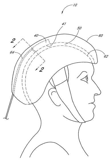
[0025] Figure 1 schematically illustrates a therapy apparatus comprising a cap
which fits securely over the patient's head.
[0026] Figi.~re 2 schematically illustrates a fragmentary cross-sectional view
talcen
along the lines 2-2 of Figure 1, showing one embodiment of a portion of a
therapy apparatus
comprising an element and its relationship to the scalp and brain.
[0027] Figl.~re 3 schematically illustrates an embodiment with an element
comprising a container coupled to an inlet conduit and an outlet conduit for
the transport of a
flowing material tluough the element.
[0028] Figv.~re 4A schematically illustrates a fragmentary cross-sectional
view
taken along the lines 2-2 of Figure 1, showing another embodiment of a portion
of a therapy
apparatus compuising an element with a portion contacting the scalp and a
portion spaced
away from the scalp.
[0029] Figure 4B schematically illustrates a fiagmentary cross-sectional view
talcen along the lines 2-2 of Figure 1, showing an embodiment of a portion of
a therapy
apparatus comprising a ph~rality of light sources and an element with portions
contacting the
scalp and portions spaced away fiom the scalp.
[0030] Figures SA and SB schematically illustrate cross-sectional views of two
embodiments of the element in accordance with Figure 4B taken along the line 4-
4.
[0031] Figures 6A-6C schematically illustrate an embodiment in which the light
sources axe spaced away from the scalp.
[0032] Figures 7A and 7B schematically illustrate the diffusive effect on the
light
by the element.
-6-
CA 02537370 2006-02-28
WO 2005/025672 PCT/US2004/029724
[0033] Figures 8A and 8B schematically illustrate two light beams having
different cross-sections impinging a patient's scalp and propagating through
the patient's
head to irradiate a portion of the patient's brain tissue.
[0034] Figure 9A schematically illustrates a therapy apparatus comprising a
cap
and a light source comprising a light blanlcet.
[0035] Figiues 9B and 9C schematically illustrate two embodiments of the light
blanket.
[0036] Figure 10 schematically illustrates a therapy apparatus comprising a
flexible strap and a housing.
[0037] Figure 11 schematically illustrates a therapy apparatus comprising a
handheld probe.
[0038] Figure 12 is a blocle diagram. of a control circuit comprising a
programmable controller.
[0039] Figure 13 schematically illustrates a therapy apparatus comprising a
light
source and a controller.
[0040] Figure 14 schematically illustrates a light source comprising a laser
diode
and a galvometer with a miiTOr and a plurality of motors.
[0041] Figures 15A and 15B schematically illustrate two irradiation patterns
tlat
are spatially shifted relative to each other.
[0042] Figure 16 schematically illustrates an exemplary therapy apparatus in
accordance with embodiments described herein.
[0043] Figure 17A is a graph of the effects of laser treatment of 7.SmW/cm2
for a
treatment duration of 2 minutes on a population of rabbits having small clot
embolic stroke.
[0044] Figure 17B is a graph of the effects of laser treatment of 25 mW/cmz
for a
treatment duration of 10 minutes on a population of rabbits having small clot
embolic strobe.
[0045] Figvue 18 is a graph showing the therapeutic window for laser-induced
behavioral improvements after small-clot embolic stTOlces in rabbits.
Detailed Description of the Preferred Embodiment
[0046] Low level light therapy ("LLLT") or phototherapy involves therapeutic
administration of light energy to a patient at lower power outputs than those
used for cutting,
CA 02537370 2006-02-28
WO 2005/025672 PCT/US2004/029724
cauterizing, or ablating biological tissue, resulting in desirable
biostimulatory effects while
leaving tissue wdamaged. W non-invasive phototherapy, it is desirable to apply
an
efficacious amount of light energy to the internal tissue to be treated using
light sources
positioned outside the body. (See, e.g., U.S. Patent No. 6,537,304 to Oron and
U.S. Patent
Application No. 10/353,130, both of which are incorporated in their entireties
by reference
herein.)
[0047] Laser therapy has been shown to be effective in a variety of settings,
including treating lymphoedema and muscular trauma, and carpal tunnel
syndrome. Recent
studies have shown that laser-generated infrared radiation is able to
penetrate various tissues,
including the brain, and modify fiuiction. In addition, laser-generated
infrared radiation can
induce angiogenesis, modify growth factor (transforming growth factor-[3)
sig~laling
pathways, and enhance protein s5nlthesis.
[0048] However, absorption of the light energy by intervening tissue can limit
the
amotmt of light energy delivered to the target tissue site, while heating the
intervening tissue.
In addition, scattering of the light energy by intervening tissue can limit
the power density or
energy density delivered to the target tissue site, Brute force attempts to
circumvent these
effects by increasing the power and/or power density applied to the outside
surface of the
body can result in damage (e.g., burning) of the intervening tissue.
[0049] Non-invasive phototherapy methods are circmnscribed by setting selected
treatment parameters within specified limits so as to preferably avoid
damaging the
intervening tissue. A review of the existing scientific literature in this
field would cast doubt
on whether a set of undamaging, yet efficacious, parameters could be fond.
However,
ceutain embodiments, as described herein, provide devices and methods which
can achieve
this goal.
(0050] Such embodiments may include selecting a wavelength of light at which
the absorption by intervening tissue is below a damaging level. Such
embodiments may also
include setting the power output of the light source at very low, yet
efficacious, poyver
densities (e.g., between approximately 100 yW/cm'' to approximately 10 W/cm2)
at the tazget
tissue site, and time periods of application of the light energy at a few
seconds to minutes to
achieve an efficacious energy density at the target tissue site being treated.
Other parameters
_g_
CA 02537370 2006-02-28
WO 2005/025672 PCT/US2004/029724
can also be varied in the use of phototherapy. These other parameters
contribute to the light
energy that is actually delivered to the treated tissue and may play lcey
roles in the efficacy of
phototherapy. W certain embodiments, the irradiated poution of the brain can
comprise the
entire brain.
Element to Inhibit Temperature Increases at the Scab
[0051] Figures 1 and 2 schematically illustrate an embodiment of a therapy
apparatus 10 for treating a patient's brain 20. The therapy apparatus 10
comprises a light
source 40 having an output emission area 41 positioned to irradiate a portion
of the brain 20
with an efficacious power density and wavelength of light. The therapy
apparatus 10 further
comprises an element 50 interposed between the light source 40 and the
patient's scalp 30.
The element 50 is adapted to inhibit temperature increases at the scalp 30
caused by the light.
[0052) As used herein, the temp "element" is used in its broadest sense,
including,
but not linuted to, as a reference to a constituent or distinct part of a
composite device. In
certain embodiments, the element 50 is adapted to contact at least a portion
of the patient's
scalp 30, as schematically illustrated in Figlues 1-4. liz certain such
embodiments, the
element 50 is in themal commuucation with and covers at least a portion of the
scalp 30. In
other embodiments, the element 50 is spaced away from the scalp 30 and does
not contact the
scalp 30.
[0053] In certain embodiments, the light passes tluough the element 50 prior
to
reaching the scalp 30 such that the element 50 is in the optical path of light
propagating from
the light source 40, tluough the scalp 30, through the bones, tissues, and
fluids of the head
(schematically illustrated in Figure 1 by the region 22), to the brain 20. In
certain
embodiments, the light passes through a transmissive medium of the element 50,
while in
other embodiments, the light passes through an aperhue of the element 50. As
described
more fully below, the element 50 may be utilized with various embodiments of
the therapy
apparatus 10.
[0054] In certain embodiments, the light source 40 is disposed on the interior
surface of a cap 60 which fits securely over the patient's head. The cap 60
provides stiwctural
integrity for the therapy apparatus 10 and holds the light source 40 and
element 50 in place.
Exemplary materials for the cap 60 include, but are not limited to, metal,
plastic, or other
_9_
CA 02537370 2006-02-28
WO 2005/025672 PCT/US2004/029724
materials with appropriate structural integrity. The cap 60 may include an
inner lining 62
comprising a stretchable fabric or mesh material, such as Lycra or nylon. In
certain
embodiments, the light source 40 is adapted to be removably attached to the
cap 60 in a
plurality of positions so that the output emission area 41 of the light source
40 can be
advantageously placed in a selected position for treatment of a strobe or CVA
in any portion
of the brain 20. In other embodiments, the light source 40 can be an integral
portion of the
cap 60.
[0055] The light source 40 illustrated by Figures 1 and 2 comprises at least
one
power conduit 64 coupled to a power source (not shown). W some embodiments,
the power
conduit 64 comprises an electrical conduit which is adapted to transmit
electrical signals and
power to an emitter (e.g., laser diode or light-emitting diode). W certain
embodiments, the
power conduit 64 comprises an optical conduit (e.g., optical waveguide) which
transmits
optical signals and power to the output emission area 41 of the light source
40. In certain
such embodiments, the light source 40 comprises optical elements (e.g.,
lenses, diffusers,
and/or waveguides) which transmit at least a portion of the optical power
received via the
optical conduit 64. In still other embodiments, the therapy apparatus 10
contains a power
source (e.g., a battery) and the power conduit 64 is substantially internal to
the therapy
app aratus 10.
[0056] In certain embodiments, the patient's scalp 30 composes hair and shin
which cover the patient's skull. In other embodiments, at least a portion of
the hair is
removed prior to the phototherapy treatment, so that the therapy apparatus 10
substantially
contacts the skin of the scalp 30.
[0057] In certain embodiments, the element 50 is adapted to contact the
patient's
scalp 30, thereby providing an interface between the therapy°apparatus
10 and the patient's
scalp 30. h1 certain such embodiments, the element 50 is coupled to the light
source 40 and
in other such embodiments, the element is also adapted to conform to the scalp
30, as
schematically illustrated in Figure 1. In this way, the element 50 positions
the output
emission area 41 of the light source 40 relative to the scalp 30. hi certain
such embodiments,
the element 50 is mechanically adjustable so as to adjust the position of the
light source 40
relative to the scalp 30. By fitting to the scalp 30 and holding the light
source 40 in place, the
-10-
CA 02537370 2006-02-28
WO 2005/025672 PCT/US2004/029724
element 50 iWibits temperature increases at the scalp 30 that would otherwise
result from
misplacement of the light source 40 relative to the scalp 30. W addition, in
certain
embodiments, the element 50 is mechanically adjustable so as to fit the
therapy apparatus 10
to the patient's scalp 30.
(0058] In certain embodiments, the element 50 provides a reusable interface
between the therapy apparatus 10 and the patient's scalp 30. W such
embodiments, the
element 50 can be cleaned or sterilized between uses of the therapy apparatus,
particularly
between uses by different patients. In other embodiments, the element 50
provides a
disposable and replaceable interface between the therapy apparatus 10 and the
patient's scalp
30. By using pre-sterilized and pre-paclcaged replaceable interfaces, certain
embodiments can
advantageously provide sterilized interfaces without undergoing cleaning or
sterilization
processing immediately before use.
[0059] In certain embodiments, the element 50 comprises a container (e.g., a
cavity or bag) containing a material (e.g., gel or liquid). The container can
be flexible and
adapted to conform to the contours of the scalp 30: Other exemplary materials
contained in
the container of the element 50 include, but are not limited to, thermal
exchange materials
such as glycerol and water. The element 50 of certain embodiments
substantially covers the
entire scalp 30 of the patient, as schematically illustrated in Figure 2. W
other embodiments,
the element 50 only covers a localized portion of the scalp 30 in proximity to
the irradiated
portion of the scalp 30.
[0060] W certain embodiments, at least a portion of the element 50 is within
an
optical path of the light from the light source 40 to the scalp 30. In such
embodiments, the
element 50 is substantially optically transmissive at a wavelength of the
light emitted by the
output emission area 41 of the light source 40 and is adapted to reduce back
reflections of the
light. By reducing baclc reflections, the element 50 increases the amount of
light transmitted
to the brain 20 and reduces the need to use a higher power light source 40
which may
otherwise create temperature increases at the scalp 30. W certain such
embodiments, the
element 50 comprises one or more optical coatings, films, layers, membranes,
etc. in the
optical path of the transmitted light which are adapted to reduce back
reflections.
-11-
CA 02537370 2006-02-28
WO 2005/025672 PCT/US2004/029724
[0061] In certain such embodiments, the element 50 reduces back reflections by
fitting to the scalp 30 so as to substantially reduce air gaps between the
scalp 30 and the
element 50 in the optical path of the light. The refractive-index mismatches
between such an
air gap and the element 50 and/or the scalp 30 would otherwise result in at
least a portion of
the light propagating from the light source 40 to the brain 20 to be reflected
baclc towards the
light source 40.
[0062] In addition, cez-tain embodiments of the element 50 comprise a matez-
ial
having, at a wavelength of light emitted by the light source 40, a refractive
index which
substantially matches the refractive index of the scalp 30 (e.g., about 1.3),
thereby reducing
any index-mismatch-generated back reflections between the element 50 and the
scalp 30.
Examples of materials with refractive indices compatible with embodiments
described herein
include, but are not limited to, glycerol, water, and silica gels. Exemplary
index-matching
gels include, but are not limited to, those available from Nye Lubricants,
Inc. of Fairhaven,
Massachusetts.
[0063] In cez-tain embodiments, the element 50 is adapted to cool the scalp 30
by
removing heat froze the scalp 30 so as to inhibit temperature increases at the
scalp 30. In
certain such embodiments, the element 50 comprises a reservoir (e.g., a
chamber or a
conduit) adapted to contain a coolant. The coolant flows through the reservoir
near the scalp
30. The scalp 30 heats the coolant, which flows away from the scalp 30,
thereby removing
heat from the scalp 30 by active cooling. The coolant in certain embodiments
circulates
between the element 50 and a heat transfer device, such as a chiller, whereby
the coolant is
heated by the scalp 30 and is cooled by the heat transfer device. Exemplazy
materials for the
coolant include, but are not limited to, water or air.
[0064] In certain embodiments, the element 50 comprises a container 51 (e.g.,
a
flexible bag) coupled to an inlet conduit 52 and an outlet conduit 53, as
schematically
illustrated in Figure 3. A flowing material (e.g., water, air, or glycerol)
can flow into the
container 51 froze the inlet conduit 52, absorb heat from the scalp 30, and
flow out of the
container 51 through the outlet conduit 53. Certain such embodiments can
provide a
mechanical fit of the container 51 to the scalp 30 and sufficient thermal
coupling to prevent
-12-
CA 02537370 2006-02-28
WO 2005/025672 PCT/US2004/029724
excessive heating of the scalp 30 by the light. In certain embodiments, the
container 51 can
be disposable and replacement containers 51 can be used for subsequent
patients.
[0065] In still other embodiments, the element 50 comprises a container (e.g.,
a
flexible bag) containing a material which does not flow out of the container
but is thermally
coupled to the scalp 30 so as to remove heat fiom the scalp 30 by passive
cooling.
Exemplary materials include, but are not limited to, water, glycerol, and gel.
In ceutain such
embodiments, the non-flowing material can be pre-cooled (e.g., by placement in
a
refrigerator) prior to the phototherapy treatment to facilitate cooling of the
scalp 30.
[0066] In certain embodiments, the element 50 is adapted to apply pressure to
at
least a portion of the scalp 30. By applying sufficient pressure, the element
50 can blanch the
portion of the scalp 30 by forcing at least some blood out the optical path of
the light energy.
The blood removal resulting fiom the pressure applied by the element 50 to the
scalp 30
decreases the corresponding absorption of the light energy by blood in the
scalp 30. As a
result, temperature increases due to absorption of the light energy by blood
at the scalp 30 are
reduced. As a further result, the fraction of the light energy transmitted to
the subdermal
target tissue of the brain 20 is increased.
[0067] Figures 4A and 4B schematically illustrate embodiments of the element
50
adapted to facilitate the blanching of the scalp 30. In the cross-sectional
view of a portion of
the therapy apparatus 10 schematically illustrated in Figure 4A, certain
element portions 72
contact the patient's scalp 30 and other element poutions 74 are spaced away
from the scalp
30. The element portions 72 contacting the scalp 30 provide an optical path
for light to
propagate from the light source 40 to the scalp 30. The element portions 72
contacting the
scalp 30 also apply pressure to the scalp 30, thereby forcing blood out from
beneath the
element portion 72. Figure 4B schematically illustrates a similar view of an
embodiment in
which the light source 40 comprises a plurality of light sources 40a, 40b,
40c. .
[0068] Figure SA schematically illustrates one embodiment of the cross-section
along the line 4-4 of Figure 4B. The element poutions 72 contacting the scalp
30 comprise
ridges extending along one direction, and the element portions 74 spaced away
from the scalp
30 comprise troughs extending along the same direction. W certain embodiments,
the ridges
are substantially parallel to one another and the troughs are substantially
parallel to one
-13-
CA 02537370 2006-02-28
WO 2005/025672 PCT/US2004/029724
another. Figure SB schematically illustrates another embodiment of the cross-
section along
the line 4-4 of Figure 4B. The element portions 72 contacting the scalp 30
comprise a
plurality of projections in the form of a grid or array. More specifically,
the portions 72 are
rectangular and are separated by element portions 74 spaced away from the
scalp 30, which
form troughs extending in two substantially perpendicular directions. The
portions 72 of the
element 50 contacting the scalp 30 can be a substantial fraction of the total
area of the
element 50 or of the scalp 30.
[0069] Figures 6A-6G schematically illustrate an embodiment in which the light
sources 40 are spaced away from the scalp 30. hi certain such embodiments, the
light emitted
by the light sources 40 propagates from the light sources 40 through the scalp
30 to the brain
20 and disperses in a direction generally parallel to the scalp 30, as shown
in Figure 6A. The
light sources 40 are preferably spaced sufficiently far apart from one another
such that the
light emitted from each light source 40 overlaps with the light emitted from
the neighboring
light sources 40 at the brain 20. Figure 6B schematically illustrates this
overlap as the
overlap of circular spots 42 at a reference depth at or below the surface of
the brain 20.
Figure 6C schematically illustrates this overlap as a graph of the power
density at the
reference depth of the brain 20 along the line L-L of Figures 6A and 6B.
Summing the power
densities from the neighboring light sources 40 (shown as a dashed line in
Figvue 6C) seines
to provide a more uniform light distribution at the tissue to be treated. In
such embodiments,
the sunnned power density is preferably less than a damage threshold of the
brain 20 and
above an efficacy threshold.
[0070] W certain embodiments, the element 50 is adapted to diffuse the light
prior
to reaching the scalp 30. Figures 7A and 7B schematically illustrate the
diffusive effect on
the light by the element 50. An exemplary energy density profile of the light
emitted by a
light source 40, as illustrated by Figlare 7A, is peaked at a particular
emission angle. After
being diffused by the element 50, as illustrated by Figure 7B, the energy
density profile of the
light does not have a substantial peals at any particular emission angle, but
is substantially
evenly distributed among a range of emission angles. By diffusing the light
emitted by the
light source 40, the element 50 distributes the light energy substantially
evenly over the area
to be illwninated, thereby inhibiting "hot spots" which would otherwise create
temperature
-14-
CA 02537370 2006-02-28
WO 2005/025672 PCT/US2004/029724
increases at the scalp 30. W addition, by diffusing the light prior to its
reaching the scalp 30,
the element 50 can effectively increase the spot size of the light impinging
the scalp 30,
thereby advantageously lowering the power density at the scalp 30, as
described more fully
below. W addition, in embodiments with multiple light sources 40, the element
50 can
diffuse the light to alter the total light output distribution to reduce
inhomogeneities.
[0071] W certain embodiments, the element 50 provides sufficient diffusion of
the
light such that the power density of the light is less than a maximum
tolerable level of the
scalp 30 and brain 20. In certain other embodiments, the element 50 provides
sufficient
diffusion of the light such that the power density of the light equals a
therapeutic value at the
target tissue. The element 50 can comprise exemplary diffusers including, but
are not limited
to, holographic diffusers such as those available from Physical Optics Corp.
of Torrance,
Califonua and Display Optics P/N SN1333 from Reflexite Coip. of Avon,
Connecticut.
Power Density
[0072] Phototherapy for the treatment of stroke is based in part on the
discovery
that power density (i.e., power per unit area or number of photons per unit
area per unit time)
and energy density (i.e., energy per mlit area or number of photons per unit
area) of the light
energy applied to tissue appear to be significant factors in detemninng the
relative efficacy of
low level phototherapy. This discovery is particularly applicable with respect
to treating and
saving suzviving but endangered neurons in a zone of danger sm-romding the
primary infarct
after a strolce or cerebrovascular accident (CVA). Preferred methods described
herein are
based at least in part on the fording that, given a selected wavelength of
light energy, it is the
power density and/or the energy density of the light delivered to tissue (as
opposed to the
total power or total energy delivered to the tissue) that appears to be
important factors in
determining the relative efficacy of phototherapy.
[0073] Without being bound by theory, it is believed that light energy
delivered
within a certain range of power densities and energy densities provides the
desired
biostimulative effect on the intracellular enviromnent, such that proper
function is retun~ed to
previously nonfunctioning or poorly functioning mitochondria in at-risk
neurons. The
biostimulative effect may include interactions with cluomophores within the
target tissue,
which facilitate production of ATP thereby feeding energy to injured cells
which have
-15-
CA 02537370 2006-02-28
WO 2005/025672 PCT/US2004/029724
experienced decreased blood flow due to the stroke. , Because strokes
correspond to
bloclcages or other interruptions of blood flow to portions of the brain, it
is thought that any
effects of increasing blood flow by phototherapy are of less importance in the
efficacy of
phototherapy for stroke victims. Further information regarding the role of
power density and
exposure time is described by Hans H.F.I. van Breugel and P.R. Dop Bar in
"Power Density
and Exposure Time of He-Ne Laser IlTadiation Are More Important Than Total
Energy Dose
in Photo-Biomodulation of Human Fibroblasts In Vitro," Lasers in Surgery and
Medicine,
Volume 12, pp. 528-537 (1992), which is incorporated in its entirety by
reference herein.
[0074] The significance of the power density used in phototherapy has
ramifications with regard to the devices and methods used in phototherapy of
brain tissue, as
schematically illustrated by Figures 8A and 8B, which show the effects of
scattering by
intervening tissue. Flu-ther information regarding the scattering of light by
tissue is provided
by V. Tllchlll In "Tissue Optics: Light Scattering Methods and Instruments for
Medical
Diagnosis," SPIE Press (2000), Bellingharn, WA, pp. 3-11, which is
incorporated in its
entirety by reference herein.
[0075] Figure 8A schematically illustrates a light beam 80 impinging a portion
90
of a patient's scalp 30 and propagating through the patient's head to
irradiate a portion 100 of
the patient's brain tissue 20. In the exemplary embodiment of Figure 8A, the
light beam 80
impinging the scalp 30 is collimated and has a circular cross-section with a
radius of 2 cm
and a cross-sectional area of approximately 12.5 cm2. For comparison purposes,
Figw-e 8B
schematically illustrates a light beam 82 having a sigluflcantly smaller cross-
section
impinging a smaller portion 92 of the scalp 30 to irradiate a portion 102 of
the brain tissue
20. The light beam 82 impinging the scalp 30 in Figure 8B is collimated and
has a circular
cross-section with a radius of 1 cm and a cross-sectional area of
approximately 3.1 cm''. The
collimations, cross-sections, and radii of the light beams 80, 82 illustrated
in Figures 8A and
8B are exemplary; other light beams with other parameters are also compatible
with
embodiments described herein. In particular, similar considerations apply to
focussed beams
or diverging beams, as they are similarly scattered by the intezvening tissue.
(0076] As shown in Figures 8A and 8B, the cross-sections of the light beams
80,
82 become larger while propagating through the head due to scattering from
interactions with
-16-
CA 02537370 2006-02-28
WO 2005/025672 PCT/US2004/029724
tissue of the head. Assuming that the angle of dispersion is 15 degrees and
the irradiated
brain tissue 20 is 2.5 cm below the scalp 30, the resulting area of the
portion 100 of brain
tissue 20 irradiated by the light beam 80 in Figure 8A is approximately 22.4
cm2. Similarly,
the resulting area of the portion 102 of brain tissue 20 irradiated by the
light beam 82 in
Figure 8B is approximately 8.8 cm2.
[0077] h-radiating the portion 100 of the brain tissue 20 with a power density
of
mW/cmz corresponds to a total power within the poution 100 of approximately
224 mW
(10 mW/cm2 x 22.4 cmz). Assuming only approximately 5% of the light beam 80 is
transmitted between the scalp 30 and the brain tissue 20, the incident light
beam 80 at the
scalp 30 will have a total power of approximately 4480 mW (224 mW / 0.05) and
a power
density of approximately 358 mW/cm2 (4480 mW / 12.5 cm2). Similarly,
irradiating the
portion 102 of the brain tissue 20 with a power density of 10 mW/cm2
corresponds to a total
power within the portion 102 of approximately 88 mW (10 mWlcm2 x 8.8 cm2), and
with the
same 5% transmittance, the incident light beam 82 at the scalp 30 will have a
total power of
approximately 1760 mW (88 mW / 0.05) and a power density of approximately 568
mW/cm''
(1760 mW / 3.1 cm2). These calculations are summarized in Table 1.
Table l:
2 cm Spot Size 1 cm Spot Size
(Fr -e 8A) (Fr a 8B)
Scalp:
Ar ea 12.5 cm 3 .1 Clll
Total ower 4480 mW 17G0 mW
Power density 358 mW/cm2 SG8 mW/cm''
Brain:
Area 22.4 cm 8.8 cm
Total ower 224 mW 88 mW
Power density 10 mW/cm2 10 mW/cm
[0078] These exemplary calculations illustrate that to obtain a desired power
density at the brain 20, higher total power at the scalp 30 can be used in
conjunction with a
larger spot size at the scalp 30. Thus, by increasing the spot size at the
scalp 30, a desired
power density at the brain 20 can be achieved with lower power densities at
the scalp 30
which can reduce the possibility of overheating the scalp 30. In certain
embodiments, the
-17_
CA 02537370 2006-02-28
WO 2005/025672 PCT/US2004/029724
light can be directed through an apeuure to define the illumination of the
scalp 30 to a
selected smaller area.
Light Source
[0079] Iii certain embodiments, a single Light source 40 is used as a light
generator to generate light, while in other embodiments, a plurality of Light
sources 40 are
used as a light generator to generate light. The light source 40 preferably
generates light in
the visible to near-infrared wavelength range. W certain embodiments, the
light solace 40
comprises one or more laser diodes, which each provide coherent light, h1
embodiments in
which the Light from the light source 40 is coherent, the emitted light may
produce
"speckling" due to coherent interference of the light. This speclcling
comprises intensity
spikes which are created by constructive interference and can occur in
proximity to the target
tissue being treated. For example, while the average power density may be
approximately 10
mWfcm~, the power density of one such intensity spire in proximity to the
brain tissue to be
treated may be approximately 300 mW/cm2. 111 certain embodiments, this
increased power
density due to speclcling can improve the efficacy of treatments using
coherent light over
those using incoherent light for illmnination of deeper tissues.
[0080] In other embodiments, the light source 40 provides incoherent tight.
Exemplary Light sources 40 of incoherent Light include, but are not limited
to, incandescent
lamps or light-emitting diodes. A heat sink can be used With the light source
40 (for either
coherent or incoherent sources) to remove heat from the light source 40 and to
inhibit
temperattue increases at the scalp 30.
[0081] W certain embodiments, the light source 40 generates light which is
substantially monocluomatic (i.e., light having one wavelength, or light
having a narrow band
of wavelengths). So that the amount of light transmitted to the brain is
maximized, the
wavelength of the light is selected in certain embodiments to be at or near a
transmission
peals (or at or near an absorption minimum) fox the intervening tissue. h1
certain such
embodiments, the wavelength corresponds to a peals in the transmission
spectrum of tissue at
about 820 manometers. In other embodiments, the wavelength of the light is
preferably
between about 630 manometers and about 1464 manometers, more preferably
between about
780 manometers and about 840 manometers, and most preferably includes
wavelengths of
-18-
CA 02537370 2006-02-28
WO 2005/025672 PCT/US2004/029724
about 785, 790, 795, 800, 805, 810, 815, 820, 825, or 830 manometers. An
intemnediate
wavelength in a range between approximately 730 manometers and approximately
750
manometers (e.g., about 739 manometers) appears to be suitable for penetrating
the skull,
although other wavelengths are also suitable and may be used.
[0082] W other embodiments, the light source 40 generates light having a
plurality
of wavelengths. W certain such embodiments, each wavelength. is selected so as
to worlc with
one or more chromophores within the target tissue. Without being boozed by
theory, it is
believed that irradiation of chromophores increases the production of ATP in
the target
tissue, thereby producing beneficial effects. W certain embodiments, the light
source 40 is
adapted to generate light having a first wavelength concurrently with light
having a second
wavelength. In certain other embodiments, the light source 40 is adapted to
generate light
having a fwst wavelength sequentially with light having a second wavelength.
[0083] In certain embodiments, the light source 40 includes at least one
continuously emitting GaAIAs laser diode having a wavelength of about 830
manometers. In
another embodiment, the light source 40 comprises a laser source having a
wavelength of
about 808 manometers. In still other embodiments, the light source 40 includes
at least one
vertical cavity surface-emitting laser (VCSEL) diode. Other light sources 40
compatible with
embodiments described herein include, but are not limited to, light-emitting
diodes (LEDs)
and filtered lamps.
[00841 The Light source 40 is capable of emitting light energy at a power
sufficient fio achieve a predetermined power density at the subdennal target
tissue (e.g., at a
depth of approximately 2 centimeters from the dura). It is presently believed
that
phototherapy of tissue is most effective when irradiating the target tissue
with power
densities of Light of at least about 0.01 mW/cm''' and up to about 1 W/cm2 at
the Ievel of the
tissue. W various embodiments, the subsurface power density is at least about
0.01, 0.05, 0.1,
0.5, 1, 5, 10, 15, 20, 30, 40, 50, 60, 70, 80, or 90 mW/cm2, respectively,
depending on the
desired clinical performance. In certain embodiments, the subsurface power
density at the
target tissue is preferably about 0.01 mW/cm2 to about 100 mW/cm2, more
preferably about
0.01 mWlcm2 to about 50 mWlcm2, and most preferably about 2 mW/cmz to about 20
-19-
CA 02537370 2006-02-28
WO 2005/025672 PCT/US2004/029724
mW/cm2. It is believed that these subsurface power densities are especially
effective at
producing the desired biostimulative effects on the tissue being treated.
j0085] Talcing into account the attenuation of energy as it propagates from
the
slcin surface, through body tissue, bone, and fluids, to the subdermal target
tissue, surface
power densities preferably between about 10 mW/cm2 to about 10 W/cm2, or more
preferably
between about 100 mW/cmz to about 500 mW/cm2, will typically be used to attain
the
selected power densities at the subdermal target tissue. To achieve such
surface power
densities, the light source 40 is preferably capable of emitting light energy
having a total
power output of at least about 25 mW to about 100 W. In various embodiments,
the total
power output is limited to be no more than about 30, 50, 75, 100, 150, 200,
250, 300, 400, or
500 mW, respectively. hi certain embodiments, the light source 40 comprises a
plurality of
sources used in combination to provide the total power output. The actual
power output of
the light source 40 is preferably controllably variable. In this way, the
power of the light
energy emitted can be adjusted in accordance with a selected power density at
the subdermal
tissue being treated.
[0086] Certain embodiments utilize a light source 40 that includes only a
single
laser diode that is capable of providing about 25 mW to about 100 W of total
power output at
the slcin surface. In certain such embodiments, the laser diode can be
optically coupled to the
scalp 30 via an optical fiber or can be configured to provide a sufficiently
large spot size to
avoid power densities which would burn or otherwise damage the scalp 30. In
other
embodiments, the light source 40 utilizes a plurality of sources (e.g., laser
diodes) arranged in
a grid or array that together are capable of providing at least about 25 mW to
about 100 W of
total power output at the shin surface. The light source 40 of ether
embodiments may also
comprise soL~rces having power capacities outside of these limits.
[0087] Figure 9A schematically illustrates another embodiment of the therapy
apparatus 10 which comprises the cap 60 and a light source comprising a light-
emitting
blanl~et 110. Figure 9B schematically illustrates an embodiment of the
blanlcet 110
comprising a flexible substrate 111 (e.g., flexible circuit board), a power
conduit interface
112, and a sheet formed by optical fibers 114 positioned in a fan-life
configuration. Figure
9C schematically illustrates an embodiment of the blancet 110 comprising a
flexible substrate
-20-
CA 02537370 2006-02-28
WO 2005/025672 PCT/US2004/029724
111, a power conduit interface 112, and a sheet formed by optical fibers 114
woven into a
mesh. The blau~et 110 is preferably positioned within the cap 60 so as to
cover an area of
the scalp 30 corresponding to a portion of the brain 20 to be treated.
[0088] W certain such embodiments, the power conduit interface 112 is adapted
to
be coupled to an optical fiber conduit 64 which provides optical power to the
blanket 110.
The optical power interface 112 of certain embodiments comprises a beam
splitter or other
optical device which distributes the incoming optical power among the various
optical fibers
114. W other embodiments, the power conduit interface 112 is adapted to be
coupled to an
electrical conduit which provides electrical power to the blancet 110. In
certain such
embodiments, the power conduit interface 112 comprises one or more laser
diodes, the output
of which is distributed among the various optical fibers 114 of the blanket
110. In ceutain
other embodiments, the blanket 110 comprises an electroluminescent sheet which
responds to
electrical signals from the power conduit interface 112 by emitting light. W
such
embodiments, the power conduit interface 112 comprises circuitry adapted to
distribute the
electrical signals to appropriate portions of the electroluminescent sheet.
[0089] The side of the blanket 110 nearer the scalp 30 is preferably provided
with
a light scattering surface, such as a roughened swface to increase the amount
of light
scattered out of the blanket 110 towards the scalp 30. The side of the
blanlcet 110 further
from the scalp 30 is preferably covered by a reflective coating so that light
emitted away from
the scalp 30 is reflected back towards the scalp 30. This configvtration is
similar to
configwations used for the "baclc illtunination" of liquid-crystal displays
~LCDs). Other
configurations of the blanket 110 are compatible with embodiments described
herein.
[0090] In certain embodiments, the light source 40 generates light which cause
eye damage if viewed by an individual. fil such embodiments, the apparatus 50
can be
configured to provide eye protection so as to avoid vietxTing of the light by
individuals. For
example, opaque materials can be appropriately placed to block the light from
being viewed
directly. In addition, interlocks can be provided so that the light source 40
is not activated
unless the apparatus 50 is in place, or other appropriate safety measures are
taken.
-21-
CA 02537370 2006-02-28
WO 2005/025672 PCT/US2004/029724
Light Delivery Apparatuses
[0091] The phototherapy methods for the treatment of stroke described herein
may be practiced and described using, for example, a low level laser therapy
apparatus such
as that shown and described in U.S. Pat. No. 6,214,035, U.S. Pat. No.
6,267,780, U.S. Pat.
No. 6,273,905 and U.S. Pat. No. 6,290,714, which are all incorporated in their
entirety by
reference herein, as are the references incorporated by reference therein.
[0092] Another suitable phototherapy apparatus in accordance with embodiments
described here is illustrated in Figure 10. The illustrated therapy apparatus
10 includes a light
source 40, an element 50, and a flexible strap 120 adapted for securing the
therapy apparatus
over an area of the patient's head. The light solute 40 can be disposed on the
strap 120
itself, or in a housing 122 coupled to the strap 120. The light source 40
preferably comprises
a plurality of diodes 40a, 40b, etc. capable of enutting light energy having a
wavelength in
the visible to near-infrared wavelength range. The element 50 is adapted to be
positioned
between the light source 40 and the patient's scalp 30.
[0093] The therapy apparatus 10 fiuther includes a power supply (not shown)
operatively coupled to the light source 40, and a prograrnlnable controller
126 operatively
coupled to the light source 40 and to the power supply. The programmable
controller 126 is
configured to control the light source 40 so as to deliver a predetermined
power density to the
brain tissue 20. W certain embodiments, as schematically illustrated in Figure
10, the light
source 40 comprises the programmable controller 126. W other embodiments the
programmable controller 126 is a separate component of the therapy apparatus
10.
[0094] hl certain embodiments, the strap 120 comprises a loop of elastomeric
material sized appropriately to fit snugly onto the patient's scalp 30. W
other embodiments,
the strap 120 comprises an elastomeric material to which is secured any
suitable securing
means 130, such as mating Velcro strips, buclcles, snaps, hoolcs, buttons,
ties, or the like. The
precise configltration of the strap 120 is subject only to the limitation that
the strap 120 is
capable of maintaining the light source 40 in a selected position so that
light energy emitted
by the light source 40 is directed towards the targeted brain tissue 20.
[0095] In the exemplary embodiment illustrated in Figlue 10, the housing 122
comprises a layer of flexible plastic or fabric that is secured to the strap
120. In other
-22-
CA 02537370 2006-02-28
WO 2005/025672 PCT/US2004/029724
embodiments, the housing 122 comprises a plate or an enarged portion of the
strap 120.
Various strap config rations and spatial distributions of the light sources 40
are compatible
with embodiments described herein so that the therapy apparatus 10 can treat
selected
portions of brain tissue.
[0096] In still other embodiments, the therapy apparatus 10 for delivering the
light energy includes a handheld probe 140, as schematically illustrated in
Figure 11. The
probe 140 includes a light source 40 and an element 50 as described herein.
[0097] Figure 12 is a block diagram of a control circuit 200 comprising a
programunable controller 126 according to embodiments described herein. The
control
circuit 200 is configured to adjust the power of the light energy emitted by
the light source 40
to generate a predetermined surface power density at the scalp 30
corresponding to a
predetermined energy delivery profile, such as a predetemnined subsurface
power density, to
the target area of the brain 20.
[0098] In certain embodiments, the programmable controller 126 comprises a
logic circuit 210, a clocl~ 212 coupled to the logic circuit 210, and an
interface 214 coupled to
the logic circuit 210. The clock 212 of certain embodiments provides a timing
signal to the
logic circuit 210 so that the logic circuit 210 can monitor and control timing
internals of the
applied light. Examples of timing intervals include, but are not limited to,
total treatment
times, pulsewidth times for pulses of applied light, and time intervals
between pulses of
applied light. In certain embodiments, the light sources 40 can be selectively
turned on and
off to reduce the themnal load on the scalp 30 and to deliver a selected power
density to
particular areas of the brain 20.
[0099] The interface 214 of certain embodiments provides signals to the logic
circuit 210 which the logic circuit 210 uses to control the applied light. The
interface 214 can
comprise a user interface or an interface to a sensor monitoring at least one
parameter of the
treatment. In certain such embodiments, the progranunable controller 126 is
responsive to
signals from the sensor to preferably adjust the treatment parameters to
optimize the
measured response. The programmable controller 126 can thus provide closed-
loop
monitoring and adjustment of various treatment parameters to optimize the
phototherapy.
The signals provided by the interface 214 fiom a user are indicative of
parameters that may
-23-
CA 02537370 2006-02-28
WO 2005/025672 PCT/US2004/029724
include, but are not limited to, patient characteristics (e.g., slclll type,
fat percentage), selected
applied power densities, target time intervals, and power density/timing
profiles for the
applied light.
[0100] In certain embodiments, the logic circuit 210 is coupled to a light
source
driver 220. The light source driver 220 is coupled to a power supply 230,
which in certain
embodiments comprises a battery and in other embodiments comprises an
alternating culTent
source. The light source driver 220 is also coupled to the light source 40.
The logic circuit
210 is responsive to the signal from the cloclc 212 and to user input from the
user interface
214 to transmit a control signal to the light source driver 220. hl response
to the control
signal from the logic circuit 210, the light source driver 220 adjust and
controls the power
applied to the light sources 40. Other control circuits besides the control
circuit 200 of
Figure 12 axe compatible with embodiments described herein.
[0101] In certain embodiments, the logic circuit 110 is responsive to signals
from
a sensor monitoring at least one parameter of the treatment to control the
applied light. For
example, certain embodiments comprise a temperature sensor thermally coupled
to the scalp
30 to provide information regarding the temperature of the scalp 30 to the
logic circuit 210.
In such embodiments, the logic circuit 210 is responsive to the infol~rlation
from the
temperature sensor to transmit a control signal to the light source driver 220
so as to adjust
the parameters of the applied light to maintain the scalp temperature below a
predetermined
level. Other embodiments include exemplary biomedical sensors including, but
not limited
to, a blood flow sensor, a blood gas (e.g., oxygenation) sensor, an ATP
production sensor, or
a cellular activity sensor. Such biomedical sensors can provide real-time
feedback
information to the logic circuit 210. In certain such embodiments, the logic
circuit 110 is
responsive to signals from the sensors to preferably adjust the parameters of
the applied light
to optimize the measured response. The logic circuit 110 can thus provide
closed-loop
monitoring and adjustment of various parameters of the applied light to
optimize the
phototherapy.
[0102] In certain embodiments, as schematically illustrated in Figure 13, the
therapy apparatus 310 comprises a light source 340 adapted to irradiate a
portion of the
patient's brain 20 with an efficacious power density and wavelength of light.
The therapy
-24-
CA 02537370 2006-02-28
WO 2005/025672 PCT/US2004/029724
apparatus 310 fuuther comprises a controller 360 for energizing said light
source 340, so as to
selectively produce a plurality of different irradiation patterns on the
patient's scalp 30. Each
of the irradiation patterns is comprised of a least one illtuninated area that
is small compared
to the patient's scalp 30, and at least one non-illmninated area.
[0103] W certain embodiments, the light soluce 340 includes an apparatus for
adjusting the emitted light to irradiate different portions of the scalp 30.
In certain such
embodiments, the apparatus physically moves the light source 40 relative to
the scalp 30. In
other embodiments, the apparatus does not move the light source 40, but
redirects the emitted
light to different portions of the scalp 30. W an exemplary embodiment, as
schematically
illustrated in Figure 14, the light source 340 comprises a laser diode 342 and
a galvometer
344, both of which are electrically coupled to the controller 360. The
galvometer 344
comprises a mirror 346 mounted onto an assembly 348 which is adjustable by a
plurality of
motors 350. Light emitted by the laser diode 342 is directed toward the mirror
346 and is
reflected to selected portions of the patient's scalp 30 by selectively moving
the mirror 346
and selectively activating the laser diode 342. W certain embodiments, the
therapy apparatus
310 comprises an element 50 adapted to inhibit temperature increases at the
scalp 30 as
described herein.
[0104] Figure 15A schematically illustrates an irradiation pattern 370 in
accordance with embodiments described herein. The irradiation pattern 370
comprises at
least one illuminated area 372 and at least one non-ilhuninated area 374. W
ceutain
embodiments, the irradiation pattern 370 is generated by scanning the mirror
346 so that the
light impinges the patient's scalp 30 in the illuminated area 372 but not in
the non-
illuminated area 374. Certain embodiments modify the illununated area 372 and
the non-
illumiliated area 374 as a function of time.
[0105] This selective irradiation can be used to reduce the themal load on
particular locations of the scalp 30 by moving the light from one illuminated
area 372 to
another. For example, by irradiating the scalp 30 with the irradiation pattern
370
schematically illustrated in Figure 15A, the illuminated areas 372 of the
scalp 30 are heated
by interaction with the light, and the non-illuminated areas 374 are not
heated. By
subsequently irradiating the scalp 30 with the complementary irradiation
pattern 370'
-25-
CA 02537370 2006-02-28
WO 2005/025672 PCT/US2004/029724
schematically illustrated in Figure 15B, the previously non-illuminated areas
374 are now
illuminated areas 372', and the previously illuminated areas 372 are now non-
illuminated
areas 374'. A comparison of the illuminated areas 372 of the irradiation
pattern 370 of Figure
15A with the illtuninated area 372' of the irradiation pattern 370' of Figure
15B shows that
the illuminated areas 372, 372' do not significantly overlap one another. In
this way, the
theunal load at the scalp 30 due to the absorption of the light can be
distributed across the
scalp 30, thereby avoiding mduly heating one or more portions of the scalp 30.
[0106] Figure 16 schematically illustrates another therapy apparatus 400 in
accordance with embodiments described herein. The therapy apparatus 400
comprises a
plurality of light sources 410 in a housing 420. Each light source 410 has an
output emission
area positioned to irradiate a corresponding portion of the brain 20 with an
efficacious power
density and wavelength of light. In certain embodiments, these portions
overlap such that the
poution of the brain 20 irradiated by two or more light sow-ces 410 overlap
one another at
least in part. As described herein, the light sources 410 can be activated by
a controller (not
shown) in concert or separately to produce a predetermined irradiation
pattern.
[0107] The therapy apparatus 400 of Figure 16 further comprises a cap 430
interposed between the light sources 410 and the patient's scalp 30, such that
light passes
through the cap 430 prior to reaching the scalp 30. In certain embodiments,
the cap 430 is
substantially optically transmissive at the wavelength and reduces baclc
reflections of the
light. The cap 430 of ceutain embodiments fits to the scalp 30 so as to
substantially reduce air
gaps between the scalp 30 and the cap 430. W certain embodiments, the cap 430
comprises a
material having a refractive index which substantially matches a refiactive
index of the scalp
30. W certain embodiments, the cap 430 comprises a meterial having a
refractive index
which substantially matches a refiactive index of the skin and/or hair of the
scalp 30.
[0108] In the embodiment schematically illustrated by Figure 16, the cap 430
is
wearable over the patient's scalp 30. In certain such embodiments, the patient
wears the cap
430 and is in a reclining position so as to place his head in proximity to the
light sources 410.
The cap 430 is adapted to inhibit temperature increases at the scalp 30 caused
by the light
from the light sources 410, as described herein (e.g., by cooling the scalp
30, by blanching a
portion of the scalp 30, by diffusing the light prior to reaching the scalp
30).
-26-
CA 02537370 2006-02-28
WO 2005/025672 PCT/US2004/029724
Methods of Light Deliyery
[0109] Preferred methods of phototherapy are based at least in part on the
finding
described above that, for a selected wavelength, the power density (light
intensity or power
per unit area, in W/cmz) or the energy density (energy per unit area, in
J/cmz, or power
density multiplied by the exposure time) of the light energy delivered to
tissue is an important
factor in determining the relative efficacy of the phototherapy, and efficacy
is not as directly
related to the total power or the total energy delivered to the tissue. In the
methods described
herein, power density or energy density as delivered to a portion of the
patient's brain 20,
which can include the area of infarct after a strobe, appears to be important
factors in using
phototherapy to treat and save surviving but endangered neurons in a zone of
danger
surrounding the ii2farcted area. Certain embodiments apply optimal power
densities or
energy densities to the intended target tissue, within acceptable margins of
error.
[0110] In certain embodiments, the apparatus and methods of phototherapy
described herein increase the cerebral blood flow of the patient. In certain
such
embodiments, the cerebral blood flow is increased by 10%, 15%, 20%, or 25%
immediately
post-irradiation, as compared to inunediately prior to irradiation.
[0111] In certain embodiments, the apparatus and methods of phototherapy
described herein are used to treat strokes or other sources of
neurodegeneration. As used
herein, the term "neurodegeneration" refers to the process of cell destniction
resulting fiom
primary destructive events such as stroke or CVA, as well as from secondary,
delayed and
progressive destructive mechanisms that are involved by cells due to the
occurrence of the
primacy destructive event. Primary destructive events include disease
processes or physical
injury or insult, including strolve, but also include other diseases and
conditions such as
multiple sclerosis, amylotrophic lateral sclerosis, heat strolve, epilepsy,
Alzheimer's disease,
dementia resulting from other causes such as AIDS, cerebral ischemia including
focal
cerebral ischemia, and physical trauma such as cnish or compression injury in
the CNS,
including a crush or compression injury of the brain, spinal cord, nerves or
retina, or any
acute injury or insult producing neurodegeneration. Secondary destructive
mechanisms
include any mechanism that leads to the generation and release of neurotoxic
molecules,
including apoptosis, depletion of cellular energy stores because of changes in
mitochondria)
-27-
CA 02537370 2006-02-28
WO 2005/025672 PCT/US2004/029724
membrane permeability, release or failure in the reuptalce of excessive
glutamate, reperfusion
injury, and activity of cytokines and inflammation. Both primary and secondary
mechanisms
contribute to forming a "zone of danger" for neurons, wherein the netuons in
the zone have at
least temporarily survived the primary destructive event, but are at risk of
dying due to
processes having delayed effect.
[0112] As used herein, the term "neuroprotection" refers to a therapeutic
strategy
for slowing or preventing the otherwise irreversible loss of neurons due to
neurodegeneration
after a puimary destructive event, whether the neurodegeneration loss is due
to disease
mechanisms associated with the primacy destructive event or secondary
destmctive
mechanisms.
[0113] The term "cognitive function" as used herein refers to cognition and
cognitive or mental processes or functions, including those relating to
lalowing, thiucing,
learning, perception, memory (including immediate, recent, or remote memory),
and judging.
Symptoms of loss of cognitive function can also include changes in
personality, mood, and
behavior of the patient. Diseases or conditions affecting cognitive function
iilclude
Alzheimer's disease, dementia, AIDS or HIV infection, Cruetzfeldt-Jalcob
disease, head
trawna (including single-event trauma and long-term trauma such as multiple
concussions or
other traumas which may result from athletic injury), Lewy body disease,
Piclc's disease,
Parlcinson's disease, Huntington's disease, drug or alcohol abuse, brain
tumors,
hydrocephalus, kidney or liver disease, strolce, depression, and other mental
diseases which
cause disruption in cognitive function, and neurodegeneration.
[0114] The teen "motor function" as used herein refers to those bodily
functions
relating to muscular movements, primarily conscious muscular movements,
including motor
coordination, performance of simple and complex motor acts, and the like.
[0115] The term "neurologic function" as used herein includes both cognitive
function and motor function.
[0116] The terms "cognitive enhancement" and "motor eWancement" as used
herein refer to the improving or heightening of congnitive function and motor
function,
r espectively.
-28-
CA 02537370 2006-02-28
WO 2005/025672 PCT/US2004/029724
[0117] The term "neurologic eWancement" as used herein includes both cognitive
enhancement and motor enhancement.
[0118] As used herein, the term "net~roprotective-effective" as used herein
refers
to a characteristic of an amount of light energy, wherein the amount is a
power density of the
light energy measured in mW/cm2. A neuroprotective-effective amount of light
energy
achieves the goal of preventing, avoiding, reducilig, or eliminating
neurodegeneration, which
should result in cognitive enhancement and/or motor eWancement.
[0119] The term "neurologic function enhancement effective" as used herein
refers to a characteristic of an amount of light energy, whet ein the amount
is a power density
of the light energy measured in mW/cm2. The amount of light energy achieves
the goal of
neuroprotection, motor enhancement, and/or cognitive enhancement.
[0120] Thus, a method for the treatment of strolce or for the enhancement of
neurologic function in a patient in need of such treatment involves delivering
a neurologic
function enhancement effective amount or a neuroprotective-effective amount of
light energy
having a wavelength in the visible to near-infrared wavelength range to a
target area of the
patient's brain 20. In certain embodiments, the target area of the patient's
brain 20 includes
the area of infarct, i.e. to neurons within the "zone of danger." In other
embodiments, the
target area includes portions of the brain 20 not within the zone of danger.
Without being
bound by theory, it is believed that irradiation of healthy tissue in
proximity to the zone of
danger increases the production of ATP and copper ions in the healthy tissue
a~ld which then
migrate to the injured cells witlvn the region surrounding the infarct,
thereby producing
beneficial effects. Additional information regarding the biomedical mechanisms
or reactions
involved in phototherapy is provided by Tiina I. Karu in "Mechanisms of Low-
Power Laser
Light Action on Cellular Level", Proceedings of SPIE Vol. 4159 (2000), Effects
of Low-
Power Light on Biological Systems V, Ed. Rachel Lubart, pp. 1-17, which is
incorporated in
its entirety by reference herein.
[0121] h1 certain embodiments, delivering the neuroprotective amount of light
energy includes selecting a surface power density of the light energy at the
scalp 30
corresponding to the predetermined power density at the target area of the
brain 20. As
described above, light propagating through tissue is scattered and absorbed by
the tissue.
-29-
CA 02537370 2006-02-28
WO 2005/025672 PCT/US2004/029724
Calculations of the power density to be applied to the scalp 30 so as to
deliver a
predetermined power density to the selected target area of the brain 20
preferably talce into
account the attenuation of the light energy as it propagates through the skin
and other tissues,
such as bone and brain tissue. Factors laiown to affect the attenuation of
light propagating to
the brain 20 from the scalp 30 include, but are not limited to, slcin
pigmentation, the presence
and color of hair over the area to be treated, amount of fat tissue, the
presence of bruised
tissue, slcull thiclaZess, and the location of the target area of the brain
20, particularly the
depth of the area relative to the surface of the scalp 30. For example, to
obtain a desired
power density of 50 mW/cm2 in the brain 20 at a depth of 3 cm below the
surface of the scalp
30, phototherapy may utilize an applied power density of 500 mW/cm2. The
higher the level
of skin pigmentation, the higher the power density applied to the scalp 30 to
deliver a
predetermined power density of light energy to a subsurface site of the brain
20.
[0122] In certain embodiments, treating a patient suffering from the effects
of
strolce comprises placing the therapy apparatus 10 in contact with the scalp
30 and adjacent
the target area of the patient's brain 20. The target area of the patient's
brain 20 can be
previously identified such as by using standard medical imaging techniques. In
certain
embodiments, treatment further includes calculating a surface power density at
the scalp 30
which corresponds to a preselected power density at the target area of the
patient's brain 20.
The calculation of certain embodiments includes factors that affect the
penetration of the light
energy and thus the power density at the target area. These factors include,
but are not
limited to, the thiclaiess of the patient's s1.u11, type of hair and hair
coloration, skin coloration
and pigmentation, patient's age, patient's gender, and the distance to the
target area witlun
the brain 20. The power density and other parameters of the applied light are
then adjusted
according to the results of the calculation.
[0123] The power density selected to be applied to the target area of the
patient's
brain 20 depends on a number of factors, including, but not limited to, the
wavelength of the
applied light, the type of CVA (ischemic or hemorrhagic), and the patient's
clinical
condition, including the extent of the affected brain area. The power density
of light energy
to be delivered to the target area of the patient's brain 20 may also be
adjusted to be
combined with any other therapeutic agent or agents, especially phamnaceutical
-30-
CA 02537370 2006-02-28
WO 2005/025672 PCT/US2004/029724
neuroprotective agents, to aclueve the desired biological effect. W such
embodiments, the
selected power density can also depend on the additional therapeutic agent or
agents chosen.
(0124] hi prefeiTed embodiments, the treatment proceeds continuously for a
period of about 10 seconds to about 2 hours, more preferably for a period of
about 1 to about
minutes, and most preferably for a period of about 1 to 5 minutes. In other
embodiments,
the light energy is preferably delivered for at least one treatment period of
at least about five
minutes, and more preferably for at least one treatment period of at least ten
minutes. The
light energy can be pulsed during the treatment period or the light energy can
be continuously
applied during the treatment period.
[0125] In certain embodiments, the treatment may be temninated after one
treatment period, while in other embodiments, the treatment may be repeated
for at least two
treatment periods. The time between subsequent treatment periods is preferably
at least
about five minutes, more preferably at least about 1 to 2 days, and most
preferably at least
about one weelc. W certain embodiments in which treatment is performed over
the course of
multiple days, the apparatus 10 is wearable over multiple concmTent days
(e.g., embodiments
of Figures 1, 3, 9A, 10, and 13). The length of treatment time and fiequency
of treatment
periods can depend on several factors, including the functional recovery of
the patient and the
results of imaging analysis of the infarct. W certain embodiments, one or more
treatment
parameters can be adjusted in response to a feedback signal fiom a device
(e.g., magnetic
resonance imaging) monitoring the patient.
. [0126] During the treatment, the light energy may be continuously provided,
or it
may be pulsed. If the light is pulsed, the pulses are preferably at least
about 10 nanosecond
long and occur at a frequency of up to about 100 lcHz. Continuous wave light
may also be
used.
[0127] The thrombolytic therapies currently in use for treatment of strolce
are
typically begun within a few hours of the stroke. However, many hours often
pass before a
person who has suffered a stroke receives medical treatment, so the short time
limit for
initiating thrombolytic therapy excludes many patients fiom treatment. In
contrast,
phototherapy treatment of strolce appears to be more effective if treatment
begins no earlier
-31-
CA 02537370 2006-02-28
WO 2005/025672 PCT/US2004/029724
than several hours after the ischemic event has occm~red. Consequently, the
present methods
of phototherapy may be used to treat a greater percentage of stroke patients.
[012] In certain embodiments, a method provides a neuroprotective effect in a
patient that had an ischemic event in the brain. The method comprises
identifying a patient
who has experienced an ischemic event in the brain. The method ftu-ther
comprises
estimating the time of the ischemic event. The method further comprises
commencing
administration of a neuroprotective effective amount of light energy to the
brain. The
achninistration of the light energy is commenced no less than about two hours
following the
time of the ischemic event. W certain embodiments, phototherapy treatment can
be
efficaciously performed preferably within 24 hours after the ischemic event
occurs, and more
preferably no earlier than two hours following the ischemic event, still more
preferably no
earlier than three hours following the ischemic event, and most preferably no
earlier than five
hours following the ischemic event. W certain embodiments, one or more of the
treatment
parameters can be varied depending on the amount of time that has elapsed
since the
ischemic event.
[0129] Without being bound by theory, it is believed that the benefit in
delaying
treatment occurs because of the time needed for induction of ATP production,
and/or the
possible induction of angiogenesis in the region surrounding the infarct.
Thus, in accordance
with one preferred embodiment, the phototherapy for the treatment of stroke
occurs
preferably about 6 to 24 hours after the onset of stroke symptoms, more
preferably about 12
to 24 hours after the onset of symptoms. It is believed, however, that if
treatment begins after
about 2 days, its effectiveness will be greatly reduced.
[0130] In certain embodiments, the phototherapy is combined with other types
of
treatments for an improved therapeutic effect. Treatment can comprise
directing light
through the scalp of the patient to a target area of the brain concurrently
with applying an
electromagnetic field to the brain. W such embodiments, the light has an
efficacious power
density at the target area and the electromagnetic field has an efficacious
field strength. For
example, the apparatus 50 can also include systems for electromagnetic
treatment, e.g., as
described in U.S. Patent No. 6,042,531 issued to Holcomb, which is
incorporated in its
entirety by reference herein. In certain embodiments, the electromagnetic
field comprises a
-32-
CA 02537370 2006-02-28
WO 2005/025672 PCT/US2004/029724
magnetic field, while in other embodiments, the electromagiletic field
comprises a radio-
frequency (RF) field. As another example, treatment can comprise directing an
efficacious
power density of light through the scalp of the patient to a target area of
the brain
concurrently with applying an efficacious amount of ultrasonic energy to the
brain. Such a
system can include systems for ultrasonic treatment, e.g., as described in
U.S. Patent No.
5,054,470 issued to Fry et al., which is iilcorporated in its entirety by
reference hereW .
Phototherap e~ples
Example 1
[0131] An in vita°o experiment was done to demonstrate one effect of
phototherapy on neurons, namely the effect on ATP production. Normal Human
Neural
Progenitor (NHNP) cells were obtained cryopresemed through Clonetics of
Baltimore,
Maryland, catalog # GC-2599. The NHNP cells were thawed and cultured on
polyethyleneimine (PEn with reagents provided with the cells, following the
manufacturers'
instructions. The cells were plated into 9G well plates (blaclc plastic with
clear bottoms,
Becton Dickinson of Franklin Lalces, New Jersey) as spheroids and allowed to
differentiate
into mature neurons over a period of two weeks.
[0132] A Photo Dosing Assembly (PDA) was used to provide precisely metered
doses of laser light to the NHNP cells in the 9G well plates. The PDA included
a Nilcon
Diaphot inverted microscope (Nikon of Melville, New Yorlc) with a LUDL
motorized x,y,z
stage (Ludl Electronic Products of Hawthome, New York). An 808 manometer laser
was
routed into the rear epi-fluorescent port on the microscope using a custom
designed adapter
and a fiber optic cable. Diffusing lenses were mounted in the path of the beam
to create a
"speckled" pattern, which was intended to mimic ifZ vivo conditions after a
laser beam passed
tluough human skin. The beam diverged to a 25 millimeter diameter circle when
it reached
the bottom of the 9G well plates. This dimension was chosen so that a cluster
of four adjacent
wells could be lased at the same time. Cells were plated in a pattern such
that a total of
12 clusters could be lased per 96 well plate. Stage positioning was controlled
by a Silicon
Graphics workstation and laser timing was perfo~zned by hand using a digital
timer. The
measured power density passing tluough the plate for the NHNP cells was 50
mW/cm2.
-33-
CA 02537370 2006-02-28
WO 2005/025672 PCT/US2004/029724
[0133] Two independent assays were used to measure the effects of
808 nanometer laser light on the NHNP cells. The first was the CellTiter-Glo
Lmninescent
Cell Viability Assay (Promega of Madison, Wisconsin). Tlus assay generates a
"glow-type"
luminescent signal produced by a luciferase reaction with cellular ATP. The
CellTiter-Glo
reagent is added in an amount equal to the volume of media in the well and
results in cell
lysis followed by a sustained luminescent reaction that was measured using a
Reporter
luminometer (Turner Biosystems of Sunnyvale, California). Amounts of ATP
present in the
NHNP cells were quantified in Relative Luminescent Units (RLUs) by the
lLUninometer.
[0134] The second assay used was the alamarBlue assay (Biosource of Camarillo,
California). The internal envirozunent of a proliferating cell is more reduced
than that of a
non-proliferating cell. Specifically, the ratios of NADPH/NADP, FADH/FAD,
FMNH/FMN
and NADH/NAD, increase during proliferation. Laser irradiation is also thought
to have an
effect on these ratios. Compounds such as alamarBlue are reduced by these
metabolic
intermediates and can be used to monitor cellular states. The oxidization of
alamarBlue is
accompanied by a measurable shift in color. In its unoxidized state,
alamarBlue appears blue;
when oxidized, the color changes to red. To quantify tlus shift, a 340PC
microplate reading
spectrophotometer (Molecular Devices of Smuryvale, California) was used to
measure the
absorbance of a well containing NHNP cells, media and alamarBlue diluted 10%
v/v. The
absorbance of each well was measured at 570 nanometers and 600 nanometers and
the
percent reduction of alamarBlue was calculated using an equation provided by
the
manufacturer.
[0135] The two metrics described above, (RLUs and %Reduction) were then used
to compare NHNP culture wells that had been lased with 50 mW/cm2 at a
wavelength of
808 nanometers. For the CellTiter-Glo assay, 20 wells were lased for 1 second
and compared
to an unlased control group of 20 wells. The CellTiter-Glo reagent was added
10 minutes
after lasing completed and the plate was read after the cells had lysed and
the luciferase
reaction had stabilized. The average RLUs measured for the control wells was
3808 +/- 3394
while the laser group showed a two-fold increase in ATP content to 7513 +/-
6109. The
standard deviations were somewhat high due to the relatively small number of
NHNP cells in
the wells (approximately 100 per well from visual observation), but a
student's unpaired t-
-34-
CA 02537370 2006-02-28
WO 2005/025672 PCT/US2004/029724
test was performed on the data with a resulting p-value of 0.02 indicating
that the two-fold
change is statistically significant.
[0136] The alamarBlue assay was performed with a lugher cell density and a
lasing time of 5 seconds. The plating density (calculated to be between 7,500-
26,000 cells
per well based on the certificate of analysis provided by the manufacturer)
was difficult to
determine since some of the cells had remained in the spheroids and had not
completely
differentiated. Wells from the same plate can still be compared though, since
plating
conditions were identical. The alamarBlue was added immediately after lasing
and the
absorbance was measured 9.5 hours later. The average measured values for
percent reduction
were 22% +/- 7.3% for the 8 lased wells and 12.4% +/- 5.9% for the 3 unlased
control wells
(p-value=0.076). These alamarBlue results support the earlier findings in that
they show a
similar positive effect of the laser treatment on the cells.
[0137] Increases in cellular ATP concentration and a more reduced state witlun
the cell are both related to cellular metabolism and are considered to be
indications that the
cell is viable and healthy. These results are novel and signficant in that
they show the
positive effects of laser irradiation on cellular metabolism in ire-vitro
neuronal cell cultures.
Example 2
(0138] In a second example, transcranial laser therapy was investigated using
a
low-energy infrared laser to treat behavioral deficits in a rabbit small clot
embolic stroke
model (RSCEM). This example is described in more detail by P.A. Lapchalc et
al.,
"Ti°anscna~zial Ijafi~ared Lase~~ Thef°apy Ir~Zpf°oves
Clinical Rating Scores Aftet° Embolic
Stf-olzes iri Rabbits," Shore, Vol. 35, pp. 1985-1988 (2004), which is
incorporated in its
entirety by reference herein.
(0139] RSCEM was produced by injection of blood clots into the cerebral
vasculatwe of anethestized male New Zealand White rabbits, resulting in
ischemia-induced
behavioral deficits that can be measured quantitatively with a dichotomous
rating scale. In
the absence of treatment, small numbers of microclots caused no grossly
apparent neurologic
dysf~.tnetion while large numbers of microclots invariably caused
encephalopathy or death.
Behaviorally normal rabbits did not have any signs of impairment, whereas
behaviorally
-35-
CA 02537370 2006-02-28
WO 2005/025672 PCT/US2004/029724
abnormal rabbits had loss of balance, head leans, circling, seizure-type
activity, or limb
paralysis.
[0140] For laser treatment, a laser probe was placed in direct contact with
the
slcin. The laser probe comprised a low-energy laser (wavelength of 808~5
nanometers) fitted
with an OZ Optics Ltd. fiber-optic cable and a laser probe with a diameter of
approximately
2 centimeters. Instrument design studies showed that these specifications
would allow for
laser penetration of the rabbit skull and brain to a depth of 2.5 to 3
centimeters, and that the
laser beam would encompass the majority of the brain if placed on the skin
surface posterior
to breglna on the nudline. Although the sL~rface skin temperature below the
probe was
elevated by up to 3°C, the focal brain temperature directly under the
laser probe was
increased by 0.8°C to 1.8°C during the 10-minute laser treatment
using the 25mW/cm2
energy setting. Focal brain temperature returned to normal within 60 minutes
of laser
treatment.
[0141] The quantitative relationship between clot dose and behavioral or
neurological deficits was evaluated using logistic (S-shaped) curves fitted by
computer to the
quantal dose-response data. These parameters are measures of the almount of
microclots (in
mg) that produced neurologic dysfunction in 50% of a group of animals (P50). A
separate
curve was generated for each treatment condition, with a statistically
significant increase in
the PSO value compared with control being indicative of a behavioral
improvement. The data
were analyzed using the t test, which included the Bonferroni collection when
appropriate.
[0142] To detelTnine if laser treatment altered physiological variables, 14
rabbits
were randomly divided into 2 groups, a control group and a laser-treated group
(25mW/cm2
for 10 minutes). Blood glucose levels were measured for all embolized rabbits
using a Bayer
Elite XL 3901B Glucometer, and body temperature was measured using a Braun
Thermoscan
Type 6013 digital thermometer. Within 60 11111111teS Of embohization, there
was an increase in
blood glucose levels in both the control group and the laser-treated group
that was maintained
for the 2 hours post-embolization observation time. Blood glucose levels
returned to control
levels by 24 hours, regardless of the extent of stroke-induced behavioral
deficits. Laser
treatment did not sigluficantly affect glucose levels at any time. Neither
embolization nor laser
treahnent sigluficantly affected body temperature in either group of rabbits.
-36-
CA 02537370 2006-02-28
WO 2005/025672 PCT/US2004/029724
[0143] Figure 17A is a graph for the percentage of the population which was
either abnomnal or dead as a function of the clot weight in milligrams for
laser treatment of
7.SmW/cm2 for a treatment duration of 2 minutes. As shown by Figure 17A, the
control
ctuve (dotted line) has a PSO value of 0.97~0.19 mg (n=23). Such laser
treatment initiated
3 hours after the stroke significantly improved behavioral perfomnance, with
the PSO value
increased to 2.21~0.54 mg (n=28, *P=0/05) (solid line). The effect was durable
and was
measurable 3 weeps after embolization. However, the same setting did not
improve behavior
if there was a long delay (24 hotus) after embolization (dashed line)
(Pso=1.23~0.15 mg,
n=32).
[0144] Figure 17B is a graph for the percentage of the population which was
either abnormal or dead as a function of the clot weight in milligrams for
laser treatment of
25 mW/cm2 for a treatment duration of 10 minutes. As shown by Figure 17B, the
control
curve (dotted line) has a PSO value of 1.10~0.17 mg (n=27). Such laser
treatment initiated
1 (dashed line) or 6 (solid line) hours after embolization also significantly
increased
behavioral performance, with the PSO value increased to 2.02~0.46 mg (n=18,
*P<0.05) and
2.98~0.65 mg (n=26, *P<0.05), respectively.
[0145] Figure 18 is a graph showing the therapeutic window for laser-induced
behavioral improvements after small-clot embolic sholces in rabbits. Results
are shown as
clinical rating score PSO (mg clot) given as mean~SEM for the number of
rabbits per time
point (number in brackets) for laser treatment initiated l, 3, 6, or 24 hours
after embolization
as shovv~nn on the x-axis. The horizontal line represents the mean of the
control PSO values
(*P<0.05).
[0146] The results in the RSCEM showed that laser treatment significantly
improved behavioral rating scores after embolic strolces in rabbits without
affecting body
temperature and blood glucose levels. In addition, laser treatment was
effective when
initiated up to 6 hours after strokes, which is later than any other
previously effective single
therapy in the same preclinical stroke model. Moreover, the effect was durable
and was
measurable up to 21 days after embolization. The magnitudes of laser-induced
improvement
in rabbits are similar to previously tested tluombollytics (alteplase,
tenecteplase, and
-37-
CA 02537370 2006-02-28
WO 2005/025672 PCT/US2004/029724
microplasinin) and neuroprotective compounds (NXY-059), which are undergoing
clinical
development.
[0147] The explanations and illustrations presented herein are intended to
acquaint others s1~i11ed in the art with the invention, its principles, and
its practical
application. Those spilled in the art may adapt and apply the invention iii
its nmnerous
foi~ris, as may be best suited to the requirements of a particular use.
Accordingly, the specific
embodiments of the present invention as set forth are not intended as being
exhaustive or
limiting of the invention.
-38-