Note: Descriptions are shown in the official language in which they were submitted.
CA 02537618 2006-03-02
WO 2005/032385 PCT/US2003/027497
1
METHOD AND APPARATUS FOR SPINAL SURGERY
Field of the Invention
This invention relates generally to surgical methods and instruments.
More specifically, the invention relates to methods and instruments for spinal
surgery. Most specifically, the invention relates to a surgical retractor and
methods for its use whereby spinal surgery may be carried out at an open
surgical field, under direct visualization, with minimal trauma to intervening
tissue.
Background of the Invention
Spinal surgeries such as laminectomies, discectomies, fusions and the
like are needed by a great many patients. The fact that the spine is a complex
construction of bone, cartilage and nerves surrounded by relatively strong
muscles makes spinal surgery difficult to perform and requires a high degree
of
skill on the part of the surgeon if successful results are to be obtained.
Initially,
all such spinal surgeries were carried out by what is referred to as "open"
procedures wherein the spinal structures being operated upon were exposed via
a relatively large skin incision that narrows down in conical fashion to the
deep, bony operative target, cutting and destroying intervening soft tissue
structures. Formation of the large open incision involved severing and
separating a large number of tendons, ligaments, and muscle fibers, and this
tissue trauma has been found to cause the patient pain, prolonged hospital
stays, prolonged recovery and permanent low back weakness. In addition,
many patients were dissatisfied with the scarring resultant from large-scale
open procedures. Open procedures and apparatus for their implementation are
disclosed in the prior art; for example, in Patents 5,052,373 and 5,363,841.
In an attempt to minimize problems associated with large-scale open
spinal surgeries, the prior art developed a number of minimally invasive
techniques. These techniques are often referred to as "percutaneous" and are
typically implemented through the use of endoscopic devices. Such
percutaneous techniques involve minimal ("puncture") incisions and are less
CA 02537618 2006-03-02
WO 2005/032385 PCT/US2003/027497
2
traumatic to the patient. However, endoscopic visualization techniques are
limiting insofar as the image provided thereby is a two-dimensional image with
compromised resolution, as for example an image displayed on a video monitor
or visualized through a fiber optic viewing device. Furthermore, many such
techniques require that the operation be carried out in a surgical field that
is
filled with a liquid such as a saline solution, or with a gas such as carbon
dioxide. Also, if the endoscope and the surgical instruments are passed, by a
cylindrical retractor, through the same access port, depth perception and the
normal, bimanual use of instruments is greatly hindered. As a consequence,
prior art percutaneous techniques are of limited utility. Such techniques are
shown in U.S. Patents 5,792,044 and 6,206,26.
As a result of the shortcomings of prior art open and percutaneous
surgical techniques, spinal surgeons have sought alternative methods whereby
spinal surgery may be carried out with minimal tissue invasion, but with
maximized visualization of, and access to, the surgical field. Toward that
end,
direct visualization techniques have been developed wherein a relatively minor
incision is formed in the patient's skin, and underlying tissues are displaced
through the use of a dilator device, which may comprise a series of dilator
mandrels having progressively greater diameters, or through the use of an
expansible cannula. The dilator device displaces muscle tissue with minimal
tearing or cutting, in a manner analogous to that of a blunt needle being
forced
through a woven cloth. Once an appropriately sized channel is dilated through
the tissue, a working cannula is disposed in the dilated channel, and surgery
takes place through the cannula. While this technique does allow for direct
visualization and an open surgical field, the dilator devices and cannulae of
the
prior art provide a cylindrical working passageway through the tissue. This
passage has a fairly high aspect ratio insofar as the diameter of the passage
is
relatively small compared to the passage length. Consequently, the surgeon
has a difficult time manipulating instruments through the long narrow channel;
furthermore, the geometry of the channel impedes binocular vision of the
CA 02537618 2006-03-02
WO 2005/032385 PCT/US2003/027497
3
surgical field. As a consequence, it is often necessary to reposition the
cannula
during surgery to provide better visualization and/or access. Such
repositioning is time consuming, and can tear muscle tissue or cause other
undesired surgical trauma. Furthermore, the cylindrical cannula still limits
the
surgeon's vision and restricts the use of instruments, since access is still
provided through a cylindrical channel.
In partial response to the shortcomings of the aforementioned
minimally invasive, cannula based techniques, the prior art has developed a
transparent walled cannula device which, following tissue dilation, is
disposed
in the dilated passage. The transparent walls of the cannula enhance
visualization of the surgical site. However, the passage defined through the
tissue is still cylindrical, and problems of access remain. Such apparatus,
and
techniques for its use are disclosed in a publication entitled Dilation
Discectofny: A Systes~a Fog Tlae Surgeon., Abram, Leon J. M.D., paper
published at the 2001 International Intradiscal Therapy Society (ZITS)
Meeting,
and republished July 2001 by Spinal Concepts Inc. as document number 1999-
0006-MKC Rev. A per DCR #1327.
In view of the foregoing, it will be appreciated that there is a need for
methods and apparatus whereby a surgeon may carry out spinal surgery under
direct visualization with minimal patient trauma. Such techniques should
provide a surgeon with a good view of, and access to, the surgical site.
Furthermore, it is desirable that such techniques and apparatus be simple and
reliable. As will be explained in detail hereinbelow, the present invention
provides surgical instruments and techniques which fulfill these requirements.
These and other advantages of the invention will be apparent from the
drawings, discussion and description which follow.
Brief Description of the Invention
There is disclosed herein a surgical retractor having a first arm and a
second arm. A first end of each arm defines a handle portion thereof, and a
second end of each arm has a retractor blade projecting therefrom. The arms
CA 02537618 2006-03-02
WO 2005/032385 PCT/US2003/027497
4
are pivotally connected together at a connection point between their
respective
first and second ends. Each retractor blade includes a junction end at which
it
is permanently affixed to its respective arm and a free end. The arms and
blades are configured and disposed so that when, in the use of the retractor,
the
arms are pivoted about the connection point, the free ends of the blades are
always at least as far apart as are the junction ends. In specific
embodiments,
the retractor is configured so that when the arms are pivoted about the
connection point in the use of the retractor, the free ends of the blades are
always farther apart than are the junction ends. In specific embodiments, the
blades may include straight and/or curved portions, and in one particular
group
of embodiments, at least one of the blades is curved transverse to its length.
In
one particular embodiment, the width dimension of each of the blades is no
greater than 20 millimeters.
Also disclosed is a method for performing spinal surgery. The method
comprises forming an opening through the skin of a patient, said opening
having a maximum dimension of no more than 20 millimeters, and being
separated from a surgical field by intervening tissue. In a subsequent step,
the
intervening tissue between the opening and the surgical field is displaced so
as
to form a keyhole channel therethrough, said keyhole channel being
characterized in that it increases in width as it progresses from the opening
to
the surgical field. In this manner, the area of the surgical field at its
depth
exposed by the channel is greater than the area of the opening through the
patient's slein. According to the method of the present invention, a surgical
procedure can be implemented under direct visualization.
In a specific embodiment, the keyhole channel is formed by providing a
passage through the intervening tissue, as for example by the use of a dilator
device, and then inserting the blades of a retractor into the passage and
separating the blades so as to displace the intervening tissue and form the
leeyhole channel. The dilator may comprise a series of dilator mandrels, each
having a different diameter, or it may comprise an expansible member. The
CA 02537618 2006-03-02
WO 2005/032385 PCT/US2003/027497
blades of the retractor may be inserted into the passage either before or
after
the dilator is withdrawn therefrom. The retractor of the present invention is
particularly well suited for use in this surgical procedure.
Brief Description of the Drawings
S Figure 1 is a perspective view of a retractor structured in accord with
the principles of the present invention;
Figure 2 is a perspective view of the retractor of Figure 1, taken from
the front end thereof, and showing the configuration of the retractor's
blades;
Figure 3 is a front perspective view of the blades of another
embodiment of retractor;
Figure 4 is a cross-sectional view, in perspective, of yet another
embodiment of retractor blade;
Figure 5 is a side elevational view of another retractor blade of the
present invention;
Figure 6 is a front, perspective view of the blades of yet another
embodiment of retractor of the present invention;
Figure 7A is a perspective view of a retractor blade of the present
invention, as used in combination with an extension member;
Figure 7B is a perspective view showing a locking mechanism for
retaining an extension member onto a retractor blade, in accord with the
present invention; and
Figure ~ is a cross-sectional view of the retractor of the present
invention as utilized in a surgical procedure.
Detailed Description of the Invention
The present invention provides a method and apparatus whereby
surgery, and in particular, spinal surgery, may be carried out in an open
surgical field with minimal patient trauma. The method and apparatus of the
present invention employs a keyhole surgical opening which accords a surgeon
a maximum operating field and necessitates only a minimal incision. The open
procedure allows direct visualization of the surgical field, either with the
naleed
CA 02537618 2006-03-02
WO 2005/032385 PCT/US2003/027497
6
eye or through the use of optical devices such as surgical microscopes and/or
loupes the like, and thereby eliminates the need to employ devices such as
endoscopes. The keyhole surgical opening permits a surgeon to utilize normal
binocular vision and provides ready access for surgical tools.
In a particular aspect of the present invention, a specialized surgical
retractor is utilized to provide the keyhole access opening. Referring now to
Figure 1, there is shown a perspective view of one embodiment of retractor 10
structured in accord with the principles of the present invention. The
retractor
of Figure 1 bears some general similarity to retractors of the prior art
10 generally known as Williams retractors, Gelpi retractors, Velpi retractors,
Caspar retractors, or Ducker retractors; however, and as will be explained
hereinbelow, the retractor 10 of the present invention differs therefrom with
regard to some significant details.
The retractor 10 of Figure 1 includes a first arm 12 and a second arm
14. Each arm 12, 14 has a first end which terminates in a handle portion; and
each handle portion as shown herein is configured as a loop 16, 18, although
it
is to be understood that the handle portion may be otherwise configured. For
example, the handle portions may be configured as handgrips; they may be
straight or curved, or otherwise shaped. Each arm 12, 14 also includes a
retractor blade 22, 24 at a second end thereof. These blades, 22, 24 are shown
in greater detail in Figure 2, and will be discussed hereinbelow with
reference
thereto. The arms 12, 14 of the retractor 10 are pivotally connected together
at
a connection point 26 disposed between their respective first and second ends.
As shown herein, each of the arms 12, 14 is bent so that when the two handle
loops 16 and 18 are brought together, the blades 22, 24 are moved apart. In
other embodiments, the arms may be otherwise configured so as to work in the
opposite manner.
As is also shown in Figure l, the retractor 10 includes a locking
mechanism for selectably immobilizing the arms 12, 14 relative to one another.
This mechanism includes a toothed locking bar 28 which projects from the arm
CA 02537618 2006-03-02
WO 2005/032385 PCT/US2003/027497
7
14. This locking bar 28 passes through an opening (not shown) in the other
arm 12, and is engageable by a locking lever 30 affixed to the other arm 12.
This locking lever 30 is preferably spring biased and engages the toothed
locking bar 28. In other embodiments, differently configured locking
engagements may be employed as is known in the art, while in yet other
embodiments the locking assembly may be further modified or eliminated.
Referring now to Figure 2, there is shown a detailed depiction of the
retractor blades 22, 24 of the Figure 1 embodiment. As specifically shown
therein, both retractor blades 22, 24 project from the second ends of their
respective retractor arms 12, 14. In this embodiment, the blades 22, 24
project
therefrom at approximate right angles; although, it is to be understood that
in
other embodiments, the blades may project at different angles. In accord with
the present invention, the blades 22, 24 are permanently and rigidly attached
to
their respective arms 12, 14. This is important to ensure the integrity of and
strength of the retractor device. Most preferably, the blades 22, 24 are
formed
integral with the arms 12, 14, as for example by forging or otherwise shaping
the unitary piece of material. In other preferred embodiments, the blades 22,
24 are fixedly attached to their respective arms by welding, brazing or other
such techniques. It has been found that such an integral structure provides
very
high strength for a given volume. Also, the integral nature of the structure
facilitates sterilization.
As shown in Figure 2, the first blade 22 is configured as a flattened,
elongated member which is joined to its respective arm 12 at a junction end 32
thereof. While in this embodiment, the junction end terminates the blade 22,
as
will be discussed hereinbelow, in some instances, the blade may project some
distance beyond the point at which the junction end 32 is joined to its
respective arm 12. The blade 22 of Figure 2 includes a relatively straight
portion which commences at the junction end and runs for a portion of the
length of the blade 22. The blade 22 also includes a curved portion 34 which
commences at the free end 36 of the blade 22.
CA 02537618 2006-03-02
WO 2005/032385 PCT/US2003/027497
In the Figure 2 illustration, the second blade 24 is shown as being a
generally cylindrical spike. This second blade 24 is also integral with its
respective arm 14, and includes a straight portion which commences at a
junction end 38 of the blade 24, and a curved portion 40 which commences at a
free end 42 of the blade 24. While the blades of the retractors of the present
invention may be variously configured, it is generally preferred that at least
one
of the blades have a flattened (i.e. non-spiked) portion. The flatted portion,
which may be of curved cross section as described below, is more effective in
displacing tissue as compared to a spike configuration, which is best employed
as a bone contacting member.
In accord with the present invention, the blades 22, 24 are configured
and disposed so that when the retractor is in use and the arms 12, 14 are
pivoted about the pivot point 26, the free ends 36, 42 of the blades 22, 24
are
always at least as far apart from one another as are the junction ends 32, 28
of
those blades. This configuration is in contrast to prior art retractors
wherein
the blades are configured so that the free ends are closer to one another than
are
the junction ends. The blades of such prior art retractors provide a generally
V-
shaped passage through tissue such that the bottommost portion of a surgical
opening, proximate the surgical field being operated in, is smaller than the
topmost portion of the access channel. Such prior art retractors limit the
surgeon's access to the surgical field, while necessitating a relatively large
opening through skin and tissue.
It is a notable feature of the retractor of the present invention that, in
contrast to prior art retractors, the blades thereof are configured so that
they are
parallel to one another along their length and/or they diverge from one
another
as they progress from their junction ends to their free ends. As such, the
retractor blades of the present invention provide a keyhole access channel for
surgery.
While Figure 1 and Figure 2 depict one particular configuration of
retractor and blades, it is to be understood that, in accord with the present
CA 02537618 2006-03-02
WO 2005/032385 PCT/US2003/027497
9
invention, other configurations may be implemented. Referring now to Figure
3, there is shown a view of a retractor generally similar to that of Figure 2.
The
Figure 3 retractor includes a first and second retractor arm 12, 14 also as
discussed and described hereinabove. A first retractor blade 44, and a second
retractor blade 46, project from their respective arms 12, 14 as described
above.
The retractor blades 44, 46 differ from the previously described blades
insofar
as they curve away from one another along their entire lengths. The first
retractor blade 44 is of a generally flat cross section; it has an elongated
shape
as described above, and it curves along its entire length. The second
retractor
blade 46 is a spike shaped blade generally similar to the blade 24 described
above, except that it also curves along its entire length. The blades 44, 46
function, as described above, to provide a keyhole access channel through
tissue.
As shown in Figures 1-3, the retractors depicted include a first,
relatively flat blade, such as blades 22 and 44, and a second, spike blade,
such
as blades 24 and 46. It is to be understood that the retractor may be
otherwise
configured. For example, a retractor may include two flat blades. As shown
above, both blades are of approximately equal length; however, in some
embodiments of the invention, the blades will differ in length. Also, the
blades
may be yet otherwise configured. For example, in some embodiments, either,
or both, of the blades may include a relatively flat portion and a
cylindrical,
spiked portion. The flat portion may form the free end of the blade so that
the
blade is spoon-like, or the cylindrical spiked portion may form the free end.
In other instances it may be desirable to replace a flat blade with a blade
which is curved in a direction transverse to its length. That is to say, a
blade
which is curved transverse to a longitudinal axis extending from its junction
and to its free end. Figure 4 depicts a cross-sectional perspective view of
one
such blade, with a cross section being taken in the direction of the
curvature,
and transverse to the longitudinal axis of the blade. The curved blade 48 of
Figure 4 has a relatively straight longitudinal axis with a terminal flare, as
does
CA 02537618 2006-03-02
WO 2005/032385 PCT/US2003/027497
the blade 22 of Figure 2, or it may be longitudinally curved, as for example
in
the blade 44 of Figure 3. The curvature of the blade is preferably disposed so
that the convex side thereof contacts and displaces the patient's tissue when
the
retractor is in use. This type of curved blade can minimize pressure damage to
5 tissue, particularly pressure damage which occurs at the edges of the blade.
One or both of the retractor blades may have a curved cross section.
Referring now to Figure 5, there is shown yet another variation of
retractor blade structured in accord with the principles of the present
invention.
Figure 5 depicts a relatively straight, flat, retractor blade 50 having a
junction
10 end 52, which is joined to a retractor arm 12 and a free end 56 with a
relatively
short curved portion 54 commencing thereat. As such, the blade 50 of Figure 5
is generally similar to the blade 22 of Figure 2. However, the blade 50 of
Figure 5 also includes an opening 58 running along a portion of its
longitudinal
axis. This opening 58 is configured to receive a portion of a corresponding
retractor blade when the retractor is in its closed position. This
configuration
will minimize the profile of the closed retractor thereby enabling it to be
inserted through a relatively small opening, while still permitting maximal
tissue displacement when the retractor is open. As depicted in Figure 5, the
opening 58 passes entirely through the blade 50; however, in other
embodiments, the opening may only pass partway through the blade and still
secure the benefits of the invention.
Referring now to Figure 6, there is shown yet another embodiment of
retractor of the present invention. This embodiment includes a first and a
second arm 12, 14 as previously described. It further includes a first and a
second retractor blade 62 and 64. Each blade in this embodiment is configured
as a curved blade, as generally described with reference to Figure 4, and each
blade has a straight longitudinal axis. In this embodiment, each blade 62, 64
projects above its respective retractor arm 12, 14 for a short distance past
its
junction end, for example junction end 66 of blade 64. In this regard, it is
to be
understood that the term "junction end" refers to that portion of the
retractor
CA 02537618 2006-03-02
WO 2005/032385 PCT/US2003/027497
11
blade which is joined to its respective arm even in those instances where a
further projection of the blade extends beyond this point. In most instances,
the blades will not include any such projection; however, relatively short
projections, typically less than 25% of the length of the remainder of the
blade,
may be desirable to aid in positioning or to maintain the integrity of the
opening.
The Figure 6 embodiment is shown as having the longitudinal axes of
each of the blades 62, 64 disposed generally parallel to one another. It is to
be
understood that other variations of this embodiment may be implemented in
which the axes diverge from one another along their entire length, or flare
apart
from one another as they progress toward the free ends 68, 70 thereof.
In certain embodiments of the present invention, one or more of the
retractor blades may be configured to receive and retain an extension portion
thereupon. This extension portion can operate to change the length and/or
profile of the blade without compromising the advantages achieved through the
use of the present invention. Referring now to Figure 7A, there is shown a
perspective view of a portion of a retractor 90 which is so configured. As
shown therein, a retractor blade 92 is permanently affixed to its respective
retractor arm 12 as described hereinabove. However, this retractor blade 92 is
configured to retainably engage a blade extender 94. In this regard, the blade
92 and extender 94 are configured to include a dovetail joint 96a, 96b
therebetween. Use of the extender 94 allows for adjustment of the length
and/or profile of the blade without comprising the integrity of the structure,
since the blade 92 remains permanently and rigidly affixed to its respective
arm
12. This represents a significant improvement over prior art retractor
structures
which include removable blades. In such retractors, the removable blades are
affixed to the arm by clamps, screws or other such releasable joining
mechanisms; and it has been found that the presence of these mechanisms, in
addition to compromising the integrity of the retractor, interferes with the
surgeon's vision and access to the surgical site, since such structures
present a
CA 02537618 2006-03-02
WO 2005/032385 PCT/US2003/027497
12
bulky impediment at the surface of the stein. In contrast, the retractor of
Figure
7 is strong and stable, since the blade 92 is permanently affixed to the arm
12 at
a point where significant stresses occur. Furthermore, the extension portion
94
joins the blade 92 at a location well within the surgical channel. In a
typical
implementation of this embodiment, the joint 96 between the blade 92 and
extension portion 94 is typically made at a point two or more centimeters from
the junction point between the blade 92 and arm 12. In this manner, a strong,
secure joint is achieved, and the surgeon's view of, and access to, the
surgical
field is not impeded. As shown in Figure 7A, the extension portion 94 is
joined to the blade 92 by a dovetail joint; however, it is to be understood
that
these members may be otherwise joined as will be apparent to one of skill in
the art. In some specific embodiments, mechanisms for effecting the junction
may be conveniently disposed on the tissue contacting side of the retractor
blade 92 so that such connector structures are not disposed in the surgical
channel.
Yet other embodiments of extender blade may be implemented in
accord with the present invention, and Figure 7B depicts one such alternative
embodiment. The Figure 7B retractor 100 includes a blade 102, a portion of
which is shown in phantom outline. The blade 102 includes a locking
mechanism comprised of a locking pin 104 having a first 106a and second
106b notch defined therein. The pin 104 engages a biasing spring 108. A
portion of the pin 104 projects from the remainder of the blade 102, and the
remainder of the pin 104 is captive in the blade 102.
The Figure 7B embodiment includes an extender 110, which has a first
and second notched pin 112a, 112b projecting therefrom. In use, the pins 112
are passed into the blade 102 through appropriately configured holes 114a,
114b; the projecting portion of the pin 104 is depressed so as to move the
notches 106a, 106b into alignment with the holes 114a, 114b so as to permit
the pins to pass fully into the blade 102. When the pin is released, the
spring
CA 02537618 2006-03-02
WO 2005/032385 PCT/US2003/027497
13
108 biases the pin 104 back to its initial location thereby locking the
extension
portion 110 onto the blade 102.
In accord with the present invention, retractors such as the
aforedescribed embodiments are employed to create the keyhole access passage
to the surgical site. In that regard, an opening which is most preferably no
more than 20 millimeters in any maximum dimension is formed through a
patient's skin. This opening is separated from the ultimate surgical operating
field by intervening tissue. Following the formation of the incision, the
intervening tissue is displaced, most preferably through the use of a
retractor of
the type described hereinabove. In this regard, most preferably, a passage is
first formed through the intervening tissue by the use of a dilator.
Appropriate
dilators are known in the art, and one type of dilator comprises a series of
mandrels configured as elongated cylindrical members. The mandrels are of
progressively larger diameter, and they are inserted serially into the tissue.
The
mandrels push apart the fibers of the tissue with minimal tearing or breakage.
The final mandrel may also include a cylindrical sleeve member or cannula
disposed about its outside circumference. After this final dilator is inserted
into the tissue, the central mandrel is withdrawn, leaving the cannula in
place.
This cannula retains the tissue in its dilated state and allows for insertion
of the
retractor, after which the cannula is withdrawn and the retractor is expanded
to
create a yet wider passage through the tissue. The retractor may be inserted
into the central bore of the cannula, or it may be slid along the outside of
the
cannula. In other instances, the cannula may have a "C" shaped cross section
which will facilitate placement and use of the retractor. In some instances, a
retractor of the type shown in Figure 6 may function dually as a cannula and
as
a retractor, in which instance the retractor of Figure 6 is first clamped
about the
final dilator mandrel and inserted into the tissue therewith, following which
it
is expanded to displace tissue and the mandrel withdrawn.
Other configurations of tissue dilator are known in the art. For
example, some dilators comprise an expansible cannula in which the diameter
CA 02537618 2006-03-02
WO 2005/032385 PCT/US2003/027497
14
thereof may be increased after insertion into the tissue. Yet other types of
dilator are known in the art, and all of such dilators may be employed in the
practice of the present invention. Also, while the invention has been
described
as being practiced in connection with the use of a cannula, in some instances
the cannula may be dispensed with.
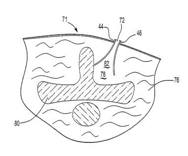
Referring now to Figure 8, there is shown a cross-sectional view of a
portion of a patient's body 71 illustrating the surgical method of the present
invention. As will be seen from Figure 8, a relatively small opening 72 is
formed through a patient's skin 74. Most preferably, this opening will not be
larger than 20 millimeters in its greatest dimension. A pair of retractor
blades
44, 46 displace tissue 76 so as to provide access to a surgical field 78 in
which
a portion of a patient's spine 80 is exposed. As will be seen from Figure 8,
the
passage 82 through the tissue 76 is flared so that it widens as it progresses
from
the skin 74 to the surgical field 78. In this manner, the cross-sectional area
of
the surgical field 78 is greater than the cross-sectional area of the opening
72 in
the patient's skin 74. This keyhole passage provides maximum visualization of
the surgical field 78 while minimizing the incision through the patient's skin
74. The keyhole passage allows for direct visualization of the surgical field
78
and maximizes the space available for surgery. The use of the retractor
minimizes tissue trauma, and the relatively small size of the opening in the
skin
74 minimizes scarring and facilitates closure of the surgical wound.
It is to be understood that while the present method and apparatus have
been described with particular reference to spinal surgery, the principles of
the
present invention may be readily adapted to other surgical procedures which
are not limited to the spine or to the formation of a leeyhole channel. As
will be
apparent, the retractor and method of the present invention may also be
utilized
in veterinary procedures, as well as in non-medical applications. Also, while
specific embodiments of retractor have been described, it is to be understood
that yet other modifications and configurations thereof may be implemented in
accord with the teaching herein. The foregoing drawings, discussion and
CA 02537618 2006-03-02
WO 2005/032385 PCT/US2003/027497
description are meant to be illustrative of specific embodiments of the
invention, but they are not meant to be limitations upon the practice thereof.
It
is the following claims, including all equivalents, which define the scope of
the
invention.