Note: Descriptions are shown in the official language in which they were submitted.
CA 02538465 2006-03-02
SYSTEM AND METHOD FOR INDUCING CONTROLLED CARDIAC
DAMAGE
SPECIFICATION
CROSS-REFERENCE TO RELATED APPLICATIONS
This application claims the benefit of United States provisional patent
application No. 60/658,351 filed on March 3, 2006, which application is hereby
incorporated by reference herein in its entirety.
Portions of this research were supported by research grant RO1
CA84588 awarded by the National Cancer Institute and the National Heart, Lung,
and
Blood Institute.
FIELD OF THE INVENTION
The present invention relates generally to systems and methods of
biomedical and genetic science. The invention in particular relates to animal
models
that are used in biomedical and genetic research.
BACKGROUND OF THE INVENTION
Animal testing (also referred to as animal research) refers to the use of
non-human animals in experiments. Animal experiments are carried out, for
example, for basic or pure research, studying diseases and developing
medicines, and
toxicology testing of chemicals. The testing is carried out inside
universities,
medical schools, pharmaceutical companies, commercial facilities that provide
animal-testing services to industry, on farms, in defense-research
establishments, and
by public-health authorities, on a variety of species from fruit flies and
mice to
non-human primates.
The particular species selected for biomedical testing is often based on
CA 02538465 2006-03-02
a suitable animal model of the biological phenomena or disease under
investigation.
Animal model refers to a non-human animal with a disease that is similar to a
human
condition. Mice are convenient in research because their physiology is similar
to
that of humans and their short life cycle makes breeding easy. Th ey are
mainly used
to model human diseases in order to develop new drugs, to test the safety of
proposed
drugs, and in basic research.
In order to serve as a useful model, a modeled disease must be similar
in etiology (mechanism of cause) and function to the human equivalent. Animal
models are used to learn more about a disease, its diagnosis and its
treatment. For
example, the marine model (i.e., mouse) is an important animal model for
studying
the cardiovascular system. The marine myocardial infarction model is widely
used
as an ischemic heart model and a heart failure model. Gene-targeted mouse
models
have been extensively used for the research on cardiovascular diseases and for
understanding the molecular mechanism of heart failure.
1 S Animal models of disease can be spontaneous, or be induced by
physical, chemical or biological means. The marine myocardial infarction model
is
generally induced by surgical ligation of the proximal left anterior
descending
coronary artery. However, opening the thoracic cavity, which is necessary for
this
purpose, may lead to infection and death. Further, the surgical ligation
technique
does not provide good control of the degree of the resulting myocardial
damage.
Consideration is now directed towards improving the marine model for
cardiac disease investigations. In particular, attention is directed to
inducing
coronary defects and cardiac failure in the marine (mouse) model.
SUMMARY OF THE INVENTION
2S A device and method is provided for inducing coronary defects and
2
CA 02538465 2006-03-02
cardiac failure in the marine model. The device is configured to generate high
intensity ultrasound waves, which are focused on a subject mouse to ablate
cardiac
tissue in-vivo and to cause cardiac damage. High intensity focused ultrasound
(HIFU) produces immediate focal lesions with ultrasound exposures within short
periods. Useful marine myocardial failure models may be created using HIFU.
The HIFU technique is a noninvasive extracorporeal technique capable
of ablating subsurface structures without injuring intervening tissues.
Ultrasonic
energy can be applied in a target volume to induce tissue necrosis. 'The HIFU
technique has an advantage over other ablative techniques because the tissue
in the
acoustic focal volume during HIFU ablation is rapidly damaged by a remote
energy
source (the ultrasonic transducer), and the intervening tissue is not damaged.
The HIFU technique can be used for targeted LU wall thinning, LU
dilatation and systolic dysfunction in animals without thoracotomy. H IFU may
be
used to nonivasively create marine or other animal myocardial failure models.
I S The HIFU technique may be modified or extended to alternately or
additionally use hyperthermia from ultrasound and other heat sources, other
focused
ultrasound ablation technologies such as tissue emulsification, and other
ablation
technologies such as ethanol injection for inducing coronary defects and
cardiac
failure.
BRIEF DESCRIPTION OF THE DRAWINGS
Further objects and advantages of the invention will be apparent from a
reading of the following description in conjunction with the accompanying
drawings
in which:
Fig. 1 A is an illustration of the High Intensity Focused Ultrasound
(HIFU) transducer device, which can be used to induce cardiac defects in
accordance
3
CA 02538465 2006-03-02
with the principles of the present invention.
Fig. 1 B is an illustration of the focal zone beam shape of the output of
a the HIFU transducer of Fig. 1 A. The output is measured using a pulse-echo
reciprocity technique with a point target.
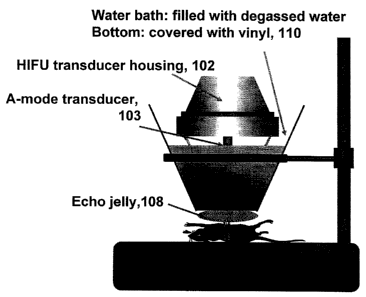
Fig. 2 is an illustration of the HIFU transducer surface (Fig. 1 A)
coupled with the intercostals muscle of a mouse using gel and water baths, in
accordance with the principles of the present invention.
Fig. 3 shows hematoxylin and eosin stains and Masson's trichrome
stains of the transverse left ventricle (LV) middle sections from an exemplary
group
of mice ("the HIFU group") treated in accordance with the principles of the
present
invention.
Fig. 4 is a table showing the weight of the body, heart, lung and liver of
the HIFU group as compared to the control group.
Fig. 5 is a table showing the LV diameter at end-diastole phase (end
I S diastolic dimension, EDD) and at end-systole phase (end systolic
dimension, ESD),
and fractional shortening (FS) transthoracically for the HIFU group and
control group
before and after ablation, in accordance with the principles of the present
invention.
Fig. 6 is an illustration of a sample PXI implementation of an
ultrasound therapy system for supplying the therapeutic ultrasound energy, in
accordance with the principles of the present invention.
Fig. 7 is a block diagram of an example of a simple gate mechanism
for an ultrasound therapy system, in accordance with the principles of the
present
invention.
Fig. 8 is an example of a simple embodiment of an ultrasound therapy
system for supplying the therapeutic ultrasound energy, in accordance with the
principles of the present invention.
4
CA 02538465 2006-03-02
DESCRIPTION OF THE INVENTION
Coronary defects and cardiac failure are obtained in an animal model
by ablating cardiac tissue using focused ultrasound energy such as HIFU. An
ablation device, which includes a high intensity ultrasound transducer, is
used to
generate and focus ultrasound energy on a subject heart. The focused high
intensity
ultrasound waves ablate cardiac tissue in localized regions and accordingly,
or
otherwise, cause coronary defects and cardiac failure.
Figs. 1 A and 2 show an exemplary ablation device 100. Ablation
device 100 includes a therapeutic focused ultrasound transducer 102, which
produces
high intensity ultrasound waves. In the exemplary device, transducer 102 is
specified to produce ultrasound waves of about 4.7 MHz focused at 90 mm, with
a
half power focal region approximately 3 mm axial and 0.4 mm transverse to the
beam.
Cone 104 is designed to contain water for coupling. (Se a Fig. 1 B).
In one version of operation, the transducer is acoustically coupled with
the intercostals muscle of a subject mouse 106 using a water path through cone
I02
and bath 110, and through echo gel 108. (Fig. 2).
In one embodiment, high intensity focused ultrasound (HIFU)
produces rapid focal lesions. Useful myocardial failure animal models may be
created using HIFU. The HIFU technique is capable of producing transmural
myocardial injury on animal hearts, resulting in animal heart failure models
that are
created noninvasively.
Post-infarct LV remodeling is a progressive process involving LV
chamber dilatation, infarcted wall thinning, fibrous change, and compensatory
thickening in the non-infarcted regions.
In a study to assess the feasibility of cardiac failure model creation
5
- CA 02538465 2006-03-02
using HIFU, a group of 30 wild type mice was selected. - The study was
designed to
assess the chronic lesions of marine myocardial tissue after HIFU ablation and
the
feasibility of a marine heart failure model induced by HIFU ablation using
conventional 2-D echocardiography.
A commercial ultrasound therapy system (Model CST 100, sold by
Sonocare Inc., Ridgewood N~, which is originally designed for clinical
glaucoma
therapy, was modified for use as the source of HIFU energy for the study. The
system includes a signal generator, a power amplifier, and a transducer
assembly.
(See Figures 1 A, 1 B, and 2). The transducer's focal length is about 90 mm
(Figure
lA). The 80-mm diameter, 90-mm focal length spherical cap PZT 4 therapy
transducer has a central 23-mm hole, which houses a 7.5 MHz A-mode diagnostic
transducer (Model MD 3657, sold by Panametrics, Inc. of Waltham, MA). The
diagnostic transducer is aligned to be coaxial and confocal with the HIFU
transducer,
In the study, the operating frequency of the HIFU transducer was 4.7 MHz and
the
ultrasound energy was applied with an acoustic power of 35 W, as determined
from
acoustic radiation force measurements. The focal zone beam shape was measured
using a pulse-echo reciprocity technique with a point target; at the half
power points
the focal zone was 3 mm in dEpth and 0.4 mm wide (Fig. 1 B).
The transducer assembly was attached to an acrylic resin coupling cone
with a 25 mm diameter exit hole. The cone was filled with degassed water, and
the
exit hole was covered with a latex membrane. Th a focus of ultrasound beam was
2.5
cm distal from the membrane at the tip of the coupling cone. The focus of
ultrasound beam was positioned at the desired tissue location by distance
measurements made with the diagnostic A-mode transducer.
Study Protocol
The 30 wild type mice, age 6 - 8 weeks and ranging in body weight
6
CA 02538465 2006-03-02
from 30 - 39 g, were housed in a facility with a 12/12 light and dark cycle,
and free
access to water and mouse pellets. The mice were randomly divided into two
p groups: a test group (20 mice) and a control group (10 mice). For each mouse
in the
subject groups, cardiac characteristics were measured transthoracically using
a high
frequency ultrasound system. The measured characteristics included, for
example,
the left ventricular (LU) diameter at end-diastole and end-systole phases
(EDD/ESD),
and ejection fraction or fractional shortening (FS).
Surgery and HIFU ablation was performed without thoracotomy on the
test group of 20 mice (HIFU group). The animals in the HIFU group were first
anesthetized using Isoflurane (for induction 3.0% and maintenance l.St2.0%),
and
their chests were shaved. The subject animals were intubated with a 20 G
intravenous catheter through the oral cavity under visualization and
ventilated with a
mixture of oxygen and room air, using a rodent ventilator at a tidal volume of
2 to 4
ml and a respiratory rate of 130 to 150 per minute.
Animals in the HIFU group underwent a left midsternal skin incision
through the fifth or sixth intercostal space. The skins were retracted by use
of 5-0 or
6-0 silk suture. Slight rotation of the subject animals to the right oriented
the heart
to better expose the left ventricle (LU). Their pectoralis major muscles and
pectoralis minor muscles were moved to the sides. The beating heart was
visible
through the nearly transparent intercostal muscle. The HIFU transducer surface
was
coupled with the intercostals muscle using echo gel and a water bath (Fig. 2).
The
depth of the heart from the chest surface was measured using the diagnostic A-
mode
transducer, and the therapeutic transducer focal point was set to the middle
of the LV
anterior wall.
Each mouse in the HIFU group was subject to three HIFU energy
discharge pulses. Ea ch pulse was about one second in duration and had a
nominal
7
- CA 02538465 2006-03-02
spatial-peak temporal-average intensity of about 19.7 kW/cm2.
A sham operation was performed on the control group of 10 mice.
Animals in control group were anesthetized and underwient only intubation and
skin
incision without use of HIFU ablation. After these procedures, the lungs were
re-expanded and the chest was closed. The control animals were taken oil the
respirator and allowed to recover from the anesthesia in a warm cage.
Transthoracic echocardiography was performed on all surviving
animals every week after the ablation or sham operation procedure. Four weeks
later
all survival animals were euthanised for morphological and histological
analysis.
Transthoracic echocardiography was performed in both groups using
a commercial echocardiographic system (Model Sequoia, sold by Acuson
Corporation
of Mountain View, California, 94039) equipped with a 13-MHz liner array
ultrasound
transducer. T he transducer was used at a depth setting of 2 cm to optimize
resolution.
This examination was performed under light anesthesia induced by
intraperitoneal
injection of 2,2,2-tribromoethanol (Avertin, 2.5% solution, O.OOSmI/g body
weight),
which produced a semiconscious state in which the animals breathed
spontaneously.
The animal chests were shaved and the animals were placed on a heating table
in a
left lateral decubitus position. Before the procedure and every week after the
procedure, the following parameters were measured in the parasternal short
axis view
of a 2-dimensional image at the level close to the papillary muscles: LV
diameter at
the end-diastolic and end-systolic phases (EDD and ESD, respectively). LV
fractional shortening (FS) was calculated as
FS = [(EDD - ESD)/EDD] X 100 %.
The statistical results for each group of animals were expressed as
mean values ~ one standard deviation. The paired t-test was used for the
comparison
8
CA 02538465 2006-03-02
within each group. The unpaired t-test was used to compare the results between
the
HIFU and control groups. Statistical significance was defined as a p-value of
less
~ than 0.05.
Morphological and histological examinations were conducted after
four weeks. Euthanasia was performed by C02 exposure or overdose of
pentobarbital (euthanasia solution, 100 mg/kg) intraperitoneally. Animal
hearts and
other organs were taken out. Each heart, lung and liver weights were obtained.
Each heart was fixed in 10% formalin, and cut in paraffin blocks. Standard
hematoxylin and eosin (H&E) stained slides and Masson's trichrome stained
slides
were evaluated for pathological evidence of injury, inflammation, and scarnng.
Study Results
The mortality of the HIFU group was 15%. H IFU ablation could be
performed on all mice hearts. The overall survival after HIFU ablation was
85%.
One animal in HIFU group died immediately after HIFU ablation as a result of a
ruptured left anterior wall and two died of severe heart failure within three
days after
HIFU ablation. All the sham -operated mice survived throughout the study.
At four weeks after the ablation and surgery procedures, the cardiac
characteristics of the mice in both the HIFU and control groups were evaluated
for
comparison with the pre-procedure characteristics.
Body weight was similar in both groups before HIFU ablation (HIFU
group vs. control group: 36.612.4 g vs. 36.42.6 g). In the HIFU group, body
weights were significantly ,decreased after HIFU ablation (36.612.4 g vs.
30.212.7 g,
p<0.01 ). The weights of whole heart and liver were not significantly
different
between the two groups (Fig. 4).
Technically adequate echocardiographic images were obtained in all
9
CA 02538465 2006-03-02
animals for LU dimension and function measurements. However, image quality was
reduced compared with echocardiographic images from non-operated animals
because
n of residual fibrinous, exudate and fibrous adhesions.
The pre-procedure LU EDD/ESD for the control mice were measured
to be 1.34 ~0.15/2.59 ~ 0.24 mm (Figure 5). Similarly, the pre-procedure
EDD/ESD
for the HIFU group were measured to be 1.35~0.17/2.67~0.2. Thus, there was no
significant difference in EDD/ESD between the two groups before the HIFU
ablation
procedure. However, after four weeks, the treated group of mice showed
considerably larger LU diameters. The post-procedure LU EDD/ESD for the HIFU
group were measured to be 2.53~0.54/3.54~0.54. The pre-procedure LU ejection
fraction or fractional shortening (FS) in the control group and the HIFU group
of mice
was similar. The post-procedure FS in the control mice did not change
significantly.
However, FS in the HIFU group was significantly reduced after HIFU ablation.
FS
in the HIFU group was measured before and after ablation to be about 48.8~2.3%
and
25.2~7.3%, respectively, p<0.01.
Histopathological analysis the HIFU group hearts showed necrosis (i.e.,
a fibroid degeneration) around the ablation site and the LU anterior wall
thinning.
Figure 3 shows H&E and trichrome stains of transverse LU middle sections from
the
HIFU group. Myocardial injuries were identified histologically as transmural
injuries in all animals of the HIFU group. At the targeted site, the
myocardial tissues
were changed into fibrous degeneration. LU wall thinning and LV chamber
enlargements were found. The histological findings show typical
characteristics of
myocardial infarction. The results of the study demonstrate that HIFU can
produce
LU dilatation and systolic dysfunction in mice. Thus, HIFU may be used to
create a
marine myocardial failure model. The marine myocardial failure model with
focal
myocardial dysfunction is created without opening marine chests.
CA 02538465 2006-03-02
Using HIFU may be superior to the use of the other techniques that are
used to create the marine heart failure mode. For example, in the previous
studies
using LAD ligation, a mortality rate in the range of 11 % to 46% with an
average of
27% has been observed. The mortality rate of HIFU treated mice in the
above-described study was 15%. Thus, HIFU has a potential to make a marine
heart
failure model with minimum invasion and a high success rate.
The advantages of using HIFU may stem from its capability of
producing lesions not only thermally but also through cavitation, acoustic
streaming,
and shear stresses. Further, the focusing ability of HIFU makes it superior to
other
l0 ablative techniques such as radio frequency (RF) ablation. RF ablation and
focused
ultrasound ablation produce lesions with similar histological injury in
myocardial
tissue. However, RF is not focused and RF energy is absorbed proportionally by
the
distance between the tissue and the RF catheter. In contrast, ultrasound
energy can
be focused, allowing smaller and more precise lesions to be created.
In-vitro study shows that the eventual size of the HIFU lesion in the
myocardial tissue depends on many factors. T he extent of HIFU induced tissue
injury and coagulative necrosis varies linearly with ablation time, exposure
number,
and acoustic intensity. By changing these factors smaller or larger lesions
may be
produced at will.
The foregoing merely illustrates the principles of the invention. It
will be appreciated that those skilled in the art will be able to devise
numerous
modifications which, although not explicitly described herein, embody the
principles
of the invention and are thus within the spirit and scope of the invention.
For
example, it will be readily understood by those skilled in the art that by
changing the
focal depth or the location of the focal point, focused ultrasound ablation
may be
obtained at any suitable subsurface tissue. Focusing in the papillary muscle
may be
11
- CA 02538465 2006-03-02
used to create a papillary muscle failure model without thoracotomy. Further,
in the
study described herein, an A-mode transducer is mounted in the center the HIFU
q therapy transducer for the measurement of the distance between the heart and
the
transducer. If a 2-D transducer is mounted in the center of the HIFU therapy
transducer instead of the A-mode transducer, it may be possible to focus in
the LV
anterior wall and perform HIFU ablation from outside the body without skin
incision.
Further, for example, the HIFU technique may be utilized to create suitable
disease
models in other animal species (e.g., canine models). Finally, the ultrasound
frequencies can be varied over a wide range to activate different defect
generation
mechanisms. When the ultrasound frequencies are in the range of several
hundred
kHz, and the ultrasound is pulsed at varying rates, tissue emulsification due
to
cavitation will dominate the ablation mechanism for defect generation.
Similarl y,
when the ultrasound frequencies are in the MHz range, thermal necrosis will
dominate
the ablation mechanism.
I 5 Simple low cost ultrasound systems may be used for HIFU application.
Fig. 8 shows an exemplary ultrasound therapy system that can be used for HIFU
application. Fig. 6 shows an exemplary PXI implementation of the ultrasound
therapy system. Further, Fig. 7 shows an example of a simple gate mechanism
that
may be used in the ultrasound therapy system.
12