Note: Descriptions are shown in the official language in which they were submitted.
CA 02538852 2006-03-10
WO 2005/031309 PCT/US2004/031643
SPECIFICATION
TO ALL WHOM IT MAY CONCERN:
BE IT KNOWN THAT WE, John K. Westwick, a resident of San Ramon, California and
citizen of USA, Brigitte Keon, a resident of Castro Valley, California and
Marnie L.
MacDonald, a resident of Pleasanton, California and a citizen of USA; have
invented certain
new and useful improvements in
FRAGMENT COMPLEMENTATION ASSAYS FOR G-PROTEIN-COiJPLED
RECEPTORS AND THEIR SIGNALING PATHWAYS
of which the following is a specification.
1
CA 02538852 2006-03-10
WO 2005/031309 PCT/US2004/031643
FRAGMENT COMPLEMENTATION ASSAYS FOR G-PROTEIN-COUPLED
RECEPTORS AND THEIR SIGNALING PATHWAYS
This application claims the priority benefit under 35 U.S.C. section 119 of
U.S.
Provisional Patent Application No. 60/505,447 entitled "Fragment
Complementation Assays For
G-Protein-Coupled Receptors And Their Signaling Pathways", filed September 25,
2003, which
is in its entirety herein incorporated by reference.
FIELD OF THE INVENTION
This invention relates generally to the fields of biology, molecular biology,
chemistry and
biochemistry. The invention is directed to a large number of novel assays for
G-protein-coupled
receptors (GPCRs) and their signaling pathways. The invention also relates to
methods for
constructing such assays for one or more steps in a GPCR pathway. The
invention can be used
for functional characterization of GPCRs, target validation, de-orphanization
of receptors, high-
throughput screening, high-content screening, pharmacological profiling, and
other drug
discovery applications. The assays can be used directly to assess whether a
compound library or
a biological extract contains an agonist or antagonist of a receptor. Assay
compositions are also
provided. The development of such assays is shown to be straightforward,
providing for a broad,
flexible and biologically relevant platform for the discovery of novel drugs
and natural ligands
that act on GPCRs or their cognate pathways. The invention is demonstrated for
a broad range
of proteins in GPCR pathways and for a range of assay formats.
BACKGROUND OF THE INVENTION
The superfamily of G-protein coupled receptors (GPCRs) represents the largest
family of
cell surface receptors, and is one of the most important sources of drug
targets for the
CA 02538852 2006-03-10
WO 2005/031309 PCT/US2004/031643
pharmaceutical industry. GP(:lts are involven m a wiae range or aisoraers ana
aisease s~aies
including ulcers, psychosis, anxiety, Parkinson's disease, Alzheimer's disease
and hypertension.
More than 20% of the bestselling prescription drugs and an estimated 50% of
all prescription
drugs interact directly with a GPCR. Also, interactions of drugs with this
class of receptors are
responsible for some of the side effects associated with these drugs.
Over 500 different GPCRs have been identified in the human genome. As many as
200
of these represent 'orphan' receptors, for which the natural ligand is
unknown. The first step for
the transformation of orphan receptors into drug targets is their
characterization, or'de-
orphanization' (AD Howard et al., 2001, Orphan G-protein-coupled receptors and
natural ligand
discovery, Trends in Pharmacol. Sci. 22(3): 132-140). De-orphanization of
GPCRs, and the
identification of synthetic agonists and antagonists, will likely lead to a
large number of new and
potent medicines for various conditions. This is a task that involves
significant efforts, both to
understand the potential importance of receptor candidates to specific
diseases and to develop
efficient drug screening tools. For example, ligands of an identified receptor
can be tested
against related orphan GPCRs to identify compounds that bind to the orphan
receptor. In
addition, extracts of tissues can be tested using functional assays to guide
ligand fractionation,
purification and molecular characterization. Finally, orphan GPCRs can be
evaluated against
arrayed families of known ligands. Sensitive, biologically relevant assays are
needed that can be
used to de-orphanize GPCRs on a large scale.
GPCRs do not share any overall sequence homology, but have in common the
presence
of seven transmembrane-spanning alpha-helical segments connected by
alternating intracellular
CA 02538852 2006-03-10
WO 2005/031309 PCT/US2004/031643
and extracellular loops, with the amino terminus on the extracelluiar sate ana
the carnoxy
terminus on the intracellular side of the cell membrane. Therefore, GPCRs are
commonly
referred to as seven-transmembrane (7TM) receptors. The GPCRs have been
divided into
different subfamilies (A-F); the major subfamilies, A, B and C, include the
beta-2-adrenergic
receptor (family A), receptors related to the glucagon receptor (family B) and
receptors related to
the neurotransmitter receptors (family C). Family B includes the receptors for
vasoactive
intestinal peptide, calcitoW n, PTH and glucagon; family C includes the
receptors for GABA,
calcium, mammalian pheromones, and taste receptors. All GPCRs signal through
guanine
nucleotide-binding proteins (G-proteins). The DNA sequences of a large number
of GPCRs can
be found in public databases, among other sources (F. Horn et al., 1998,
GPCRDB: an
Information system for G protein-coupled receptors, Nucleic Acids Res. 26:275-
279). The
public Gl'CR database can be found on the worldwide web at http://www.~pcr.or
/~ 7tm/ and
corresponding cDNAs and pairwise sequence alignments can be found at
http://www.g cr.or~/7trnlseq/dna.html. This database is incorporated herein by
reference.
The general mechanism of action of GPCRs in cell signaling has been elucidated
over the
last 20 years, although many details remain to be discovered. Hundreds of
scientific and review
articles have been written on the topic (for reviews see GB Downes & N
Gautham, 1999, The G-
protein subunit gene families, Genomics 62: 544-552; Hermans, 2003,
Biochemical and
pharmacological control of the multiplicity of coupling at G-protein-coupled
receptors,in:
Pharmacology & Therapeutics 99: 25-44; and U Gether, 2000, Uncovering
molecular
mechanisms involved in activation of G-protein-coupled receptors, in:
Endocrine Reviews 21:
90-113; EM Hur & KT Kim, 2002, G-protein-coupled receptor signaling and cross
talk:
CA 02538852 2006-03-10
WO 2005/031309 PCT/US2004/031643
achieving rapidity and specificity, Cell Signal 14: 397-405 ). Some of the
known elements of the
various G-protein-coupled pathways are described herein for the purposes of
placing the present
invention in the context of the prior art. Further elucidation of the
signaling pathways linked to
GPCRs would allow the construction of a large number of assays for
intracellular events linked
to GPCR activation. In turn, such assays would allow drug discovery to
identify drug candidates
capable of activating or blocking GPCR signaling.
For drug discovery, there is a need to quickly and inexpensively screen large
numbers of
chemical compounds to identify new drug candidates, including agonists,
antagonists and
inhibitors of GPCRs and GPCR-dependent pathways. These chemical compounds are
collected
in large libraries, sometimes exceeding one million distinct compounds. The
use of the term
chemical compound is intended to be interpreted broadly so as to include, but
not be limited to,
simple organic and inorganic molecules, proteins, peptides, antibodies,
nucleic acids and
oligonucleotides, carbohydrates, lipids, or any chemical structure of
biological interest.
Traditional, biochemical approaches to assaying GPCRs have relied upon
measurements of
ligand binding, for example with scintillation proximity assays or with
surface plasmon
resonance (C. Bieri et al., 1999, Micropatterned immobilization of a G-protein-
coupled receptor
and direct detection of G protein activation, Nature Biotech. 17: 1105-1109).
Although such
assays are inexpensive to perform, they can take 6 months or longer to
develop. A major
problem is that the development of an in vitro assay requires specific
reagents for every target of
interest, including purified protein for the target against which the screen
is to be run. Often it is
difficult to express the protein of interest and/or to obtain a sufficient
quantity of the protein in
pure form. Moreover, although in vitro assays are the gold standard for
pharmacology and
CA 02538852 2006-03-10
WO 2005/031309 PCT/US2004/031643
studies of structure activity relationships (SAR) it is not possible to
perform target validation
with an in vitro assay, in vivo assays are necessary in order to obtain
information about the
biological availability and cellular activity of the screening hit.
The increased numbers of drug targets identified by genomics approaches has
driven the
development of 'gene to screen' approaches to interrogate poorly defined
targets, many of which
rely on cellular assay systems. Speculative targets are most easily screened
in a format in which
the target is expressed and regulated in the biological context of a cell, in
which all of the
necessary components are pre-assembled and regulated. Cell-based assays are
also critical for
assessing the mechanism of action of new biological targets and the biological
activity of
chemical compounds. In particular, there is a need to 'de-orphanize' those
GPCRs for which the
natural activating ligand has not been identified. Various approaches to de-
orphanization have
been reviewed (AD Howard et al., 2001, Orphan G-protein-coupled receptors and
natural ligand
discovery, Trends in Pharinacological Sciences 22: 132-140). For example,
extracts of tissues
can be tested using functional assays to guide ligand fractionation,
purification and molecular
characterization. Alternatively, orphan GPCRs can be evaluated against arrayed
families of
known ligands.
Current cell-based assays for GPCRs include measures of pathway activation
(Ca2+
release, cAMP generation, or transcriptional activity); measurements of
protein trafficking by
tagging GPCRs and downstream elements with GFP; and direct measures of
interactions
between proteins using fluorescence resonance energy transfer (FRET) or
bioluminescence
CA 02538852 2006-03-10
WO 2005/031309 PCT/US2004/031643
resonance energy transfer (BRET) or yeast two-hybrid approaches (e.g. King et
al., US Patent
Application 20020022238). These approaches are described below.
The majority of cell-based assays for GPCRs rely upon measurements of
intracellular
calcium. Calcium release from intracellular stores is stimulated by specific
classes of GPCRs
upon their activation; in particular, those GPCRs that couple to Gq.
Fluorescent and
luminescent assays of calcium release have been generated by loading cells
with dyes that act as
calcium indicators. Fluorescent Ca2+ indicators such as fura-2, indo-1, fluo-
3, and Calcium-
Green have been the mainstay of intracellular Ca2+ measurement and imaging
(see for example
U.S. Pat. Nos. 4,603,209 and 5,049,673). Such indicators and associated
instrumentation
systems (FLIPR system) are sold, for example, by Molecular Devices of
Sunnyvale California
(www.rnoleculardevices.com). Luminescent assays of calcium flux can be
accomplished by
introducing aequorin into cells. Aequorin emits blue light in the presence of
calcium, and the
rate of photon emission is proportional to the free Ca2+ concentration within
a specific range.
Cells ea~pressing the GPCR of interest are loaded first with coelenterazine to
activate the
aequorin, and then the compounds to be tested are added to the cells and the
results quantitated
with a luminometer. To extend these assays to non-Gq-coupled receptors,
various strategies
have been employed, including the use of a promiscuous Ga protein such as Gal6
that is
capable of coupling a wide range of GPCRs to phospholipase C (PLC) activity
and calcium
mobilization (Milligan et al., 1996, Trends in Pharmacological Sciences 17:
235-237).
Fluorescent dyes, and fluorescent proteins such as GFP, YFP, BFP and CFP, have
also
been used as cellular sensors of cAMP or Ca2+. The first fluorescent protein
indicator for CAMP
CA 02538852 2006-03-10
WO 2005/031309 PCT/US2004/031643
consisted of the cyclic AMP-dependent protein kinase, PKA, in which the
catalytic anct
regulatory subunits were labelled with fluorescein and rhodamine,
respectively, so that cAMP-
induced dissociation of the subunits disrupted FRET (S.R. Adams et al., 1991,
Fluorescence ratio
imaging of cyclic AMP in single cells, Nature 349: 694-697). Replacement of
the dyes by GFP
and BFP made this system genetically encodable and eliminated the need for in
vitro dye
conjugation and microinjection (M. Zaccolo et al., 2000, A genetically encoded
fluorescent
indicator for cyclic AMP in living cells, Nature Cell Biol. 2: 25-29). A
variety of other GFP-
based techniques have been used to create cellular sensors. For example, two
GFP molecules
joined by the kinase iriducible domain (K>D) of the transcription factor CREB
(cyclic AMP-
responsive element binding protein) exhibit a decrease in fluorescence
resonance energy transfer
upon phosphorylation of the KIl? by the cyclic AMP-dependent protein kinase,
PKA (~. Nagai
et al., 2000, A fluorescent indicator for visualizing CAMP-induced
phosphorylation in vivo,
Nature Biotech. 18: 313-316). Calmodulin, a calcium-sensitive protein, has
been inserted into
~'FP, resulting in calcium sensors ('camgaroos') that increase fluorescence
sevenfold upon
binding of calcium (GS Baird et al., 1999, Circular permutation and receptor
insertion within
green fluorescent proteins, Proc. Natl. Acad. Sci. USA 96: 11241-11246).
Similarly, insertion of
a circularly permuted GFP between calmodulin and M13- a peptide that binds
calmodulin in a
calcium-sensitive manner - yields calcium indicators that are known
as'pericams' (T. Nagai et
al., 2001, Circularly permuted green fluorescent proteins engineered to sense
Ca2+, Proc. Natl.
Acad. Sci USA 98: 3197-3202). Alternative calcium indicators known as
'cameleons' have been
created by sandwiching calmodulin, a peptide linker, and M13 between CFP and
YFP (A.
Miyawaki et al., 1997, Fluorescent indicators for Ca2+ based on green
fluorescent proteins and
calmodulin, Nature 388: 882-887).
CA 02538852 2006-03-10
WO 2005/031309 PCT/US2004/031643
Transcriptional reporter assays provide a measurement of pathway
activationlinhibition
in response to an agonistlantagonist and have been used extensively in GPCR
studies (see Klein
et al., US 6,255,059 and references therein). Reporter assays couple the
biological activity of a
receptor to the expression of a readily detected enzyme or protein reporter.
Synthetic repeats of
a particular response element can be inserted upstream of the reporter gene to
regulate its
expression in response to signaling molecules generated by activation of a
specific pathway in a
live cell. Such drug screening systems have been developed with a variety of
enzymatic and
fluorescent reporters, including [3-galactosidase (H Brauner-Osborne & MR
Brann, 1996, Eur.
J. Pharmacol. 295: 93-102), luciferase, alkaline phosphatase, GFP, (3-
lactamase (G. Zlokarnik et
al., 1998, Quantitation of transcription and clonal selection of single living
cells with beta-
lactamase as reporter, Science 279: 84-88) and other reporters. Transcription
reporter assays are
highly sensitive screening tools; however, they do not provide information on
the mechanism of
action of the compound, enable mapping of the components of the pathway
leading to
transcription, or enable studies of individual steps within signaling
cascades.
Subcellular compartmentalization of signaling molecules is an important
phenomenon in
cell signaling, not only in defining how a biochemical pathway is activated
but also in
influencing the desired physiological consequence of pathway activation. High-
content
screening (HCS) is an approach that relies upon imaging of cells to detect the
subcellular
location and trafficking of proteins in response to stimuli or inhibitors of
cellular processes.
Fluorescent probes can be used in HCS. For example, GTP has been labeled with
the fluorescent
dye, BODIPY, and used to study the on and off rates of GTP hydrolysis by G-
proteins, and
CA 02538852 2006-03-10
WO 2005/031309 PCT/US2004/031643
tluorescein-labeled myristoylated Galpha-I has been used as the ligand bound
to Gbeta-gamma
in order to study the association and dissociation of G-protein subunits (NA
Sarvazyan et al.,
2002, Fluorescence analysis of receptor-G protein interactions in cell
membranes, Biochemistry
41: 1258-12867).
Increasingly, green fluorescent protein (GFP) has been used to analyze key
signaling events
within cells. By fusing in-frame a cDNA for GFP to a cDNA coding for a protein
of interest, it
is possible to examine the function and fate of the resulting chimera in
living cells. This strategy
has now been applied to nearly all known elements of G-protein coupled
pathways including the
receptors themselves; G-protein subunits such as Ga; beta-arrestin; RGS
proteins; protein kinase
C; and numerous other intracellular components of G-protein-coupled pathways
(M Zaccolo and
T. Pozzan, 2000, Imaging signal transduction in living cells with GFP-based
probes, IUBMB
Life 49: 1-5, 2000.)
For example, G-protein-coupled receptors have been tagged with GFP in order to
monitor
receptor internalization. A fusion protein comprising GFP- beta-arrestin has
been shown to co-
localize with thyrotropin-releasing hormone receptor 1 in response to agonist
(T Drmota et al.,
1999, Visualization of distinct patterns of subcellular redistribution of the
thyrotropin-releasing
hormone receptor-l and Gqalpha/G1 lalpha induced by agonist stimulation,
Biochem. J. 340:
529-53 ~). GFP has been introduced internally to G- proteins, creating a
Galpha/GFP chimera,
which has been shown to translocate to the cell membrane upon GPCR activation
(J-Z Yu ~ M
Rasenick, 2002, Real-time visualization of a fluorescent GalphaS dissociation
of the activated G
protein from plasma membrane, Mol. Pharmacol. 61: 352-359; P Coward et al.,
1999, Chimeric
to
CA 02538852 2006-03-10
WO 2005/031309 PCT/US2004/031643
G proteins allow a high-throughput signaling assay of Gi-coupiect receptors,
Anal. .tiiocnem.
270: 242-248). GFP tagging has also been used to monitor intracellular
signaling events. GFP-
tagged Regulator of G protein Signaling (RGS2 and RGS4) proteins were
selectively recruited to
the plasma membrane by G proteins and their cognate receptors (AA Roy et al.,
2003,
Recruitment of RGS2 and RGS4 to the plasma membrane by G proteins and
receptors reflects
functional interactions. Mol. Pharmacol. 64: 587-593). GFP-tagged protein
kinase C (PKC),
which is activated by release of diacylglycerol from cell membranes, has been
used to monitor
translocation of the kinase in response to cell signaling (E. Oancea et al.,
1998, Green fluorescent
protein (GFP)-tagged cysteine-rich domains from protein kinase C as
fluorescent indicators for
diacylglycerol signaling in living cells, J. Cell Biol. 140: 485-498). GFP-
tagged connexin has
been used to monitor intracellular calcium flux (K Paemeleire et al., 2000,
Intercellular calcium
waves in HeLa cells expressing GFP-labeled connexin, Mol. Biol. Cell 11: 1815-
1827). GFP-
tagged beta-arrestin has been used to monitor GPCR activation by imaging the
subcellular
redistribution of beta-arrestin in reponse to GPCR agonist. The latter assay,
k3zown as
TransFluor, is marketed by Norak Bioscience (www.norakbio.com) and is the
subject of US
5,891,646 amd US 6,110,693. All the above assays and inventions involve fusing
a protein of
interest (receptor, beta-arrestin, G-protein, connexin, RGS, kinase etc.) to
an optically detectable
molecule such as GFP; expressing the fusion construct in cells; and then
detecting the quantity,
and/or the subcellular location, of the chimeric protein in response to a
stimulus or inhibitor.
Measurements of protein-protein interactions between GPCRs and cognate
intracellular
signaling proteins represent an alternative to the above-mentioned techniques.
In contrast to
monitoring a single protein by tagging it with GFP, a protein-protein
interaction assay is capable
11
CA 02538852 2006-03-10
WO 2005/031309 PCT/US2004/031643
of measuring the aynarnic association aria aissociation of two proteins. The
most widespread
cell-based assays for protein-protein interactions are based on fluorescence
resonance energy
transfer (FRET) or bioluminescence resonance energy transfer (BRET). With
FRET, the genes
for two different fluorescent protein reporters are separately fused to genes
encoding of interest,
and the two chimeric proteins are co-expressed in live cells. When a protein
complex forms
between two proteins of interest, the two fluorophores are brought into close
proximity. If the
two proteins possess overlapping emission and excitation wavelengths, the
emission of photons
by the first "donor" fluorophore, results in the efficient absorption of the
emitted photons by the
second "acceptor" fluorophore. The FRET pair fluoresces with a unique
combination of
excitation and emission wavelengths that can be distinguished from those of
either fluorophore
alone in living cells. Quantifying FRET or BRET can be technically challenging
its use in
imaging protein-protein interactions is limited by the very weak FRET signal.
The signal is
often weak because the acceptor fluorophore is excited only indirectly,
through excitation of the
donor. The fluorescence wavelengths of the donor and acceptor must be quite
close for FRET to
work, because FRET requires overlap of the donor emission and acceptor
excitation. Newer
methods are in development to enable deconvolution of FRET from bleed-through
and from
autofluorescence. In addition, fluorescence lifetime imaging microscopy
eliminates many of the
artifacts associates with quantifying simple FRET intensity.
For example, FRET has been used to study GPCR-mediated activation of G-
proteins in
living cells (C. Janetopoulos, 2001, Receptor-mediated activation of
heterotrimeric G-proteins in
living cells, Science 291:2408-2411) and to study the association of PKA with
AKAPs (ML
Ruehr et al., 1999, Cyclic AMP-dependent protein kinase binding to A-kinase
anchoring proteins
12
CA 02538852 2006-03-10
WO 2005/031309 PCT/US2004/031643
in living cells by fluorescence resonance energy transfer of green fluorescent
protein fusion
proteins, J. Biol. Chem. 274: 33092- 33096). A variety of GFP variants,
including cyan, citrine,
enhanced green and enhanced blue fluorescent proteins, have been used to
construct FRET
assays. With BRET, a luminescent protein, such as the enzyme Renilla
luciferase (Rluc) is used
as the energy donor and a green fluorescent protein (GFP) is used as the
acceptor. Upon addition
of a compound that serves as the substrate for Rluc, the FRET signal is
measured by comparing
the amount of blue light emitted by Rluc to the amount of green light emitted
by GFP. The ratio
green/blue ratio increases as the two proteins are brought into proximity.
FRET and BRET have
been applied to studies of GPCR oligomerization for oligomers of the (32-
adrenergic, 8-opioid,
thyrotropin releasing hormone and melatonin receptors. BRET has also been used
for studies of
the agonist-dependent association of beta2-arrestin with the beta2-adrenergic
receptor in live
cells (S Angers et al., 2000, Detection of beta-2-adrenergic receptor
dimerization in living cells
using bioluminescence resonance energy transfer, Proc. Natl. Acad. Sci. USA
97: 3684-3689).
Receptor ligands, coupled to fluorophores, have also been used as FRET
partners to monitor
oligomerization of GPCRs.
In principle, cell-based assays of protein-protein interactions can be used
both to monitor
the activity of a biochemical pathway in the living cell and to directly study
the effects of
chemicals on targets and pathways. Unlike transcriptional reporter assays, the
information
obtained from perturbation of a specific pathway is what is happing
specifically in a particular
branch or node of that pathway, not its endpoint. Protein-fragment
complementation assays
(PCAs) and enzyme-fragment complementation assays represent an alternative to
FRET-based
methods. PCA involves tagging of proteins with polypeptide fragments derived
by fragmenting
13
CA 02538852 2006-03-10
WO 2005/031309 PCT/US2004/031643
a suitable reporter. Unlike intact fluorescent proteins or holoenzymes, the
PCA fragments have
no intrinsic activity or fluorescence. However, if two proteins that are
tagged with
complementary fragments interact, the fragments are brought into close
proximity. The
complementary fragments can then fold into an active conformation and re-
constitute the activity
of the reporter from which the fragments were derived. Unlike FRET or BRET,
PCA-based
fluorescent or luminescent assays provide for signals with large dynamic
range. Moreover,
PCAs do not require specialized optics or equipment. Using a similar approach,
naturally-
occurring subunits of a multimeric protein - beta-galactosidase - have been
used to construct
complementation assays for the measurement of protein-protein interactions
(Rossi, et al. ,1997,
Monitoring protein-protein interactions in intact eukaryotic cells by beta-
galactosidase
complementation. P~oc Natl Acad Sci USA 94: 8405-8410).
Fluorescent PCAs based either on dihydrofolate reductase or beta-lactamase
have been
used to quantify the effects of the drug rapamycin on its target in living
cells (Remy, I. and
Michnick, S.W., Clonal Selection and In Vivo Quantitation of Protein
Interactions with Protein
Fragment Complementation Assays. Proc Natl Acad Sci USA, 96: 5394-5399, 1999;
Galarneau,
A., Primeau, M., Trudeau, L.-E. and Michnick, S.W., A Protein fragment
Complementation
Assay based on TEM1 13-lactamase for detection of protein-protein
interactions, Nature Biotech.
20: 619-622, 2002 and to study phosphorylation-dependent interactions of two
domains of the
cyclic AMP response element binding protein, CREB (JM Spotts, RE Dolmetsch, &
ME
Greenberg, 2002, Time-lapse imaging of a dynamic phosphorylation-dependent
protein-protein
interaction in mammalian cells, Proc. Natl. Acad. Sci. USA 99: 15142-15147.)
PCA has also
been used to construct quantitative and high-content assays for a variety of
proteins in the insulin
14
CA 02538852 2006-03-10
WO 2005/031309 PCT/US2004/031643
ana growin racior-aepenaent patnways m mammalian cells (Kerry, 1. and
Michnick, S.W.,
Visualization of Biochemical Networks in Living Cells, Proc Natl Acad Sci USA,
98: 7678-
7683, 2001 and US Patent Application 20030108869).
With regard to direct assays of receptor activation, PCA has been used to
construct
fluorescent assays of the erythropoietin (EPO) receptor in living cells (Remy,
L, Wilson, LA. and
Michnick, S.W., Erythropoietin receptor activation by a ligand-induced
conformation change,
Science 283: 990-993, 1999; Remy, I. and Michnick, S.W., Clonal selection and
in vivo
quantitation of protein interactions with protein fragment complementation
assays, Proc Natl
Acad Sci USA, 96: 5394-5399, 1999; and US 6,294,330). These assays were
quantitative,
demonstrating dose dependence and showing a differential response to
erythropoietin or EMP1
consistent with the EC50 of the two agonists. Similarly, enzyme-fragment
complementation
assays based on low-affinity subunits of (3-galactosidase have been used to
study EGF receptor
dimerization in living cells (Rossi, et al., Monitoring protein-protein
interactions in intact
eukaryotic cells by beta-galactosidase complementation, Proc Natl Acad Sci USA
94: 8405-
8410, 1997; and US 6,342,345). However, the prior art is silent on the use of
either protein-
fragment or enzyme-fragment complementation assays for G-protein-coupled
receptors or G-
protein-coupled signaling pathways.
At its basic level, fragment complementation is a general and flexible
strategy that allows
measurement of the association and dissociation of protein-protein complexes
in intact, living cells.
In particular, PCA has unique features that make it an important tool in drug
discovery:
CA 02538852 2006-03-10
WO 2005/031309 PCT/US2004/031643
~ Molecular interactions are detected directly, not through secondary events
such as
transcription activation or calcium release.
~ Tagging of proteins with large molecules, such as intact, fluorescent
proteins, is not
required.
~ With in vivo PCAs, proteins are expressed in the relevant cellular context,
reflecting the
native state of the protein with the correct post-translational modifications
and in the
presence of intrinsic cellular proteins that are necessary, directly or
indirectly, in
controlling the protein-protein interactions that are being measured by the
PCA.
~ PCA allows a variety of reporters to be used, enabling assay design specific
for any
instrument platform, automation setup, cell type, and desired assay format.
Reporters
suitable for PCA include fluorescent, phosphorescent and luminescent proteins
(GFP,
YFP, CFP, BFP, RFP and variants thereof, and photoproteins (aequorin or
obelin);
various luciferases; (3-lactamase; dihydrofolate reductase; beta-
galactosidase; tyrosinase;
and a wide range of other enzymes.
~ Depending upon the choice of reporter, either high-content or high-
throughput assays can
be constructed with PCA, allowing flexibility in assay design depending on the
specific
target and the way in which it responds to agonist or antagonist in the
cellular context.
16
CA 02538852 2006-03-10
WO 2005/031309 PCT/US2004/031643
~ With high-content PCAs, the sub-cellular location of protein-protein
complexes can be
determined, whether in the membrane, cytoplasm, nucleus or other subcellular
compartment; and the movement of protein-protein complexes can be visualized
in
response to a stimulus or inhibitor.
~ With high-throughput PCAs, the assays are quantitative and can be performed
either by
flow cytometry or in multi-well, microtiter plates using standard fluorescence
microplate
readers.
~ PCA can be used to 'map' proteins into signaling pathways and validate novel
targets by
detecting the interactions that a particular protein makes with other proteins
in the context
of a mammalian cell, and then determining whether the protein-protein complex
can be
modulated in response to an agonist, antagonist or inhibitor.
OBJECTS AND ADVANTAGES OF THE INVENTION
It is an object of the present invention to provide methods for functional
annotation and
screening of GPCRs and GPCR-dependent pathways on a large scale. It is an
object of this
invention to allow the rapid construction of screening assays for GPCRs,
starting with genes
encoding the receptors or their downstream signaling elements. Another object
of this invention
is to enable either high-throughput assays or high-content assays for GPCRs
and GPCR-
dependent pathways. A further object of this invention is to provide
compositions useful for
17
CA 02538852 2006-03-10
WO 2005/031309 PCT/US2004/031643
such assays, including suitable signaling proteins that can ne usea m assay
consuucmon. m is also
an object of this invention to provide for a variety of detection options,
including fluorescent,
luminescent, phosphorescent or colorimetric readouts. Advantages of the
invention include the
ability to screen for agonists, antagonists and inhibitors for any GPCR or
GPCR-dependent
pathway, in any cell type of interest. Another advantage of the invention is
the ability to de-
orphanize GPCRs for drug discovery. A further advantage is the ability to
construct assays
suitable for a wide range of detection methods, laboratory instrumentation and
automation. An
additional advantage is the ability to construct assays for any assay format,
either in vitro or in
vivo, and in any cell type.
SUMMARY OF THE INVENTION
The present invention seeks to provide the above-mentioned needs for drug
discovery.
The present invention provides a general strategy for constructing and
employing high-
throughput assays for GPCRs and GPCR-dependent pathways based on reporter
complementation strategies, including protein-fragment complementation assays
(PCAs) and
enzyme-fragment complementation assays. The present invention also teaches how
such assays
can be applied to de-orphanization of receptors, mapping of pathways, and
screening of
compound libraries in order to identify natural products, small molecules,
peptides, nucleic acids,
or other pharmacologically active agents that can inhibit or activate specific
biochemical
pathways in live cells.
is
CA 02538852 2006-03-10
WO 2005/031309 PCT/US2004/031643
The present application teaches general approaches to aeve~opxng a tirt;tc
assay,
including methods for identifying and selecting an interacting protein pair
for the construction of
assays for GPCRs and G-protein-coupled signaling pathways. Multiple methods
for identifying
or selecting an interacting protein pair are described, including cDNA library
screening, gene-
by-gene interaction mapping. These methods can be used to map the
intracellular signaling
elements linked to a specific GPCR, thereby generating additional assays for
drug screening.
Prior knowledge or hypothesis regarding a pathway or a protein-protein
interaction can also be
used to design and construct such assays.
Methods and compositions are provided both for high-throughput screens (HTS)
and for
high-content screens (HCS). These assays can be used for the screening of
compound libraries
to identify compounds of potential therapeutic value, and for the screening of
biological
compounds or extracts for natural ligands. These assays can be used to
identify the native ligand
of a GPCR (de-orphanization) and to identify the signaling pathways) for
'orphan' GPCRs.
Signals that are optically detectable in live cell assays, such as
fluorescence, luminescence or
phosphorescence, can be generated. Cell lysates and fixed cells can also be
used in the present
invention. Cell fixation offers advantages over live cell assays for purposes
of laboratory
automation, since entire assay plates can be fixed at a specific time-point
after cell treatment,
loaded into a plate starker or carousel, and read at a later time.
Alternatively, in vitro assays can
be constructed using the strategies and methods described herein. While in
vitro assays have the
disadvantage of being de-coupled from live-cell signaling events, they may
offer certain
advantages for ultra high-throughput, low-cost screening.
19
CA 02538852 2006-03-10
WO 2005/031309 PCT/US2004/031643
In the case of purely quantitative assays, the signal generated in the assay
is quantified
with a microtiter fluorescence plate reader, flow cytometer, fluorimeter,
microfluidic device, or
similar devices. The intensity is a measure of the quantity of the protein-
protein complexes
formed and allows for the detection of changes in protein-protein complex
formation in live cells
in response to agonists, antagonists and inhibitors. In the case of high-
content assays, cells are
imaged by automated microscopy, confocal, laser-based, or other suitable high-
resolution
imaging systems. The total fluorescence/cell as well as the sub-cellular
location of the signal
(membrane, cytosol, nucleus, endosomes, etc.) can be detected. The choice of
HTS or HCS
formats is determined by the biology and biochemistry of the signaling event
and the functions
of the proteins being screened. It will be understood by a person skilled in
the art that the HTS
and HCS assays that are the subject of the present invention can be performed
in conjunction
with any instrument that is suitable for detection of the signal that is
generated by the chosen
reporter.
The general characteristics of reporters suitable for PCA, methods of
engineering reporters for PCA,
and various uses of PCA, have been described in detail (US 6,270,964 which is
incorporated herein by
reference). Examples of reporters suitable for the present invention are shown
in Table I. The present
invention teaches that any reporter suitable for PCA can be utilized to create
an assay for a GPCR or a G-
protein-coupled pathway. The present application also explains the rationale
for selecting a particular
reporter. Preferred embodiments of the invention include the creation of
assays based on fragments of
the following reporters (see Table 1): beta-lactamase; beta-galactosidase;
Gaussia, Renilla or firefly
luciferase; yellow fluorescent protein; the YFP mutant known as Venus;
kindling fluorescent protein;
(I~FP1); photoactivatable GFP (PA-GFP); aequorin; and obelin. Alternate
embodiments of the invention
include the following reporters: dihydrofolate reductase; cyan fluorescent
protein; monomeric red
CA 02538852 2006-03-10
WO 2005/031309 PCT/US2004/031643
fluorescent protein; a large number of alternative PCA reporters that have
been described by Michnick et
al. (ITS 6,270,964) and in Table 1; and the split ubiquitin and split intein
systems (J. N. Pelletier, I. Remy
and S. W. Michnick (1998) Protein-Fragment Complementation Assays: a General
Strategy for the in
vivo Detection of Protein-Protein Interactions. .Iou~»al of Biotsaolecular
Teclaraiques, accession number
50012; T. Ozawa, TM Takeuchi, A. Kaihara, M. Sato and Y. Umezawa (2001)
Protein splicing-based
reconstitution of split green fluorescent protein for monitoring protein-
protein interactions in bactexia:
improved sensitivity and reduced screening time. (2001) Anal. Chem. 73: 5866-
5874) which rely upon
DHFR, GFP or similar proteins to generate an optically detectable signal.
TABLE 1. Examples of reporters suitable for the present invention
Protein Nature of Signal ~ Reference
Aequorin monomericLuminescence, requiresUngrin et. al. (1999) An
calcium cell automated aequorin
activated photoproteinpermeable coelenterazineluminescence-based functional
luciferin calcium assay for G-
and calcium protein-coupled receptors,
Anal Biochem. 272, 34-42;
Rizzuto et. al. (1992) Rapid
changes of mitochondriai
calcium revealed by specifically
targeted recombinant
ae uorin, Nature 358 6384
: 325-327
AsFP499 and relatedFluorescence Weidenmann et al. (2000)
Cracks in the beta-can:
fluorescent proteins fluorescent proteins from
from the anemonia Sulcata Proc.
Natl.
sea anemone Anemonia Acad. Sci. USA 97 (26 :
sulcata 14091-14096
Beta-galactosidaseFluorescence - Rossi, et al. (1997) Monitoring
protein-protein
Interactions in Intact eukaryotic
cells by beta-
galactosidase complementation.
Proc Natl Acad Sci
USA 94: 8405-8410.
Beta-lactamase Fluorescence, CCF2/AMMichnick et. al. (2002)
or other Nature Biotechnology 20;
619-
cell-permeable cephalosporin622
substrate
Blue fluorescent Fluorescence Pavlakis et. al. Mutant
proteins, BFPs Aequorea victorea fluorescent
proteins having increased
cellular fluorescence,
US
Patent 6,027,881
"Citrine" a novelFluorescence Griesbeck et. al. (2001
engineered ) Reducing the environmental
version of YFP sensitivity of yellow fluorescent
protein. J. Biol Chem.,
31: 29188-29194
Cyan fluorescent Fluorescence Zhang et al. (2002) Creating
protein: ECFP new fluorescent probes
and enhanced GFP for cell biology, Nature
and YFP: Reviews Mol. Cell Biology
3:,
EGFP, EYFP 906-918; Tsien (1998) Annu.
Rev. 8iochem. 67: 509-
544.
Dihydrofotate Fluorescence, bindingRemy & Michnick (2001).
reductase (DHFR) of fluorophore- Visualization of Biochemical
methotrexate to Networks in Living Cells.
reconstituted DHFR Proc Nafl Acad Sci USA,
98:
7678-7683.
DsRed a tetramericFluorescence Matz et al. (1999) Fluorescent
red proteins from
fluorescent protein nonbioluminescent anthozoa
from species. Nature
discosoma coral Biotechnolo , 17 10): 969-973
EqFP611 a red Fluorescence Wiedenmann et al. (2002)
fluorescent A far-red fluorescent protein
protein from the with fast maturation and
sea anemone reduced oligomerization
Entacmaea quadricoior tendency from Entacmaea
quadricolor. Proc. Natl.
Acad. Sci. USA 99(18 : 11646-11651
21
CA 02538852 2006-03-10
WO 2005/031309 PCT/US2004/031643
Firefly luciferaseLuminescence, requiresRutter et al. (1995) Involvement
D luciferin of MAP kinase in
insulin signaling revealed
by non-invasive imaging of
luciferase gene expression
in living cells, Current
Biology 5 (8): 890-899; De
Wet et. al. (1985) Proc.
Natl. Acad. Sci., USA 82:
7870-7873; de Wet et, al.
(1986) Methods in Enzymology,
133, 3; US Patent
4,968,613.
GFP Fluorescence _
Remy et al. (2000) Protein
interacflons and
Library screening with protein
fragment
complementation strategies,
in: Protein-protein
interactions: a molecular
cloning manual.
Cold Spring Harbor Laboratory
Press. Chapter 25,
449-475; and US Patent 6,270,964
"Kaede" a new fluorescentFluorescence; green Ando et al. (2002) An optical
to red marker based on the uv-
protein isolated photoconversion induced green-red photoconversion
from coral of a fluorescent
protein, Proc. Natl. Acad.
Sci. USA 99 (20): 12651-
12656
m-RFP monomeric Fluorescence Campbell et al. (2002) A monomeric
red red fluorescent
fluorescent protein protein. Proc. Natl. Acad.
derived by Sci. USA 99 (12): 7877-
engineering DsRed. 7882
Obelin a 22 kd Calcium activated Campbell et al. (1988) Formation
monomeric photoprotein also of the calcium
calcium activated requires coelenterazineactivated photoprotein obelin
photoprotein luciferin from apo-obelin and
mRNA in human neutrophils,
Biochem J. 252 (1): 143-
149
PA-GFP a new mutantFluorescence; photoactivatablePatterson et al. (2002) A
of YFP photoactivatable GFP for
selective labeling of proteins
and cells. Science 297:
1873-1877.
Recombinant monomericFluorescence Such enzymes can produced
either by protein
glucuronidaseslglycosidases engineering of the subunit
interface of existing
symmetrical multimeric enzymes
or suitable naturally
occurring monomeric giycosyl
hydrolases and
detected using cell permeable
fluorescent substrates
such as e.g. the lipophilic
substrate: lmaGene Green
C12 FDGIcU available from
Molecular Probes; Catalog
number I-2908
Reef coral AnthozoanFluorescence Labas et al. (2002) Diversity
derived and evolution of the green
GFPs fluorescent protein family,
Proc. Natl. Acad. Sci., USA
99(7):4256-4262; Matz et al.
(1999) Fluorescent
. proteins from nonbioluminescent
anthozoa species.
Nature Biotechnolo 17 10 :
969-973.
Renilla and PtilosarcusFluorescence Luciferases, fluorescent proteins,
Green nucleic acids
fluorescent proteins encoding the luciferases and
fluorescent proteins and
the use thereof In diagnostics,
high throughput
screening and novelty items.
US Patent 6,436,682 B1,
Aug. 20, 2002 assi ned to
Prolume, Ltd.
Renilla fuciferase.Luminescence. RenillaBaumik et al. (2002) Optical
monomeric luciferase imaging of renilla
luminescent photoproteinrequires cell-permeableluciferase reporter gene
expression
and in living mice,
Firefly luciferasecoelenterazine luciferin.Proc. Natl. Acad. Sci., USA
Firefly 99 (1 ): 377-382; Lorenz
et
luciferase requires al. (1991 ) Isolation and
D-luciferin. expression of a cDNA
encoding renilla reniformis
tuciferase, Proc. Nati. Acad.
Sci., USA 88: 4438-4442.
"Venus" and super-enhancedFluorescence Nagai et al. (2002) A variant
of yellow fluorescent
YFP (SEYFP) protein with fast and efficient
maturation for cell-
biological applications. Nature
Biotechnology 20: 87-
90
Renilla mulleri, Luminescence Luciferases, fluorescent proteins,
Gaussia and nucleic acids
Pleuromma luciferases encoding the luciferases and
fluorescent proteins and
the use thereof in diagnostics,
high throughput
screening and novelty items.
US Patent 6,436,682 B1,
Au . 20, 2002
22
CA 02538852 2006-03-10
WO 2005/031309 PCT/US2004/031643
BRIEF DESCRIPTION OF THE DRAWINGS
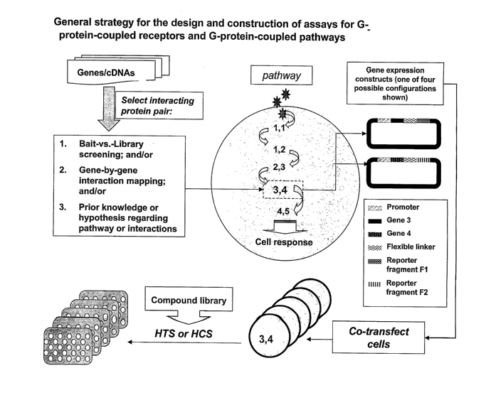
Figure 1 illustrates the general strategy for the design and construction of
an assay
according to the present invention.
Figure 2 shows a live cell assay for self association of a G-protein-coupled
receptor.
Figure 3 shows an assay for the association of a G-protein-coupled receptor
with a G-
protein a subunit (Goci) in two different human cell types.
Figure 4 shows an assay for the association of a G-protein-coupled receptor
with a G-
protein ~3 subunit (G~i 1 )
Figure 5 shows a quantitative assay for the association of a G-protein-coupled
receptor
with beta-arrestin, demonstrating an increase in signal intensity in response
to agonst.
Figure 6 shows a time course for the effect of agonist on the association of a
G-protein-
coupled receptor with beta-arrestin, showing both an increase in signal
intensity with time and a
redistribution of the protein-protein complex into intracellular granules.
Figure 7 shows that individual fragment fusions do not generate an optically
detectable
signal; signal generation depends upon fragment complementation.
23
CA 02538852 2006-03-10
WO 2005/031309 PCT/US2004/031643
Figure 8 demonstrates the effects of drugs on the association of beta-
arrestin2 with the
beta-2 adrenergic receptor . In the absence of agonist (upper left panel)
there is no YFP signal;
only the Hoechst-stained cell nuclei are seen. In the presence of
isoproterenol the two proteins
associate, causing the reassociation of YFP fragments to generate a bright
punctuate
fluorescence. The antagonist, propanolol, blocks the effect of isoproterenol.
The assay can be
used to screen for novel agonists and antagonists of the beta-adrenergic
receptor. This assay
principle can be applied to any GPCR that binds to an arrestin molecule.
Figure 9 shows the quantitative results obtained from the assay shown in FIG.
8.
Fluorescence intensity of the YFP channel is shown as a percent of control
(vehicle alone). The
effects of several different agonists (clenbuterol, salbumatomol and
isoproterenol) are shown.
The antagonist, propanolol, completely blocks the effect of isoproterenol.
Figure 10 shows the results of using the assay of FIG. 8 to screen for agents
that activate
the GPCR pathway at different time points. 98 different drugs were tested for
their ability to
increase the interaction of the beta-adrenergic receptor with beta-arrestin2
at either 30 minutes,
90 minutes or 480 minutes. Fluorescence intensity of the YFP channel is shown
as a percent of
control (vehicle alone). The direct agonists isoproterenol and salbutamol had
effects at early time
points whereas other drugs (BAY 11-7082, clozapine and pertussis toxin) had
effects only at
later time points consistent with their different mechanisms of action.
Figure 11 shows an assay for the detection of proteasomal regulation of GPCR
pathways.
The ubiquitination of beta-arrestin2 was measured. Signal was apparent only in
the presence of
24
CA 02538852 2006-03-10
WO 2005/031309 PCT/US2004/031643
the proteasom.e inhibitor ALLN (upper left panel). In the presence of ALLN,
isoproterenol
caused a significant increase in signal at 60' or 120'. These assays can be
used to identify novel
proteasome inhibitors.
Figure 12 demonstrates the effects of drugs on the beta-arrestin2-ubiquitin
assay. The
proteasome inhibitor MG132 and the histone deacetylase inhibitor, Trichostatin
A, increased the
signal fourfold over the negative control (vehicle alone). Assay fluorescence
is shown as a
percent of control for the positive pixel mean fluorescence intensity (PPM).
Figures 13 (A-C) demonstrates a variety of high-content fragment
complementation
assays for GPCRs and their cognate pathways. Fluorescence micrographs for
specific protein-
fragment complementation assays are shown. Fluorescence from the Hoechst
nuclear staining of
HEK cells is in blue whereas the YFP PCA signals from transient transfections
are in
yellow/green. Subcellular localization of the protein-protein complexes can
also be seen.
Assays for the following protein-protein pairs are shown: Frizzled4/G-alpha-I;
Frizzled
4lRGS2; Frizzled 4/GRK2; PKCalpha/GRK; PKCalpha/Chemokine Receptor 5; GRKIc-
Src;
GRK/ERK2; VIPR2/G-beta-l; and the Somatostatin Receptor/G-beta-1.
DETAILED DESCRIPTION OF THE INVENTION
Assay Construction
An overview of the process of constructing an assay for a GPCR or a G-protein-
coupled
pathway is shown in Fig. 1. The genes to be used in the assay may code either
for known or for
2s
CA 02538852 2006-03-10
WO 2005/031309 PCT/US2004/031643
novel interacting proteins. The interacting proteins are selected by one or
methods that include
bait-versus-library screening; pairwise (gene by gene) interaction mapping;
and/or prior
knowledge or a hypothesis regarding a pathway or an interacting protein pair.
A F1/F2 reporter fragment pair is generated by fragmentation of a suitable
reporter
(examples of additional reporters are in Table l and in US 6,270,964). Two
expression
constructs comprising the complementary fragments are made, in which the
expression of a
fusion protein is driven by a suitable promoter. One of the expression
constructs comprises a
first gene fused in frame to reporter fragment F1, and the other comprises a
second gene fused in
frame to reporter fragment F2. Optimally, a flexible linker, such as that
described in Example 1
below, is fused between the fluorescent protein fragment and the gene of
interest to facilitate
fragment complementation. Therefore, each expression vector codes for a fusion
protein
consisting of an operably linked gene ofinterest, a flexible linker, and
either F1 or F2 of the
chosen reporter. Such compositions are a subject of the present invention.
Fragments may be
fused at either the 3' or 5' end of the gene of interest with the linker
between the fragment and the
gene of interest. Selection of the fusion orientation may be based on a prior
understanding of a
particular protein function or based on empirical evidence of optimal fragment
orientations. As
shown in Figure 1, since either F1 or F2 can be fused to the gene of interest
and the orientation
of the fusion can either be 5' or 3' relative to the gene of interest, four
different DNA constructs
are possible for any single gene of interest.
To generate the PCA for a pair of proteins e.g. A and B, constructs encoding A
and B
fused to complementary reporter fragments F1 and F2, respectively, are co-
transfected into cells
26
CA 02538852 2006-03-10
WO 2005/031309 PCT/US2004/031643
using transfection methods suitable for the particular vector and cell type.
If proteins A and B
interact, fragments F 1 and FZ are brought into close proximity where they are
capable of folding
and reconstituting an active reporter. The resulting signal can then be
detected, quantified,
visualized or imaged by a variety of standard methods for optical detection of
the chosen
reporter. All of these methods can be used in automated, high-throughput
formats using
instrumentation well known to those skilled in the art.
It will be apparent to one skilled in the art that the choice of expression
vector depends on
the cell type for assay construction, whether bacterial, yeast, mammalian, or
other cell type; the
desired expression level; the choice of transient versus stable transfection;
and other typical
molecular and cell biology considerations. A wide variety of other useful
elements can be
incorporated into appropriate expression vectors, including but not limited to
epitope tags,
antibiotic resistance elements, and peptide or polypeptide tags allowing
subcellular targeting of
the assays to different subcellular compartments (e.g. A Chiesa et al.,
Recombinant aequorin and
green fluorescent protein as valuable tools in the study of cell signaling).
The incorporation of a
different antibiotic resistance marker into each of the two complementary
constructs would allow
for the generation of stable cell lines through double antibiotic selection
pressure, whereas
subcellular targeting elements would allow for the creation of assays for G-
protein-coupled
pathway events that occur within a particular subcellular compartment, such as
the mitochondria,
Golgi, nucleus, or other compartments.
A variety of standard or novel expression vectors can be chosen based on the
cell type
and desired expression level; such vectors and their characteristics will be
well known to one
2~
CA 02538852 2006-03-10
WO 2005/031309 PCT/US2004/031643
skilled in the art and include plasmid, retroviral, and adenoviral expression
systems. In addition,
there is a wide range of suitable promoters including constitutive and
inducible reporters that can
be used in vector construction. If an inducible promoter is used, the signal
generated in the assay
will be dependent upon activation of an event that turns on the transcription
of the genes encoded
by the PCA constructs.
Reporter Fragmentation
The principles of PCA assay construction have been described in detail in the
References
incorporated herein. While fragmentation of proteins for PCA is generally
based on rational
dissection of the polypeptide chain, a number of other engineering approaches
can be used that
will be well known to one skilled in the art. Fragments of a suitable reporter
protein can be
generated by starting with a cDNA encoding a full-length reporter of interest
and using PCR to
amplify fragments of interest. Alternatively, random fragmentation of a
reporter can be
performed, e.g. using 5' exonucleases to generate libraries of fragments to
search for optimal
pairs (Michnick, et al. 6,270,964). In addition, oligonucleotides encoding
fragments can simply
be synthesized using standard oligonucleotide synthesis techniques.
Mutant fragments can also be generated in order to generate assays tailored to
the
biological application and the instrumentation to be used. For example, site-
directed
mutagenesis of reporters, followed by fragmentation (or alternatively, site-
directed mutagenesis
of previously-created fragments) can be used to obtain fragments that provide
altered
fluorescence properties, or superior folding or maturation rates and
stabilities upon fragment
2s
CA 02538852 2006-03-10
WO 2005/031309 PCT/US2004/031643
complementation. Site-directed mutagenesis is achieved by any of a number of
approaches that
are well known to one skilled in the art (see MM Ling & BH Robinson, 1997,
Approaches to
DNA mutagenesis: an overview. Anal Biochem 254:157-78). Selected examples of
such
methods are provided here; however, these examples are not intended to be
limiting for the
practice of this invention. Suitable methods could include combinations of
random mutagenesis
and directed evolution or DNA shuffling schemes (A.L. Kurtzman et al., 2001,
Advances in
directed protein evolution by recursive genetic recombination: applications to
therapeutic
proteins, Curr Opin Biotechnol 2001 Aug;l2(4):361-70; SW Santoro et al., 2002,
Directed
evolution of the site specificity of Cre recombinase. Proc Natl Acad Sci U S A
2002 99:4185-90;
Z. Shao et al., 1996, Engineering new functions and altering existing
functions, Curr Opin Struct
Biol 6:513-8; S. Harayarna, 1998, Artificial evolution by DNA shuffling,
Trends Biotechnol
1998, 16:76-82); assembly PCR or gene synthesis approaches (WP Stemmer et al.,
1995, Single-
step assembly of a gene and entire plasmid from large numbers of
oligodeoxyribonucleotides,
Gene 164(1):49-53; RM Horton et al. 1993, Gene splicing by overlap extension.
Methods
Enzymol. 217:270-9), or fragmentation by exo- or endo-nuclease digestion (M.
Kitabatake and
H. Inokuchi, 1993, A simplified method for generating step-wise deletions
using PCR, Gene
123:59-61; S. Henikoff, 1990, Ordered deletions for DNA sequencing and in
vitro mutagenesis
by polymerase extension and exonuclease III gapping of circular templates,
Nucleic Acids Res
18(10):2961-6). A particularly powerful method is based on 5'-template-
assisted long-range
plasmid polymerization as exemplified by a number of commercial mutagenesis
kits, for
example the QuickChangeTM system (Stratagene). In addition, various forms of
directed
evolution based on DNA shuffling could also be used to generate completely
novel PCAs.
29
CA 02538852 2006-03-10
WO 2005/031309 PCT/US2004/031643
Although it is expedient to carry out the engineering and construction of PCAs
at the DNA level
and then either allow a cell to produce the fusion proteins, it is not
essential. The assays
described herein can be performed in vivo or in vitro. In vitro assays may
facilitate
characterization of a large number of GPCRs. For example, fusion proteins can
be made in vitro
using in vitro expression techniques that are well known to those skilled in
the art such as
baculovirus expression systems and alternative approaches to polypeptide
expression. In
addition, for in vitro PCAs, fusion polypeptides could be produced
synthetically by peptide
synthesis, or by ligation of peptide fragments encoding molecules of interest
to create peptide
fusions with the reporter fragments. Such assays could be used, for example,
to characterize the
binding of orphan GPCRs to peptide ligands. The GPCRs could each be tagged
with a F1
fragment of a reporter and immobilized on a solid surface such as a chip. The
chip could then be
exposed to a library, such as a peptide library, in which each peptide is
tagged with a
complementary (F2) fragment of a reporter. Binding of a peptide to a GPCR
would be detected
by reconstitution of the reporter activity through the association of Fl and
F2. The identity of
the binding peptide could then be assessed, for example by any one of a
variety of proteomic
methods including mass spectroscopy. Such assays for screening and de-
orphanization are also a
subject of the present invention. The present invention is not limited to the
assay format used or
the detection method for the assay.
Selection of an Appropriate Reporter
The general characteristics of reporters suitable for PCA have previously been
described
(References incorporated herein). A preferred embodiment of the present
invention involves
CA 02538852 2006-03-10
WO 2005/031309 PCT/US2004/031643
cell-based assays generating a fluorescent or luminescent signal are
particularly useful.
Examples of reporters that can be used in the present invention are listed in
Table 1. It will be
appreciate by one skilled in the art that the choice of reporter is not
limited. Rather, it will be
based on the desired assay characteristics, format, cell type, spectral
properties, expression, time-
course and other assay specifications. For any reporter of interest various
useful pairs of
fragments can be created, for example using the methods taught in US 6,270,964
arid the
References incorporated herein, and then engineered in order to generate
fragments that produce
a brighter signal or a specific color readout upon fragment reassembly. It
will be obvious to one
skilled in the art that various techniques of genetic engineering can be used
to create useful
fragments and fragment variants of any of the reporters that are the subject
of this invention.
It will be appreciated by a person skilled in the art that the ability to
select from among a
wide variety of reporters makes the invention particularly useful for drug
discovery on a large
scale. In particular, reporters can be selected that emit light of a specific
wavelength and
intensity that may be suitable for a range of protein expression levels, cell
types, and detection
modes. The flexibility is an important feature of the invention because of the
wide range of
signaling events, or biochemical processes, linked to GPCRs. For some
biochemical events,
activation of a GPCR - for example, by the binding of an agonist - will lead
to an increase in the
association of a GPCR and a cognate binding protein, or of two
elements'downstream' in the G-
protein-coupled pathway, such as a kinase and its substrate. An increase in
the association of the
two proteins that form the PCA pair leads to an increase in the signal
generated by the
reassembled reporter fragments. In that case, a high-throughput assay format
can be used to
measure the fluorescent signal that is proportional to the amount of the
complex of interest. For
31
CA 02538852 2006-03-10
WO 2005/031309 PCT/US2004/031643
quantitative assays, where the readout is an increase or decrease in signal
intensity, any of the
reporters discussed in the present invention can be used and each reporter has
various pros and
cons that are well understood by those skilled in the art of cell biology.
Enzymes - for which the
catalytic reaction generates a fluorescent, phosphorescent, luminescent or
other optically
detectable signal - may be best suited for purely quantitative assays. Upon
fragment
complementation, the reconstituted enzyme acts upon a substrate to generate a
fluorescent or
luminescent product, which accumulates while the reporter is active. Since
product accumulates,
a high signal-to-noise can be generated upon fragment complementation. Such
assays are
particularly amenable to scale-up to 384-well or 1536-well formats and beyond,
and are
compatible with standard and ultra high-throughput laboratory automation.
Preferred reporters for the present invention include but are not limited to a
beta-
lactamase PCA or a luciferase PCA such as with a firefly luciferase or Renilla
luciferase. Each
of these enzymes has been successfully used as a cell-based reporter in
mammalian systems (S
Baumik & SS Gambhir, 2002, Optical imaging of renilla luciferase reporter gene
expression in
living mice, Proc. Natl. Acad. Sci., USA 2002, 99(1): 377-382; Lorenz et al.,
1991, Isolation and
expression of a cDNA encoding renilla reniformis luciferase, Proc. Natl. Acad.
Sci. USA 88:
4438-4442; G. Zlokarnik et al., 1998, Quantitation of transcription and clonal
selection of single
living cells with beta-lactamase as reporter, Science 279: 84-88). As an
example of the
construction of a PCA, beta-lactamase PCAs have been constructed with cell-
permeable
substrates that generate a high signal to background upon cleavage (A
Galarneau et al., 2000,
Nature Biotechnol. 20: 619-622). The beta-lactamase PCA is a sensitive and
quantitative assay
suitable for HTS. This PCA has been used with CCF2/AM, a green fluorescent
molecule which
32
CA 02538852 2006-03-10
WO 2005/031309 PCT/US2004/031643
becomes blue upon cleavage of the beta-lactam ring by beta-lactamase; the blue-
green ratio is
therefore a measure of the activity of beta-lactamase which is reconstituted
upon protein-
fragment complementation. Luciferase PCAs can also be used with cell-permeable
substrates to
generate HTS assays suitable for the present invention (e.g. R Paulmurugan et
al., 2002,
Noninvasive imaging of protein-protein interactions in living subjects by
using reporter protein
complementation and reconstitution strategies, Proc. Natl. Acad. Sci. USA 99:
15608-15613).
With suitable modifications, any of these PCAs can also be used in vivo or in
vitro for the
present invention. It will be apparent to one skilled in the art that PCAs
based on inherently
fluorescent, phosphorescent or bioluminescent proteins can be read either in
high-content
formats or in high-throughput formats. These PCAs have the advantage of not
requiring the
addition of substrate; however, the signal generated is usually lower than
that generated by an
enzymatic reporter.
Calcium-sensitive photoproteins would be particularly useful as PCAs for GPCR
assays.
These could be based on fragments of aequorin, obelin; or any other calcium-
sensitive protein
(e.g. MD Ungrin et al., 1999, An automated aequorin luminescence-based
functional calcium
assay for G-protein-coupled receptors, Anal Biochem. 272: 34-42; Rizzuto et
al., 1992, Rapid
changes of mitochondrial calcium revealed by specifically targeted recombinant
aequorin,
Nature 358 (6384): 325-327; Campbell et al., 1988, Formation of the calcium
activated
photoprotein obelin from apo-obelin and mRNA in human neutrophils, Biochem J.
252 (1):143-
149). Aequorin, a calcium-sensitive photoprotein derived from the jellyfish
Aequorea victoria, is
composed of an apoprotein (molecular mass ~21 kDa) and a hydrophobic
prosthetic group,
coelenterazine. Calcium binding to the protein causes the rupture of the
covalent link between
33
CA 02538852 2006-03-10
WO 2005/031309 PCT/US2004/031643
the apoprotein and the coelenterazine, releasing a single photon. The rate of
this reaction depends
on the calcium concentration to which the photoprotein is exposed. Intact
aequorin with
coelenterazine has been used to monitor calcium flux in cell-based assays for
GPCRs. Obelin is
a 22-kDa monomeric protein that also requires coelenterazine for signal
generation. Construction
of an aequorin PCA or an obelin PCA would enable assays for G-protein-coupled
receptors and
pathways in which photon release only occurs if the reporter fragments are
associated as a result
of a ligand-protein interaction or a protein-protein interaction. Such an
assay would combine
measures of pathway activation with calcium flux, making the assays
extraordinarily sensitive
for GPCR studies.
Although small monomeric reporters are preferred for this invention due to the
small size
of the reporter fragments, it will be apparent from the prior art that
multimeric enzymes such as
beta-galactosidase, beta-glucuronidase, tyrosinase, and other reporters can
also be used in the
present invention. A number of mu.ltimeric enzymes suitable for PCA have
previously been
described (US 6,270,964). Fragments of multimeric proteins can be engineered
using the
principles of PCA described in the prior art; alternatively, naturally-
occurring fragments or
engineered low-affinity subunits of multimeric enzymes can be used including
the widely-used
beta-galactosidase alpha and omega complementation systems (see References).
The naturally-
occurring fragments of beta-galactosidase and protocols for their use have
been developed and
distributed by DiscoveRx, Inc. (Fremont, CA; www.discoverx.com ) and by
distributors
including Stratagene (Q-Tag detection kit, www.stratagene.com) and can be used
in conjunction
with any of the fragment complementation assays and GPCR signaling proteins
taught in the
present invention. The engineered low-affinity subunits of beta-gal are sold
by Applied
34
CA 02538852 2006-03-10
WO 2005/031309 PCT/US2004/031643
Biosystems, Inc. (www.ap 1p ledbiosystems.com). Substrates suitable for the
generation of
fluorescent and luminescent signals upon beta-gal complementation are also
widely available
(see for example www.prome~a.com for Beta-Glo protocols and reagents) and can
be used in
conjunction with the present invention.
For some G-protein-coupled events, activation of the pathway leads to the
translocation
of a pre-existing protein-protein complex from one sub-cellular compartment to
another, without
an increase in the total number of protein-protein complexes. In that case,
the fluorescent signal
generated by the reassembled reporter at the site of complex formation within
the cell can be
imaged, allowing the trafficking of the complex to be monitored. Such "high-
content" PCAs can
be engineered for any suitable reporter for which the signal remains at the
site of the protein-
protein complex. Examples include the DHFR PCA, which has been used for high-
content
assays of signal transduction pathways (I Remy & S Michnick, 2001,
Visualization of
Biochemical Networks in Living Cells, Proc Natl Acad Sci USA, 98: 7678-7683)
and also for
high-throughput assays (I Remy et al., 1999, Erythropoietin receptor
activation by a ligand-
induced conformation change, Science 283: 990-993). Reconstituted DHFR binds
methotrexate
(MTV); if the MTX is conjugated to a fluorophore such as fluorescein, Texas
Red, or BODIPY,
the PCA signal can be localized within cells. Additional reporters
particularly useful for high-
content assays are described in US 6,270,964 and include the green fluorescent
protein (GFP)
from Aequorea victoria.
PCAs based on GFP, YFP, and other inherently fluorescent, luminescent or
phosphorescent protein reporters are preferred embodiments of the present
invention. Any
CA 02538852 2006-03-10
WO 2005/031309 PCT/US2004/031643
number of fluorescent proteins have been described in the scientific
literature (e.g. RY Tsien,
1998, The Green Fluorescent Protein, in: Annual Reviews of Biochemistry 67:
509-544; 3~
Zhang et al., 2000, Creating new fluorescent probes for cell biology, Nature
Reviews 3: 906-
918). Any mutant fluorescent protein can be engineered into fragments for use
in the present
invention. Suitable reporters include YFP, CFP, dsRed, mRFP, 'citrine', BFP,
PA-GFP, 'Venus',
SEYFP and other AFPs; and the red and orange-red fluorescent proteins from
Anemonia and
Anthozoa.
Reporters generating a high signal in a cellular background are preferred for
the present
invention. For example, PCAs based on YFP, SEYFP, or 'Venus' (T Nagai et al.,
2002, A variant
of yellow fluorescent protein with fast and efficient maturation for cell-
biological applications,
Nature Biotech. 20: 87-90) are particularly suitable for the present
invention. PCAs based on
proteins for which the signal can be triggered, such as a kindling fluorescent
protein (KFP1)
(DM Chudakov et al., 2003, Kindling fluorescent proteins for precise in vivo
photolabeling,
Nat. Biotechnol. 21, 191-194), a photo-converting fluorescent protein such as
Kaede (R Ando et
al., 2002, An optical marker based on the uv-induced green-red photoconversion
of a fluorescent
protein, Proc. Natl. Acad. Sci. USA, 202, 99 (20): 12651-12656), or a
photoactivatable protein
such as PA-GFP (GH Patterson et al., 2002, A photoactivatable GFP for
selective labeling of
proteins and cells, Science 297: 1873-1877) may have advantages, particularly
in cases where it
is necessary to capture very rapid signaling events. KFP 1 is derived from a
unique GFP-like
chromoprotein asCP from the sea anemone Anemonia sulcata. asCP is initially
nonfluorescent,
hut in response to intense green light irradiation it becomes brightly
fluorescent (kindles) with
emission at 595 nm. Kindled asCP relaxes back to the initial nonfluorescent
state with a half life
36
CA 02538852 2006-03-10
WO 2005/031309 PCT/US2004/031643
of <10 seconds. Alternatively, fluorescence can be "quenched" instantly and
completely by a
brief irradiation with blue light. The mutant (asCP A148G, or KFP 1 ) is
capable of unique
irreversible photoconversion from the nonfluorescent to a stable bright-red
fluorescent form that
has 30 times greater fluorescent intensity than the unkindled protein, making
it particularly
suitable for live cell PCAs.
It will be apparent to one skilled in the art that any of these reporters can
be used to
construct assays based on the principles and methods described herein by
simply generating
fragment pairs for the reporter that is to be used; substituting the fragment
pairs for the desired
reporter in the fusion constructs; expressing them at a level suitable for
detection of the signal of
interest; and performing the assay under conditions suitable for the detection
of the signal that is
generated upon fragment complementation for that particular reporter. Such
assay conditions
can be found in the biochemical literature and are well known to those skilled
in the art.
37
CA 02538852 2006-03-10
WO 2005/031309 PCT/US2004/031643
Instrumentation
The high-throughput assays described above generate optically detectable
signals that can
be read on commercially available instrumentation, including fluorescence
plate readers,
lurninometers, and flow cytometers. Such instrumentation is widely available
from commercial
manufacturers, including Molecular Devices, Packard, Perkin Elmer, Becton
Dickinson,
Beckman Coulter, and others. All such assays can be constructed in multiwell
(96-well and 384-
well) formats. The high-content assays described above generate optically
detectable signals that
can be spatially resolved within subcellular compartments. The resulting
images can be captured
with automated microscopes, confocal imaging systems, and similar devices.
Suitable imaging
instrumentation is widely available from a variety of commercial manufacturers
including
Molecular Devices (Universal Imaging), Amersham Bioscience, Cellomics, Evotec,
Zeiss,
Q3DM, Atto, and others. Image analysis software such as MetaMorph, the
publicly available
IMAGE software from the National Institutes of Health
(http://rsb.info.nih.gov/nih-image/) and
various proprietary software packages are used to distinguish the signal
emanating from different
subcellular compartments (membrane, cytosol, nucleus) and to quantitate the
total fluorescence
per cell. In addition, mufti-well PCA formats for the present invention can be
constructed for
array-based assay formats, including reverse transfection methods such as
those provided by
Akceli Inc., allowing the rapid, simultaneous processing of a large number of
different PCAs on
a single array.
38
CA 02538852 2006-03-10
WO 2005/031309 PCT/US2004/031643
Selection of G-protein-coupled pathway elements for assay construction
The present invention can be used to construct assays for G-protein-coupled
receptors
themselves, for example by tagging the intracellular (C-terminal) portion of
the GPCR with a
reporter fragment and assaying its self interactions, receptor interactions
with ligands, with other
receptors, with kinases or phosphatases, writh receptor activated modulator
proteins, or with other
intracellular proteins. For example, the various interactions among GPCRs and
G-proteins can
be assayed using the methods provided herein. The advantage is the ability to
understand the
intracellular machinery linked to a particular GPCR and to construct an assay
for any step in a
pathway. The advantages of being able to construct screening assays for
various steps in a G-
protein-coupled pathway, starting with the receptor and moving down the entire
signaling
cascade, include the ability to screen for compounds that may act at any one
of various
potentially druggable targets iri a G-protein-coupled pathway and to de-
orphanize receptors by
identifying the signaling machinery to which the receptor is linked.
Suitable pairs of interacting molecules for assay construction can be
identified by any one
of the methods outlined in Figure 1. PCA enables a systematic characterization
of the
interactions made among the GPCRs in living cells by first examining whether
different pairs of
GPCRs generate a PCA signal in a cell type of interest and then determining
whether the signal
amount or subcellular location is affected by agents that modify cell
signaling. In this way, the
functional interactions among the hundreds of GPCRs can be 'mapped' using PCA.
For example,
a GPCR tagged with one complementary fragment of a reporter can be used as
'bait' to screen a
cDNA library tagged with a second complementary fragment of a reporter (bait-
vs.-library
39
CA 02538852 2006-03-10
WO 2005/031309 PCT/US2004/031643
screening). In this respect PCA is an alternative to the widely-used yeast-two-
hybrid system for
the identification of interactions among G-protein-coupled signaling
components. Systematic
screening can also be performed to identify pathway elements; for example,
novel or orphan
GPCRs tagged with F1 of a suitable reporter can be tested individually against
other proteins
tagged with complementary fragment F2 (gene-by-gene analysis). The presence of
a PCA signal
indicates an interaction between the two proteins tagged with the
complementary fragments. For
example, the cognate G-proteins linked to particular GPCRs can be identified
or the kinases
linked to specific GPCRs can be identified by testing each protein against the
other in a PCA.
The presence of a PCA signal indicates an interaction between the two assay
components. The
advantage of the present invention is that, once an interaction has been
identified, an assay is in
hand that can be used to screen for pathway modulation, to de-orphanize
receptors, or to screen
for compounds that modulate the pathway of interest by using a high-content or
high-throughput
PCA as a screen.
The components of G-protein-coupled pathways have been partially elucidated,
and the
known or hypothesized interactions can readily be used to design assays
according to the present
invention. The present invention encompasses assays for a variety of steps in
G-protein-coupled
pathways. A number of these steps are described in detail below based on the
biochemical
literature, and are listed in Table 2. Any of the protein-protein interactions
reported to date can
be used as the basis for the construction of protein-fragment complementation
assays or enzyme-
fragment complementation assays. Such assays can include the GPCRs themselves;
G-proteins
(alpha, beta and gamma subunits); adenylyl cyclase (R and C subunits); protein
kinases
(including but not limited to GRK, PCA, PI~C, MAPK, ERK, and others); protein
phosphatases
CA 02538852 2006-03-10
WO 2005/031309 PCT/US2004/031643
(PPP2A and others) ; phospholipases, such as phospholipase C (PLC); receptor-
activity-
modifying proteins (RAMPS); lipid transferases (farnesyl transferase,
myristoyl transferase,
palmitoyl transferase); ion exchange regulatory factors; GIRKs; beta-
arrestins; E3 ligases;
Ubiquitin monomer or polypeptide; RDG proteins; phosphodiesterases such as
PDE4; cytokine
and growth factor receptors that exhibit cross-tally with GPCRs; and
transcription factors such as
ELK, CREB and CBP.
All of the assays that are the subject of the present invention are of general
use as
validation assays or in basic experimental biology research as well as in drug
discovery. For
example, these assays can be used in conjunction with RNA interference methods
to link specific
genes to G-protein-coupled pathways. This can be accomplished by introducing,
for example, a
small interfering RNA (siRNA) into a cell-based fragment complementation assay
and
determining whether the siRNA reduces the signal generated by the interacting
protein pair. Any
number of other cellular probes can be used in a similar manner, including
dominant negative
genes, drugs, peptides, antibodies and other biochemical or biological
reagents. In addition to
functional annotation, target validation and pathway mapping, the assays
described herein are all
amenable to high-throughput screening and drug discovery.
41
CA 02538852 2006-03-10
WO 2005/031309 PCT/US2004/031643
Table 2. Interacting elements of Gprotein-coupled pathways suitable for the
construction
of fragment complementation assays
First roteinSecond roteinRe orted event Reference
GPCR GPCR receptor homo- S. Angers et al., 2002,
or hetero- Dimerization: an emerging
dimerization concept for GPCR ontogeny
and function, Annu. Rev.
Pharmacol. Toxicol. 42:
409-435.
GPCR ligand (eitherligand binding U Gether, 2000, Uncovering
a to receptor molecular mechanisms
peptide involved in activation of
or a small- G Protein-Coupled Receptors,
molecule in: Endocrine Reviews 21:
ligand 90-I 13.
could be
used in
PCA)
GPCR G-protein Ga, G(3, or Gy NA Sarvazyan et al., 2002,
subunit binding to Fluorescence Analysis of
intracellular portionReceptor-G protein Interactions
of G-protein in Cell membranes,
coupled receptor; Biochemishy 41: 12858-12867;
specificity variesE Hermans, 2003,
based on receptor,Biochemical and Pharmacological
ligand, and control of the
cognate Ga homologuemultiplicity of coupling
at GPCRs, Pharmacology
&
Thera eutics 99:25-44.
GPCR beta-arrestinb-arrestin binds LM Luttrell & RJ Lefkowitz,
to agonist-occupied2002, The role of beta-
receptor arrestins in the termination
and transduction of G-
protein-coupled receptor
signals, J. Cell. Sci.
1 I5:
455-465.
GPCR GRKs (GPCR binding and phosphorylationAE Brady & LE Limbird, 2002,
of G-protein-coupled
kinases); GPCR by GRK promotesreceptor interacting proteins:
multiple binding emerging roles in
homologues of beta-arrestin localization and signal
to GPCRs transduction, Cell Signal
14:
297-309
GPCR PKA, PKC, phosphorylation Daaka et al., 1997, Switching
Src, of the beta-2 of the coupling of the
Casein kinases;adrenoreceptor beta-2-adrenergic receptor
Raf, by the cyclic to different G-proteins
AMP- by
MEK, ERK dependent protein protein kinase A, Nature
kinase, PKA, 390: 88-91;G Fan et al.,
2001,
switches its couplingC-src tyrosine kinase binds
from Gs to Gi the beta-2-adrenergic
and induces receptorreceptor via phospho-Tyr-350,
phosphorylates GRK2
internalization; and mediates agonist-induced
binding ofc-Src receptor desensitization,
tyrosine kinase J. Biol. Chem. 276: 13240-13247;
to GPCR mediates N Yuan et al., 1994,
5re-dependent phosphorylationcAMP-dependent protein kinase
of A and protein kinase
GRK C consensus site mutations
of the beta-adrenergic
receptor, J. Biol. Chem.
269: 23032-23038; R Winstel
1996, Protein kinase cross-talk:
membrane targeting of
the beta-adrenergic receptor
kinase by protein kinase
C, Proc. Natl. Acad. Sci.
USA 93: 2105-2109.
GPCR E3 ligase; ubiquitlnation A. Ciechanover, 1998, The
Ubiquitin of GPCR by an ubiquitin-proteasome
E3
ligase targets pathway: on protein death
the receptor for and cell life, The EMBO
downregulation Journal 17: 7151-7160.
GPCR Protein dephosphorylation JA Pitcher, 1995, The G-protein
phosphataseof coupled receptor
(PP2A and phosphorylated phosphatase: a protein phosphatase
GPCR type 2A with a
homologues) distinct subcellular distribution
and substrate
s ecificity, Proc. natl.
Acad. Sci. USA 92: 8343-8347
GPCR RAMPS (receptor-RAMPs are single-transmembraneJA Fischer et al.,
2002,
Functional relevance of
G-
activity-modulatingproteins that transportprotein-coupled-receptor-associated
receptors to proteins
proteins); the cell surface exemplified by receptor-activity-modifying-
proteins
several
homologues (RAMPS , Biochem. Soc. Trans.
30(4): 455-460.
GPCR PDZ-containing,binding of C-terminal3Ciang, 2002, The PDZ-binding
GPCR motif of the (32-
EVH-containingsequences and regulationadrenergic receptor modulates
and of receptor trafficking and
Homer familyreceptor function;signaling in cardiac myocytes,
some GPCRs J. Biol. Chem. 277:
proteins interact directly,33783-33790; RA Hall 1999,
via their C- Heptahelical receptor
terminal domain, signaling: beyond the G
with proteins protein paradign, J. Cell
Biol.
containing PDZ 145: 927-932.
and
Enabled/VASP homology
(EVH)-
like domains.
GPCR growth factorcross-talk betweenML Grimes & HM Miettinen,
or receptors 2003, Receptor tyrosine
cytokine kinase and G-protein coupled
receptors receptor signaling and
(EGFR, PDGFR, sorting within endosomes,
j. Neurochem. 84: 905-918.
VEGFR, HGH
receptor,
GM-CSF
receptor,
IL
rece tors,
etc.
42
CA 02538852 2006-03-10
WO 2005/031309 PCT/US2004/031643
GPCR Na+/H+ exchangerexchange protein RA Hall, 1998, The beta-2
interactions that adrenergic receptor
regulatory control cell signalinginteracts with the Na+/H+
factors; linked to exchanger regulatory factor
GIRKs (G-proteincertain GPCRs; to control Na+/H+ exchange,
certain GPCFa Nature 392: 626-630; MJ
activated including the alpha-2a-adrenergicMahon et al., 2002, Na+/H=
inwardly exchanger regulatory
rectifying receptor couple factor 2 directs parathyroid
K+ to GIRKs hormone I receptor
channels si alin , Nature 417: 858-861.
G-protein G-protein Association or J-Z. Yu & MM Rasenick,
subunit subunit dissociation of 2002, Real-time
Ga, G(3, Ga, G(3, Ga/G(i, G(3/Gy, visualization of a fluorescent
or Gy or Gy; GalGy Gas: dissociation of the
AGS
proteins activated G-protein from
(Activators the plasma membrane, Mol.
of G Proteins) Pharmacol. 61: 352-359-
NA Sarvazyan et al., 2002,
Fluorescence Analysis of
Receptor-G protein
Interactions in Cell membranes,
Biochemistry 41:
12858-12867.
G-protein Adenylyl G-proteins coupledRK Sunahara et al., 1996,
subunit Cyclase through Complexity and Diversity
of
Ga, G(3, adenylyl cyclase mammalian adenylyl cyclases,
or Gy to generate the Annu. Rev. Pharmacol.
second messen er Toxicol. 36: 461-480.
cAMP
_ _ GPCRs that couple JL Blank et al., 1992,
G-protein Phospholipaseto Gq family Activation of cytosolic
subunit C
Ga, G(i, members stimulate phosphoinositide phospholipase
or G~y PLC resulting C by G-protein ~i/y
in release of IP3 subunits, J. Biol. Chem.
and diacylglycerol267: 23069-23075; M
Schmidt et al., 2000, G
Protein-coupled receptor-
induced sensitization ofphospholipase
C stimulation
by receptor tyrosine kinases,
3. Biol. them. 275:
32603-32610.__
Beta-arrestinsGPCR beta-arrestins RH Oakley et al., 2000,
as desensitizers differential affinities
for of visual
GPCRs arrestin, beta-arrestinl
, and beta-Arrestin2 for
G
Protein-coupled receptors
delineate two major classes
of rece tors, J. Biol.
them. 275: 17201-17210.
Beta-arrestinsGPCRs; Src beta-arrestins LM Luttrell & RJ Leflcowitz,
kinase; as scaffolds for 2002, The role of beta-
5hc; MAPK; intracellular proteinarrestins in the termination
JNK3; kinase and transduction of G-
ASK; MKK4; cascades protein-coupled receptor
Raf; signals, J. Cell. Sci.
115:
MEK; ERKI 455-465.
/2; AKT
G-protein _ G-protein-modulatedEM Hur & KT Kim, 2002,
subunit GPCRs; GRK;kinase G-protein-coupled receptor
Src
Ga, G(3, kinase; signaling cascadessignalling and cross-talk
or Gy Shc; MAPK; (ras/raf, MAPK, : achieving rapidity and
JNK3; ASK; JNK, AKT, ERKIELK specificity, Cell. Signaling
MKK4; and other 14: 397-405.
Raf; MEK; signaling cascades)
ERKI/2;
AKT
_
Beta-awestinsclathrin; Beta-arrestin promotesLM Luttrell & RJ Leflcowitz,
NSF; AP2; 2002, The role of beta-
Arf 6 internalization arrestins in the termination
of receptors via and transduction of G-
clathrin-coated protein-coupled receptor
vesicles; interactssignals, J. Cell. Sci.
115:
with clathrin, 455-465.
adaptorprotein
Al'-2,
N-ethylmaleimide-sensitive
fusion
rotein (NSF) and
Arf 6
Beta-amestinsUbiquitln; beta-arrestins SK Shenoy et al., 2001,
MDM2 as substrates Regulation of receptor
for fate by
ubiquidnatlon by ubiquitination of activated
E3 ligases; in (3-2-adrenergic receptor
and
particular, MDM2 beta-arrestin, Science
binds to and 294: 1307-1313.
ubiquifinates beta-arrestin
_ dishevelledbeta-attestinl W Chen et al., 2001, beta-arrestinl
Beta-arrestins(DSH) interacts with modulates
DSH
in a phosphorylation-dependentlymphoid enhancer factor
transcriptional activity
manner through interaction with
phosphorylated dishevelled
roteins, Proc. Natl. Acad.
Sci. USA 98: 14889-14894
Beta-arrestinsPDE4 beta-arrestins GS Baillie et al., 2003,
recruit the cAMP- beta-arrestin-mediated
PDE4
degrading PDE4 CAMP phosphodiesterase
recruitment regulates
(3-
phosphodiesterasesadrenoceptor switching
to the from Gs to Gi, Proc, Natl.
2adrener is rece Acad. Sci. USA 100(3):
for 940-945
RGS (regulatorsGa; GPCR RGS as desensitizerSP Heximer et al., 2001,
of signaling Mechanisms governing
of G-protein components; bind subcellular location and
directly to Ga function of human RGS2,
J.
signaling) Biol. Chem. 276: 14195-14203;
RJ Kimple et al.,
2003, Established and emerging
fluorescence-based
assays for G-Protein Function,
in: Combinatorial
Chemistry & Hi h-Through
ut Screenin , 6: t-9.
GRKs tubulin, _ 3LR Freeman, 2.002, Adrenergic
ribosomal GRK-mediated phosphorylationreceptor-stimulated,
of
protein intracellular substratesGRK2-mediated phos-phorylation
P2, synucleins, of ribosomal
synucleins,phosducins, ribosomalprotein P2, Biochemistry
proteins, 41:12850-12857; S Sarnago,
phosducins;tubulins; c-src 1999, Agonist-dependent
c-src phosphorylates phosphorylation of the
G-
GRK2 on tyrosine protein-coupled receptor
residues, kinase 2 (GRK2) by Src
iri erin GRK2 de osine kinase, J. Biol.
adation Chem. 274: 34411-34-116
Farnesyl GRKs, GPCRs,Membrane targetingY Okamoto,1997, Paimitoylation
Ga of GRKs of human endothelin
transferase(s), GPCRs, and Ga by B, J. Biol. Chem. 272;
farnesylation, 21589-21596; BF O'Dowd,
palmitoyl almito lation, 1989, Palmitoylation of
and/or the human beta-2 adrenergic
43
CA 02538852 2006-03-10
WO 2005/031309 PCT/US2004/031643
transferase(s), myristoylation receptor, J. Biol. Chem.
264: 7564-7569; P Svoboda
myristoyl and J Novotny, 2002, Hormone-induced
subcellular
transferase(s) redistribution of irimeric
G proteins, Cell. Mol.
Life
Sci. 59: 501-512
_ _ _ _ _ _ M Schmidt et al., 2000,
Phospholipase(s)Gaq, Gai, GPCRs activate G Protein Coupled Receptor-
G[3, Gy> PLC via G-
C (PLC) GPCR, RTK proteins; GPCRs induced sensitization of
sensitize PLC phospholipase C stimulation
stimulation by by Receptor Tyrosine Kinases;
RTKs; Gy activatesJ. Biul. Chem. 275:
phospholipase C 32603-32610; JH Exton,1996,
Regulation of
phosphoinositide phospholipases
by hormones,
neurotransmitters, and
other agonists linked
to G-
roteins, Annu. Rev. Pharmacol.
Toxicol. 36: 481-509
PKA (cyclic__ _ p~ regulatory subunitsX Fang et al., 2002, Convergence
PKA (RI/RII),(R) of multiple signaling
PKA
AMP-dependent(C) GSK3, cascades at glycogen synthase
GPCRs, interact and regulatekinase 3, Mol. Cell.
catalytic
protein AKAPs, PKC Biol. 22: 2099-2110; ML
kinase) subunits (C) in Ruehr et al., 1999, Cyclic
response to cAMP;
AMP-dependent protein kinase
PKA phosphorylatesbinding to A-kinase
GPCRs and
anchoring proteins in living
GSK3; A-kinase cells by fluorescence
anchoring proteins
resonance energy transfer
(AKAPs) interact of green fluorescent protein
with PKA~
fusion proteins J. Biol.
AKAPs can recruit Chem. 274: 33092- 33096
both PKA and SR
Adams et al., 1991, Fluorescence
PKC to specific ratio imaging of
subcellular
cyclic AMP in single living
locations via interactionscells, Nature 349: 694-697;
with the
GA Perkins et al., 2001,
various isoforms PICA, PKC, and AKAP
of each family
of
localization in and around
kinases. Each of the neuromuscular junction,
these proteins
has
numerous homologues.Neuroscience 2: 17.
Also see review articles
in References listed herein.
TranscriptionpenultimateGPCR-dependent JC Chrivia et al., 1993,
activation of Phosphorylated CREB binds
factors components oAMP-responsive specifically to the nuclear
e.g. ELK, of element binding protein CBP, Nature 365:
CREB, CBP,signaling protein (CREB) 855-859
of GPCRs via PICA-
NFkB via second dependent phosphorylation;
messengers
(calcium,
cAMP, IP3)
and via
kinase cascades
(ERKIELK
In order to exemplify these principles, we used various known pairs of
interacting
proteins to construct assays far GPCRs and G-protein-coupled pathways,
including
receptor/receptor assays, receptor/G-protein assays, and receptor/beta-
arrestin assays. For the
latter, we demonstrated both quantitative, agonist-dependent association of
proteins as well as
high-content, kinetic assays in living cells.
44
CA 02538852 2006-03-10
WO 2005/031309 PCT/US2004/031643
Example 1. Dimerization of GPCRs
A growing number of studies have shown that GPCRs are capable of forming
homodimers or heterodimers (S. Angers et al., 2002, Dimerization: an emerging
concept for
GPCR ontogeny and function, Annu. Rev. Pharmacol. Toxicol. 42: 409-435; MK
Dean et al.,
2001, Dimerization of G-protein-coupled receptors, J. Med. Chem. 44: 4594-
4614). Receptor
self association and subsequent changes in receptor activity have been
reported for the beta2-
adrenergic receptor (T.E. Hebert, 1996, A peptide derived from a beta2-
adrenergic receptor
transmembrane domain inhibits both receptor dimerization and activation, J.
Biol. Chem. 271:
16384-16392), in addition to the delta-opioid receptors, the dopamine
receptors, and other
GPCRs. It has been shown that agonists can stabilize the dimeric forms of
different GPCRs (U.
tether et al., 2000, Uncovering molecular mechanisms involved in activation of
G-protein-
coupled receptors, Endocrine Reviews 21:90-113), suggesting that
homodimerization may play a
role directly in receptor activation or, alternatively, in the subsequent
agonist-dependent
desensitization and internalization process (He et al., 2002, Regulation of
opioid receptor
trafficking and morphine tolerance by receptor oligornerization, Cell 108: 271-
282). In addition
to homo-dimerization, evidence has accumulated demonstrating the possible
importance of
hetero-dimerization between closely related receptor subtypes, which may also
be critical for
targeting functional receptors to the cell surface and for drug tolerance.
Standard biochemical
methods have been used to study receptor dimerization, including co-
immunoprecipitation and
gel shift assays. In addition, FRET or BRET have been used to monitor homo-
oligomerization
and hetero-oligomerization of GPCRs (McVey et al., 2001, Monitoring receptor
oligomerization
using fluorescence resonance energy transfer and bioluminescence resonance
energy transfer, J.
CA 02538852 2006-03-10
WO 2005/031309 PCT/US2004/031643
Biol. Chem. 276: 14092-14099). However, the prior art is silent on the use of
fragment-
complementation assays to study GPCR dimerization.
A high-content assay suitable for live cells would enable monitoring of
trafficking of
receptor complexes between the cell membrane, endocytic vesicles, cytoplasm
and other
subcellular compartments in response to agonists and antagonists. We
postulated that fragment
complementation could be used to construct fluorescence assays for the
detection of receptor-
receptor dimers. Accordingly, in Example 1 we constructed a high-content,
fluorescence PCA
to measure the ø2-adrenergic receptor. An enhanced YFP was selected as the
reporter. To
generate fusion constructs for (32AR, the full coding sequence of the cDNA for
the (3zAR was
amplified by PCR from a sequence-verified full-length cDIVA. The resulting PCR
products were
cleaned up by vacuum filtration (MultiScreen PCR, Amicon) and digested with
appropriate
restriction enzymes to allow directional cloning. The PCR products were fused
in-frame to the 5'
end of either YFP[1] or YFP[2] to generate the following constructs in a
pcDNA3.1 (Invitrogen)
backbone: (32AR-YFP[1] and (32AR-YFP[2]. Fragments YFP[1] and YFP[2] had the
following
nucleotide sequences:
>YFP{1]
atggtgagcaagggcgaggagctgttcaccggggtggtgcccatcctggtcgagctggacggcgacgtaaacggcca
caagttcagcgtgtccggcgagggcgagggcgatgccacctacggcaagctgaccctgaagttcatctgcaccaccg
gcaagctgcccgtgccctggcccaccctcgtgaccaccttcggctacggcctgcagtgcttcgcccgctaccccgac
ca catgaagcagcacgacttcttcaagtccgccatgcccgaaggctacgtccaggagcgcaccatcttcttcaagga
cgacggcaactacaagacccgcgccgaggtgaagttcgagggcgacac cctggtgaaccgcatcgagctgaagggca
tcgacttcaaggaggacggcaacatcctggggcacaagctggagtaca actacaacagccacaacgtctatatcatg
gccgacaagcagtaa
46
CA 02538852 2006-03-10
WO 2005/031309 PCT/US2004/031643
YFP fragment 1 translation:
MVSKGEELFTGVVPILVELDGDVNGHKFSVSGEGEGDA
TYGKLTLKFICTTGKLPVPWPTLVTTFGYGLQCFARYPDHMKQHDFFKSA
MPEGYVQERTIFFKDDGNYKTRAEVKFEGDTLVNRIELKGIDFKEDGNIL
GHKLEYNYNSHNVYIMADKQ
»FP (27
aagaacggcatcaaggtgaacttcaagatccgccacaacatcgag gacggcagcgtgcagctcgccgaccactacca
gcagaacacccccatcggcgacggccccgtgctgctgcccgacaa ccactacctgagctaccagtccgccctgagca
aagaccccaacgagaagcgcgatcacatggtcctgctggagttcgtgaccgccgccgggatcactctcggcatggac
gagctgtacaagtaa
YFP fragment 2 translation:
KNGIKVNFKIRHNIEDGSVQLADHYQQNTPIGDGPVLLPDNHYLSYQSAL
SKDPNEKRDHMVLLEFVTAAGITLGMDELYK
YFP[1] corresponded to amino acids 1 to 158 and YFP[2] to amino acids 159 to
239 of EYFP.
The above sequences are the subject of US patent application No. 10/724,178
filed December 1,
2004 (published as US 2004/0137528 on July 15, 2004) and US patent application
No. 10/
772021 filed February 5, 2004 ( published as 2004/0161787 on August 19, 2004).
The above two
applications are owned by the same assignee of the present application and
electronic format
sequences were submitted with the above-identified applications. All fusions
were through a
flexible linker encoding a 10-amino acid peptide (Gly.Gly.Gly.Gly.Ser)2. The
use of a flexible
linker between the gene of interest and the reporter fragment assures that the
orientation and
arrangement of the fusions is optimal to bring the protein fragments into
close proximity (J.N.
47
CA 02538852 2006-03-10
WO 2005/031309 PCT/US2004/031643
Pelletier, F.-X. C.-Valois & S.W. Michnick, 1998, Proc Natl Acad Sci USA 95:
12141-12146).
DNAs from recombinant constructs were isolated on a Beckman FX robotic
workstation
(Beckman Coulter, Fullerton, CA) using Qiagen Turbo BioRobot Prep kits or
manually using
Qiagen Midi Prep kits. Isolated DNAs were quantitated and then normalized to a
concentration
of 50 ngl~,l. (32AR- YFP1 and (32AR- YFP2 fusion genes were transiently
expressed fox 48 hours
in HEK293E cells. For transient expression of the fusion constructs, HEK293E
cells were plated
(9,000 cells per well) in 96-well plates coated with poly-lysine and 24 hours
later were co-
transfected with 30ng of DNA using Fugene transfection reagent (Roche
Diagnostics,
Indianapolis, IN) according to the manufacturer's recommendations. Following
48 hrs of
expression, cells were washed once with PBS and acquired using a 40x objective
on a Nikon TE-
2000 equipped with a CoolSnap HQ CCD camera (excitation: 4-60-500nm;
emission:505-560
nm; dichroic mirror:505LP). As shown in FIG. 2 the receptors self associated
in human cells
and generated an intense fluorescence signal at the cell membrane,
demonstrating localization of
the receptor-receptor complexes at the membrane in actively growing cells.
Example 2: Associations of GPCRs with subunits of guanine nucleotide binding
proteins
(G-proteins)
GPCRs are coupled to their second messenger systems by heterotrimeric guanine
nucleotide-binding proteins (G-proteins) comprised of subunits Galpha, Gbeta
and Ggamma(CC
Malbon & AJ Morris, 1999, Physiological regulation of G protein-linked
signalilag, Physiol. Rev.
79: 1373-1430; T Gudermann et al., 1996, Diversity and selectivity of receptor-
G-protein
interaction, Annu. Rev. Pharmacol. Toxicol. 36: 429-459). When an
extracellular ligand or
48
CA 02538852 2006-03-10
WO 2005/031309 PCT/US2004/031643
agonist binds to a GPCR, the receptor exerts guanine nucleotide exchange
factor activity,
promoting the replacement of bound guanosine diphosphate (GDP) for guanosine
triphosphate
(GTP) on the Ggammasubunit. Upon binding GTP, conformational changes within
the three
flexible 'switch' regions of Galphaallow the release of Gbeta-gammaand the
subsequent
engagement of downstream effectors that are specific to each Galphasubtype.
The intrinsic
GTPase activity of Galpha returns the protein to the GDP-bound state.
Reassociation of Gbeta-
gamrnawith Galpha obscures critical effector contact sites, thereby
terminating all effector
interactions. Accordingly, the duration of G-protein-coupled signaling is
determined by the
lifetime of the Galpha subunit in its GTP-bound state. G-proteins have been
classified into four
protein familities based on alpha-subunit composition: Gs, Gi, Gq and 612/13.
The major
effectors regulated by Galphainclude adenylyl cyclase (Gs is stimulatory and
Gi is inhibitory),
phopholipase C (6q is stimulatory) and K+ channels (6i is stimulatory). Free
G(3y can also
engage specific effector systems including phospholipase C. These events could
be studied by
simple fluorescence assays in living cells, which would allow investigation of
the associations
between GPCRs and their cognate G-proteins, and between G-proteins and
downstream effectors
in G-protein-coupled pathways.
To generate PCA expression constructs for Gai and G~ 1, the full coding
sequences of
each gene were amplified by PCR from the corresponding sequence-verified full-
length cDNAs
using methods described in Example 1. The following constructs were prepared:
Gai-YFP[1]
and YFP[1)-6(31. Each of these constructs was used to construct a transient
PCA to assess the
association of the G-protein with the (32-adrenergic receptor. The (3~AR-
YFP[2) construct
prepared as in Example 1 was co-transfected with Gai-YFP[1] and transiently
expressed for 48
49
CA 02538852 2006-03-10
WO 2005/031309 PCT/US2004/031643
hours in U-2 OS (human osteosarcoma) cells (FIG. 3a) or HEK293E (human
embryonic kidney)
cells (FIG. 3b). Images of the reconstituted fluorescent signal in live cells
were acquired as
described in Example 1. In a separate experiment, YFP[1]-G(31 was co-
transfected with the
(32AR-YFP2 in HEK 293E cells as described in Example 1 (FIG 4). The results
showed a bright
fluorescent signal in living cells, demonstrating that the GPCR associates
with both the alpha and
beta subunits of the G-protein and that the resulting protein-protein complex
is localized at the
plasma membrane, as expected from the known biochemistry of these proteins.
These assays can
now be constructed for any GPCR using its cognate G-protein as a
complementation partner in
the PCA. Such assays can be used to monitor the association, dissociation and
subcellular
redistribution of receptor/G-protein complexes in response to agoriists and
antagonists; to screen
for novel compounds that increase or decrease coupling of GPCRs to G-proteins;
and to de-
orphanize receptors, by identifying natural compounds that induce G-protein
coupling to orphan
receptors and identifying the specific Goc subtype that couples to the orphan
receptor.
Example 3. Intracellular events involving beta-arrestin
Beta-arrestins are adapter proteins that form complexes with most GPCRs and
play a
central role in receptor desensitization, sequestration and downregulation
(for a review see
Luttrell and Leflcowitz, J. Cell Science 115 (3): 455-465, 2002). Beta-
arrestin binding to GPCRs
both uncouples receptors from their cognate G-proteins and targets the
receptors to clathrin-
coated pits for endocytosis. Beta-arrestins may also function as GPCR signal
transducers. They
can form complexes with other signaling proteins, including Src framily
tyrosine kinases and
components of the ERK1/2 and JNK3 MAP kinase cascades. Beta-arrestin/Src
complexes have
so
CA 02538852 2006-03-10
WO 2005/031309 PCT/US2004/031643
been proposed to modulate receptor endocytosis and to act as as scaffolds for
several kinase
cascades. Beta-arrestin movement from the plasma membrane to intracellular
vesicles has been
visualized by tagging beta-arrestin with GFP and monitoring the subcellular
distribution of the
fluorescence in living cells (LS Barak et al., 2001, A beta-arrestin/green
fluorescent protein
biosensor for detecting G-protein-coupled receptor activation. J. Biol. Chem.
272: 27497-
27500).
Beta-arrestin is phosphorylated on Ser412 by an unidentified protein kinase.
Upon
translocation to the membrane, Beta-arrestin-1 is rapidly dephosphorylated by
an unidentified
protein phosphatase. Fragment complementation assays can be used to identify
the kinase and
phosphatase that act upon beta-arrestin. For example, beta-arrestin conjugated
to a first fragment
of a reporter could be tested against a large number of ser/thr kinases, each
conjugated to a
second fi agment of a reporter. These fusion constructs could then be co-
transfected into cells to
identify whether complexes form. If an interaction occurs, the effects of GPCR
agonists and
antagonists could be tested to determine if receptor activation increases or
decreases the binding
of a kinase to beta-arrestin. Alternatively, siRNAs for specific protein
kinases could be used for
gene silencing in cell-based fragment complementation assays. In this way it
can be determined
if silencing of a specific protein kinase diminishes or enhances the binding
of beta-arrestin to
other elements of the G-protein-coupled pathway. Once a kinase is linked to
beta-arrestin, the b-
arrestin/kinase assay can be used in high-throughput screening to identify
compounds that bloclc
the pathway at that step. All of these approaches are made possible by the
fragment
complementation assays that are the subject of the present invention.
s1
CA 02538852 2006-03-10
WO 2005/031309 PCT/US2004/031643
Ubiquitination is a biochemical process that targets proteins for degradation
in the
proteasome. Beta-arrestin ubiquitination is apparently required for
internalization of GPCRs.
Beta-arrestin-2 is ubiquitinated by an E3 ubiquitin ligase Mdm2, which has
been shown to bind
directly to beta-arrestin. GPCRs are also ubiquitinated by an as-yet-
unidentified ubiquitin ligase.
The construction of PCAs either for assays of beta-arrestin/Mdm2 or for assays
of beta-
arrestin/ubiquitin; for GPCR/ubiquitin; or for a GPCR/E3 ligase would allow
characterization,
quantitation and monitoring of the cellular events controlling receptor
desensitization. As an
example, we demonstrate an assay for beta-arrestin/ubiquitin.
Many GPCRs activate MAP kinases, leading to the activation of protein kinase
ERK 1/2.
Beta-arrestin binds directly to ERK and may function as a scaffold for the
ERKl/2 MAP kinase
cascade. ERK directly phosphorylates nuclear transcription factors (ELK) and
other substrates
(Pearson 2001). These events can all be studied using fragment complementation
assays to
localize and quantitate the protein-protein complexes and their responses to
pathway agonists
and antagonists.
To illustrate these examples, we first constructed a cell-based protein-
fragment
complementation assay to measure the formation of complexes between beta-
arrestin2 and the
beta-2-adrenergic receptor (see Figs. 5-6 and 8-10 ). First, we sought to
create a quantitative
fluorescence assay for which changes in beta2AR activation would be detected
by an increase or
decrease in the reconstituted fluorescent signal generated by binding of the
receptor to beta-
arrestin 2.
52
CA 02538852 2006-03-10
WO 2005/031309 PCT/US2004/031643
Fusion constructs for betaaAR and beta-arrestin2 were prepared as described
for Example
1, generating the following constructs: beta2AR-YFP[2] and YFP1-beta-
arrestin2. YFP1-beta-
arrestin and beta2AR-YFP2 constructs were transfected into HEK293E cells.
Following 48 hrs
of expression, cells were washed once with Hank's Balanced Salt Solution
(I3BSS) and treated
with 0 to 10~M of isoproterenol (Sigma-Aldrich Corp., St. Louis, MO) for 3 0
minutes.
Treatments were terminated by washing once in HBSS and sequentially fixing and
staining the
cells with 2% formaldehyde and 3 p,g/ml Hoechst dye, respectively, for 15
minutes each. Cells
were then washed with HBSS and fluorescence was quantified on a Gemini
fluorescence plate
reader (Molecular Devices). The mean fluorescence value from untreated cells
was subtracted as
background from the treatment values. Fig. 5 shows the dose-response for the
association of the
beta2AR with beta-arrestin2, demonstrating a dose-dependent increase in
fluorescence intensity
in response to the known receptor agonist (isoproterenol).
To generate a high-content kinetic assay for the association of the beta2AR
with beta-
arrestin2, a fusion construct for beta-arrestin2 was generated using a mutated
version of YFP[1]
which we designated as IFP [ 1 ], generating the construct beta-arrestin2-1FP
[ 1 ] . IFP[ 1 ]
incorporates the mutations F46L, F64L and M153T which have been shown to
increase the
signal intensity of YFP (Nagai et al. (2002) A variant of yellow fluorescent
protein with fast and
efficient maturation for cell-biological applications. Nature Biotechnology
20: 87-90). The
resulting novel fragment IFP[1] is shown below. The mutations in IFP[1]
relative to YFP[1] are
capitalized and underlined for emphasis.
53
CA 02538852 2006-03-10
WO 2005/031309 PCT/US2004/031643
>IFPI
atggtgagcaagggcgaggagctgttcaccggggtggtgcccatcctggtcgagctggacggcgacgtaaacggcca
caagttcagcgtgtccggcgagggcgagggcgatgccacctacggcaagctgaccctgaagttGatctgcaccaccg
gcaagctgcccgtgccctggcccaccctcgtgaccaccCtcggctacggcctgcagtgcttcgcccgctaccccgac
cacatgaagcagcacgacttcttcaagtccgccatgcccgaaggctacgtccaggagcgcaccatcttcttcaagga
cgacggcaactacaagacccgcgccgaggtgaagttcgagggcgacaccctggtgaaccgcatcgagctgaagggca
tcgacttcaaggaggacggcaacatcctggggcacaagctggagtacaactacaacagccacaacgtctatatcaC;g
gccgacaagcagtaa
>IFP1 translation
MVSKGEELFTGVVPILVELDGDVNGHKFSVSGEGEGDATYGKLTLKLICT
TGKLPVPWPTLVTTLGYGLQCFARYPDHMKQHDFFKSAMPEGYVQERTTF
FKDDGNYKTRAEVKFEGDTLVNRIELKGIDFKEDGNILGHKLEYNYNSHN
VYITADKQ
The above sequences are the subject of US patent application No. 10/724,178
filed
December l, 2004 (published as US 2004/0137528 on July 15, 2004) and US patent
application
No. l Ol '772021 filed February 5, 2004 ( published as 2004/0161787 on August
19, 2004). The
above two applications are owned by the same assignee of the present
application and electronic
format sequences were submitted with the above-identified applications.
The constructs beta-arrestin2-IFP[1 j and beta2AR-YFP[2) were co-transfected
into cells
following the methods described for Example 1. After 48 hours cells were
treated at 37°C with 1
micromolar isoproterenol for 1-30 minutes. Cells were fixed and stained as
described for Fig. 5.
Images of the reconstituted fluorescent signal were acquired on a Discovery-1
instrument
(Universal Imaging, Downingtown PA) using a 20x objective. Fig. 6 demonstrates
a time-
54
CA 02538852 2006-03-10
WO 2005/031309 PCT/US2004/031643
dependent change in signal intensity and location in response to agonist as
the signal progresses
from membrane to intracellular granules. The appearance of the protein-protein
complex in
intracellular granules is consistent with the process of receptor
internalization in response to
agonist. Fig. 8 shows the effects of agonist (isoproterenol) and antagonist
(propanolol) on the
PCA signal at 30', further demonstrating that these high-content assays are
dynamically
responsive to receptor activation and inhibition and that the known
antagonist, propanolol,
blocks the effects of isoproterenol. Fig. 9 shows quantitative results for the
high-content assay
and demonstrates that known agonists increase the amount of the protein-
protein complex and
that propanolol almost completely blocks the formation of the complex. These
assays will be
useful in high-content screening to identify novel agonists and antagonists of
the beta-adrenergic
receptor. Moreover, the assay principle can also be used to construct assays
for any GPCR that
couples to beta-arrestin, in a manner similar to that shown for the beta-
adrenergic receptor, by
simply constructing the assay with a cDNA encoding any GPCR of interest fused
to a fragment
of a suitable reporter, together with a beta-arrestin fused to a complementary
fragment of the
reporter; and establishing transfection conditions that allow the detection of
a robust and
reproducible signal. This latter step primarily involves establishing the
amount of DNA to be
used in the transient transfection, that gives a robust assay signal over
background while
avoiding massive overexpression of the fusion constructs. Alternatively,
stable cell lines can be
generated, using for example the methods described below.
Example 4: Establishment of stable cell lines and their use in drug screening
For the betaARR2/beta2AR, stable cell lines were generated. HEK293T cells were
transfected with the yFP[1)-betaARR2 fusion vector and stable cell lines were
selected using
ss
CA 02538852 2006-03-10
WO 2005/031309 PCT/US2004/031643
1000 ~,g/ml Zeocin. Selected cell lines were subsequently transfected with the
beta.2AR-YFP[2]
fusion vector and stable cell lines expressing YFP[1]-betaARR2lbeta2AR-YFP[2]
were isolated
following double antibiotic selection with 200 ~,g/ml Hygromycin B and 500
~.g/ml Zeocin. The
fluorescence signals were stable over at least 25 passages (data not shown).
Approximately 24
hours prior to drug treatments, cells were seeded into 96 well ploy-I~-Lysine
coated plates
(Greiner) using a Multidrop 384 peristaltic pump system (Thermo Electron
Corp., Waltharn,
Mass). The assay was screened in duplicate against a panel of 98 drugs (names,
sources and
doses are listed in the table below.
56
CA 02538852 2006-03-10
WO 2005/031309 PCT/US2004/031643
Table 3. Drugs and concentrations used in proof of principle screening assays
Concentrat
D ' a c ConcentrationDRUG Source ConcentrationDRUG Source ion
(S)-(+)-
Cam Si ma 500 nM GGTI-2133Calbiochem5 microMQuetia Se ueia2 microM
tothecin ine
LKT
17-AAG Tocris 5 microMGleevecNovartis10 microMRaloxifeneLabs, 500
Inc. nM
Acetyl
ceramideSi ma 10 microMGo 6976Calbiochem100 RapamycinCalbiochem250
nM nM
ALLN Calbiochem25 microMGSK-3 Calbiochem1 microMRisperidoneSe uoia2 microM
Inh.
II
Aminoglutethi -
mide Microsource30 microMGW1929 Alexis 3 microMRofecoxibSe uoia10
microM
An ~ogeninSi 100 n H-89 Calbiochem2 microMRolipramCalbiochem25
/ml microM
An iotensinCalbiochem300 nM I3A14-ITocris 2 microMRoscovitineCalbiochem5
nvcroM
II
Indirubin-3'- LKT
Api Calbiochem50 microMMonvximeCalbiochem10 microMRosi Labs, 15
enin 1i Ine. mieroM
tazo
ne.
Arsenic(III) _
Oxide Si ma 5 microMIso Si a 2 microM_ Se uoia30
roterenol Rosuvastatin microM
ATRA Si ma 5 microMKetoconazoleSigma 30 microMRotenoneSigma 300
nM
BAY Calbiochem10 microML-744,832Si a 10 microMSalbutamolSi a 2 nvcroM
11-7082
Sarafotoxin
BicalutamideSe uoia 500 nM Le tom Si a 10 n S6b Calbiochem100
cin ml nM
B
BrefeldinSi a 50 mg/m1LetrvzoleSe uoia1.50 SB 203580Calbiechem25
A microM microM
Lithium
CaffeineSi a 50 microMChlorideSigma 1000 5C- Calbiochem250
microM 560 nM
CalyculinCalbiochem2 nM LvvastatinCalbiochem30 micrvM_ Se uoiat
microM
A Sildenafil
CelecoxibSe uoia 10 microMLPA Si a 5 microMSimvastatinCalbiochem30
microM
CerivistatinSequoia 30 microMLY 294002Calbiochem25 microMTadalafilSequoia1
microM
CiglitazoneCalbiochem15 microMMevastatinCalbiochem30
microMTamoxifenCalbiochem500
nM
CilostazolSi ma 2 microMMG 132 Tocris I microMTaxol Calbiochem2.5
microM
250
Ci rofibrateSigma 30 microMMilrinoneSi ma 200
ThalidomideCalbiochemmicroM
nM
ClenbuterolSigma 2 microMMS-275 Calbiechem10 microMToremifeneSequoia500
nM
ClofibrateSi ma 30 microMOlanza Se uoia2 microMTRAIL Si ma 50
ine n
/ml
ClozapineSe uoia 2 microMParoxetineSe uoia10 microMTrichostatinCalbiochem5
microM
A
DBH Calbiochem5 microMPatulinSigma 10 microMTro litazoneCalbiochem15
microM
Dexamethason Tyiphostin
a Si ma 500 nM PD 158780CalbiochemI microMAG 1296 Calbiochem5 microM
Tyrphostin
EpothiloneCalbiochem100 nM PD 98059Calbiochem20 microMAG Calbiochem25
A 143 microM
3
EstrogenCalbiochem500 nM PDI53035Calbiochem200 _ Sigma 10
nM _ microM
Valdecoxib
Pertussis
ExemestaneSe uoia 1.50 Toxin Si ma 100 VardenafilSe uoiaI nvcroM
microM n /rnl
FluvastattnCalbiochem30 microMPifithrin-aCalbiochem50
microMWortmanninCalbiochem500
nM
FulvestrantTocris 500nM Pio CalbiochemIS microMY-27632 Calbiochem25
litazone microM
GeldanamycinCalbiechem2.5 microMPravastatinCalbiochem30 microMZi rasidoneSe
uoia2 microM
GemfibrozilSi ma 30 microMPro Calbiochem2 microMZM 336372Calbiochem5
microM
anolol
~ GenisteinCalbiochem12.5 PTPBS Calbioehem500
~ ~ microM ~ ~ nM
~
Drug concentrations initially were chosen based on literature references and
were further
refined to ensure lack of toxicity in HEK293 cells, based on lactate
dehydrogenase (LDH)
toxicity analyses. All liquid handling steps were perforlned using the Biomek
FX platform
(Beckman Instruments, Fullerton, CA). Cells expressing the betaARR2lbeta2AR
PCA were
57
CA 02538852 2006-03-10
WO 2005/031309 PCT/US2004/031643
incubated in cell culture medium containing drugs for 30 min., 90 min., and 8
hours. Following
drug treatments cells were simultaneously stained with 33 micrograms/ml
Hoechst 33342
(Molecular Probes) and 15 micrograms/ml TexasRed-conjugated Wheat Germ
Agglutinin
(WGA; Molecular Probes), and fixed with 2°I° formaldehyde (Ted
Pella) for 10 minutes. Cells
were subsequently rinsed with HBSS (Invitrogen) and maintained in the same
buffer during
image acquisition. YFP, Hoechst , and Texas Red fluorescence signals were
acquired using the
Discovery-1 automated fluorescence imager (Molecular Devices, Inc.) equipped
with a robotic
arm (CRS Catalyst Express; Thermo Electron Corp., Waltham, Mass). The
following filter sets
were used to obtain images of 4 non-overlapping populations of cells per well:
excitation filter
480/40nm, emission filter 535/SOnm (YFP); excitation filter 360/40nm, emission
filter 465/30nm
(Hoechst); excitation filter 560/SOnm, emission filter 650/40nm (Texas Red).
All treatment
conditions were run in duplicate yielding a total of 8 images for each
wavelength and treatment
condition.
Fluorescence image analxsis:
For these high-content assays, image analysis is needed to convert the
acquired images
into fluorescence intensity and, if desired, to resolve the subcellular
distribution of the
fluorescence signal. A variety of high-content image analysis programs are
commercially
available; we used the publicly-available program, Image). Raw images in 16-
bit grayscale
TIFF format were analyzed using Image) API/library
(http://rsb.info.nih.gov/ij/, NIH, MD).
First, images from all 3 fluorescence channels (Hoechst, YFP, and Texas Red)
were normalized
using the Image) built-in rolling-ball algorithm [S.R. Sternberg, Biomedical
image processing.
Computer, 16(1), January 1983. Next a threshold was established to separate
the foreground
ss
CA 02538852 2006-03-10
WO 2005/031309 PCT/US2004/031643
from background. An iterative algorithm based on Particle Analyzer from Image)
was applied to
the thresholded Hoechst chaimel image (HI) to obtain the total cell count. The
nuclear region of
a cell (nuclear mask) was also derived from the thresholded HI. A WGA mask was
generated
similarly from the thresholded Texas Red image. The positive particle mask was
generated from
the thresholded YFP image (YI). To calculate the global background (gBG), a
histogram was
obtained from the un-thresholded YI and the pixel intensity of the lowest
intensity peak was
identified as gBG. The Hoechst mask, WGA mask and YFP mask were overlapped to
define the
correlated sub-regions of the cell. The mean pixel intensity for all positive
particles within each
defined sub- region was calculated, resulting in 4 parameters: total positive
pixel mean (MT, the
mean intensity of the total particle fluorescence); Hoechst mean (M1, the mean
intensity of the
Hoechst defined region); Texas Red mean (M2, the mean intensity of the WGA-
defined region);
and Subtracted mean (M3, the mean intensity of the pixels excluded from the
WGA- and
Hoechst -defined regions). All means were corrected for the corresponding gBG.
For each set of experiments (assay + drug treatment + treatment time),
positive pixel data
from eight images were pooled. For each parameter, an outlier filter was
applied to filter out
those particles falling outside the range (mean ~ 3SD) of the group. Next the
sample mean or
control mean for each parameter was obtained from each filtered group.
The assay demonstrated a high degree of sensitivity, selectivity and
reproducibility.
Among the 98 drugs that were tested only a handful of drugs showed an effect
which was
consistent with known mechanisms of action of those drugs. At early time
points the direct
agonists such as isoproterenol and salbutamol resulted in an increase in
signal intensity. At later
times (480 minutes) other agents had effects, including BAY 11-7082; pertussis
toxin; and
clozapine, which are known to affect GPCR signaling pathways.
59
CA 02538852 2006-03-10
WO 2005/031309 PCT/US2004/031643
Example 5. Regulation of GPCR signaling by the proteasome: ubiquitination of
beta-
arrestin
To illustrate another example of a novel assay constructed using the methods
provided
herein, we developed a protein-fragment complementation assay to measure the
association of
beta-arrestin2 and ubiquitin (see Figs 11-12) using the methods described
here. Ubiquitin is a
highly conserved 76-amino acid polypeptide. Since its discovery in the mid-
1970s, ubiquitin
has been associated with cellular house-keeping functions such as eliminating
damaged proteins.
It has recently become clear that ubiquitin is involved in a variety of other
vital processes at
different subcellular locations ranging from the plasma membrane to the
nucleus, including cell-
cycle progression, signal transduction, transcriptional regulation, receptor
down-regulation, and
endocytosis. Ubiquitin is covalently attached to proteins through an
isopeptide bond between its
carboxy-terminal glycine and the epsilon-amino group of lysines in the target
protein. This
attachment is catalyzed by enzymes that activate and ultimately conjugate the
ubiquitin moiety to
a lysine residue in the substrate. This can be followed by further additions
of ubiquitin to
specific lysine residues within the linked ubiquitin itself, resulting in a
poly-ubiquitin chain.
This covalent modification can be reversed by unique proteases specific for
the iso-peptide
linkage. Although ubiquitin is the best-characterized polypeptide modifier,
other polypeptides
(often referred to as Ubiquitin-like, or Ubl) are also conjugated to targets
in analogous reactions.
These 'alternative' modifiers, which differ from ubiquitin in sequence
similarity but which are
structurally similar to ubiquitin, include SUMO; NeddB; Hubl, ISG15 or UCRP;
and Apg 12
(reviewed in Aguilar).
CA 02538852 2006-03-10
WO 2005/031309 PCT/US2004/031643
Ubiquitinated proteins are recognized by the 19S regulatory subunit of the
proteasome,
which removes the ubiquitin chain for recycling and denatures the doomed
protein. The
denatured protein is then fed into the core of the proteasome and reduced to
short peptides (less
than 22 residues).
A number of proteins that are ubiquitinated have already been identified.
These include
cyclins and related proteins (cyclins A, B, D, E and cyclin-dependent kinase
inhibitors); tumor
suppressors, including p53; oncogenes, including c-fos, c-jun, c-myc and N-
myc; inhibitory
proteins, including IkappaB nad p130; and enzymes, including cdc25
phosphatase, tyrosine
arninotransferase, and topoisomerases (I and IIalpha). Copies of two protein
motifs - the F-box
and the Ring forger, which are believed to identify targets for protein
turnover - number in the
hundreds in the eukaryotic genome suggesting a large number of proteins whose
tunlover is
regulated by the ubiquitin system.
In addition to the proteasome machinery itself, the regulatory events upstream
of the
proteasome (that is, phosphorylation and ubiquitination of proteasome
substrates and their
regulators) are being actively explored for drug discovery. The selectivity of
protein degradation
is determined mainly at the stage of ligation to ubiquitin. Briefly, ubiquitin-
protein ligation
requires the sequential action of three enzymes. Ubiquitin must first become
attached to a
member of the family of E2 ubiquitin-conjugating enzymes (an El ubiquitin-
activating enzyme
provides the initial ATP-dependent activation). Subsequently, the E2 enzyme
itself, or, more
typically, an E3 ligase, provides the specificity for the transfer of
ubiquitin onto the targeted
protein (ligase substrate). Usually there is a single E1, but there are many
species of E2s and
multiple families of E3s or E3 multiprotein complexes. Specific E3s appear to
be responsible
mainly for the selectivity of ubiquitin-protein ligation (and thus, of protein
degradation). They
61
CA 02538852 2006-03-10
WO 2005/031309 PCT/US2004/031643
do so by binding specific protein substrates that contain specific recognition
signals. In some
cases, binding of the substrate protein to an E3 is indirect, via an adaptor
protein. The
identification of the E3 ubiquitin ligases as proteins containing protein-
protein interaction
domains that couple to the ubiquitin-charged E2 (ubiquitin-conjugating) enzyme
provided the
link between substrate recognition and the catalytic steps for ubiquitin chain
formation.
Agonist-stimulated ubiquitination of the beta-2-adrenergic receptor, and of
beta-arrestin2,
has been reported as essential for receptor internalization. Proteasomal
inhibitors such as
lactacystin and ALLN are cell-permeable compounds that specifically block the
activity of the
26S proteasome and cause accumulation of those ubiquitinated proteins that are
degraded by
proteasomes.
Previously, assays for ubiquitination have relied upon irnmunoblotting of
proteins with
anti-ubiquitin antibodies, or on labelling of proteins with a fluorophore-
tagged ubiquitin
polypeptide. Here we demonstrate the construction of cell-based assays for the
direct
measurement of ubiquitination by measuring the formation of a complex between
ubiquitin and
beta-arrestin-2. Assays were constructed using the methods described above for
the YFP PCA
in which the protein-protein pair used in the transfection was the betaAR2
construct described
above (beta-arrestin2-IFP[1]) together with the ubiquitin monomer (Genbank
identifier
NM 021009 -CDS 69..296) with the F2 fragment of YFP fused at the N-terminus of
the
Ubiquitin cDNA. Figs 11-12 show the results of the transient transfection of
the PCA
constructs. To demonstrate that the assay was capable of detecting the effects
of drugs, we tested
the effects of the beta-adrenergic agonist, isoproterenol, and the proteasome
inhibitor, ALLN, on
the signal intensity. Since the process of ubiquitination targets proteins for
degradation, we used
ALLN (see Table 3 for concentration) to inhibit the proteasome activity of the
cell in order to
62
CA 02538852 2006-03-10
WO 2005/031309 PCT/US2004/031643
ensure that the arrestin/ubiquitin complex would accumulate in the cells and
be detectable. The
assay signal was clearly visible in the presence of ALLN (Fig 11, upper left
panel) even in the
absence of isoproterenol. However, treatment of the cells with isoproterenol
for 60-120 minutes
caused a significant increase in fluorescence intensity (Fig. 11). Fig 12
shows the effects of the
MG132, a proteasome inhibitor, and Trichostatin A, an inhibitor of histone
deacetylase
(HDAC), on the fluorescence signal as compared with the negative (vehicle-
only) control.
Both drugs significantly increased the assay signal, as shown. The effects of
Trichostatin A are
particularly interesting in light of recent findings linking HDAC inhibition
with the attenuation
of cardiac hypertrophy induced by isoproterenol or by overexpression of
proteins linked to
cardiac growth and proliferation (Cardiac hypertrophy and histone deacetylase-
dependent
transcriptional repression mediated by the atypical homeodomain protein Hop;
H. Kook, et al., J
Clin Invest. 2003 Sep;112(6):863-71). These authors discuss the finding that
chromatin
remodeling and repression of otherwise active transcriptional processes can
result in hypertrophy
and heart failure, and that this process can be blocked with chemical HDAC
inhibitors
suggesting a connection between HDAC and GPCR-dependent signaling events. HDAC
inhibitors are already in clinical trials for a variety of noncardiac
disorders and could potentially
influence normal or pathological cardiac function. The identification and
further elucidation of
antihypertrophic transcriptional pathways will offer novel therapeutic targets
for the treatment of
congestive heart failure. Our results show an increase in ubiquitin/arrestin
complexes in the
presence of Trichostatin A and suggest that these assays will be useful in
high-throughput
screening to further elucidate the pathways leading to cardiac hypertrophy and
to identify novel
inhibitors of the proteasome and of HDACs; in addition to identifying other
ubiquitinated
proteins and other components of the proteasome and HDAC regulatory pathways.
63
CA 02538852 2006-03-10
WO 2005/031309 PCT/US2004/031643
Example 6. PCA fragments are not optically detectable molecules.
It is important to note the key distinctions between the present invention and
previous
cell-based assays involving a (3-arrestin tagged with GFP. In the latter
assays, a single fusion
construct comprising a beta-arrestin tagged with an optically detectable
molecule, such as an
intact GFP, is introduced into a cell. Therefore, what is visualized or
quantified is the amount of
the expressed beta-arrestin protein.
In contrast, in the present invention, (3-arrestin is tagged with an inactive
fragment of a
reporter. To demonstrate the lack of optical activity of the fragments,
HEK293E cells were
transfected with equal amounts of either the p-arrestin2-IFP[1] or (32AR-
YFP[2] fusion construct
DNA, or co-transfected with both beta-arrestin2-IFP[1] and /32AR-YFP[2]. After
48 hours cells
were treated at 37°C with 10 micromolar isoproterenol for 30 minutes.
Cells were fixed and
stained as described for Fig. 5. Images of the reconstituted fluorescent
signal (upper panel) and
the Hoechst-stained nuclei (lower panel) were acquired on a Discovery-1
instrument (Universal
Imaging, Downingtown PA) using a 20x obj ective.
As shown in Fig. 7, when transfected singly, this beta-arrestin-fragment
fusion does not
generate a detectable signal (compare to No DNA control). Neither does the
complementary
fragment, fused to the beta-2-adrenergic receptor, generate a signal.
Therefore, the PCA
fragment is not an optically detectable molecule.
The same is true for all previously-created PCAs that are referenced in the
present
specification. PCA requires the co-expression of two molecules that form a
complex. When
complementary fragments are brought into proximity by the molecules to which
they are fused,
64
CA 02538852 2006-03-10
WO 2005/031309 PCT/US2004/031643
the fragments are capable of folding and reassembling, or complementing. It is
a characteristic
of PCA that a signal is generated only upon assisted complementation.
Moreover, the present invention is not equivalent in any aspect to previous
inventions in
which a single protein is tagged with an optically detectable molecule, such
as described by
Barak et al. (US 5,891,646 and US 6,110,693). These inventions measure
completely different
phenomena. In the case of tagging a protein such as beta-arrestin with a
fluorescent protein or
some other optically detectable molecule, what'is imaged or quantified in the
assay is the amount
of an individual, tagged protein and/or the subcellular location of an
individual, tagged protein.
This is not possible with fragment complementation, since the individual
proteins tagged with
fragments do not generate an optically detectable signal (Fig. 7). In
contrast, what is quantified
with PCA as in Fig. 5 is the association of two proteins; and what is imaged
with PCA as in Fig.
6 and Fig. 8 is the subcellular location of the protein-protein complex.
Example 7. Assays for other elements of G-protein-coupled pathways
Table 1 describes a large number of assays that can be created for GPCR
pathways. Fig
13 (A-C) demonstrates fragment complementation assays for some of these
elements; transient
transfections are shown. We teach that the methods provided here are not
limited to any
particular GPCR or downstream event. Here we provide examples of fragment
complementation
assays for a variety of 7TM receptors including the Frizzled homolog 4
(Frizzled4) (Fig. 13A);
the Chemokine Receptor 5 (Fig. 13B); the vasoactive intestinal peptide
receptor 2 (VIPR2) and
the somatostatin receptor (Fig 13C) along with various 'downstream' elements
of GPCR
signaling pathways.
CA 02538852 2006-03-10
WO 2005/031309 PCT/US2004/031643
Frizzled homolog 4 is a member of the frizzled gene family. Members of this
family
encode seven-transmembrane domain proteins and current evidence indicates that
the Frizzled
family of proteins act as receptors for Wnt family ligands. Recent studies
have shown that
increased activity of the Wnt signaling pathway, mediated by stabilization of
~-catenin, is an
important aspect of carcinogenesis in human melanomas and colorectal cancers,
making this an
important pathway for drug discovery. Fig.13A shows that Frizzled homolog 4
interacts with
G-alpha-T, RGS2, and GRK2 in human cells and that robust fluorescence assays
can be generated
for these protein-protein complexes based on the fragment complementation
assays taught
herein.
Chemokine Receptor 5 (Fig. 13B): Chemokine receptors were recently shown to
play an
important role in human immunodeficiency virus type 1 (HIV-1) infection by
serving as essential
cofactors for HIV-1 entry. Chemokines are 70-90 amino acid major inflammatory
peptides that
have been implicated in migration and activation of leukocytes. They can be
subdivided into
CXC and CC subfamilies according to the position of conserved cysteine
residues, which are
either separated by one amino acid (X) or are adjacent to one another . CXC
chemokines
predominantly activate neutrophils and appear to be important in acute
inflammatory responses,
whereas CC chemokines generally target myeloid and lymphoid cells as well as
basophils and
eosinophils and are thought to be involved in chronic and allergic
inflammation. Chemokines
bind to a family of G protein-coupled receptors (GPCRs) that are
differentially expressed in
blood cells. CXC chemokinesbind CXC-specific receptor subtypes (CXCR-1, CXCR-
2, CXCR-
3 and CXCR-4) and CC chemokines recognize a second subgroup of chemokine
receptors (CCR-
1, CCR-2a, CCR-2b, CCR-3, CCR-4 and CCR-5), each of which shows distinct but
overlapping
ligand binding specificity. Although identification of CCR-5 as a cofactor for
HIV-1 infection
66
CA 02538852 2006-03-10
WO 2005/031309 PCT/US2004/031643
represents a breakthrough, little is known about signal transduction and
regulation of this
chemokine receptor. In general, agonist binding to GPCRs activates a signaling
cascade mediated
by intracellular second messengers, which is counteracted by intrinsic
cellular mechanisms
which rapidly attenuate receptor signaling. The process involves
phosphorylation by second
messenger-dependent proteinkinases and receptor-specific G protein-coupled
receptor kinases
(GRKs), which facilitate binding of arresting proteins (beta-arrestins) to the
receptor, resulting in
further uncoupling of receptor-G protein interactions. In addition, beta-
arrestins participate in
receptor sequestration/internalization, the process responsible for re-
establishment of normal
responsiveness, by serving as GPCR adaptor proteins. GRK-mediated
phosphorylation may
represent a common mechanism by which chemokine receptor desensitization is
achieved.
Previous studies have shown that CCR-5 can be phosphorylated by GRK2, -3, -5
and -6. We
show a direct interaction between the chemokine receptor and GRK. Beta-
arrestin/CCR assays
can also be constructed, in addition to assays for a number of known or novel
proteins
downstream of CCRs in the signaling cascade (e.g. Table 2). Fig. 13B shows
that CCRS
interacts with PI~Calpha generating a bright fluorescence assay. The assays we
teach in the
present invention will be useful in identifying the signaling pathways
controlled by chemokine
receptors and in screening drug candidates that act on these pathways.
Vasoactive intestinal peptide receptor 2 (VIPR2): Vasoactive intestinal
peptide (VIP) is a
28 amino acid peptide (human, chr 6q26-q27). It is expressed and secreted by
neurons
innervating primary and secondary immune organs such as lymph nodes with a
molecular weight
of 20kD. VIP is a potent neurotrophic factor causes vasodilation, lowers
arterial blood pressure,
and relaxes the smooth muscle of trachea, stomach and gall bladder. VIP also
modulates several
T-lymphocyte activities including motility, cytokine production, proliferation
and apoptosis.
67
CA 02538852 2006-03-10
WO 2005/031309 PCT/US2004/031643
VIPR2, a 438aa (chr.7q36.3) protein in human (437aa in rat and mouse). The VIP
receptor2
(VIPR2) is also termed as type III PACAP receptor, mainly expressed in neural
tissues. This is a
receptor for VIP as well as PACAP 38 and 27. The activity of VIPR2 is mediated
by G proteins,
which activate adenyly cyclase and can be coupled to phospholipase C. Fig. 13C
shows that
VIPR2 interacts with the G-protein, G-beta-l, generating a robust fluorescence
assay. The
assays we teach in the present invention will be useful in identifying the
signaling pathways
controlled by VIPRZ and in identifying novel drug candidates that act on these
pathways.
Somatostatin receptor Type 2 (Fig 13C): SSTR2 is the receptor for
somatostatins-14 and -28.
This receptor is coupled via pertussis toxin sensitive G proteins to
inhibition of adenylyl cyclase.
Somatostatin is growth-inhibitory for certain tumor cells. Somatostatin is a
widely expressed
hormone that exerts pleiotropic biological actions, including
neurotransmission, inhibition of
hormonal and hydroelectrolytic secretions, and cell proliferation. This
neuropeptide acts by
interacting with specific receptors that belong to the G protein-coupled seven-
transrnembrane
domain receptor (GPCR) superfamily. Five subtypes of somatostatin receptors
have been thus
far cloned (SSTRI-5). They mediate a variety of signal transduction pathways,
including
inhibition of adenylate cyclase and guanylate cyclase, modulation of ionic
conductance channels
and protein phosphorylation, activation of mitogen-activating protein lcinase
and phospholipase
C. Among somatostatin receptors, sst2 has been found to play a critical role
in the negative
control of cell growth and to act as a tumor suppressor gene for pancreatic
cancer. The signaling
pathways, for SSTR2 receptor-mediated cell growth inhibition have not been
fully elucidated.
Fig. 13C shows that SSTR2 interacts with G-beta-1, generating a robust
fluorescence assay. The
68
CA 02538852 2006-03-10
WO 2005/031309 PCT/US2004/031643
assays we teach in the present invention will be useful in identifying the
signaling pathways
controlled by SSTR2 and in identifying novel drug candidates that act on these
pathways.
Details of various GPCR signaling proteins
1) RGS proteins (Regulators of G-protein Signaling)
A large superfamily of GTPase-accelerating proteins that numbers over 30
members has been
identified and named "RGS" (Regulators of G-protein Signaling) (RJ Kimple et
al., 2003,
Established and emerging fluorescence-based assays for G-protein function:
heterotrimeric G-
protein alpha subunits and RGS proteins, Combinatorial Chem. & HTS 6: 1-9).
Each RGS
protein contains a hallmark domain, or RGS-box which contacts the Ga switch
regions. Many
RGS proteins have been shown to catalyze rapid GTP hydrolysis by isolated Ga
subunits and to
attenuate agonist/GPCR-stimulated cellular responses in vivo. RGS proteins may
play various
roles, functioning as key desensitizers of GPCR pathways; as heterotrimeric G-
protein effectors;
effector antagonists; and/or scaffolds that regulate the kinetics and
specificity of GPCR signal
transduction. Moreover, RGS proteins represent some of the best drug discovery
targets in
GPCR pathways. They are a highly diverse protein family, have unique tissue
distributions, are
strongly regulated by signal transduction events, and will likely play diverse
functional roles in
living cells. Drugs targeting RGS proteins can be divided into five groups: 1)
potentiators of
endogenous agonist function, 2) potentiators/desensitization blockers of
exogenous GPCR
agonists, 3) specificity enhancers of exogenous agonists, 4) antagonists of
effector signaling by
an RGS protein, and 5) RGS agonists.
Previously, interactions between G-alpha subunits and RGS proteins were
measured by
biochemical methods including affinity chromatography or surface plasmon
resonance. More
recently, FRET has been used to study the Ga/RGS interactions. For example,
interactions
69
CA 02538852 2006-03-10
WO 2005/031309 PCT/US2004/031643
between RGS and Ga have been studied by fusing CFP to Gail and fusing YFP to
RGS4 and
measuring FRET. The present invention encompasses PCAs for the measurement of
RGS
interactions with G-proteins and other elements of GPCR pathways. As for other
PCAs, either
high-throughput or high-content assays can be constructed, enabling studies of
the induction or
inhibition of protein-protein complexes as well as their trafficking between
subcellular
compartments.
Fig. 13A (middle panel) demonstrates a novel assay for RGS2 in association
with the
Frizzled homolog 4 protein. These assays should be useful not only in drug
discovery but in
understanding the biological mechanisms underlying wntlfrizzled signaling. The
known
elements of the Frizzled signaling pathway are poorly understood including the
mechanisms by
which signals from the ligand (Wnt) and receptor (Frizzled) to downstream
components such as
Axin, glycogen synthase kinase 3 (GSK3), adenornatous polyposis coli protein
(APC), and ~-
catenin are transmitted. APC, the gene for adenomatous polyposis coli, is a
known tumor
suppressor, and ~-catenin is an oncogene. Axin, in complex with APC and GSK3,
negatively
regulates the transcription factor ~-catenin, in part by causing its
ubiquitination and degradation.
Wnt binding to its receptor, Frizzled, relieves this inhibitory effect thus
increasing levels of active
~-catenin. The recent crystal structure of an Axin/APC complex revealed that
APC binds to the
RGS domain of Axin. The assay we present here may be useful in discovering
drug candidates
capable of blocking or activating the wnt/frizzled pathway and in elucidating
the components of
the pathway important for a variety of disorders in man.
2) RAMPS (receptor-activity-modifying proteins)
A family of accessory single-transmembrane proteins, RAMPS (receptor-activity-
modifying
~o
CA 02538852 2006-03-10
WO 2005/031309 PCT/US2004/031643
proteins) has been identified and found to complex with the calcitonin-
receptor-like receptor
(CRLR). The association of CRLR with RAMPs plays a role not only in targeting
the receptor to
the cell surface, but also in modifying the pharmacological properties of the
receptor. Various
homologues of RAMP proteins have been identified. While RAMP 1 converted CRLR
into a
calcitonin-gene-related peptide (CGRP) receptor, RAMP2-associated receptors
display the
properties of an adrenomedullin receptor (Gether 282). PCAs for the detection
and quantitation
of complexes betyveen GPCRs and RAMPS can readily be constructed using the
principles and
methods described herein, which will enable a thorough functional
characterization of these
mechanisms and their responses to biological agents and novel compounds.
3) Phospholipase C
Stimulation of phosphoinositide-hydrolyzing phospholipase C (PLC) is a
cellular response to
activation of a wide variety of membrane receptors, including numerous GPCRs
as well as
several receptor tyrosine kinases (RTKs). These two types of membrane
receptors generally
stimulate distinct PLC isoenzymes. GPCRs activate PLC(3 isoenzymes, either via
GTP-liganded
alpha-subunits of the Gq class of G proteins or by beta-gamma dimers liberated
from Gi-type G
proteins. In contrast, RTKs, such as those for EGF and PDGF receptors,
activate PLCy
isoenzymes by recruitment of these PLCs to the autophosphorylated RTKs and
subsequent
tyrosine phosphorylation.
~1
CA 02538852 2006-03-10
WO 2005/031309 PCT/US2004/031643
4) Protein kinases and protein phosphatases
A variety of protein kinases are integral to G-protein-coupled receptor
regulation and pathway
activity. First, GPCRs are phosphorylated by G-protein-coupled receptor
kinases (GRKs).
GRKs are serine/threonine kinases that preferentially phosphorylate receptors
that are occupied
by agonists. Fig. 13A shows a novel assay for the association of GRK2 with
Frizzled 4.
GRKs participate in homologous receptor desensitization and the subsequent
binding of
beta-arrestin. There are seven known GRKs. Rhodopsin kinase (GRKl) and GRK7, a
candidate
for a conde opsin kinase, are retinal kinases involved in the regulation of
thodopsin
photoreceptors, whereas GRI~2-GRK6 are more widely expressed. Membrane
targeting of all
the GRKs is apparently critical to their function and is conferred by a C-
terminal tail domain.
GRI~1 and GRI~7 each possess a C-terminal CAAX motif. Light-induced
translocation of
GRK1 from the cytosol to the membrane is facilitated by the post-translational
farnesylation of
this site. The beta-adrenergic receptor kinases (GRK2 and GRK3) have C-
terminal G-beta-
gamma subunit binding and pleckstrin-homology domains, and they translocate to
the membrane
as a result of interactions between these domains and free G-beta-gamma
subunits and inositol
phospholipids. Palmitoylation of GRK4 and GRK6 on C-terminal cysteine residues
leads to
constitutive membrane localization. Targeting of GRKS to the membrane is
thought to involve
the interaction of a 46-resude C-terminal domain with membrane phospholipids.
Assays for GRK interactions with their cognate proteins and substrates are a
subject of
the present invention. GRK interactions with GPCRs, with G-protein subunits,
with beta-
arrestin, with farnesyl transferase or other lipid transferases, and with a
variety of downstream
~2
CA 02538852 2006-03-10
WO 2005/031309 PCT/US2004/031643
kinases and other signaling proteins can all be investigated using fragment
complementation
assays. Due to the post-translational modifications of the C-terminus of GRK
and its role in
proper subcellular localization, reporter fragments should be fused at the
amino terminus of
GRK.
The Ras-dependent activation of the ERKll2 MAP kinase pathwayby many GPCRs
requires activity of the tyrosine kinase, c-Src. In some cases, the known
interaction between 13-
arrestin and Src appears to be important for GPCR-mediated ERK1/2 activation.
In HEIR-293
cells, overexpression of beta-arrestinl mutants that exhibit either impaired
Src binding or are
unable to target receptors to clathrin-coated pits blocks beta-2-adrenergic
receptor-mediated
activation of ERK1/2. In I~NRK cells, activation of NKl receptor by substance
P leads to
assembly of a scaffolding complex containing the internalized receptor,13-
arrestin, Src and
ERKl/2. Expression of either a dominant-negative f3-arrestin 1 mutant or a
truncated NKl
receptor that fails to hind to 13-arrestinblocks complex formation and
inhibits both substance-P-
stimulated endocytosis of the receptor and activation of ERl~.ll2.
Taken together these results suggest that it might be possible to demonstrate
direct
interactions between the kinases GRK, PI~C and Src. Fig. 13B demonstrates that
this is indeed
the case. We show novel assays for GRK association with protein kinase C
(PKCalpha) and
with the c-Src kinase. In addition Fig. 13C demonstrates a novel assay for
GRID association
with transcription factor ERI~2.
Small-molecule inhibitors of GRKs have not yet been reported. Assays for GRID
could
be used in high-throughput ox high-content screening to identify inhibitors of
GRKs. Such
inhibitors would be expected to produce prolonged activation of their cognate
GPCRs.
73
CA 02538852 2006-03-10
WO 2005/031309 PCT/US2004/031643
5) Protein phosphatases
Shortly after stimulation, phosphorylated beta-2 adrenergic receptors appear
in an endosomal
vesicle fraction that is enriched in GPCR-specific protein phosphatase 2A
(Pitcher et al 1995)
which dephosphorylates the receptor. The association and dissociation of PPP2A
and other
phosphatases with GPCRs can be measured using fragment complementation assays.
6) Other G-protein-coupled pathway elements suitable for fragment
complementation assays
A large number of other G-protein-coupled pathway elements are suitable for
use under
the present invention. This includes those proteins listed in Table 2 and in
the references
provided throughout this application, and any homologues thereof. The
invention can be applied
to any 7-transmembrane receptor from any species, including but not limited to
those found in
human GPCR databases. The present invention can be applied to novel or orphan
GPCRs and
novel elements of G-protein-coupled pathways that may be identified by the
methods provided
herein or by other methods for mapping pathways and identifying protein-
protein interactions.
Such alternative methods are well known by those skilled in the art and may
include yeast two-
hybrid approaches, phage display, and mass spectroscopy for the analysis of
protein-protein
complexes.
The entire contents including the references cited therein of the following
patents
including all their foreign equivalents and publications are incorporated by
reference in their
entirety for all purposes to the same extent as if each individual patent,
patent application or
publication were so individually denoted.
US Patent Documents
6,270,964 Michnick, et al.
74
CA 02538852 2006-03-10
WO 2005/031309 PCT/US2004/031643
6,294,330 Michnick, et al.
6,428,951 Michnick, et al.
US Patent Application 20030108869 Michnick, et al.
US Patent Application 20020064769 Michnick, et al.
6,342,345 Blau, et al.
5,891,646 Barak, et al.
6,110,693 Barak, et al.
6,255,059 Klein, et al.
US Patent Application 20020022238 King et al.
Other Publications
LS Barak, SS Ferguson, J Zhang and MG Caron, 2001, A beta-arrestin/green
fluorescent protein
biosensor for detecting G-protein-coupled receptor activation. J. Biol. Chem.
272: 27497-27500.
JN Pelletier, I Remy, I. and SW Michnick, 1998, Protein-Fragment
Complementation Assays: a General
Strategy for the irz vivo Detection of Protein-Protein Interactions. Journal
of Biomolecular Techniques
10: 32-39.
I Remy, JN Pelletier, A Galarneau & SW Michnick, 2002, Protein Interactions
and Library
Screening with Protein Fragment Complementation Strategies. in: Protein-
protein interactions:
A molecular cloning manual. E.A. Golemis, editor. Cold Spring Harbor
Laboratory Press.
Chapter 25, 449-475.
CA 02538852 2006-03-10
WO 2005/031309 PCT/US2004/031643
SW Michnick, I Remy, FX C-Valois, F.X., A Vallee-Belisle, A. Galarneau & JN
Pelletier, 2000,
Detection of Protein-Protein Interactions by Protein Fragment Complementation
Strategies, Parts
A and B (John N. Abelson, Scott D Emr and Jeremy Thorner, editors) Methods in
Enzymology
328: 208-230.
I Remy, IA Wilson & SW Michnick, 1999, Erythropoietin receptor activation by a
ligand-
induced conformation change. Science 283: 990-993.
I Remy & SW Michnick, 2001, Visualization of Biochemical Networks in Living
Cells. Proc
Natl Acad Sci USA 98: 7678-7683.
Rossi, 1997, Monitoring protein-protein interactions in intact eukaryotic
cells by beta-
galactosidase complementation. Proc Natl Acad Sci USA 94: 8405-8410, 1997.
JM Spotts, RE Dolmetsch, & ME Greenberg, 2002, Time-lapse imaging of a dynamic
phosphorylation-dependent protein-protein interaction in mammalian cells,
Proc. Natl. Acad. Sci.
USA 99: 15142-15147.
Although the present invention has been described with reference to specific
details of
certain embodiments thereof, it is not intended that such detail should be
regarded as limitations
upon the scope of the invention, except as and to the extent that they are
included in the
accompanying claims.
76