Note: Descriptions are shown in the official language in which they were submitted.
CA 02539510 2006-03-17
WO 2005/026191 PCT/KR2004/002383
Size-Controlled Macromolecule
BACKGROUND OF THE INVENTION
a) Field of the Invention
The present invention relates to the field of hyperbranched macromolecules.
The present invention relates to the field of functionalized substrates on
which is
bound the macromolecules. The present invention also relates to the field of
functionalized size-controlled dendrimers and dendrons that are used to bind
to a
functionalized substrate at one end of the dendron and to a target-specific
ligand on
the other end. The present invention also relates to the field of
combinatorial
chemistry, specific protein detection methods, specific nucleic acid or
nucleic
acid/peptide hybrid detection methods using a functionalized substrate to
which is
bound a hyperbranched polymer linked to a probe biomolecule.
b) Description of the Related Art
Since the first report (Fodor et al., Nature 364, 555-556 (1993); Saiki et
al.,
Proc. Natl. Acad. Sci. USA 86, 6230-6234 (1986)), DNA microarrays have
attracted
a great deal of attention because they allow high-throughput analysis of the
DNA
sequence, genetic variations, and gene expression. It is known that this
methodology requires improvement in terms of fidelity, reproducibility, and
spot
homogeneity that are essential for the standardization and application to
human
gene diagnosis (Hackett et al., Nature Biotechnology 21, 742-743 (2003)).
These
shortcomings are caused mainly by the variations in the nature of the surface
and
molecular interlayer structures that are far from ideal. Likewise, the field
of high-
throughput target detection systems encompasses bioassays utilizing
immobilized
bioactive molecules and biomolecules.
Here we show that DNA microarrays fabricated on a nanoscale-controlled
surface discriminates single mismatched pairs as effectively as DNA does in
solution. This approach provides an ideal DNA-microarray in which each probe
DNA
strand is given ample space enough to interact with an incoming target DNA
with
minimal steric hindrance. The dramatically increased discrimination efficiency
promises the very reliable diagnosis of human genes. Moreover, the approach is
general enough to be applied to various bioassays utilizing immobilized
bioactive
molecules and biomolecules.
1
CA 02539510 2006-03-17
WO 2005/026191 PCT/KR2004/002383
Affinity purification is a well-known technique for the separation and
identification of ligand-binding proteins (Cuatrecasas et al., Proc. Natl.
Acad. Sci.
U.S.A. 1968, 61, 636-643). A unique interaction between a ligand covalently
attached to an insoluble matrix and the complementary target protein provides
the
specificity required for the isolation of biomolecules from complex mixtures.
However, its widespread use has been hampered by the limited choice and
instability of conventional matrices. Significant nonspecific binding of
proteins to
many solid supports has been a persistent problem in establishing new matrices
(Cuatrecasas, P. J. Biol. Chem. 1970, 245, 3059-3065). It is therefore
desirable to
find new matrices that are comparable to the traditional matrices in terms of
the
specificity while exhibiting environmental stability and capability of well-
defined and
facile attachment of ligands.
Aminopropyl-controlled pore glass (or AMPCPG) that is originally used for
the solid-phase peptide synthesis appears to have many desirable features.
However, the controlled pore glass (or CPG) surface is polar and retains
partial
negative charge even when coated (Hudson, D. J. Comb. Chem. 1999, 1, 403-457).
The feature plays a key role in significant nonspecific binding of proteins.
Therefore,
application on both affinity chromatography and solid-phase peptide synthesis
has
been limited. Once the obstacles are eliminated, widespread use of the
materials
can be expected.
Accessibility of ligands is a key factor in determining binding capacity. The
traditional approaches are introducing a spacer molecule and increasing the
ligand
concentration for better exposition of the ligand on the surface (Rusin, et
al.,
Biosensors & Bioelectronics 1992, 7, 367-373; Suen et al., Ind. Eng. Chem.
Res.
2000, 39, 478-487; Penzol et al., Biotechnol and Bioeng. 1998, 60, 518-523;
Spinke
et al., J. Chem. Phys. 1993, 99, 7012-7019). The approach works to a certain
degree, but insufficient space between the ligands and random distribution of
capture molecules over the surfaces are the issues yet to be solved (Hearn et
al., J.
Chromatogr. A. 1990, 512, 23-39; Murza et al., J. Chromatogr. B. 2000, 740,
211-
218; Xiao et al., Langmuir 2002, 18, 7728-7739). By far two methods have been
employed to improve these shortcomings. One way is to utilize a big molecule
such
as protein as a placeholder. The protein is conjugated onto the matrix, and
the
placeholder molecule was cleaved off and washed out. In this way, certain
distance
between the linkers left on the matrix is secured. Nevertheless, choice of the
2
CA 02539510 2006-03-17
WO 2005/026191 PCT/KR2004/002383
placeholder molecule and design of the deprotection route have to be
elaborately
optimized for every different situation (Hahn et al., Anal. Chem. 2003, 75,
543-548).
Another way is to employ a cone-shape dendron that gives a highly ordered self-
assembled monolayer and utilize an active functional group at the apex of the
dendron (Xiao et al., Langmuir 2002, 18, 7728-7739; Whitesell et al., Langmuir
2003, 19, 2357-2365).
Here we present modification of AMPCPG with dendrons, further
attachment of GSH at the apex of the dendrons, and characteristics of the
surface
materials in terms of GST proteins binding. A dendron featuring three or nine
carboxylic acid groups at the termini and one amine group at the apex has been
introduced into the matrices. Their carboxylic groups were covalently linked
with the
soiid surface. Due to wide use and understating of glutathione S-transferase
(or
GST) gene fusion system, glutathione was chosen as a ligand to be tethered on
the
dendron-treated matrix. Ligand binding property of the matrix has been
investigated
with GST and two fusion proteins (GST-PXP47, GST-Munc-18) (Smith et al., Gene
1988, 67, 31-40; Sebastian et al., Chromatogr. B. 2003, 786, 343-355; Wu et
al.,
Chromatogr. B. 2003, 786, 177-185; De Carlos et al., J. Chromatogr. B. 2003,
786,
7-15).
SUMMARY OF THE INVENTION
The present invention provides a substrate bound thereon size-controlled,
preferably cone shaped molecules linked to a ligand.
The present invention is directed to a substrate comprising a molecular
layer of regularly spaced size-controlled macromolecules comprising a polymer
comprising branched and linear regions in which a plurality of termini on the
branched region are bound to the substrate, and a terminus of the linear
region is
functionalized. On the substrate, the macromolecules may be spaced at regular
intervals. In particular, the macromolecules may be spaced at regular
intervals
between about 0.1 nm and about 100 nm between the linear functionalized
groups.
In particular, the macromolecules may be spaced at regular intervals of about
10
nm.
In the above-described substrate, the terminus of the branched region may
be functionalized with -COZ, -NHR, -OR', or -PR"3, wherein Z may be a leaving
group, wherein R may be an alkyl, wherein R' may be alkyl, aryl, or ether, and
R"
3
CA 02539510 2006-03-17
WO 2005/026191 PCT/KR2004/002383
may be H, alkyl, alkoxy, or O. In particular, COZ may be ester, activated
ester, acid
halide, activated amide, or CO-imiazoyl; R may be CI-C4 alkyl, and R' may be
Cl-C4
alkyl. Further, in the above described substrate, the polymer may be a
dendron. Still
further, the linear region of the polymer may be comprised of a spacer region.
And
the spacer region may be connected to the branched region via a first
functional
group. Such first functional group may be without limitation -NH2, -OH, -PH3, -
COOH, -CHO, or -SH. Still further, the spacer region may comprise a linker
region
covalently bound to the first functional group.
In the substrate described above, the linker region may comprise a
substituted or unsubstituted alkyl, alkenyl, alkynyl, cycloalkyl, aryl, ether,
polyether,
ester, or aminoalkyl group. Still further, spacer region may comprise a second
functional group. The second functional group may include without limitation -
NH2, -
OH, -PH3, -COOH, -CHO, or -SH. The second functional group may be located at
the terminus of the linear region. And a protecting group may be bound to the
terminus of the linear region. Such protecting group may be acid labile or
base
labile.
In another embodiment of the invention, in the substrate as described
above, a target-specific ligand may be bound to the terminus of the linear
region. In
particular, the target-specific ligand may be a chemical compound, DNA, RNA,
PNA,
aptamer, peptide, polypeptide, carbohydrate, antibody, antigen, biomimetics,
nucleotide analog, or a combination thereof. Further, the distance between the
target-specific ligands bound to the linear region of the macromolecules may
be
from about 0.1 to about 100 nm.
In yet another embodiment of the invention, the substrate described above
may be made of semiconductor, synthetic organic metal, synthetic
semiconductor,
metal, alloy, plastic, silicon, silicate, glass, or ceramic. In particular,
the substrate
may be without limitation a slide, particle, bead , micro-well, or porous
material. The
porous material may be a membrane, gelatin or hydrogel. And further in
particular,
the bead may be a controlled pore bead.
The invention is also directed to a method for manufacturing a molecular
layer of regularly spaced size-controlled macromolecules comprising a polymer
comprising branched and linear regions in which a plurality of termini on the
branched region are bound to the substrate, and a terminus of the linear
region is
functionalized, comprising:
4
CA 02539510 2006-03-17
WO 2005/026191 PCT/KR2004/002383
(i) functionalizing the substrate so that it will react with the termini of
the
macromolecules; and
(ii) contacting the macromolecules to the substrate so that the termini and
the substrate form a bond.
In this method, the substrate may be made of without limitation
semiconductor, synthetic organic metal, synthetic semiconductor, metal, alloy,
plastic, membrane, silicon, silicate, glass, or ceramic. The substrate may be
a slide,
bead, microwell, or porous material. The porous material may be a hydrogel,
gelatin,
or membrane. The bead may be a controlled pore bead.
Further, in the method described above, a target-specific ligand is fixed to
the terminus of the linear region, comprising the steps of
i) removing protecting group from the terminus of the linear region of the
macromolecules on the substrate; and
ii) contacting the target-specific ligand or a linker molecule linked to the
target-specific ligand to the terminus of the linear region of the
macromolecules on
the substrate so that the ligand or the linker molecule and the terminus form
a bond,
wherein the linker molecule is a homobifunctional or heterobifunctional
linker.
In this method, the presence of the macromolecules on the substrate
results in minimal interference in the binding of the target-specific ligand
to the
linear termini. Further in this method, the presence of the macromolecules on
the
substrate results in minimal interference in the detection of a target
specific to the
target-specific ligand. Still further, the target-specific ligand may be
spaced at
regular intervals. In particular, the target-specific ligands may be placed on
the
substrate at a low density. In the above-described method, the target-specific
ligand
may be a chemical compound, DNA, RNA, PNA, aptamer, peptide, polypeptide,
enzyme, carbohydrate, polysaccharide, antibody, antigen, biomimetics,
nucleotide
analog, or a combination thereof.
In another embodiment, the invention is also directed to a diagnostic
system for detecting a mutation in a gene, comprising the above-described
substrate, wherein the terminus of the linear region is fixed with target
specific
oligonucleotides. Such oligonucleotides may be specific for cancer related
genes. In
particular, the cancer related gene may be p53.
In still another embodiment, the invention is directed to a method for
detecting presence of a mutation in a gene, comprising contacting the above-
5
CA 02539510 2006-03-17
WO 2005/026191 PCT/KR2004/002383
described substrate with a sample containing the gene to be assayed, wherein
the
terminus of the linear region is fixed with a target specific oligonucleotide.
In this
method, the gene may be a cancer related gene. Further, the gene may be p53.
These and other objects of the invention will be more fully understood from
the following description of the invention, the referenced drawings attached
hereto
and the claims appended hereto.
BRIEF DESCRIPTION OF THE DRAWINGS
The present invention will become more fully understood from the detailed
description given herein below, and the accompanying drawings which are given
by
way of illustration only, and thus are not limitative of the present
invention, and
wherein;
FIGURE 1 shows Formula I, which is a branched/linear polymer or a size-
controlled macromolecule.
FIGURE 2 shows a reaction scheme for producing a dendron. X represents
a protecting group.
FIGURES 3a-3c show detection of a dendron-modified surface. Fig. 3a
shows a scheme for surface modification and hybridization. Fig. 3b shows the
molecular structure of the employed dendron. Fig. 3c shows the DNA sequence of
the probe and target DNA strands. Probe oligonucleotides include Probe 1: 5'-
NH2-
C6-CAT TCC GNG TGT CCA-3' (SEQ ID NO:1) and Probe 2: 5'-NH2-C6-(T)30-CAT
TCC GNG TGT CCA-3' (SEQ ID NO:2). Target nucleotides include Target 1: 5'-
Cy3-TGG ACA CTC GGA ATG-3' (SEQ ID NO:3) and Target 2: 5'-Cy3-CCT ACG
AAA TCT ACT GGA ACG AAA TCT ACT TGG ACA CTC GGA ATG-3' (SEQ ID
NO:4).
FIGURES 4a-4b show UV spectroscopic analysis. (a) shows UV spectrum
after each reaction step. EG/GPDS and Dendron signify spectra acquired before
and after the introduction of the dendron on the ethylene glycol-modified
substrate,
and Deblock corresponds to the spectrum after the deprotection step. (b) shows
stability test. A spectrum obtained after stirring in DMF at room temperature
for 1d
is signified by "Washing".
FIGURE 5 shows tapping mode atomic force microscopy (AFM) image of
the dendron-modified surface. A Nanoscope Illa AFM (Digital Instruments)
equipped with an "E" type scanner was employed. The scanned area is 1.0 x 1.0
6
CA 02539510 2006-03-17
WO 2005/026191 PCT/KR2004/002383
Pm2=
FIGURES 6a-6d show fluorescence images after hybridization. 6a-6b show
images obtained after the hybridization between (a) probe 1 and target 1 or
(b)
probe 1 and target 2 on the dendron-modified surface. 6c-6d show images
recorded
after the hybridization between (c) target 1 and probe 1 or (d) target 1 and
probe 2
on an APDES-modified surface.
FIGURES 7a-7f show differences in intensity between matched and the
internally mismatched pairs of oligonucleotides. Upper images (a-c) are 4x4
array
fluorescence images and lower images (d-f) show one spot sampled from the 16
spots. (a) and (d) are for a dendron-modified microarray with DSC linker, (b)
and (e)
for an APDES-modified microarray with PDITC linker, and (c) and (f) for an
APDES-
modified microarray with DSC linker. Fluorescence images for a dendron-
modified
microarray with DSC linker and a APDES-modified microarray with PDITC linker
show less then 10% coefficient variance (CV) value and homogeneous
fluorescence signal in a single spot. On the other side, fluorescence images
for an
APDES-modified microarray with DSC linker show much smaller spot size, over
20% CV value, and non-uniform fluorescence signal in a single spot. Each pixel
size
is10x10Pm2.
FIGURES 8a-8b show fluorescence images after hybridization of p53
specific oligonucleotide probe to target DNA sample for detection of single
mutation
in p53 using (a) [9]-acid dendron; and (b) [27]-acid dendron.
FIGURES 9a-9b show simultaneous detection of 7 hotspots of p53 Gene.
FIGURE 10 shows a schematic presentation of sample El (Fmoc-(3)acid)
and E3 (Fmoc-(9)acid) preparation with the dendrons on AMPCPG matrices,
deprotection of Fmoc group by 20 % piperidine in DMF and the incorporation of
glutathione.
FIGURE 11 shows binding of purified GST and GST lysate using three
types of beads. M: markers. For comparison, GST lysate is run directly (lane
1). As
controls, binding of the purified GST was tested for the matrices (A, El, and
E3)
(lane 2, 3, 4). Finally, binding of cell lysate was examined to investigate
efficiency of
the matrices (A, El, and E3) (lane 5, 6, 7).
FIGURE 12 shows a protected first generation functionalized dendron (El,
Fmoc-(3)acid), and a protected second generation functionalized dendron (E3,
Fmoc-(9)acid).
7
CA 02539510 2006-03-17
WO 2005/026191 PCT/KR2004/002383
FIGURE 13 shows binding of GST from cell lysate was recorded for two
control beads, CL and CS in comparison with El and E3. M: markers; Lane 1: CL;
Lane 2: CS; Lane 3: E1; Lane 4: E3.
FIGURE 14 shows three fused GST proteins (GST (28 kDa) , GST-PXP47
(41 kDa), and GST-Mucnc18 (98 kDa)) were employed to examine change of the
binding capacity. Relative binding capacity of three matrices was measured
with a
densitiometer. Binding capacity of all matrices is set to be 100 % for GST.
Sepharose-4B (filled circle); El (filled square); E3 (open triangle).
DETAILED DESCRIPTION OF THE PREFERRED EMBODIMENTS
In the present application, "a" and "an" are used to refer to both single and
a plurality of objects.
As used herein, "aptamer" means a single-stranded, partially single-
stranded, partially double-stranded or double-stranded nucleotide sequence,
advantageously replicatable nucleotide sequence, capable of specifically
recognizing a selected nonoligonucleotide molecule or group of molecules by a
mechanism other than Watson-Crick base pairing or triplex formation.
As used herein, "bifunctional," "trifunctional" and "multifunctional," when
used in reference to a synthetic polymer or multivalent homo- or
heteropolymeric
hybrid structure, mean bivalent, trivalent or multivalent, as the case may be,
or
comprising two, three or multiple specific recognition elements, defined
sequence
segments or attachment sites.
As used herein, "biomimetic" means a molecule, group, multimolecular
structure or method that mimics a biological molecule, group of molecules,
structure.
As used herein, "dendritic molecule" is a molecule exhibiting regular
dendritic branching, formed by the sequential or generational addition of
branched
layers to or from a core.
As used herein, "dendritic polymer" is a polymer exhibiting regular dendritic
branching, formed by the sequential or generational addition of branched
layers to
or from a core. The term dendritic polymer encompasses "dendrimers", which are
characterized by a core, at least one interior branched layer, and a surface
branched layer (see, e.g., Petar et al. Pages 641-645 In Chem. in Britain,
(August
1994). A "dendron" is a species of dendrimer having branches emanating from a
focal point, which is or can be joined to a core, either directly or through a
linking
8
CA 02539510 2006-03-17
WO 2005/026191 PCT/KR2004/002383
moiety to form a dendrimer. Many dendrimers comprise two or more dendrons
joined to a common core. However, the term dendrimer may be used broadly to
encompass a single dendron.
Dendritic polymers include, but are not limited to, symmetrical and
asymmetrical branching dendrimers, cascade molecules, arborols, and the like.
In a
preferred embodiment, the branch arms may be of equal length. The branching
may
typically occur without limitation at the hydrogen atoms of a terminal -NH2
group on
a preceding generation branch, for example. However, it is also contemplated
that
asymmetric dendimers may also be used. For instance, lysine-based dendrimers
are asymmetric, in that the branch arms are of a different length. One branch
occurs at the epsilon nitrogen of the lysine molecule, while another branch
occurs at
the alpha nitrogen, adjacent to the reactive carboxy group which attaches the
branch to a previous generation branch.
Further, it is understood that even though not formed by regular sequential
addition of branched layers, hyperbranched polymers, e.g., hyperbranched
polyols,
may be equivalent to a dendritic polymer where the branching pattern exhibits
a
degree of regularity approaching that of a dendrimer.
As used herein, "hyperbranched" or "branched" as it is used to describe a
macromolecule or a dendron structure is meant to refer to a plurality of
polymers
having a plurality of termini which are able to bind covalently or ionically
to a
substrate. In one embodiment, the macromolecule comprising the branched or
hyperbranched structure is "pre-made" and is then attached to a substrate.
Accordingly, the inventive macromolecule excludes polymer cross-linking
methods
as disclosed in U. S. Patent No. 5,624,711 (Sundberg et al.).
As used herein, "immobilized" means insolubilized or comprising, attached
to or operatively associated with an insoluble, partially insoluble,
colloidal,
particulate, dispersed, suspended and/or dehydrated substance or a molecule or
solid phase comprising or attached to a solid support.
As used herein, "library" refers to a random or nonrandom mixture,
collection or assortment of molecules, materials, surfaces, structural shapes,
surface features or, optionally and without limitation, various chemical
entities,
monomers, polymers, structures, precursors, products, modifications,
derivatives,
substances, conformations, shapes, or features.
As used herein, "ligand" means a selected molecule capable of specifically
9
CA 02539510 2006-03-17
WO 2005/026191 PCT/KR2004/002383
binding to another molecule by affinity-based attraction, which includes
complementary base pairing. Ligands include, but are not limited to, nucleic
acids,
various synthetic chemicals, receptor agonists, partial agonists, mixed
agonists,
antagonists, response-inducing or stimulus molecules, drugs, hormones,
pheromones, transmitters, autacoids, growth factors, cytokines, prosthetic
groups,
coenzymes, cofactors, substrates, precursors, vitamins, toxins, regulatory
factors,
antigens, haptens, carbohydrates, molecular mimics, structural molecules,
effector
molecules, selectable molecules, biotin, digoxigenin, crossreactants, analogs,
competitors or derivatives of these molecules as well as library-selected
nonoligonucleotide molecules capable of specifically binding to selected
targets and
conjugates formed by attaching any of these molecules to a second molecule.
As used herein, "linker molecule," and "linker" when used in reference to a
molecule that joins the branched portion of a size-controlled macromolecule
such as
a branched/linear polymer to a protecting group or a ligand. Linkers may
include, for
instance and without limitation, spacer molecules, for instance selected
molecules
capable of attaching a ligand to a dendron.
As used herein, "low density" refers to about 0.01 to about 0.5 probe/nm2,
preferably about 0.05 to about 0.2, more preferably about 0.075 to about 0.15,
and
most preferably about 0.1 probe/nm2.
As used herein, "molecular mimics" and "mimetics" are natural or synthetic
nucleotide or nonnucleotide molecules or groups of molecules designed,
selected,
manufactured, modified or engineered to have a structure or function
equivalent or
similar to the structure or function of another molecule or group of
molecuies, e.g., a
naturally occurring, biological or selectable molecule. Molecular mimics
include
molecules and multimolecular structures capable of functioning as
replacements,
alternatives, upgrades, improvements, structural analogs or functional analogs
to
natural, synthetic, selectable or biological molecules.
As used herein, "nucleotide analog" refers to molecules that can be used in
place of naturally occurring bases in nucleic acid synthesis and processing,
preferably enzymatic as well as chemical synthesis and processing,
particularly
modified nucleotides capable of base pairing and optionally synthetic bases
that do
not comprise adenine, guanine, cytosine, thymidine, uracil or minor bases.
This
term includes, but is not limited to, modified purines and pyrimidines, minor
bases,
convertible nucleosides, structural analogs of purines and pyrimidines,
labeled,
CA 02539510 2008-11-06
derivatized and modified nucleosides and nucleotides, conjugated nucleosides
and
nucleotides, sequence modifiers, terminus modifiers, spacer modifiers, and
nucleotides with backbone modifications, including, but not limited to, ribose-
modified nucleotides, phosphoramidates, phosphorothioates, phosphonamidites,
methyl phosphonates, methyl phosphoramidites, methyl phosphonamidites, 5'-f3-
cyanoethyl phosphoramidites, methylenephosphonates, phosphorodithioates,
peptide nucleic acids, achiral and neutral intemucleotidic linkages and
nonnucleotide bridges such as polyethylene glycol, aromatic polyamides and
lipids.
As used herein, "polymer" or "branched/linear polymer" refers to a molecule
having a branched structure at one end of the molecule and a linear portion at
the
other end so that the branched portion binds to a substrate and the linear
portion
binds to a ligand, probe or a protecting group.
As used herein, "polypeptide", "peptide" and "protein" are used
interchangeably herein to refer to a polymer of amino acid residues. The terms
apply to amino acid polymers in which one or more amino acid residue is an
artificial
chemical analogue of a corresponding naturally occurring amino acid, as well
as to
naturally occurring amino acid polymers. The term may also include variants on
the
traditional peptide linkage joining the amino acids making up the polypeptide.
As used herein, "protecting group" refers to a group that is joined to a
reactive group (e.g., a hydroxyl or an amine) on a molecule. The protecting
group is
chosen to prevent reaction of the particular radical during one or more steps
of a
chemical reaction. Generally the particular protecting group is chosen so as
to
permit removal at a later time to restore the reactive group without altering
other
reactive groups present in the molecule. The choice of a protecting group is a
function of the particular radical to be protected and the compounds to which
it will
be exposed. The selection of protecting groups is well known to those of skill
in the
art. See, for example Greene et al., Protective Groups in Organic Synthesis,
2nd
ed., John Wiley & Sons, Inc. Somerset, N.J. (1991),
As used herein, "protected amine" refers to an amine that has been reacted
with an amino protecting group. An amino protecting group prevents reaction of
the
amide function during attachment of the branched termini to a solid support in
the
situation where the linear tip functional group is an amino group. The amino
protecting group can be removed at a later time to restore the amino group
without
11
CA 02539510 2006-03-17
WO 2005/026191 PCT/KR2004/002383
altering other reactive groups present in the molecule. For example, the
exocyclic
amine may be reacted with dimethylformamide diethylacetal to form the
dimethylaminomethylenamino function. Amino protecting groups generally include
carbamates, benzyl radicals, imidates, and others known to those of skill in
the art.
Preferred amino protecting groups include, but are not limited to, p-
nitrophenylethoxycarbonyl or dimethyaminomethylenamino.
As used herein, "regular intervals" refers to the spacing between the tips of
the size-controlled macromolecules, which is a distance from about 1 nm to
about
100 nm so as to allow room for interaction between the target-specific ligand
and
the target substantially without steric hindrance. Thus, the layer of
macromolecules
on a substrate is not too dense so that specific molecular interactions may
occur.
As used herein, "solid support" refers to a composition comprising an
immobilization matrix such as but not limited to, insolubilized substance,
solid phase,
surface, substrate, layer, coating, woven or nonwoven fiber, matrix, crystal,
membrane, insoluble polymer, plastic, glass, biological or biocompatible or
bioerodible or biodegradable polymer or matrix, microparticle or nanoparticle.
Solid
supports include, for example and without limitation, monolayers, bilayers,
commercial membranes, resins, matrices, fibers, separation media,
chromatography supports, polymers, plastics, glass, mica, gold, beads,
microspheres, nanospheres, silicon, gallium arsenide, organic and inorganic
metals,
semiconductors, insulators, microstructures and nanostructures.
Microstructures
and nanostructures may include, without limitation, microminiaturized,
nanometer-
scale and supramolecular probes, tips, bars, pegs, plugs, rods, sleeves,
wires,
filaments, and tubes.
As used herein, "spacer molecule" refers to one or more nucleotide and/or
nonnucleotide molecules, groups or spacer arms selected or designed to join
two
nucleotide or nonnucleotide molecules and preferably to aiter or adjust the
distance
between the two nucleotide or nonnucleotide molecules.
As used herein, "specific binding" refers to a measurable and reproducible
degree of attraction between a ligand and its specific binding partner or
between a
defined sequence segment and a selected molecule or selected nucleic acid
sequence. The degree of attraction need not be maximized to be optimal. Weak,
moderate or strong attractions may be appropriate for different applications.
The
specific binding which occurs in these interactions is well known to those
skilled in
12
CA 02539510 2006-03-17
WO 2005/026191 PCT/KR2004/002383
the art. When used in reference to synthetic defined sequence segments,
synthetic
aptamers, synthetic heteropolymers, nucleotide ligands, nucleotide receptors,
shape recognition elements, and specifically attractive surfaces. The term
"specific
binding" may include specific recognition of structural shapes and surface
features.
Otherwise, specific binding refers explicitly to the specific, saturable,
noncovalent
interaction between two molecules (i.e., specific binding partners) that can
be
competitively inhibited by a third molecule (i.e., competitor) sharing a
chemical
identity (i.e., one or more identical chemical groups) or molecular
recognition
property (i.e., molecular binding specificity) with either specific binding
partner. The
competitor may be, e.g., a crossreactant, or analog of an antibody or its
antigen, a
ligand or its receptor, or an aptamer or its target. Specific binding between
an
antibody and its antigen, for example, can be competitively inhibited either
by a
crossreacting antibody or by a crossreacting antigen. The term "specific
binding"
may be used for convenience to approximate or abbreviate a subset of specific
recognition that includes both specific binding and structural shape
recognition.
As used herein, "substrate," when used in reference to a substance,
structure, surface or material, means a composition comprising a
nonbiological,
synthetic, nonliving, planar, spherical or flat surface that is not heretofore
known to
comprise a specific binding, hybridization or catalytic recognition site or a
plurality of
different recognition sites or a number of different recognition sites which
exceeds
the number of different molecular species comprising the surface, structure or
material. The substrate may include, for example and without limitation,
semiconductors, synthetic (organic) metals, synthetic semiconductors,
insulators
and dopants; metals, alloys, elements, compounds and minerals; synthetic,
cleaved,
etched, lithographed, printed, machined and microfabricated slides, devices,
structures and surfaces; industrial polymers, plastics, membranes; silicon,
silicates,
glass, metals and ceramics; wood, paper, cardboard, cotton, wool, cloth, woven
and
nonwoven fibers, materials and fabrics; nanostructures and microstructures
unmodified by immobilization probe molecules through a branched/linear
polymer.
As used herein, "target-probe binding" means two or more moiecules, at
least one being a selected molecule, attached to one another in a specific
manner.
Typically, a first selected molecule may bind to a second molecule that either
indirectly, e.g., through an intervening spacer arm, group, molecule, bridge,
carrier,
or specific recognition partner, or directly, i.e., without an intervening
spacer arm,
13
CA 02539510 2006-03-17
WO 2005/026191 PCT/KR2004/002383
group, molecule, bridge, carrier or specific recognition partner,
advantageously by
direct binding. A selected molecule may specifically bind to a nucleotide via
hybridization. Other noncovalent means for conjugation of nucleotide and
nonnucleotide molecules include, e.g., ionic bonding, hydrophobic
interactions,
ligand-nucleotide binding, chelating agent/metal ion pairs or specific binding
pairs
such as avidin/biotin, streptavidin/biotin, anti-fluorescein/fluorescein, anti-
2,4-
dinitrophenol (DNP)/DNP, anti-peroxidase/peroxidase, anti-
digoxigenin/digoxigenin
or, more generally, receptor/ligand. For example, a reporter molecule such as
alkaline phosphatase, horseradish peroxidase, R-galactosidase, urease,
luciferase,
rhodamine, fluorescein, phycoerythrin, luminol, isoluminol, an acridinium
ester or a
fluorescent microsphere which is attached, e.g., for labeling purposes, to a
selected
molecule or selected nucleic acid sequence using avidin/biotin,
streptavidin/biotin,
anti-fluorescein/fluorescein, anti-peroxidase/peroxidase, anti-DNP/DNP, anti-
digoxigenin/digoxigenin or receptor/ligand (i.e., rather than being directly
and
covalently attached) may be conjugated to the selected molecule or selected
nucleic
acid sequence.by means of a specific binding pair.
Macromolecule Polymer Formulation
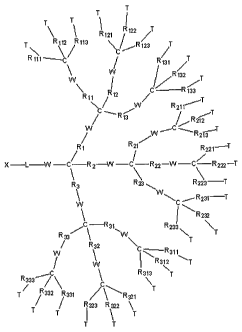
Figure 1 diagram may be referred to in describing the inventive polymer.
Various R, T, W, L, and X group variables are noted in Figure 1. The inventive
macromolecule polymer may comprise any branched or hyperbranched,
symmetrical or asymmetrical polymer. The branched termini of the polymer may
bind to the substrate preferably by a plurality of the termini. The linear end
of the
polymer may end with a functional group to which may be attached a protecting
group or a target-specific ligand. The distance between the probes among the
plurality of polymers on a substrate may be from about 0.1 nm to about 100 nm,
preferably about 1 nm to about 100 nm, preferably, about 2 nm to about 70 nm,
more preferably about 2 nm to about 60 nm, most preferably about 2 nm to about
50 nm.
R-Group
Referring to Formula I set forth in Figure 1, the polymer generally
comprises a branched section, wherein a plurality of the ends are
functionalized to
bind to a substrate. Within this branched section, the first generation group
of
branches Rx (R1, R2, R3) is connected to a second generation group of branches
RX,
(R11l R12, R137 R21, R22, R23, R31I R32, R33) by a functional group, W. The
second
14
CA 02539510 2006-03-17
WO 2005/026191 PCT/KR2004/002383
generation group of branches is connected to a third generation group of
branches
Rx>a (R111, R,112, R113, R121, R122, R123, R131, R132, R133, R211, R212, R213,
R221, R222, R223,
R231, R232, R233, R311, R312, R313, R321, R322, R323, P331, R332, R333) by a
functional
group W. And further fourth generation may be connected to the third
generation
branches in like fashion. The terminal R group is functionalized so that it is
capable
of binding to the substrate.
The R groups of all generations may be the same or different. Typically, the
R group may be a repeating unit, a linear or branched organic moiety, such as
but
not limited to alkyl, alkenyl, alkynyl, cycloalkyl, aryl, ether, polyether,
ester,
aminoalkyl, and so on. However, it is also understood that not all of the R
groups
need to be the same repeating unit. Nor do all valence positions for the R
group
need be filled with a repeating unit. For instance, in the first generation
branch, Rx,
R1, R2, R3 all of the R groups at this branch level may be the same repeating
units.
Or, R1 may be a repeating unit, and R2 and R3 may be H or any other chemical
entity. Or, R2 may be a repeating unit, and R1 and R3 may be H or any other
chemical entity. Likewise, for the second and third generation branches, any R
group may be a repeating unit, H or any other chemical entity.
Thus, a variety of shapes of polymers may be made in this way, for
instance, if R1, R11, R111, R112 and R113 are the same repeating units, and
all other R
groups are H's or any number of small neutral molecule or atom, then a fairly
long
and thin polymer having a branch with three functional group termini for R111,
R112
and R113 is made. A variety of other optional chemical configurations are
possible.
Thus, it is possible to obtain from about 3 to about 81 termini having a
functional
group capable of binding to a substrate. A'preferable number of termini may be
from about 3 to about 75, from about 3 to about 70, from about 3 to about 65,
from
about 3 to about 60, from about 3 to about 55, from about 3 to about 50, from
about
3 to about 45, from about 3 to about 40, from about 3 to about 35, from about
3 to
about 30, from about 3 to about 27, from about 3 to about 25, from about 3 to
about
21, from about 3 to about 18, from about 3 to about 15, from about 3 to about
12,
from about 3 to about 9, or from about 3 to about 6.
T-Terminal Group
Terminal groups, T, are functional groups that are sufficiently reactive to
undergo addition or substitution reactions. Examples of such functional groups
include without limitation amino, hydroxyl, mercapto, carboxyl, alkenyl,
allyl, vinyl,
CA 02539510 2006-03-17
WO 2005/026191 PCT/KR2004/002383
amido, halo, urea, oxiranyl, aziridinyl, oxazolinyl, imidazolinyl, sulfonato,
phosphonato, isocyanato, isothiocyanato, silanyl, and halogenyl.
W-Functional Group
In Formula I in Figure 1, W may be any functional group that may link a
polymer to another (or any other divalent organic moiety), such as but not
limited to
ether, ester, amide, ketone, urea, urethane, imide, carbonate, carboxylic acid
anhydride, carbodiimide, imine, azo group, amidine, thiocarbonyl, organic
sulphide,
disulfide, polysulfide, organic sulphoxide, sulphite, organic sulphone,
sulphonamide,
sulphonate, organic sulphate, amine, organic phosphorous group, alkylene,
alkyleneoxide, alkyleneamine and so on.
L - Spacer or Linker Group
In Figure 1, the linear portion of the polymer may include a spacer domain
comprised of a linker region optionally interspersed with functional groups.
The
linker region may be comprised of a variety of polymers. The length of the
linker
may be determined by a variety of factors, including the number of branched
functional groups binding to the substrate, strength of the binding to the
substrate,
the type of R group that is used, in particular, the type of repeating unit
that is used,
the type of the protecting group or target specific ligand that is to be
attached at the
apex of the linear portion of the polymer. Therefore, it is understood that
the linker is
not to be limited to any particular type of polymer or of any particular
length.
However, as a general guideiine, the length of the linker may be from about
0.5 nm
to about 20 nm, preferably, about 0.5 nm to about 10 nm, and most preferabiy
about 0.5 nm to about 5 nm.
The chemical construct of the linker may include without limitation, a linear
or branched organic moiety, such as but not limited to substituted or
unsubstituted
alkyl, alkenyl, alkynyl, cycloalkyl, cycloalkenyl, aryl, ether, polyether,
ester,
aminoalkyl, polyalkenylglcol and so on. The linker may further include
functional
groups such as those described above, and as such is not limited to any
particular
structure.
The linker group functionalized at the tip may comprise a protective group.
Thus, in one aspect, the present invention is directed to a substrate to which
is
attached a plurality of branched/linear polymers comprising linear tip
attached to a
protective group. Such a substrate may be chemically reacted to strip off the
protective group to be replaced with a target specific ligand. Therefore, in a
16
CA 02539510 2006-03-17
WO 2005/026191 PCT/KR2004/002383
functional use of the present inventive system, a substrate bound with a
population
of branched/linear polymers linked to a library of target specific ligands is
provided.
X - Protecting Group
The choice of protecting group depends on numerous factors such as the
desirability of acid-labile or base-lability. Therefore, the invention is not
limited to
any particular protecting group so long as it serves the function of
preventing the
reaction of the functional group to another chemical entity, and that it is
capable of
being stripped under desired specified conditions. Preferably, the protecting
group
is easily stripped away. Examples of such protecting groups that may be used
in the
present invention include without limitation the following:
Amino acid protecting groups: Methyl, Formyl, Ethyl, Acetyl, t-Butyl,
Anisyl, Benzyl, Trifluroacetyl, N-hydroxysuccinimide, t-Butyloxycarbonyl,
Benzoyl, 4-
Methylbenzyl, Thioanizyl, Thiocresyl, Benzyloxymethyl, 4-Nitrophenyl,
Benzyloxycarbonyl,2-Nitrobenzoyl, 2-Nitrophenylsulphenyl, 4-Toluenesulphonyl,
Pentafluorophenyl, Diphenylmethyl (Dpm), 2-Chlorobenzyloxycarbonyl, 2,4,5-
trichlorophenyl, 2-bromobenzyloxycarbonyl, 9-Fluorenylmethyloxycarbonyl,
Triphenylmethyl, 2,2,5,7,8-pentamethyl-chroman-6-sulphonyl, Phthaloyl, 3-
Nitrophthaloyl, 4,5-dichlorophthaloyl, tetrabromophthaloyl,
tetrachlorophthaloyl.
Protecting groups for alcohols: p-Anisyloxymethyl (p-AOM),
Benzyloxymethyl (BOM), t-Butoxymethyl, 2-Ch lorotetrahyd rofu ran (THF),
Guaiacolmethyl (GUM), (1 R)-Menthoxymethyl (MM), p-Methoxybenzyloxymethyl
(PMBM), metoxyethoxymethyl (MEM), Methoxymethyl (MOM), o-
Nitrobenzyloxymethyl, (Phenyldimethylsilyl)methoxymethyl (SMOM), 2-
(Trimethylsilyl)ethoxymethyl (SEM).
DNA, RNA protecting reagent: 2'-OMe-Ac-C-CE Phosphoramidite, 2'-
OMe-Ac-RNA CPG, 2'-OMe-l-CE Phosphoramidite, 2'-OMe-5-Me-C-CE
Phosphoramidite, Ac-C-CE Phosphoramidite, Ac-C-RNA 500, dmf-dG-CE
Phosphoramidite, dmf-dG-CPG 500, 2-Amino-dA-CE Phosphoramidite, (M.P.
Reddy, N.B. Hanna, and F. Farooqui, Tetrahedron Lett., 1994, 35, 4311-4314;
B.P.
Monia, et al., J. Biol. Chem., 1993, 268, 14514-14522).
Common Protecting Reagents in Organic Syntheses: (Dimethyl-t-
butylsilyloxy)methyl chloride (SOMCI), Ethoxyethyl chloride (EECI), -chloro
ethers,
o-Nitrobenzyloxymethyl chloride, b,b,b-Trichloroethoxymethyl chloride
(TCEMCI), (-
)-Menthyl ester, (P)-Benzyl ester, 1,1,1,3,3,3-Hexafluoro-2-phenyl-2-propyl
ether,
17
CA 02539510 2006-03-17
WO 2005/026191 PCT/KR2004/002383
1,1,3,3-Tetramethyl-1,3,2-disilazane, 1,2,4-Dithiazolidine-3,5-dione, 1,2-
Dibromide,
1,2-Dichloride, 1,2-Diol mono-4-methoxybenzyl ether, 1,2-Diol mono-t-butyl
ether,
1,2-Diol monoacetate ester, 1,2-Diol monoallyl ether, 1,2-Diol monobenzoate
ester,
1,2-Diol monobenzyl ether, 1,2-Diol monotosylate, 1,3-Benzodithiolan, 1,3-
Benzodithiolan-2-yl ether, 1,3-Diol mono-4-methoxybenzyl ether, 1,3-Diol
monobenzoate ester, 1,3-Diol monobenzyl ether, 1,3-Dioxan, 1-(2-
(Trimethoxysilyl)ethoxy)ethyl ether, 1-Adamantyl ester, 1-Benzoyl-l-propen-2-
yl
amine, 1-Ethoxyethyl ether, 1-Methoxyethylidene acetal, 1-Methyl-1-
methoxyethyl
ether, 1-Phenyl-3,5-di-t-butylcyclohexadien-4-onyl amine, 1-Phenylethyl ester,
2,2,2-
Trichloroethoxymethyl ether, 2,2,2-Trichloroethyl carbonate, 2,2,2-
Trichloroethyl
ester, 2,2,2-Trichloroethyl phosphate, 2,2,5,7,8-Pentamethylchroman-6-
sulphonamide, 2,2-Dimethyl-4-pentenoate ester, 2,3,6-Trimethyl-4-
methoxybenzenes6lphonamide, 2,4,6-Trimethylbenzenesulphonamide, 2,4-DNP
hydrazone, 2,5-Dichlorophenyl phosphate, 2,5-Dimethylpyrrole, 2-(2-
Methoxyethoxy)ethyl ester, 2-(4-Nitrophenyl)ethyl ether, 2-(4-
Nitrophenyl)ethyl
phosphate, 2-(4-Toluenesulphonyl)ethyl ester, 2-(Dibromomethyl)benzoate ester,
2-
(Trimethylsilyl)ethyl carbonate, 2-(Trimethylsilyl)ethyl ester, 2-
(Trimethylsilyl)ethyl
ether, 2-Benzenesulphonylethyl thioether, 2-Bromoethyl ester, 2-Chloroethyl
ester,
2-Chlorophenyl phosphate, 2-Cyanoethyl phosphate, 2-Methoxyethyl ester, 2-
Nitrobenzenesulphenamide, 2-Nitrobenzenesulphonamide, 2-Oxazoline, 2-
Phenylethyl ester, 2-Pyridyl disulphide, 2-Tetrahydropyranyl amine, 4-
Chlorobenzoate ester, 4-Chlorobutyl ester, 4-Methoxybenzamide, 4-
Methoxybenzoate ester, 4-Methoxybenzyl amine, 4-Methoxybenzyl ester, 4-
Methoxybenzyloxymethyl ether, 4-Nitrobenzamide, 4-Nitrobenzoate ester, 4-
Nitrobenzyl ester, 4-Nitrobenzyl ether, 4-Nitrobenzyl phosphate, 4-Nitrophenyl
ester,
4-Nitrophenyl hydrazone, 4-Toluenesulphonamide, 4-Toluenesulphonate, 9-
Fluorenylmethyl carbonate, 9-Fluorenylmethyl ester, Allyl carbonate, Allyl
ester,
Benzenesulphonamide, Benzenesulphonate, Benzyl carbonate, Benzyl ester, BOM
ether, DMTr ether, MEM ether, Methanesulphonamide, Methanesulphonate, ethyl
carbonate, MMTr ether, MOM carbonate, MOM ester, MOM ether, MTHP ether,
MTM ester, MTM ether, N-4-Methoxybenzyl amide, N-4-Tolyl amide, N-
Benzenesulphonyl amide, N-Benzyl imine, n-Butyl ester, n-Butyl ether, 0-4-
Methoxybenzyl carbamate, 0-9-Fluorenylmethyl carbamate, Phenyl thioether,
Phenyl thiolesterPiperidinamide, PMB ether, SEM ester, SEM ether, Succinate
ester,
18
CA 02539510 2008-11-06
t-Butyl carbonate, t-Butyl ester, t-Butyl ether,t-Butyl phosphate, t-Butyi
thioether, t-
Butyl thiolester, TBDMS ester, TBDMS ether, TBDPS ether, TES ether, , THF
ether,
THP ether, TIPDS diether, TIPS ether, TMS ester,TMS ether, TMS thioether,
Tosyl
hydrazone, TPS ether, Trifluoroacetamide.
A list of commercially available protecting groups may be found in Sigma-
Aldrich (2003) Catalog.
in general, in one aspect of the invention, the protecting groups used in the
present invention may be those that are used in the sequential addition of one
or
more amino acids or suitably protected amino acids to a growing peptide chain.
Normally, either the amino or carboxyl group of the first amino acid is
protected by a
suitable protecting group.
In a particularly preferred method the amino function is protected by an acid
or base sensitive group. Such protecting groups should have the properties of
being
stable to the conditions of linkage formation, while being readily removable
without
destruction of the growing branched/linear polymer. Such suitable protecting
groups
may be without limitation 9-fluorenylmethyloxycarbonyl (Fmoc), t-
butyloxycarbonyl
(Boc), benzyloxycarbonyl (Cbz), biphenylisopropyl-oxycarbonyl, t-
amyloxycarbonyl,
isobornyloxycarbonyl, (a, a)-dimethyl-3,5-dimethoxybenzyloxycarbonyl, o-
nitrophenyisulfenyl, 2-cyano-t-butyloxycarbonyl, and the like.
Particularly preferred protecting groups also include 2,2,5,7,8-
pentamethylchroman-6-suffonyl (pmc), p-toluenesulfonyl, 4-
methoxybenzenesulfonyl, adamantyloxycarbonyl, benzyl, o-
bromobenzyloxycarbonyl, 2,6-dichlorobenzyl, isopropyl, t-butyl (t-Bu),
cyclohexyl,
cyclophenyl and acetyl (Ac), 1-butyl, benzyl and tetrahydropyranyl, benzyl, p-
toluenesulfonyl and 2,4-dinitrophenyl.
In the addition method, the branched termini of the linear/branched polymer
is attached to a suitable solid support. Suitable solid supports useful for
the above
synthesis are those materials which are inert to the reagents and reaction
conditions of the stepwise condensation-deprotection reactions, as well as
being
insoluble in the media used.
The removal of a protecting group such as Fmoc from the linear tip of the
branched/linear polymer may be accomplished by treatment with a secondary
amine, preferably piperidine. The protected portion may be introduced in about
3-
19
CA 02539510 2006-03-17
WO 2005/026191 PCT/KR2004/002383
fold molar excess and the coupling may be preferably carried out in DMF. The
coupling agent may be without limitation O-benzotriazol-1-yl-N,N,N',N'-
tetramethyluroniumhexafluorophosphate (HBTU, 1 equiv.) and 1-hydroxy-
benzotriazole (HOBT, 1 equiv.).
The polymer may be deprotected, either in succession or in a single
operation. Removal of the polypeptide and deprotection can be accomplished in
a
single operation by treating the substrate-bound poiypeptide with a cleavage
reagent, for example thianisole, water, ethanedithiol and trifluoroacetic
acid.
Table 1 below lists various types of exemplified compounds. However, it is
to be understood that variations in X, L, W, R and T are encompassed by the
present invention.
Table 1- Representative and Exemplified Macromolecule Compounds
Compo X L W R T
und No.
3-1 A NH-(CH2)3C(O) NH CHaO(CH2)2C(O) OH
3-2 A NH-(CH2)3C(O) NH CH2O(CH2)2C(O) OMe
3-3 Boc NH-(CH2)3C(O) NH CHZO(CHa)aC(O) OH
3-4 Boc NH-(CH2)3C(O) NH CHZO(CHa)2C(O) OMe
3-5 A NH-(CH2CH2O)ZCH2C(O) NH CH2O(CH2)2C(O) OH
3-6 A NH-(CH2CH2O)2CH2C(O) NH CHZO(CHO2C(O) OMe
6-1 A NH-(CH2)3C(O) NH CH2O(CHZ)AO) OH
6-2 Boc NH-(cyclohexyl)(CO) CH2 (CH2)2-(cyclohexyl)-C(O) NH2
6-3 Boc NH-(CH2CH2O)2CH2C(O) NH CH2O(CH2)2C(O) OH
6-4 Fmoc NH-(CH2)6NHC(O) NH CH2-C=C-CH2C(O) OH
6-5 Fmoc NH-(CH2)7C(O) 0 CH2-C=C-CH2C(O) OMe
6-6 NS NH-(cyclohexyl)(CO) 0 CH2O(CH2)ZC(O) NH2
6-7 NS NH-(CH2)6NHC(O) NH (CH2)7 NH2
8-1 A NH-(CH2)3C(O) NH CHACH2)AO) OH
8-2 Boc NH-(CH2)7C(O) NH (CH2)2C(O) OH
8-3 NS NH-(CH2)6 (CO) NH (CH2)2-(cyclohexyi)-C(O) OH
8-4 Fmoc NH-(CHZ)s (CO) 0 CHZ-C=C-CH2C(O) NH2
8-5 Fmoc NH-(CH2)6NH(CO) 0 (CH2)2-(cyclohexyl)-C(O) OH
8-6 NS NH-(cyclohexyl)(CO) 0 CH2OCH(CH3)CH2C(O) NH2
8-7 Boc NH-(cyclopropyll)(CO) 0 CH2-C=C-CH2C(O) NH2
9-1 A NH-(CH2)3C(O) NH CHACHZ)AO) OH
9-2 A NH-(CH2)3C(O) NH CHZO(CH2)ZC(O) OMe
CA 02539510 2006-03-17
WO 2005/026191 PCT/KR2004/002383
9-3 A NH-(CH2CH2O)2CHaC(O) NH CHZO(CH2)ZC(O) OH
9-4 A NH-(CH2CH2O)2CHaC(O) NH CH2O(CH2)2C(O) OMe
9-5 Fmoc NH-(CH2)6C(O) NH CH2O(CHZ)2C(O) OH
9-6 Fmoc NH-(CH2)6C(O) NH CH2O(CH2)2C(O) OMe
9-7 Boc NH-(CH2)3C(O) NH CH2O(CH2)2C(O) OH
9-8 Boc NH-(CH2)3C(O) NH CH2O(CH2)2C(O) OMe
9-9 Ns NH-(CH2)3C(O) NH CHZO(CH2)2C(O) OH
9-10 Ns NH-(CH2)3C(O) NH CH2O(CH2)2C(O) OMe
9-11 A NH-(CH2)6NHC(O)CH2 CH2 (CH2)7 OBzI
12-1 A NH-(CH2)3C(O) NH CHZO(CHZ)AO) OH
12-2 Fmoc NH-(CH2)6NHC(O) NH (CH2)2-(cyclohexyl)-C(O) NH2
12-3 Boc NH-(cyclohexyl)(CO) 0 CH2-C=C-CH2C(O) OMe
12-4 Boc NH -(CH2)5 NH CH2OCH(CH3)CHZC(O) NH2
12-5 NS NH-(cyclopropyl)(CO) CH2 (CH2)2 NH2
12-6 NS NH-(CH2)6C(O) 0 CH2OCH2CH(CH3)C(O) NH2
12-7 Fmoc NH-(CH2)6NHC(O) 0 CH2OCH(CH3)CHAO) NH2
16-1 Boc NH-(CH2)3C(O) NH CHACH02C(O) NH2
16-2 Boc NH-(cyclohexyl)(CO) CH2 (CH2)2-(cyclohexyi)-C(O) OH
16-3 Fmoc NH-(CH2CHZO)2CH2C(O) 0 CHACH02C(O) OH
16-4 Fmoc NH-(CH2)6NHC(O) NH (CH2)2-(cyclohexyl)-C(O) NH2
16-5 NS NH-(cyclohexyl)(CO) NH CH2-C=C-CH2C(O) OH
16-6 NS NH-(cyclopropyl)(CO) CH2 CHZO(CH2)2C(O) OMe
16-7 A NH-(cyclopropyl)(CO) CH2 CH2OCH(CH3)CH2C(O) OH
16-8 A NH-(cyclopropyl)(CO) CH2 CH2OCHaCH(CH3)C(O) NH2
16-9 A NH -(CH2)5 0 CH2OCH2CH(CH3)C(O) OH
18-1 A NH-(CH2)3C(O) NH CHZO(CHOZC(O) OH
18-2 Fmoc NH-(cyclohexyl)(CO) 0 CH2OCH(CH3)CHZC(O) NH2
18-3 Boc NH-(cyclopropyl)(CO) 0 CH2OCH2CH(CH3)C(O) NH2
18-4 Fmoc NH-(CH2)6NHC(O)CH2 NH (CH2)2-(cyclohexyl)-C(O) OH
18-5 NS NH-(CH2)6NHC(O) CH2 CH2-C=C-CHaC(O) OMe
18-6 Boc NH -(CH2)5 0 CH2OCHZCH(CH3)C(O) NH2
27-1 A NH-(CH2)3C(O) NH CHZO(CHZ)2C(O) OH
27-2 A NH-(CH2)6NHC(O)CH2 CH2 (CH2)7 OH
27-3 Fmoc NH-(CHaCH2O)2CHzC(O) 0 (CH2)2-(cyclohexyl)-C(O) NH2
27-4 NS NH-(cyclopropyl)(CO) NH (CH2)2-(cyclohexyl)-C(O) NH2
27-5 Boc NH-(cyclohexyl)(CO) CH2 CH2OCH(CH3)CHAO) OMe
27-6 Fmoc NH -(CH2)5 0 CH2OCH2CH(CH3)C(O) NH2
21
CA 02539510 2006-03-17
WO 2005/026191 PCT/KR2004/002383
Target-Specific Ligand or Probe
The target-specific ligand, also known as probe, which is to be attached to
the linear end of the branched/linear polymer may include a variety of
compounds,
including chemicals, biochemicals, bioactive compounds and so on. In this
regard,
the ligand may be nucleic acid, oligonucleotide, RNA, DNA, PNA, or aptamer.
The
oligonucleotide may be a naturally occurring nucleic acid or an analog
thereof. Thus,
the ligand may be a polypeptide composed of naturally occurring amino acids or
synthetic amino acids. The ligand may be a combination of nucleic acid, amino
acid,
carbohydrate or any other chemical so long as it is capable of being attached
to the
linear portion of the branched/linear polymer. In particular, the ligand may
also be a
chemical, such as based on a triazine backbone, which may be used as a
component in a combinatorial chemistry library, in particular, a triazine
tagged
library.
Substrate
The substrate may be any solid surface to which the branched/linear
polymer may bind through either covalent or ionic bond. The substrate may be
functionalized so that binding may occur between the branched termini of the
branched/linear polymer. The surface of the substrate may be a variety of
surfaces
according to the needs of the practitioner in the art. If a microarry or
biochip format
is desired then typically oxidized silicon wafer, fused siiica or glass may be
the
substrate. Preferably, the substrate may be a glass slide. Other substrates
may
include membrane filters such as but not limited to nitrocellulose or nylon.
The
substrate may be hydrophilic or polar, and may possess negative or positive
charge
before or after coating.
Microarray
In order to improve the performance of DNA microarrays, issues such as
probe design, reaction conditions during spotting, hybridization and washing
conditions, suppression of non-specific binding, distance between the
biomolecules
and the surface, and the space between the immobilized biomolecules should be
considered. Because most of these factors are associated with the nature of
the
microarray surface, surface optimization has become one of the major goals in
microarray research. Whitesell and Chang showed that an alpha- helix formation
of
22
CA 02539510 2006-03-17
WO 2005/026191 PCT/KR2004/002383
immobilized oligopeptides was encouraged on a space-controlled gold surface
(Whitesell et al., Science 261, 73-76 (1993)). We now report that a surface
modified
with the cone-shaped dendron can provide DNA microarrays with single
nucleotide
polymorphism (or SNP) discrimination efficiency close to the solution value
(1:0.01)
while concurrently reducing DNA non-specific binding.
Figure 2 is a scheme showing the synthesis of a dendron. Various starting
material, intermediate compounds, and dendron compounds, wherein "X" may be
any protecting group, including anthracenemethyl (A), Boc, Fmoc, Ns and so
forth.
Figure 3(a) shows modification of glass surface with a dendron (Fig. 3b) and
selective hybridization of a fluorophore-tagged target oligonucleotide with a
matched oligonucleotide probe while discriminating effectively a single base
mismatched pair out on the dendron-modified surface.
A second generation branch dendron having surface reactive functional
groups at the branch termini may be used, which self assembles and provides
appropriate space between them. Previous studies showed that multiple ionic
attractions between cations on a glass substrate and anionic carboxylates at
the
dendron's termini successfully generated a well-behaved monolayer, and
guaranteed an inter-ligand space over 24 A (Hong et al., Langmuir 19, 2357-
2365
(2003)). To facilitate deprotection and increase the deprotected apex amine's
reactivity, we modified the structure as in Fig. 3b. We also observed that
covalent
bond formation between the dendron's carboxylic acid groups and the surface
hydroxyl groups is as effective as the ionic attraction, while also providing
enhanced
thermal stability. Moreover, an oligoetheral interlayer was effective for
suppressing
non-specific oligonucleotide binding.
The hydroxylated substrate was prepared by using a previously reported
method (Maskis et al., Nucleic Acids Res. 20, 1679-1684 (1992)). Substrates
including oxidized silicon wafer, fused silica, and glass slide, were modified
with (3-
glycidoxypropyl)methyldiethoxysilane (GPDES) and ethylene glycol (EG). The
dendron was introduced to the above substrates through a coupling reaction
between the dendron's carboxylic acid group and the substrate's hydroxyl group
using 1-[3-(dimethylamino)propyl]-3- ethylcarbodiimide hydrochloride (EDC) or
1,3-
dicyclohexylcarbodiimide (DCC) in the presence of 4-dimethylaminopyridine
(DMAP) (Boden et al., J. Org. Chem. 50, 2394-2395 (1985); Dhaon et al., J.
Org.
Chem. 47, 1962-1965 (1982)). The increase in thickness after the dendron
23
CA 02539510 2006-03-17
WO 2005/026191 PCT/KR2004/002383
introduction was 11 2 A, which was comparable to the previous value observed
for
the ionic bonding (Hong et al., Langmuir 19, 2357-2365 (2003)). After the
immobilization, an absorption peak arising from the anthracene moiety of the
dendron was observed at 257 nm. The molecular layer is stable enough to show
no
change in terms of thickness and absorption characteristics upon stirring in
dimethylformamide for 1 d (Fig. 4). The topographical images obtained by
tapping
mode atomic force microscope (AFM) also showed that the resulting layer was
very
smooth and homogeneous without any aggregates or holes (Fig. 5).
To be ready for DNA microarrays, the immobilized dendron was activated to
generate a primary amine group through deprotection process. After the
deprotection in 1.0 M trifluoroacetic acid (TFA) (Kornblum et al., J. Org.
Chem. 42,
399-400 (1977), the absorption peak at 257 nm disappeared without any other
detrimental change of the surface properties (Fig. 4a). This observation
demonstrated that the protecting group was removed successfully without
chemically damaging the layer while thickness was slightly decreased due to
the
elimination of the protecting group.
After modification with di(N-succinimidyl)carbonate (DSC) according to a
previously established method (Beier et al., Nucleic Acids Res. 27, 1970-1977
(1999)), probe oligonucleotides were immobilized onto the activated surface of
glass slide by spotting 50 mM sodium bicarbonate buffer (10% dimethylsulfoxide
(DMSO), pH 8.5) solution of the appropriate amine-tethered oligonucleotide (20
,uM)
using a Microsys 5100 Microarrayer (Cartesian Technologies, Inc.) in a class
10,000
clean room. Typically, for substrates with a reactive amine surface group, a
thiol-
tethered oligonucleotide and a heterobifunctional linker such as succinimidyl
4-
maleimido butyrate (SMB) or sulphosuccinimidyl-4-(N -
maleimidomethyl)cyclohexane-l-carboxylate (SSMCC) are employed (Oh et al.,
Langmuir 18, 1764-1769 (2002); Frutos et al., Langmuir 16, 2192-2197 (2000)).
In
contrast, because the dendron-modified surface guarantees a certain distance
among the amine functional groups, use of homobifunctional linkers such as DSC
is
not problematic. As a result, an amine-tethered oligonucleotide can be
utilized for
spotting. Apart from the cost effectiveness, use of easily oxidized thiol-
tethered
oligonucleotide can be avoided, although it is possible that such thiol-
tethered
oligonucleotides may be useful under certain conditions.
The DNA microarrays were fabricated to evaluate the discrimination
24
CA 02539510 2006-03-17
WO 2005/026191 PCT/KR2004/002383
efficiency between a complementary pair (A:T) and three internal single-base
mismatched pairs (T:T, G:T, C:T). After spotting the probe oligonucleotides
side by
side in a 4 by 4 format, the microarray was incubated in a humidity chamber
(80%
humidity) for 12 h to give the amine-tethered DNA sufficient reaction time.
Slides
were then stirred in a buffer solution (2x SSPE buffer (pH 7.4) containing 7.0
mM
sodium dodecylsulfate) at 37 C for 3 h and in boiling water for 5 min to
remove the
non-specifically bound oligonucleotides. Finally, the DNA-functionalized
microarray
was dried under a stream of nitrogen for the next step. For a fair comparison,
different kinds of probes were spotted in a single plate.
For hybridization, a 15-base oligonucleotide (Target 1) or 45-base
oligonucleotide (Target 2) was used (Fig. 3c). Hybridization was performed in
the
above washing buffer solution containing a target oligonucleotide (1.0 nM)
tagged
with a Cy3 fluorescent dye at 50 C for 1 h using a GeneTACTM HybStation
(Genomic Solution, Inc.). The microarray was rinsed with buffer solution at 37
C
four times for 1 min in order to remove excess target oligonucleotide and
dried with
nitrogen. The fluoresecence signal on each spot was measured with a ScanArray
Lite (GSI Lumonics) and analyzed by Imagene 4.0 (Biodiscovery).
In the case of the 15-base target oligonucleotide, the image shows that
there is a dramatic difference in the intensity between the matched and the
internal
mismatched pairs (Fig. 6a). The normalized fluorescence signal ratios (or
intensity
ratios for one base internal-mismatched pair versus the perfectly matched
pair, i.e.,
MM/PM) were 0.005, 0.008, and 0.006 (T:T, G:T, and C:T internal mismatches)
(Fig.
6a and Table 2). The observed selectivity is significantly improved over
conventional
methods, and a large increase of the selectivity (20 - 82 times) is recorded
in
comparison with DNA microarrays on the generic surface (Table 2). Previously,
we
also observed a selectivity factor of 1:0.19 - 0.57 for microarrays fabricated
on
various amine surfaces including a mixed self-assembled monolayer (i.e., mixed
SAM)( Oh et al., Langmuir 18, 1764-1769 (2002)). In addition, other
investigators
improved the performance of DNA microarrays by modifying their surface and
inventing better detection process, but none has reached this significantly
improved
ratio as far as a fluorescence detection method is concerned (Zhao et al., J.
Am.
Chem. Soc. 125, 12531-12540 (2003); Chakrabarti et al., J. Am. Chem. Soc. 125,
12531-12540 (2003); Benters et al., Nucleic Acids Res. 30, elO (2002); Guschin
et
al., Analytical Biochemistry 250, 203-211 (1997); Taton et al., Science 289,
1757-
CA 02539510 2006-03-17
WO 2005/026191 PCT/KR2004/002383
1760 (2000); Wang et al., Nucleic Acids Res. 30, e61 (2002)). For example,
successful discrimination ratio of 1:0.07 was reported for a three component
hybridization/detection system (capture/target/probe)( Zhao et al., J. Am.
Chem.
Soc. 125, 12531-12540 (2003)). Even when peptide nucleic acids (PNAs) capable
of increasing the selectivity were used, the selectivity on gold thin film and
gold
nanoparticle were 1:0.14 and 1:0.07, respectively (Chakrabarti et al., J. Am.
Chem.
Soc. 125, 12531-12540 (2003)).
Table 2
Normalized fluorescence si nal ratio
Matched Mismatched Mismatched Mismatched
A:T (T:T) (G:T) (C:T)
Dendron-modified surface, 15- 1 0.005 0.008 0.006
mer (Target 1& Probe 1)
Dendron-modified surface, 45- 1 0.006 0.009 0.009
mer (Target 2 & Probe 1)
APDES-modified surface, C6 1 0.41 0.38 0.26
spacer (Target 1& Probe 1)
APDES-modified surface, (T)30 1 0.17 0.18 0.12
spacer (Target 1& Probe 2)
To simulate a more realistic system, a 45-base target oligonucleotide was
employed. The MM/PM ratios for T:T, G:T, and C:T internal mismatches were
0.006,
0.009, and 0.009 (Fig. 6b and Table 2). This result shows that outstanding
selectivity holds for the longer target oligonucleotides. It is believed that
the efficacy
of this DNA microarray should be attributed 'to the peculiarity of the dendron-
modified surface, mesospacing between immobilized DNA strands.
For comparison, a DNA microarray was fabricated on the substrate
modified with (3-aminopropyl)diethoxymethylsilane (APDES)( Oh et al., Langmuir
18,
1764-1769 (2002)), which is a typical substrate for DNA or protein
microarrays. Its
selectivity was tested using the same procedure and oligonucleotides as those
for
the dendron-modified DNA microarray, except for the use of 1,4-
phenylenediisothiocyanate (PDITC) linker. Amine-tethered oligonucleotides were
employed as described by Guo (Guo et al., Nucleic Acids Res. 22, 2121-2125
(1994)). The observed MM/PM ratios for T:T, G:T, and C:T cases were 0.41,
0.38,
and 0.26 (Fig. 6c and Table 2). Use of DSC linker on the APDES-modified
substrate
resulted in high coefficient variance (CV) value (> 20 %), which represents
the
degree of variation among the spots, and non-uniform fluorescence intensity
within
26
CA 02539510 2006-03-17
WO 2005/026191 PCT/KR2004/002383
each spot. On the other hand, PDITC linker assured better coefficient variance
(CV)
value (< 15 %) and homogeneous fluorescence intensity within a single spot
like
those of the dendron-modified substrate with DSC linker (Fig. 7).
For additional comparison, probe 2 oligonucleotides having an extra (T)3o
spacer at the 5' end of oligomer were utilized for SNP discrimination test.
For this
case, the probe with the extra spacer was immobilized on an APDES-modified
surface. The observed MM/PM ratios for T:T, G:T, and C:T cases were 0.17,
0.18,
and 0.12 (Fig. 6d and Table 2). The selectivity was significantly enhanced in
comparison with the case of probe DNA with a C6 spacer, but still was largely
inferior to the dendron-modified DNA microarray.
Hybridization on the surface poses various complications, hurdles to control
and predict the microarray's screening performance precisely. Non-specific
binding,
steric and electrostatic effects, and environmental changes during the washing
process should be considered in addition to the melting temperature (Tm) of
the
duplex and the Gibbs free energy for the duplex formation. Difference between
the
Gibbs free energy of the internal-mismatched pairs (T:T, G:T, and C:T internal
mismatches of the 15-mer) and that of the perfectly matched pair in soiution
is 2.67,
1.75, and 3.05 kcal/mol at 50 C. Gibbs free energy was calculated with
HYTHERT""
Software (http://ozone2.chem.wayne.edu). Therefore, the theoretical
fluorescence
ratios (MM/PM) are 0.016, 0.065, and 0.009 respectively. Also, study in
solution
phase with a molecular beacon showed that SNP discrimination ratio was as low
as
1:0.01 (Taton et al., Science 289, 1757-1760 (2000)). These data strongly
demonstrate that our dendron-modified DNA microarray represents an ideal case
that reaches or even surpasses the thermodynamic limit. In particular, for the
G:T
case, the discrimination efficiency in the microarray format is better than
the value
calculated for the solution phase. The answer to which factors are main
reasons for
the selectivity increase is yet to be investigated, but washing stringency may
play a
role.
p53 SNP Detection
In biological systems, the p53 tumor-suppressor gene plays key roles in cell
regulation, gene transcription, genomic stability, DNA repair, and apoptosis
(see
Velculescu et al, 1996, Clin. Chem., 42: 858-868, Harris et al, 1996, 88: 1442-
1455,
Sidransky et al, Annu, Rev. Med., 1996, 47: 285-301). It has been reported
that loss
of wild-type function of p53 can lead to cancer and p53 mutations are the most
27
CA 02539510 2006-03-17
WO 2005/026191 PCT/KR2004/002383
frequent genetic changes in human cancer such as colon, and lung cancer
(Greenblatt, 1994, 54: 4855-4878).
DNA microarrays on [9]-acid dendron modified substrates were applied to
the detection of single mutation of p53 tumor suppressor gene in cancer cell
line.
Target DNA samples (-200-400 bases) which contain 175 codon were prepared by
random priming the genomic DNA templates and allowed to hybridize with dendron-
modified substrates on which 18mer probe oligonucleotides had been immobilized
in a 10 by I format. The MM/PM ratio for A:C, T:C, and C:C internal mismatches
were 0.028, 0.031, and 0.007 (Fig. 8a). This result shows that the outstanding
selectivity holds for real target DNAs.
The DNA microarrays on [27]-acid dendron modified substrates were
prepared using the same method as in the case of [9]-acid dendron which is
described above and applied to the detection of single mutation of 175 codon
of p53
tumor suppressor gene. The MM/PM ratio for A:C, T:C, and C:C internal
mismatches were 0.066, 0.01, and 0.005 (Fig. 8b). This result indicates that
the
DNA microarrays on [27]-acid dendron modified substrates also show outstanding
selectivity for the detection of single mutation of real target DNAs.
Detection of 7 hot spot mutations of p53 gene using single dendron-
modified surface.
The dendron-modified substrates were applied to the detection of single
mutation of p53 tumor suppressor gene in cancer cell line. Target DNA samples
(200-400 mer) which span 7 hot spot codons (175, 215, 216, 239, 248, 273, and
282) were amplified from the DNA extracted from cancer cells by random priming
and allowed to hybridize with capture probes (oligonucleotides of 15-25 mer)
corresponding to 7 hot spot codons that had been immobilized (Figs. 9a and
9b).
Excellent SNP discrimination efficiency was obtained.
We fabricated successfully DNA microarray of the highest fidelity by
providing mesospacing among the probe DNA, and found that SNP discrimination
efficiency could be enhanced to reach or even surpass the solution value. The
observed discrimination efficiency will make this methodology widely
acceptable for
very reliable high throughput gene diagnosis. It is expected that this
strategy can be
applied to various bioassays utilizing immobilized biomolecules.
Controlled Pore Glass Bead
Natural polymers such as dextran and agarose are the most frequently
28
CA 02539510 2006-03-17
WO 2005/026191 PCT/KR2004/002383
used chromatography supports for affinity chromatography. Sepharose 6B, 4B,
and
2B are chromatographic materials composed of cross-linked agarose, which
exhibit
extremely low nonspecific adsorption. In spite of their wide use, agarose gel,
typically in a bead shape, suffers some drawbacks. For instance, the flow (or
elution) rates are moderate due to their soft nature, they cannot be dried or
frozen
since they shrink severeiy and essentially irreversibly, and they do not
tolerate some
organic solvents (Cuatrecasas, P. J. Biol. Chem. 1970, 245, 3059-3065; Kim et
al.,
Biochemistry 2002, 41, 3414-3421). In comparison, controlled pore glass (CPG)
exhibits many exceptional properties for the support: 1) it is mechanically
stable, 2)
it has a fixed three dimensional structure; it does not swell or shrink upon
change of
environment, 3) it is chemically stable from pH 1 to pH 14, 4) it is inert to
a broad
range of nucleophilic and electrophilic reagents, 4) it is stable against
heating, 5) it
exhibits excellent flow (or elution) properties, 6) it shows less tendency to
adhere to
surface of containers. In addition, after a modification step, removal of
reagents and
byproducts through washing is rapid and efficient. All of these
characteristics
support potential usefulness in many fields such as permeation chromatography,
solid phase synthesis, affinity purification, and so on.
Pore size: Effective porosity of CPG toward an adsorbed molecule is
determined by the accessibility of the guest to the host surface. To a first
approximation, the accessibility of CPG to a guest depends on geometric
factors,
which are related to the relative size of the pores of the host compared to
the size of
the guest. If a guest has a molecular size that is larger than the pore
openings
leading to the internal surface, adsorption and interactions can only occur
with the
external surface, which is much smaller than the internal surface area of the
investigated porous materials (Poschalko et al., J. Am. Chem. Soc. 2003, 125,
13415-13426; Ottaviani et al., J. Phys Chem. B. 2003, 107, 2046-2053). From
these
considerations, the extent and strength of adsorption of a guest onto CPG is
expected to depend on the following parameters: pore size of CPG, the total
surface area of the host, and the chemical composition of accessible surface
of the
host. In our investigation, three kinds of GST fused protein (GST (28 kDa),
GST-
PXP47 (41 kDa), and GST-Munc18 fragment (98 kDa)) were employed. Molecular
dimension of GST-Munc18 should be similar to that of a fused GST of 100 kDa,
GST-DREF (140X140X93 A) (Hirose et al., J. Biol. Chem. 1996, 271, 3930-3937;
Zhan et al., Gene 2001, 218, 1-9). To achieve the balance between pore size
and
29
CA 02539510 2006-03-17
WO 2005/026191 PCT/KR2004/002383
surface area, porosity of the support has to be optimized for each specific
protein.
Because it is known that CPG with a pore size of approximately 50 nm allows
the
inclusion of complexes of the complete range of molecular subunits normally
found
in proteins our investigation had been carried out with 50 nm CPG.
Simultaneously,
use of CPG with a larger pore (300 nm) confirmed the effectiveness of the
former
CPG as far as the above proteins are concerned (Collins et al., Anal. Biochem.
1973, 54, 47-53; Haller, W. J. Chromatogr. 1973, 85, 129-131).
Modification of Glutathione CPG (Sample El, E3, A, CS, and CL): A key
concern of affinity matrices is degree of nonspecific binding (or NSB). It is
a
ubiquitous problem in affinity purification and solid-phase synthesis. In
general, key
factors to suppress nonspecific binding are to avoid the hydrogen bond donor
groups and increase the hydrophilicity of matrices (Sigal et al, J. Am. Chem.
Soc.
1998, 120, 3464-3473; Chapman et al., Langmuir 2000, 16, 6927-6936; Chapman
et al., J. Am. Chem. Soc. 2000, 122, 8303-8304; Holmlin et al., Langmuir 2001,
17,
2841-2850; Ostuni et al., Langmuir 2001, 17, 6336-6343; Chapman et al.,
Langmuir
2001, 17, 1225-1233; Ostuni et al., Langmuir 2001, 17, 5605-5620). CPG
surface,
even when modified with an aminoalkyl group, is polar and retains partial
negative
charge (Hudson, D. J. Comb. Chem. 1999, 1, 403-457). Use of a diepoxide as a
spacer had been reported to be responsible for the hydrophilic character of
the
matrix and the minimal nonspecific binding (Suen et al., Ind. Eng. Chem. Res.
2000,
39, 478-487; Sundberg et al., J. Chromatogr. B. 1974, 90, 87-98; Shimizu et
al.,
Nature Biotechnology 2000, 18, 877-881). Therefore, 1,4-butanediol diglycidyl
ether
(or BUDGE) was employed for the modification leading to sample El and E3. The
key features of the incorporation of BUDGE include generation of very stable
ethereal bond against hydrolysis, the enhanced flexibility through a long
spacer arm,
full distance from the surface, and suppression of nonspecific binding to a
certain
extent. The last advantage can be explained by resembled structural motif with
that
of polyethylene glycol. Diepoxides can be utilized to link a molecule and a
surface
having a nucleophile, such as amine and thiol. During the ring opening
process,
stable carbon-heteroatom bond is generated as well as a(3-hydroxy group. Use
of
the linker before and after dendron modification guarantees flexibility of the
tethered
GSH. The summarized modification steps are outlined in Figure 10. For
incorporation of the dendrons on the matrices, common reagents called EDC and
NHS were used. After modification with the dendrons, acetic anhydride was
CA 02539510 2006-03-17
WO 2005/026191 PCT/KR2004/002383
introduced into system to cap the remained amine functionality. Finally,
matrices
were treated by 20 % piperidine for 30 min to deblock the Fmoc group of the
dendrons for the further modification. After elongation with BUDGE one more
time,
GSH was immobilized by utilizing reaction between the thiol and the epoxide.
As a control, sample A was prepared. Sequential modification with BUDGE,
1,3-diaminopropane, and BUDGE gave surface materials that is exactly same as
El
and E3 except absence of the dendrons. As before, GSH was immobilized by ring
opening reaction between the epoxide and the thiol. Other control beads
(Sample
CL and CS) were prepared by using a heterobiofunctional linker called GMBS to
link
GSH and AMCPG or LCAA-CPG. While, AMPCPG has a short arm consisting C3
hydrocarbon at the surface, LCAA-CPG has a long arm of C15 aliphatic chain.
After
amide formation with GMBS was allowed, the beads were treated with GSH.
Addition of thiol group into maleimido group generated a covalent bond between
carbon and sulfur atoms. The two-step treatment produced GSH immobiiized
controlled pore glass beads, i.e. CS and CL, with covalent bonds.
Ligand Density Measurement: Due to the difficulties in measuring the
amount of immobilized glutathione directly, an indirect method that the ligand
density was determined by measuring amount of dibenzofulvene released during
the deprotection step was employed. 9-Fluorenylmethoxycarbonyl (Fmoc)
protecting
group at the apex of the dendrons is stable against acids but is readily
cleaved by a
variety of bases. In this study 20 % piperidine in DMF is employed to
deprotect the
Fmoc functional group. Piperidine forms an adduct with the dibenzofulvene, and
the
adduct absorbs at 301 nm (Q1ye et al., J. Phys. Chem. B. 2003, 107, 3496-
3499).
On the other hand, when the absorbance of the collected solution appeared at
301 nm during the deprotection step with 20 % piperidine, it indicated that
the
deprotection proceeded as intended.
Ligand density obtained with this method is 8.3 ,umol/g for El, 5.6 ,umol/g
for E3. The density is reduced by a factor of 11.1 upon modification with F-
moc(3)acid and the value is further reduced by a factor of 1.5 upon use of a
larger
dendron. Thus, in a specific embodiment of the invention, smaller dendrons
were
more effective at obtaining higher density than using larger dendrons.
GST Binding Assay: Binding characteristics of sample A, El, and E3 were
examined using purified GST and cell lysate (lane 2, 3, and 4 in Figure 11).
Lane 1
shows successful preparation of lysate. It is evident that the three matrices
bind
31
CA 02539510 2006-03-17
WO 2005/026191 PCT/KR2004/002383
purified GST effectively. When cell lysate was introduced into the beads (lane
5, 6,
and 7), a significant difference was observed between A and El or E3. For
sample
A, in spite of incorporation of BUDGE linkers, serious nonspecific binding was
observed. Interestingly, when the dendrons were introduced on the matrix,
nonspecific protein binding was effectively suppressed. It is noteworthy that
self-
assembly of either the dendron of the first generation or the one of the
second
generation effectively suppresses nonspecific binding of the solid support,
while an
extended spacer between the dendron and GSH retains the activity of the
tethered
tripeptide.
In Figure 12, in one aspect of the invention, etheral and amide groups
constitute the main backbone of the structure, and immobilization of the
dendrons
generates again amide bonds. Also, high coverage of the dendrons is also an
important factor for the success.
The ligand density for El is 1.48 times higher than that for E3. In other
words, 148 % of the ligand concentration was recorded for El (Table 3). In
order to
examine the binding efficiency of both beads, the weight of the samples was
adjusted to have the same number of GSH in each sample. Densitometer showed
that the ligand utilization for both cases was quite close (29 %, 31 %). The
larger
spacing of E3 does not enhance the binding efficiency of GST, probably because
the examined protein is larger than the spacing of both El and E3 anyway.
Table 3: Ligand concentration and ligand utilization of sample El and E3.
Samples Ligand density Ratio of the ligand Percentage of ligand
umol/ concentration % utilization %
El 8.3 148 29
E3 5.6 100 31
Control experiment: We found that density of GSH was 14.5 /imol/g for
CS, 11.9 ,umol/g for CL. To compare efficacy of the beads in terms of specific
binding of GST, captured proteins with CS (5.7 mg) and CL (7.0 mg) beads were
analyzed along with samples from El (10.0 mg) and E3 (14.8 mg) beads. The
utilized quantity was adjusted to have the same number of the GSH roughly. It
is
evident in the chromatogram (Figure 13) CS and CL beads display poor
selectivity
as well as low binding capacity. The result stresses again importance of the
dendron to guarantee not only improved accessibility of GST towards
immobilized
32
CA 02539510 2006-03-17
WO 2005/026191 PCT/KR2004/002383
GSH but effective suppression of nonspecific binding.
Molecular Weight Dependence. Because the dendron modification
generates a surface of controlled spacing between the immobilized ligands,
binding
capacity towards proteins of various molecular weights is intriguing. In
particular, it
is known that use of the second generation dendron guarantees a spacing over
24
angstrom (Cardona et al., J. Am. Chem. Soc. 1998, 120, 4023-4024). For this
particular test, GST protein (28 kDa), GST-PXp47 (41 kDa), and GST-munc-18
fragment (98 kDa) from the wild-type lysate were prepared. As shown in Fig.
14,
binding capacity of the beads (El, E3, and Sepharose 4B) decreases sharply as
molecular weight of proteins increases. It is interesting to note that the
degree of
decrease holds same for the three different cases. When binding capacity of El
is
set at 100 % for GST, GST-PXp47 has a relative biding capacity of 92% and 22 %
for
GST-munc18. For E3 bead, 85 % for former protein and 23 % for the latter
protein
are recorded. This strong dependence on protein molecular weight was also
observed with glutathione Sepharose-4B. For glutathione Sepharose-4B, the
binding efficiencies are 104 % and 17 % for GST-PXP47 and GST-munc18,
respectively. The only notable difference is a rather constant capacity for
GST and
GST-PXp47 for this commercially available matrix. The difference might reflect
heterogeneous spacing in Sepharose 4B. In this material, diverse spacings
between
GSH exist so that the matrix binds the fused GST as efficiently as the
pristine GST.
For the much bigger protein, GST-munc18, the spacings should be too small. In
this
regard, constant decrease of binding capacity of the dendron-treated beads
supports again the regular spacing of GSH on the surface.
In summary, the dendron-modified matrix demonstrates selectivity as high
as that of the commercial matrix (for example, Sepharose 4B), and almost same
molecular weight dependence as the commercial one. The incorporation of the
dendrons on AMPCPG matrix not only reduces the nonspecific binding
effectively,
but retains binding activity of GSH. Constant decrease of the binding capacity
as
increase of protein molecular weight was observed, and the phenomenon seems in
harmony with the regular spacing between the immobilized GSH. In addition to
the
well-controlled spacing, favorable aspects such as mechanical stability, wide
compatibility with various chemical environment, and easiness to handle
promise
interesting applications.
The present invention is not to be limited in scope by the specific
33
CA 02539510 2006-03-17
WO 2005/026191 PCT/KR2004/002383
embodiments described herein. Indeed, various modifications of the invention
in
addition to those described herein will become apparent to those skilled in
the art
from the foregoing description and accompanying figures. Such modifications
are
intended to fall within the scope of the appended claims. The following
examples
are offered by way of illustration of the present invention, and not by way of
limitation.
EXAMPLES
Numbering scheme is used for compounds throughout the Examples such
as compound 1, compound 2, I, II, III, IV, V and so on. It is to be understood
however, that the compound numbering scheme is consistent with and is confined
to the particular Example section to which it is recited. For instance,
compound 1 as
recited in Example 2 may not necessarily be the same compound 1 as found in
Example 3.
Example 1- Methods For Making Microarray Using Size-Controlled
Macromolecule
In Example 1, designations I, II, III, IV, and V refer to various compounds
and intermediate compounds as shown in Figure 2.
Example 1.1 - Materials. The silane coupling reagents, (3-
glycidoxypropyl)methyldiethoxysilane (GPDES) and (3-
aminopropyl)diethoxymethylsilane (APDES), were purchased from Gelest, Inc. and
all other chemicals were of reagent grade from Sigma-Aldrich. Reaction
solvents for
the silylation are anhydrous ones in Sure/Seal bottles from Aldrich. All
washing
solvents for the substrates are of HPLC grade from Mallinckrodt Laboratory
Chemicals. The UV grade fused silica plates (30 mm x 10 mm x 1.5 mm) were
purchased from CVI Laser Corporation. The polished prime Si(100) wafers
(dopant,
phosphorus; resistivity, 1.5-2.1 4-cm) were purchased from MEMC Electronic
Materials, Inc. Glass slides (2.5 x 7.5 cm) were purchased from Corning Co.
All of
the oligonucleotides were purchased from Metabion. Ultrapure water (18 M 0/cm)
was obtained from a Milli-Q purification system (Millipore).
Example 1.2 - Instruments. The film thickness was measured with a
spectroscopic ellipsometer (J. A. Woollam Co. Model M-44). UV-vis spectra were
recorded on a Hewlett-Packard diodearray 8453 spectrophotometer. Tapping mode
34
CA 02539510 2006-03-17
WO 2005/026191 PCT/KR2004/002383
AFM experiments were performed with a Nanoscope Illa AFM (Digital Instruments)
equipped with an "E" type scanner.
Example 1.3 - Cleaning the substrates. Substrates such as oxidized
silicon wafer, fused silica, and glass slide, were immersed into Piranha
solution
(conc. H2SO4:30% H202 = 7:3 (v/v)) and the reaction bottle containing the
solution
and the substrates was sonicated for an hour. (Caution: Piranha solution can
oxidize organic materials explosively. Avoid contact with oxidizable
materials.) The
plates were washed and rinsed thoroughly with a copious amount of deionized
water after the sonication. The clean substrates were dried in a vacuum
chamber
(30-40 mTorr) for the next steps.
Example 1.4 - Preparing the hydroxylated substrates. The above clean
substrates were soaked in 160 ml toluene solution with 1.0 ml (3-
glycidoxypropyl)methyldiethoxysilane (GPDES) for 10 h. After the self-
assembly, the
substrates were washed with toluene briefly, placed in an oven, and heated at
110 C for 30 min. The plates were sonicated in toluene, toluene-methanol (1: 1
(v/v)), and methanol in a sequential manner for 3 min at each washing step.
The
washed plates were dried in a vacuum chamber (30-40 mTorr). GPDES-modified
substrates were soaked in a neat ethylene glycol (EG) solution with two or
three
drops of 95 % sulfuric acid at 80 - 100 C for 8 h. After cooling, the
substrates were
sonicated in ethanol and methanol in a sequential manner each for 3 min. The
washed plates were dried in a vacuum chamber (30-40 mTorr).
Example 1.5 - Preparing the dendron-modified substrates. The above
hydroxylated substrates were immersed into a methylene chloride solution
dissolving the dendron (1.2 mM) and a coupling agent, 1-[-3-
(dimethylamino)propyl]-3- ethylcarbodiimide hydrochloride (EDC) or 1,3-
dicyclohexyicarbodiimide (DCC) (11 mM) in the presence of 4-
dimethylaminopyridine (DMAP) (0.82 mM). After 3 days at room temperature, the
plates were sonicated in methanol, water, and methanol in a sequential manner
each for 3 min. The washed plates were dried in a vacuum chamber (30-40 mTorr)
for the next step.
Example 1.6 - Preparing the NHS-modified substrates. The dendron-
modified substrates were immersed into a methylene chloride solution with 1.0
M
trifluoroacetic acid (TFA). After 3 h, they were again soaked in a methylene
chloride
solution with 20% (v/v) diisopropylethylamine (DIPEA) for 10 min. The plates
were
CA 02539510 2006-03-17
WO 2005/026191 PCT/KR2004/002383
sonicated in methylene chloride and methanol each for 3 min. After being dried
in a
vacuum chamber, the deprotected substrates were incubated in the acetonitrile
solution with di(N-succinimidyl)carbonate (DSC) (25 mM) and DIPEA (1.0 mM).
After 4 h reaction under nitrogen atmosphere, the plates were placed in a
stirred
dimethylformamide solution for 30 min and washed briefly with methanol. The
washed plates were dried in a vacuum chamber (30-40 mTorr) for the next step.
Example 1.7 - Arraying oligonucleotides on the NHS-modified
substrates. Probe oligonucleotides in 50 mM NaHCO3 buffer (pH 8.5) were
spotted
side by side in a 4 by 4 format on the NHS-modified substrate. The microarray
was
incubated in a humidity chamber (80 % humidity) for 12 h to give the amine-
tethered
DNA sufficient reaction time. Slides were then stirred in a hybridization
buffer
solution (2x SSPE buffer (pH 7.4) containing 7.0 mM sodium dodecylsulfate) at
37
C for 1 h and in boiling water for 5 min to remove non-specifically bound
oligonucleotides. Finally, the DNA-functionalized microarray was dried under a
stream of nitrogen for the next step. For a fair comparison, different kinds
of probes
were spotted in a single plate.
Example 1.8 - Hybridization. Hybridization was performed in the
hybridization buffer solution containing a target oligonucleotide (1.0 nM)
tagged with
a Cy3 fluorescent dye at 50 C for 1 h using a GeneTACTM HybStation (Genomic
Solutions, Inc.). The microarray was rinsed with the hybridization buffer
solution in
order to remove excess target oligonucleotide and dried with nitrogen. The
fluorescence signal on each spot was measured with a ScanArray Lite (GSI
Lumonics) and analyzed by Imagene 4.0 (Biodiscovery).
ExAMPLE 1.9 - Synthesis of the dendron
EXAMPLE 1.9.1 - Preparation of 9-anthrylmethyl N-(3-
carboxylpropyl)carbamate (I) - Compound I.
4-Aminobutyric acid (0.50 g, 4.8 mmol, 1.0 equiv) and triethylamine (TEA)
(1.0 ml, 7.3 mmol, 1.5 equiv) were dissolved in N,N-dimethylformamide (DMF)
and
stirred at 50 C. 9-Anthrylmethyl p-nitrophenyl carbonate (1.81 g, 4.8 mmol,
1.0
equiv) was slowly added while stirring. After stirring at 50 C for 2 h, the
solution
was evaporated to dryness, and the solution was basified with 0.50 N sodium
hydroxide (NaOH) solution. The aqueous solution was washed with ethyl acetate
(EA), stirred in an ice bath and acidified with dilute hydrochloric acid
(HCI). After the
product was extracted with EA, the organic solution was dried with anhydrous
36
CA 02539510 2006-03-17
WO 2005/026191 PCT/KR2004/002383
MgSO4, filtered and evaporated. The total weight of the resulting yellow
powder was
1.06 g and the yield was 65 %.
'H NMR(CDCI3)
6 11.00-9.00(br, CH2COOH, 1 H), 8.41 (s, C14H9CH2, 1 H), 8.31 (d,
C14H9CH2, 2H), 7.97 (d, Cl4H9CH2, 2H), 7.51 (t, C14H9CH2, 2H), 7.46(t,
C14H9CH2,
2H), 6.08(s, C14H9CH20, 2H), 5.01 (t, OCONHCH2,1 H), 3.23(q, NHCH2CH2, 2H),
2.34(t, CH2CH2COOH, 2H), 1.77(m, CH2CH2CH2, 2H).
13C NMR(CDCI3)
6 178.5(CH2COOH), 157.9(OCONH), 132.1 (C14H9CH2), 131.7(Cl4H9CH2),
129.7(C14H9CHa), 129.7(Cl4H9CH2), 127.3(C14H9CH2), 126.8(ClaHsCHz),
125.8( C14H9CH2), 124.6( C14H9CH2), 60.2(C14H9CH2), 41.0(NHCH2CH2),
31.7(CH2CH2COOH),25.6(CH2CH2CH2).
EXAMPLE 1.9.2 - Preparation of 9-anthrylmethyl N-{[(tris{[2-
(methoxycarbonyl)ethoxy]methyl}methyl) amino]carbonyl}propylcarbonate (II)
- Compound II.
9-Anthrylmethyl N-(3-carboxylpropyl)carbamate (0.65 g, 1.93 mmol, 1.5
equiv), 1-[3-(dimethylamino)propyl]-3-ethylcarbodiimide hydrochloride (EDC)
(0.37 g,
1.93 mmol, 1.5 equiv), and 1-hydroxybenzotriazole hydrate (HOBT) (0.261 g,
1.93
mmol, 1.5 equiv) were dissolved in acetonitrile and stirred at room
temperature.
Tris{[(methoxycarbonyl)ethoxy]methyl} aminomethane (0.49 g, 1.29 mmol, 1.0
equiv) dissolved in acetonitrile was added with stirring, After stirring at
room
temperature for 12 h, the acetonitrile was evaporated. The crude product was
dissolved in EA and washed with 1.0 N HCI and saturated sodium bicarbonate
solution. After being dried with anhydrous MgSO4, filtered, and evaporated,
the
crude product was loaded in a column packed with silica gel. Purification by
'column
chromatography (eluent: ethyl acetate:hexane = 5:1 (v/v)) resulted in a
viscous
yellow liquid. The total weight of the yellow liquid was 0.67 g, and the yield
was
74%.
'H NMR(CDCI3)
6 8.43(s, C14H9CH2, 1 H), 8.36(d, CI4H9CH2i 2H), 7.99 (d, Cl4H9CH2, 2H),
7.53(t, C14H9CH2, 2H), 7.47(t, Cl4H9CH2, 2H), 6.15(s, CONHC, 1 H), 6.08(s,
CI4H9CH20, 2H), 5.44(t, OCONHCH2,1 H), 3.63-3.55(m, CH2OCH2CH2COOCH3, 21
H), 3.27(q, NHCH2CH2, 2H), 2.46(t, CH2CH2COOCH3, 6H), 2.46(t, CH2CH2CONH,
2H), 1.81 (m, CH2CHZCH2, 2H).
37
CA 02539510 2006-03-17
WO 2005/026191 PCT/KR2004/002383
13C NMR(CDCI3)
6173.2(CH2CONH), 172.7(CH2COOCH3), 157.4(OCONH), 132.9(C14H9CH2),
131.5(C14H9CH2), 129.5(C14H9CH2), 129.4(CI4H9CH2), 127.5(C14H9CH2),
127.0(C14H9CH2), 125:6(Cl4H9CH2), 124.7(C14H9CH2), 69.6(NHCCH2O),
67.2(C14H9CH2), 60.1 (OCH2CH2), 59.4(NHCCH2), 52.1(OCH3), 40.8(NHCH2CH2),
35.1 (OCH2CH2), 34.7(CH2CH2CONH), 26.3(CH2CH2CH2).
Anal. Calcd for C3sHasN2012 ' 0.5 H20: C 61.18, H 6.65, N 4.03; Found:
C61.09, H 6.69, N 3.96.
EXAMPLE 1.9.3 - Preparation of 9-anthrylmethyl N-[({tris[(2-
carboxyethoxy)methyl]methyl}amino) carbonyl]propylcarbamate (III) -
Compound Ill.
9-Anthrylmethyl N-{[(tris{[2-
(methoxycarbonyl)ethoxy]methyl}methyl)amino]carbonyl}propyi- carbonate (0.67
g,
0.93 mmol) was dissolved in acetone (30 ml) and 0.20 N NaOH (30 ml, 6 mmol).
After being stirred at room temperature for 1 d, the acetone was evaporated.
The
aqueous solution was washed with EA, stirred in an ice bath and acidified with
dilute
HCI. After the product was extracted with EA, the organic solution was dried
with
anhydrous MgSO4, filtered and evaporated. Solidification in acetone and ether
solution at -20 C resulted in a yellow powder. The total weight of the final
pale
yellow powder was 0.54 g with a yield of 88%.
'H NMR(CDCI3)
6 11.00-9.00(br, CH2COOH, 3H), 8.61(s, C14H9CH2, 1 H), 8.47(d, C14H9CH2,
2H), 8.11 (d, C14H9CH2, 2H), 7.60(t, C14H9CH2, 2H}, 7.52(t, C14H9CH2, 2H),
6.63(s,
CONHC, 1 H), 6.36(t, OCONHCH2, 1 H), 6.12(s, C14H9CH2O, 2H). 3.40-363(m,
CH2OCH2CH2COOH, 12H), 3.20(q, NHCH2CH2, 2H), 2.52(t, CH2CH2COOH, 6H),
2.17(t, CH2CH2CONH, 2H), 1.75(m, CH2CH2CH2, 2H).
13C NMR(CDCI3)
6 172.2(CH2COOH), 172.0(CH2CONH), 156.7(OCONH), 131.2(C14H9CH2),
130.7(C14H9CH2), 128.6(C14H9CH2), 128.4(C14H9CH2), 127.3(C14H9CH2),
126.2(C14H9CH2), 124.8(C14H9CH2), 124.0(C14H9CH2), 68.6(NHCCH2O),
66.5(C14H9CH2), 59.5(OCH2CH2), 58.0(NHCCH2), 40.0(NHCH2CH2),
34.0(OCH2CH2)133.5(CH2CH2CONH), 25.8(CH2CH2CH2).
Anal. Calcd for C33H40N2012' 1.5 H20: C 57.97, H 6.34, N 4.10; Found: C
57.89, H 6.21, N 4.09.
38
CA 02539510 2006-03-17
WO 2005/026191 PCT/KR2004/002383
EXAMPLE 1.9.4 - Preparation of 9-anthryimethyl N-[({tris[(2-{[(tris{[2-
(methoxycarbonyl)ethoxy]methyl}
(methyl)amino]carbonyl}ethoxy)methyl]methyl}amino)carbonyl]propylcarbam
ate (IV) - Compound IV.
9-Anthrylmethyl N-[({tris[(2-carboxyethoxy)methyl]methyl}amino)carbonyl]
propylcarbamate (0.54 g, 0.82 mmol, 1.0 equiv), EDC (0.55 g, 2.87 mmol, 3.5
equiv), and HOBT (0.39 g, 2.89 mmol, 3.5 equiv) were dissolved in acetonitrile
and
stirred at room temperature. Tris{[(methoxycarbonyl)ethoxy]methyl}
aminomethane
(0.96 g, 2.53 mmol, 3.1 equiv) dissolved in acetonitrile was added with
stirring. After
stirring at room temperature for 36 h, the acetonitrile was evaporated. The
crude
product was dissolved in EA and washed with 1.0 N HCI and saturated sodium
bicarbonate solution. After drying with anhydrous MgSO4, filtered, and
evaporated,
the crude product was loaded in a column packed with silica gel. Column
purification (eluent: ethyl acetate:methanol = 20:1 (v/v)) resuited in a
viscous yellow
liquid. The total weight of the yellow liquid was 1.26 g with an 88% yield.
'H NMR(CDCI3)
6 8.47(s, C14H9CH2, 1 H), 8.39(d, C14H9CH2, 2H), 8.02 (d, C14H9CH2, 2H),
7.53(t, C14H9CH2, 2H), 7.47(t, C14H9CH2, 2H), 6.60(s, CH2CH2CH2CONHC, 1 H),
6.13(s, OCH2CH2CONHC, 3H), 6.11 (s, C14H9CH20, 2H), 5.79(t, OCONHCH2,1 H),
3.65-3.60(m, CH2OCH2CH2CONH, CH2OCH2CH2COOCH3, 75H), 3.29(q,
NHCH2CH2, 2H), 2.50(t, CH2CH2COOCH3, 18H), 2.36(t, OCH2CH2CONH, 6H),
2.27(t, CH2CH2CHZCONH, 2H), 1.85(m, CH2CH2CH2, 2H).
13C NMR(CDCI3)
b 173.3(OCH2CH2CONH), 172.5(CH2CH2CHaCONH), 171.6(CH2COOCH3),
157.2(OCONH), 131.8(C14H9CH2), 131.5(C14H9CH2), 129.4(Cl4H9CH2),
129.3(C14H9CH2), 127 .6(Cl4H9CH2), 127.0(Cl4H9CH2), 125.6(Cl4H9CHz),
124.7(C14H9CHA 69.5(NHCCHZOCH2CH2COOCH3),
67.9(NHCCH2OCH2CH2CONH), 67.2(C14H9CH2), 60.3(OCH2CH2CONH),
60.2(OCH2CH2COOCH3), 59.2(NHCCH2OCH2CH2COOCH3,
NHCCH2OCH2CH2CONH), 52.1(OCH3), 41.0(NHCH2CH2), 37.6(OCH2CH2CONH),
35.1(OCHZCHZCOOCH3), 34.7(CH2CH2CH2CONH), 26.3(CH2CH2CH2).
Anal. Calcd for C81H121N5036 ' H2O: C 55.31, H 7.05, N 3.98; Found: C
55.05, H 7.08, N 4.04.
MALDI- TOF-MS: 1763.2 (MNa+), 1779.2 (MK+).
39
CA 02539510 2006-03-17
WO 2005/026191 PCT/KR2004/002383
EXAMPLE 1.9.5 - Preparation of 9-anthrylmethyl N-({[tris({2-[({tris[(2-
carboxyethoxy)methyl]methyl}
amino)carbonyl]ethoxy}methyl)methyl]amino}carbonyl)propylcarbamate (V) -
Compound V.
9-Anthrylmethyl N-[({tris[(2-{[(tris{[2-
(methoxycarbonyl)ethoxy]methyl}methyl)amino]carbonyl}
ethoxy)methyl]methyl}amino)carbonyl]propylcarbamate (0.60 g, 0.34 mmol) was
dissolved in acetone (30 ml) and 0.20 N NaOH (30 ml). After stirring at room
temperature for 1 d, the acetone was evaporated. The aqueous solution was
washed with EA, stirred in an ice bath and acidified with dilute HCI. After
the
product was extracted with EA, the organic solution was dried with anhydrous
MgSO4, filtered and evaporated. The total weight of the final yellow powder
was
0.37 g and the yield was 68 %.
'H NMR(DMSO)
6 13.00-11.00(br, CH2COOH, 9H), 8.66(s, C14H9CH2, 1 H), 8.42(d,
C14H9CH2, 2H), 8.13 (d, C14H9CH2, 2H), 7.62(t, Cl4H9CH2, 2H), 7.54(t,
C14H9CH2,
2H), 7.12(t, OCONHCH2, 1 H), 7.10(s, OCH2CH2CONHC, 3H), 7.06(s,
CH2CH2CH2CONHC, 1 H), 6.06(s, C14H9CH20, 2H), 3.57-3.55(m,
CH2OCH2CH2CONH, CH2OCH2CH2COOH, 48H), 3.02(q, NHCH2CH2, 2H), 2.42(t,
CH2CH2COOH, 18H), 2.32(t, OCH2CH2CONH, 6H), 2.11(t, CH2CH2CH2CONH, 2H),
1.60(m, CH2CH2CH2, 2H).
13 C NMR(DMSO)
6 172.8(CH2COOH), 172.2(CH2CH2CH2CONH), 170.5(OCH2CH2CONH),
156.5(OCONH), 131.0(C14H9CH2), 130.6(CI4H9CH2), 129.0(C14H9CH2),
128.7(C14H9CH2), 127.6(C14H9CH2), 126.7(Cl4H9CH2), 125.4(CI4H9CH2),
124.3(CI4H9CH2), 68.3(NHCCH2OCH2CH2COOH), 67.4(NHCCH2OCH2CH2CONH),
66.8(C14H9CH2), 59.8(OCH2CH2COOH), 59.6(OCH2CH2CONH),
57.9(NHCCH2OCH2CH2CONH), 55.9(NHCCH2OCH2CH2COOH), 36.4(NHCH2CH2),
34.6(OCH2CH2COOH), 30.8(OCH2CH2CONH), 29.7(CH2CH2CH2CONH),
25.9(CH2CH2CH2).
ExAMPLE 2- Methods of producing alternative starting material dendron
macromolecule - Fmoc-Spacer-[9] -acid
CA 02539510 2006-03-17
WO 2005/026191 PCT/KR2004/002383
In Example 2, various indicated compounds are referred to as compound 1,
2 and so forth.
First, we synthesized a spacer, 6-azidohexylamine (1) from 1,6-
dibromohexane according to Lee, J. W.; Jun, S. I.; Kim, K. Tetrahedron Lett.,
2001,
42, 2709.
Br B~ NaN3 _10 -~~N3 Triphenylphosphine N NH2
N3 3
(1)
This spacer was attached to repeating unit (2) through unsymmetric urea
formation and made N3-spacer-[3]ester (3). The repeating unit was synthesized
by
condensation of TRIS with tert-butyl acrylate, which had been reported in
Cardona,
C. M.; Gawley, R. E. J. Org. Chem. 2002, 67, 141.
O O/'<
oH o NaOH o ~ ~
H2N ~OH + v_O I~MSO/H2O H2N O o O
OH ----/CO-~
(2)
o~-OR
triphosgene 0 0r' O
(1) + (2) N3 H~H O'~-OR
O 0
OR
(3) R=t-butyl
Formic Acid
(4) R=H
41
CA 02539510 2006-03-17
WO 2005/026191 PCT/KR2004/002383
This triester was transformed to N3-spacer-[3]acid (4) through hydrolysis
and coupled with triester (2) under peptide coupling conditions, which led to
N3-
spacer-[9]ester. After reduction of azide to amine and protection of amine
with
Fmoc group, hydrolysis of nonaester afforded Fmoc-spacer-[9]acid (5).
OOH
OH
7
0 O
KOO .~~OH
~NH Oj~OH
H 0 O
O~N N~NNpOH
0 H H O-~~O H O-~O
HN OOH
00
-p'OH
(5) V oH
HO
N-(6-Azidohexyl)-N'-tris{[2-(tert-butoxycarbonyl)ethoxy]methyl}-
methylurea (3). Triphosgene (1.3 g, 4.3 mmol) was dissolved in anhydrous
CH2CI2
(20 mL). A mixture of 6-azidohexylamine (1) (1.6 g, 12 mmol) and N,N-
diisopropylethylamine (DIEA, 2.4 mL, 13.8 mmol) in anhydrous CH2CI2 (35 mL)
was
added dropwise to the stirred solution of triphosgene over a period of 7h
using a
syringe pump. After further stirring for 2h, a solution of (2) (6.4 g, 13
mmol) and
DIEA (2.7 mL, 15.2 mmol) in anhydrous CH2C12 (20 mL) was added. The reaction
mixture was stirred for 4 h at room temperature under nitrogen, and washed
with
0.5 M HCI and brine. The organic layer was then dried over anhydrous MgSO4,
and
the solvent was removed by evacuation. Purification with column chromatography
(silica, 1:1 EtOAc/hexane) yielded colorless oil (3.0 g, 40 %).
'H NMR (CDCI3, 300 MHz): S 1.45 (s, (CH3)3C, 27H); 1.36-1.58 (m,
CH2CH2CH2CH2, 8H); 2.46 (t, CH2CHZO, J = 6.4 Hz, 6H), 3.13 (m, CONHCH2, 2H),
3.26 (t, N3CH2, J = 6.9 Hz, 2H), 3.64-3.76 (m, CCH2O and CH2CH20, 12H); 5.00
(t,
CH2NHCO, J=6.7 Hz, 1 H), 5.29 (s, CONHC, 1 H).
13 C NMR (CDCI3, 75 MHz): 6 26.52, 26.54, 28.81, 30.26 (CH2CH2CH2CH2);
28.14((CH3)3C); 36.20 (CH2CH2O); 39.86 (CONHCH2); 51.40 (N3CH2); 58.81
(CCH2O); 67.16 (CHzCHZO); 69.23 (CCH2O); 80.58 ((CH3)3C); 157.96 (NHCONH);
171.26 (COOt-Bu).
FAB-MS: 674.26 (M+).
42
CA 02539510 2006-03-17
WO 2005/026191 PCT/KR2004/002383
N-(6-Azidohexyl)-N'-tris{[2-carboxyethoxy]methyl}methylurea (4). N3-
spacer-[3]ester (3) (0.36 g, 0.56 mmol) was stirred in 6.6mL of 96 % formic
acid for
24 h. The formic acid was then removed at reduced pressure at 50 C to produce
colorless oil in a quantitative yield.
'H NMR (CD3COCD3, 300 MHz): 6 1.34-1.60 (m, CH2CH2CH2CH2, 8H);
2.53 (t, CH2CH2O, J = 6.4 Hz, 6H), 3.07 (t, CONHCH2, J = 6.9 Hz, 2H), 3.32 (t,
N3CH2, J = 6.9 Hz, 2H), 3.67-3.73 (m, CCH2O and CH2CH2O, 12H).
13C NMR (CD3COCD3, 75 MHz): 6 27.21, 29.54, 31.02 (CH2CH2CH2CH2);
35.42 (CH2CH2O); 40.27 (CONHCH2); 52.00 (N3CH2); 59.74 (CCH2O); 67.85
(CH2CH2O); 70.96 (CCHZO); 158.96 (NHCONH); 173.42 (COOH).
FAB-MS: 506.19 (MH+).
N-(6-Azidohexyl)-N'-tris((2-{[(tris{[2-(tert-butoxycarbo nyl)ethoxy]-
methyl}methyl)amino]carbonyl}ethoxy)methyl]methylurea (4.1).
The HOBt (0.20 g, 1.5 mmol), DIEA (0.30 mL, 1.8 mmol), and EDC (0.33 g,
1.8 mmol) were added to (4) (0.25 g, 0.50 mmol) in 5.0 mL of dry acetonitrile.
Then,
the amine (2) (1.14 g, 2.3 mmol) dissolved in 2.5 mL of dry acetonitrile was
added,
and the reaction mixture was stirred under N2 for 48 h. After removal of the
solvent
at reduced pressure, the residue was dissolved in MC and washed with 0.5 M HCI
and brine. The organic layer was then dried over MgSO4, the solvent was
removed
in vacuo, and column chromatography (Si02, 2:1 EtOAc/hexane) yielded a
colorless oil (0.67 g, 70%).
'H NMR (CDCI3, 300 MHz): 6 1.45 (s, (CH3)3C, 81H); 1.36-1.58 (m,
CH2CH2CH2CH2, 8H); 2.40-2.47 (m, CH2CH2O gen. 1& 2, 24H), 3.13 (m,
CONHCH2, 2H), 3.26 (t, N3CH2, 6.9 Hz, 2H), 3.62-3.69 (m, CCHzO gen. 1& 2,
CH2CH2O gen. 1& 2, 48H); 5.36 (t, CH2NHCO, J=6.7 Hz, 1 H), 5.68 (br, CONHC,
1 H), 6.28 (br, amide NH, 3H).
13C NMR (CDCI3, 75 MHz): 6 26.59, 26.69, 28.91, 30.54 (CH2CH2CH2CH2);
28.22 ((CH3)3C); 36.20 (CH2CH2O gen. 2); 37.43 (CH2CH2O gen. 1); 39.81
(CONHCH2); 51.47 (N3CH2); 58.93 (CCH2O gen. 1); 59.89 (CCH2O gen. 2); 67.15
(CH2CH2O gen. 2); 67.68 (CH2CH2O gen. 1); 69.23 (CCH2O gen. 2); 70.12 (CCH2O
gen. 1); 80.57 ((CH3)3C); 158.25 (NHCONH); 171.01 (COOt-Bu) 171.41 (CONH
amides).
MALDI-MS: 1989.8 (MNa+), 2005.8 (MK+).
43
CA 02539510 2006-03-17
WO 2005/026191 PCT/KR2004/002383
N-(6-Aminohexyl)-N =tris[(2-{[(tris{[2-(tert-butoxycarbonyl)ethoxy]-
methyl}methyl)amino]carbonyl}ethoxy)methyl]methylurea (4.2).
Nona-tert-butyl ester (4.1) (0.37 g, 0.20 mmol) was stirred with 10 % Pd/C
(37.0 mg) in ethanol (20.0 mL) under H2 at room temperature for 12 h. After
checking completion of the reaction with TLC, the mixture was filtered with a
0.2 ,um
Millipore filter. After the filter paper was rinsed with CH2CI2, the combined
solvent
was removed in vacuo, and colorless oil was recovered.
N-{6-(9-fluorenylmethoxycarbonyl)aminohexyl}-N =tris[(2-{[(tris{[2-(tert-
butoxycarbonyl)ethoxy]methyl}methyl)amino]carbonyl}ethoxy)methyl]methylu
rea (4.3).
The amine (4.2) (0.33 g, 0.17 mmol) and DIEA (33 pL, 0.19 mmol) were
dissolved in 5.0 mL of CH2CI2, and stirred for 30 min under nitrogen
atmosphere. 9-
Fluorenylmethyl chloroformate (48 mg, 0.19 mmol) in 2.0 mL of CH2CI2 was
added,
and the reaction mixture was stirred for 3 h at room temperature. The solvent
was
removed under reduced pressure and washed with 0.5 M HCI and brine. The
residue was purified with column chromatography (silica, EtOAc) to yield
colorless
oil (0.18 g, 64 %).
'H NMR (CDCI3, 300 MHz): 6 1.45(s, (CH3)3C, 81H); 1.23-1.58 (m,
CH2CH2CH2CH2, 8H); 2.37-2.47 (m, CHaCH2O gen. 1& 2, 24H); 3.10-3.22 (m,
CONHCH2, 4H); 3.62- 3.70 (m, CCHZO gen. 1& 2, CHZCH2O gen. 1& 2, 48H); 4.22
(t, CH(fluorenyl)-CH2, J=7.1 Hz, IH); 4.36 (d, fluorenyl-CH2, J=7.1 Hz, 2H);
5.27-
5.35 (m, CH2NHCO, 2H); 5.67 (br, CONHC, 1 H); 6.25 (br, amide, 3H); 7.28 -7.77
(fluorenyl, 8H).
13C NMR (CDCI3, 75 MHz): 6 26.85, 27.02, 30.27, 30.88 (CH2CH2CH2CH2);
28.49 ((CH3)3C); 36.48 (CH2CH2O gen. 2); 37.73 (CH2CH2O gen. 1); 40.03, 41.34
(CONHCH2); 47.68 (CH(fluorenyl)-CHZ); 59.22 (CCH2O gen. 1); 60.16 (CCHzO gen.
2); 66.87 (fluorenyl-CH2); 67.43 (CH2CH2O gen. 2); 67.98 (CH2CH2O gen. 1);
69.52
(CCH2O gen. 2); 70.42 (CCH2O gen.1); 80.84 ((CH3)3C); 120.28, 125.52, 127.38,
127.98, 141.65, 144.48 (fluorenyl); 156.88 (OCONH); 158.52 (NHCONH); 171.27
(COOt-Bu) 171.65(amide CONH).
MALDI-MS : 2186.8 (MNa+), 2002.8 (MK+).
N-{6-(9-fluorenylmethoxycarbonyl)aminohexyl}-N =tris[(2-{[(tris{[2-
carboxyethoxy]methyl}methyl)amino]carbonyl}ethoxy)methyl]-methylurea (5).
44
CA 02539510 2006-03-17
WO 2005/026191 PCT/KR2004/002383
Nona-tert-butyl ester having a protecting group (4.3) (0.12 g, 72 mmol) was
stirred in 10 mL of 96 % formic acid for 18 h. The formic acid was then
removed at
reduced pressure at 50 C to produce colorless oil in a quantitative yield.
'H NMR (CD3COCD3i 300 MHz): 6 1.23-1.51 (m, CH2CH2CH2CH2, 8H);
2.44-2.58 (m, CH2CH2O gen. 1& 2, 24H); 3.15-3.18 (m, CONHCH2, 4H); 3.61-3.75
(m, CCH2O gen. 1& 2, CH2CH2O gen. 1& 2, 48H); 4.23 (t, CH(fluorenyl)-CH2,
J=7.0 Hz, 1 H); 4.35 (d, fluorenyl-CH2, J=7.0 Hz, 2H); 5.85, 6.09 (br,
CH2NHCO,
2H); 6.57 (br, CONHC, 1 H); 6.88 (br, amide NH, 3H); 7.31-7.88 (fluorenyl,
8H).
13C NMR (CD3COCD3, 75 MHz): 6 27.21, 27.33, 30.69, 30.98
(CH2CH2CH2CH2); 35.31 (CH2CH2O gen. 2); 37.83 (CH2CH2O gen. 1); 40.56, 41.54
(CONHCH2); 48.10 (CH(fluorenyl)-CH2); 59.93 (CCH2O gen. 1); 61.10 (CCH2O gen.
2); 66.86 (fluorenyl-CH2); 67.81 (CH2CH2O gen. 2); 68.37 (CH2CH2O gen. 1);
69.80
(CCH2O gen. 2); 70.83 (CCH2O gen.1); 120.84, 126.13, 127.98, 128.56, 142.10,
145.16 (fluorenyl); 157.50 (OCONH); 159.82 (NHCONH); 173.20 (amide CONH);
173.93 (COOH).
ExannPLE 3- Additional Dendron Compounds
It is to be noted that while a particular protecting group may be shown with
a macromolecule, the compounds are not limited to the specific protecting
groups
shown. Moreover, while various chains and spacers are depicted indicating an
exact
molecular structure, modifications are possible according to accepted chemical
modification methods to achieve the function of a density controlled,
preferably low
density, array on a substrate surface. As a point of reference for the short-
hand
description of the compounds, the left most letter(s) indicates the protecting
group;
the numeral in brackets indicates the number of branched termini; and the
right
most chemical entity indicates the chemistry on the branched termini. For
example,
"A-[27]-acid" indicates anthrylmethyl protecting group; 27 termini, and acid
groups at
the termini.
CA 02539510 2006-03-17
WO 2005/026191 PCT/KR2004/002383
A-[27]-acid
OHHO O p~O~7OH
~ OH
O~~ OO l, ~O
~ O
`''~ O
NH O 11'OH
O'~ p O
p p lz~peJLOH
NH OJrt~p p`~(OH
r4p O p~OH
o~-OH
p NH O ~ OH
0 p-/,40
p NH p
0 ~ p~pOH
O
O N~~ N p~N~FN~p'J-HN O~~OH
p H 'p' H O p O
p '-~rOH
0
0 IHrO p
O HN OpH
p%H OH
OH
Tor%_ - HN O~l
~ ~O O
p O 0 _~OH
y2 y2 OH
O~p~ 0
~t-p
HO p
00
HO p
HO ~ p OH
p pHHO
46
CA 02539510 2006-03-17
WO 2005/026191 PCT/KR2004/002383
Boc-[1]-acid
BocHN"~~COOH
Boc-[3]-ester
O yOMe
O
O
BocHN-,-~N O~OMe
O
O
OMe
O
Boc-[3]-acid
OH
O
O
BocHN~r-N O~OH
O
O OH
47
CA 02539510 2006-03-17
WO 2005/026191 PCT/KR2004/002383
Boc-[9]-ester
O 7 OMe
O OMe
O ~ OMe
O O
~ ~
O NH
~ O
OMe
O O O 0
J~
BocHNN O~II '~'HN O-_v `OMe
O O
O -\r OMe
0
OHN
l\` ~O
O O
1,O OMe
O OMe OMe
Boc-[9]-acid
O OH
O OH
OH
y O
~
O NH O
f,- /-~ OH
O O O 0
BocHNN O,,jLHN O" v _OH
O O
O OH
0
HN
O
(\` ~O
O O
)YO OH
O OH OH
48
CA 02539510 2006-03-17
WO 2005/026191 PCT/KR2004/002383
Ns-[9]-ester
p OMe
p OMe
p ~ OMe
~zO-/4O
O NH
~ O
~OMe
0 p 0 0
NsHNN p,,,,)LHN O" v _OMe
O O
p ')r OMe
0
HN
O
O O
)y p OMe
p OMe OMe
Ns-[9]-acid
p 7' OH
O OH
p ~ OH
O
~
p ~O
NH
~ O
~OH
0 p 0 p
NsHNN p~HN O" v OH
O O
O -)-OH
0
HN
O
p p NO2
OH
p Ns: S02
0 OH pH
49
CA 02539510 2006-03-17
WO 2005/026191 PCT/KR2004/002383
Fmoc-[9]-ester (R=t-butyl)
O OR
O
~ OR
O 0
KO~OR
O O 0
~NH ~OR
Ou N NN o Q N p D OR
II HO~~ H~O
9~Y O
O HN O OR
o p0 IoOR
R
VO O
RO
Fmoc-[9]-acid
O OH
7 OOH
OO
O ~O~OH
O O
0 ~NH ~OH
~ N N-~`~'J-N`p'~/~OH
9-Y~ O N O O
O H H O-O H O~~O
HN O OH
o p0 ~OH
OO H
HO
AE-[1]-acid
N~
O-TFH"~O~~O^COOH
O
CA 02539510 2006-03-17
WO 2005/026191 PCT/KR2004/002383
AE-[3]-acid
0
OH
0
0
O-FH~~O~\O~HN O" v _OH
0 O
0
O OH
AE-[9]-acid
p OH
7 O OH
~
p OH
O ~~p
p NH
~ O
/-~ OH
O p 0
OII
p-~HO_--N HN O" v'OH
O O p
p '-)r OH
O
HN
O
O p
~p OH
p OH
OH
51
CA 02539510 2006-03-17
WO 2005/026191 PCT/KR2004/002383
A-[6]-acid
0
OH
r- O
O ( -~OH
O
\ \ ~O
~N ^ OH
p H Ov~(
O
O-TFN"-"--N~
O H O O
~HN O O
H
O
-\--~/,o
~ OH
O/~OH
p OH
7
p H
~~O--/-40
p NH
~
I \ \ \ ~
OH
/ / O p
p
O'I
O~-N~~N HN
O H O O
p \-)r OH
O
HN
O ro~O
O
OH
O OH
52
CA 02539510 2006-03-17
WO 2005/026191 PCT/KR2004/002383
A-[8]-Acid
Oy
O rJ OH
J OH
p ~p0
NH
~
O
O
~ N~ O~LOH
I \ \ \ \ HO O ~
OH
p-iFH-~N 0 0
0 O p IJI-OH
O 0
O)LIH
O N~ ~OH
O O
~y2
O ~O~O
p OH
0-~OH
A-[12]-Acid
O~o 0
OH
O OH
O ~O O
~NH
0 O
p-,)'-OH
O
I \ \ \ /--H O -ill OH
O
"-~OH
O-T-N p 0
O O p )~-OH
~ p O O
O'~L N~p~~OH
p N~ H O
`-IrOH
0
ti2
p ~O~O
p O~ OH
O
OH
p OH
53
CA 02539510 2006-03-17
WO 2005/026191 PCT/KR2004/002383
A-[16]-Acid
py
O rJ OH
J OH
~SO~p Oj~-OH
OHN
0
O -~N~O ~O~
H
~NH O O~'OH H 0 ~~N
O p
\ \ \ p ~~ ~OH
~-H O~ O O
O O "--H O~i
H O N~ OH
O~H~ O ~N
O O `O
p OH
0
O NH O-~ /O' o O~`OH
I I~N p~LN{
0 H
O
0 ~-OH
0 2y "-' p
}-~ p N~O-~
OH
O ~O O
O NH 1-
~-
NH ("-\O O OH
r-4\ ~O O
p
HO
O OH ~~ OH
HO
54
CA 02539510 2006-03-17
WO 2005/026191 PCT/KR2004/002383
A-[18]-Acid
pHO OH
O~ ~ HO
OH
p~ ~ o1OH
NH
O
~ O p OH
O ry
O
O11/HN O
C?o O~' O'~LOH
0 O p 0
H OH
p o H Ipl N p OH
O
0
O ~~OH
N
O NH O 0
~ H~O~`OH
O O
1--,--lo \-~OH
O ,LO
-\--~
p O O p OH
HO
HO~ 0 q..~p OH OH
CA 02539510 2006-03-17
WO 2005/026191 PCT/KR2004/002383
G. R. Newkome J. Org. Chem. 1985, 50, 2003
HO OH OH OH
HO Yi H
HOH
HN O NO
N~OH
OH
\ \ \ O OHOH
O OH
p HN OH
H O OH
N O
p N OH
O-FNH 0
O NHH OH
p OH
O pWH
NH
HO NH p K--OH
HO HN
OH OH
HO OH
OH OH
J. -J. Lee Macromolecules 1994, 27, 4632
H2N
H2N NH2
~ O f
O
O
\ \ \ NH2
o
07-NH ON O 0-11~NH2
O p
O
O
~ ~ NH2
H2N NH2
NH2
56
CA 02539510 2006-03-17
WO 2005/026191 PCT/KR2004/002383
L. J. Twyman Tetrahedron Lett. 1994, 35, 4423
OH
O
OH
NH O
\ \ \ O OH
O NH NH O
H
O~ NH 0 N O OH
O
NH O
HN O
4-1-
O O-~~OH
NH O OH
HO
O OH
57
CA 02539510 2006-03-17
WO 2005/026191 PCT/KR2004/002383
D. A. Tomalia Polym. J. 1985, 17, 117
OH
N
NH ~OH
O
NH N OH
O N
Boc-N N
H OH
O
HN~- ~N /O/
N H~-N~`OH
O
O OH
O
N~LOH
O
OH
58
CA 02539510 2006-03-17
WO 2005/026191 PCT/KR2004/002383
E. Buhleier. Synthesis 1978, 155
NH2
H2N
H2 N
N,_,-,,~,NH2
r-r NH2
r,rN
NH2
N
"-~~~y N~~~NH2
Boc-H N \-~NH
NH2
,-\, rf 2
N
N
N~\N1--\
NH2
N
N J N \-~NH2
H2N HN
H2N H2N H2N
A. W. van der Made J. Chem. Soc., Chem. Commun. 1992, 1400
59
CA 02539510 2006-03-17
WO 2005/026191 PCT/KR2004/002383
SiCI(CH3)
(H3C)2CISi 2
SiCI(CH3)2
I \ \ \ Si SiCi(CH3)2
SISI~~SICI(CH3)2
~
0 SiCI(CH3)2
Si--'\,glCi(CH3)2
H C CISiJ ~
( 3 )2 SiCI(CH3)2
G. R. Newkome Angew. Chem. Int. Ed. Engl. 1991, 30, 1176
OBzI OBzI
OBzI
I \ \ \ ~ OBzI
OBzI
-T-H
0
OBzI
OBzI
OBzI
OBzI
CA 02539510 2006-03-17
WO 2005/026191 PCT/KR2004/002383
G. R. Newkome Angew. Chem. Int. Ed. Engl. 1991, 30, 1176
HOOC COOH
COOH
HOOC COOH
COOH
COOH
COOH
COOH
COOH
COOH -/~~ COOH
COOH
O p H COOH
COOH
COOH
COOH
COOH
COOH
COCOHOH
COOH
COOHOH
COOH
HOOC COOH
61
CA 02539510 2006-03-17
WO 2005/026191 PCT/KR2004/002383
COOH
COOH
O 5s
O
O COOH
O
~ ~ llzz~ A O
O
0
O-T-N-O COOH
0 H O
O O
COOH
O O
blo COOH
O
ZCOOH
COOH
62
CA 02539510 2006-03-17
WO 2005/026191 PCT/KR2004/002383
K. L. Wooley J. Chem. Soc., Perkin Trans.1 1991, 1059
cl
cl
o
o
0
- cl
o \ /
- o -
p---N~-O cl
0 H
/ CI
O 0
~ CI
O
bllo'~' I \
CI
CI
63
CA 02539510 2006-03-17
WO 2005/026191 PCT/KR2004/002383
EXaMPLE 3.1 - Preparation Methods
1. A-[3]-OEt (3)
.~. ~..~ O o
O-J
~-w~~ +
O O H Br NaC(CO2CH2CH5)3 oON H O
O O
1 2 3
Compound 1 reacted with NaC(CO2Et)3 2 in C6H6/DMF at 80 C.
2. A-[3]-OMe (5)
o
. .
p O~ OH ~ i i i O 0
O -~ -~ O--Nl~ p~pi
0 O H~~ ~~ ~~ ~- p O H~ \ OH H
O p O p
H
0
3 4 5
A-[3]-OEt 3 was reduced with LiAIH4 or LiBH4 in ether, reacted with
chloroacetic acid in the presence of t-BuOK/t-BUGH, and esterified with MeOH.
3. A-[3]-OTs (7)
O/ oH OTs
. . a~ p , l p
~/~OTs
\ O~p~ ' O~-OH O-rN p
OOH O OH O OH 0
-\I-o --OH ~OTs
O \
5 6 7
Reduction of A-[3]-OMe 5 with LiAIH4 in ether yields triol compound 6, which
is tosylated to compound 7.
4. A-[9]-OEt (8)
64
CA 02539510 2006-03-17
WO 2005/026191 PCT/KR2004/002383
`oO
O
0 0-/
IOTs /
O I
0 O 00~
I~? 0 O
o-/~OTs + NaC(COZCH2CH5)3 -~ o,~N o
O~H O OH O
'~,-OTs O \
O O~
(O 0 ~
7 2 8
A-[3]-OTs 7 was treated with NaC(CO2Et)3 in C6H6-DMF to afford the
desired nonaester (compound 8)
5. A-[27]-OH (9)
HO OH OHOH
HO~ O ~OH
` HN 2Np OH
0 N-~ OH
O O~ O O O ,OH
O ~ I "1 OH
~ O N
~ i i 0 o 00~ 0 OH
C~Hõ`~O -~ 0-~NH O NOH
0 O O O NW OH
0 OI fiOH
o O OFQH
O-'-, NH
O HO NHN O OH
O O ~ H OFPH
hi OHOOH
8 9
A-[9]-OEt 8 was treated with tris(hydroxymethyl)aminomethane and K2C03
in DMSO at 70 C.
CA 02539510 2006-03-17
WO 2005/026191 PCT/KR2004/002383
EXAMPLE 3.2
1. Boc-[2]-OMe (3)
0
OMe
J-
Boc-H NH2 + ~OMe Boc-N N
O H
OMe
O
1 2 3
Compound 1 was reacted with methyl acrylate 2 in methanol solvent at
temperature below 50 C. Excess reagents and solvent were removed under high
vacuum at temperature below 55 C.
2. Boc-[4]-NH2 (5)
NHz
0 OMe 0
N
~/~/~ ~ ~iNH2 ~ ~- ~ NH2
Boc-H N + HZN Boc-H N NH
~~ 2
C//~OMe O N
NH2
3 4 5
Boc-[2]-OMe 3 was reacted with large excesses of ethylenediamine (EDA)
4 in methanol solvent at temperature below 50 C. Excess reagents and solvent
were removed under high vacuum at temperature below 55 C.
3. Boc-[8]-OMe (6)
66
CA 02539510 2006-03-17
WO 2005/026191 PCT/KR2004/002383
OMe
Me
ODOO
N
O
H2 ',-~OMe
OMe
N
Boc-N~~N NHa + 8 -:-~YOMe )W Boc-H~^-N~ 0
H f-NHZ 0 O
O N"'~N-Ij~
OMe
O ~
NH2 OMe
N~OO
~
O OMe
OMe
2 6
5 Boc-[4]-NH2 5 was reacted with methyl acrylate 2 in methanol solvent at
temperature below 50 C. Excess reagents and solvent were removed under high
vacuum at temperature below 55 C.
ExAMPLE 3.3
1. Boc-[2]-OH (3)
0
H OEt H OH
Boc-N OH Boc-N N Boc-N N
H O H O H O
O O
OEt OH
1 2 3
Compound 1, 1-[3-(dimethylamino)propyl]-3-ethylcarbodiimide
hydrochloride (EDC), and 1-hydroxybenzotriazole hydrate (HOBT) were dissolved
in
acetonitrile and stirred at room temperature. L-glutamic acid-diethyl ester
(H2NCH(CO2Et)CH2CH2CO2Et) dissolved in acetonitrile was added with stirring,
After stirring at room temperature for 12 h, the acetonitrile was evaporated.
The
crude product was dissolved in EA and washed with 1.0 N HCI and saturated
sodium bicarbonate solution. After being dried with anhydrous MgSO4, filtered,
and
evaporated, the crude product was loaded in a column packed with silica gel.
Purification by column chromatography (eluent: ethyl acetate : haxane)
resulted in a
viscous yellow liquid.
67
CA 02539510 2006-03-17
WO 2005/026191 PCT/KR2004/002383
Compound 2 was hydrolyzed by NaOH solution. After being stirred at room
temperature for 1 d, the organic liquid was evaporated. The aqueous solution
was
washed with EA, stirred in an ice bath and acidified with dilute HCI. After
the product
was extracted with EA, the organic solution was dried with anhydrous MgSO4,
filtered and evaporated.
2. Boc-[4]-OH (3)
O OH O HO OEt O N~
Boo-H~~ N ~ Boo-H'w~-N NO Boo-H'~~N
O OEt ~O OH
P
OH HN HN
0'J OEt ~OH
EtOY HO
3 4 5
Compound 3, 1-[3-(dimethylamino)propyl]-3-ethylcarbodiimide
hydrochloride (EDC), and 1-hydroxybenzotriazole hydrate (HOBT) were dissolved
in
acetonitrile and stirred at room temperature. L-glutamic acid-diethyl ester
(H2NCH(CO2Et)CH2CH2CO2Et) dissolved in acetonitrile was added with stirring,
After stirring at room temperature for 12 h, the acetonitrile was evaporated.
The
crude product was dissolved in EA and washed with 1.0 N HCI and saturated
sodium bicarbonate solution. After being dried with anhydrous MgSO4, filtered,
and
evaporated, the crude product was loaded in a column packed with silica gel.
Purification by column chromatography (eluent: ethyl acetate : haxane)
resulted in a
viscous yellow liquid.
Compound 4 was hydrolyzed by NaOH solution. After being stirred at room
temperature for 1 d, the organic liquid was evaporated. The aqueous solution
was
washed with EA, stirred in an ice bath and acidified with dilute HCI. After
the product
was extracted with EA, the organic solution was dried with anhydrous MgSO4,
filtered and evaporated.
3. Boc-[8]-OH (3)
68
CA 02539510 2006-03-17
WO 2005/026191 PCT/KR2004/002383
OEt O OH
OOEt ~OH
OHOOH OHO N}}H~~O OHH O
Boc-H^~^-pN~OOH Boc-H oN~ ~N O t ~ Boc-H oN HN OOH
O
HN O HN O O OEt HN O O OH
O~OH O~NH O~N
HO
Q~'j HN OO O HN O O
Et0 OEt Et HO OH OH
O
EtO HO
6 7
5 Compound 5, 1-[3-(dimethylamino)propyl]-3-ethylcarbodiimide
hydrochloride (EDC), and 1-hydroxybenzotriazole hydrate (HOBT) were dissolved
in
acetonitrile and stirred at room temperature. L-glutamic acid-diethyl ester
(H2NCH(CO2Et)CH2CH2CO2Et) dissolved in acetonitrile was added with stirring,
After stirring at room temperature for 12 h, the acetonitrile was evaporated.
The
crude product was dissolved in EA and washed with 1.0 N HCI and saturated
sodium bicarbonate solution. After being dried with anhydrous MgSO4, fiitered,
and
evaporated, the crude product was loaded in a column packed with silica gel.
Purification by column chromatography (eluent: ethyl acetate : haxane)
resulted in a
viscous yellow liquid.
Compound 6 was hydrolyzed by NaOH solution. After being stirred at room
temperature for 1 d, the organic liquid was evaporated. The aqueous solution
was
washed with EA, stirred in an ice bath and acidified with dilute HCI. After
the product
was extracted with EA, the organic solution was dried with anhydrous MgSO4,
filtered and evaporated.
Ew4nnPLE 3.4
1. Boc-[2]-CN (3)
CN
/
Boc-H NH2 + 2 /CN )W Boc-H ~
CN
1 2 3
69
CA 02539510 2006-03-17
WO 2005/026191 PCT/KR2004/002383
Compound 1 was dissolved at room temp. in acrylonitrile. Glacial acetic
acid was added and the solution is heated under reflux for 24 h. Excess
acrylonitrile
was distilled off under vacuum, the residue was extracted with chloroform, and
added to concentrated ammonia solution. The organic phase was separated,
washed with water, and dried with sodium sulfate.
2. Boc-[2]-NH2 (4)
N rj-NHZ
rj
Boc-N N )MI Boc-N N
H H
CN NH2
3 4
Boc-[2]-CN 3 was dissoived in methanol and cobalt(II) chloride hexahydrate
was added. Sodium borohydride was added in portions. The resultant mixture was
stirred for 2 h at room temp. and then cautiously acidified with concentrated
hydrochloric acid. The solvent was removed under vacuum and concentrated. The
organic phase was separated, washed with water, and dried with sodium sulfate.
3. Boc-[4]-CN (5)
CN
~~CN
~NHZ ~
Boc-H N + 4~CN ~ Boc-N N
H
~
NH2 Nl^-~CN
NC
4 2 5
Boc-[2]-NH2 4 was dissolved at room temp. in acrylonitrile. Glacial acetic
acid was added and the solution is heated under reflux for 24 h. Excess
acrylonitrile
was distilled off under vacuum, the residue was extracted with chloroform, and
added to concentrated ammonia solution. The organic phase was separated,
washed with water, and dried with sodium sulfate.
4. Boc-[4]-NH2 (6)
CA 02539510 2006-03-17
WO 2005/026191 PCT/KR2004/002383
NH2
CN
NHZ
N~CN N~
/ r-I
Boc-N N )P- Boc-N N
H H
N-'~CN ~ ~i~NH
2
NC
NH2
6
5
Boc-[4]-CN 5 was dissolved in methanol and cobalt(II) chloride hexahydrate
was added. Sodium borohydride was added in portions. The resultant mixture was
stirred for 2 h at room temp. and then cautiously acidified with concentrated
hydrochloric acid. The solvent was removed under vacuum and concentrated. The
organic phase was separated, washed with water, and dried with sodium sulfate.
5. Boc-[8]-CN (7)
N~/- CN
NH2 NI CN
NHa
Boc-N'-'-""-"-"-N + 8 -:,~'CN Boc-H^~-N
H N
~ N
NHZ ~
~NH2 -"I-CN CN
NC
6 2 7
Boc-[4]-NH2 6 was dissolved at room temp. in acrylonitrile. Glacial acetic
acid was added and the solution is heated under reflux for 24 h. Excess
acrylonitrile
was distilled off under vacuum, the residue was extracted with chloroform, and
71
CA 02539510 2006-03-17
WO 2005/026191 PCT/KR2004/002383
added to concentrated ammonia solution. The organic phase was separated,
washed with water, and dried with sodium sulfate.
6. Boc-[8]-NH2 (8)
NH2
NC NH2
/CN
N N N ,_rNHZ
N
~/~/~ ~ CN ~ J' `_LNHZ
Boc_N N Boc-N"~~N
H CN H -NHZ N N
N CN NH2
N
~CN ~
HZ
NC
NH2
7 8
Boc-[8]-CN 7 was dissolved in methanol and cobalt(II) chloride hexahydrate
was added. Sodium borohydride was added in portions. The resultant mixture was
stirred for 2 h at room temp. and then cautiously acidified with concentrated
hydrochloric acid. The solvent was removed under vacuum and concentrated. The
organic phase was separated, washed with water, and dried with sodium sulfate.
7. Boc-[16]-CN (9)
72
CA 02539510 2006-03-17
WO 2005/026191 PCT/KR2004/002383
NC
\/) /CN
NH2 NJ ~CN
NH2 CN
N NHz Nj CN
1 ~ 1 CN
N_rN` LNHz N\/iN`'1-N
CN f-i
Boc-N~~N + 16 ~CN 10 Boc-N--^-~-N CN
H H
-NHZ ti` N ~~N
N N \-CN
N I-A-NHZ N1--~NCN
NH2 / N ;\CN
\\ \\\ CN
NHz N
cCN CN NC
8 2 9
Boc-[8]-NH2 8 was dissolved at room temp. in acrylonitrile. Glacial acetic
acid was added and the solution is heated under reflux for 24 h. Excess
acrylonitrile
was distilled off under vacuum, the residue was extracted with chloroform, and
added to concentrated ammonia solution. The organic phase was separated,
washed with water, and dried with sodium sulfate.
7. Boc-[16]-NH2 (10)
N CNCN NH2 NH2 NH2
C~ f
C) NH2
6-CN NH2
N CN N
N-"j `NCN N~ NH2
1 / CN 1 ~ NHz
N ~n/~ rC`~N\-NCN ~(_rN`'\-N~-NHz
Boc_N N /--CN Boc-N-"~N /-~NHz
H N'~`N~N\ -~CN H N'~^N ~~N\-NHz
N "-~N--CN ~N ~N'NH
z
/ N CN \\\CN J N~~NHz
\N~ ~ N ~ NH2
CN CN NH2
NC ~NH2
NH2
9 10
Boc-[16]-CN 9 was dissolved in methanol and cobalt(II) chloride
hexahydrate was added. Sodium borohydride was added in portions. The resultant
73
CA 02539510 2006-03-17
WO 2005/026191 PCT/KR2004/002383
mixture was stirred for 2 h at room temp. and then cautiously acidified with
concentrated hydrochloric acid. The solvent was removed under vacuum and
concentrated. The organic phase was separated, washed with water, and dried
with
sodium sulfate.
EXAMPLE 3.5
1. A-[3]-Alkene (3)
/\/\- + /\iM9x
O-~-H SiCl3 3 / O H
O
1 2 3
A-[1]-SiCI3 1 was refluxed with 10% excess of allylmagnesium bromide in
diethyl ether for 4 h, and cooled to 0 C and hydrolyzed with 10 % aqueous
NH4CI.
The organic layer was washed with water, dried MgSO4 and concentrated.
2. A-[3]-SiC13 (4)
$O3
3HSiCI3 + Pt catalyst o o Hti~ s~a,
zx SiC13
3 4
A mixture of A-[3]-Alkene 3, HSiCl3, and a common platinum-based
hydrosilylation catalyst, e.g. H2PtCI6 in propan-2-ol (Speier's catalyst) or
platinum
divinyisiloxane complecx (Karstedt's catalyst), was stirred for 24 h at room
temp.
When the reaction was completed, excess HSiCl3 was removed under vacuum.
74
CA 02539510 2006-03-17
WO 2005/026191 PCT/KR2004/002383
3. A-[9]-Alkene (5)
CliJ~-
I SiCl3
-SiC13 + 9/'~M9X
O H O \\
SiG3
4 2 5
A-[3]-SiCI3 4 was refluxed with 10% excess of allylmagnesium bromide in
diethyl ether for 4 h, and cooled to 0 C and hydrolyzed with 10 % aqueous
NH4CI.
The organic layer was washed with water, dried MgSO4 and concentrated.
4. A-[9]-SiCI3 (6)
SiC13SiC13
SiCl3
I \ \ \ ~~ I ~ \ \ ~~~
siCl3
+ 3HSiCI3 + Pt catalyst ~ sicl,
O N Si \-~ O N Si
C H C H ~ ~SiCl3
Sl~ Si~SiCI3
(1~SiC13
II SICI3
5 6
A mixture of A-[9]-Alkene 5, HSiCI3, and a common platinum-based
hydrosilylation cataiyst, e.g. H2PtCI6 in propan-2-ol (Speier's catalyst) or
platinum
divinyisiloxane complecx (Karstedt's catalyst), was stirred for 24 h at room
temp.
When the reaction was completed, excess HSiCI3 was removed under vacuum.
EXAMPLE 3.6
1. [1 ]-acid-[3]-triol (3)
OH OH OH
a
OH
02N NC OH b HOOC OH
OH OH OH
CA 02539510 2006-03-17
WO 2005/026191 PCT/KR2004/002383
1 2 3
(a) The triol 1 was cyanoethylated affording the nitrile compound 2.
Acrylonitrile, nBu3SnH, and azobisisobutyronitrile was added in PhCH3
including
compound 1 at 110 C. (b) The nitirle compound 2 was hydrolyzed to give
compound
3 with carboxylic acid cleanly in such condition as KOH, EtOH/H2O, H202, A.
2. A-[3]-triol (5)
OH OH
C H O OH
O 0 HNHz + HOOC OH O O H
OH OH
4 3 5
(c) [1]-acid-[3]-triol was linked with compound 4 through an amide coupling
reaction using 1-[3-(dimethylamino)propyl]-3-ethylcarbodiimide hydrochloride
(EDC)
and 1-hydroxybenzotriazole hydrate (HOBT).
3. A-[3]-tribromide (6)
~ ~ OH ~ ~ Br
I/ // I// O
H 0 OH H Br
O 0 H~/~iN 0 0 HN
OH Br
5 6
(d)The alcohol was used to synthesize tribromide by bromination with
HBr/H2SO4at 100 C.
4. [1]-CN-[3]-OBzl (8)
OH OBzI
OBzI
10. OzN OBzI NC OBzI
OzN OH e
OH OBzI
OBzI
76
CA 02539510 2006-03-17
WO 2005/026191 PCT/KR2004/002383
1 7 8
(e) The triol I was treated with benzyl chloride to give trisether using
Me2SO and KOH. (f) The trisether 8 was cyanoethylated affording the nitrile
compound 9. Acrylonitrile, nBu3SnH, and azobisisobutyronitrile was added in
PhCH3
including compound 8 at 110 C.
5. [1]-OH-[3]-OBzI (11)
OBzl OBzI OBzI
NC OBzl 9 -)Nl- HOOC OBzl h10 HOHzC OBzI
OBzI OBzI OBzI
9 10 11
(g) The nitirle compound 9 was hydrolyzed to give compound 10 with
carboxylic acid cleanly in such condition as KOH, EtOH/H2O, H202, A. (h) The
compound 10 with a carboxylic acid was proceeded with excess 1.0 M BH3=THF
solution to converse the acid into alcohol.
6. [1]-Aikyne-[3]-OBzi (13)
oBzl OBzl oBzl
OBzl
HOH2C OBzI CIH2C OBzl H
OBzI OBzI OBzI
11 12 13
(i) The alcohol was transformed into chloride (CH2CI2) with excess SOCI2
and a catalytic amount of pyridine. (j) The chloride was reacted with lithium
acetylide
ethylenediamine complex in dimethylsulphoxide at 40 C.
7. A-[3]-Alkyne-[9]-OBzl (14)
77
CA 02539510 2006-03-17
WO 2005/026191 PCT/KR2004/002383
OBzI
<OBz'
BzI
BoBzl I. . . OBzl
. . .
~ = = ~ 0 oszI k H OBzI
H O~Nti~~N Br 3 H= ~ O-irN^^i-N
OH OH
Br OBzI OBzI
OBzI
BzIO BzI
6 13 14
(k) The A-[3]-OBzl 6 was alkylated with 4 equivalents of terminal alkyne
building block 13, hexamethylphosphoric rtriamide (HMPA), lithium
diisopropylamide
(LDA), and tetramethylethylenediamine (TMED) at 0-40 C for 1.5 h.
EXAMPLE 3.7
1. A-[9]-OH (15)
OH
OBzi OBzI OH
OH
OBzIOBzI
OH
H O OBzI H O
O~NN O~NOH
OH OH
OBzI OH
OBzI OH
BzlO OBzI OH
OH
14 15
A-[3]-Alkyne-[9]-OBzl 14 was reduced and deprotected with Pd-C/H to
produce A-[9]-OH 15 in EtOH and THF solution including 10% Pd-C/H at 60 C for
4d.
78
CA 02539510 2006-03-17
WO 2005/026191 PCT/KR2004/002383
2. A-[27]-COOH (17)
OH
OH
OH OBZI
~ ~ ~ ~ OH OBzJ
H O + 9 H - ='
07~-NN OH
OH
OH OBzI
OH
OH
OH
15 13
COOH
Bzl Bzl COOH
"COOH COOH
bBal
OBZI COOH
OBzI COOH
OBzI COOH
OBzI COOH
OBZI OBZI CC-OH COOH
/ OBzI COOH
OBzI COOH
OBzI (~q COOH
0?0~~N 0 _ OBZI
~ylslr ~ COOH
0 OBZI -~ OrN
OH COOH
~ OBZI COOH
OBzI
COOH
OBzI COOH
II \\ OBzI
COOH
OBzI
OBzI HOOC COOH
OBZI
OBzI
Bz10 OBzI HOOC ~ COOH OH
16 17
The alcohol was smoothly converted into the nonabromide employing
SOBr2 in CH2CI2 at 40 C for 12 h. And then the nonabromide compound was
alkylated with 12 equivalents of [1]-Alkyne-[3]-OBzl 13 to give 49% of A-[9]-
Alkyne-
[27]- OBzl 16. A-[9]-Alkyne-[27]- OBzl 16 were reduced and deprotected in one
step
with Pd-C/H in EtOH and THF solution including 10% Pd-C/H at 60 C for 4d
yielding
79
CA 02539510 2006-03-17
WO 2005/026191 PCT/KR2004/002383
89% of A-[27]-OH. A-[27]-OH was oxidized by Ru04 treating with NH4OH or
(CH3)4NOH to achieve 85% of A-[27]-COOH 17.
EXAMPLE 3.8
1) [G1]-(OMe)2 (3)
OMe
OMe
O O H~-OH B~ 0 Q H
OMe
1 2 3
A mixture of compound 1(1.05 mol equiv.), 3,5-dimethoxybenzyl bromide
(1.00 mol equiv. 2), potassium carbonate (1.1 mol equiv.) and 18-c-6 (0.2 mol
equiv.) in dry acetone was heated at reflux under nitrogen for 48h. The
mixture was
cooled and evaporated to dryness, and the residue was partitioned between
CH2CI2
and water. The aqueous layer was extracted with CH2CI2 (3 x), and the combined
organic layers were dried and evaporated to dryness. The crude product was
purified by flash chromatography with EtOAc-CH2CI2 as eluent to give compound
3.
2) [G1]-(OH)2 (4)
OMe OH
0 o Ho 0 o
OMe OH
3 4
Methyl ether group of compound 3 was deprotected by BBr3 in EtOAc
solution for 1 h, and the crude product was purified by flash chromatography
with
MeOH-EtOAc as eluent to give compound 4.
3) [G2]-(OMe)4 (5)
CA 02539510 2006-03-17
WO 2005/026191 PCT/KR2004/002383
OMe
\ \ \ \ \ \
I / / / OH OMe ~ I
OMe
O~H'~p + 2 er ~
p OH OMe O H OMe
OMe
4 2 5
A mixture of [G1]-(OH)2 (1.00 mol equiv. 4), 3,5-dimethoxybenzyl bromide
(2.00 mol equiv. 2), potassium carbonate (2.1 mol equiv.) and 18-c-6 (0.2 mol
equiv.) in dry acetone was heated at reflux under nitrogen for 48h. The
mixture was
cooled and evaporated to dryness, and the residue was partitioned between
CH2CI2
and water. The aqueous layer was extracted with CH2CI2 (3 x), and the combined
organic layers were dried and evaporated to dryness. The crude product was
purified by flash chromatography with EtOAc-CH2CI2 as eluent to give compound
5.
4) [G2]-(OH)4 (6)
OMe OH
\ \ \ \ \ \
I
^/\i0 OMe OH
O D H OMe 0 H ~~O OH
~ ~
OMe OH
5 6
Methyl ether group of compound 5 was deprotected by BBr3 in EtOAc
solution for 1 h, and the crude product was purified by flash chromatography
with
MeOH-EtOAc as eluent to give compound 4.
5) [G3]-(OMe)8 (7)
OH
\ \ ~
OMe
O
OH
O~-H'~/O + 4 Br
O OH OMe
OH
6 2
81
CA 02539510 2006-03-17
WO 2005/026191 PCT/KR2004/002383
MeO
P-OMe
0 OMe
~
~ i i i - O ~~ O I
^n~0 OMe
OpH \ / O OMe
O
OMe
O
OMe
MeO
7
A mixture of [G2]-(OH)4 (1.00 mol equiv. 6), 3,5-dimethoxybenzyl bromide
(4.00 mol equiv. 2), potassium carbonate (4.1 mol equiv.) and 18-c-6 (0.2 mol
equiv.) in dry acetone was heated at reflux under nitrogen for 48h. The
mixture was
cooled and evaporated to dryness, and the residue was partitioned between
CH2CI2
and water. The aqueous layer was extracted with CH2CI2 (3 x), and the combined
organic layers were dried and evaporated to dryness. The crude product was
purified by flash chromatography with EtOAc-CH2CI2 as eluent to give compound
7.
6) [G3]-(OH)$ (8)
Me0 HO
OMe P OH
0 OMe 0
O OH
~ ~ ~~ ~ - O~~ O ~~O
OMe OH
O O H~~O \/O _ OMe O p H~/ O _ OH
I ~ O / ~ ~ O /
i OMe i OH
O ~ OMe VOH
MeO HO
7 8
82
CA 02539510 2006-03-17
WO 2005/026191 PCT/KR2004/002383
Methyl ether group of compound 7 was deprotected by BBr3 in EtOAc
solution for 1 h, and the crude product was purified by flash chromatography
with
MeOH-EtOAc as eluent to give compound 8.
ExannPLE 4- Assembly of the Dendron on a Substrate
TMAC (N-trimethoxysilylpropyl-N,N,N-trimethylammonium chloride) was
self-assembled on oxide glass instead of APDES. The dendrimer layer on TMAC
layer did not need to cap the residual amine.
Aminosilylation with TMAC. Clean substrates (slide glass) were placed
into a solution of TMAC (2mL) and acetone (100mL) for 5 h. After the self-
assembly,
the substrates were taken out of the flask, washed with acetone. The
substrates
were placed in an oven, and heated at 110 C for 40 min. After immersion in
acetone, the substrates were sonicated for 3 min. The washed substrate was
placed in a Teflon vessel, and placed in a glass container with a big screw
cap lined
with an 0-ring, and eventually the container was evacuated (30-40 mTorr) to
dry the
substrate.
\N+~
CI"
MeO---si\OMe
OMe
Structure of TMAC (N-trimethoxysilylpropyl-N,N,N-trimethylammonium
chloride).
Self-assembly of the Fmoc-spacer-[9]acid was performed in same condition
to the case of CBz-[9]acid with exception of capping of the residual amines by
acetic anhydride
Self-Assembly of the Fmoc-spacer-[9] acid (5). A certain amount of the
Fmoc-spacer-[9]acid (5) was dissolved in a mixed solvent (DMF:deionized water
=
1:1 (v/v)) to make a solution of 20 mL. The solution was added into a Teflon
vessel,
and subsequently pieces of the above prepared aminosilylated slide glass were
placed in the solution. While allowing the flask at room temperature to self-
83
CA 02539510 2006-03-17
WO 2005/026191 PCT/KR2004/002383
assemble, each piece of the substrate was taken out of the solution after 1
day.
Right after being taken out, the plate was washed with a copious amount of
deionized water. Each substrate was sonicated for 3 min in deionized water, a
mixture of deionized water-methanol (1:1 (v/v)), and methanol in a sequential
manner. After sonication, the substrates were placed in a Teflon vessel, and
placed
in a glass container with a big screw cap lined with an 0-ring, and eventually
the
container was evacuated (30-40 mTorr) to dry the substrate.
Deprotection of Fmoc from the Self-Assembled Fmoc-spacer-[9]acid
(5). Teflon vessels containing 5 % piperidine in DMF were prepared. The self-
assembled substrates were immersed in the vessels, and stirred for 20 min.
Each
substrate was sonicated for 3 min in acetone, and MeOH in a sequential manner
and evacuated in a vacuum chamber (30-40 mTorr).
ExAMPLE 5- p53 Microarray on Dendron (9-acid and 27-acid) Modified Surface
Seven codons, 175, 215, 216, 239, 248, 273, and 282 which are already
known to be missense mutational hotspots with unusually high frequency were
selected for this study. Codons 175, 248, 273, and 282 of 7 codons were taken
from
the international TP53 mutation database (IARC, http//:www-
p53.iarc.fr/p53DataBase.htm) and the other three codons 215, 216, and 239 from
Korean p53 mutational hotspot database. The capture probe sequences (the DNA
immobilized on dendron-modified surface) for seven codons were designed by
software and their lengths were 15-23 mer varied from codon to codon to set Tm
to
around 55 C.
Ew4MPLE 5.1 - Detection of 7 hot spot mutations of p53 gene using
single dendron-modified surface The dendron-modified substrates were applied
to the detection of single mutation of p53 tumor suppressor gene in cancer
cell line.
Target DNA samples (100-200 mer) which span 7 hot spot codons (175, 215, 216,
239, 248, 273, and 282) were amplified from the DNA extracted from cancer
cells
by random priming (See Ex,4MPLE 5.8) and allowed to hybridize with the capture
probe (oligonucleotides of 15-25 mer) corresponding to the 7 hot spot codons
that
had been immobilized. The fluorescence intensity of each hybridized spot was
determined with confocal laser scanner and the SNP discrimination efficiency
was
calculated. This study shows the quality of DNA microarray on dendron-modified
surface for the detection of single mutation in real target sample.
84
CA 02539510 2006-03-17
WO 2005/026191 PCT/KR2004/002383
ExAMPLE 5.2 - Effect of length of probe oligonucleotide with T30 on
hybridization efficiency and SNP discrimination
The effect of the length of capture probe for the SNP discrimination
efficiency was tested by varying the length of capture probes with T30. After
immobilizing capture oligonucleotides corresponding to codons 175 and 239
containing T30 by linking the 5' end of the specific sequence and the terminal
primary amino group on dendron-modified surface, p53 target DNA was hybridized
and fluorescence intensity was measured. This study shows dependence of the
SNP discrimination efficiency and signal intensity on the length of the
capture probe.
EXAMPLE 5.3 - Concentration of capture probe vs. intensity; and
Concentration of capture probe vs. SNP discrimination
Dependence of signal intensity and SNP discrimination efficiency on the
concentration of capture probes was investigated. Capture probes on dendron-
modified surface, at various concentrations, were allowed to hybridize with
target
DNA and the fluorescence intensity and SNP discrimination efficiency were
determined. Optimal concentration of capture probe for p53 was determined.
EXAMPLE 5.4 - Concentration of target probe vs. intensity; and
Concentration of target probe vs. SNP discrimination
Dependence of signal intensity and SNP discrimination efficiency on the
concentration of target probes was investigated. Target DNAs of various
concentration were applied to hybridization and the fluorescence intensity and
SNP
discrimination efficiency were determined. This work provides the dynamic
range of
DNA microarray on dendron-modified surface.
EXAMPLE 5.5 - Detection of mutation in mixed target samples
Point mutations with target samples in which the mutated target sequences
exist in a small portion compared with normal sequence (5 or 10%) may be
detected. Samples containing two kinds of target DNAs were prepared with
different
molar ratio and used for hybridization to detect single point mutation in
certain
codon in mixtures of normal as well as mutated target DNA. This work has
clinical
importance for detecting early stage cancer.
EXAMPLE 5.6 - Detection of mutation in ten unknown colon cancer cell
lines
The inventive system is used to detect mutations in unknown cancer cell
lines.
CA 02539510 2006-03-17
WO 2005/026191 PCT/KR2004/002383
ExAMPLE 5.6.1 - Cell cultures and genomic DNA extraction. The colon
cancer cell lines SNU-C1, SNU-C5, COLO 201, COLO 205, DLD-1, LS 513, HCT-
15, LS 174T, HCT 116, and SW480 were purchased from KCLB (Korea Cell Line
Bank, Seoul, Korea). Cells were cultured in RPMI 1640 supplemented with 10%
fetal bovine serum (FBS), 100 Ng/mI streptomycin and 100 U penicillin
(GibcoBRL,
Carlsbad, CA) and incubated in 5% CO2 at 37 C. The colon cancer cells (2x10⁶
cells) were harvested for genomic DNA extraction by Invisorb spin cell mini
kit
(Invitek, Berlin, Germany) following the manufacturer's instructions. From
these
genomic DNAs, p53 target DNAs were prepared (see ExAMPLE 5.8.2) and DNA
microarray experiment were performed using the same procedure described above.
ExAmPLE 5.7 - Effect of length of target probe on hybridization
efficiency and SNP discrimination
By preparing different lengths of target DNAs by several different methods
such as random priming, PCR, and DNase degradation the effect of length of
target
probe on hybridization and SNP discrimination efficiency was investigated.
EXAMPLE 5.8 -Experimental Protocol
Ex,4MPLE 5.8.1 - Genomic DNA samples
Genomic DNAs of SNU-cell lines (SNU-61, 216, 475, 563, 601, 668, 761,
and 1040) were kind gifts from Jae-Gab Park, College of Medicine in Seoul
National
University. The provided SNU-cell lines were human carcinoma cell lines from
individual Korean patients. The characteristics of these cell lines were
previously
described and have been used in various studies (Bae IS et al., 2000, Park JG
et al.,
1997, Kang MS et al., 1996, Yuan Y et al., 1997, 378-87).
Ex,4MPLE 5.8.2 - Subcioning and sequencing
p53 genes, especially between exon 5 and exon 8, for each cell lines were
amplified by PCR with 2 pairs of synthetic oligonucleotide primers used in the
previous report: Exon 5 Fwd I, 5'- CTG ACT TTC AAC TCT GTC TCC T - 3' (SEQ
ID NO:5); Exon 5 Fwd II, 5'- TAC TCC CCT GCC CTC AAC AA - 3' (SEQ ID NO:6);
Exon 8 Rev I, 5'- TGC ACC CTT GGT CTC CTC CAC - 3' (SEQ ID NO:7); Exon 8
Rev li, 5'- CTC GCT TAG TGC TCC CGG G - 3' (SEQ ID NO:8) (Kang MS et al.,
1996). Each genomic DNA was amplified with 10 pmoles of first primer pair
(exon 5
Fwd I and Exon 8 Rev I, corresponding to intron 4 and intron 8), 250 pM dNTP
mix,
2.5U Taq polymerase (NEB) in lx ThermoPol buffer (supplemented with Taq
polymerase) for 20 iai of total reaction volume in Multiblock System (Hybaid,
UK)
86
CA 02539510 2006-03-17
WO 2005/026191 PCT/KR2004/002383
using the following settings: initiation activation of the polymerase at 95 C
for 1
minute, then 20 cycles of 95 C for 30 sec, 58 C for 30 sec, 72 C for 90 sec,
followed by final elongation step at 72 C for 5 min. First PCR products were
diluted
and used as template for second PCR. The amplified genomic DNA PCR products
were diluted 20 fold and used for the second nested PCR under the same
conditions as the previous step except PCR was performed with 10 pmoles of the
second primer pair (exon 5 Fwd II and exon 8 Rev II, corresponding to exon 5
and
exon 8) and the cycle for amplification was increased to 25 cycles. The final
nested
PCR products were purified by gel extraction method. PCR products from genomic
DNA were ligated into pGEM T-easy vector (Promega) and transformed to DH5a
cells. Subcloned plasmid was purified by QIAGEN Plasmid Min kit (QIAGEN Inc.,
Valencia, CA) for sequencing analysis. Bidirectional sequencing was performed
using pUC/M13 Forward and Reverse Sequencing Primer as follows: M13 FWD 5'-
GTT TTC CCA GTC ACG ACG TTG -3' (SEQ ID NO:9) and M13 REV 5' - TGA
GCG GAT AAC AAT TTC ACA CAG -3' (SEQ ID NO:10).
ExAMPLE 5.8.3 - Preparation of target probe
DNA target probes spanning SNP sites were random primed and labeled in
a Multiblock System (Hybaid, UK) using 50 ng of template DNA with 50 U Klenow
enzyme (NEB), lx EcoPol buffer supplemented with Kienow enzyme, 6pg of
random octamer (synthesized by Bionics), low dT dNTP mix (100pM dA,G,CTP /
50pM dTTP) and 5OpM Cyanine3-dUTP (NEN) in 20 pl of total reaction volume at
37 C for 2 hours. Unincorporated nucleotides were separated by QIAGEN MinElute
PCR purification kit (QIAGEN Inc., Valencia, CA). After quantitative and
qualitative
(specific activity, number of nucleotide per an incorporated fluorescent dye)
analysis
using UVNis spectrophotometer, qualified products were applied to the
hybridization.
EXAMPLE 5.8.4 - Cell cultures and genomic DNA extraction. The colon
cancer cell lines SNU-C1, SNU-C5, COLO 201, COLO 205, DLD-1, LS 513, HCT-
15, LS 174T, HCT 116, and SW480 were purchased from KCLB (Korea Cell Line
Bank, Seoul, Korea). Cells were cultured in RPMI 1640 supplemented with 10%
fetal bovine serum (FBS), 100 pg/mI streptomycin and 100 U penicillin
(GibcoBRL,
Carlsbad, CA) and incubated in 5% CO2 at 37 C. The colon cancer cells (2x10⁶
cells) were harvested for genomic DNA extraction by Invisorb spin cell mini
kit
(Invitek, Berlin, Germany) following the manufacturer's instructions.
87
CA 02539510 2006-03-17
WO 2005/026191 PCT/KR2004/002383
ExAMPLE 6- Fixing Protein Probe on the Dendron
EXAMPLE 6.1 - Arraying NHS-biotin to the dendrimer modified slide
glass. Produce the spotting solution of succinimidyl D-biotin (1.0mg) in 1 mL
sodium
bicarbonate buffer 50 mM and DMSO (40 % v/v). Arraying NHS-biotin to the
dendrimer modified slide glass was performed using Microsys 5100 microarrayer
(Cartesian Technologies, Inc, USA) in a class 10,000 clean room. After
arraying and
incubating for 1 h in a humidified chamber (- 75 % humidity), the biotin
microarrays
were subsequently washed for 2 h each with DMF (50 C) THF and aqueous wash
with MBST (50 mM MES, 100 mM NaCI, 0.1% Tween- 20, pH 6.0). Slides were
rinsed with double-distilled water, dried, and either used immediately or
stored at
room temperature for several days.
EXAMPLE 6.2 - Detection of protein/ligand interactions. This method
according to Hergenrother, P. J.; Depew, K. M.; Schreiber, S. L. J. Am. Chem.
Soc.
2000, 122, 7849 was followed. Before adding Cy3-labeled streptavidin solution,
the
slides were blocked for 1 h with MBST supplemented with 3% bovine serum
albumin (BSA). After a brief rinse, the slides were exposed to Cy3-labeled
streptavidin solution for 30 min at room temperature. This solution was
prepared by
diluting stock solutions of the appropriate protein(s) with MBST supplemented
with
1% BSA at a concentration of 1 g/mL. After incubation, the slides were rinsed
once
with MBST and then gently agitated with four changes of MBST over the course
of
12 min. The slides was dried and scanned using a commercial confocal laser
scanner, ScanArray Lite (GSI Lumonics). Quantitative microarray analysis
software,
ImaGene (BioDiscovery, Inc.) was used for image acquisition and fluorescence
intensity analysis.
EXAMPLE 7 - Methods For Making Controlled Pore Glass Bead That Includes
Size-Controlled Macromolecule
Aminopropyl group tethered controlled pore glass beads (AMPCPG; 80-120
mesh; mean pore diameter, 50 nm or 300 nm) and controlled pore glass beads
modified with a long chain aminoalkyl group (LCAA-CPG; 80-120 mesh; mean pore
diameter, 50 nm) were purchased from CPG, Inc. 1,4-Butanediol diglycidyl
ether,
1,3-diaminopropane, reduced glutathione (GSH), N-(3-methylaminopropyl)-N'-
ethylcarbodiimide (EDC), N-hydroxysuccinimide (NHS), N-(9-
fluorenylmethoxycarbonyloxy)chloride (Fmoc-CI), piperidine, 4-maleimidobutyric
88
CA 02539510 2006-03-17
WO 2005/026191 PCT/KR2004/002383
acid N-hydroxysuccinimide ester (GMBS), phosphate buffered saline tablets
(PBS)
were obtained from Sigma-Aldrich. All other chemicals were of analytical
reagent
grade and were used without further purification. Deionized water (18 MSZ=cm)
was
obtained by passing distilled water through a Barnstead E-pure 3-Module
system.
UV-vis spectra were recorded on a Hewlett-Packard diode-array 8453
spectrophotometer.
EXAMPLE 7.1 - Immobilization of Glutathione on the Dendron-modified
CPG (Sample El and E3). (i) Modification with Fmoc-(3)acid : AMPCPG (dry
weight 0.70 g) was washed thoroughly with acetone with a glass filter. After
drying
in vacuum, a mixture of 1,4-butanediyl diglycidyl ether (1.0 mL) and carbonate
buffer solution (2.0 mL, pH=11) was added to AMPCPG (surface capacity: 91.8
,umol/g, surface area: 47.9 m2/g). After shaking for 24 h at room temperature,
the
resulting beads were separated from the solution by filtration and washed
thoroughly with deionized water and subsequently with acetone. Then a vial
containing this sample was shaken with a mixture of 1,3-diaminopropane (1.0
mL)
and carbonate buffer solution (pH=1 1) for 24 h at room temperature. After
washing
thoroughly, a mixture of 2-mercaptoethanol (1.0 mL) and aqueous sodium
bicarbonate solution (2.0 mL, pH=8.5) was employed for blocking the residual
epoxy
group on the surface. Subsequently, an aqueous solution of dimethylformamide
(30 % DMF (v/v)) dissolving Fmoc-(3)acid (14 mg, 21.3 ,umol), N-(3-
methylaminopropyl)-N'-ethylcarbodiimide (15 mg, 77 ,umol) and N-
hydroxysuccinimide (9.0 mg, 77 ,umol) was introduced into a vial containing
the
beads. After shaking for 11 h at room temperature, the beads were washed
thoroughly with deionized water and subsequently with acetone. (ii) Blocking
step:
Acetic anhydride (1.0 mL) in anhydrous methylene chloride (2.0 mL) was allowed
to
react with the residual amine overnight at room temperature. (iii)
Deprotection step:
After washing the beads with methylene chloride and subsequently with acetone,
20 % piperidine in DMF (3.0 mL) was added in a vial holding the beads, and the
vial
was shaken for 30 min. (iv) Ligand-immobilization step: A mixture of 1, 4-
butanediyl
diglycidyl ether (1.0 mL) and carbonate buffer solution (2.0 mL, pH=11) was
added
again into the vial, and the mixture was shaken for another 24 h at room
temperature. After washing the beads with deionized water and subsequently
with
acetone, the reduced glutathione (GSH, 5.4 mg, 17.6 ,umol) in sodium
bicarbonate
solution (3.0 mL, pH 8.5) was added into a vial containing the beads, and the
vial
89
CA 02539510 2006-03-17
WO 2005/026191 PCT/KR2004/002383
was shaken for 12 h at room temperature. After washing the beads, a mixture of
2-
mercaptoethanol (1.0 mL) and aqueous sodium bicarbonate solution (2.0 mL,
pH=3.5) was added into the vial containing the beads. Finally, the beads were
separated, washed, dried in vacuum, and stored at 4 C under dry nitrogen
atmosphere. The same steps were followed exactly to prepare the sample E3 as
described above, except that Fmoc-(9) acid was used instead of Fmoc-(3) acid.
EXAMPLE 7.2 - Preparation of Other GSH tethered Matrices for Control
Experiment. (Sample CS, CL, and A): (i) Sample CS and CL: GSH was
immobilized directly on both AMPCPG and LCAA-CPG through GMBS linker. The
beads (0.10 g) were washed thoroughly with acetone with a glass filter. After
being
dried in vacuum, a mixture of DMF and sodium bicarbonate buffer (1.0 mL, 3:7
(v/v),
pH=8.5) dissolving 4-maleimidobutyric acid N-hydroxysuccinimide ester (GMBS,
3.0
mg, 11 ymol) was added into a vial containing the beads. After four hours of
shaking at room temperature, the resulting beads were separated from the
solution
by filtration and washed thoroughly with deionized water and subsequently with
acetone. Finally, acetic anhydride (1.0 mL) in anhydrous methylene chloride
(2.0
mL) was allowed to react with residual amine group on the matrix. After
thorough
washing, glutathione (GSH, 3.4 mg, 11 /umol) in PBS buffer (1.0 mL) was added
into
a vial containing the beads, and the vial was shaken for 12 h at room
temperature.
After 2-mercaptoethanol (1.0 mL) was used to block the residual maleimido
group,
the beads were separated, washed, dried in vacuum. (ii) Sample A: The same
modification steps for El and E3 were followed to modify AMPCPG with 1,4-
butanediyl diglycidyl ether and 1,3-diaminopropane. After the capping with 2-
mercaptoethanol, 1,4-butanediyl diglycidyl ether was employed to generate an
epoxy group. Finally, glutathione was immobilized, and 2-mercaptoethanol was
used to open the remaining epoxy group on the beads.
EXAMPLE 7.3 - Determination of Amine Density on the Modified
Beads: Either modified beads on the way to El or E3 or beads for control
experiments (10 mg) were taken into an e-tube. In parallel, 9-fluorenylmethyl
chloroformate (Fmoc-Cl, 1.75 mg) and Na2CO3 (1.45 mg) were placed into a
separate glass vial, and a mixed solvent (2:1(v/v) 1,4-dioxane and water, 2.5
mL)
was added to dissolve the reagents. One fifth of the solution was taken and
transferred into the e-tube containing the beads. The tube was placed into a
vial,
and the vial was shaken for 12 h at room temperature. The beads were separated
CA 02539510 2008-11-06
with a glass filter, and the porous materials were washed with deionized water
and
subsequentiy with acetone. After being dried in vacuum, 20 % piperidine in DMF
(0.50 mL) was added Into an e-tube containing the beads. The beads were
allowed
to react with piperidine for 30 min. Then the resulting solution from the tube
was
transferred carefully into a new e-tube, and the beads were washed with 20 %
piperidine in DMF (0.25 mL) twice. All of the solution was added into the
previous e-
tube. Then the solution was mixed with a certain volume of methanol to adjust
the
absorbance. The absorbance at 301 nm was measured using a UVNis
spectrometer, and a relevant solvent was used for the background correction.
To
increase reliability, the measurements were carried out with five different
samples.
For calibration, we prepared a series of the solution of N-Fmoc-
ethanolamine (or 9-fluorenylmethyl N-(2-hydroxyethyl)carbamate) (30 M - 70
M) in 20 % piperidine in DMF. After allowing 30 min for the reaction, the
solutions
containing dibenzofulvene were utilized for measuring absorbance, and
calcuiating
the absorption coefficient.
ExAMPLE 7.4 - Preparation of GST Fusion Protein Lysate: -. GST-fusion
proteins were prepared as described before, Kim, J. H.; Lee, S.; Kim, J. H.;
Lee, T.
G.; Hirata, M.; Suh, P.-G.; Ryu, S. H.; Biochemistry 2002, 41, 3414-3421.
For large scale cultures, the single
colony containing a recombinant pGEX plasmid was incubated into 200 ml of 2X
YTA medium. After growing to log phase, gene expression was induced with IPTG
for another 6 h. Subsequently, cells were pelleted by centrifugation and
washed with
IX PBS. Then E. coil was lysed in 10 mL hypotonic buffer (20 mM Tris, 150 mM
NaCI, 1.0 mM MgCiZ, 1.0 mM EGTA, pH 7.4) containing 0.50 mM PMSF by the
sonicator. The proteins were obtained by the removal of insoluble material.
EXAMPi.E 7.5 '- Binding assays: (i) The effect of chain length: The
prepared beads CL (5.72 mg), CS (6.97 mg), El (10.0 mg), and E3 (14.8 mg) were
incubated separately with the mixed solution including GST lysates in 0.8 mL
of the
incubation buffer (20 mM Tris, 150 mM NaCI, 1.0 mM MgC12, 1.0 mM EGTA, 1%
TX-100, 0.10 mM PMSF, pH 7.4, 0.50 mM PMSF) for I h at 4 C, washed with the
10 bed volume of incubation buffer for three times and then 100 pL of the SDS-
sample buffer was added. After the tubes were cooked for 5 min at 95 C, 20 pL
sampies were utilized for SDS-PAGE and the gel was stained by CBB G-250
staining solution. (ii) Selectivity of the dendron-treated matrices: 10 mg of
samples
91
CA 02539510 2008-11-06
A, El, and E3, as well as 100 pg of purified GST or GST-fused protein lysate
were
used in this experiment. The other steps were same as described above.
ExAmpLE 7.6 - Elution of GST Fusion Proteins from Glutathione
Sepharose-4B, El and E3: Glutathione Sepharose-4B, El, and E3 were processed
as described in `Binding assays (i)'. The amount of the protein bound to beads
was
determined using Image gauge V3.12 (FUJI PHOTO FILM CO., LTD.). The same
steps were followed for PX domain of p470" and Munc-18 fragment lysates (Fig.
13).
Those skilled In the art will recognize, or be able to ascertain using no more
than routine experimentation, many equivalents to the specific embodiments of
the
invention specifically described herein. Such equivalents are intended to be
encompassed in the scope of the claims.
92
CA 02539510 2006-03-17
SEQUENCE LISTING
<110> POSCO
POSTECH FOUNDATION
<120> SIZE-CONTROLLED MACROMOLECULE
<130> 428-166
<140> PCT/KR2004/002383
<141> 2004-09-17
<150> PCT/KR03/01913
<151> 2003-09-18
<150> PCT/KR03/02261
<151> 2003-10-24
<150> US 60/567,844
<151> 2004-05-03
<150> US 60/571,052
<151> 2004-05-14
<160> 10
<170> KopatentIn 1.71
<210> 1
<211> 16
<212> DNA
<213> Artificial sequence
<220>
<223> Probe
<220>
<221> miscfeature
<222> (9) ._(9)
<223> n can be a, t, g, or c.
<400> 1
ccattccgng tgtcca 16
<210> 2
<211> 45
<212> DNA
<213> Artificial sequence
<220>
<223> Probe
<220>
<221> misc feature
1
CA 02539510 2006-03-17
<222> (38) (38)
<223> n can be a, t, g, or c.
<400> 2
tttttttttt tttttttttt tttttttttt cattccgngt gtcca 45
<210> 3
<211> 15
<212> DNA
<213> Artificial sequence
<220>
<223> Target
<400> 3
tggacactcg gaatg 15
<210> 4
<211> 45
<212> DNA
<213> Artificial sequence
<220>
<223> Target
<400> 4
cctacgaaat ctactggaac gaaatctact tggacactcg gaatg 45
<210> 5
<211> 22
<212> DNA
<213> Artificial sequence
<220>
<223> Primer
<400> 5
ctgactttca actctgtctc ct 22
<210> 6
<211> 20
<212> DNA
<213> Artificial sequence
<220>
<223> Primer
<400> 6
tactcccctg ccctcaacaa 20
<210> 7
<211> 21
2
CA 02539510 2006-03-17
<212> DNA
<213> Artificial sequence
<220>
<223> Primer
<400> 7
tgcacccttg gtctcctcca c 21
<210> 8
<211> 19
<212> DNA
<213> Artificial sequence
<220>
<223> Primer
<400> 8
ctcgcttagt gctcccggg 19
<210> 9
<211> 21
<212> DNA
<213> Artificial sequence
<220>
<223> Primer
<400> 9
gttttcccag tcacgacgtt g 21
<210> 10
<211> 24
<212> DNA
<213> Artificial sequence
<220>
<223> Primer
<400> 10
tgagcggata acaatttcac acag 24
3