Note: Descriptions are shown in the official language in which they were submitted.
CA 02539950 2006-03-23
WO 2005/030072 PCT/US2004/030730
PROBE ASSEMBLY FOR CREATING CIRCUMFERENTIAL LESIONS WITHIN OR
AROUND A VESSEL OSTIUM
FIELD OF THE INVENTION
This invention relates to medical ablation systems, and more particularly to
systems for ablating tissue in and around vessels ostiums.
BACKGROUND OF THE INVENTION
Surgical methods of treating atrial fibrillation by interrupting pathways for
reentry circuits include the so-called "maze procedure," which relies on a
prescribed
pattern of incisions to anatomically create a convoluted path, or maze, for
electrical
propagation within the left and right atria. Maze-like procedures have also
been
developed utilizing catheters, which can form lesions on the endocardium (the
lesions being 1 to 15 cm in length and of varying shape) to effectively create
a maze
for electrical conduction in a predetermined path. The formation of these
lesions by
soft tissue coagulation (also referred to as "ablation") can provide the same
therapeutic benefits that the complex incision patterns that the surgical maze
procedure provides, but without invasive, open heart surgery.
One lesion that has proven to be difficult to form with conventional devices
is
the circumferential lesion that is used to isolate the pulmonary vein and cure
ectopic
atrial fibrillation. Lesions that isolate the pulmonary vein may be formed
within the
pulmonary vein itself or in the tissue surrounding the pulmonary vein.
Ablation of
pulmonary veins is currently performed by placing a diagnostic catheter (such
as
Biosense Webster's LassoT"" circular ECG catheter, Irvine Biomedical's
AfocusT"'
circular ECG catheter, or Boston Scientific Corporation's Constellation T""
ECG
catheter) into the pulmonary vein to be treated, and then ablating the
pulmonary
CA 02539950 2006-03-23
WO 2005/030072 PCT/US2004/030730
tissue adjacent to the distal end of the selected diagnostic catheter with a
standard,
commercially available ablation catheter. The diagnostic catheter is used to
determine if the lesion created by the ablation catheter has been successful
in
electrically isolating the pulmonary vein.
Some physicians may alternatively use a standard linear diagnostic catheter
with 2-20 ECG electrodes to evaluate pre-ablation electrocardiogram (ECG)
recordings, then swap the diagnostic catheter with a standard ablation
catheter
either through the same sheath, or in conjunction with the ablation catheter
through a
second sheath, ablating the pulmonary tissue, and then swapping the ablation
catheter with the diagnostic catheter to evaluate post-ablation ECG
recordings.
In any event, the circumferential lesion must be iteratively formed by placing
the ablation electrode into contact with a tissue region, ablating the tissue
region,
moving the ablation electrode into contact with another tissue region, and
then
ablating again. In a standard procedure, placement of the electrode and
ablation of
tissue may be repeated from 15-25 times to create the circumferential lesion.
It is
often difficult to form an effective circumferential lesion, however, by
forming a
pattern of relatively small diameter lesions. More recently, inflatable
balloon-like
devices that can be expanded within or adjacent to the pulmonary vein have
been
introduced. Although the balloon-like devices are generally useful for
creating
circumferential lesions, these devices have the undesirable effect of
occluding blood
flow through the pulmonary vein.
In response to these problems, a corkscrew-type ablation catheter has been
recently designed. This catheter comprises a helical distal end on which a
plurality
of ablation electrodes are mounted. The helical distal end can be inserted
into a
pulmonary vein to be treated and operated to efficiently produce a
circumferential
2
CA 02539950 2006-03-23
WO 2005/030072 PCT/US2004/030730
lesion, while allowing passage of blood. The ablation electrodes on the
corkscrew-
type ablation catheter can also be used to generate ECG recordings as a frame
of
reference for the ablation procedure. In use, it has been noted that ECG drops
in
amplitude are an indicator of potential pulmonary vein electrical isolation.
Although
this technique has proven successful, the ablation device does not offer as
high of
an ECG signal resolution as would a dedicated ECG catheter.
SUMMARY OF THE INVENTION
In accordance with a first embodiment of the invention, a probe for ablating
tissue, e.g., the tissue within a pulmonary vein, is provided. The probe
comprises an
outer elongate probe body (e.g., an intravascular catheter body or a surgical
probe
body) including a distal ablative structure having an open architecture
defining an
interior space. For example, the distal ablative structure can be a loop
structure or
an open helical structure. The probe further comprises a lumen extending
through
the outer probe body. The lumen is configured to slidably receive an inner
elongate
probe body, and comprises an exit port out which the inner probe body can
extend
within the interior space of the ablative structure. The probe further
comprises one
or more ablative elements mounted to the distal ablative structure, wherein
the one
or more ablative elements are arranged to create a circumferential lesion. For
example, the ablative structure of the outer probe body can be configured to
be
disposed within or around the ostium of a pulmonary vein, in which case, the
ablative
elements) can be configured to circumferential contact tissue within or around
the
ostium of the pulmonary vein.
In accordance with another embodiment of the invention, a probe assembly
for ablating tissue is provided. The probe assembly comprises an outer probe
body
3
CA 02539950 2006-03-23
WO 2005/030072 PCT/US2004/030730
that includes an ablative structure, e.g., a loop structure, open helical
structure, or
expandable balloon. The probe assembly further comprises an inner probe
configured to be slidably disposed within the lumen of the outer probe. The
inner
probe includes an elongate probe body having a distal diagnostic structure
configured to extend out the exit port, and one or more diagnostic elements
(e.g.,
electrophysiology mapping elements) mounted to the distal diagnostic
structure. The
diagnostic structure may be formed of a single spline to provide a low
profile, or
some other structure, such as a basket structure. The diagnostic structure may
be
configured to assume an curvilinear shape in order to provide a firm and
stable
contact between the diagnostic elements and the tissue.
BRIEF DESCRIPTION OF THE DRAWINGS
The drawings illustrate the design and utility of embodiments of the
invention,
in which similar elements are referred to by common reference numerals, and in
which:
Fig. 1 is a plan view of one embodiment of a medical treatment system,
constructed in accordance with the invention;
Fig. 2 is a plan view of an ablation/mapping catheter assembly that can be
used in the medical treatment system of Fig. 1;
Fig. 3 is a cross-sectional view of the catheter assembly of Fig. 2, taken
along
the lines 3-3;
Fig. 4 is a plan view of another ablation/mapping catheter assembly that can
be used in the medical treatment system of Fig. 1;
Fig. 5 is a profile view of the catheter assembly of Fig. 4;
4
CA 02539950 2006-03-23
WO 2005/030072 PCT/US2004/030730
Fig. 6 is a plan view of still another ablation/mapping catheter assembly that
can be used in the medical treatment system of Fig. 1, wherein a balloon
electrode
structure is particularly shown in a collapsed geometry;
Fig. 7 is a plan view of the catheter assembly of Fig. 6, wherein the balloon
electrode structure is particularly shown in an expanded geometry; and
Fig. 8 is a plan view showing yet another ablation/mapping catheter assembly
to create a circumferential lesion within the ostium of a vessel.
DETAILED DESCRIPTION OF THE ILLUSTRATED EMBODIMENTS
The embodiments disclosed herein may be used within body lumens,
chambers or cavities for diagnostic or therapeutic purposes in those instances
where
access to interior bodily regions is obtained through, for example, the
vascular
system or alimentary canal and without complex invasive surgical procedures.
For
example, the embodiments herein have application in the diagnosis and
treatment of
arrhythmia conditions within the heart. The embodiments herein also have
application in the diagnosis or treatment of ailments of the gastrointestinal
tract,
prostrate, brain, gall bladder, uterus, and other regions of the body.
With regard to the treatment of conditions within the heart, the embodiments
herein are designed to produce intimate tissue contact with target substrates
associated with various arrhythmias, namely atrial fibrillation, atrial
flutter, and
ventricular tachycardia. For example, the distal portion of a catheter used in
the
embodiments herein can be used to create lesions within or around the
pulmonary
vein to treat ectopic atrial fibrillation.
Although the embodiments illustrated herein are catheter-based, the
embodiments are adaptable for use with probes other than catheter-based
probes.
For example, the structures disclosed herein may be used in conjunction with
hand
5
CA 02539950 2006-03-23
WO 2005/030072 PCT/US2004/030730
held surgical devices (or "surgical probes"). The distal end of a surgical
probe may
be placed directly in contact with the targeted tissue area by a physician
during a
surgical procedure, such as open heart surgery. Here, access may be obtained
by
way of a thoracotomy, median sternotomy, or thoracostomy. Exemplary surgical
probes are disclosed in U.S. Pat. No. 6,142,994.
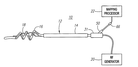
Referring to Fig. 1, an exemplary ablation/diagnostic system 10 constructed in
accordance with the invention is shown. The system 10 may be used within body
lumens, chambers or cavities for diagnostic or therapeutic purposes in those
instances where access to interior bodily regions is obtained through, for
example,
the vascular system or alimentary canal and without complex invasive surgical
procedures. For example, the system 10 has application in the diagnosis and
treatment of arrhythmia conditions within the heart. The system 10 also has
application in the diagnosis or treatment of ailments of the gastrointestinal
tract,
prostrate, brain, gall bladder, uterus, and other regions of the body. As an
example,
the system 10 will be described hereinafter for use in pulmonary veins, and
specifically, to electrically isolate a pulmonary vein from the left atrium of
the heart in
order to treat ectopic atrial fibrillation.
The system 10 generally comprises an ablation/diagnostic catheter assembly
12, which includes a guide sheath 14, an ablation catheter 16 that can be
guided
through the guide sheath 14, and a diagnostic catheter, and specifically, a
mapping
catheter 18, integrated within the ablation catheter 16. As will be described
in further
detail below, the catheter assembly 12 is configured to be introduced through
the
vasculature of the patient, and into the left atrium of the heart, where it
can be used
to map and ablate the tissue within and around a selected pulmonary vein. The
6
CA 02539950 2006-03-23
WO 2005/030072 PCT/US2004/030730
treatment system 10 further comprises an ablation source, and specifically, a
radio
frequency (RF) generator 20, and an mapping processor 22.
The mapping processor 22 is configured to record and process ECG signals
obtained from the mapping catheter 18 to determine irregular electrical
signals within
the heart, and specifically electrical signals adjacent the ostia of the
pulmonary
veins. Recording ECG signals is well known in the art, and thus for purposes
of
brevity, the mapping processor 22 will not be described in further detail. The
RF
generator 20 is configured to deliver ablation energy to the ablation catheter
16 in a
controlled manner in order to ablate the area around the ostium of the
pulmonary
vein identified by the mapping processor 22. Alternatively, other types of
ablative
sources besides the RF generator 20 can be used, e.g., a microwave generator,
an
ultrasound generator, a cryoablation generator, and a laser or other optical
generator. Ablation of tissue within the heart is well known in the art, and
thus for
purposes of brevity, the RF generator 20 will not be described in further
detail.
Further details regarding RF generators are provided in U.S. Patent No.
5,383,874.
The ablation catheter 16 is not a steerable catheter and, accordingly, may be
advanced though the conventional steerable guide sheath 14 to the target
location.
The sheath 14, which should be lubricious to reduce friction during movement
of the
ablation catheter 16, may be advanced over a guidewire in conventional
fashion.
Alternatively, a steerable sheath may be provided. With respect to materials,
the
proximal portion of the sheath 14 is preferably a Pebax~ material and
stainless steel
braid composite, and the distal portion is a more flexible material, such as
unbraided
Pebax~, for steering purposes. The sheath 14 should also be stiffer than the
ablation catheter 16. A sheath introducer (not shown), such as those used in
combination with basket catheters, may be used when introducing the ablation
7
CA 02539950 2006-03-23
WO 2005/030072 PCT/US2004/030730
catheter 16 into the sheath 14. The guide sheath 14 preferably includes a
radio-
opaque compound, such as barium, so that the guide sheath 14 can be observed
using fluoroscopic or ultrasound imaging, or the like. Alternatively, a radio-
opaque
marker (not shown) can be placed at the distal end of the guide sheath 14.
Referring now to Fig. 2, the ablation catheter 16 comprises a flexible
catheter
body 24 formed of a proximal member 28 and a distal member 26. The proximal
member 26 is relatively long (e.g., 80-100 cm) ,while the distal member 26 is
relatively short (e.g., 2-10 cm). The proximal member 26 is preferably formed
from a
biocompatible thermoplastic material, such as a Pebax~ material (polyether
block
amide) and stainless steel braid composite, which has good torque transmission
properties. In some implementations, an elongate guide coil (not shown) may
also
be provided within the proximal member 26. A handle assembly 31 (shown in Fig.
1 )
is mounted to the proximal end of the proximal member 26. The distal member 26
is
preferably formed from a softer, more flexible biocompatible thermoplastic
material
such as unbraided Pebax~ material, polyethylene, or polyurethane. The proximal
and distal members, which are about 5 French to about 9 French in diameter,
are
preferably either bonded together at interface 30 with an overlapping thermal
bond or
adhesively bonded together end to end over a sleeve in what is referred to as
a "butt
bond."
The distal member 26 of the ablation catheter body 24 forms an
unconstrained open helically-shaped ablative structure 32 on which ablation
electrodes 34 are mounted. The ablative structure 32 defines a longitudinal
axis
coincident with the longitudinal axis of the remainder of the catheter body
24. The
number of revolutions (or "coils"), length, diameter, orientation and shape of
the
helical structure will vary from application to application. In the
illustrated
8
CA 02539950 2006-03-23
WO 2005/030072 PCT/US2004/030730
embodiment, the ablative structure 32 revolves around the longitudinal axis of
the
catheter body 24 two and one-half times in its relaxed state, and can be
defined with
a proximal coil 36, medial coil 38, and distal coil 40.
Although the diameter of the ablative structure 32 can alternatively be
substantially constant over its length, as illustrated in Fig. 2, the ablative
structure 32
preferably has a generally frusto-conical shape, where the diameter decreases
in the
distal direction. Specifically, when used in pulmonary veins, the proximal
coil 36 of
the ablative structure 32 preferably has an outer diameter that will cause it
abut the
pulmonary vein ostium (e.g., between about 15 mm and about 35 mm), and the
distal coil 40 of the ablative structure 32 preferably has an outer diameter
suitable for
placement within the pulmonary vein (e.g., between about 5 mm and about 10
mm).
The ablative structure 32 will, therefore, be self-centering when inserted
into the
pulmonary vein, because the tapered ablative structure 32 will wedge itself
against
the pulmonary vein ostium and the internal wall of pulmonary vein itself. Not
only
does this result in proper positioning of the electrodes 34, the wedging
effect also
prevents beating related movement of the heart from the knocking the ablation
catheter 16 out of position once it is in place.
The distal member 26 of the catheter body 24 also forms a distal anchoring
structure 42, which allows the ablative structure 32 to be precisely located
relative to
the pulmonary vein. More specifically, advancing the anchoring structure 42
into the
pulmonary vein aligns the ablative structure 32 with the pulmonary vein. In
the
illustrated embodiment, the anchoring structure 42 is simply the portion of
the distal
member 26 that is distal to the ablative structure 32. Alternatively, a
separate
structure may be secured to the distal end of the distal member 26. The
exemplary
9
CA 02539950 2006-03-23
WO 2005/030072 PCT/US2004/030730
anchoring structure 42 is approximately 1 to 2 inches in length, although
other
lengths may be used to suit particular applications.
Referring to Fig. 3, the shape of the ablative structure 32 is achieved
through
the use of a center support 44 that is positioned inside of and passes within
the
length of the distal member 26. In the illustrated embodiment, the center
support 44
is a rectangular wire formed from resilient inert wire, such as Nickel
Titanium
(commercially available under the trade name Nitinol) or 17-7 stainless steel
wire,
with a portion thereof heat set into the desired helical configuration.
Alternatively, the
center support 44 can be circular. The thickness of the rectangular center
support 44
is preferably between about 0.010 inch and about 0.015 inch. Resilient
injection
molded plastic can also be used. Although other cross sectional configurations
can
be used, such as a round wire, a rectangular cross section arranged such that
the
longer edge extends in the longitudinal direction is preferred for at least
the ablative
structure 32.
Such an orientation reduces the amount of torsional force, as compared to a
round wire, required to unwind the ablative structure 32 into an expanded
configuration and collapse the ablative structure 32 into a linear structure.
The
center support 44 is preferably housed in an insulative tube 46 formed from
material
such as Teflon. or polyester. Additional details concerning the placement of a
center support within the distal member of a catheter can be found in commonly
assigned U.S. Patent No. 6,287,301.
Preferably, the distal portion of the distal member 26 is more flexible than
the
proximal portion of the distal member 26 in order to prevent tissue damage
when
attempts are made to insert the ablative structure 32 into a pulmonary vein.
In
addition, the ablative structure 32 will be more predisposed to easily uncoil
for
CA 02539950 2006-03-23
WO 2005/030072 PCT/US2004/030730
placement within the sheath 14, remain uncoiled and slide though the sheath 14
until
it exits through the distal end of the sheath and re-coils, and then easily
uncoil again
when pulled back into the sheath after the procedure is completed. Also, the
stiffer
proximal portion of the distal member 26 allows the physician to press the
ablation
electrodes 34 against the tissue with more force when lesions are being
created.
The flexibility of the distal portion of the distal member 26 can be increased
in variety
of ways, e.g., by using a core wire (not shown) having a varying stiffness, or
by
constructing the distal member 26 from different materials. Further details on
the
construction of helical structures with varying flexibility are disclosed in
U.S. Patent
No.6,745,080.
The ablation catheter 16 may comprise an optional stylet (not shown) that
enables the physician to manipulate the ablative structure 32 and adjust its
shape by
longitudinally and/or rotating the stylet. Further details on the construction
and use
of the stilette, along with a handle assembly specifically designed to
manipulate the
stilette, are disclosed in U.S. Patent Application Ser. No. 09/832,612.
The ablation catheter 16 comprises a lumen 48 (in addition to other lumens
for providing ablation and signals wires described below) for slidably
receiving the
mapping catheter 18 (shown in Fig. 2). The lumen 48 proximally terminates in
the
handle assembly 31 at an insertion port 50 (shown in Fig. 1) and distally
terminates
in the distal member 26 at an exit port 52 (shown in Fig. 2) just proximal to
the
ablative structure 32. Thus, the mapping catheter 18 can be introduced into
the
insertion port 50 on the handle assembly 31, through the lumen 48, and out the
exit
port 52, so that it extends within an interior space 54 created by the
ablative
structure 32.
11
CA 02539950 2006-03-23
WO 2005/030072 PCT/US2004/030730
The spaced ablation electrodes 34 are preferably in the form of wound, spiral
coils. The coils are made of electrically conducting material, like copper
alloy,
platinum, or stainless steel, or compositions such as drawn-filled tubing
(e.g. a
copper core with a platinum jacket). The electrically conducting material of
the coils
can be further coated with platinum-iridium or gold to improve its conduction
properties and biocompatibility. A coil electrode is disclosed in U.S. Patent
No.
5,797,905. The electrodes 34 are electrically coupled to individual wires 55
(shown in
Fig. 3) to conduct coagulating energy to them. The wires are passed in
conventional
fashion through a lumen extending through the associated catheter body into a
PC
board (not shown) in the handle assembly 31, where they are electrically
coupled to
a connector (not shown) that is received in a port on the handle assembly 31.
The
connector plugs into the RF generator 20 (shown in Fig. 1 ).
As an alternative, the ablation electrodes 34 may be in the form of solid
rings
of conductive material, like platinum, or can comprise a conductive material,
like
platinum-iridium or gold, coated upon the device using conventional coating
techniques or an ion beam assisted deposition (IBAD) process. For better
adherence, an undercoating of nickel or titanium can be applied. The
electrodes 34
can also be in the form of helical ribbons. The electrodes 34 can also be
formed with
a conductive ink compound that is pad printed onto a nonconductive tubular
body.
One such conductive ink compound is a silver-based flexible adhesive
conductive
ink (polyurethane binder), however other metal-based adhesive conductive inks
such
as platinum-based, gold-based, copper-based, etc., may also be used to form
electrodes 34. Such inks are more flexible than epoxy-based inks.
The flexible electrodes 34 are preferably about 4 mm to about 20 mm in
length. In one embodiment, the electrodes are 12.5 mm in length with 1 mm to 3
mm
12
CA 02539950 2006-03-23
WO 2005/030072 PCT/US2004/030730
spacing, which will result in the creation of continuous lesion patterns in
tissue when
coagulation energy is applied simultaneously to adjacent electrodes 34. For
rigid
electrodes 34, the length of the each electrode can vary from about 2 mm to
about
mm. Using multiple rigid electrodes 34 longer than about 10 mm each adversely
5 effects the overall flexibility of the device, while electrodes 34 having
lengths of less
than about 2 mm do not consistently form the desired continuous lesion
patterns.
The portion of the electrodes 34 that are not intended to contact tissue (and
be exposed to the blood pool) may be masked through a variety of techniques
with a
material that is preferably electrically and thermally insulating. This
prevents the
10 transmission of coagulation energy directly into the blood pool and directs
the energy
directly toward and into the tissue. For example, a layer of UV adhesive (or
another
adhesive) may be painted on preselected portions of the electrodes 34 to
insulate
the portions of the electrodes not intended to contact tissue. Deposition
techniques
may also be implemented to position a conductive surface only on those
portions of
the assembly intended to contact tissue. Alternatively, a coating may be
formed by
dipping the electrodes 34 in PTFE material.
The electrodes 34 can include a porous material coating, which transmits
coagulation energy through an electrified ionic medium. For example, as
disclosed in
U.S. Pat. No. 5,991,650, electrodes 34 may be coated with regenerated
cellulose,
hydrogel or plastic having electrically conductive components. With respect to
regenerated cellulose, the coating acts as a mechanical barrier between the
surgical
device components, such as electrodes, preventing ingress of blood cells,
infectious
agents, such as viruses and bacteria, and large biological molecules such as
proteins, while providing electrical contact to the human body. The
regenerated
cellulose coating also acts as a biocompatible barrier between the device
13
CA 02539950 2006-03-23
WO 2005/030072 PCT/US2004/030730
components and the human body, whereby the components can now be made from
materials that are somewhat toxic (such as silver or copper).
The electrodes 34 may be operated in a uni-polar mode, in which the soft
tissue coagulation energy emitted by the electrodes 34 is returned through an
indifferent patch electrode (not shown) externally attached to the skin of the
patient.
Alternatively, the electrodes 34 may be operated in a bi-polar mode, in which
energy
emitted by one or more electrodes 34 is returned through other electrodes 34.
The
amount of power required to coagulate tissue~ranges from 5 to 150 W.
Although ablation electrodes 34 have been described as the operative
elements that create the lesion, other operative elements, such as lumens for
chemical ablation, laser arrays, ultrasonic transducers, microwave electrodes,
and
ohmically heated hot wires, and such devices may be substituted for the
electrodes
34.
The ablation catheter 16 further comprises temperature sensors (not shown),
such as thermocouples or thermistors, which may be located on, under, abutting
the
longitudinal end edges of, or in between, the electrodes 34. Preferably, the
temperature sensors are located at the longitudinal edges of the electrodes 34
on
the distally facing side of the ablative structure 32. In some embodiments, a
reference thermocouple (not shown) may also be provided. For temperature
control
purposes, signals from the temperature sensors are transmitted to the source
of
coagulation energy by way of wires 60 (shown in Fig. 3) that are also
connected to
the aforementioned PC board in the handle assembly 31. Suitable temperature
sensors and controllers which control power to electrodes based on a sensed
temperature are disclosed in U.S. Pat. Nos. 5,456,682, 5,582,609 and
5,755,715.
14
CA 02539950 2006-03-23
WO 2005/030072 PCT/US2004/030730
The mapping catheter 18 comprises a flexible catheter body 62 formed of a
flexible spline composed of a resilient, biologically inert material, like
Nitinol metal or
silicone rubber. Thus, the mapping catheter body 62 is configured to bend and
conform to the endocardial and pulmonary vein tissue surface its contacts. In
the
illustrated embodiment, the diameter of the mapping catheter body 62 is
relatively
small (e.g., 3-4F), so that the ablation catheter 16 that houses the mapping
catheter
18 assumes a small profile.
The distal end of the mapping catheter body 62 forms a mapping structure 64.
The mapping catheter 18 comprises mapping electrodes 58 extending along the
mapping structure 64. In the illustrated embodiment, the mapping electrodes 58
are
ring electrodes that are composed of a solid, electrically conducting
material, like
platinum or gold, attached about the catheter body 62. Alternatively, the
mapping
electrodes 58 can be formed by coating the exterior surface of the catheter
body 62
with an electrically conducting material, like platinum or gold. The coating
can be
applied using sputtering, ion beam deposition, or equivalent techniques. The
mapping electrodes 58 can have suitable lengths, such as between 0.5 and 5 mm.
In use, the mapping electrodes 58 sense electrical events in myocardial tissue
for the creation of electrograms, and are electrically coupled to the mapping
processor 22 (see Fig. 1 ). A signal wire (not shown) is electrically coupled
to each
mapping electrode 58. The wires extend through the catheter body 62 into an
external multiple pin connector 66. The connector 66 electrically couples the
mapping electrodes 58 to the mapping processor 22.
As illustrated in Fig. 2, the mapping structure 64 extends distally within an
interior space 54 of the ablative structure 32, and is curved in an undulated
fashion
that will resiliently place the mapping electrodes 58 in contact with the
tissue of the
CA 02539950 2006-03-23
WO 2005/030072 PCT/US2004/030730
vessel in which the ablation and mapping catheters 16 and 18 are placed.
Specifically, the total transverse distance that the mapping structure 64
curves is
greater than the diameter of the selected vessel, so that placement of the
mapping
structure 64 within the vessel will cause the vessel wall to provide a
compressive
force mapping structure 64, thereby resiliently lodging the mapping structure
64
within the vessel.
In the illustrated embodiment, the mapping structure 64 comprises first and
second curved sections 68 and 70 having apexes that point in the same
direction
away from the longitudinal axis of the ablation catheter 16, and an mediate
curved
section 72 between the proximal and distal curved sections 68 and 70 having an
apex that points towards the longitudinal axis of the ablation catheter 16. In
this
manner, when the mapping structure 64 is deployed from the exit port 52 of the
ablation catheter lumen 48, the proximal curved section 68 may come in contact
with
tissue between the proximal and medial coils 36 and 38 of the ablative
structure 32,
and the distal curved section 70 may come in contact with tissue between the
mediate and distal coils 38 and 40 of the ablative structure 32. The medial
curved
section 72 provides clearance between the mapping structure 64 and the medial
coil
38 of the ablative structure 32.
The distal end of the mapping catheter body 62 further forms a straight distal
section 74 that is configured to stabilize the mapping structure 64 by
contacting the
portion of the vessel wall opposite the portion in which the proximal and
distal curved
sections 68 and 70 contact. Preferably, the distal section 74 is floppy,
similar to the
tip of a guidewire, thereby minimizing tissue trauma.
It should be noted that although the mapping catheter body 62 has been
described as a linear resilient spline, the catheter body 62 can also have
other
16
CA 02539950 2006-03-23
WO 2005/030072 PCT/US2004/030730
shapes, such as, e.g., a spiral, basket, etc., that allow the mapping catheter
body 62
to collapse into the lumen 48 of the ablation catheter body 62.
Referring now to Fig. 4, an alternative embodiment of a catheter assembly
112 that can be used in the treatment system 10 of Fig. 1 is shown. The
catheter
assembly 112 is similar to the previously described catheter assembly 12, with
the
exception that the ablation catheter body forms a loop-shaped, rather than a
helically-shaped, ablative structure. Specifically, an ablation catheter 116
comprises
a flexible elongate catheter body 124 having a distal member 126 that forms a
loop-
shaped ablative structure 132. The ablation catheter 116 further comprises a
pull
wire 134, which extends from the tip of the distal member 126 back through the
sheath 32. The pull wire 134 is used to pull the distal member 116 into a loop
configuration. The pull wire 134 also maintains the shape of the ablative
structure
132 (thereby insuring good tissue contact) when the loop structure 132 is
urged
against tissue, such as a pulmonary vein ostium.
In the illustrated embodiment, the ablative structure 132 forms a curved
portion with a radius of about 0.5 inch. The curved portion lies in a plane
between
about 30 and about 60 degrees, and preferably about 45 degrees, out of the
horizontal catheter plane, as illustrated in Fig. 5. The preset curvature may
be
accomplished in a variety of ways. Preferably, the curved portion is preset
through
the use of a thermal forming technique (100° C for 1 hour). The preset
curvature
may also be accomplished through the use of a pre-shaped core wire (not shown)
formed from Nitinol or 17-7 stainless steel. The curved portion will typically
be bent
out of its pre-bent orientation when the ablative structure 132 is urged
against tissue
(note the dashed lines in Fig. 5). As a result, a spring force that urges the
ablative
17
CA 02539950 2006-03-23
WO 2005/030072 PCT/US2004/030730
structure 132 against the tissue is generated, thereby improving
tissue/electrode
contact.
The pull wire 134 is preferably a flexible, inert cable constructed from
strands
of metal wire material, such as Nitinol or 17-7 stainless steel, that is about
0.012 inch
to about 0.025 inch in diameter. Alternatively, the pull wire 134 may be
formed from
a flexible, inert stranded or molded plastic material. The pull wire 134 is
also
preferably round in cross-section, although other cross-sectional
configurations can
be used. Further details on the construction of loop-shaped ablative
structures and
pull wires are disclosed in U.S. Patents Nos. 6,745,080 and 6,048,329.
The catheter assembly 112 further comprises a mapping catheter 118 that
includes a catheter body 164 with a mapping structure 164 that extends
distally
through an interior space 156 of the ablative structure 132. The mapping
structure
164 is similar to the previously described mapping structure 64, with the
exception
that is forms a single curve that will extend along one side of the pulmonary
vein
contralaterally to a straight distal section 174.
Referring now to Figs. 6 and 7, another alternative embodiment of a catheter
assembly 212 that can be used in the treatment system 10 of Fig. 1 is shown.
The
catheter assembly 212 is similar to the previously described catheter assembly
12,
with the exception that an expandable-collapsible ablative structure, rather
than a
helically-shaped ablative structure, is formed at the distal end of the
ablation catheter
body.
Specifically, an ablation catheter 216 comprises a flexible elongate catheter
body 224 having a distal member 226 on which there is mounted an expandable-
collapsible ablative structure 232. The ablative structure 232 is formed by a
"balloon-like" wall suitably bonded to and disposed about the distal member
226.
18
CA 02539950 2006-03-23
WO 2005/030072 PCT/US2004/030730
The geometry of the ablative structure 232 can be altered between a collapsed,
low
profile geometry (Fig. 6), and an expanded, high profile geometry (Fig. 13).
The
ablation catheter body 224 comprises inflation and venting lumens (not shown)
that
extend from a handle assembly (not shown) to the interior region of the
ablative
structure 232. The catheter body 224 comprises a lumen (not shown) that
terminates in an exit port 252 out which the previously described mapping
structure
164 of the mapping catheter 118 extends. The exit port 252, in this case, is
distal to
the ablative structure 232.
In order to inflate the ablative structure 232, a liquid inflation medium,
such as
water, saline solution, or other bio-compatible fluid, is conveyed under
positive
pressure through a port on the handle assembly, through the inflation lumen
extending through the catheter body 224. The liquid medium fills the interior
of the
ablative structure 232 and exerts pressure on the inside of the ablative
structure 232
to urge the ablative structure from its collapsed geometry (Fig. 6) to its
expanded
geometry (Fig. 7). Constant exertion of pressure through the inflation lumen
maintains the ablative structure 232 in its expanded geometry. The venting
lumen is
used to vent any air or excess fluid from the ablative structure 232.
Alternatively, the
inflating fluid medium can comprise a gaseous medium, such as carbon dioxide.
Preferably, the ablative structure 232 is less than 8 French diameter when in
a
collapsed geometry for ease of manipulation through the vasculature, and about
2.0
cm in circumference around its largest portion when in its expanded geometry
and
located in a desired ablation region within the pulmonary vein. The ablative
structure
232 is preferably made of a suitable biocompatible, thermoplastic or
elastomeric
material, and can be configured to have any one of many shapes in its expanded
19
CA 02539950 2006-03-23
WO 2005/030072 PCT/US2004/030730
geometry, such as the shape shown in Fig. 7, depending on the desired
resulting
geometry.
Proximate the center of the ablative structure 232 is a pronounced
circumferential region 236 having a larger circumference than that of the rest
of the
ablative structure 232. In this manner, expansion of the ablative structure
232 within
the pulmonary vein provides a force that is concentrated between the enlarged
circumferential region 236 and the interior surface of a pulmonary vein in
which the
ablative structure 232 is situated, thus enhancing the lesion creating
characteristics
of the ablative structure 232.
The ablation catheter 216 comprises an electrode that takes the form of a
conductive shell 234 made of a material having a relatively high electrical
and
thermal conductivity that is suitably deposited on the outer surface of the
ablative
structure 232 over the enlarged circumferential region 236 using ion
deposition or
equivalent techniques. Materials possessing these characteristics include,
among
others, gold, platinum, platinum/iridium, conductive ink epoxy, or a
combination
thereof. In particular, noble metals are preferred.
The area of the ablative structure 232 located immediately proximal and distal
to the enlarged circumferential region 236 is preferably masked prior to the
deposition of the conductive material, so that resulting non-conductive
regions 238
and 240 are formed on either side of the conductive shell 234. In particular,
the
masking of the regions on either side on the conductive region assures that
the
maximum current density will be distributed at the enlarged circumferential
region
236 of the ablative structure 232, thereby allowing the ablative structure 232
to
efficiently form annular lesions within the pulmonary vein. In order to
deliver current,
CA 02539950 2006-03-23
WO 2005/030072 PCT/US2004/030730
the conductive shell 234 is coupled to a plurality of insulated ablation wires
(not
shown), that are in turn coupled to the handle assembly (not shown).
There are many modifications that can be made to the ablative structure 232.
For example, the conductive shell 234 may be segmented instead of continuous.
The ablative structure 232 may be microporous, allowing ions to pass from an
interior electrode, through the pores, into the tissue. The ablative structure
232 may
have an interior support structure (such as resilient splines or mesh or a
foam
substance) arranged to apply an outward force against the electrode structure
232 to
augment, or replace, the outward force caused by a pressurized liquid medium.
The
ablative structure 232 may comprise blood lumens for allowing the flow of
blood
through the ablative structure 232 when expanded within the pulmonary vein.
The
ablative structure 232 may be shaped, such that a portion of the conductive
shell 234
engages the ostium of the pulmonary vein.
Other types of ablative structures, besides balloon-like ablative structures,
can
be envisioned. For example, a basket-type ablative structure having a
plurality of
resilient splines with ablation electrodes mounted thereon can be used to
create a
circumferential lesion within the pulmonary vein. Pre-shaped ablative loop
structures
that are either coplanar with, or orthogonal to, the longitudinal axis of the
catheter
body can also be used to create lesions within or around the pulmonary vein.
In
each case, a lumen for housing the mapping catheter 118, and an exit port out
which
the mapping electrode structure 164 extends, can be incorporated into the
design.
In all of the previously described embodiments, the mapping catheters are
configured to be introduced through lumens contained within the ablation
catheters.
Alternatively, as illustrated in Fig. 8, the ablation catheter 16 and mapping
catheter
18 can be independently introduced through the lumen of the guide sheath 14.
21