Note: Descriptions are shown in the official language in which they were submitted.
CA 02540177 2006-03-24
WO 2005/030139 PCT/US2004/031504
Seneca Valley Virus Based Compositian~~and Meth~~s'~for Treafrn~~~Disease
[0001] This disclosure contains material that is subject to copyright
protection.
The copyright owner has no objection to the facsimile reproduction by anyone
of the
patent document or the patent disclosure, as it appears in the U.S. Patent and
Trademark Office patent file or records, but otherwise reserves any and all
copyright
rights.
[0002] All patent applications, published patent applications, issued and
granted patents, texts, and literature references cited in this specification
are hereby
incorporated herein by reference in their entirety to more fully describe the
state of the
art to which the present invention pertains.
[0003] This application claims priority to U.S. Serial No. 601506,182, which
was filed on September 26, 2003, which is hereby incorporated in its
erstirety.
BACKGROUND OF THE INVENTION
[0004] Virotherapy holds great promise for treating cancer. Oncolytic viruses,
which aim to specifically bind and kill cancer cells, whether native and/or
engineered,
may be more efficacious and less toxic than alternative treatments, such as
chemotherapy and radiation. In addition, oncolytic virus therapy is the only
therapy
known that can amplify the therapeutic at the pharmacologically desired site.
[0005] A key aspect of cancer therapy is to achieve a high rate of killing of
cancer cells versus normal cells. Accomplishing this goal has been difficult
for many
reasons, including the wide array of cell types involved, the systemic
dissemination of
cancer cells due to metastases, and the narrow biological differences between
normal
and cancer cells. While progress has been made, much still needs to be done to
improve upon current cancer therapies.
[0006] In the past, surgeons have tried to remove tumors surgically without
substantially harming the patient. Even complete removal of a primary tumor
does
not ensure survival since earlier metastases to unknown sites in the body are
left
undetected. There is also some research suggesting that surgical intervention
may
enhance the growth of distant metastases due to removal of tumor cells
producing
angiogenesis inhibitors. Finally, in many cases, the tumor grows back at the
original
site after surgical removal. Radiation aims to selectively destroy the most
rapidly
CA 02540177 2006-03-24
WO 2005/030139 PCT/US2004/031504
proliferating cells at the expense of the others. However, tumor cells can
escape
radiation therapy either by becoming resistant or by being in a non-dividing
state
during treatment. In addition, radiation is not always selective in that many
normal
cells are actively dividing and killed by the treatment (gastrointestinal
cells, hair
follicles, etc.).
[0007] Like radiation, chemotherapy is not completely selective and thus
destroys many normal cells, and does not kill all tumor cells due to drug
resistance
and/or division state of the cell. Thus, chemotherapy and radiation therapies
exploit a
small differential sensitivity that exists between normal and cancer cells,
giving them
a narrow therapeutic index. A small therapeutic index is clearly an
undesirable
property of any modality to treat cancer. Therefore, novel cancer therapeutic
approaches overcoming these limitations are desired.
[0008] One such novel approach is oncolytic virus therapy. Initially,
replication-defective viruses carrying cytotoxic transgenes were utilized in
attempts to
treat cancer. However, they were found to be inefficient in transduction of
tumors
and not adequately selective toward cancers. To overcome this limitation,
viruses
were either modified to replicate selectively in tumor cells or viruses were
discovered
to have natural tumor-selective properties. These oncolytic viruses thus had
the
properties to replicate, spread, and kill tumor cells selectively through a
tumor mass
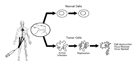
by locally injecting the virus or by systemically delivering the virus (Figure
1).
[0009] Despite the early promise of this newly defined class of anti-cancer
therapeutics, several limitations remain that may limit their use as a cancer
therapeutic.
Therefore, there is an ongoing need for novel oncolytic viruses that can be
utilized for
cancer therapy.
SUMMARY OF THE INVENTION
[0010] A novel RNA picornavirus has been discovered (hereafter referred to
as Seneca Valley virus ("SVV")) whose native properties include the ability to
selectively kill some types of tumors. As demonstrated below in the examples,
SVV
selectively kills tumor lines with neurotropic properties, in most cases with
a greater
than 10,000 fold difference in the amount of virus necessary to kill 50% of
tumor cells
versus normal cells (i.e., the ECso value). This result also translates in
vivo, where
2
CA 02540177 2006-03-24
WO 2005/030139 PCT/US2004/031504
tumor explants in mice are selectively eliminated. Further, irz vivo results
indicate that
SVV is not toxic to normal cells, in that up to 1x10¹⁴ vp/kg (vector or virus
particles
per kilogram) systemically administered causes no mortality and no visible
clinical
symptoms in immune deficient or immune competent mice.
[0011] SVV elicits efficacy at doses as low as 1x10⁸ vp/kg; therefore, a very
high therapeutic index of >100,000 is achieved. Efficacy is very robust in
that 100%
of large pre-established tumors in mice can be completely eradicated (see
Example
11). This efficacy may be mediated with a single systemic injection of SVV
without
any adjunct therapy. Furthermore, SVV injected mice show neither clinical
symptoms nor recurrence of tumors for at least 200 days following injection.
SVV
can also be purified to high titer and can be produced at >200,000 virus
particles per
cell in permissive cell lines. SVV-based viral therapy therefore shows
considerable
promise as a safe, effective and new line of treatment for selected types of
cancers.
Further, SVV has a small and easily manipulatable genome, simple and fast
lifecycle,
and a well-understood capsid, and thus is amenable to modification. These
properties,
at least in part, allow for methods that generate modified SVVs that have new
cell or
tissue specific tropisms, such that SVV-based therapy can be directed to new
tumor
types resistant to infection by the original SVV isolate.
[0012] Accordingly, the present invention provides an isolated nucleic acid
comprising a nucleic acid sequence having at least 65%, 70%, 75%, 80%, 85%,
90%,
95% or 99% sequence identity to SEQ ~ NOs: 1, 3, 5, 7, 9, 11, 13, 15, 17, 19,
21, or
a contiguous portion of any one of these sequences that is at least 50
nucleotides in
length, or 95% identical to a contiguous portion of any one of these sequences
that is
at least least 10, 15 or 20 nucleotides in length. The isolated nucleic acids
of the
invention can be RNA or DNA.
[0013] In other aspects, the invention provides an isolated nucleic acid that
hybridizes under conditions of high, moderate stringency or low stringency to
SEQ m
NO: 1, 3, 5, 7, 9, 1 l, 13, 15, 17, 19, 21, or to a contiguous portion of any
one of these
sequences that is at least 50 nucleotides in length.
[0014] In another aspect, the invention provides a vector comprising a nucleic
acid sequence having at least 65%, 70%, 75%, 80%, 85%, 90%, 95% or 99%
3
CA 02540177 2006-03-24
WO 2005/030139 PCT/US2004/031504
sequence identity to SEQ m NQs 1, 3, 5, 7, 9, 11, 13, 15, 17, 19, 21, or to a
contiguous portion of any one of these sequences that is at least 50
nucleotides in
length.
[0015] The present invention also provides an isolated polypeptide encoded
by a nucleic acid having at least 65%, 70%, 75%, 80%, 85%, 90%, 95% or 99%
sequence identity to a nucleic acid sequence comprising SEQ m NOs: 1, 3, 5, 7,
9, 11,
13, 15, 17, 19, 21, or to a contiguous portion of any one of these sequences
that is at
least 50 nucleotides in length.
[0016] In one aspect, the invention provides an isolated polypeptide
comprising an amino acid sequence having at least 65%, 70%, 75%, 80%, 85%,
90%,
95% or 99% sequence identity to SEQ m NOs: 2, 4, 6, 8, 10, 12, 14, 16, 18, 20,
22,
or to a contiguous portion of any one of these sequences that is at least 10
amino acids
in length.
[0017] In another aspect, the invention provides an isolated antibody which
specifically binds a polypeptide comprising an amino acid sequence having at
least
65%, 70%, 75%, 80%, 85%, 90%, 95% or 99% sequence identity to SEQ m NOs: 2,
4, 6, 8, 10, 12, 14, 16, 18, 20, 22, or to a contiguous portion of any one of
these
sequences that is at least 10 amino acids in length. The isolated antibody can
be
generated such that it binds to any protein epitope or antigen of SEQ m N0:2.
Further, the antibody can be a polyclonal antibody, a monoclonal antibody or a
chimeric antibody.
[0018] In one aspect, the invention provides an isolated SVV or derivative or
relative thereof, having a genomic sequence comprising a sequence that is at
least
65%, 70%, 75%, 80%, 85%, 90%, 95% or 99% identical to SEQ m NO:1.
[0019] In another asepct, the invention provides an isolated virus having all
the identifying characteristics and nucleic acid sequence of American Type
Culture
Collection (ATCC) Patent Deposit number PTA-5343. Some of the viruses of the
present invention are directed to the PTA-5343 isolate, variants, homologues,
relatives, derivatives and mutants of the PTA-5343 isolate, and variants,
homologues,
derivatives and mutants of other viruses that are modified in respect to
sequences of
4
CA 02540177 2006-03-24
WO 2005/030139 PCT/US2004/031504
SVV (both wild-type and mutant) that are determined to be responsible for its
oncolytic properties.
[0020] The present invention further provides an isolated SVV comprising the
following characteristics: a single stranded RNA genome (positive (+) sense
strand)
of ~7.5 kilobases (kb); a diameter of ~27 nanometers (nm); a capsid comprising
at
least 3 proteins that have approximate molecular weights of about 31 kDa, 36
kDa
and 27 kDa; a buoyant density of approximately 1.34 g/mL on cesium chloride
(CsCI)
gradients; and replication competence in tumor cells. In this aspect, the 31
kDa
capsid protein (VP1) can comprise an amino acid sequence that is at least 65%,
70%,
75%, 80%, 85%, 90%, 95% or 99% identical to SEQ m N0:8; the 36 kDa capsid
protein (VP2) can comprise an amino acid sequence that is at least 65%, 70%,
75%,
80%, 85%, 90%, 95% or 99% identical to SEQ m N0:4; and the 27 kDa capsid
protein (VP3) can comprise an amino acid sequence that is at least 65%, 70%,
75%,
80%, 85%, 90%, 95% or 99% identical to SEQ m N0:6.
[0021] In another aspect, the invention provides an isolated SVV derivative or
relative comprising the following characteristics: replication competence in
tumor
cells, tumor-cell tropism, and lack of cytolysis in normal cells. In another
aspect, the
virus is replication competent in tumor cell types having neuroendocrine
properties.
[0022] In other aspects, the present invention provides: a pharmaceutical
composition comprising an effective amount of a virus of the invention and a
pharmaceutically acceptable carrier; a cell comprising a virus of the
invention; a viral
lysate containing antigens of a virus of the invention; and an isolated and
purified
viral antigen obtained from a virus of the invention.
[0023] In yet another aspect, the invention provides a method of purifying a
virus of the invention, comprising: infecting a cell with the virus;
harvesting cell
lysate; subjecting cell lysate to at least one round of gradient
centrifugation; and
isolating the virus from the gradient.
[0024] In another aspect, the invention provides a method for treating cancer
comprising administering an effective amount of a virus or derivative thereof,
so as to
treat the cancer, wherein the virus has a genomic sequence that comprises a
sequence
CA 02540177 2006-03-24
WO 2005/030139 PCT/US2004/031504
that is at least 65%, 70%, 75%, 80%, 85 Jo, 90%, 95% or 99% identical to SEQ m
NOs: 1, 3, 5, 7, 9, 11, 13, 15, 17, 19, 21 or to a portion of SEQ m N0:1.
[0025] In another aspect, the invention provides a method for treating cancer
comprising adminstering an effective amount of a virus comprising a capsid
encoding
region that comprises a sequence that is at least 65%, 70%, 75%, 80%, 85%,
90%,
95% or 99% identical to SEQ ~ N0:3, S, 7 or a contiguous portion thereof. The
invention also provides a method for treating cancer comprising administering
an
effective amount of a virus comprising a capsid that comprises an amino acid
sequence that is at least 65%, 70%, 75%, 80%, 85%, 90%, 95% or 99% identical
to
SEQ m N0:4, 6, 8 or a contiguous portion thereof.
[0026] In one aspect, the present invention provides a method for inhibiting
cancer progression comprising contacting a cancer cell with a virus or
derivative
thereof, wherein the virus or derivative thereof specifically binds to the
cancerous cell,
wherein the virus has a genomic sequence that comprises a sequence that is at
least
65%, 70%, 75%, 80%, 85%, 90%, 95% or 99% identical to SEQ m NO: 1, 3, 5, 7, 9,
11, 13, 15, 17, 19 or 21.
[0027] In another aspect, the present invention provides a method for killing
cancer cells comprising contacting a cancer cell with an effective amount of a
virus or
derivative thereof, wherein the virus has a genomic sequence that comprises a
sequence that is at least 65%, 70%, 75%, 80%, 85%, 90%, 95% or 99% identical
to
SEQ m NO: 1, 3, 5, 7, 9, 11, 13, 15, 17, 19 or 21.
[0028] In these methods directed to cancer, the virus can be a picornavirus.
The picornavirus can be a cardiovirus, erbovirus, aphthovirus, kobuvirus,
hepatovirus,
parechovirus, teschovirus, enterovirus or rhinovirus. The cardiovirus can be
selected
from the group consisting of: vilyuisk human encephalomyelitis virus,
Theiler's
murine encephalomyelitis virus, encephalomyocarditis virus and SVV. The
encephalomyocarditis virus can be selected from the group of isolates
consisting of:
CA-131395, LA-97-1278,1L-92-48963, IA-89-47752, NJ-90-10324, MN-88-36695,
and NC-88-23626. The SVV can be a virus having the ATCC deposit number PTA-
5343 or a virus comprising a nucleic acid sequence that is at least 65%, 70%,
75%,
6
CA 02540177 2006-03-24
WO 2005/030139 PCT/US2004/031504
80%, 85%, 90%, 95% or 99% identical to SEQ ID NO:1 or a contiguous portion
thereof.
[0029] The present invention also provides a method of killing an abnormally
proliferative cell comprising contacting the cell with a virus of the
invention. In one
aspect, the abnormally proliferative cell is a tumor cell. In various aspects
of this
method, the tumor cell is selected from the group consisting of: human small
cell
lung cancer, human retinoblastoma, human neuroblastoma, human medulloblastoma,
mouse neuroblastoma, Wilms' tumor, and human non-small cell lung cancer.
[0030] The present invention also provides a method of treating a neoplastic
condition in a subject comprising administering to the subject an effective
amount of
a virus of the invention to the mammal. In one aspect, the neoplastic
condition is a
neuroendocrine cancer. In another aspect, the subject is a mammal. In another
aspect,
the mammal is a human.
[0031] The present invention also provides a method of producing a virus of
the invention, comprising: culturing cells infected with the virus under
conditions
that allow for replication of the virus and recovering the virus from the
cells or the
supernatant. In one aspect of this method, the cells are PER.C6 cells. In
another
aspect of this method, the cells are H446 cells. In the various aspects of
this method,
the cells may produce over 200,000 virus particles per cell.
[0032] In another aspect, the present invention provides a method for
detecting a virus of the invention, comprising: isolating RNA from test
material
suspected to contain the virus of the invention; labeling RNA corresponding to
at least
15 contiguous nucleotides of SEQ ID NO:1; probing the test material with the
labeled
RNA; and detecting the binding of the labeled RNA with the RNA isolated from
the
test material, wherein binding indicates the presence of the virus. In another
aspect,
the present invention provides a nucleic acid probe comprising a nucleotide
sequence
corresponding to at least 15 contiguous nucleotides of SEQ ID N0:1 or its
complement.
[0033] The present invention also provides a method for making an oncolytic
cardiovirus, the method comprising: (a) comparing a SVV genomic sequence with
a
test virus genomic sequence; (b) identifying at least a first amino acid
difference
7
CA 02540177 2006-03-24
WO 2005/030139 PCT/US2004/031504
between a polypeptide encoded by the SVV genomic sequence and a polypeptide
encoded by the test virus genomic sequence; (c) mutating the test virus
genomic
sequence such that the polypeptide encoded by the test virus genomic sequence
has at
least one less amino acid difference to the polypeptide encoded by the SVV
genomic
sequence; (d) transfecting the mutated test virus genomic sequence into a
tumor cell;
and (e) determining whether the tumor cell is lytically infected by the
mutated test
virus genomic sequence. In one aspect, the amino acids) mutated in the test
virus are
amino acids in a structural region, such as in the capsid encoding region. In
another
aspect, the amino acids mutated in the test virus are amino acids in a non-
structural
region.
[0034] In one aspect of the method for making an oncolytic virus, the SVV
genomic sequence is obtained from the isolated SVV having the ATCC deposit
number PTA-5343 or from a virus comprising a sequence that is at least 65%,
70%,
75%, 80%, 85%, 90%, 95% or 99% identical to SEQ ID NOs: 1, 3, 5, 7, 9, 11, 13,
15,
17, 19, 21 or a contiguous portion thereof. In another aspect of this method,
the step
of mutating the test virus genomic sequence comprises mutating a cDNA having
the
test virus genomic sequence. In another aspect of this method, the step of
transfecting
the mutated test virus genomic sequence comprises transfecting RNA, wherein
the
RNA is generated from the cDNA having the mutated test virus genomic sequence.
[0035] In another aspect of the method for making an oncolytic cardiovirus,
the test virus is a picornavirus. The test picornavirus can be a teschovirus,
enterovirus,
rhinovirus, cardiovirus, erbovirus, apthovirus, kobuvirus, hepatovirus,
parechovirus or
teschovirus. In another aspect, the test virus is a cardiovirus. In another
aspect, the
amino acid differences identified in the methods for making an oncolytic virus
are
between a SVV capsid protein and a test virus capsid protein sequence. In
another
aspect for making an oncolytic virus, the test virus genomic sequence is
selected from
the group consisting of: Vilyuisk human encephalomyelitis virus, Theiler's
murine
encephalomyelitis virus, and encephalomyocarditis virus. In another aspect,
the test
virus genomic sequence is selected from an encephalomyocarditis virus. The
encephalomyocarditis virus can be selected from the group of isolates
consisting of:
CA-131395, LA-97-1278, IL-92-48963,1:A-89-47752, NJ-90-10324, MN-88-36695,
and NC-88-23626. In yet another aspect, the encephalomyocarditis virus or any
other
8
CA 02540177 2006-03-24
WO 2005/030139 PCT/US2004/031504
test virus can be selected from an isolate having a nucleic acid sequence
comprising at
least 65%, 70%, 75%, 80%, 85%, 90%, 95% or 99% sequence identity to SVV of
ATCC deposit number PTA-5343 or SEQ D7 NO:1, 3, 5, 7, 9, 11, 13, 15, 17, 19,
21
or a contiguous portion thereof.
[0036] In another aspect of the method for making an oncolytic cardiovirus,
the amino acid difference between the test virus and SVV is in a capsid
protein region
of SVV, wherein the amino acid difference is aligned within SVV SEQ m N0:4, 6
or
8.
[0037] The present invention also provides a method for making a mutant
virus having an altered cell-type tropism, the method comprising: (a) creating
a
library of viral mutants comprising a plurality of nucleic acid sequences; (b)
transfecting the library of viral mutants into a permissive cell, such that a
plurality of
mutant viruses is produced; (c) isolating the plurality of mutant viruses; (d)
incubating
a non-permissive cell with the isolated plurality of mutant viruses; and (e)
recovering
a mutant virus that was produced in the non-permissive cell, thereby making a
mutant
virus having an altered tropism. In one aspect, this method further comprises
the
steps of: (f) incubating the recovered mutant virus in the non-permissive
cell; and (g)
recovering a mutant virus that that was produced in the non-permissive cell.
In
another aspect, the method further comprises iteratively repeating steps (f)
and (g). In
another aspect, the library of viral mutants is created from a parental
sequence
comprising SEQ >D NO: 1, 3, 5, 7, 9, 11, 13, 15, 17, 19, 21 or a contiguous
portion
thereof.
[0038] In one aspect of the method for making a mutant virus having an
altered cell-type tropism, the incubating is conducted in a multi-well high-
throughput
platform wherein the platform comprises a different non-permissive cell-type
in each
well. In this aspect, the method can further comprise screening the platform
to
identify which wells contain a mutant virus that kills the cells. In another
aspect, the
screening is conducted by analyzing light absorbance in each well.
[0039] In another aspect of the method for making a mutant virus having an
altered cell-type tropism, the non-permissive cell is a tumor cell.
9
CA 02540177 2006-03-24
WO 2005/030139 PCT/US2004/031504
[0040] In another aspect of the method for making a mutant virus having an
altered cell-type tropism, the step of creating the library of viral mutants
comprises: (i)
providing a polynucleotide having a sequence identical to a portion of a
genomic
sequence of a virus; (ii) mutating the polynucleotide in order to generate a
plurality of
different mutant polynucleotide sequences; and (iii) ligating the plurality of
mutated
polynucleotides into a vector having the genomic sequence of the virus except
for the
portion of the genomic sequence of the virus that the polynucleotide in step
(i)
contains, thereby creating the library of viral mutants. In one aspect, the
genomic
sequence of a virus is from a picornavirus. In another aspect, the genomic
sequence
of a virus comprises a sequence that is at least 65%, 70%, 75%, 80%, 85%, 90%,
95%
~or 99% identical to SEQ ~ NO: 1, 3, 5, 7, 9, 11, 13, 15, 17, 19, 21 or a
contiguous
portion thereof. In another aspect, in the step of creating the library of
viral mutants,
the mutating of step (ii) is conducted by random insertion of nucleotides into
the
polynucleotide. In one aspect, the random insertion of nucleotides is
conducted by
trinucleotide-mutagenesis (TRIM). In another asepct, at least a portion of the
nucleotides inserted into the polynucleotide encodes an epitope tag. In
another aspect,
in the step of creating the library of viral mutants, the mutating of step
(ii) is
conducted in a capsid encoding region of the polynucleotide.
[0041] The present invention also provides a method for making a mutant
cardiovirus having an altered cell-type tropism, the method comprising: (a)
creating a
library of mutant polynucleotide sequences of a cardiovirus, wherein the
creating
comprises: providing a polynucleotide encoding a capsid region of the
cardiovirus;
mutating the polynucleotide in order to generate a plurality of different
mutant capsid-
encoding polynucleotide sequences; and ligating the plurality of mutated
capsid-
encoding polynucleotides into a vector having the genomic sequence of the
cardiovirus except for the capsid-encoding region, thereby creating the
library of
mutant polynucleotide sequences of the cardiovirus; (b) transfecting the
library of
mutant polynucleotide sequences into a permissive cell, such that a plurality
of mutant
viruses is produced; (c) isolating the plurality of mutant viruses; (d)
incubating a non-
permissive cell with the isolated plurality of mutant viruses; and (e)
recovering a
mutant virus that that was produced in the non-permissive cell, thereby making
a
mutant cardiovirus having an altered tropism. In one aspect, the method
further
CA 02540177 2006-03-24
WO 2005/030139 PCT/US2004/031504
comprises the steps of: (f) incubating the recovered mutant virus in the non-
permissive cell; and (g) recovering a mutant virus that that was produced in
the non-
permissive cell. In another aspect, the method further comprises iteratively
repeating
steps (f) and (g). In another aspect, the mutating is conducted by random
insertion of
nucleotides into the capsid-encoding polynucleotide. In another aspect, at
least a
portion of the nucleotides randomly inserted into the capsid-encoding
polynucleotide
encodes an epitope tag. In another aspect, the random insertion of nucleotides
is
conducted by TRIM. In another aspect, the plurality of different mutant capsid-
encoding polynucleotide sequences comprises greater than 10g or 109different
capsid-
encoding polynucleotide sequences.
[0042] In one aspect, a method for making a mutant SVV having an altered
cell-type tropism comprises: (a) creating a cDNA library of SVV mutants; (b)
generating SVV RNA from the cDNA library of SW mutants; (c) transfecting the
SVV RNA into a permissive cell, such that a plurality of mutant SVV is
produced; (d)
isolating the plurality of mutant SVV; (e) incubating a non-permissive tumor
cell with
the isolated plurality of mutant SVV; and (f) recovering a mutant SVV that
lytically
infects the non-permissive tumor cell, thereby making a mutant SVV having an
altered tropism. In another aspect, the method further comprises the steps of:
(g)
incubating the recovered mutant SVV in the non-permissive cell; and (h)
recovering a
mutant SVV that lytically infects the non-permissive tumor cell. In another
apsect,
the method further comprises iteratively repeating steps (g) and (h). In one
aspect, the
incubating is conducted in a multi-well high-throughput platform wherein the
platform comprises a different non-permissive tumor cell-type in each well. In
another aspect, the method further comprises screening the platform to
identify which
wells contain a mutant SVV that lytically infects the cells. In another
aspect, the
screening is conducted by analyzing light absorbance in each well. In one
aspect, the
cDNA library of SVV mutants comprises a plurality of mutant SVV capsid
polynucleotide sequences. In another aspect, the plurality of mutant SVV
capsid
polynucleotide sequences is generated by random insertion of nucleotides. In
another
aspect, at least a portion of the sequence of the nucleotides randomly
inserted encodes
an epitope tag. In another aspect, the random insertion of nucleotides is
conducted by
TRIM. In another aspect, the cDNA library of SVV mutants is generated from a
SVV
11
CA 02540177 2006-03-24
WO 2005/030139 PCT/US2004/031504
of ATCC deposit number PTA-5343. In another aspect, the cDNA library of SVV
mutants is generated from a SVV comprising a sequence having at least 99%,
95%,
90%, 85%, 80%, 75%, 70%, or 65% sequence identity to SEQ m N0:1, 3, 5, 7, 9,
11,
13, 15, 17, 19 or 21. In another aspect, the non-permissive tumor cell is a
tumor cell-
line or a tumor cell-type isolated from a patient.
[0043] The present invention also provides a method for making a mutant
virus having a tumor cell-type tropism in vivo, the method comprising: (a)
creating a
library of viral mutants comprising a plurality of nucleic acid sequences; (b)
transfecting the library of viral mutants into a permissive cell, such that a
plurality of
mutant viruses is produced; (c) isolating the plurality of mutant viruses; (d)
administering the isolated plurality of mutant viruses to a mammal with a
tumor,
wherein the mammal is not a natural host of the unmutated form of the mutant
virus;
and (e) recovering a virus that replicated in the tumor, thereby making a
mutant virus
having a tumor cell-type tropism in vivo. In one aspect, the step of creating
a library
of viral mutants comprises: providing a polynucleotide encoding a capsid
region of a
virus; mutating the polynucleotide in order to generate a plurality of
different mutant
capsid-encoding polynucleotide sequences; and ligating the plurality of
mutated
capsid-encoding polynucleotides into a vector having the genomic sequence of
the
virus except for the capsid-encoding region, thereby creating the library of
viral
mutants. In another aspect, the virus recovered in step (e) lytically infects
cells of the
tumor. In another aspect for a method for making a mutant virus having a tumor
cell-
type tropism in vivo, the tumor is a xenograft, a syngeneic tumor, an
orthotopic tumor
or a transgenic tumor. In another aspect, the mammal is a mouse.
[0044] For all the methods of the present invention, the virus can be a
picornavirus. The picornavirus can be a cardiovirus, erbovirus, aphthovirus,
kobuvirus, hepatovirus, parechovirus, teschovirus, entrovirus, or rhinovirus.
The
cardiovirus can be SVV. The SVV can be a SVV having the ATCC Patent Deposit
No. PTA-5343 or a SVV comprising a sequence that is at least 65%, 70%, 75%,
80%,
85%, 90%, 95% or 99% identical to SEQ m NO:l, 3, 6, 7, 9, 11, 13, 15, 17, 19,
21 or
a contiguous portion thereof. Further, the cardiovirus can be selected from
the group
consisting of: vilyuisk human encephalomyelitis virus, Theiler's marine
encephalomyelitis virus, and encephalomyocarditis virus. In one aspect, the
12
CA 02540177 2006-03-24
WO 2005/030139 PCT/US2004/031504
encephalomyocarditis virus is selected from the group of isolates consisting
of: CA-
131395, LA-97-1278, IL-92-48963, IA-89-47752, NJ-90-10324, MN-88-36695, and
NC-88-23626. In another aspect, the present invention encompasses any virus
that is
selected from an isolate having at least 99%, 95%, 90%, 85%, 80%, 75%, 70%, or
65% sequence identity to SVV of ATCC deposit number PTA-5343 or SEQ ID NO:1,
3, 5, 7, 9, 11, 13, 15, 17, 19, 21 or a contiguous portion thereof or is
otherwise
considered related to SVV to by sequence homology.
[0045] The present invention also provides an oncolytic virus made by any of
the methods for making a mutant virus elisclosed herein. In one aspect, the
present
invention provides a method for treating a patient with an oncolytic virus,
the method
comprising: (a) inactivating an oncolytic virus made by any of the methods for
making a mutant virus disclosed herein, such that the oncolytic virus is non-
infectious
and the tropism of the oncolytic virus is unaffected; and (b) administering
the
irradiated oncolytic virus to a patient afflicted with a tumor. In another
aspect, the
method for treating a patient further comprises attaching a toxin to the
inactivated
oncolytic virus.
[0046] In another aspect, the present invention provides a method for treating
a patient with a tumor with SVV, the method comprising: (a) inactivating a SVV
such that the virus is non-infectious and the tropism is unaffected; and (b)
administering the inactivated SVV in a patient afflicted with a tumor. In
another
aspect, the method for treating a patient with a tumor with SVV further
comprises
attaching a toxin to the inactivated SVV.
[0047] In another aspect, the present invention provides a SVV composition
comprising an inactivated SVV. In another aspect, the present invention
provides a
SVV comprising an epitope tag incorporated in the capsid region.
[0048] The present invention also provides a method for treating a patient
with a tumor with SVV, the method comprising: (a) creating a mutant SVV
comprising an epitope tag encoded in the capsid; (b) attaching a toxin to the
epitope
tag; and (c) administering the mutant SVV with the attached toxin to a patient
afflicted with a tumor. In one aspect, the creating comprises: inserting an
oligonucleotide encoding an epitope tag into a capsid-encoding region
polynucleotide
13
CA 02540177 2006-03-24
WO 2005/030139 PCT/US2004/031504
of SVV. In one aspect, the mutant SVV does not have an altered cell-type
tropism.
In another aspect, the method further comprises inactivating the mutant SVV
such
that the mutant SVV is not infectious.
[0049] The present invention also provides a method for detecting a tumor cell
in a sample comprising: (a) isolating a tumor sample from a patient; (b)
incubating
the tumor sample with an epitope-tagged SVV; and (c) screening the tumor
sample
for bound SVV by detecting the epitope tag.
[0050] In one aspect, the present invention provides a method for detecting a
tumor cell ih vivo comprising: (a) administering to a patient an inactivated
epitope-
tagged SVV, wherein a label is conjugated to the epitope-tag; and (b)
detecting the
label in the patient. In the methods for detecting a tumor cell of the present
invention,
the SVV can be a mutant SVV generated by the methods disclosed herein.
[0051] Further, the present methods for treating neoplastic conditions, for
detecting neoplastic conditions and for producing SVV, apply to wild-type SVV,
mutant (including modified or variant) SVV, relatives of SVV, and other tumor-
specific viruses of the invention.
[0052] The viruses of the present invention, and the compositions thereof, can
be used in the manufacture of a medicament for treating the diseases mentioned
herein. Further, the viruses and composition thereof of the invention can be
used for
the treatment of the diseases mentioned herein. Thus, in one aspect of the
present
invention, the present invention provides the use of SVV (or mutants,
derivatives,
relatives, and compositions thereof) for the treatment of cancer or in the
manufacture
of a medicament for treating cancer.
Deposit Information
[0053] The following material has been deposited with the American Type
Culture Collection (ATCC), 10801 University Blvd., Manassas, Virginia, 20110-
2209,
U.S.A., under the terms of the Budapest Treaty on the International
Recognition of
the Deposit of Microorganisms for the Purposes of Patent Procedure. All
restrictions
on the availability of the deposited material will be irrevocably removed upon
the
granting of a patent. Material: Seneca Valley Virus (SVV). ATCC Patent Deposit
Number: PTA-5343. Date of Deposit: July 25, 2003.
14
CA 02540177 2006-03-24
WO 2005/030139 PCT/US2004/031504
BRIEF DESCRIPTION OF THE DRAWINGS
[0054] Figure 1 shows a schematic of virotherapy using oncolytic viruses.
Oncolytic viruses have the properties to replicate, spread and kill tumor
cells
selectively through a tumor mass by locally injecting the virus or by
systemically
delivering the virus.
[0055] Figure 2 shows purified SVV stained with uranyl acetate and
examined by transmission electron microscopy. Spherical virus particles are
about 27
nm in diameter.
[0056] Figure 3 is an electron micrograph of an SVV-infected PER.C6 cell
that has a large crystalline inclusion and large vesicular bodies.
[0057] Figure 4A shows an analysis of SVV RNA. SVV genomic RNA is
extracted using guanidium thiocyanate and a phenol extraction method using
Trizol
(Invitrogen Corp., Carlsbad, CA). RNA is resolved through a 1.25% denaturing
agarose gel. The band is visualized by ethidium bromide (EtBr) staining and
photographed. In lane 2, a predominant band of SVV genomic RNA is observed,
indicating that the size of the full-length SVV genome is about 7.5 kilobases
[confirm].
[0058] Figure 4B is a schematic showing the genome structure and protein
products generated from polyprotein processing for picornaviruses, including
SVV.
[0059] Figures 5A-5E presents the nucleotide sequence of SVV (SEQ m
N0:1) and the encoded amino acid sequence (SEQ m N0:2). The stop codon is
depicted by a "*" at positions 5671-3. As a general note, in sequence
disclosures that
include positions where the exact nucleotide is being confirmed, these
positions are
represented by an "n". Therefore, in codons that possess an "n", the relevant
amino
acid is depicted by a "x".
[0060] Figures 6A-6D presents the nucleotide sequence (SEQ m NO:l) of
the majority of the full-length genome of SVV. The nucleotide sequence was
derived
from the SVV isolate having the ATCC Patent Deposit Number: PTA-5343. Date of
Deposit: July 25, 2003.
CA 02540177 2006-03-24
WO 2005/030139 PCT/US2004/031504
[0061] Figures 7A-7B presents the amino acid sequence (SEQ m NO:2)
encoded by SEQ m NO:1.
[0062] Figure 8 presents the nucleotide sequence (SEQ m N0:3) of the
partial 1B or VP2 encoding region of SVV. This sequence is identical to
nucleotides
4-429 of SEQ ID NO:l.
[0063] Figure 9 presents the amino acid sequence (SEQ ID N0:4) of the
partial SVV VP2 protein that is encoded by SEQ ID N0:3. The sequence listed in
SEQ m N0:4 is identical to amino acids 2-143 of SEQ III N0:2.
[0064] Figure 10 presents the nucleotide sequence (SEQ m N0:5) of the 1C
or VP3 encoding region of SVV. This sequence is identical to nucleotides 430-
1146
of SEQ m NO:1.
[0065] Figure 11 presents the amino acid sequence (SEQ m N0:6) of the
SVV VP3 protein that is encoded by SEQ ID N0:5. The sequence listed in SEQ m
NO:6 is identical to amino acids 144-382 of SEQ m N0:2.
[0066] Figure 12 presents the nucleotide sequence (SEQ m N0:7) of the 1D
or VPl encoding region of SVV. This sequence is identical to nucleotides 1147-
1923
of SEQ )17 NO:1.
[0067] Figure 13 presents the amino acid sequence (SEQ m N0:8) of the
SVV VP1 protein that is encoded by SEQ >D N0:7. The sequence listed in SEQ m
N0:8 is identical to amino acids 383-641 of SEQ m N0:2.
[0068] Figure 14 presents the nucleotide sequence (SEQ m N0:9) of the 2A
encoding region of SVV. This sequence is identical to nucleotides 1924-1965 of
SEQ
m N0:1.
[0069] Figure 15 presents the amino acid sequence (SEQ m NO:10) of the
SVV 2A protein that is encoded by SEQ m N0:9. The sequence listed in SEQ m
NO:10 is identical to amino acids 642-655 of SEQ m NO:2.
[0070] Figure 16 presents the nucleotide sequence (SEQ m NO:11) of the 2B
encoding region of SVV. This sequence is identical to nucleotides 1966-2349 of
SEQ
m N0:1.
16
CA 02540177 2006-03-24
WO 2005/030139 PCT/US2004/031504
[0071] Figure 17 presents the amino acid sequence (SEQ ID N0:12) of the
SVV 2B protein that is encoded by SEQ ID NO:11. The sequence listed in SEQ ID
N0:12 is identical to amino acids 656-783 of SEQ m N0:2.
[0072] Figure 18 presents the nucleotide sequence (SEQ m N0:13) of the 2C
encoding region of SVV. This sequence is identical to nucleotides 2350-3315 of
SEQ
m NO:1.
[0073] Figure 19 presents the amino acid sequence (SEQ m N0:14) of the
SVV 2C protein that is encoded by SEQ ~ N0:13. The sequence listed in SEQ m
N0:14 is identical to amino acids 784-1105 of SEQ m N0:2.
[0074] Figure 20 presents the nucleotide sequence (SEQ m N0:15) of the 3A
encoding region of SVV. This sequence is identical to nucleotides 3316-3585 of
SEQ
m NO:1.
[0075] Figure 21 presents the amino acid sequence (SEQ m N0:16) of the
SVV 3A protein that is encoded by SEQ m N0:15. The sequence listed in SEQ m
N0:16 is identical to amino acids 1106-1195 of SEQ >D N0:2.
[0076] Figure 22 presents the nucleotide sequence (SEQ m N0:17) of the 3B
encoding region of SVV. This sequence is identical to nucleotides 3586-3651 of
SEQ
m NO:1.
[0077] Figure 23 presents the amino acid sequence (SEQ ~ N0:18) of the
SVV 3B protein that is encoded by SEQ ID N0:17. The sequence listed in SEQ ll~
NO:18 is identical to amino acids 1196-1217 of SEQ m N0:2.
[0078] Figure 24 presents the nucleotide sequence (SEQ m N0:19) of the 3C
encoding region of SVV. This sequence is identical to nucleotides 3652-4284 of
SEQ
m NO:1.
[0079] Figure 25 presents the amino acid sequence (SEQ m N0:20) of the
SVV 3C protein that is encoded by SEQ ll~ N0:19. The sequence listed in SEQ m
N0:20 is identical to amino acids 1218-1428 of SEQ m N0:2.
[0080] Figure 26 presents the nucleotide sequence (SEQ m N0:21) of the 3D
encoding region of SVV. This sequence is identical to nucleotides 4285-5673 of
SEQ
m N0:1.
17
CA 02540177 2006-03-24
WO 2005/030139 PCT/US2004/031504
[0081] Figure 27 presents the amino acid sequence (SEQ 1D N0:22) of the
SVV 3D protein that is encoded by SEQ >D N0:21. The sequence listed in SEQ m
N0:22 is identical to amino acids 1429-1890 of SEQ m N0:2.
[0082] Figures 28A-28H present an amino acid sequence alignment between
SVV SEQ m N0:2 and various members of the Cardiovirus genus, such as
Encephalomyocarditis virus (EMCV; species Encephalojnyocarditis virus),
Theiler's
murine encephalomyocarditis virus (TMEV; species Theilovirus), a rat TMEV-like
agent (TLV; species Theilovirus), and Vilyuisk human encephalomyelitis virus
(VHEV; species Theilovirus). The specific sequences of the various
Cardioviruses
are presented in: SEQ m NOs: 23 (EMCV-R), 24 (EMCV-PV21), 25 (EMCV-B), 26
(EMCV-Da), 27 (EMCV-Db), 28 (EMCV-PV2), 29 (EMCV-Mengo), 30
(TMEV/DA), 31 (TMEV/GDVII), 32 (TMEVBeAn8386), 33 (TLV-NGS910) and 34
(VHEVISiberia-55).
[0083] Number positions in Figure 28 do not correspond to the numbering of
the sequence listings. The "/" symbol indicates cleavage sites where the
polyprotein
is cleaved into its final functional products. For example, the alignment
between
positions 1 and 157 is in the 1A (VP4) region. The alignment between
positions: 159
and 428 is in the 1B (VP2) region; 430 and 668 is in the 1C (VP3) region; 670
and
967 is in the 1D (VPl) region; 969 and 1111 is in the 2A region; 1112 and 1276
is in
the 2B region; 1278 and 1609 is in the 2C region; 1611 and 1700 is in the 3A
region;
1702 and 1723 is in the 3B region; 1725 and 1946 is in the 3C region; 1948 and
2410
is in the 3D region. The alignment also shows regions of potential
conservation or
similarity between the viral sequences as can be determined by standard
sequence
analysis programs. The alignments were generated using BioEdit 5Ø9 and
Clustal W
1.81.
[0084] Figure 29 lists the final polypeptide products of SVV. Some
conserved motifs are bolded and underlined: 2A/2B "cleavage" (NPGP); 2C ATP-
binding (GxxGxGKS/T and hyhyhyxxD); 3B (VPg~/RNA attachment residue (Y); 3C
(pro) active site residues (H, C, H); 3D (pol) motifs (KDEL/IR, PSG, YGDD,
FLKR).
18
CA 02540177 2006-03-24
WO 2005/030139 PCT/US2004/031504
[0085] Figure 30 lists the picornavirus species that were used in sequence
analyses to determine the phylogenetic relationship between SVV and these
picornaviruses see Example 4).
[0086] Figure 31 shows the phylogenetic relationship between SVV (SEQ m
N0:4) and other picornaviruses in view of VP2 sequence analyses. The figure
shows
a bootstrapped neighbor joining tree see Example 4).
[0087] Figure 32 shows a bootstrapped neighbor joinining tree for VP3
between SVV (SEQ m N0:6) and other picornaviruses see Example 4).
[0088] Figure 33 shows a bootstrapped neighbor joinining tree for VP1
between SVV (SEQ m NO:B) and other picornaviruses see Example 4).
[0089] Figure 34 shows a bootstrapped neighbor joinining tree for P1 (i.e.,
1A, 1B, 1C and 1D) between SVV (i.e., partial P1 - amino acids 2-641 of SEQ m
N0:2) and other picornaviruses see Example 4).
[0090] Figure 35 shows a bootstrapped neighbor j oinining tree for 2C
between SVV (SEQ ~ N0:14) and other picornaviruses see Example 4).
[0091] Figure 36 shows a bootstrapped neighbor joinining tree for 3C (pro)
between SVV (SEQ m N0:20) and other picornaviruses see Example 4).
[0092] Figure 37 shows a bootstrapped neighbor joinining tree for 3D (pol)
between SVV (SEQ m N0:22) and other picornaviruses see Example 4).
[0093] Figure 38 presents the actual amino acid percent identities of VP2
between SVV (SEQ m N0:4) and other picornaviruses see Example 4).
[0094] Figure 39 presents the actual amino acid percent identities of VP3
between SVV (SEQ m N0:6) and other picornaviruses see Example 4).
[0095] Figure 40 presents the actual amino acid percent identities of VP1
between SVV (SEQ m N0:8) and other picornaviruses see Example 4).
[0096] Figure 41 presents the actual amino acid percent identities of P1
between SVV (partial capsid or P1 - amino acids 2-641 of SEQ m NO:2) and other
picornaviruses see Example 4).
19
CA 02540177 2006-03-24
WO 2005/030139 PCT/US2004/031504
[0097] Figure 42 presents the actual amino acid percent identities of 2C
between SW (SEQ 1D N0:14) and other picomaviruses see Example 4).
[0098] Figure 43 presents the actual amino acid percent identities of 3C (pro)
between SVV (SEQ ll7 N0:20) and other picomaviruses see Example 4).
[0100] Figure 44 presents the actual amino acid percent identities of 3D (pol)
between SVV (SEQ ll~ N0:22) and other picornaviruses see Example 4).
[0101] Figure 45 shows the VP2 (~36 kDa), VP1 (~31 kDa) and VP3 (~27
kDa) proteins of SVV as analyzed by SDS-PAGE. Purified SVV was subjected to
SDS-PAGE and proteins were visualized by silver stain. Lane "MWt" is molecular
weight markers; lane "SVV" contains structural proteins of SVV. The sizes of
three
molecular weight markers and the names of viral proteins are also given.
[0102] Figures 46A-46B show the amounts of SVV in blood and tumor
following systemic administration (Example 7). H446 tumor bearing nude mice
were
treated with SVV at a dose of 1x10¹² vp/kg by tail vein injection. The mice
were bled
at 0, l, 3, 6, 24, 48, 72 hours and at 7 days post-injection, and the plasma
was
separated from the blood immediately after collection, diluted in infection
medium,
and used to infect PER.C6 cells. The tumors were harvested at 6, 24, 48, 72
hours
and at 7 days post-injection. The tumors were cut into small sections and
suspended
in one mL of medium and CVL was made.
[0103] Figures 46C-46D presents data relating to SVV clearance ih vivo. The
figures show that SVV exhibits a substantially longer resident time in the
blood
compared to similar doses of i.v. adenovirus (Example 7), and therefore SVV
has a
slower clearance rate than adenovirus in vivo. Following a single intravenous
(i.v.)
dose, SVV remains present in the blood for up to 6 hours (Figure 46C; Figure
46C is a
duplication of Figure 46A for comparison purposes to Figure 46D), whereas
adenovirus is cleared or depleted from the blood in about an hour (Figure
46D).
[0104] Figure 47 shows immunohistochemistry and hematoxylin and eosin
(H&E) staining of H446 xenograft sections (Example 7). H446 tumor bearing nude
mice were treated with Hank's balanced salt solution (HBSS) or SVV at a dose
of
1x10¹² vp/kg by tail vein injection. The mice were sacrified at 3 days post-
injection
and the tumors were collected. The virus proteins in the tumor cells are
visualized by
CA 02540177 2006-03-24
WO 2005/030139 PCT/US2004/031504
immunohistochemistry using SVV-specific mouse antibodses (upper panels). The
general morphology of H446 tumor cells collected from HBSS or SVV treated mice
were stained by H&E stain (lower panels).
[0105] Figure 48 shows SVV mediated cytotoxicity in primary human
hepatocytes (Example 9). Primary human hepatocytes plated in collagen coated
12-
well plates were infected with SVV at 1, 10 and 100 and 1000 particles per
cell (ppc).
Three days after infection, the cell associated lactate dehydxogenase (LDH)
and LDH
in the culture supernatant were measured separately. Percent cytotoxicity was
determined as a ration of LDH units in supernatant over maximal cellular LDH
plus
supernatant LDH.
[0106] Figure 49 shows virus production by SVV in selected cell lines. To
assess the replicative abilities of SVV, selected normal cells and tumor cells
were
infected with SVV at one virus particle per cell (ppc) (Example 9). After 72
hours,
cells were harvested and CVL was assayed for titer on PER.C6 cells. For each
cell
line, the efficiency of SVV replication was expressed as plaque forming units
per
milliliter (pfu/ml).
[0107] Figure 50 shows toxicity in nude and CD 1 mice according to body
weights (Example 10). The mean body weight of mice in each treatment group
were
measured different days post virus administration. lVlice were injected with a
single
dose of SVV or PBS by tail vein on day 1.
[0108] Figure 51 shows efficacy in a H446 xenograft model. H446 tumors
are established in nude mice and the mice are sorted into groups (n=10) and
treated
via tail vein injection with either HBSS or six different doses of SVV
(Example 11).
On study day 20, five mice from the HBSS group that bear large tumors (mean
tumor
volume = 2000 mm3) were injected with 1x10¹¹ vp/kg (indicated by an arrow).
Data
is expressed as mean tumor volume + standard deviation (SD).
[0109] Figure 52 shows a picture of H446 xenograft nude mice that have been
untreated or treated with SVV (Example 11). The efficacy of SVV is very robust
in
that 100% of large pre-established tumors were completely eradicated. SVV-
treated
mice show neither clinical symptoms nor recurrence of tumors for at least 200
days
following injection.
21
CA 02540177 2006-03-24
WO 2005/030139 PCT/US2004/031504
[0110] Figure 53 presents data relating to SVV tumor specificity and efficacy
ifi vitro (Example 11). The graphs show cell survival following incubation of
either
H446 human small cell lung carcinoma (SCLC) tumor cells (top graph) or normal
human H460 cells (bottom graph). SVV specifically killed the tumor cells with
an
ECSO of approximately 10-3 particles per cell. In contrast, normal human cells
were
not killed at any concentration of SVV.
[0111] Figure 54 depicts a representative plasrnid containing the complete
genome of SVV (Example 15). The presence of the T7 promoter on the vector
upstream of the SVV sequence allows for the i~ vitro transcription of the SVV
sequence such that SVV RNA molecules can be generated.
[0112] Figure 55 depicts a schematic for the construction of a full-length and
functional genomic SVV plasmid and subsequent SVV virus production (Example
16). To obtain a functional genomic SVV clone, the complete genome of a SVV
can
be cloned into a vector with a T7 promoter. This can be accomplished by making
cDNA clones of the virus, sequencing them and cloning contiguous pieces into
one
plasmid, resulting in the plasmid depicted "pSVV". The plasmid with the full
genome of SVV can then be reverse-transcribed to generate SVV RNA. The SVV
RNA is then transfected into permissive mammalian cells and SVV virus
particles can
then be recovered and purified.
[0113] Figure 56 depicts a schematic for the construction of a vector ("pSVV
capsid") containing the coding sequence (i.e., coding regions for lA-1D) for
the SVV
capsid (Example 16). The pSVV capsid can then be used to generate a library of
SVV capsid mutants.
[0114] Figure 57 shows one method of mutating the SVV capsid for the
generation of a library of SVV capsid mutants (Example 16). The figure
illustrates
the insertion of an oligonucleotide sequence at random sites of the plasmid.
The
oligonucleotides can be random oligonucleotides, oligonucleotides with known
sequences, or an oligonucleotide encoding an epitope tag. In the figure, the
restriction
enzyme CviJI randomly cleaves the pSVV capsid DNA. Linearized pSVV capsid
DNA that has been cut at a single site is isolated and purified from a gel,
and ligated
with oligonucleotides.
22
CA 02540177 2006-03-24
WO 2005/030139 PCT/US2004/031504
[0115] Figure 58 presents a scheme to generate a library of full-length SVV
mutants comprising sequence mutations in the capsid encoding region (Example
16).
For example, the capsid encoding region from a pSVV capsid mutant library
(generated according to the scheme depicted in Figure 57, for example) is
isolated by
restriction digestion and gel purification. The vector containing the full-
length SVV
sequence is also digested such that the capsid encoding region is cut out. The
capsid
encoding region from the pSVV capsid mutant library is then ligated to the
pSVV
vector that is missing its wild-type capsid sequence, thereby generating a
library of
full-length SVV mutants (the "pSVVFL" vector) having a plurality of mutations
in
the capsid encoding region.
[0116] Figure 59 presents a general method for producing the SVV virus
particles comprising a library of capsid mutations (Example 16). The pSVVFL
vector
is reverse-transcribed to generate SVV RNA. The SVV RNA is transfected into
permissive cells, wherein SVV mutant virus particles are produced. The virus
particles lyse the cells and a population of SVV virus particles comprising a
plurality
of capsid variants, "SVV capsid library," are isolated.
[0117] Figure 60 shows a general method for screening SVV capsid mutants
that can specifically infect tumor cells while being unable to infect non-
tumor cells.
The SVV capsid library is incubated with a tumor cell line or tissue of
interest. After
an initial incubation period, the cells are washed such that SVV virus
particles that
were unable to gain entry into the cells are eliminated. The cells are then
maintained
in culture until viral lysis is observed. Culture supernatant is then
collected to isolate
SVV capsid mutants that were able to lytically infect the tumor cell. These
viruses
can then be grown-up by infecting a known permissive cell-line prior to a
counter-
screen. A counter-screen is performed by incubating the SVV capsid mutant
viruses
that were able to infect the tumor cell with normal cells. Only those viruses
that
remain unbound in the supernatant are collected, thereby isolating mutant SVV
viruses that have tumor-specificity. This process can be repeated to further
refine the
isolation of SVV tumor-specific viruses.
[0118] Figure 61 shows a traditional method for testing whether virus mutants
can bind and/or infect cell lines. Traditional methods require what are often
inefficient methods for growing cell-lines, i.e. flasks, such that a mass-
screen of a
23
CA 02540177 2006-03-24
WO 2005/030139 PCT/US2004/031504
library of virus mutants in relation to a number of different cell-lines
becomes
burdensome.
[0119] Figure 62 shows a high-throughput method of the invention for
screening virus mutants that have the ability to specifically infect different
cell-lines
(Example 16). In this figure, a number of different tumor cell-lines are grown
in a
384 well-plate., To each well, a sample of a virus is added (for example, a
sample of a
SVV capsid library). From those wells which show cytopathic effects, the media
is
collected such that any viruses in the media can be amplified by infecting
permissive
cell lines (for example, for SVV, H446 or PER.C6) in flasks or large tissue
culture
plates. The viruses are grown such that the RNA can be isolated and the
sequence
analyzed to determine the encoded peptide sequence inserted by the
oligonucleotide-
insertion mutagenesis of the capsid. The peptide itself can then be tested to
determine
whether it can bind to a tumor cell-type specifically.
[0120] Figure 63 shows another high-throughput screening schematic
(Example 16). Tumor and normal cell lines are grown in multi-well plates.
Viruses
are added to each well to test whether the cells are killed by virus-mediated
lysis.
Cytopathic effects can be quickly assayed by reading the light-absorbance in
each
well. Viruses from the wells showing cytopathic effects are grown up and
tested in
further i~ vitro (re-testing of tumor and normal cell lines) and in vivo
models (testing
whether the virus can kill explanted tumors in mice).
[0121] Figure 64 shows that SVV capsid mutants having new tumor-specific
tropisms can be analyzed to generate tumor-selective peptides. Those SVV
capsid
mutants that enable the specific infection of a tumor cell line are sequenced
to
determine the peptide encoded by the oligonucleotide insertion. An amino acid
consensus sequence can thereby be determined from the successful capsid
mutants.
Peptides having the consensus sequence can then be tested to determine whether
they
can bind specifically to the tumor cell-type in question. Tumor-selective
peptides can
then be attached to toxins or drugs in order to serve as tumor-specific
targeting
vehicles.
[0122] Figure 65 illustrates that an SVV capsid library can be first tested
iii
vivo. Mice (including normal, athymic, nude, CD-1 transgenics, ete.) can be
24
CA 02540177 2006-03-24
WO 2005/030139 PCT/US2004/031504
explanted with a specific tumor. These mice are then injected with a SVV
derivative
library, such as a SVV capsid library. At certain time points, tumor cells are
recovered from the mice, such that in those mice that display the elimination
of a
tumor, viruses will be isolated from initial tumor samples and grown-up in
permissive
cell lines.
[0123] Figure 66 shows a clinical testing program for the SVV derivatives of
the presentinvention.
[0124] Figure 67 illustrates that SVV derivatives (with new .tumor tropisms)
encoding epitope tags in their capsid can be used for a variety of purposes.
They can
be used as a screening reagent to detect whether a specific tumor cell is
present in
tissue samples by assaying for the presence of the epitope. Alternatively,
toxins or
other therapeutics can be attached to the epitope tag, and the virus then
administered
to patients. Further, wild-type or derivative SVV can be irradiated or
inactivated such
that the virus particle itself is used as a therapeutic device. Either the
virus particle
induces cellular apoptosis due to the presence of apoptosis-inducing peptides,
or the
particle can have a toxin or some other therapeutic attached such that the
virus is used
a specific-targeting delivery device.
[0125] Figure 68 shows the basic life-cycle of the picornavirus.
[0126] Figure 69 shows a comparison of the polypeptide lengths of SVV
compared to other picornaviruses.
[0127] Figure 70 lists an amino acid comparison of the picornavirus 2A-like
NPG/P proteins. The sequence for SVV is listed at residues 635-656 of SEQ >D
N0:2.
[0128] Figure 71 lists the amino acid sequence (SEQ m N0:23) for EMCV-
R.
[0129] Figure 72 lists the amino acid sequence (SEQ 1D N0:24) for EMCV-
PV21 (Accession CAA52361).
[0130] Figure 73 lists the amino acid sequence (SEQ ll~ N0:25) for EMCV-
B (Accession P17593).
CA 02540177 2006-03-24
WO 2005/030139 PCT/US2004/031504
[0131] Figure 74 lists the amino acid sequence (SEQ 1D N0:26) for EMCV-
Da (Accession P17594).
[0132] Figure 75 lists the amino acid sequence (SEQ m N0:27) for EMCV-
Db.
[0133] Figure 76 lists the amino acid sequence (SEQ 1D N0:28) for EMCV-
PV2 (Accession CAA60776).
[0134] Figure 77 lists the amino acid sequence (SEQ 1D N0:29) for EMCV-
mengo (Accession AAA46547).
[0135] Figure 78 lists the amino acid sequence (SEQ 1D N0:30) for
TMEV/DA (Accession AAA47928).
[0136] Figure 79 lists the amino acid sequence (SEQ 1D N0:31) for
TMEV/GDVII (Accession AAA47929).
[0137] Figure 80 lists the amino acid sequence (SEQ ID N0:32) for
TMEV/BeAn8386 (Accession AAA47930).
[0138] Figure 81 lists the amino acid sequence (SEQ m N0:33) for TLV-
NGS910 (Accession BAC58035).
[0139] Figure 82 lists the amino acid sequence (SEQ 1D N0:34) for
VHEV/Siberia-55 (Accession AAA47931). .
DETAILED DESCRIPTION OF THE INVENTION
[0140] The terms "virus," "viral particle," "virus particle," "vector
particle,"
"viral vector particle," and "virion" are used interchangeably and are to be
understood
broadly - for example - as meaning infectious viral particles that are formed
when,
e.g., a viral vector of the invention is transduced or transfected into an
appropriate cell
or cell line for the generation of infectious particles.
[0141] The terms "derivative," "mutant," "variant" and "modified" are used
interchangeably to generally indicate that a derivative, mutant, variant or
modified
virus can have a nucleic acid or amino acid sequence difference in respect to
a
template viral nucleic acid or amino acid sequence. For example, a SVV
derivative,
mutant, variant or modified SVV rnay refer to a SVV that has a nucleic acid or
amino
26
CA 02540177 2006-03-24
WO 2005/030139 PCT/US2004/031504
acid sequence difference with respect to the wild-type SVV nucleic acid or
amino
acid sequence of ATCC Patent Deposit Number PTA-5343.
[0142] As used herein, the terms "cancer," "cancer cells," "neoplastic cells,"
"neoplasia," "tumor," and "tumor cells," are used interchangeably, and refer
to cells
that exhibit relatively autonomous growth, so that they exhibit an aberrant
growth
phenotype characterized by a significant loss of control of cell
proliferation.
Neoplastic cells can be malignant or benign. According to the present
invention, one
type of preferred tumor cells are those with neurotropic properties.
[0143] The terms "identical" or percent "identity" in the context of two or
more nucleic acid or protein sequences, refer to two or more sequences or
subsequences that are the same or have a specified percentage of amino acid
residues
or nucleotides that are the same, when compared and aligned for maximum
correspondence, as measured using a sequence comparison algorithm such as
Protein-
Protein BLAST (Protein-Protein BLAST of GenBank databases (Altschul, S.F.,
Gish,
W., Miller, W., Myers, E.W. & Lipman, D.J. (1990) "Basic local alignment
search
tool." J. Mol. Biol. 215:403-410)) or by visual inspection. The BLAST
algortihm is
described in Altschul et al., J. Mol. Biol., 215:403-410 (1990), and publicly
available
BLAST software is provided through the National Center for Biotechnology
Information (NCBI) (http://www.ncbi.nlm.nih.gov/).
[0144] For example, as used herein, the term "at least 90% identical to"
refers
to percent identities from 90 to 100 relative to the reference polypeptides
(or
polynucleotides). Identity at a level of 90% or more is indicative of the fact
that,
assuming for exemplification purposes a test and reference polypeptide length
of 100
amino acids are compared, no more than 10% (i.e., 10 out of 100) amino acids
in the
test polypeptide differs from that of the reference polypeptide. Similar
comparisons
can be made between a test and reference polynucleotide. Such differences can
be
represented as point mutations randomly distributed over the entire length of
an amino
acid sequence or they can be clustered in one or more locations of varying
length up
to the maximum allowable, e.g., 10 out of 100 amino acid difference (90%
identity).
Differences are defined as nucleic acid or amino acid substitutions,
insertions or
deletions. At the level of identities above about 85-90%, the result should be
27
CA 02540177 2006-03-24
WO 2005/030139 PCT/US2004/031504
independent of the program and gap paramaters set; such high levels of
identity can
be assessed readily, often without relying on software.
[0145] In the context of the present invention, the term "isolated" refers to
a
nucleic acid molecule, polypeptide, virus or cell that, by the hand of man,
exists apart
from its native environment. An isolated nucleic acid molecule or polypeptide
may
exist in a purified form or may exist in a non-native environment, such as,
for
example, a recombinant host cell. An isolated virus or cell may exist in a
purified
form, such as in a cell culture, or may exist in a non-native environment such
as, for
example, a recombinant or xenogeneic organism.
[0146] The term "naturally occurring" or "wildtype" is used to describe an
entity that can be found in nature as distinct from being artificially
produced by man.
For example, a protein or nucleotide sequence present in an organism
(including a
virus), which can be isolated from a source in nature and which has not been
intentionally modified by man in the laboratory, is naturally occurring.
[0147] The concepts of "high stringency," "intermediate stringency" or "low
stringency" refer to nucleic acid hybridization conditions. High stringency
conditions
refers to conditions that require a greater identity between a target's
nucleic acid
sequence and a probe's nucleic acid sequence in order for annealing or
hybridization
to occur between the target and the probe. Low stringency conditions refer to
conditions that require a lower identity between a target's nucleic acid
sequence and a
probe's nucleic acid sequence in order for annealing or hybridization to occur
between the target and the probe. Stringency conditions can be controlled by
the salt
concentration of the buffer or by the temperature at which the hybridization
is carried
out, where higher salt concentrations result in less stringent conditions and
where
higher temperatures result in more stringent conditions. Although stringency
conditions will vary based on the length and nucleic acid content of the
sequences
undergoing hybridization, representative conditions of high, intermediate and
low
stringency are described in the following exemplary conditions_ A commonly
used
hybridization buffer is SSC (sodium chloride sodium citrate) with a 20X stock
concentration corresponding to 0.3 M trisodium citrate and 3 M NaCI. For high
stringency conditions, the working concentration of SSC can be O.1X - 0.5X
(1.5 -
7.5 mM trisodium citrate, 15 - 75 mM NaCI) with the hybridization temperature
set at
28
CA 02540177 2006-03-24
WO 2005/030139 PCT/US2004/031504
65°C. Intermediate conditions typically utilize a 0.5X - 2X SSC
concentration (7.5 -
30 mM trisodium citrate, 75 - 300 mM NaCI) at a temperature of 55 -
62°C.
Hybridizations conducted under low stringency conditions can use a 2X - 5X SSC
concentration (30 - 75 mM trisodium citrate, 300 - 750 mM NaCI) at a
temperature
of 50 - 55°C. Note that these conditions are merely exemplary and are
not to be
considered limitations.
Seneca Valley Virus (SVV):
[0148] SVV is a novel, heretofore undiscovered RNA virus, most closely
related to members from the Cardiovirus genus in the Picorhaviridae family.
Thus,
for purposes of the present invention, SVV is considered to be a member of the
Cardiovirus genus and the Picornaviridae family. Cardioviruses are
distinguished
from other picornaviruses by special features of their genome organization,
common
pathological properites, and the dissociability of their virions at pHs
between 5 and 7
in 0.1M NaCI (Scraba, D. et al., "Cardioviruses (Picornaviridae)," in
Enc~pedia of
Virolo~y, 2nd Edition, R.G. Webseter and A. Granoff, Editors, 1999). The
results of
sequence analyses between SVV and other Cardioviruses are discussed
hereinbelow.
[0149] The genome of SVV consists of one single-stranded positive (+) sense
strand RNA molecule having a predicted size of about 7.5 kb see Figure 4A). As
SVV is a picornavirus, it has a number of features that are conserved in all
picornaviruses: (i) genomic RNA is infectious, and thus can be transfected
into cells
to bypass the virus-receptor binding and entry steps in the viral life cycle;
(ii) a long
(about 600-1200 bp) untranslated region (UTR) at the 5' end of the genome and
a
shorter 3' untranslated region (about 50-100 bp); (iii) the 5' UTR contains a
clover-
leaf secondary structure known as the internal ribosome entry site (IRES);
(iv) the rest
of the genome encodes a single polyprotein and (v) both ends of the genome are
modified, the 5' end by a covalently attached small, basic protein, "Vpg," and
the 3'
end by polyadenylation.
[0150] The present invention provides the isolated SVV virus (ATCC Patent
Deposit number PTA-5343) and the complete genomic content of SVV therefrom.
Presently, the largest SVV genomic fragment that has been sequenced is an
isolated
SVV nucleic acid, derived from the PTA-5343 isolate, that comprises the
majority of
29
CA 02540177 2006-03-24
WO 2005/030139 PCT/US2004/031504
the SVV genomic sequence, and is listed in Figures 5A-5E and Figures 6A-6D,
and
has the designation of SEQ m NO:1 herein. Translation of this nucleotide
sequence
shows that the majority of the single polyprotein of SVV is encoded by SEQ D7
NO:1. The amino acid sequence encoded by nucleotides 1 to 5673 of SEQ m NO: l
is listed in Figures 5A-E and Figures 7A-7B has the designation of SEQ m N0:2
herein. The present invention therefore provides isolated portions of SEQ m
NO:1,
including SEQ m NOs:3, 5, 7, 9, 1 l, 13, 15, 17, 19 and 21, that can be
subcloned into
expression vectors such that polypeptides encoded by these portions of SEQ m
NO:l
can be isolated. Further encompassed by the invention are isolated nucleic
acids that
can hybridize to SEQ m NO:1, or any portion thereof, under high, moderate or
low
stringency conditions.
[0151] The present invention also provides an isolated partial SVV VP2 (1B)
protein with the amino acid sequence of SEQ m N0:4, as listed in Figure 9
(which
corresponds to amino acids 2-143 of SEQ m N0:2). The amino acid sequence of
the
partial SVV VP2 protein is encoded by the nucleic acid sequence of SEQ m N0:3,
as
listed in Figure 8 (which corresponds to nucleotides 4-429 of SEQ m N0:1).
[0152] The present invention also provides an isolated SVV VP3 (1C) protein
with the amino acid sequence of SEQ m N0:6, as listed in Figure 11 (which
corresponds to amino acids 144-382 of SEQ m N0:2). The amino acid sequence of
the SVV VP3 protein is encoded by the nucleic acid sequence of SEQ m N0:5, as
listed in Figure 10 (which corresponds to nucleotides 430-114.6 of SEQ m
NO:1).
[0153] The present invention also provides an isolated SVV VPl (1D) protein
with the amino acid sequence of SEQ m N0:8, as listed in Figure 13 (which
corresponds to amino acids 383-641 of SEQ ~ N0:2). The amino acid sequence of
the SVV VP1 protein is encoded by the nucleic acid sequence of SEQ m N0:7, as
listed in Figure 12 (which corresponds to nucleotides 1147-1923 of SEQ m
NO:1).
[0154] The present invention also provides an isolated SVV 2A protein with
the amino acid sequence of SEQ m N0:10, as listed in Figure 15 (which
corresponds
to amino acids 642-655 of SEQ m N0:2). The amino acid sequence of the SVV 2A
protein is encoded by the nucleic acid sequence of SEQ m N0:9, as listed in
Figure
14 (which corresponds to nucleotides 1924-1965 of SEQ m N0:1).
CA 02540177 2006-03-24
WO 2005/030139 PCT/US2004/031504
[0155] The present invention also provides an isolated SVV 2B protein with
the amino acid sequence of SEQ m N0:12, as listed in Figure 17 (which
corresponds
to amino acids 656-783 of SEQ m N0:2). The amino acid sequence of the SVV 2B
protein is encoded by the nucleic acid sequence of SEQ m NO:11, as listed in
Figure
16 (which corresponds to nucleotides 1966-2349 of SEQ m NO:l).
[0156] The present invention also provides an isolated SVV 2C protein with
the amino acid sequence of SEQ m N0:14, as listed in Figure 19 (which
corresponds
to amino acids 784-1105 of SEQ m N0:2). The amino acid sequence of the SVV 2B
protein is encoded by the nucleic acid sequence of SEQ m N0:13, as listed in
Figure
18 (which corresponds to nucleotides 2350-3315 of SEQ m NO:~1).
[0157] The present invention also provides an isolated SVV 3A protein with
the amino acid sequence of SEQ m N0:16, as listed in Figure 21 (which
corresponds
to amino acids 1106-1195 of SEQ m N0:2). The amino acid sequence of the SVV
3A protein is encoded by the nucleic acid sequence of SEQ m N0:15, as listed
in
Figure 20 (which corresponds to nucleotides 3316-3585 of SEQ m NO:1).
[015] The present invention also provides an isolated SVV 3B (VPg) protein
with the amino acid sequence of SEQ m N0:18, as listed in Figure 23 (which
corresponds to amino acids 1196-1217 of SEQ m N0:2). The amino acid sequence
of the SVV 3B protein is encoded by the nucleic acid sequence of SEQ m N0:17,
as
listed in Figure 22 (which corresponds to nucleotides 3586-3651 of SEQ ID
NO:l).
[0159] The present invention also provides an isolated SVV 3C ("pro" or
"protease") protein with the amino acid sequence of SEQ m N0:20, as listed in
Figure 25 (which corresponds to amino acids 1218-1428 of SEQ m N0:2). The
amino acid sequence of the SVV 3C protein is encoded by the nucleic acid
sequence
of SEQ m N0:19, as listed in Figure 24 (which corresponds to nucleotides 3652-
4284 of SEQ m NO:1).
[0160] The present invention also provides an isolated SVV 317 ("pol" or
"polymerase") protein with the amino acid sequence of SEQ m N0:22, as listed
in
Figure 27 (which corresponds to amino acids 1429-1890 of SEQ m N0:2). The
amino acid sequence of the SVV 3C protein is encoded by the nucleic acid
sequence
of SEQ m N0:19, as listed in Figure 24 (which corresponds to nucleotides 4285-
31
CA 02540177 2006-03-24
WO 2005/030139 PCT/US2004/031504
5673 of SEQ ID NO:l; nucleotides 5671-5673, "tga," code for a stop-codon,
which is
depicted in the amino acid sequence listings as an asterisk "*").
[0161] The nucleic acids of the present invention include both RNA and DNA
forms, and implicitly, the complementary sequences of the provided listings.
[0162] Thus, the isolated SVV nucleic acid depicted by SEQ ID NO:1 has a
length of 5,752 nucleotides that encodes a polypeptide with the amino acid
sequence
depicted by SEQ ID N0:2. The SVV genomic sequence is translated as a single
polyprotein that is cleaved into various downstream "translation products."
The
present invention encompasses all nucleic acid fragments of SEQ ID NO:1 and
all
polypeptides encoded by such fragments.
[0163] The majority of the full-length SVV polyprotein amino acid sequence
is encoded by nucleotides 1-5673 of SEQ ID NO:1. The polyprotein is cleaved
into
three precursor proteins, P1, P2 and P3 see Figure 4B). P1, P2 and P3 are
further
cleaved into smaller products. The cleavage products of the structural region
Pl
(lABCD; or the capsid region) are lABC, VPO, VP4, VP2, VP3 and VPl. The
cleavage products of the non-structural protein P2 (2ABC) are 2A, 2BC, 2B and
2C.
The cleavage products of the non-structural region P3 polyprotein (3ABCD) are
3AB,
3CD, 3A, 3C, 3D, 3C', and 3D'. In certain embodiments, the present invention
provides isolated nucleic acids that comprise: (i) the sequence of 2ABC
(nucleotides
1924-3315 of SEQ m N0:1) and the protein encoded therefrom; (ii) the sequence
of
2BC (nucleotides 1966-3315 of SEQ m N0:1) and the protein encoded therefrom;
(iii) the sequence of 3ABCD (nucleotides 3316-5673 of SEQ m NC~:1) and the
protein encoded therefrom; (iv) the sequence of 3AB (nucleotides 3316-3651 of
SEQ
ID NO:1) and the protein encoded therefrom; and (v) the sequence of 3CD
(nucleotides 3652-5673 of SEQ m N0:1) and the protein encoded therefrom.
[0164] The basic capsid structure of picornaviruses consists of a densely
packed icosahedral arrangement of 60 protomers, each consisting of 4.
polypeptides,
VP1, VP2, VP3 and VP4, all of which are derived from the cleavage of the
original
protomer, VPO. The SVV virus particle is about 27 nm in diameter (see Figure
2),
which is consistent with the size of other picornavirus particles, which are
about 27-
30 nm in diameter.
32
CA 02540177 2006-03-24
WO 2005/030139 PCT/US2004/031504
[0165] The kinetics of picornavirus replication is rapid., the cycle being
completed in about 5-10 hours (typically by about 8 hours) (see Figure 68 for
a
schematic of the picornavirus replication cycle). Upon receptor binding, the
genomic
RNA is released from the particle into the cytoplasm. Genomic RNA is then
translated directly by polysomes, but in about 30 minutes after infection,
cellular
protein synthesis declines sharply, almost to zero. This phenomenon is called
"shutoff," and is a primary cause of cytopathic effects (cpe). Shutoff appears
to be
due to cleavage of the host cell's 220 kDa cap-binding complex (CBC) that is
involved in binding the m7G cap structure at the 5' end of all eukaryotic mRNA
during initiation of translation. The cleavage of the CBC appears to be caused
by the
2A protease.
[0166] The 5' UTR contains the IRES. Normally, eukaryotic translation is
initiated when ribosomes bind to the 5' methylated cap and then scans along
the
mRNA to find the first AUG initiation codon. The IRES overcomes this process
and
allows Picornavirus RNA's to continue to be translated after degradation of
CBC.
[0167] The virus polyprotein is initially cleaved by the 2A protease into
polyproteins P1, P2 and P3 (see Figure 4B). Further cleavage events are then
carried
out by 3C, the main picornavirus protease. One of the cleavage products made
by 3C
is the virus RNA-dependent RNA polymerase (3D), which copies the genomic RNA
to produce a negative (-) sense strand. The (-) sense strand forms the
template for the
(+) strand (genomic) RNA synthesis. Some of the (+) strands are translated to
produce additional viral proteins are some (+) strands are packaged into>
capsids to
form new virus particles.
[0168] The (+) strand RNA genome is believed to be packaged into preformed
capsids, although the molecular interactions between the genome and the capsid
are
not clear. Empty capsids are common in all picornavirus infections. The capsid
is
assembled by cleavage of the Pl polyprotein precursor into a protomer
consisting of
VPO, VP3, and VP1, which join together enclosing the genome. Maturation and
infectivity of the virus particle relies on an internal autocatalytic cleavage
of VPO into
VP2 and VP4. Release of newly formed virus particles occurs when the cell
lyses.
33
CA 02540177 2006-03-24
WO 2005/030139 PCT/US2004/031504
[0169] The present invention also provides an isolated virus having all the
identifying characteristics and nucleic acid sequence of ATCC Patent Deposit
number
PTA-5343. Viruses of the present invention can be directed to the PTA-5343
isolate,
variants, homologues, derivatives and mutants of the PTA-5343 isolate, and
variants,
homologues, derivatives and mutants of other picornaviruses that are modified
in
respect to sequences of SVV (both wild-type as disclosed herein and mutant)
that are
determined to be responsible for its oncolytic properties.
[0170] The present invention further provides antibodies that are specific
against: the isolated SVV having the ATTC Patent Deposit number PTA-5343, and
epitopes from the isolated SVV proteins having the amino acid sequences SEQ
ll7
NOs: 2, 4, 6, 8, 10, 12, 14, 16, 18, 20 and 22. The invention also includes
antibodies
that are specific against epitopes from the proteins that are encoded by
fragments or
portions of SEQ ID NO:1.
[0171] Comparative analyses of the RNA sequences from a variety of
cardiovirus isolates have shown >45 % nucleotide identity between genornes.
Cardioviruses can be subclassified into the EMC-like viruses ("EMCV" - such
as,
Mengo, B, R; and also MM, ME, Columbia-SK.), the Theiler's-like viruses
("TMEV"
- such as, BeAn, DA and GD VII strains), and the Vilyuisk viruses.
[0172] In analyzing the SVV sequence to other viruses, it appears that SVV is
a cardiovirus see Example 4 and Figures referenced therein). If EMCV and TMEV
are taken as the standard cardioviruses, SVV is clearly not a typical
cardiovirus.
However, even these two viruses have their differences, notably in the 5' UTR
(Pevear et al., 1987, J. Virol., 61: 1507-1516). Phylogenetically SVV clusters
with
EMCV -and TMEV in much of its polyprotein (P1, 2C, 3Cp'° and
3DP°1 regions; see
Figures 31-37), indicating that SVV is most likely a cardiovirus.
Methods for Treating Cancer:
[0173] The present invention provides methods for cancer therapy using
viruses modified in view of the oncolytic properties of SVV, including
picornaviruses, derivatives, variants, mutants or homologues thereof. The
present
invention shows that wild-type SVV (i.e., ATTC Patent Deposit number PTA-5343)
has the ability to selectively kill some types of tumors. For example, SVV can
34
CA 02540177 2006-03-24
WO 2005/030139 PCT/US2004/031504
selectively kill tumor cells that have neurotropic or neuroendocrine
properties,
including small cell lung cancer (SCLC) and neuroblastomas. Other examples of
neuroendocrine tumors that are contemplated to be treated by the viruses of
the
present invention include, but are not limited to: adrenal pheochromocytomas,
gastrinomas (causing Zollinger-Ellison syndrome), glucagonomas, insulinomas,
medullary carcinomas (including medullary thyroid carcinoma), multiple
endocrine
neoplasia syndromes, pancreatic endocrine tumors, paragangliomas, VlPomas
(vasoactive intestinal polypeptide tumor), islet cell tumors, and
pheochromocytoma.
[0174] Also encompassed in the present invention are the four types of
neuroendocrine lung tumors. The most serious type, small cell lung cancer
(SCLC), is
among the most rapidly growing and spreading of all cancers. Large cell
neuroendocrine carcinoma is a rare cancer that, with the exception of the size
of the
cells forming the cancer, is very similar to SCLC in its prognosis and in how
patients
are treated. Carcinoid tumors, also known as carcinoids, comprise the other 2
types of
lung neuroendocrine cancer. These two types are typical carcinoid and atypical
carcinoid.
[0175] Not being bound by theory, the ability of SVV to specifically kill
tumor cells may include, but is not limited to: selective replication,
apoptosis, lysis
via tumor-selective cell entry, tumor-selective translation, tumor-selective
proteolysis,
tumor-selective RNA replication, and combinations thereof:
[0176] SVV has many advantageous characteristics over other oncolytic
viruses, including modified adenoviruses, for example: (i) SVV has a very high
selectivity for cancers with neural properties, including SCLC, Wilms' tumor,
retinoblastoma, and neuroblastoma - for example, SVV shows a greater than
10,000-
fold selectivity toward neuroendocrine tumor cells; (ii) SVV has been shown to
have
a 1,000 fold better cell-killing specificity than chemotherapy treatments;
(iii) SVV
exhibits no overt toxicity in mice following systemic administration with as
high as
1014 viral particles per kilogram; (iv) the efficacy of SVV is very robust in
that 100%
of large pre-established tumors can be eradicated in mice, with no recurrence
of tumor
growth; (v) SVV can be purified to high titer and can be produced at more than
200,000 particles per cell in permissive cell lines; (vi) SVV has a small size
(the SVV
virus particle is less than 30 nm in diameter) enabling better penetration and
spread in
CA 02540177 2006-03-24
WO 2005/030139 PCT/US2004/031504
tumors than other oncolytic viruses, (vii) SVV replicates quickly (less than
12 hours)
and (viii) no modification of SVV is necessary for its use as a specific anti-
tumor
agent.
[0177] Further, initial studies (see Example 6) indicate some additional
factors
that make SVV an advantageous tool for oncolytic viral therapy: (i) human
serum
samples do not contain neutralizing antibodies directed against SW; (ii) SVV
is not
inhibited by complement; and (iii) SVV is not inhibited by hemagglutination.
All of
these factors contribute to the fact that SVV exhibits a longer circulation
time if2 vivo
than other oncolytic viruses (for example, see Example 7).
[0178] The present invention provides methods for selectively killing a
neoplastic cell in a cell population that comprises contacting an effective
amount of
SVV with said cell population under conditions where the virus can transduce
the
neoplastic cells in the cell population, replicate and kill the neoplastic
cells. Besides
methods where SVV kills tumor cells itz vivo, the present methods encompass
embodiments where the tumors can be: (1) cultured ire vitro when infected by
SVV;
(2) cultured in the presence of non-tumor cells; and (3) the cells are
mammalian (both
tumor and non-tumor cells), including where the cells are human cells. The ifa
vitro
culturing of cells and infection by SVV can have various applications. For
example,
ifz vitro infection be used as a method to produce large amounts of SVV, as
method
for determining or detecting whether neoplastic cells are present in a cell
population,
or as a method for screening whether a mutant SVV can specifically target and
kill
various tumor cell or tissue types.
[0179] The present invention further provides an ex vivo method of treating
cancer wherein cells are isolated from a human cancer patient, cultured irc
vitro,
infected with a SVV which selectively kills the cancer cells, and the non-
tumor cells
are introduced back to the patient. Alternatively, cells isolated form a
patient can be
infected with SVV and immediately introduced back to the patient as a method
for
administering SVV to a patient. In one embodiment, the cancer cells are of a
hematopoietic origin. Optionally, the patient may receive treatment (e.g.,
chemotherapy or radiation) to destroy the patient's tumor cell in vivo before
the
cultured cells are introduced back to the patient. In one embodiment, the
treatment
may be used to destroy the patient's bone marrow cells.
36
CA 02540177 2006-03-24
WO 2005/030139 PCT/US2004/031504
[0180] SVV possesses potent antitumor activity against tumor cell-types with
neural characteristics. SVV does not exhibit cytolytic activity against normal
human,
rat mouse, bovine or ovine cell lines or non-neural tumor cell lines. Further
SVV is
not cytotoxic to primary human hepatocytes. Table 1 below summarizes studies
that
have been conducted to determine the ih vitro cytolytic potency of SVV against
selected tumor cell types.
Table 1: SVV Cytolytic Potency Against Selected Tumor Cell-Types
Cell Line Cell Type ECSO (VPlcell)
H446 Human SCLC 0.0012
PER.C6 Human Embryonic Retinoblast 0.02
H69AR SCLC-Multidrug Resistant 0.035
293 AD5 DNA Transformed Human Kidney0.036
Y79 Human Retinoblastoma 0.00035
IMR32 Human Brain Neuroblastsoma 0.035
D283 Med Human Brain Cerebellar 0.25
Medulloblastoma
SK-N-AS Human Brain Neuroblastoma 0.474
N1E-115 Mouse Neuroblastoma 0.0028
BEKPCB3E1 Bovine embryonic Kidney cells 0.99
transformed with AdSEl
H1299 Human non-SCLC 7.66
ST Porcine Testis 5.9
DMS153 Human SCLC 9.2
BEK Bovine Embryonic Kidney 17.55
M059K Human Brain Malignant Glioblastoma1,061
PK15 Porcine Kidney 1,144
FBRC Fetal Bovine Retina 10,170
HCN-lA Human Brain 23,708
H460 Human LCLC >30,000 (inactive)
Neuro 2A Mouse Neuroblastoma >30,000 (inactive)
DMS79 Human SCLC >30,000 (inactive)
H69 Human SCLC >30,000 (inactive)
C8D30 Mouse Brain >30,000 (inactive)
37
CA 02540177 2006-03-24
WO 2005/030139 PCT/US2004/031504
Cell Line Cell Type ECSQ (VP/cell)
MRC-5 Human Fetal Lung Fibroblast >30,000 (inactive)
HMVEC Neonatal vascular endothelial >30,000 (inactive)
cells
HMVEC Adult vascular endothelial cells>30,000 (inactive)
A375-S2 Human Melanoma >30,000 (inactive)
SK-MEL-28 Melanoma >30,000 (inactive)
PC3 Human prostate cancer >30,000 (inactive)
PC3M2AC6 Human prostate cancer >30,000 (inactive)
LnCap Human Prostate cancer >30,000 (inactive)
DU145 Human prostate cancer >30,000 (inactive)
[0181] Murine studies (see Examples) show that SW can specifically kill
tumors with great efficacy and specificity iyi vivo. These ifz vivo studies
show that
SVV has a number of advantages over other oncolytic viruses. For example, one
important factor affecting the ability of an oncolytic tumor virus to
eradicate
established tumors is viral penetration. In studies with adenoviral vectors,
Ad5 based
vectors had no effect on SCLC tumor development in athymic mice. Based on
immunohistochemical results, adenovirus did not appear to penetrate the
established
tumors. In contrast, SVV was able to eliminate H446 SCLC tumors in athymic
nude
mice following a single systemic administration. SVV has a small size (<30 nm
in
diameter) enabling better penetration and spread in tumor tissue than other
viruses,
and thus, the small size of SVV may contribute to its ability to successfully
penetrate
and eradicate established tumors.
[0182] Chemoresistance is a major issue facing any patient that receives
chemotherapy as a facet of cancer therapy. Patients that become chemoresistant
often, if not always, have a much poorer prognosis and may be left with no
alternative
therapy. It is well known that one of the major causes of chemoresistance is
the
expression, over expression, or increased activity of a family of proteins
called
Multiple Drug Resistant proteins (MRPs). Applicants have found that a
sensitivity of
certain tumor cells for SVV is also correlated with the chemoresistant state
of cancer
cells and MRP expression. H69 is a chemosensitive (adriamycin) cell line that
is
resistant to SVV in vitro, whereas H69AR is a chemoresistant cell line that
38
CA 02540177 2006-03-24
WO 2005/030139 PCT/US2004/031504
overexpresses MRPs and is sensitive to SVV (see Table 1). Evidence indicates
that
overexpression of MRPs, including MDR, correlates with sensitivity of cells to
SVV
killing. Thus, in one embodiment, the present invention provides a method for
treating cancer wherein SVV kills cells overexpressing an MRP.
[0183] The present invention also provides methods for treating diseases that
are a result of abnormal cells, such as abnormally proliferative cells. The
method
comprises contacting said abnormal cells with SVV in a manner that results in
the
destruction of a portion or all of the abnormal cells. SVV can be used to
treat a
variety of diseases that are a result of abnormal cells. Examples of these
diseases
include, but are not limited to, cancers wherein the tumor cells display
neuroendocrine
features and neurofibromatosis.
[0184] Neuroendocrine tumors can be identified by a variety of methods. For
example, neuroendocrine tumors produce and secrete a multitude of peptide
hormones
and amines. Some of these substances cause a specific clinical syndrome:
carcinoid,
Zollinger-Ellison, hyperglycenuc, glucagonoma and WDHA syndrome. Specific
markers for these syndromes are basal and/or stimulated levels of urinary 5-
HIAA,
serum or plasma gastrin, insulin, glucagon and vasoactive intestinal
polypeptide,
respectively. Some carcinoid tumors and about one third of endocrine
pancreatic
tumors do not present any clinical symptoms and are called 'nonfunctioning'
tumors.
Therefore, general tumor markers such as chromogranin A, pancreatic
polypeptide,
serum neuron-specific enolase and subunits of glycoprotein hormones have been
used
for screening purposes in patients without distinct clinical hormone-related
symptoms.
Among these general tumor markers chromogranin A, although its precise
function is
not yet established, has been shown to be a very sensitive and specific serum
marker
for various types of neuroendocrine tumors. This is because it may also be
elevated in
many cases of less well-differentiated tumors of neuroendocrine origin that do
not
secrete known hormones. At the moment, chromogranin A is considered the best
general neuroendocrine serum or plasma marker available both for diagnosis and
therapeutic evaluation and is increased in 50-100% of patients with various
neuroendocrine tumors. Chromogranin A serum or plasma levels reflect tumor
load,
and it may be an independent marker of prognosis in patients with midget
carcinoids.
39
CA 02540177 2006-03-24
WO 2005/030139 PCT/US2004/031504
[0185] The present invention also provides a pharmaceutical composition
comprising SVV and a pharmaceutically acceptable carrier. Such compositions,
which can comprise an effective amount of SVV in a pharmaceutically acceptable
carrier, are suitable for local or systemic administration to individuals in
unit dosage
forms, sterile parenteral solutions or suspensions, sterile non-parenteral
solutions or
oral solutions or suspensions, oil in water or water in oil emulsions, and the
like.
Formulations for parenteral and non-parenteral drug delivery are known in the
art.
Compositions also include lyophilized and/or reconstituted forms of SVV.
Acceptable pharmaceutical carriers are, for example, saline solution,
protamine
sulfate (Elkins-Sinn, Inc., Cherry Hill, NJ), water, aqueous buffers, such as
phosphate
buffers and Tris buffers, or Polybrene (Sigma Chemical, St. Louis, MO) and
phosphate-buffered saline and sucrose. The selection of a suitable
pharmaceutical
carrier is deemed to be apparent to those skilled in the art from the
teachings
contained herein. These solutions are sterile and generally free particulate
matter
other than SVV. The compositions may contain pharmaceutically acceptable
auxiliary substances as required to approximate physiological conditions such
as pH
adjusting and buffering agents, toxicity adjusting agents and the like, for
example,
sodium acetate, sodium chloride, potassium chloride, calcium chloride, sodium
lactate, etc. Excipients that enhance infection of cells by SVV may be
included.
[0186] SVV is administered to a host or subject in an amount that is effective
to inhibit, prevent or destroy the growth of the tumor cells through
replication of the
virus in the tumor cells. Methods that utilize SVV for cancer therapy include
systemic, regional or local delivery of the virus at safe, developable, and
tolerable
doses to elicit therapeutically useful destruction of tumor cells. Even
following
systemic administration, the therapeutic index for SVV is at least 10,
preferably at
least 100 or more preferably at least 1000. In general, SVV is administered in
an
amount of between 1x10⁸ and 1x10¹⁴ vp/kg. The exact dosage to be administered
is
dependent upon a variety of factors including the age, weight, and sex of the
patient,
and the size and severity of the tumor being treated. The viruses may be
administered
one or more times, which may be dependent upon the immune response potential
of
the host. Single or multiple administrations of the compositions can be
carried out
with dose levels and pattern being selected by the treating physician. If
necessary, the
CA 02540177 2006-03-24
WO 2005/030139 PCT/US2004/031504
Immune response may be diminished by employing a variety of
immunosuppressants,
so as to permit repetitive administration and/or enhance replication by
reducing the
immune response to the viruses. Anti-neoplastic viral therapy of the present
invention
may be combined with other anti-neoplastic protocols. Delivery can be achieved
in a
variety of ways, employing liposomes, direct injection, catheters, topical
application,
inhalation, etc. Further, a DNA copy of the SVV genomic RNA, or portions
thereof,
can also be a method of delivery, where the DNA is subsequently transcribed by
cells
to produce SVV virus particles or particular SVV polypeptides.
[0187] A therapeutically effective dose refers to that amount of the virus
that
results in amelioration of symptoms or a prolongation of survival in a
patient.
Toxicity and therapeutic efficacy of viruses can be determined by standard
procedures
in cell cultures or experimental animals, e.g., for determining the LDso (the
dose lethal
to 50% of the population of animals or cells; for viruses, the dose is in
units of vp/kg)
and the EDso (the dose - vp/kg - therapeutically effective in 50% of the
population of
animals or cells) or the ECso (the effective concentration - vp/cell (see
Table 1 for
example) - in 50% of the population of animals or cells). The dose ratio
between
toxic and therapeutic effects is the therapeutic index and it can be expressed
as the
ratio between LDso and EDso or ECso. Viruses which exhibit high therapeutic
indices
are preferred. The data obtained from these cell culture assays and animal
studies can
be used in formulating a range of dosage for use in human. The dosage of
viruses lies
preferably within a range of circulating concentrations that include the EDso
or ECSo
with little or no toxicity. The dosage may vary within this range depending
upon the
dosage form employed and the route of administration utilize
[0188] In yet another aspect, a method for treating a host organism having a
neoplastic condition is provided, comprising administering a therapeutically
effective
amount of a viral composition of the invention to said host organism. In one
embodiment, the neoplastic tissue is abnormally proliferating, and the
neoplastic
tissue can be malignant tumor tissue. Preferably, the virus is distributed
throughout
the tissue or tumor mass due to its capacity for selective replication in the
tumor
tissue. Neoplastic conditions potentially amenable to treatment with the
methods of
the invention include those with neurotropic properties.
41
CA 02540177 2006-03-24
WO 2005/030139 PCT/US2004/031504
Methods for Producing the Viruses of the Present Invention:
[0189] Methods for producing the present viruses to very high titers and
yields
are additional aspects of the invention. As stated, SVV can be purified to
high titer
and can be produced at more than 200,000 particles per cell in permissive cell
lines.
Cells that are capable of producing high quantities of viruses include, but
are not
limited to, PER.C6 (Fallaux et al., Human Gene Therapy, 9:1909-1917, 1998),
H446
(ATCC# HTB-171) and the other cell lines listed in Table 1 where the ECSO
value is
less than 10.
[0190] For example, the cultivation of picornaviruses can be conducted as
follows. The virus of interest is plaque purified once and a well-isolated
plaque is
picked and amplified in a permissive cell line, such as PER.C6. A crude virus
lysate
(CVL) from the infected cells can be made by multiple cycles of freeze and
thaw, and
used to infect large numbers of permissive cells. The permissive cells can be
grown in
various tissue culture flasks, for example, 50x150 cm2 flasks using various
media,
such as Dulbecco's modified Eagle medium (DMEM, Invitrogen, Carlsbad, CA))
containing 10% fetal bovine serum (Biowhitaker, Walkersvile, MD) and 10 mM
magnesium chloride (Sigma, St Louis, MO). The infected cells can be harvested
between 24-48 hours after infection or when complete cytopathic effects (CPE)
are
noticed, and are collected by centrifugation at 1500 rpm for 10 minutes at
4°C. The
cell pellet is resuspended in the cell culture supernatant and is subjected to
multiple
cycles of freeze and thaw. The resulting CVL is clarified by centrifugation at
1500
rpm for 10 minutes at 4°C. Virus can be purified by gradient
centrifugation. For
example, two rounds of CsCI gradients can suffice for SVV purification: a one-
step
gradient (density of CsCI 1.24 g/ml and 1.4 g/ml) followed by one continuous
gradient centrifugation (density of CsCI 1.33 g/ml). The purified virus
concentration
is determined spectrophotometrically, assuming 1A260 = 9.5 x 10¹² particles
(Scraba
D.G., and Palmenberg, A.C. 1999. Cardioviruses (Picornaviridae).
In:Encyclopedia of
Virology, Second edition, R.G. Webster and A Granoff Eds). Titers of purified
virus
are also determined by a standard plaque assay using PER.C6 cells. The yield
of SVV
from PER.C6 cells are greater than 200, 000 particles per cell with particles
to PFU
ratio of about 100. The yields of SVV from other permissive cells (H446-ATCC#
HTB-171) may be at least this high or higher.
42
CA 02540177 2006-03-24
WO 2005/030139 PCT/US2004/031504
[0191] In addition, several steps in a commercially attractive large scale
Good
Manufacturing Processes (GMP) are applicable to the purification of SVV. The
invention also contemplates methods for purifying SVV that are based on
methods for
purifying adenoviruses. These methods include isolating SVV based on its
density,
since SVV has a very similar density to adenovirus and can be co-purified with
adenovirus.
Methods for Detectinx and Studyin~ Tumors:
[0192] The present invention provides methods for detecting tumor or
neoplastic cells in a patient using the viruses of the present invention.
Cellular
samples can be obtained from a patient and screened by incubating the sample
with an
epitope-tagged SVV (or other tumor-specific viruses provided by the invention,
i.e.,
tumor-specific mutant cardioviruses), and then screening the sample for bound
SVV
by detecting the epitope tag. Alternatively, the sample can be screened by
detecting
whether the SVV causes any cellular lysis. If SVV does cause cellular lysis,
or if
SVV can bind specifically to cells in the sample, this would indicate the
possibility
that the sample contains neoplastic or tumor cells known to be capable of
being bound
and/or infected by SVV.
[0193] Additionally, SVV can be used in a method for detecting a tumor cell
in vivo. In such a method, epitope-tagged SVV can first be inactivated in a
manner
where SVV can still bind to tumor cells specifically but cannot replicate.
Tumor cells
that have bound SVV can be detected by assaying for the epitope tag. Detection
of
the epitope tag can be accomplished by antibodies that specifically bind the
epitope,
where the antibodies are either labeled (for example, fluorescently) or where
the
antibodies can then be detected by labeled secondary antibodies.
[0194] The present methods of detection encompass detecting any type of
tumor or neoplastic cell that is specifically targeted by any virus of the
present
invention. Specific tumor types include, for example, neuroendocrine-type
tumors,
such as retinoblastomas, SCLC, neuroblastomas glioblastomas and
medulloblastomas.
[0195] The present invention also provides the use of SVV as a tool to study
tumor cells. SVV selectively destroys some tumor cell types, and has very
little, if
any, toxic effects on non-tumor cells. Because of these characteristics, SVV
can be
43
CA 02540177 2006-03-24
WO 2005/030139 PCT/US2004/031504
used to study tumors and possibly discover a new tumor specific gene and/or
pathway. In other words, there is some characteristic of the tumor cells that
allows
replication of SVV, wherein normal cells do not exhibit said characteristic.
Upon
identification of a new tumor specific gene and/or pathway, therapeutic
antibodies or
small molecules can then be designed or screened to determine whether these
reagents
are anti-tumor agents.
[0196] The present invention also provides a method for identifying all types
of cancers that respond to SVV. In one embodiment, the method for identifying
SVV-responsive cells comprises obtaining cells, contacting said cells with SVV
and
detecting cell killing or detecting viral replication. Cell killing can be
detected using
various methods known to one skilled in the art (e.g., MTS assay, see High-
Throughput section herein). Methods of detecting virus replication are also
known to
one skilled in the art (e.g., observance of CPE, plaque assay, DNA
quantification
methods, FAGS to detect quantity of virus in the tumor cells, RT-PCR assays to
detect
viral RNA, etc.). In one embodiment, the cells are cancer cells. Examples of
cancer
cells include, but are not limited to, established tumor cell lines and tumor
cells
obtained from a mammal. In one embodiment, the mammal is a human. In a further
embodiment, the cells are cancer cells obtained from a human cancer patient.
[0197] The method for identifying SVV-responsive cancer cells may be used
to discover tumor cell lines or tumor tissues that are permissive for SVV
replication.
Also, by determining the characteristics of permissive tumor cells, one may be
able to
identify characteristics of tumor cells that cause the cells to be selectively
killed by
SVV. The discovery of these characteristics could lead to new targets for
cancer
drugs. Also, the methods for identifying SVV responsive cancer cells could be
used
as a screen for human cancer patients who would benefit from treatment with
SVV.
[0198] Since the natural host of SVV has not yet been determined, there is a
need for an assay to detect SVV. Thus, the present invention provides methods
of
detecting SVV. In one embodiment, the detection assay is based on antibodies
specific to SVV polypeptide epitopes. In another embodiment, the detection
assay is
based on the hybridization of nucleic acids. In one embodiment, RNA is
isolated
from SVV, labeled (e.g., radioactive, chemiluminsecence, fluorescence, etc.)
to make
a probe. RNA is then isolated from test material, bound to nitrocellulose (or
a similar
44
CA 02540177 2006-03-24
WO 2005/030139 PCT/US2004/031504
or functionally equivalent substrate), probed with the labeled SVV RNA, and
the
amount of bound probe detected. Also, the RNA of the virus may be directly or
indirectly sequenced and a PCR assay developed based on the sequences. In one
embodiment, the PCR assay is a real time PCR assay.
Methods for Making Viruses with Altered Tro ip~sm:
[0199] The present invention provides methods for constructing SVV mutants
(or variants or derivatives) where these mutants have an altered cell-type
tropism.
Specifically, SVV mutants are selected for their ability to specifically bind
and/or kill
tumor or neoplastic cells that are known to be resistant to wild-type SVV
binding.
[0200] The native or wild-type SVV has a simple genome and structure that
allow the modification of the native virus to target other cancer indications.
These
new derivatives have an expanded tropism toward non-neural cancers and still
maintain the high therapeutic index found in the native SVV. One possible
means of
targeting is the inclusion of tissue-specific peptides or ligands onto the
virus.
[0201] To select cancer-targeting viral candidates, the present invention
provides methods to construct and screen an oncolytic virus library with a
genetic
insertion that encodes a random peptide sequence in the capsid region of the
native
SVV. A random peptide library with a diversity of 108 is believed to be
sufficient and
should yield peptides that specifically direct the virus to tumor tissue.
[0202] Various studies have shown that tumor cells exhibit different
characteristics from normal cells such as: (1) tumor cells have more permeable
cell
membranes; (2) tumors have specific stromal cell types such as angiogenic
endothelial cells which have previously been shown to express cell type
specific
receptors; and (3) tumor cells differentially express certain receptors,
antigens and
extracellular matrix proteins (Arap, W. et al., Nat. Med., 2002, 8(2): 121-
127;
I~olonin, M. et al., Curr. Opin. Chem. Biol., 2001, 5(3): 308-313; St. Croix,
B. et al.,
Science, 2000, 289(5482): 1997-1202). These studies demonstrated that tumor
and
normal tissues are distinct at the molecular level and targeted drug delivery
and
treatment of cancer is feasible. Specifically, several peptides selected by
homing to
blood vessels in mouse models have been used for targeted delivery of
cytotoxic
drugs (Arap, W. et al., Science, 1998, 279(5349): 377-380), pro-apoptotic
peptides
CA 02540177 2006-03-24
WO 2005/030139 PCT/US2004/031504
(Ellerby, H.M. et al., Nat. Med., 1999, 17(8): 768-774), metalloprotease
inhibitor
(I~oivunen, E. et al., Nat. Biotechnol, 1999, 17(8): 768-774), cytokine
(Curnis, F. et
al., Nat. Biotechnol., 2000, 18(11): 1185-1190), fluorophores (Hong. F.D. and
Clayman, G.L., Cancer Res., 2000, 60(23): 6551-6556) and genes (Trepel, M. et
al.,
Hum. Gene Ther., 2000, 11(14): 1971-1981). The tumor-targeting peptides have
proven to increase the efficacy and lower the toxicity of the parental drugs.
[0203] A library of SVV derivatives can be generated by the insertion of a
random peptide sequence into the capsid region of the virus. As shown in
Figure 57,
a vector is first generated that contains the SVV capsid region, i.e., "pSVV
capsid."
This capsid vector can then be mutagenized, for example, by cutting the vector
with a
restriction enzyme that cuts DNA at random positions, i.e., CviJI (a blunt
cutter). The
vector is cut at numerous positions, and DNA that has been cut only once by
CviJI
can be isolated by gel-purification (see Figure 57). This isolated population
of DNA
contains a plurality of species that have been cut in the capsid region at
different
locations. This population is then incubated with oligonucleotides and ligase,
such
that a percentage of the oligonucleotides will be ligated into the capsid
region of the
vector at a number of different positions. In this manner, a library of mutant
SVV
capsids can be generated.
[0204] The oligonucleotides that are inserted into the capsid encoding region
can be either random oligonucleotides, non-random oligonucleotides (i.e., the
sequence of the oligonucleotide is pre-determined), or semi-random (i. e., a
portion of
the oligonucleotide is pre-determined and a portion has a random sequence).
The
non-random aspect of the contemplated oligonucleotides can comprise an epitope-
encoding region. Contemplated epitopes include, but are not limited to, c-myc -
a 10
amino acid segment of the human protooncogene myc (EQKLISEEDL (SEQ ~ NO:
35); HA - haemoglutinin protein from human influenza hemagglutinin protein
(YPYDVPDYA (SEQ ID NO: 36)); and His6 - a sequence encoding for six
consecutive histidines.
[0205] The library of mutant capsid polynucleotides (for example, "pSVV
capsid library" in Figure 57) can then be digested with restriction enzymes
such that
only the mutant capsid encoding region is excised. This mutant capsid encoding
region is then ligated into a vector containing the full-length genomic
sequence minus
46
CA 02540177 2006-03-24
WO 2005/030139 PCT/US2004/031504
the capsid encoding region (see Figure 58, for example). This ligation
generates a
vector having a full-length genomic sequence, where the population (or
library) of
vectors comprise a plurality of mutant capsids. In Figure 58, this library of
SVV
mutants comprising different capsids is denoted as "pSVVFL capsid." The pSVVFL
capsid vector library is then linearized and reverse-transcribed in order to
generate
mutant SVV RNA (see Figure 59). The mutant SVV RNA is then transfected into a
permissive cell line such that those SVV sequences that do not possess a
dehabilitating mutation in its capsid are translated by the host cells to
produce a
plurality of mutant SVV particles. In Figure 59, the plurality of mutant SVV
particles
are denoted as a "SVV capsid library."
[0206] The peptide encoded by the oligonucleotide inserted into the capsid
encoding region can serve as a targeting moiety for specific viral infection.
The
viruses that target a specific type of cancer would selectively infect only
those cancer
cells that have a receptor to the peptide, replicate in those cells, kill
those cells, and
spread to only those same types of cells. This methodology enables the
identification
of novel tumor-targeting peptides and ligands, tumor-selective receptors,
therapeutic
SVV derivatives and other virus derivatives, including picornavirus
derivatives.
[0207] Ih vitro and in vivo screening of SVV mutant libraries have several
advantages over other technologies such as peptide bead libraries and phage
display.
Unlike these other technologies, the desirable candidate here, i.e. an SVV
derivative
that selectively binds to a cancer cell, will replicate i~r situ. This
replication-based
library approach has numerous advantages over prior methods of discovering new
cell
binding moieties, such as phage display. First, the screening of a SVV library
is based
on replication. Only the desired viral derivatives can replicate in the target
tissue, in
this case certain cancer cells. The screening/selection process will yield
very specific
viral candidates that have both the targeting peptide moiety and may be a
cancer
therapeutic itself. On the contrary, phage display screens will only result in
binding
events and yields only the targeting peptide candidates. Thus, SVV library
screening
offers a much faster and selective approach. Second, during in vitro or in
vivo phage
display screening, phages recovered from the target cells have to be amplified
in
bacteria, while SVV derivatives can be directly recovered and purified from
infected
cells (or from the culture supernatant of lytically infected cells). Third,
SVV has a
47
CA 02540177 2006-03-24
WO 2005/030139 PCT/US2004/031504
smaller genome that renders easier manipulability; thus it is possible to
randomly
insert the genetic information into the capsid region to ensure an optimized
insertion.
Therefore, construction and screening of the SVV library has a high
possibility to
produce highly effective viral derivatives. These derivatives are designed and
screened to specifically infect cancers with non-neural properties.
[0208] The insertion of oligonucleotides into the capsid encoding region will
result in the generation of some defective mutants. Mutants may be defective
in the
sense that the insertion of an oligonucleotide sequence can result in a stop
codon, such
that the viral polyprotein will not be produced. Also, mutants may be
defective in the
sense that the insertion of an oligonucleotide sequence may result in the
alteration of
the capsid structure such that capsid can no longer be assembled. To decrease
the
probability that the insertion of oligonucleotide sequences may result in stop
codon or
untenable capsid structure, random oligonucleotides can be designed such that
they do
not encode for stop codons or for certain amino acids using methods such as
TRIM.
[0209] To determine whether there is an optimal insertion point in the capsid
region for oligonucleotides, one can generate an RGD-SVV library (see Example
16).
The polynucleotide encoding the SVV capsid is randomly cut, for example, with
CviJI. The randomly linearized capsid polynucleotides are then ligated to
oligonucleotides encoding at least the RGD amino acid sequence (arginine-
glycine-
aspartic acid). These RGD-capsid sequences are then ligated into SVV full-
length
sequence vectors that are missing the capsid sequence. RGD-SVV derivatives
viruses
are produced and tested for their ability to infect and replicate in certain
integrin-
expressing cell lines (as the RGD peptide has been shown to target entities to
integrin
receptors). The RGD-SVV derivatives that are successful in infecting the
integrin-
expressing cell lines are then analyzed to determine whether there is a
predominant
insertion site for the RGD oligonucleotide. This site can then be used for
site-directed
insertion of random, non-random or semi-random oligonucleotides.
[0210] Further, in comparing portions of the capsid encoding region between
SVV and other picornaviruses (see Figure 28), there are various non-boxed
regions
between the viruses where the sequence similarity is at its lowest. These
regions may
be important in contributing to the different tropisms between the viruses.
Thus, these
48
CA 02540177 2006-03-24
WO 2005/030139 PCT/US2004/031504
regions may be candidate locations for oligonucleotide insertion mutagenesis
of the
SVV capsid (and for other viral capsids).
Inactivated SVV as a Tumor-Specific Thera en utic:
[0211] Since SVV and SVV-capsid derivatives can target specific tumor cell-
types and/or tissues, the SVV particle itself can be used as a delivery
vehicle for
therapeutics. In such a method, the need for the oncolytic abilities of SVV
becomes
optional, as the delivered therapeutic can kill the targeted tumor cell.
[0212] For example, the wild-type SVV can be inactivated such that the virus
no longer lyses infected cells, but where the virus can still specifically
bind and enter
targeted tumor cell-types. There are many standard methods known in the art to
inactivate the replicative functions of viruses. For example, whole virus
vaccines are
inactivated by formalin or (3-propiolactone such that the viruses cannot
replicate. The
wild-type SVV may itself contain peptides that cause the apoptosis of cells.
Alternatively, SVV can be irradiated. However, irradiated viruses should first
be
tested to ensure that they are still capable of specifically targeting tumor
cells, as
certain irradiation conditions may cause protein, and thus capsid,
alterations. Further,
mutant SVVs can be generated where the packaging signal sequence is deleted.
These SVV mutants are able to specifically bind and enter target cells, but
replicated
SVV genomic RNA will not be packaged and assembled into capsids. However, this
method may prove to be useful as initial entry of these mutant SVVs will cause
host-
protein synthesis shut-off such that tumor-cell death is still achieved.
[0213] Derivative SVVs having mutant capsids can also be inactivated and
used to kill cancer cells. Derivative SVVs with oligonucleotides encoding
epitope
tags inserted into the capsid region can be used as vehicles to deliver toxins
to tumor
cells. As described herein, derivative SVVs can be randomly mutagenized and
screened for tumor-specific tropisms. Toxins can be attached to the epitope
tags, such
that the virus delivers the toxin to tumor cells. Alternatively, therapeutic
antibodies
that specifically bind to the epitope tag can be used, such that the virus
delivers the
therapeutic antibody to the tumor cell.
High-Throug-hput Screening:
49
CA 02540177 2006-03-24
WO 2005/030139 PCT/US2004/031504
[0214] The present invention encompasses high-throughput methods for
screening viruses that have the ability to specifically infect different cell-
lines. The
specificity of infection can be detected by assaying for cytopathic effects.
For
example, a number of different tumor cell-lines can be grown in different
wells of a
multi-well plate that is amenable for high-throughput screening, for example a
384
well-plate. To each well, a sample of virus is added to test whether the cells
are killed
by virus-mediated lysis. From those wells that show cytopathic effects, the
media is
collected such that any viruses in the media can be amplified by infecting
permissive
cell lines in flasks or large tissue culture plates. The viruses are grown
such that the
RNA can be isolated and the sequence analyzed to determine sequence mutations
that
may be responsible for providing a tumor cell-type specific tropism for a
virus.
[0215] Various colorimetric and fluorometric methods can quickly assay
cytopathic effects, including fluorescent-dye based assays, ATP-based assays,
MTS
assays and LDH assays. Fluorescent-dye based assays can include nucleic acid
stains
to detect dead-cell populations, as cell-impermeant nucleic acid stains can
specifically
detect dead-cell populations. If it is desired to simultaneously detect both
live-cell
and dead-cell populations, nucleic acid stains can be used in combination with
intracellular esterase substrates, membrane-permeant nucleic acid stains,
membrane
potential-sensitive probes, organelle probes or other cell-permeant indicators
to detect
the live-cell population. For example, Invitrogen (Carlsbad, CA) offers
various
SYTOXTM nucleic acid stains that only penetrate cells with compromised plasma
membranes. Ethidium bromide and propidium iodide can also be used to detect
dead
or dying cells. These stains are high-affinity nucleic acid stains that can be
detected
by any light-absorbance reader
[0216] For example, lysis can be based on the measurement of lactate
dehydrogenase (LDH) activity released from the cytosol of damaged cells into
the
supernatant. To detect the presence of LDH in cell culture supernatants, a
substrate
mixture can be added such that LDH will reduce the tetrazolium salt INT to
formazan
by a coupled enzymatic reaction. The formazan dye can then be detected by a
light-
absorbance reader. Alternatively, an MTS assay [3-(4,5-dimethylthiazol-2-yl)-
5(3-
carboxymethoxyphenyl)-2-(4-sulfophenyl)-2H-tetrazolium, inner salt] using
phenzine
methosulfate (PMS) as the electron coupling reagent can also be used to detect
CA 02540177 2006-03-24
WO 2005/030139 PCT/US2004/031504
cytotoxicity. Promega (Madison, WI] offers a CellTiter 96~ AQ"eous One
Solution
Cell Proliferation Assay kit where the solution reagent is added directly to
culture
wells, incubated for 1-4 hours and then absorbance is recorded at 490 nm. The
quantity of formazan product as measured by the amount of 490 nm absorbance is
directly proportional to the number of living cells in culture.
[0217] There are numerous high-throughput devices for reading light-
absorbance. For example, SpectraMax Plus 384 Absorbance Platereader (Molecular
Devices) can detect wavelengths from 190-1000 nm in 1 nm increments. The
device
can read 96-well microplates in 5 seconds and 384-well microplates in 16
seconds for
ultra fast sample throughput.
[0218] Virus replication can also be assayed as an indication of successful
infection, and such detection methods can be used in a high-throughput manner.
For
example, real-time RT-PCR methods can be used to detect the presence of virus
transcripts in cell-culture supernatants. Upon reverse-transcription of viral
RNA into
cDNA, the cDNA can be amplified and detected by PCR with the use of double-
stranded DNA-binding dyes (for example, SYBR° Green, Qiagen GmbH,
Germany).
The amount of PCR product can then be directly measured using a fluorimeter.
[0219] Viruses from the wells showing cytopathic effects are grown up and
tested in further ih vitro (re-testing of tumor and normal cell lines) and ih
vivo models
(testing whether the virus can kill explanted tumors in mice).
Antibodies:
[0220] The present invention is also directed to antibodies that specifically
bind to the viruses of the present invention, including the proteins of the
viruses.
Antibodies of the present invention include naturally occurring as well as non-
naturally occurring antibodies, including, for example, single chain
antibodies,
chimeric antibodies, bifunctional antibodies and humanized antibodies, as well
as
antigen-binding fragments thereof. Such non-naturally occurnng antibodies can
be
constructed using solid phase peptide synthesis, can be produced recombinantly
or
can be obtained, for example, by screening combinatorial libraries consisting
of '
variable heavy chains and variable light chains (Huse et al., Science 246:1275-
1281,
1989). These and other methods of making, for example, chimeric, humanized,
CDR-
51
CA 02540177 2006-03-24
WO 2005/030139 PCT/US2004/031504
grafted, single chain, and bifunctional antibodies are well known to those
skilled in
the art (Winter and Hams, T_m_m__unol. Today 14:243-246, 1993; Ward et al.,
Nature
341:544-546, 1989; Harlow and Lane, Antibodies: A laboratory manual, Cold
Spring
Harbor Laboratory Press, 1988); Hilyard et al., Protein En ing Bering: A
practical
ap rp oach, lRL Press 1992; Borrabeck, Antibody En_ineering, 2d Bd., Oxford
University Press 1995). Antibodies of the invention include intact molecules
as well
as fragments thereof, such as Fab, F(ab')2, and Fv which are capable of
binding to an
epitopic determinant present in a polypeptide of the present invention.
[0221] Where a peptide portion of a SVV polypeptide of the invention (i.e.,
any peptide fragment from SEQ m N0:2) or peptide portion of another viral
polypeptide of the invention used as an immunogen for antibody generation is
non-
immunogenic, it can be made immunogenic by coupling the hapten to a carrier
molecule such as bovine serum albumin (BSA) or keyhole limpet hemocyanin
(KLH),
or by expressing the peptide portion as a fusion protein. Various other
carrier
molecules and methods for coupling a hapten to a carrier molecule are well
known in
the art (for example, by Harlow and Lane, supra, 1988). Methods for raising
polyclonal antibodies, for example, in a rabbit, goat, mouse or other mammal,
are well
known in the art (see, for example, Green et al., "Production of Polyclonal
Antisera,"
in Ilnmunochemical Protocols, Manson, Bd., Humana Press 1992, pages 1-5;
Coligan
et al., "Production of Polyclonal Antisera in Rabbits, Rats, Mice and
Hamsters," in
Curr. Protocols Immunol. (1992), section 2.4.1).
[0222] Monoclonal antibodies also can be obtained using methods that are
well known and routine in the art (Kohler and Milstein, Nature 256:495, 1975;
Coligan et al., supra, 1992, sections 2.5.1-2.6.7; Harlow and Lane, supra,
1988). For
example, spleen cells from a mouse immunized with a virus, viral polypeptide
or
fragment thereof, can be fused to an appropriate myeloma cell line to produce
hybridoma cells. Cloned hybridoma cell lines can be screened using, for
example,
labeled SVV polypeptide to identify clones that secrete monoclonal antibodies
having
the appropriate specificity, and hybridomas expressing antibodies having a
desirable
specificity and affinity can be isolated and utilized as a continuous source
of the
antibodies. Polyclonal antibodies similarly can be isolated, for example, from
serum
of an immunized animal. Such antibodies, in addition to being useful for
performing a
52
CA 02540177 2006-03-24
WO 2005/030139 PCT/US2004/031504
method of the invention, also are useful, for example, for preparing
standardized kits.
A recombinant phage that expresses, for example, a single chain antibody also
provides an antibody that can used for preparing standardized kits. Monoclonal
antibodies, for example, can be isolated and purified from hybridoma cultures
by a
variety of well established techniques, including, for example, affinity
chromatography with Protein-A SEPHAROSE gel, size exclusion chromatography,
and ion exchange chromatography (Barnes et al., in Meth. Mol. Biol. 10:79-104,
Humana Press 1992); Coligan et al., supra, 1992, see sections 2.7.1-2.7.12 and
sections 2.9.1-2.9.3).
[0223] An antigen-binding fragment of an antibody can be prepared by
proteolytic hydrolysis of a particular antibody, or by expression of DNA
encoding the
fragment. Antibody fragments can be obtained by pepsin or papain digestion of
whole
antibodies by conventional methods. For example, antibody fragments can be
produced by enzymatic cleavage of antibodies with pepsin to provide a 5S
fragment
denoted F(ab')2. This fragment can be further cleaved using a thiol-reducing
agent,
and optionally a blocking group for the sulfhydryl groups resulting from
cleavage of
disulfide linkages, to produce 3.55 Fab' monovalent fragments. Alternatively,
an
enzymatic cleavage using pepsin produces two monovalent Fab' fragments and an
Fc
fragment directly (see, for example, Goldenberg, U.S. Pat. Nos. 4,036,945 and
4,331,647; Nisonhoff et al., Arch. Biochem. Biophys. 89:230. 1960; Porter,
Biochem.
J. 73:119, 1959; Edelman et al., Meth. Enzymol., 1:422 (Academic Press 1967);
Coligan et al., supra, 1992, see sections 2.8.1-2.8.10 and 2.10.1-2.10.4).
[0224] Another example of an antigen binding fragment of an antibody is a
peptide coding for a single complementarity determining region (CDR). CDR
peptides can be obtained by constructing polynucleotides encoding the CDR of
an
antibody of interest. Such polynucleotides can be prepared, for example, using
the
polymerase chain reaction to synthesize a variable region encoded by RNA
obtained
from antibody-producing cells (for example, Larrick et al., Methods: A
Companion to
Methods in Enzymolo~y 2:106, 1991).
[0225] The antibodies of the invention are suited for use, for example, in
immunoassays in which they can be utilized in liquid phase or bound to a solid
phase
carrier. In addition, the antibodies in these immunoassays can be detectably
labeled in
53
CA 02540177 2006-03-24
WO 2005/030139 PCT/US2004/031504
various ways. Examples of types of immunoassays which can utilize antibodies
of the
invention are competitive and non-competitive immunoassays in either a direct
or
indirect format. Examples of such immunoassays are the radioimmunoassay (RIA)
and the sandwich (immunometric) assay. Detection of the antigens using the
antibodies of the invention can be done utilizing immunoassays which are run
in
either the forward, reverse, or simultaneous modes, including
immunohistochemical
assays on physiological samples. Those of skill in the art will know, or can
readily
discern, other immunoassay formats without undue experimentation.
[0226] There are many different labels and methods of labeling antibodies
known to those of ordinary skill in the art. Examples of the types of labels
which can
be used in the present invention include enzymes, radioisotopes, fluorescent
compounds, colloidal metals, chemiluminescent compounds, phosphorescent
compounds, and bioluminescent compounds. Those of ordinary skill in the art
will
know of other suitable labels for binding to the antibody, or alternatively to
the
antigen, or will be able to ascertain such, using routine experimentation.
[0227] As various changes can be made in the above methods and
compositions without departing from the scope and spirit of the invention as
described, it is intended that all subject matter contained in the above
description,
shown in the accompanying drawings, or defined in the appended claims be
interpreted as illustrative, and not in a limiting sense.
EXAMPLES
[0228] The examples described below are provided to illustrate the present
invention and are not included for the purpose of limiting the invention.
Example 1
Amplification and Purification of Virus
[0229] Cultivation of SVV in PER.C6 sells: SVV is plaque purified once and
a well isolated plaque is picked and amplified in PER.C6 cells (Fallaux et
al., 1998).
A crude virus lysate (CVL) from SVV infected PER.C6 cells is made by three
cycles
of freeze and thaw and used to infect PER.C6 cells. PER.C6 cells are grown in
50 x
150 cm~' T.C. flasks using Dulbecco's modified Eagle medium (DMEM, Invitrogen,
54
CA 02540177 2006-03-24
WO 2005/030139 PCT/US2004/031504
Carlsbad, CA, USA)) containing 10% fetal bovine serum (Biowhitaker,
Walkersvile,
MD, USA) and 10 mM magnesium chloride (Sigma, St Louis, MO, USA). The
infected cells harvested 30 hr after infection when complete CPE is noticed
and are
collected by centrifugation at 1500 rpm for 10 minutes at 4 C. The cell pellet
is
resuspended in the cell culture supernatant (30 ml) and is subjected to three
cycles of
freeze and thaw. The resulting CVL is clarified by centrifugation at 1500 rpm
for 10
0
minutes at 4 C. Virus is purified by two rounds of CsCl gradients: a one-step
gradient
(density of CsCI 1.24 g/ml and 1.4 g/ml) followed by one continuous gradient
centrifugation (density of CsCl 1.33 glml). The purified virus concentration
is
determined spectrophotometrically, assuming 1A26o = 9.5 x 10¹² particles
(Scraba
D.G., and Palmenberg, A.C. 1999. Cardioviruses (Picornaviridae).
In:Encyclopedia of
Virology, Second edition, R.G. Webster and A Granoff Eds). Titers of purified
virus
are also determined by a standard plaque assay using PER.C6 cells. The yield
of SVV
fiom PER.C6 cells are greater than 200, 000 particles per cell with particles
to PFU
ratio of about 100. The yields of SVV from other permissive cells (H446-ATCC#
HTB-171) may be at least this high or higher.
Example 2
Electron Microscopy
[0230] SVV is mounted onto formvar carbon-coated grids using the direct
application method, stained with uranyl acetate, and examined in a
transmission
electron microscope. Representative micrographs of the virus are taken at high
magnification. For the transmission electron microscope, ultra-thin sections
of SVV-
infected PER.C6 cells are cut from the embedded blocks, and the resulting
sections
are examined in the transmission electron microscope.
[0231] The purified SVV particles are spherical and about 27 nm in diameter,
appearing singly or in small aggregates on the grid. A representative picture
of SVV
is shown in Figure 2. In some places, broken viral particles and empty capsids
with
stain penetration are also seen. Ultrastructural studies of infected PER.C6
cells
revealed crystalline inclusions in the cytoplasm. A representative picture of
PER.C6
cells infected with SVV is shown in Figure 3. The virus infected cells
revealed a few
large vesicular bodies (empty vesicles).
CA 02540177 2006-03-24
WO 2005/030139 PCT/US2004/031504
Example 3
Nucleic Acid Isolation of SVV
[0232] RNA Isolation: SVV genomic RNA was extracted using guanidium
thiocyanate and a phenol extraction method using Trizol (Invitrogen).
Isolation was
performed according to the supplier's recommendations. Briefly, 250 ~,l of the
purified SVV was mixed with 3 volumes TRIZOL and 240 ~.1 of chloroform. The
aqueous phase containing RNA was precipitated with 600 ~,1 isopropanol. The
RNA
pellet was washed twice with 70% ethanol, dried and dissolved in DEPC-treated
water. The quantity of RNA extracted was estimated by optical density
measurements at 260 nm. An aliquot of RNA was resolved through a 1.25%
denaturing agarose gel (Cambrex Bio Sciences Rockland Inc., Rockland, ME USA)
and the band was visualized by ethidium bromide staining and photographed
(Figure
4).
[0233] cDNA synthesis: cDNA of the SVV genome was synthesized by RT-
PCR. Synthesis of cDNA was performed under standard conditions using 1 ~,g of
RNA, AMV reverse transcriptase, and random 14-mer oligonucleotide or oligo-dT.
Fragments of the cDNA were amplified, cloned into plasmids and the clones are
sequenced
Example 4
SVV Sequence Analysis:
[0234] The nucleotide sequence of SVV SEQ m NO:1 was analyzed to
determine its evolutionary relationship to other viruses. The translated
product (SEQ
ID N0:2) for this ORF was picornavirus-like and reached from the middle of VP2
to
the termination colon at the end of the 3D polymerase and was 1890 amino acids
in
length (Fig. 5A-5E and 7A-7B). The 3' untranslated region (UTR), nucleotides
5671-
5734, which follows the ORF is 64 nucleotides (nt) in length, including the
termination colon and excluding the poly(A) tail of which 18 residues are
provided
(Fig. 5E).
56
CA 02540177 2006-03-24
WO 2005/030139 PCT/US2004/031504
(0235] Preliminary comparisons (not shown) of three partial genome segments
of SVV had revealed that SVV was most closely related members of the genus
Cardiovirus (family Picornaviridae). Therefore an alignment of the polyprotein
sequences of SVV, encephalomyocarditis virus (EMCV; species
Erzcephalomyocarditis virus, Theiler's murine encephalomyelitis virus (TMEV;
species Theilovirus), Vilyuisk human encephalomyelitis virus (VHEV; species
Theilovirus) and a rat TMEV-like agent (TLV; species Theilovirus) was
constructed
(Fig. 28). From this alignment, the SVV polyprotein processing was compared to
the
polyprotein processing of the most closely related members of the Cardiovirus
genus.
Cleavage sites between the individual polypeptides is demarcated by the "/"
character
in Fig. 28.
[0236] In picornaviruses, most polyprotein cleavages are carried out by one or
more virus-encoded proteases, although in cardio-, aphtho-, erbo- and
teschoviruses
the cleavage between P1-2A and 2B is carried out by a poorly understood cis-
acting
mechanism related to the 2A sequence itself and critically involving the
sequence
"NPG/P", where "/" represents the break between the 2A and 2B polypeptides
(Donnelly et al., 1997, J. Gen. Virol. 78: 13-21). One of the parechoviruses,
Ljungan
virus, has this sequence (NPGP) present upstream of a typical parechovirus 2A
and is
either an additional 2A or is the C-terminal end of the P1 capsid region. In
all nine
currently recognised picornavirus genera, 3Cpr° carries out all but the
cis-acting self
cleaving reactions (i.e. 2A cleaves at its N-terminus in entero- and
rhinoviruses and L
cleaves at its C-terminus in aphthoviruses and erboviruses). The post-assembly
cleavage of the capsid polypeptide VPO to VP4 and VP2 is not carried out by
3Cpro,
but by an unknown mechanism which may involve the virus RNA. The VPO cleavage
does not occur in parechoviruses and kobuviruses. The normal cardiovirus
3CPr°
cleavage site has either a glutamine (Q) or glutamate (E) at the -1 position
and glycine
(G), serine (S), adenine (A) or asparagine (N) at the +1 position (Table 2).
The
cleavages of the SVV polyprotein conform to this pattern except for the
VP3/VP1 site
which is histidine (H)/serine (S) (Table 2); however, H/S is probably present
as the
cleavage site between 3A and 3B~g in at least one strain of equine rhinitis A
virus
(ERAV; genus Aphthovirus) (Wutz et al., 1996, J. Gen. Virol. 77:1719-1730).
Table 2. Cleavage sites of SVV and cardioviruses
57
CA 02540177 2006-03-24
WO 2005/030139 PCT/US2004/031504
Between SW EMCV TMEV Rat VHEV
TLV
L VP4 Not knownLQ/GN PQ/GN PQ/GN PQ/GN
VP4 VP2 Not knownLA/DQ LL/DQ LL/DQ LL/DE
LM/DQ
VP2 VP3 EQ/GP RQ/SP AQ/SP PQ/SP PQ/SP
VP3 VP1 FH/ST PQ/GV PQ/GV PQ/GV PQ/GV
PQ/GI
PQ/GS
VP1 2A KQ/KM LE/SP LE/NP LQ/NP LE/NP
2A 2B NPG/P* NPG/P* NPG/P* NPG/P* Nk
2B 2C MQ/GP QQ/SP PQ/GP AQISP Nk
2C 3A LQ/SP AQ/GP AQ/SP AQ/SP Nk
AQ/AP
3A 3B SE/NA EQ/GP EQ/AA EQ/AA Nk
3B 3C MQ/QP IQ/GP IQ/GG IQ/GG Nk
VQ/GP
3C 3D MQ/GL PQ/GA PQ/GA PQ/GA Nk
*, the break between 2A and 2B is not a cleavage event
[0237] Primary cleavages (P1/P2 and P2/P3) of SVV: These primary
cleavage events are predicted to occur in a similar fashion to cardio-, aphtho-
, erbo-
and teschoviruses, involving separation of P1-2A from 2B by a novel mechanism
involving the sequence NPG/P and a traditional cleavage event by 3CP~°
between 2BC
and P3 (Table 2).
[0238] P1 cleavages: Cleavages within the SVV P1 capsid coding region
were relatively easy to predict by alignment with sequence with EMCV and TMEV
(Table 2).
[0239] P2 cleavages: The 2C protein is involved in RNA synthesis. The 2C
polypeptide of SVV contains NTP-binding motifs GxxGxGKS/T (domain A) and
hyhyhyxxD (in which by is any hydrophobic residue; domain B) present in
putative
helicases and all picornavirus 2Cs (Fig. 29).
[0240] P3 cleavages: Prediction of the P3 cleavage sites was also relatively
straightforward. Little is known about the function of the 3A polypeptide.
However,
all picornavirus 3A proteins contain a putative transmembrane alpha-helix.
Primary
sequence identity is low in this protein between SVV and cardioviruses (See
Fig. 28
between positions 1612 to 1701).
58
CA 02540177 2006-03-24
WO 2005/030139 PCT/US2004/031504
[0241] The genome-linked polypeptide, VPg, which is encoded by the 3B
region, shares few amino acids in common with the other cardioviruses,
however, the
third residue is a tyrosine, consistent with its linkage to the 5' end of the
virus genome
(Rothberg et al., 1978). See Fig. 28 between positions 1703 and 1724.
[0242] The three-dimensional structure of four picornavirus 3C cysteine
proteases have been solved and the active-site residues identified (HAV,
Allaire et al.,
1994, Nature, 369: 72-76; Bergmann et al., 1997, J. Virol., 71: 2436-2448; PV-
1,
Mosimann et al., 1997, J. Mol. Biol., 273: 1032-1047; HRV-14, Matthews et al.,
1994, Cell, 77: 761-771; and HRV-2, Matthews et al., 1999, Proc. Natl. Acad.
Sci.
USA, 96: 11000-11007). The cysteine bolded in Fig. 29 is the nucleophile,
while the
first bolded histidine is the general base and the specificity for glutamine
residues is
defined mainly by the second bolded histidine; all three residues are
conserved in the
SVV sequence (Fig. 29) and all other known picornaviruses (Fig. 28; for 3C
sequence
comparison see between positions 1726 and 1946).
[0243] The 3D polypeptide is the major component of the RNA-dependent
RNA polymerase and SVV contains motifs conserved in picorna-like virus RNA-
dependent RNA polymerases, i.e. KDEL/IR, PSG, YGDD and FLIER (Fig. 3; Fig. 28
between positions 1948 and 2410).
[0244] Myristoylation of the N-terminus of Pl: In most picornaviruses the P1
precursor polypeptide is covalently bound by its N-terminal glycine residue
(when
present the N-terminal methionine is removed) to a molecule of myristic acid
via an
amide linkage (Chow et al., 1987, Nature, 327: 482-486). Consequently the
cleavage
products VPO and VP4 which contain the P1 N-terminus are also myristoylated.
This
myristoylation is carried out by myristoyl transferase which recognises an
eight amino
acid signal beginning with glycine. In picornaviruses, a five residue
consensus
sequence motif, G-x-x-x-T/S, has been identified (Palmenberg, 1989, In
Molecular
Aspects of Picornavirus Infection and Detection, pp. 211-241, Ed. Semler &
Ehrenfeld, Washington D.C., Amer. Soc. for Micro.). Parechoviruses (Human
parechovirus and Ljungan virus) as well as not having a maturation cleavage of
VPO
are apparently not myristoylated, however, there appears to be some type of
molecule
blocking the N-terminus of VPO for these viruses.
59
CA 02540177 2006-03-24
WO 2005/030139 PCT/US2004/031504
Comparisons of the i~zdividual SW polypeptides with the public sequence
databases
[0245] Each of the SVV polypeptides (SEQ ID NOs: 4, 6, 8, 10, 12, 14, 16,
18, 20 and 22) were compared to the public protein sequence databases using
the
FASTA online program at the European Bioinformatics Institute (EBI;
http:llwww.ebi.ac.uk/). The results (best matches) of these comparisons are
shown in
Table 3. The capsid polypeptides (VP2, VP3 and VP1) taken as a whole, along
with
2C, 3Cgr° and 3Dp°I are most closely related to members of the
cardiovirus genus,
however, the short predicted 2A sequence is closer to that of Ljungan virus
(genus
Parechovirus). A more detailed comparison of the SVV 2A nucleotide sequence
with
similar sequences is shown in Fig. 28 see also Fig. 70 for 2A-like NPG/P
protein
comparison).
Table 3. Database matches of individual predicted polypeptides of Seneca
Valley
virus
SVV Length% % as Matched
identity Organism i
polypeptide(aa) identity overlap p
n
d rote
ungapp
L (Leader)No - - - - -
data
VP4 (1A) No - - - _ _
data
VP2 (1B) >142 42.857 44.037 112 TMEV WW VP2
51 - ~80 EMCV BEL- VP2
2887Af91
VP3 (1C) 239 44.068 46.637 236 EMCV ATCC Vp3
VR-129B
VP1 (1D) 259 31.086 36.404 267 EMCV Vpl
M100/1l02
2A 14 429 71.429 14 Ljungan virus2A1
71
. 174F
Multiple
2B 128 286 41.509 56 ZJreaplasma banded
39
. urealyticum antigen
2C 322 38.602 40.190 329 EMCV PV21 2C
3A 90 838 41.791 74 ClalorobiuuzEnolase
37 2~
. tepidum TLS
3B~g 22 No - - _ _
matches
3C
3Cpr 211 37.089 38.537 213 EMCV-R protease
3D
3DP1 462 58.009 58.515 462 EMCV-PV21 polymerase
CA 02540177 2006-03-24
WO 2005/030139 PCT/US2004/031504
fi a pnotosyntnettc, anaerobic, green-sulfur bacterium
t 2-phosphoglycerate dehydratase 2) (2-phospho-D-glycerate hydro-lyase 2
[0246] The significance of the matches of SVV 2B with Ureaplasma
urealyticum multiple banded antigen or 3A with Chlorobium tepiduna endolase 2
is
not clear, however, these relationships maybe worthy of further investigation.
Phylogeuetic cornparisou of SW polypeptides with other picornaviruses
[0247] Those SVV polypeptides which could be aligned with the
cardioviruses (VP2, VP3, VP1, 2C, 3C and 3D) were compared with the same
proteins of representative members of each of the picornavirus species (Table
4). The
programs BioEdit v5Ø9 (Hall, 1999, Nucl. Acids. Sym . Ser., 41: 95-98) and
Clustal
X v1.83 (Thompson et al., 1997, Nucl. Acids Res., 25:4876-4882) were used to
make
the alignments and to construct distance matrices and uprooted Neighbor
joining trees
according to the algorithm of Saitou and Nei (Satiou and Nei, 1987, Mol. Biol.
Evol.,
4: 406-425). Confidence limits on branches were accessed by bootstrap
resampling
(1000 pseudo-replicates). The trees were drawn using TreeView 1.6.6 (Page,
1996)
(Figs. 31 to 37). The distance matrices used to construct the trees used
values
corrected for multiple substitutions, while Figures 38-44 show the actual
percentage
amino acid identities. Table 4 shows the current classification of the family
Picornaviridae and the representative virus sequences used in these
comparisons.
Table 4. The taxonomic classification of the picornaviruses used in the
comparisons with SVV.
61
CA 02540177 2006-03-24
WO 2005/030139 PCT/US2004/031504
Genus Species Representative virusAbbrev.Acc.
No.
EnterovirusPoliovirus Poliovirus 1 PV-1 V01149
Human enterovirusCoxsackievirus A16 CV-A16 U05876
A
Hurnart enterovirusCoxsackievirus BS CV-BS X67706
B
Human enterovirusCoxsackievirus A21 CV-A21 D00538
C
Human erzterovirusEnterovirus 70 EV-70 D00820
D
Simian enterovirusASimian enterovirus SEV-A AF201894
A1
Bovine enterovirusBovine enterovirus BEV-1 D00214
1
Porcine enterovirusPorcine enterovirus PEV-9 AF363453
B 9
New genus?Not yet designatedSimian virus 2* SV2 AY064708
Porcine enterovirusPorcine enterovirus PEV-8 AF406813
A 8*
RhinovirusHunzan rhinovirusHuman rhinovirus HRV-2 X02316
A 2
Humarz rhinovirusHuman rhinovirus HRV-14 K02121
B 14
CardiovirusEncephalomyocarditisEncephalomyocarditisEMCV M81861
vints virus
Theilovirus Theiler's murine TMEV M20562
encephalomyelitis
virus
AphtlzovintsFoot-and-rnoutlz Foot-and-mouth diseaseFMDV-O X00871
disease virus virus O
Equine rhinitis Equine rhinitis A ERAV X96870
A virus virus
HepatovirusHepatitis A virusHepatitis A virus HAV M14707
Avian encephalomyelitis-likeAvian encephalomyelitisAEV AJ225173
virus
viruses
PareclaovirusHurnan parechovirusHuman parechovirus HPeV-1 L02971
1
Ljungan virus Ljungan virus LV AF327920
KobuvirusAichi virus Aichi virus AiV AB040749
Bovine kobuvirus Bovine kobuvirus BKV AB084788
ErbovirusEquine rhinitis Equine rhinitis B ERBV-1 X96871
B virus virus 1
TeschovirusPorcine teschovirusPorcine teschovirus PTV-1 AJ011380
1
* the current taxonomic status of SV2 and PEV-8 places them in the enterovirus
genus, however, it has been suggested that they
may be reclassified in a new genus (Krumbholz et al., 2002; Oberste et al.,
2003).
[0248] The trees of the individual capsid proteins (Figs. 31 to 33) are not
all
representative of the tree produced when the data from all tree polypeptides
is
combined (Fig. 34). This is probably the result of difficulties in aligning
the capsid
polypeptides, particularly when they are not full length as is the case for
VP2 (Fig.
31). However, the P1, 2C, 3Cpr° and 3Dp°l trees are all in
agreement and show that
SVV clusters with EMCV and TMEV.
Seneca Valley virus as a member of the cardiovirus ge~lus
[0249] Clearly the 3Dp°l of SVV is related to the cardioviruses, almost
as
closely as EMCV and TMEV are to each other (Fig. 37; Fig. 44). In the other
polypeptides which are generally considered as being relatively conserved in
picornaviruses, 2C and 3C, SVV is also most closely related to the
cardioviruses
although it is not as closely related to EMCV and TMEV as they are to each
other
(Fig. 42 and Fig. 43, respectively). In the outer capsid proteins (taken as a
whole),
SVV is also most closely related to the cardioviruses and has approximately
the same
relationship as the two aphthovirus species, Foot-a>zd-mouth disease virus and
Equine
rhinitis A virus (~33%). SVV diverges greatly from the cardioviruses in the 2B
and
3A polypeptides and has no detectable relationship with any known
picornavirus.
62
CA 02540177 2006-03-24
WO 2005/030139 PCT/US2004/031504
However, this is not without precedent; avian encephalomyelitis virus differs
considerably from hepatitis A virus (HAV) in 2A, 2B and 3A (Marvil et al.,
1999, J.
Gen. Virol., 80:653-662) but is tentatively classified within the genus
Flepatovirus
along with HAV.
[0250] Seneca Valley virus is clearly not a typical cardiovirus if EMCV and
TMEV are taken as the standard. However, even these two viruses have their
differences, notably in the 5' UTR (Pevear et al., 1987, J. Gen. Virol., 61:
1507-
1516). However, phylogenetically SVV clusters with EMCV and TMEV in much of
its polyprotein (P1, 2C, 3Cpr° and 3DP°1 regions). Ultimately,
the taxonomic position
of SVV within the Picornaviridae will be decided by the Executive Committee
(EC)
of the International Committee for the Taxonomy of Viruses (ICTV) following
recommendations by the Picoryzaviridae Study Group and supporting published
material. There are two options: i) include SVV as a new species in the
cardiovirus
genus; or ii) assign SVV to a new genus. At this stage, and for purposes of
the present
invention, SVV is in the cardiovirus genus.
Example 4
SDS-PAGE and N-Terminal Sequence Analysis of SVV Capsid Proteins
[0251] Purified SVV is subjected to electrophoresis using NuPAGE pre-cast
Bis-Tris polyacrylamide mini-gel electrophoresis system (Novex, San Diego, Ca,
USA). One half of the gel is visualized by silver stain while the other half
is used to
prepare samples for amino acid sequencing of the N-termini of the capsid
proteins.
Prior to transfer of proteins to membrane, the gel is soaked in 10 mM CAPS
buffer,
pH 11, for 1 hour, and a PVDF membrane (Amersham) is wetted in methanol.
Proteins are transferred to the PVDF membrane. After transfer, proteins are
visualized by staining with Amido black for approximately 1 minute, and bands
of
interest are excised with a scalpel and air dried. The proteins can be
subjected to
automated N-terminal sequence determination by Edman degradation using a
pulsed
phase sequences.
[0252] Three major structural proteins of the purified SVV are shown in
Figure 45 (approximately 36 kDa, 31 kDa, and 27 kDa).
Example 5
63
CA 02540177 2006-03-24
WO 2005/030139 PCT/US2004/031504
Assay for Neutralization Antibodies to SVV in Human Serum Samples
[0253] Preexisting antibodies to particular viral vectors may limit the use of
such vectors for systemic delivery applications such as for treatment of
metastatic
cancer, because preexisting antibodies may bind to systemically delivered
vectors and
neutralize them before the vectors have a chance to transduce the targeted
tissue or
organ. Therefore, it is desirable to ensure that humans do not carry
neutralization
antibodies to viral vectors selected for systemic delivery. To determine
whether
human sera samples contain SVV-specific neutralizing antibodies,
neutralization
assays are carried out using randomly collected human sera samples.
[0254] Tissue culture infective dose 50: One day before the experiment, 180
~.1 of PER.C6 cell suspension containing 1x10⁴ cells are plated in 96-well
tissue
culture dish. The crude virus lysate (CVL) of SVV is diluted in log steps from
10-° to
10-11 in DMEM medium (Dulbecco's Modified Eagle's Medium) and 20 ~.l of each
dilution is transferred to three wells of a Falcon 96-well tissue culture
plate containing
PER.C6 cells. The plates are incubated at 37 C in 5% C02 and read at 3 days
for
microscopic evidence of cytopathic effect (CPE), and the tissue culture
infective dose
50 (TCmso) is calculated.
[0255] Neutralization assay: First, 40 ~.l of medium is placed in all the
wells
and then 40 ~.1 of heat-inactivated serum is added to the first well and mixed
by
pipeting, making a 1:4 dilution used for screening purposes. 40 ~.1 is then
transferred
to the next well to perform a two-fold dilution of the serum samples. 40 ~.1
of SVV
virus, containing 100 TCll~SO, is added to wells containing diluted serum
samples.
Plates are incubated at 37 C for 1 hour. 40 ~1 of the mix is taken and
transferred to a
plate containing PER.C6 cells (1x10⁴ cells/160 ~.l/well). The plates are
incubated at
0
37 C for 3 days. After this time, the cultures are read microscopically for
CPE.
[0256] In a representative neutralization assay performed as described above,
twenty-two human sera samples randomly collected from USA, Europe and Japan
were examined for SVV specific neutralizing antibodies. The serum samples were
serially diluted and mixed with a fixed amount of SVV containing 100 TCmso.
Serum-virus mixtures were then used to infect PER.C6 cells and incubated for
24
64
CA 02540177 2006-03-24
WO 2005/030139 PCT/US2004/031504
hours. Neutralizing antibody titer was determined as the reciprocal of the
highest
dilution of serum able to block CPE formation. In this experiment, no dilution
of
serum blocked CPE formation indicating that the human serum samples did not
contain SVV neutralizing antibodies.
[0257] Further SVV infection of PER.C6 was not inhibited by incubation with
human blood (see Example 6), indicating that SVV infection was not inhibited
by
complement or by hemagglutination. As a result, S V V exhibits a longer
circulation
time in vivo than other oncolytic viruses, which is a significant problem with
the use
of oncolytic adenoviruses.
Example 6
Binding of SVV to Human Erythrocytes and Hema~lutination
[0258] Various viral serotypes have been shown to cause ira vitro
hemagglutination of erythrocytes isolated from blood of various animal
species.
Hemagglutination or binding to erythrocytes may cause toxicity in vivo and may
also
affect itZ vivo biodistribution and the efficacy of a viral vector. Therefore,
it is
desirable to analyze the erythrocyte agglutination properties of a viral
vector selected
for systemic administration to treat metastatic cancers.
[0259] Hemagglutination assay: To determine whether SVV causes
agglutination of human erythrocytes, hemagglutination assays are carried out
in U-
bottom 96-well plates. Purified SVV is serially diluted in 25 ~,1 PBS
(Phosphate
Buffered Saline) in duplicates, and an equal volume of 1% erythrocyte
suspension is
added to each well. Blood samples used for isolation of erythrocytes are
obtained
from healthy individuals with heparin as an anticoagulant. Erythrocytes are
prepared
by washing the blood three times in cold PBS to remove the plasma and the
white
blood cells. After the last wash, erythrocytes are suspended in PBS to make a
1%
(V/V) cell suspension. The virus and erythrocytes are gently mixed and the
plates are
incubated at room temperature for 1 hour and monitored for a hemagglutination
pattern.
[0260] Whole blood inactivation assay: To rule out direct inactivation of SVV
by blood components, aliquots of virus are incubated with heparinized human
blood
belonging to A, B, AB and O blood groups or PBS for 30 minutes or 1 hour at
room
CA 02540177 2006-03-24
WO 2005/030139 PCT/US2004/031504
temperature prior to separation of plasma, after which PER.C6 cells are
infected and
titers are calculated.
[0261] In representative assays performed as described above, no
hemagglutination of human erythrocytes of different blood groups (A, B, AB and
O)
was seen at any tested dilutions of SVV. A slight increase in the virus titer
is noticed
when SVV is mixed with blood human samples and incubated for 30 minutes and 1
hour, indicating that the virus is not inactivated by blood components but
becomes
more infectious under tested conditions.
Examule 7
Ih vivo Clearance
[0262] Blood circulation time: To determine the blood circulation time and the
amount of the virus in the tumor, H446 tumor bearing nude mice were treated
with
SVV at a dose of 1x10¹² vp/kg by tail vein injection. The mice were bled at 0,
l, 3, 6,
24, 48, 72 hours and 7 days (189 hours) post-injection and the plasma was
separated
from the blood immediately after collection, diluted in infection medium, and
used to
infect PER.C6 cells. The injected mice were sacrificed at 6, 24, 48, 72 hours
and 7
days post-injection and the tumors were collected. The tumors were cut into
small
sections and suspended in one ml of medium and subjected to three cycles of
freeze
and thaw to release the virus from the infected cells. Serial log dilutions of
supernatants were made and assayed for titer on PER.C6 cells. SVV titers were
expressed as pfu/ml. The tumor sections were also subjected to H&E staining
and
immunohistochemistry to detect the virus capsid proteins in the tumor.
[0263] The circulating levels of virus particles in the blood were determined
based on the assumption that 7.3% of mouse body weight is blood. In
representative
assays performed as essentially as described above, within 6 hours of virus
administration, the circulating levels of SVV reduced to zero particles and
SVV was
not detectable at later time points (Fig. 46A). In the tumor, SVV was
detectable at 6
hours post-injection, after which the amount of the virus increased steadily
by two
logs (Fig. 46B). The virus was detectable in the tumor as late as 7 days
postinjection
(Fig. 46B). The tumor sections when subjected to immunohistochemistry,
revealed
66
CA 02540177 2006-03-24
WO 2005/030139 PCT/US2004/031504
SVV proteins in the tumor cells (Figure 47, top panels). When stained by H&E,
the
tumor sections revealed several rounded tumor cells (Figu're 47, bottom
panels).
[0264] SVV also exhibits a substantially longer resident time in the blood
compared to similar doses of i.v. adenovirus. Following a single i.v. dose, SW
remains present in the blood for up to 6 hours (Figure 46C; Figure 46C is a
duplication of Figure 46A for comparison purposes to Figure 46D), whereas
adenovirus is cleared from the blood in about an hour (Figure 46D).
Example 8
Tumor Cell Selectivity
[0265] In vitro cell killing activity of SVV: To determine the susceptibility
of
human, bovine, porcine, and mouse cells, normal and tumor cells were obtained
from
various sources and infected with SVV. All cell types were cultured in media
and
under the conditions recommended by the supplier. Primary human hepatocytes
may
be purchased from In Vitro Technologies (Baltimore, MD) and cultured in
Hepatocye
Culture Media (HCM~, BioWhittaker/Clonetics Inc., San Diego, CA).
[0266] Ifi vitro cytopathic assay: To determine which types of cells are
susceptible to SVV infection, monolayers of proliferating normal cells and
tumor cells
were infected with serial dilutions of purified SVV. The cells were monitored
for
CPE and compared with uninfected cells. Three days following infection, a MTS
cytotoxic assay is performed and lethal dose-50 percent (LDSO) values in
particles per
cell are calculated. See Tables 5 and 6 below.
Table 5. Cell lines with ECSO values less than 100
Cell lines with EC50 < 1 EC50 number
H446 (human sclc) 0.001197
PERC6 0.01996
H69AR (sclc-multidrug resisitant)0.03477
293 (human kidney transformed0.03615
with ad5E1)
Y79 (human retinoblastoma) 0.0003505
IMR32 (human brain; neuroblastoma)0.03509
D283med (human brain; cerebellum;0.2503
medulloblastoma)
SK-N-AS (human brain; neuroblastoma)0.474
NlE-115 (mouse neuroblastoma)0.002846
67
CA 02540177 2006-03-24
WO 2005/030139 PCT/US2004/031504
Cell lines with EC50 < 1 EC50 number
SK-NEP-1 (kidney, wilms' 0.03434
tumor, pleural
effusion, human)
BEKPCB3E1 (bovine embryonic0.99
kidney cells
transformed with adSEl
Cell Lines with EC50 < 10 EC50 number
(1-10)
H1299 (human-non sclc) 7.656
ST (pig testes) 5.929
DMS 153 (human sclc) 9.233
Cell lines with EC50 < 100 EC50 number
(10-100)
BEK (bovine embryonic kidney)17.55
Table 6. Cell lines with ECSO values more than 1000
M059K (human HUVEC (human vein endothelialCMT-64 (mouse-sclc)
brain; cells)
mali nant glioblastoma)
KK (human glioblastoma)HAEC (human aortic LLC-1 (mouse-LCLC))
endothelial
cells)
U-118MG (human WI38 (human lung fibroblast)RM-1 (mouse-prostate)
glioblatoma)
DMS 79 (human MRC-5 (human lung fibroblast)RM-2 (mouse- rostate)
sclc)
H69 (human sclc)IMR90 (human lung fibroblast)RM-9 (mouse-prostate)
DMS 114 (human HMVEC (human microvascularMLTC-1 (mouse-testes)
sclc) endothelial cells-adult)
DMS 53 (human HMVEC (human microvascularKLN-205 (mouse-sqcc)
sclc) endothelial cells-neonatal)
H460 (human-LCLC)HCN-lA (human brain) CMT-93 (mouse-rectal)
A375-S2 (human HRCE (human renal corticalB 16F0 (mouse
melanoma) epithelial cells) melanoma)
SK-MEL-28 (human Neuro-2A (mouse
melanoma) neuroblastoma)
PC3 (human prostate) C8D30 (mouse brain)
PC3M2AC6 (human PKlS (pig-kidney)
prostate)
LNCaP (human FBRC (fetal bovine
prostate) retina)
DU145 (human MDBK (bovine kidney)
prostate)
Hep3B (human CSL 503 (sheep
liver lung cells
carcinoma) transformed with
ad5E1)
Hep2G ( human OFRC (ovine fetal
liver retina
carcinoma) cells)
SW620 (human-colon)
SW839 (human
kidney)
5637 (human bladder)
HeLa S3
S8
68
CA 02540177 2006-03-24
WO 2005/030139 PCT/US2004/031504
[0267] The MTS assay was performed according to the manufacturer's
instructions (CellTiter 96~ AQueous Assay by Promega, Madison, WI). The
CellTiter
96~ AQ"eous Assay preferably uses the tetrazolium compound (3-(4,5-
dimethylthiazol-
2-yl)-5-(3-carboxymethoxyphenyl)-2-(4-sulfophenyl)-2H-tetrazolium, inner salt;
MTS) and an electron coupling reagent, phenazine methosulfate (PMS). Contact-
inhibited normal human cells evaluated in the study include: HUVEC (human
umbilical vein endothelial cells), HAEC (human aortic endothelial cells,
Clonetics/BioWhittaker # CC-2535), Wi38 (normal human embryo lung fibroblasts,
ATCC # CCL-75), IMR90 (human normal lung fibroblasts, ATCC CCL-186), MRC-
(human normal lung fibroblasts, ATCC, # CCL-171) and HRCE (human renal
cortical epithelial cells, Clonetics/EioWhittaker # CC-2554).
[0268] SVV does not produce CPE in any of the above contact-inhibited
normal cells. No virus-induced CPE was seen in the following human tumor cell
lines: Hep3B (ATCC # HB-8064), HepG2 (human hepatocellular carcinoma, ATCC #
HB-8065), LNCaP (human prostate carcinoma, ATCC # CRL-10995), PC3M-2AC6,
SW620 (human colorectal adenocarcinoma, ATCC # CCL-227), SW 839 (human
kidney adenocarcinoma, ATCC # HTB-49), 5637 (human urinary bladder carcinoma,
ATCC # HTB-9), DMS-114 (small cell lung cancer, ATCC # CRL-2066), DMS 153
(human small cell lung cancer, ATCC # CRL-2064), A549 (human lung carcinoma,
ATCC # CCL-185), HeLa S3 (human cervical adenocarcinoma, ATCC # CCL-2.2),
NCI-H460 (human large cell lung cancer, ATCC # HTB-177), KK (glioblastoma),
and U-118 MG (human glioblastoma, ATCC # HTB-15). Note - the cell lines in
Table 6 with ECSO values greater than 1000 are most likely not permissive for
SVV
replication and/or virion production; although the possibility remains that
SVV can
bind and enter into these cells but CPE is not observed because SVV
replication
cannot occur inside the cell or that replication does occur but CPE is not
observed
because there is some other post-entry block (i.e., no packaging of replicated
SVV
genomes into virions). However, considering the absence of CPE in these cell
lines,
these cell-lines, and potentially tumor-types thereof, are good candidates to
test which
cell and tumor-types are permissive or non-permissive for SVV replication.
Although
wild-type SVV is tumor-specific, and has been shown to target neuroendocrine
tumors, including small cell lung cancer and neuroblastomas, there may be
individual
69
CA 02540177 2006-03-24
WO 2005/030139 PCT/US2004/031504
patients that have types of etiologies such that SVV is not permissive in
their form of
neuroendocrine tumor. Therefore, the invention does contemplate the generation
of
SVV derivatives that can kill tumor cell-types isolated from individual
patients where
the tumors are non-permissive to the wild-type SVV, and the tumor-types
isolated
from these individuals can include, for example, glioblastoma, lymphoma, small
cell
lung cancer, large cell lung cancer, melanoma, prostate cancer, liver
carcinoma, colon
cancer, kidney cancer, colon cancer, bladder cancer, rectal cancer and
squamous cell
lung cancer.
[0269] SVV-mediated cytotoxicity on primary human hepatocytes (In Vitro
Technologies) was determined by LDH release assay (CytoTox~ 96 Non-Radioactive
Cytotoxicity Assay, Promega, # G1780). Primary human hepatocytes plated in
collagen coated 12-well plates were infected with SVV at l, 10 and 100 and
1000
particles per cell (ppc). After 3 hours of infection, the infection medium was
replaced
with 2 ml of growth medium and incubated for 3 days in a C02 incubator. The
cell
associated lactate dehydrogenase (LDH) and LDH in the culture supernatant was
measured separately. Percent cytotoxicity is determined as a ratio of LDH
units in
supernatant over maximal cellular LDH plus supernatant LDH.
Percent cytotoxicity = LDH units in culture supernatant X 100
Sum of LDH units in supernatant and cell lysate
The data shown in Figure 48 illustrates the absence of SVV mediated
hepatoxicity at
all tested multiplicity of infections.
Examule 10
Virus Production Assay
[0270] To assess the replicative abilities of SVV, several selected contact-
inhibited normal cells and actively dividing tumor cells were infected with
SVV at
one virus particle per cell (ppc). After 72 hours, cells and the medium were
subjected
to three freeze-thaw cycles and centrifuged to collect the supernatant. Serial
log
dilutions of supernatants were made and assayed for titer on PER.C6 cells. For
each
cell line, the efficiency of SVV replication was expressed as pfu/ml (Figure
49).
Example 10
Toxicity
CA 02540177 2006-03-24
WO 2005/030139 PCT/US2004/031504
[0271] The maximum tolerated dose (MTD) is defined as the dosage
immediately preceding the dose at which animals (e.g. mice) demonstrate a dose
limiting toxicity (DLT) after the treatment with SVV. DLT is defined as the
dose at
which the animals exhibit a loss in body weight, symptoms, and mortality
attributed
to SVV administration during the entire duration of the study. Neutralizing
antibodies
to SVV were assessed at baseline, day 15, and day 21. Neutralization assays
were
carried as described earlier.
[0272] Escalating doses (1x10⁸- 1x10¹⁴vplkg) of SVV were administered
intravenously into both immune deficient nude and caesarean derived-1 (CD-1)
out-
bred immune competent mice purchased from Harlan Sprague Dawley (Indianapolis,
IN, USA) to determine the MTD with 10 mice per dose level. The virus was well-
tolerated at all tested dose levels without exhibiting any clinical symptoms
and
without loss in body weight (Figure 50). Mice were bled at day 15 and 21 and
the
sera was monitored for the presence of SVV-specific neutralizing antibodies in
neutralization assays. SVV injected CD1 mice develop neutralizing antibodies
and
the titers range from 1/1024 to greater than 1/4096.
[0273] Another toxicity study was conducted on the immunocompetent mouse
strain (A/J). It has been demonstrated that SVV exhibits cell killing activity
and
replication in N1E-115 cells see Table 1). The murine cell line N1E-115 (a
neuroblastoma cell line, i.e., neuroendocrine cancer) is derived from the A/J
mouse
strain. Thus, a syngeneic mouse model was established where N1E-115 cells were
implanted subcutaneously in A/J mice to form tumors, and the mice were then
treated
with SVV to investigate its efficacy and toxicity.
[0274] In the A/J study, mice were i.v. injected with SVV to determine
whether A/J mice can tolerate systemic administration of SVV. Blood hematology
results were obtained to look for signs of toxicity, and serum chemistry
results can
also be obtained. The study design is shown in Table 7 below:
Table 7: A/J Study Design
Group Animals Test Dosage LevelDosage Dosing Necropsy
#
(Female)Article (particles/kg)Volume regimen Day
(mL/k
)
1 5 Vehicle 0 10 IV on Day 15
71
CA 02540177 2006-03-24
WO 2005/030139 PCT/US2004/031504
Day 1
2 5 SVV 10 10 IV on Day 15
Day 1
3 5 SVV 1011 10 IV on Day 15
Day 1
4 5 SVV 101'" 10 IV on Day 15
Day 1
[0275] The A/J mice were 8-10 week old females obtained from The Jackson
Laboratory (Bar Harbor, Maine). SVV was prepared by storing isolated virions
at
-80°C until use. SVV was prepared fresh by thawing on ice and diluting
with HBSS
(Hank's balanced salt solution). SVV was diluted to concentrations of 107
particles/mL for group 2, 101° particles/mL for group 3, and 1013
particle/mL for
group 4. HBSS was used as the vehicle control for group 1. All dosing
solutions
were kept on wet ice until dosing.
[0276] SVV was administered to animals intravenous injection via the tail
vein at a dose volume of 10 mL/kg body weight. Animals were weighed on the day
of dosing and dose volumes were adjusted based on body weight (i.e., a 0.0200
kg
mouse gets 0.200 mL of dosing solution). Mice were monitored twice daily for
morbidity and mortality. Mice were weighed twice weekly. Information relating
to
moribund animals and animals exhibiting any unusual symptoms (physically or
behaviorally) are recorded immediately.
[0277] Post-mortem observations and measurements entail the collection of
blood from all surviving animals at terminal sacrifice for standard hematology
and
serum chemistry (AST, ALT, BUN, CIA, LDH). The following organs are to be
collected at sacrifice: brain, heart, lung, kidney, liver, and gonads. Half of
each
organ sample is snap frozen on dry ice and the other half will be placed in
formalin.
[0278] Initial blood hematology results (CBC, differential) were obtained two
weeks after SVV injection and the results are summarized below in Table 8
below.
Five mice were tested from each test group see Table 7):
Table 8: A/J Toxicit~Results - Blood Hematolo~y
Test Group Test Group Test Group Test Group
1 2 ~ 3 4
Sody Weight
Result SD (~):
Day,O 21.48 0.8821.98 1.9322.58 0.87 21.04 1.67
72
CA 02540177 2006-03-24
WO 2005/030139 PCT/US2004/031504
Day 14 20.26 0.9320.92 1.7121.44 0.84 21.26 1.45
CBC Wet (Result
SD (ref range)):
White blood 3.63 1.57 4.5 1.57 4.26 0.94 4.72 0.62
count
(THSN/LTL) (2.60-10.69)(2.60-10.69)(2.60-10.69)(2.60-10.69)
Red blood count9.87 0.03 9.49 0.07 9.76 0.37 9.71 0.32
(MILL/UL) (6.4-9.4) (6.4-9.4) (6.4-9.4) (6.4-9.4)
Hemoglobin 15.37 0.0614.78 0.2915.12 0.66 15.02 0.63
(GM/DL) (11.5-16.1)(11.5-16.1)(11.5-16.1) (11.5-16.1)
Hematocrit (%) 46.03 0.4044.52 0.4945.7 1.82 45.28 1.69
(36.1-49.5)(36.1-49.5)(36.1-49.5) (36.1-49.5)
MCV (FL) 46.67 0.5847.00 0.0 47.0 0.0 46.6 0.55
(45.4-60.3)(45.4-60.3)(45.4-60.3) (45.4-60.3)
MHC (PICO GM) 15.57 0.0615.70 0.1715.37 0.06 15.43 0.15
(14.1-19.3)(14.1-19.3)(14.1-19.3) (14.1-19.3)
MCHC (%) 33.37 0.1233.14 0.4833.08 0.22 33.14 0.25
(25.4-34.1)(25.4-34.1)(25.4-34.1) (25.4-34.1)
Platelet 885.33 758.2 146.2874.8 56.7 897.2 105.4
28.6
(THSN/LTL) (592-2972) (592-2972) (592-2972) (592-2972)
Differential
(Result SD
(ref
ran a
Bands (THSN/LTL)0.0 0.0 0.0 0.0
(0.0-0.1) (0.0-0.1) (0.0-0.1) (0.0-0.1)
Seg. Neutrophils0.92 0.27 1.16 0.37 1.09 0.38 0.96 0.20
(THSN/LJL) (0.13-2.57)(0.13-2.57)(0.13-2.57) (0.13-2.57)
Lymphocytes 2.64 1.26 2.98 1.41 3.10 0.56 3.70 0.41
(THSN/LJL) ( 1.43-9.94)( 1.43-9.94)( 1.43-9.94)( 1.43-9.94)
Monocytes 0.06 0.04 0.15 0.05 0.06 0.03 0.05 0.02
(THSN/UL) (0.0-0.39) (0.0-0.39) (0.0-0.39) (0.0-0.39)
Eosinophils 0.01 0.01 0.01 0.01 0.01 0.01 0.003 0.01
(THSN/LJL) (0.0-0.24) (0.0-0.24) (0.0-0.24) (0.0-0.24)
Basophils 0.0 0.004 0.0050.0 0.0
(THSN/LJL) (0.0-0.0) (0.0-0.0) (0.0-0.0) (0.0-0.0)
Atypical LymphoØ0 0.0 0.0 0.0
(THSN/LTL) (0.0-0.0) (0.0-0.0) (0.0-0.0) (0.0-0.0)
Metamyelocytes 0.0 0.0 0.0 0.0
(THSN/LJL) (0.0-0.0) (0.0-0.0) (0.0-0.0) (0.0-0.0)
Myelocytes 0.0 0.0 0.0 0.0
(THSN/LJL) (0.0-0.0) (0.0-0.0) (0.0-0.0) (0.0-0.0)
NRBC (/100WBC) 0.0 0.0 0.0 0.0
(0.0-0.0) (0.0-0.0) (0.0-0.0) (0.0-0.0)
Other (Result
SD (ref range)):
AST (SGOT) 1762.8 899.0 234.6779.8 312.2843.2 653.4
(U/L) 1129.8 (72-288) (72-288) (72-288)
(72-288)
ALT (SGPT) 2171.8 535.2 272.8555 350.8 380.2 385.7
(U/L) 2792.9 (24-140) (24-140) (24-140)
73
CA 02540177 2006-03-24
WO 2005/030139 PCT/US2004/031504
(24-140)
BUN (MG/DL) 27.2 0.8 24.8 1.9 24.6 5.5 28.2 12.8
(9-28) (9-28) (9-28) (9-28)
Creatine phospho-28312.8 12194.4 10157 11829
kinase (U/L) 20534.4 4049.2 5420.5 10363.9
(0-800) (0-800) (0-800) (0-800)
LDH (U/L) 6650.2 3661.6 3450.8 2808.4
4788.6 933.6 972.6 1709.1
(260-680) (260-680) (260-680) (260-680)
Hemolytic Index706.6 423.4477.6 195.7589.6 198.6 496.4 321.1
(MG/DL HGB) (0-70) (0-70) (0-70) (0-70)
[0279] These results show that there are no abnormalities in blood hematology
profiles obtained from mice treated with low, medium and high doses of SVV
compared to blood hematology profiles obtained from untreated mice. From this
study, it can be concluded that there are no measureable signs of toxicity
following
systemic administration of SVV, indicating that SVV is tolerated by A/J mice
following i.v. injection.
Example 11
Efficacy
[0280] Athymic female nude mice (nu/nu) aged 6-7 weeks purchased from
Harlan Sprague Dawley (Indianapolis, IN) were used in efficacy studies. Mice
were
injected subcutaneously with 5x10⁶ H446 cells into the right flank using
manual
restraint. Tumor sizes were measured regularly, and the volumes were
calculated
using the formula ~/6 x W x L2, where L= length and W = width of the tumor.
When
the tumors reach approximately 100-150 mm3, mice (n=10) were randomly divided
into groups. Mice were injected with escalating doses of SVV by tail vein
injections
at a dose volume of 10 ml/kg. A control group of mice was injected with an
equivalent volume of HBSS. Dose escalation proceeds from lx 107 to 1x10¹³
particles
per kilogram body weight. Antitumoral efficacy was determined by measuring
tumor
volumes twice weekly following SVV administration. Complete response was
defined
as complete disappearance of xenograft; partial response as regression of the
tumor
volume by equal to or more than 50%; and no response as continuous growth of
tumor as in the control group.
74
CA 02540177 2006-03-24
WO 2005/030139 PCT/US2004/031504
[0281] Tumors from mice treated with HBSS grew rapidly and the tumor
volumes reached more than 2000 mm3 by study day 20 (Figure 51; see line with
open
diamond). In contrast, mice given one systemic injection of SVV at all tested
doses
(with the exception of the lowest dose) became tumor free by study day 20. In
the
lowest dose group, 8 mice became tumor free, one mouse had a very large tumor
and
the other had a small palpable tumor (25 mm3) by study day 31. To evaluate the
antitumor activity of SVV on large sized tumors, five mice from HBSS group
bearing
tumors >2000 mm3 were systemically injected with a single dose of 1x10¹¹ vp/kg
on
study day 20. For the duration of the follow-up period (11 days of after SVV
injection), a dramatic regression of the tumor volumes were noted (Figure 51).
[0282] Figure 52 shows a picture of mice that were "untreated" with SVV
(i.e., treated with HBSS) or "treated" with SVV. As can be seen, the untreated
mice
had very large tumors and the treated mice showed no visible signs of tumor.
Further,
for unsacrified mice treated with SVV, no tumor regrowth was observed for the
duration of the study, 200 days.
[0283] Ifz vitro efficacy data for SVV for specific tumor cell lines is shown
in
Tables 1 and 5. The data shows that SVV specifically infects particular tumor
cell
types and does not infect normal adult cells, a significant advantage over any
other
known oncolytic virus. SVV has been shown to have 1,000 times better cell
killing
specificity than chemotherapy treatments (cell killing specifity values for
SVV have
been shown to be greater than 10,000, whereas cell killing specificity values
for
chemotherapy are around 10).
[0284] Specific cytotoxic activity of SVV was demonstrated in H446 human
SCLC cells. Following a two-day incubation with increasing concentrations of
SVV,
cell viability was determined. The results are shown in Figure 53. Figure 53
shows
cell survival following incubation of SVV with either H446 SCLC tumor cells
(top
graph) or normal human H460 cells (bottom graph). SVV specifically killed the
tumor cells with an ECSO of approximately 10-3 particles per cell. In
contrast, normal
human cells were not killed at any concentration of SVV. Further, as
summarized in
Tables 1-3, SVV was also cytotoxic toward a number of other tumor cell lines,
including SCLC-multidrug resistant tumor cells. The ECSO values for SVV
cytotoxicity for the other tumor cell lines ranged from 10-3 to greater than
20,000
CA 02540177 2006-03-24
WO 2005/030139 PCT/US2004/031504
particles per cell. SVV was non-cytotoxic against a variety other non-neural
tumors
and normal human tissues. Additionally, SVV was not cytotoxic to primary human
hepatocytes, as measured by LDH release at up to 1000 particles per cell (see
Figure
48).
Example 12
Biodistribution and Pharmacokinetic Study in Rodents
[0285] Pharmacokinetic and biodistribution study of SVV is performed in
normal mice and immunocompromised athymic nude mice bearing H446 SCLC
tumors. This study evaluates the biodistribution, elimination and persistence
of SVV
following a single intravenous administration to both normal and
immunocompromised tumor-bearing mice. Groups of mice each receive a single
i.v.
dose of control buffer or one of three doses of SVV (108, 101°, or 1012
vp/kg) and are
monitored for clinical signs. Blood samples are obtained from groups of 5 mice
at 1,
6, 24 and 48 hours post dose, and at l, 2, 4, and 12 weeks post dose. Dose
levels
include a known low efficacious dose and two higher dose levels to determine
linearity of virus elimination. Groups of mice are sacrificed at 24 hours, and
2, 4 and
12 weeks post dose. Selected tissues, including liver, heart, lung, spleen,
kidney,
lymph nodes, bone marrow, brain and spinal cord tissues are aseptically
collected and
tested for the presence of SVV RNA using a validated RT-PCR assay.
[0286] Samples of urine and feces are obtained at sacrifice, at 24 hours, and
at
2, 4 and 12 weeks post dose and are examined for the presence of infectious
virus.
The design of the experiments in this Example are shown in Table 9 below:
Table 9: Biodistribtuion of SVV in CD-1 Mice and Ath~mic Nude Mice Bearing
SCLC Tumors
Group Treatment Dose Route # of # of
Level Mice/TimepointMice/Timepoint
(vp/kg) for Blood for PCR Tissue
Sam lin Distribution
Normal
CD-1
Mice
1 Saline 0 i.v. 5 5
2 SVV 10 i.v. 5 5
3 SVV 10 i.v. 5 5
4 SVV 10 i.v. 5 5
Athymic
Tumor
Bearing
Mice
Saline 0 i.v. 5 5
~
76
CA 02540177 2006-03-24
WO 2005/030139 PCT/US2004/031504
6 SVV 10 i.v. 5
7 SVV 101 i.v. 5
8 SVV 10 i.v. 5
~
[0287] Acute i.v. toxicology studes were also performed in both normal and
immunocompromised athymic nude mice bearing H446 SCLC tumors. Preliminary
i.v. studies in normal and SCLC tumor bearing mice indicate safety of SVV at
doses
up to 1014 vplkg. No adverse clinical signs were observed and there was no
loss of
body weight up to 2 weeks following a single i.v. dose of 1014 vp/kg.
Example 13
Viral Transmission Study in Normal Adult and Pregnant Mice
[0288] The purpose of this Example is to determine if SVV is transmissible
following cohabitation of noninfected normal mice with mice injected with a
high
concentration of SVV. Because SVV does not replicate in normal, non-tumor
bearing
mice, tumor bearing mice can also be injected with high concentrations of SVV
and
subsequently exposed to normal, healthy animals to better simulate the
clinical
scenario. A secondary purpose is to assess the potential transmissibility of
SVV from
an infected female to an uninfected pregnant DAM, and subsequently to the
developing fetus.
[0289] Three groups of five naive male and female CD-1 mice are exposed to
a single mouse of the same sex infected with either 10g, 101° or 1012
vplkg, and are
monitored for the presence of SVV by blood sampling.
[0290] Similarly, an SVV exposed female is co-mingled with a number of
timed pregnant females, and the ability of the virus to transmit from the
infected
female to an uninfected pregnant female, and subsequently to the developing
fetus is
determined.
Example 14
Non-Human Primate Studies
[0291] The safety, toxicity and toxicokinetics of SVV are also determined in
non-human primates. In a dose range-finding phase, individual monkeys receive
a
single i.v. dose of SVV at 108 vplkg and are closely monitored for clinical
signs of
infection or toxicity. If this dose is well tolerated, additional animals are
treated with
77
CA 02540177 2006-03-24
WO 2005/030139 PCT/US2004/031504
a higher i.v. dose until a dose of 1012 vp/kg is achieved. Subsequently, the
main study
consists of groups of three male and female monkeys, and each monkey is dosed
once
weekly for six weeks with either vehicle alone or one of three doses of SVV
and
monitored for signs of toxicity. An additional two monkeys per sex are dosed
with
the vehicle alone and with the high dose level of SVV for six weeks, and are
allowed
an additional four weeks recovery prior to sacrifice.
[0292] Blood samples are obtained following dosing during week 1 and week
6. . Clinical pathological and hematology blood samples are obtained prior to
the
initial dose and prior to sacrifice. Additional blood samples are obtained
following
each dose for assessing the presence of neutralizing antibodies to SVV.
[0293] Surviving monkeys are euthanized and subjected to a full gross
necropsy and a full tissue list is collected from the main study and recovery
monkeys.
Tissues from the control and high dose groups are evaluated
histopathologically.
Urine and fecal samples are collected following dosing on weeks 1 and 6 and
are
evaluated for presence of infectious SVV. The overall design of this Example
is
shown in Table 10 below.
Tablel0: Multiple Dose Toxicolo~~y of SVV in Primates
Dose Ran
e-findin
Phase
Group Treatment Dose Route Males Females
(vp~g)
1 SVV 10 IV 1 1
2 SVV 10 IV 1 1
3 SVV 10 ~ IV 1 1
Main Phase
Main
Phase
Recovery
Group Treatment Dose Route Male FemaleMale Female
wpb )
1 Control - IV 3 3 2 2
2 SVV 10 ~~ IV 3 3 - -
3 SVV 10 IV 3 3 -
4 SVV 10 IV 3 3 2 2
Doses can vary based on results of Dose Rage-finding phase
Example 15
Construction of a Full-Length and Functional Genomic SVV Plasmid
78
CA 02540177 2006-03-24
WO 2005/030139 PCT/US2004/031504
[0294] To date, only about 1.5-2 Kb of the 5' genomic sequence of SVV
remains to be sequenced, representing the nucleotide region covering the 5'
UTR, 1A
(VP4) and part of 1B (VP2). Additional SVV cDNAs are prepared from isolated
SVV of ATCC deposit number PTA-5343. SVV particles are infected into a
permissive cell line, such as PER.C6, and viruses are isolated. Viral RNA is
then
recovered from the virus particles such that cDNA copies are made therefrom.
Individual cDNA clones are sequenced, such that selected cDNA clones are
combined
into one full-length clone in a plasmid having a T7 promoter upstream of the
5' end of
the SVV sequence. The full-length SVV from this plasmid is reverse-
transcribed, by
utilizing T7 polymerase and an i~ vitro transcription system, in order to
generate full-
length RNA (see Figure 55). The full-length RNA is then transfected into
permissive
cell lines to test the infectivity of the full-length clone (see Figure 55).
[0295] The methodology is as follows. RNA Isolation: SVV genomic RNA is
extracted using guanidium thiocyanate and a phenol extraction method using
Trizol
(Invitrogen). Briefly, 250 ~.1 of the purified SVV is mixed with 3 volumes
Trizol and
240 ~.l of chloroform. The aqueous phase containing RNA is precipitated with
600 ~.1
isopropanol. The RNA pellet is washed twice with 70°Io ethanol, dried
and dissolved
in DEPC-treated water. The quantity of RNA extracted is estimated by optical
density measurements at 260 nm. An aliquot of RNA is resolved through a 1.25%
denaturing agarose gel (Cambrex Bio Sciences Rockland Inc., Rockland, ME USA)
and the band visualized by ethidium bromide staining and photographed.
[0296] cDNA synthesis: cDNA of the SVV genome is synthesized by RT-
PCR. Synthesis of cDNA is performed under standard conditions using 1 ~.g of
RNA,
AMV reverse transcriptase, and random 14-mer oligonucleotide or oligo-dT.
Fragments of the cDNA are amplified, cloned into the plasmid and the clones
are
sequenced. It is possible that more extensive measures are necessary to
sequence the
extreme 5' end of the genome.
[0297] Clofiif~g of full lengtl2 genon2e: Once the sequence is known routine
molecular biology enables the construction of a full-length clone of SVV
downstream
of a T7 polymerase promoter (for example, see Figure 54).
79
CA 02540177 2006-03-24
WO 2005/030139 PCT/US2004/031504
[0298] Recovery of SW.' The plasmid with the full-length genome of SVV is
reverse-transcribed following standard protocols. The viral RNA (100 ng) is
used to
transfect H446 cells, a cell line known to be permissive for the native SVV,
but the
most efficient cell line for viral RNA transfection can be empirically
determined
among a variety of cell lines.
Example 16
Construction of an RGD-Displaying SVV Library
[0299] To find the optimal insertion position for the construction of SVV
capsid mutants generated with random with oligonucleotides encoding random
peptide sequences, a simple model system (RGD) is employed. RGD (arginine,
glycine, aspartic acid) is a short peptide ligand that binds to integrins. A
successful
RGD-SVV derivative should contain the following characteristics: (1) the
genetic
insertion should not alter any of SVV's unique and desirable properties; and
(2,) a
successful RGD derivative virus should have tropism toward aVb5 integrin
containing
cells.
[0300] A SVV plasmid containing just the contiguous capsid region will be
singly cleaved at random positions and a short model peptide sequence,
referred to as
RGD, will be inserted at each position. The virus SVV-RGD library will be
constructed from this plasmid library utilizing the general technology
described in
Figures 56 and 57.
[0301] Random insertion of the cRGD oligonucleotide into the capsid region
is conducted. In brief, a plasmid is constructed that just encodes the
contiguous 2.1
Kb capsid region of SVV (see Figure 56, "pSVVcapsid"). A single random
cleavage
is made in pSVVcapsid by partially digesting the plasmid utilizing either
CviJI or an
endonuclease V method as described below (see Figure 57). After isolating the
single
cleaved plasmid the RGD oligonucleotide will be inserted to create a
pSVVcapsid-
RGD library.
[0302] The restriction enzyme CviJI has several advantages over other
random cleavage methods such as sonication or shearing. First, as CviJI is a
blunt
ended cutter no repair is necessary. Second, CviJI has been demonstrated to
cleave at
random locations such that no hot spots will occur. The procedure is also
simple and
rapid. Briefly, the concentration of CviJI and/or time of digestion are
increasingly
CA 02540177 2006-03-24
WO 2005/030139 PCT/US2004/031504
lowered until the majority of cleaved DNA is a linearized plasmid, i.e. a
single
cleavage. This can be observed by standard agarose gel electrophoresis as
depicted in
Figure 57. The band is then isolated, purified and ligated with the RGD oligo.
[0303] Another method that may be utilized to randomally cleave DNA is the
endonuclease V method (Kiyazaki, K., Nucleic Acids Res., 2002, 30(24): e139).
Endonuclease V nicks uracil-containing DNA at the second or third
phosphodiester
bond 3' to uracil sites. This method is also expected to randomly cleave DNA,
the
frequency is simply determined by adjusting the concentration of dUTP in the
polymerase chain reaction. Although the cleavage sites are always two or three
bases
downstream of a thymidine (substituted by uracil) site, this method is
expected to
produce much fewer hot and cold spots than other methodologies.
[0304] The randomly linearized plasmids are ligated with the cRGD
oligonucleotides. The resultant pSVV capsid library is then amplified, such
that a
population of polynucleotides encoding the capsid region with randomly
inserted
cRGD regions can be purified (see Figures 57 and 58). The population of capsid
polynucleotides is then subcloned into a vector containing the full-length SVV
sequence minus the capsid region, such that a library of full-length SVV
sequences
are generated (where the library manifests sequence diversity in the capsid
region as
the cRGD sequence is randomly inserted). This library is then reverse
transcribed
into RNA, and the RNA is transfected into a permissive cell line to generate a
population of SVV particles having different capsids (see Figure 59). Once
this RGD-
SVV population of virus particles is recovered ("RGD-SVV library"), a number
of
viruses (i.e., 10 or more) will be randomly picked for sequencing to confirm
the
insertion of the RGD sequence and diversity of insertion site.
[0305] In vitro selectio~z of the RGD-displayifag SW library. The SVV-RGD
library is screened to determine which insertion site enabled an expanded
tropism of
SVV. The RGD-SVV library is allowed to infect ocv(35 integrin-expressing NSCLC
lines (non-small cell lung cancer cell lines, i. e., A549 expressing ocv(35).
Only those
SVV derivatives that contain a functional and properly displayed RGD motif can
infect these cells and replicate.
81
CA 02540177 2006-03-24
WO 2005/030139 PCT/US2004/031504
[0306] In vitf-o screening is carried out by a high throughput automation
system (TECAN) that is capable of liquid handling, concurrent incubation of 20
cell
lines and measurement in 384-well plates (see Figure 62 and Figure 63). The
cells are
harvested 30 hr after infection when complete CPE is noticed and then cells
are
0
collected by centrifugation at 1500 rpm for 10 minutes at 4 C. The cell
pellets are then
resuspended in the cell culture supernatant and subjected to three cycles of
freeze and
thaw. The resulting suspension is clarified by centrifugation at 1500 rpm for
10
minutes at 4 C. Virus is purified by two rounds of CsCI gradients: a one-step
gradient
(density of CsCI 1.24 g/ml and 1.4 g/ml) followed by one continuous gradient
centrifugation (density of CsCI 1.33 g/ml). The purified virus concentration
is
determined spectrophotometrically, assuming 1A26o = 9.5 x 10¹² particles
(Scraba,
D.G. and Palmenberg, A.C., 1999). The process may be repeated multiple times
until
a sufficient amount of virus is recovered from av~35 cells.
[0307] A~zalysis of recovered RGD-SW derivatives. A small pool of
individual RGD-displaying SVV derivatives (about 10-50 different derivataives)
are
analyzed. The viral mixture is diluted and single viral particles are expanded
for
analysis. Each derivative is tested to determine whether they have gained the
ability
to infect av(35-expressing cells efficiently and specifically. The capsid
region of each
derivative with this property is then be sequenced to determine the site of
RGD
insertion. The recovered cRGD-displaying SVV derivatives should possess the
following properties: (1) the original properties of the virus are still
intact; and (2) the
del-ivatives have gained the ability to infect cells that express high levels
of integrins
that bind to RGD. This approach aims to identify one or more sites that enable
an
expanded tropism with RGD insertion, such that random oligonucleotides can be
inserted into these sites to generate SVV derivatives with altered tropism.
[0308] The sequenced cRGD-SVV derivatives are numbered and ranked by
their binding abilities to integrin. To test the binding activity, recombinant
(32 integrin
is immobilized on a 96-well microtiter plate in PBS, washed twice with PBS,
blocked
with 3% BSA in PBS, and then incubated with a unique RGD-displaying virus. The
native virus without peptide insertions is used as a negative control. After 1-
5 hr of
incubation, the wells are washed at least three times with PBS. Then, the
viruses that
are bound to the plate will be detected by anti-SVV antibodies. RGD peptide or
82
CA 02540177 2006-03-24
WO 2005/030139 PCT/US2004/031504
antibodies against integrin should be able to compete with the binding of the
RGD-
SW derivatives to the integrin-bound plate.
[0309] The cRGD-SVV derivatives (20) that have the strongest binding to
integrin are analyzed to determine the 'successful' locations) of cRGD
oligonucleotide insertion. The insertion sites provide insights into the
tropism of
SVV. Based on the analysis of the insertion sites and other known structures,
an ideal
location to place a random peptide library can be determined (this method is
an
alternative method for generating SVV derivatives, where oligonucleotides
(known
sequence or random sequence) are inserted into random locations in the
capsid). SVV
derivatives generated with random sequence oligonucleotides are constructed in
essentially the same manner as described above for the RGD-SVV library, except
for
two additional and novel methodologies. To avoid unwanted stop codons and
deleterious amino acid insertions (e.g. cysteines or prolines) within a
desired coding
region, TRIM (trinucleotide-mutagenesis) technology developed by Morphosys
(Munich, Germany) can be used to generate random oligonucleotides for capsid
insertion. TRIM utilizes tri-nucleotides which only code for amino acids at
the desired
position (Virnekas, B. et al., Nucleic Acids Res, 1994, 22(25): 5600-5607).
The
random-peptide displaying SVV with a diversity of 108 is believed to be
sufficient
and expected to yield peptides that specifically direct the virus to targeted
tumor
tissues. Random-peptide displaying SVV is tested ire vitro as described above,
or in
vivo using tumor-bearing mice.
83