Note: Descriptions are shown in the official language in which they were submitted.
CA 02540910 2006-03-23
INTRODUCER SEAL ASSEMBLY
BACKGROUND
1. Technical Field
The present disclosure relates to a sealing apparatus and method for
facilitating percutaneous access of a surgical instrument into a body cavity.
More
particularly, the present disclosure relates to a seal apparatus for forming a
fluid tight seal
between a surgical instrument and an internal passageway of an access or
cannula
assembly.
=
2. Description of the Related Art . ..= = ,
Minimally invasive and laparoscopic procedures generally require that any
instrumentation inserted into the body is sealed, i.e., provisions must be
made to ensure
that gases and/or fluids do not enter or exit the body through an endoscopic
incision, such
as, for example in surgical procedures where the surgical region is
insufflated. For such
procedures, access to anatomical cavities, such as the peritoneal cavity, is
usually
accomplished by use of a system incorporating a trocar and cannula assembly.
Since the
cannula is in direct communication with the interior of the peritoneal cavity,
the cannula
should be adapted to maintain a fluid tight interface between the abdominal
cavity and
= the outside atmosphere. In view of the need to maintain the atmospheric
integrity of the
inner area of the cavity, a seal assembly for a cannula, which permits
introduction of a
1
CA 02540910 2006-03-23
wide range of surgical instrumentation and maintains the atmospheric integrity
of the
inner area of the cavity is desirable. In this regard, there have been a
number of attempts
in the prior art to achieve such sealing requirements. A difficulty
encountered with
conventional seal assemblies, however, is the inability of accommodating the
wide range
of sizes of instrumentation. In addition, angulation and/or manipulation of
instrumentation within the cannula often present difficulties with respect to
maintaining
seal integrity.
SUMMARY
Accordingly, the present disclosure provides a unique surgical system and
method of use, including a seal apparatus which is mountable about a surgical
instrument.
The surgical instrument with mounted seal apparatus is thereafter positioned
within a
cannula assembly. The seal apparatus forms a fluid tight seal within the
interior of the
cannula assembly while also forming a fluid tight seal about the surgical
instrument.
In one preferred embodiment, the surgical method includes the steps of
accessing a body cavity with a surgical portal having a longitudinal passage
extending
therethrough, mounting a seal apparatus onto an instrument shaft of a surgical
instrument
whereby the instrument shaft is received within an aperture of the seal
apparatus with
inner seal portions defining the aperture forming a substantial seal about the
instrument
= shaft, at least partially positioning the instrument shaft with mounted
seal apparatus
within the longitudinal passage of the surgical portal, and establishing, with
the seal
apparatus, a substantial seal within the longitudinal passage of the surgical
portal.
2
CA 02540910 2006-03-23
Preferably, the step of establishing includes contacting the seal apparatus
with interior
surfaces of the surgical portal adjacent the longitudinal passage to form the
substantial
seal within the longitudinal passage.
The surgical method may further include the step of angulating the
instrument shaft within the surgical portal to cause corresponding angulations
of the seal
apparatus within the surgical portal. Preferably, the seal apparatus defines
an arcuate
outer surface portion and wherein, during the step of angulating, the arcuate
outer surface
portion traverses the interior surfaces of the surgical portal while
maintaining the
substantial seal therewith.
The surgical method may also include the step of magnetically coupling or
resiliently coupling the seal apparatus with the interior surfaces of the
surgical portal to
facilitate retention of the seal apparatus within the surgical portal.
The surgical method may further include the step of substantially closing
the longitudinal passage of the surgical portal when the instrument shaft is
removed
therefrom. The step of substantially closing may include disposing a zero-
closure valve
within the surgical portal. The zero closure valve is adapted to substantially
close in the
absence of the instrument shaft of the surgical instrument.
In another embodiment, a surgical instrument and seal system for use with
a surgical portal is provided. The system includes a surgical instrument
adapted to
3
CA 02540910 2006-03-23
perform a surgical task and having an elongated shaft, and a seal apparatus
mounted to
the surgical instrument. The seal apparatus includes inner seal portions
defining an
aperture therein adapted to receive the elongated shaft of the surgical
instrument in
substantial sealed relation therewith. The surgical instrument and the mounted
seal
apparatus are dimensioned and configured to be at least partially positionable
within the
surgical portal and whereby, upon positioning, the seal apparatus is adapted
to form a
substantial seal within a longitudinal passageway of the surgical portal. The
seal
apparatus may define an arcuate outer surface. Preferably, the seal apparatus
defines a
spherical portion having the arcuate outer surface. More preferably, the seal
apparatus is
in the general shape of a sphere. The seal apparatus may include one of a
magnetic
material and a ferromagnetic material.
In another alternate embodiment, a surgical kit is provided. The surgical
kit includes a cannula and a removable seal apparatus. The cannula includes a
cannula
housing and a cannula sleeve extending from the housing. The cannula defines a
longitudinal passage extending between the cannula housing and the cannula
sleeve. The
cannula housing defines an inner surface adjacent the longitudinal passage
within the
cannula housing. The cannula sleeve is adapted to access an underlying body
cavity
insufflated with gases. The instrument is at least partially positionable
within the cannula
housing. The seal apparatus includes an outer portion defining an outer
arcuate surface
= and an inner portion defining an aperture for seal reception of a
surgical instrument.
Upon at least partial positioning of the seal apparatus within the cannula
housing, the
=
outer arcuate surface of the seal apparatus engages the inner surface of the
cannula
4
CA 02540910 2006-03-23
housing in substantial sealed relation therewith.
The surgical kit may also include a surgical instrument adapted to perform
a surgical task. The surgical instrument includes an elongated shaft where the
seal
apparatus is mounted on the elongated shaft.
The cannula may include a zero-closure valve for substantially sealing the
longitudinal passage of the cannula in the absence of the surgical instrument.
Magnetic coupling means for facilitating retention of the seal apparatus
within the cannula housing may be provided. One of the seal apparatus and the
cannula
housing includes a magnetic element and wherein the other of the seal
apparatus and the
cannula housing includes a ferromagnetic material. The magnetic element and
the
ferromagnetic material cooperate to facilitate retention of the seal apparatus
within the
cannula housing. Alternatively, resilient coupling means may be provided for
facilitating
retention of the seal apparatus within the cannula housing.
CA 02540910 2006-03-23
BRIEF DESCRIPTION OF THE DRAWINGS
The foregoing features of the present disclosure will become more readily
apparent and will be better understood by referring to the following detailed
description
of preferred embodiments, which are described hereinbelow with reference to
the
drawings wherein:
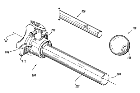
FIG 1 is a perspective view with parts separated of the surgical system in
accordance with the principles of the present disclosure, illustrating the
seal assembly,
cannula assembly and surgical instrument;
FIG 2 is an enlarged side cross-sectional view of the surgical system in
accordance with the present disclosure illustrating the seal assembly
positioned about the
surgical instrument and mounted within the cannula assembly;
FIG 3 is an enlarged side cross-sectional view similar to the view of FIG
2 illustrating an alternate embodiment of the seal assembly; and
FIG 4 is an enlarged side cross-sectional view of the surgical system in
accordance with the present disclosure illustrating angulation of the surgical
instrument
and corresponding movement of the seal assembly;
FIG 5 is an enlarged side cross-sectional view of an alternate embodiment
of a surgical system in accordance with the present disclosure illustrating
the seal
= assembly prior to mounting within the cannula assembly; and
FIG 6 is an enlarged side cross-sectional view in accordance with the
alternate embodiment of FIG 5 illustrating the seal assembly mounted within
the resilient
coupling of the cannula assembly.
6
CA 02540910 2013-05-08
=
DETAILED DESCRIPTION OF THE PREFERRED EMBODIMENTS
The surgical system of the present disclosure provides a substantial seal
between a body cavity of a patient and the outside atmosphere during an
endoscopic or
laparoscopic surgical procedure. The surgical system contemplates the
introduction and
manipulation of endoscopic or laparoscopic instrumentation, and maintains a
fluid tight
interface about the instrumentation to preserve the atmospheric integrity of a
surgical
procedure from gas and/or fluid leakage. Examples of instrumentation include
clip
appliers, graspers, dissectors, retractors, staplers, laser probes,
photographic devices,
endoscopes and laparoscopes, tubes, and the like. Several of these instruments
are
disclosed in commonly assigned U.S. Patent Nos. 6,716,232, 6,450,391,
6,231,565,
6,152,872, 5,938,668, 5,913,870 and 5,860,987. Such instruments will be
collectively
referred to herein as "instruments or instrumentation". The surgical system of
the
present disclosure is well adapted to accommodate angular manipulation of the
surgical
instrument. This feature desirably minimizes the entry and exit of gases
and/or fluids
to/from the body cavity.
In the following description, as is traditional, the term "proximal" refers to
the portion of the instrument closest to the operator while the term "distal"
refers to the
portion of the instrument remote from the operator.
Referring now to the drawings, in which like reference numerals identify
7
CA 02540910 2006-03-23
identical or substantially similar parts throughout the several views, FIGS. 1-
2 illustrate
the surgical system 10 of the present disclosure. Surgical system 10 includes
seal
assembly 100, cannula assembly 200 and surgical instrument 300. Seal assembly
100 has
seal element 102 with an inner portion 104 and an outer portion 106. Inner
portion 104
forms a seal and defines aperture 108 which is adapted to receive surgical
instrument 300.
In one preferred embodiment, inner portion 104 is fabricated from a resilient
material
whereby portions of the inner portion 104 adjacent aperture 108 engage
surgical
instrument 300 in fluid tight relation. Suitable materials for inner portion
104 include
elastomeric materials such as, e.g., polyisoprene, silicone, rubber, urethane,
soft urethane
gel, silicon gel, etc. Preferably, the selected material has compressible
characteristics to
permit inner portion 104 to conform and form a substantial seal about the
outer surface of
the instrument 300 during manipulation about the operative site. The inner
portion 104
and/or outer portion 106 may comprise a compressible foam. It is further
envisioned that
inner portion 104, outer portion 106 or both, may be a bladder or balloon
filled with
fluids such as water, saline, gel, etc. . . .
Outer portion 106 may comprise an elastomeric material or, in one
embodiment, include a magnetic material or a ferromagnetic metal. In another
embodiment, outer portion 106 may be coated with a magnetic coating or a
coating of a
ferromagnetic material. The use of magnetic material and/or ferromagnetic
material
facilitates the establishment of a magnetic coupling to assist in removably
retaining seal
assembly 100 within cannula assembly 200. The magnetic coupling will be
discussed in
greater detail hereinbelow. It is also envisioned that outer portion 106 may
be fabricated
8
CA 02540910 2006-03-23
from an elastomeric material and be monolithically formed with inner portion
104. Inner
portion 104 and outer portion 106 may be fabricated from the same or different
material.
In certain embodiments, inner portion 104 is formed from an elastomeric
material
whereas outer portion 106 is formed from a relatively rigid polymeric
material.
In the embodiment shown in FIG 2, outer portion 106 is generally
spherical and has a cylindrical opening 103 in which inner portion 104 is
disposed. Inner
portion 104 has a generally cylindrical shape forming aperture 108 for receipt
of surgical
instrument 300. Outer portion 106 and inner portion 104 may have other shapes,
such as
elliptical, polygonal, partially truncated sphere or elliptoid, etc. The
spherical shape
facilitates removable coupling of seal assembly 100 with cannula assembly 200.
It is also
envisioned that seal element 102 may be a partial or truncated sphere as shown
in FIG 3.
With continued reference to FIGS. 1-2, outer portion 106 has an outer
surface 110 which is arcuate. As to be appreciated, the arcuate configuration
of outer
surface 110 of seal element 102 permits the seal element 102 to angulate
within cannula
assembly 200.
Seal assembly 100 is preferably mountable about surgical instrument 300
preferably, about elongated shaft 302 of the instrument, with the elongated
shaft 302
being received within aperture 108 of seal element 202. Such mounting is
preferably
performed prior to positioning seal assembly 100 within cannula assembly 200.
Once
seal assembly 100 and the instrument 300 are positioned within cannula
assembly 200,
9
CA 02540910 2006-03-23
seal element 102 forms a substantial fluid tight seal within the internal
structure of
cannula assembly 200 to prevent or substantially minimize the passage of
fluids through
the cannula assembly.
Referring still to FIGS. 1-2, cannula assembly 200 of the surgical system
will be described. Cannula assembly 200 is intended to access a body cavity
and
permit introduction of instruments required to perform the desired surgical
procedure at a
remote tissue site. Cannula assembly 200 is particularly adapted for use in
laparoscopic
surgery where the peritoneal cavity is insufflated with a suitable gas, e.g.,
CO2, to raise
the cavity wall from the internal organs therein. Cannula assembly 200 is
typically used
with a trocar obturator (not shown) which is a sharp pointed instrument
positionable
within the passageway of the cannula assembly 200. The trocar obturator is
utilized to
penetrate the abdominal wall and is then subsequently removed from the cannula
assembly 200 to permit introduction of the surgical instrumentation utilized
to perform
the procedure. In the alternative, a blunt obturator may be used, such as, for
example, in
a Hasson technique. Semi blunt or dilating obturators may also be used to gain
access to
the abdominal cavity.
Cannula assembly 200 includes cannula sleeve 202 and cannula housing
204 mounted to a proximal end of the sleeve 202. Cannula sleeve 202 defines a
longitudinal axis "a" extending along the length of sleeve 202. Sleeve 202
further
defines an internal longitudinal passage 206 dimensioned to permit passage of
surgical
instrumentation. Sleeve 202 may be formed of stainless steel or other rigid
materials,
CA 02540910 2006-03-23
including polymeric materials that are medical grade material, such as
surgical steel,
titanium, polycarbonate, etc. Sleeve 202 may be clear or opaque. The diameter
of
sleeve 202 may vary, but, typically ranges from 10 to 15 mm for use with the
seal
assembly 100 of the present disclosure.
In one preferred embodiment, cannula housing 204 includes two
components, specifically, main housing 208 which is attached to the proximal
end of
cannula sleeve 202 and seal housing 210. Seal housing 210 may be connectable
to main
housing 208 through a bayonet coupling, a snap fit coupling, ultrasonic
welding or any
other means envisioned by one skilled in the art including, e.g., adhesive
means.
Alternatively, seal housing 210 and main housing 208 may be formed integrally
with one
another. Main housing 208 further includes diametrically opposed housing grips
212
(FIG 1) dimensioned and arranged for gripping engagement by the fingers of the
user.
Although shown and described as two components, cannula housing 204 may be a
single
component and attached to cannula sleeve 202 by any of the aforementioned
means or
may incorporate multiple components.
With reference to FIG 3, in conjunction with FIGS. 1-2, main housing 208
further includes duck bill or zero closure valve 214 which tapers distally and
inwardly to
a sealed configuration as shown. Valve 214 opens to permit passage of the
surgical
= instrument 300 and closes in the absence of the instrumentation and/or in
response to the
pressurized gases communicating from the insufflated body cavity. Other zero
closure
valves are also contemplated including single or multiple slit valve
arrangements, trumpet
11
CA 02540910 2006-03-23
valves, flapper valves, etc. Valve 214 may be secured within main housing 208
by any
conventional means. In one embodiment, main housing 208 includes internal
circumferential recess 216 which receives the outer peripheral flange 218 of
valve 214. A
valve mount 220 may be positioned to secure the flange area of valve 214
within main
housing 208.
Seal housing 210 has a substantially cylindrical configuration as shown.
Seal housing 210 includes seal mount 222 disposed within the interior of the
seal housing
210 concentrically arranged about longitudinal axis "a". Seal mount 222 is
adapted to
support seal assembly 100 in the assembled condition of the components. Seal
mount
222 has arcuate support surface 224 defining a concavity as shown. Support
surface 224
engages outer surface 110 of seal element 102. In a preferred embodiment, the
configuration of arcuate support surface 224 corresponds to the configuration
of outer
surface 110 of seal element 102. For example, in one preferred embodiment, the
radius
of curvature of each of arcuate support surface 224 and outer surface 110 of
seal element
102 are substantially equivalent. In this regard, seal element 102 is free to
swivel or
angulate relative to seal mount 220. The term "angulate" is to be interpreted
to include at
least two types of movement, namely, rotation of seal element 102 about
longitudinal axis
"b" and pivotal movement of the seal element 102 about a pivot axis "p". FIG 4
illustrates angulation of seal element 102 during manipulation of surgical
instrument 300.
Seal mount 222 preferably is formed of a rigid material such as a metal or
polymeric material. Seal mount 222 may have a lubricious coating to facilitate
12
CA 02540910 2006-03-23
angulation of seal element 102. Similarly, outer surface 110 of seal element
102 may
have a lubricious coating. Alternatively, seal mount 222 may have an
elastomeric layer
defining arcuate support surface 224. Irregardless of the materials utilized,
positioning of
seal element 102 within seal mount 222 establishes a fluid-tight relation
between the seal
element 102 and support surface 224, which substantially minimizes passage of
gases
through cannula assembly 200 during use in a laparoscopic procedure. In one
preferred
embodiment, seal mount 222 includes a magnetic material or a ferromagnetic
material
and cooperates with corresponding magnetic or ferromagnetic material at outer
surface
110 of seal element 102. In this manner, positioning of seal element 102
within seal
mount 222 establishes a magnetic coupling which functions to retain the seal
element 102
within the seal mount 222. Preferably, the strength of the magnetic coupling
is
selectively controlled to permit seal assembly 100 to angulate within seal
mount 222
while maintaining the mounted condition of the seal assembly 100 relative to
cannula
assembly 200. The strength of the magnetic coupling should be selected to
allow
convenient removal of seal assembly 100 from seal mount 222 while retaining
the seal
assembly 100 during manipulation. In any of the above, lubricating coatings
may be used
to further seal the cannula seal housing 210 and facilitate manipulation of
instrument.
Surgical system 10 may be part of a surgical kit incorporating at least one
seal assembly 100, corresponding cannula assembly 200 and/or surgical
instrument 300.
For example, the kit could be packaged incorporating a seal assembly 100 and
corresponding cannula assembly 200. A plurality of seal assemblies 100 of
different sizes
(e.g., seal apertures with different diameters) for various instrumentation
could be
13
CA 02540910 2006-03-23
incorporated in the kit. Alternatively, the kit could include surgical
instrument 300 and
seal assembly 100 with the seal assembly 200 mounted about the surgical
instrument 300,
either through a permanent or detachable connection.
Referring now to FIGS. 5-6, there is illustrated another embodiment of the
present disclosure. This embodiment of surgical system incorporates seal
assembly 100
as described hereinabove. Cannula housing 204 includes resilient coupling 226
incorporating first and second resilient legs 228 which receive and mount seal
assembly
100 within cannula 200. Resilient legs 228 are adapted to flex or pivot
outwardly in the
direction of directional arrows "p" to receive seal assembly 100 and then
return under the
influence of their natural resiliency to retain seal assembly 100 within
resilient coupling
226. Resilient legs 228 each include a central arcuate portion 230 defining an
arcuate
configuration approximating the general arcuate shape of the outer surface 110
of seal
element 102. In the mounted condition of seal assembly 100, the seal assembly
100 may
angulate within cannula assembly 200 in the manner described in connection
with the
embodiment of FIGS. 1-4. The contacting surfaces of resilient legs 228 and
seal element
102 may incorporate lubricious coatings to facilitate rotational and pivotal
movement of
the seal assembly 100. Resilient legs 228 may comprise a polymeric material or
resilient
metal such as spring steel. In other regards, seal assembly 100 is utilized in
the
aforedescribed manner.
While the invention has been particularly shown, and described with
reference to the preferred embodiments, it will be understood by those skilled
in the art
14
CA 02540910 2013-05-08
that various modifications and changes in form and detail may be made therein.
The
scope of the claims should not be limited by the preferred embodiments set
forth
herein, but should be given the broadest interpretation consistent with the
description as
a whole.