Note: Descriptions are shown in the official language in which they were submitted.
CA 02541073 2013-05-02
OPTICAL OBTURATOR
BACKGROUND
1. Technical Field
The present disclosure relates to an apparatus for penetrating body tissue
during
minimally invasive surgical procedures, such as endoscopic or laparoscopic
procedures. More
particularly, the present disclosure relates to an access assembly having a
transparent window for
providing visual observation during penetration of the peritoneum or other
body tissue.
2. Background of the Related Art
Minimally invasive surgical procedures including endoscopic and laparoscopic
procedures permit surgery to be performed on organs, tissue and vessels far
removed from an
opening within the tissue. Laparoscopic and endoscopic procedures are
performed in the interior
of the abdomen through a small incision such as, for example, a narrow
endoscopic tube or
cannula inserted through a small entrance incision in the skin. Typically,
after the abdominal
cavity is insufflated, a trocar is used to puncture the cavity wall, i.e., the
peritoneal lining, to
create a pathway to the underlying surgical site. Generally, the trocar
includes a stylet or
obturator having a sharp tip for penetrating the body cavity, which is
positioned coaxially within
an outer cannula. The obturator is removed, leaving the outer cannula in place
for reception of
instrumentation utilized to perform the surgical procedure. An example of a
known trocar is
described in commonly assigned U.S. Patent No. 6,319,266 to Stellon, which
issued November
21, 2001. However, with known trocars, advancement of the obturator
through tissue is typically performed blind,
CA 02541073 2006-03-27
i.e., without visualization of the tissue being entered. Obturators allowing
visualization include
U.S . Patent No. 5,334,150, 5,431,151 and 5,441,041.
Accordingly, the present disclosure provides an optical access assembly which
permits direct visualization of body tissue during penetration of the body
cavity. Moreover, the
optical access assembly of the present disclosure provides an improved
structure for direct
visualization of the body tissue being penetrated and serves as a conduit for
subsequent
introduction of surgical instrumentation required for performance of the
surgical procedure.
SUMMARY
In one preferred embodiment, an optical obturator apparatus includes an
obturator
sleeve defining a longitudinal axis and having a longitudinal bore for
receiving surgical
instrumentation and a transparent window mounted to the obturator sleeve and
being
dimensioned and configured to pass through tissue. The transparent window is
mounted for
movement between a first position in general alignment with the longitudinal
axis of the
obturator sleeve and a second position radially displaced from the
longitudinal axis to thereby
expose the longitudinal bore of the obturator sleeve to permit passage of the
surgical
instrumentation. The transparent window may include a cutting blade, or
alternatively two
cutting blades, adapted to penetrate tissue.
A control member is connected to the transparent window and at least partially
extends along the obturator sleeve. The control member is actuable to move the
transparent
window between the first position and the second position. The control member
is adapted to
2
CA 02541073 2006-03-27
rotate about an axis of rotation to cause movement of the transparent window
between the first
position and the second position. In this regard, the transparent window is
adapted for pivotal
movement about the axis of rotation to move between the first position and the
second position
thereof. The control member may be adapted to move in a longitudinal direction
from a normal
position to an extended position to displace the transparent window relative
to the obturator
sleeve.
An anti-rotation member may be associated with the transparent window to
prevent pivotal movement of the transparent window when the transparent window
is in the
normal position thereof. The anti-rotational member includes a key extending
from one of the
transparent window and the obturator sleeve, the key receivable within a keyed
port defined in
the other of the transparent window and the obturator sleeve. The key is
removed from the
keyed port upon movement of the control member to the extended position.
A manually manipulative member may be operatively connected to the control
member. The manually manipulative member is movable to move the control
member.
In another preferred embodiment, a surgical optical viewing system includes an
optical obturator having an obturator sleeve defining a longitudinal axis and
a longitudinal bore
for reception of surgical instrtunentation,. The optical obturator includes a
transparent window
for permitting passage of light into the obturator sleeve. The transparent
window has at least two
separable window sections. The at least two separable window sections are
adapted for radial
displacing movement to expose the longitudinal bore and to permit passage of
the surgical
3
CA 02541073 2006-03-27
instrumentation used for performing a surgical procedure. The transparent
window may define
a tapered configuration and at least one cutting blade adapted to penetrate
tissue.
The optical viewing system may further include a surgical instrument
positionable
within the longitudinal bore of the obturator sleeve. The at least two
separable sections of the
transparent window are adapted for radially displacing movement in response to
longitudinal
movement of the surgical instrument relative to the obturator sleeve. In this
regard, the surgical
instrument is engageable with interior surfaces of the at least two separable
sections of the
transparent window upon relative longitudinal movement of the surgical
instrument and the
obturator sleeve to radially displace the at least two separable sections.
BRIEF DESCRIPTION OF THE DRAWINGS
Preferred embodiments of the present disclosure are described hereinbelow with
references to the drawings, wherein:
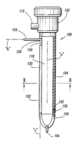
FIG. 1 is a side view in partial cross-section of one embodiment of an optical
access assembly constructed in accordance with the present disclosure;
FIG. 2 is an enlarged side cross-sectional view of the housing of the access
assembly in accordance with the embodiment of FIG. 1 illustrating the sealing
system for
forming a fluid tight seal about a surgical instrument;
FIG. 3 is a cross-sectional view of the obturator sleeve of the optical access
assembly in accordance with the embodiment of FIGS. 1-2 and taken along
section lines 3-3 of
FIG. 1;
4
CA 02541073 2006-03-27
FIG. 4 is a view illustrating a bevel gear arrangement associated with the
sleeve
of the optical access assembly in accordance with the embodiment of FIGS. 1-3;
FIG. 5 is a an axial view illustrating the transparent window of the optical
access
assembly in accordance with the embodiment of FIGS. 1-4;
FIG. 6 is view of an alternative embodiment of the transparent window of the
optical access assembly;
FIG. 7 is a side view in partial cross-section of the optical access assembly
in
accordance with the embodiment of FIGS. 1-5, illustrating movement of the
transparent window
from a first positioned aligned with the sleeve and a second position
displaced from the sleeve;
FIG. 8 is an axial view further illustrating movement of the transparent
window
from the first position to the second position in accordance with the
embodiment of FIGS. 1-5
and 7;
FIG. 9 is a view similar to the view of FIG. 7 illustrating advancement of a
surgical instrument through the sleeve and beyond the transparent window for
performing a
surgical procedure;
FIG. 10 is a side view in partial cross-section of an alternative embodiment
of an
optical access assembly of the present disclosure;
FIG. 11 is an axial view illustrating the transparent window in accordance
with
the embodiment of FIG. 10;
FIG. 12 is a view similar to the view of FIG. 10 illustrating radial movement
of
the window segments of the transparent window to open the sleeve of the
optical access
assembly in accordance with the embodiment of FIGS. 10-11;
CA 02541073 2006-03-27
FIG. 13 is axial view further illustrating the radial movement of the window
segment of the transparent window; and
FIG. 14 is a side view in partial cross-section of another embodiment of an
optical
access assembly of the present disclosure.
DETAIL DESCRIPTION OF PREFERRED EMBODIMENTS
Referring now in detail to the drawing figures, in which, like reference
numerals
identify similar or identical elements, there is illustrated in FIG. 1, an
optical access assembly
constructed in accordance with a preferred embodiment of the present
disclosure, and designated
generally by reference numeral 100. Optical access assembly 100 contemplates
the direct
visualization of body tissue during penetration of the peritoneal cavity or
other tissue portions.
In addition, optical access assembly 100 facilitates the introduction of
various types of surgical
instruments such as, for example, an endoscopic clip applier, grasper,
dissector, retractor, stapler,
photographic device, tube, and the like. Optical access assembly 100 is
dimensioned to pass
through body tissue and may incorporate structure to cut, puncture, or pierce
the body tissue.
Generally, optical access assembly 100 includes housing 102, sleeve 104
secured
to housing 102 and extending distally therefrom and window 106 operatively
connected to the
distal end of' sleeve 104. Housing 102 may incorporate several components
connected to each
= other by conventional means or may be a single component. As best
depicted in FIGS. 1-2,
housing 102 is advantageously dimensioned to be gasped by the surgeon. Housing
102 includes
an internal sealing system to receive a surgical instrument in substantial
sealed relation therewith
while also providing a substantial seal between the body cavity and the
outside atmosphere both
6
CA 02541073 2013-05-02
during, and subsequent to, insertion of the surgical instrument through sleeve
104.
One exemplative sealing system suitable for use in optical obturator assembly
100
is shown in FIG. 2. This sealing system is disclosed in commonly assigned
U.S. Published Application No. 2004/0066008 to Smith. The
sealing system includes instrument seal 108 and zero-closure seal 110.
Instrument seal 108 is
formed of a resilient material and has an aperture 112 for sealed reception of
a surgical
instrument. A fabric layer 114 is juxtaposed relative to the resilient
material and may be
disposed on either, or both of, the proximally facing surface or distally
facing surface of
instrument seal 108. The preferred fabric includes a SPANDEXTM material
containing 20%
LYCRA from Milliken. Zero closure valve 110 is preferably a duck bill valve
which opens to
permit passage of the surgical instrument. Duck bill valve 110 desirably
closes in the absence of
the surgical instrument and/or in response to the pressure of the insufflation
gases. Housing 102
further includes insufflation connector or port 116 (FIG. 1). Insufflation
connector 116 is
adapted for connection to a supply of insufflation gases for introduction
within the peritoneal
cavity as is conventional in the art. Further details of the sealing system
may be ascertained by
reference to the Smith '008 publication.
With reference to FIGS. 1 and 3, sleeve 104 defines longitudinal axis "x" and
has
longitudinal bore 118 extending the length of the sleeve 104. Longitudinal
bore 118 permits the
introduction of a surgical instrument utilized in the surgical procedure.
Sleeve 104 preferably
has a diameter of between about 4 millimeters to about 14 millimeters. Sleeve
104 may be
constructed of a medical grade metal including stainless steel or titanium or
a suitable
biocompatible polymeric material. Sleeve 104 further includes control member
120 (shown
7
CA 02541073 2006-03-27
partially in phantom in FIG. 1) which extends through passage 122 defined in
the sleeve 104.
Control member 120 serves to mount transparent window 106 and also moves the
transparent
window 106 between a first position aligned with longitudinal axis and a
second position
displaced from the longitudinal axis "x". In this regard, control member 120
is adapted to rotate
within passage 122 (i.e., about an axis of rotation "c" which is in general
parallel to axis "x" of
obturator sleeve 104) and is also adapted for limited longitudinal movement
within passage 122.
A manual manipulative handle 124 extends radially outwardly from the proximal
end of control member 120. Handle 124 is advantageously dimensioned for
gripping
engagement by the user and is actuable to effectuate rotational and/or
longitudinal movement of
control member 120 and thus corresponding movement of window 106. More
specifically,
handle 124 is mechanically connected to control member 120 in a manner whereby
rotational
movement of the handle 124 about handle axis "h" causes corresponding
rotational movement of
the control member 120 about axis "c". Any means for transferring this
rotational motion are
envisioned including, e.g., the bevel gear arrangement depicted in FIG. 4. In
this regard, handle
124 may be integrally formed with first gear 126 and control member 120 may be
integrally
formed with second gear 128. First and second gears 126, 128 cooperate whereby
rotational
movement of handle 124 and first gear 126 about handle axis "h" causes
corresponding
rotational movement of control member 120 about control axis "c". The bevel
gear arrangement
may be appropriately miniaturized to reside within passage 122 of the wall of
obturator sleeve
104 or may be disposed within housing 102. In addition, handle 124 is adapted
to move in the
longitudinal direction within slot 130 of obturator sleeve 104 in response to
proximal and distal
applications of force by the surgeon.
8
CA 02541073 2006-03-27
Sleeve 104 further defines keyed notch 132 in its distal end. Keyed notch 132
serves to prevent rotation of window 106 during introduction within the body
tissue.
Referring now to FIGS. 1 and 5, window 106 will be discussed in detail. Window
106 permits visualization during penetration of the body tissue. Window 106
may comprise a
transparent or translucent polymeric material and be fabricated via known
injection molding
techniques. Alternatively, window 106 may comprise an optical glass. The term
"transparent" is
to be interpreted as having the ability to permit the passage of light with or
without clear imaging
capabilities. Moreover, the transparent material includes any material which
is not opaque. It is
also to be appreciated that only a portion of transparent window 106 needs to
be transparent.
Thus, a portion of, or the entire window 106, may be transparent or
translucent. Window 106
may have a unitary construction or be comprised of multiple parts.
Window 106 is generally tapered in configuration, e.g., bulbous,
hemispherical, or
pyramidal conically-shaped, to facilitate passage through body tissue. Window
106 may include
an image directing member (not shown) for directing optical images into
longitudinal bore 118
of sleeve 104 or back to an image apparatus. The image directing member may be
a lens, an
optical prism, an optical mirror, or like image directing medium.
As best depicted in FIG. 3, transparent window 106 preferably has at least one
cutting blade 134. Cutting blade 134 is preferably centered with respect to
the outer surface 136
of window 106, as shown. Thus during visualization, cutting blade 134 is seen
as a thin line
9
CA 02541073 2013-05-02
through the center, i.e. bisecting the viewing field so as not to
substantially obstruct viewing of
the body tissue. Cutting blade 134 may be an independent member secured to
outer surface 136
by conventional means including welding, cements, etc. Alternatively, cutting
blade 134 may be
integrally formed with window 106 during, e.g., in a molding process. In this
embodiment,
cutting blade 134 is made of a polymeric material and is integrally formed
with window 106.
form. In one embodiment, cutting blade 134 includes a single cutting blade.
Alternatively, two
intersecting cutting blades 134 arranged in an X pattern, may be provided as
shown in FIG. 6.
Other arrangements of cutting blade 134 are envisioned, such as, for example,
arrangements of
three, four, etc., of the cutting blades 134. One or more cutting blades 134
may be disposed
along lateral sides of window 106.
Window 106 further includes anti-rotation key 136. Anti rotation key 136
resides
within keyed notch 134 of sleeve 104 to prevent rotation of the sleeve 104
during introduction of
window 106.
In operation, the peritoneal cavity is insufflated to raise the cavity wall to
provide
greater access to tissue and organs therewithin. An endoscope 200 is inserted
into optical access
assembly 100, i.e., through housing 102 and into longitudinal bore 118, as
shown in FIG. 2. One
suitable endoscope for use with optical access assembly 100 is disclosed in
commonly assigned
U.S. Patent No. 5,718,664 to Peck etal. Instrument seal 108 of housing 102
forms
a fluid tight seal about the endoscope 200. As appreciated, endoscope 200 is
advanced within sleeve 104 until the distal end of the endoscope 200 is
adjacent
window 106. In this position, the distal lens element of the endoscope
CA 02541073 2006-03-27
200 is capable of viewing the tissue being entered. Endoscope 200 may be
secured relative to
optical obturator assembly 100 with a locking system (not shown), at the
proximal end of the
optical access assembly 100, at some location along sleeve 104, or at the
distal end of sleeve 104.
For example, the locking mechanism may comprise a cam mechanism, or a ledge at
the distal
end of sleeve 104.
The procedure is continued by positioning window 106 against the body tissue
"t"
and advancing the assembly 100 to permit cutting blade 134 to penetrate the
tissue. A skin
incision may be made before pressing window 106 against the tissue, if
desired. During
penetration of the body tissue, the surgeon observes the underlying tissue
through the endoscope
200 to ensure there is no undesired contact with organs, tissue, etc. lying
beneath the peritoneal
lining. In instances where a video system is utilized, the surgeon simply
observes the penetration
of body tissue "t" via any known video monitor. Once the surgeon penetrates
the body tissue "t"
as observed through the endoscope 200, the surgeon discontinues the
application of force. For
example, in penetration of the abdominal wall, the surgeon can observe the
peritoneum and
penetration thereof.
After penetration into the underlying body cavity, handle 124 is moved in the
distal direction within slot 130 of obturator sleeve 104 to move control
member 120 from its
normal operative position depicted in FIG. 1 to the extended position depicted
in FIG. 7. During
this distal movement, anti-rotation key 136 of transparent window 106 clears
keyed notch 134
within obturator sleeve 104. Once key 136 is cleared from its containment
within keyed notch
134, handle 124 is rotated about handle axis "h" (FIG. 4) which causes
corresponding rotation of
11
CA 02541073 2006-03-27
control member 126. The rotational movement of control member 120 causes
transparent
window 106 to rotate around axis of rotation "c" to the radially displaced
position depicted in
FIGS. 7 and 8. In this position, longitudinal bore 118 of sleeve 104 is
exposed. Endoscope 200
may then removed from longitudinal bore 120 for insertion of other desired
surgical instruments
300 to carry out the desired procedure as shown in FIG. 9. Although FIG. 7
shows an endoscope
200 with an eye piece, the endoscope 200 may be additionally or alternatively
connected to an
imaging system, which may include a computer.
With reference to FIGS. 10-13, an alternative embodiment of an optical
obturator
assembly of the present disclosure is illustrated. In FIG. 10, optical access
assembly 400 is
shown with a cannula assembly 500 at least partially positioned therein and a
conventional
endoscope 200 introduced within the cannula assembly 500. Optical access
assembly 400
generally includes handle 402 and sleeve 404 extending distally from the
handle 402. Handle
402 and sleeve 404 may be separate components or integrally formed during
manufacture.
Adjacent the distal end of sleeve 404 is window 406. Window 406 is transparent
or translucent
as discussed hereinabove and preferably is integrally formed with sleeve 404.
Window 406
includes a plurality of individual separable window sections 408 (FIG. 12)
which are capable of
radial displacement to permit passage of a surgical instrument. Desirably,
window 406 and/or
the entire sleeve 404 is made from a relatively flexible material. In the
preferred embodiment,
four window segments 408 are provided with each of the segments 408 separated
from adjacent
segment 408 by respective slits 410. Window 406 further includes a pair of
intersecting cutting
blades 412. Cutting blades 412 function in penetrating or piercing body
tissue.
12
CA 02541073 2006-03-27
Cannula assembly 500 may be similar to any conventional cannula assembly
adapted for use in laparoscopic surgery. Carmula assembly 500 includes cannula
housing 502
and cannula 504 extending from the cannula housing 502. An internal seal
assembly may be
mounted within carmula housing 502 for sealed reception of a surgical
instrument such as an
endoscope. One suitable seal system is disclosed hereinabove in connection
with the discussion
of FIG. 2 any suitable seal system for cannula assemblies may be used.
In operation, cannula assembly 500 is positioned within optical access
assembly
400 followed by insertion of an endoscope 200 within the cannula assembly 500.
Endoscope
200 is positioned within access assembly 400 such that distal lens element 202
of endoscope 200
does not extend beyond the distal end of access assembly 400 as shown in FIG.
10. Thereafter,
the surgeon penetrates the body tissue "t" while observing the penetration
through the eyepiece
of the endoscope 200 (or while observing the same on an imaging and/or
computer screen), as
described in detail hereinabove. Endoscope 200 may then be removed if desired.
Subsequent to
penetration of the body cavity, the surgeon engages handle 402 to move sleeve
404 in the
proximal direction as depicted in FIG. 11. Proximal movement of obturator
sleeve 404 causes
the distal end of cannula 504 to engage internal surfaces 414 of window
sections 408.
Accordingly, window sections 408 are biased outwardly to the arrangement shown
in FIG. 13.
In this position, window sections 408 are radially displaced from longitudinal
axis "X" to
thereby expose cannula 504, thus permitting passage of surgical
instrumentation through the
cannula 504 and into the underlying body cavity.
13
CA 02541073 2006-03-27
With reference to FIG. 14, another embodiment of an optical access assembly of
the present disclosure is illustrated and designated generally as optical
access assembly 600.
Access assembly 600 generally includes handle 602 and sleeve 604 defining a
longitudinal axis
"X". A window 606 having cutting blade 608 is formed at the distal end of
sleeve 604.
Desirably, the window 606 is transparent or translucent, as discussed above in
connection with
FIGS. 1,5 and 10. Endoscope 200 is positioned within sleeve 604 to provide for
observation of
the body tissue being penetrated. Endoscope 200 includes an endoscopic shaft
202 which is
frictionally engaged by the internal surfaces of sleeve 604, i.e., sleeve 604
may be dimensioned
to form a friction fit with the endoscopic shaft 202 to thereby retain
endoscope in a desired
position relative to sleeve 604 with the distal lens component adjacent window
606.
Alternatively, a locking system, as discussed above in connection with FIG. 2,
may be used. In
use of this embodiment, endoscope 200 in positioned within access assembly 600
and the desired
frictional relationship is established between endoscopic shaft 202 and access
sleeve 604. The
system is advanced through the tissue with visualization provided by endoscope
200 through
window 206.
In further embodiments, the optical access assembly is as discussed above in
connection with Figs. 1-5 and 7-9, except that the window has window sections
that are arranged
as jaws pivotally mounted on the distal end of the sleeve. An elongated member
extending
proximally, along the sleeve, cooperates with a handle or other structure for
actuating the jaws.
During actuation, the jaws move from a closed position to an open position.
The jaws may or
may not include blades. The jaws, when in a closed position, may have any
shape, such as
pyramidal, conical, dolphin-nosed, hemispherical, etc. The pivotable jaws are
translucent or
14
CA 02541073 2006-03-27
transparent, as discussed above, and the user of the optical access assembly
views tissue before,
during and after advancement of the optical access assembly. The jaws may be
actuated while
the assembly is advanced through tissue, or after the tissue has been
penetrated and the body
cavity accessed.
In further embodiments, the optical access assembly is as discussed above in
connection with Figs. 1-5 and 7-9, except that the window comprises a closed,
pyramidal,
conical, dolphin-nosed, and/or preferably hemispherical shape. An elongated
member extending
proximally, along the sleeve, cooperates with a handle or other structure for
rotating the window
away from the distal end of the sleeve. The window rotates from a position
closing the distal end
of the sleeve, to a position that leaves the distal end of the sleeve open.
The window may or may
not include one or more blades. The user of the optical access assembly views
tissue before,
during and after advancement of the optical access assembly.
In each of the embodiments discussed above, the window may or may not include
cutting blades. The window may have any shape, such as pyramidal, conical,
dolphin-nosed,
hemispherical, etc. In each of the embodiments discussed above, the endoscope
may include an
eyepiece, and/or a connection to imaging equipment which may include a
computer. In each of
the embodiments discussed above, the movement of the window, jaws, or window
sections is
driven by an electric motor, hydraulic driver or manual drive and may be
controlled utilizing
electrical or mechanical methods.
CA 02541073 2013-05-02
It will be understood that various modifications can be made to the
preferred embodiments of the present invention herein disclosed. For example,
various
diameters for the obturator assembly, cannula assembly, as well as various
diameters
for the surgical instruments are contemplated. Also, various modifications may
be
made in the configuration of the parts. The scope of the claims should not be
limited
by the preferred embodiments set forth herein, but should be given the
broadest
interpretation consistent with the description as a whole.
16