Note: Descriptions are shown in the official language in which they were submitted.
CA 02541616 2006-03-28
WO 2005/032362 PCT/US2004/032293
1
SENSOR WITH INCREASED BIOCOMPATIBILITY
Background
The present invention relates to in vivo measurement. More specifically, 'the
present invention relates to sensing, and sensors for sensing, the
concentration of
particular substances in bodily fluids.
Measurement of the concentration of particular chemicals in bodily fluids is
useful for many types of medical diagnosis and treatment. Insulin-dependent
diabetic patients, for example, might measure the concentration of glucose in
their
blood multiple times per day. In vivo sensors have been developed and are
useful
in some situations for repeated or continuous testing, but are limited in
durability,
accuracy, ease of manufacture, and potential lifetime in use. There is thus a
need
for improved in vivo sensors and sensing techniques.
Some sensors have been developed that limit a reaction between analyte and
reagent by using membranes to control the flow of analyte therethrough. Using
these membranes adds to the design cost, manufacturing cost, and difficulty in
use
of such sensors. There is thus a further need for improved irc vivo sensors
and
sensing techniques.
CA 02541616 2006-03-28
WO 2005/032362 PCT/US2004/032293
2
Summary
It is thus an object of various embodiments of the present invention to
provide
sensors and techniques for sensing with improved characteristics of cost,
accuracy,
simplicity, durability, and irz vivo lifetime.
These objects and others are achieved in some embodiments of the present
invention by limiting the flow of the sample to or into the electrode using
the
geometric configuration of the sensor, for example, by providing a small
opening
into a three-dimensional cavity containing a conductive matrix including a
reagent.
One embodiment of the present invention is an electrode for use in vivo to
electrochemically detect or measure particular compounds. A first (substrate)
layer
has, at or adjacent to one end, a contact that is adapted for electrical
connection to
a meter. That layer and a second layer, the top surface of which is
substantially
adjacent the bottom surface of the first layer, together define a cavity with
an
opening through the top surface of the first layer, the opening being spaced
apart
from the first end of the first layer. A reagent fills at least 20 percent of
the cavity,
comprises a conductive matrix, and is electrically connected to the contact.
In
some variations of this embodiment, the first layer is polyimide, and in
others the
first layer has a thickness between about two mil and about ten mil, or about
50 ~,m
and about 250 ~.m.
In still other variations of this embodiment, the cavity has a particular
relationship with the opening through the top surface of the first layer. For
example, in some variations, each cross-section of the cavity taken parallel
to the
opening, but above the reagent-filled portion, has.an area no smaller than the
area
of the opening. In others, cross-sections of the cavity that intersect the
reagent-
filled portion also have an area at least as large as the opening. In
refinements of
this variation, the area of these cross-sections monotonically increases as
they axe
taken farther from the opening. In other variations of this embodiment, either
the
volume of the electrode or the volume of the containment cavity has a
particular
numeric relationship with the area of the opening.
Another form of the present invention is a strip for testing the concentration
or
presence of an analyte that includes a first layer with a top and bottom
surface, a
contact end and a sensing end, two contacts at or near the contact end, an
electrode
CA 02541616 2006-03-28
WO 2005/032362 PCT/US2004/032293
3
location at or near the sensing end, another electrode location near the first
electrode location, and a cavity within and defined by the main layer at the
first
electrode location. The cavity has an opening through the top surface, and is
at
least about twenty percent filled by a conductive matrix comprising a reagent.
A
conductor electrically connects the cavity and one of the contacts, while
another
conductor electrically 'connects the other electrode location and the second
contact:
A reference electrode is positioned at the second electrode location.
In variations of this embodiment, the cavity is substantially surrounded,
except
at the opening, by one or more materials that are non-permeable by the
analyte. In
a refinement of this embodiment, at least one of these materials is permeable
to a
cofactor of the reagent contained in the cavity. This may be for example
oxygen in
the case of a glucose sensor in which the reagent comprises glucose oxidase.
In
some of these variations, one or more of the cofactor-permeable materials form
a
second layer with one side disposed adjacent to the bottom surface of the main
layer.
In other variations in this embodiment, at least a portion of the cavity is
defined by a material that is cofactor-permeable. That material may be
adjacent to
the bottom surface of the first layer. In other variations, the conductive
matrix fills
at least about eighty percent of the cavity's volume.
In yet other variations on this embodiment, the conductor that reaches the
cavity extends into the cavity to at least partially define it. In others, the
conductor
is disposed along the top surface, while in yet others, the conductor is
disposed
along the bottom surface of the main layer. In still other variations, the
conductive
matrix substantially fills the cavity.
In another embodiment of the present invention, an electrochemical sensor
includes a substrate, a reference electrode on the substrate, and a working
electrode
that substantially fills a cavity that is substantially defined by the
substrate. The
working electrode includes a conducting matrix and an enzyme. In a variation
of
this embodiment, the conducting matrix comprises carbon particles, and in
others,
the enzyme is glucose oxidase. In still other variations of this embodiment,
the
working electrode also includes a catalyst, such as manganese dioxide. In yet
other
variations, the electrode also includes a binding agent, such as a polymer,
and may
CA 02541616 2006-03-28
WO 2005/032362 PCT/US2004/032293
4
further include a catalyst, such as manganese dioxide. The binder in some of
these
variants is a polymer.
In variations of this embodiment, the cavity has a substantially cylindrical
shape, while in others it has substantially the shape of a pyramidal frustum
or
conical frustum. In some of the latter variations, the cavity has a smaller
circular
surface that is open sufficiently to allow analyte to pass into the cavity,
and a larger
circular surface that is adjacent an oxygen-permeable material. In yet further
variations of this embodiment, one surface of the working electrode is open
such
that a sample can enter into the electrode without the sample passing through
a
layer that limits diffusion of the analyte.
CA 02541616 2006-03-28
WO 2005/032362 PCT/US2004/032293
Brief Description of the Drawings
FIG. 1 is a plan view of the substrate layer of a sensor according to one
embodiment of the present invention.
FIGS. 2A-2,G are cross-sectional views of the sensor shown in FIG. 1 at
5 various stages of fabrication, according to another embodiment of the
present
invention.
FIGS. 3A-3G are cross-sectional views of the sensor shown in FIG. 1 at
various stages of fabrication, according to another embodiment of the present
invention.
FIG. 4 is a perspective view of an end of a sensor strip according to one
embodiment of the present invention.
FIG. 5 is a perspective view of an alternative cavity configuration for use in
the sensor of FIG. 4.
FIG. 6 is a perspective view of another alternative cavity configuration for
use
in the sensor of FIG. 4.
FIG. 7 is a cross-sectional view of a sensor according to another embodiment
of the present invention.
FIG. ~ is a plan view of a sensor according to still another embodiment of the
present invention.
FIG. 9 is a cross-sectional view of a sensor according to the embodiment
shown in FIG. 8.
FIG. 10 is a cross-sectional view of a sensor according to yet another
embodiment of the present invention.
CA 02541616 2006-03-28
WO 2005/032362 PCT/US2004/032293
6
Description
For the purpose of promoting an understanding of the principles of the present
invention, reference will now be made to the embodiment illustrated in the
drawings and specific language will be used to describe the same. It will,
nevertheless, be understood that no limitation of the scope of the invention
is
thereby intended. Any alterations and further modifications of the described
or
illustrated embodiments, and any further applications of the principles of the
invention as illustrated therein, are contemplated as would normally occur to
one
skilled in the art to which the invention relates.
Various embodiments of the present invention provide an analyte sensor that
utilizes the geometry of the sensor to provide an advantageous control of the
disposition of analyte and interferants at the sensor's "active area," which
herein
refers (1) when a substantially planar electrode is used, to the substantially
planar
region in which analyte reaction and electrochemical detection take place, and
(2)
when a porous, conductive reagent matrix is used, to the substantially planar
area
of the opening connecting the volume containing the porous, conducting matrix
to
the volume of the bodily fluid. The sensor is implanted beneath the skin, and.
includes a portion that is in contact with the surrounding body fluid, which
contains the analyte to be measured. In general, the sensor includes a porous,
conductive matrix that has one surface in contact with the body fluid, and a
second
surface in contact with the surface of a conductive trace that communicates
back to
a meter operable to assess the analyte based on the electrical signal received
from
the sensor. The operational area of the porous reagent is significantly larger
than
the fluid-contacting surface or the surface of the conductive trace, thereby
providing greater surface area for reaction of analyte and the electrochemical
detection reaction, and for the capture of toxic byproducts of the measured
reaction
at the electrodes, especially compared to a more planar design.
In a particular embodiment, the volume occupied by the conductive matrix
generally increases in cross-sectional area as one proceeds away from the
fluid
contacting surface toward the conductive trace surface. In others having
partially
filled cavities, the cavity generally increases in cross-sectioiZal area as
one
proceeds away from the fluid-contacting opening down to the volume of reagent.
CA 02541616 2006-03-28
WO 2005/032362 PCT/US2004/032293
7
In still others, the cavity generally increases in cross-sectional area as one
proceeds
away from the fluid-contacting surface.
In some embodiments, a certain volume is opened through the substr ate, and
reagent is placed therein. A membrane over one opening to the cavity is
permeable
to the analyte, but not to certain interferants. Another membrane covers the
other
opening, and is non-permeable to the analyte. In variations of this
embodiment,
the second membrane is selectively permeable to exclude the analyte, but allow
passage of one or more cofactors (such as oxygen) in the fluid to pass into
the
reaction cavity.
Some embodiments of the present invention are useful for the subcutaneous
detection of a wide variety of analytes measurable by electrochemical means.
For
purposes of example, the discussion herein is provided with reference to a
glucose
sensor, and commensurate chemistries and other components are identified.
However, it will be appreciated by those skilled in the art that other
analytes may
be readily detected using the present invention, with corresponding changes in
the
chemistries and the like as are well known in the art.
Referring in particular to the figures, FIG. 1 shows the components of a
sensor
according to one embodiment of the present invention. Sensor strip 10 has head
portion 12 and body portion 14. Head portion 12 includes contacts 16, 18, and
20,
for electrical connection to a volt meter, a potentiostat, an ammeter, and/or
other
detection or display components. The contacts may be directly or indirectly
connected with such devices which operate to control the potential or current
in the
sensor, and to receive and evaluate the electrical signal from the sensing
portion of
the sensor, as is well known in the art of electrochemical biosensors.
Body portion 14, includes reference electrode 28, working electrode 30, and
counter electrode 29. Conductor trace 22 connects contact 16 to reference
electrode 28, conductor trace 24 connects contact 18 to working electrode 30,
and
conductor trace 26 connects contact 20 to counter electrode 29. As discussed
in
examples below, each of these structures is fabricated in or on substrate 32,
which
is preferably a flexible layer having a thickness between about two and about
ten
mil (between about 50 ~,m and about 250 ~,m) of a material, such as polyimide
or
polyester, that is non-permeable to the analyte(s) of interest. Traces 22, 24,
and 26
CA 02541616 2006-03-28
WO 2005/032362 PCT/US2004/032293
8
are preferably made of gold or carbon, but other conductive materials may also
be
used.
In one form of this embodiment, body portion 14 of sensor strip 10 is
approximately rectangular in shape, being about 25 mm long and 450 ~,m wide,
and is placed within a hollow fiber membrane (not shown) to enhance
biocompatibility while in use. Working electrode 30 is rectangular (at least
when
viewed from above, as in FIG. 1), and is about 100 ~,m wide and 325 ~.m long.
Working electrode 30 contains a reagent mixture suitable for the application.
In
one form of this embodiment, the reagent mixture comprises a conductive matrix
(of carbon particles), a catalyst (manganese dioxide), an enzyme (glucose
oxidase),
a polymeric binder, and a solvent for the polymeric binder. This reagent
mixture,
on removal of the solvent, forms a porous, conductive matrix that fills, or at
least
substantially fills, a cavity in substrate 32 to form electrode 30.
Fabrication of
these structures is discussed below. In these forms, the porous reagent matrix
exposes a great deal of reagent surface area for reaction, even though the
planar
portion of the electrode is quite small. The opening to the containment cavity
regulates diffusion by the analyte into and out of the cavity, which in some
embodiments provides improved control of variables in the reaction
measurement,
and a corresponding improvement in measurement accuracy.
When the sensor is in place, biological fluid enters the cavity containing the
working electrode 30, and the glucose in the fluid reacts with the enzyme,
changing the electrical impedance characteristics of the working electrode 30.
A
driver circuit is put in electrical communication with electrode 30 via
contact 18
and trace 24, and with reference electrode 28 and counter electrode 29 via
contacts
16 and 20, and traces 22 and 26, respectively. The electrical potential at one
or
more electrodes is controlled and the resulting currents) is/are analyzed (or
vice
versa) to determine the concentration of glucose in the fluid, as is known in
the art.
In various alternative embodiments, more or fewer electrodes are included on
sensor strip 10, as would be understood by those skilled in the art.
The fluid is in contact with a cavity that is sized a particular way to
achieve a
particular result. Some of these embodiments include a containment volume
("cavity" elsewhere herein) that is approximately cylindrical in shape. In
other
CA 02541616 2006-03-28
WO 2005/032362 PCT/US2004/032293
9
embodiments, one end of the containment volume may be substantially wider than
the other (such as a circular opening having a diameter that is twice the
diameter of
the circular opening at the other end), wherein analyte permeable membrane is
over the smaller opening, and a co-reactant-permeable membrane is over the
larger
opening, so that transfer of the analyte may be controlled on one side, but
sufficient co-reactant may be acquired from the fluid through the other side.
Within the cavity, reagent and in some cases a co-factor react with a
component of the biological fluid from the surrounding volume. Electrical
potential is created at the locus of this reaction, and must be carried to
measurement circuitry to measure the concentration of analyte in the sample.
In
these preferred embodiments, the cavity's volume is at least about 20%
(preferably
at least about 50%, more preferably at least about 80%, and most preferably
about
100%) filled with a porous, conductive matrix that presents reagent throughout
a
significant part of the cavity, and furthermore (because of the conductive
nature of
the matrix) carries the charge produced at the reaction locus to a conductive
trace
that extends into the cavity, preferably at its surrounding surface). The
conductive
trace extends to the surface of the substrate and on to a contact pad, which
comes
into electrical contact with a meter unit or other testing circuitry.
Turning to FIGS. 2A-2G, with continuing reference to certain structures in
FIG. 1, there is shown in somewhat diagrammatic form one method of fabricating
one kind of sensor according to the present invention. FIG. 2A illustrates
substrate
32, which may be a substance of more or less rigidity as would occur to one
skilled
in the art. For example, substrate 32 may be polyimide, a ceramic material, or
another material.
FIG. 2B shows that a layer 34 of conductive material has been deposited on
substrate 32. In various embodiments, conductive layer 34 is deposited by
sputtering, vapor deposition, or another method as would occur to one skilled
in
the art. Conductive layer 34 is then patterned, using lithographic or laser
ablation
techniques, for example, to define conductor traces 22, 24, and 26 on
substrate 32,
as, shown in FIG. 2C. In other embodiments, the conductor traces 22, 24, and
26
are printed or otherwise formed onto substrate 32 using a screen printing or
other
patterning technique.
CA 02541616 2006-03-28
WO 2005/032362 PCT/US2004/032293
FIG. 2D shows that a layer of relatively non-conducting material 36 has been
deposited over conductors 22, 24, and 26. Material 36 might, for example, be
PYRALUX or VACREL, each sold by E.I. DuPont de Nemours and Company
("DuPont" herein), or the like, as would occur to one skilled in the art.
5 FIG. 2E shows that recess 38 has been fabricated into layer 36. Recess 38 is
created, for example, by selective chemical etching, laser ablation, or other
techniques. Reagent 40 is then deposited over structure 31E, including recess
38,
to yield structure 31F as shown in FIG. 2F. Reagent 40 comprises a conductive
matrix such as carbon particles, a catalyst such as manganese dioxide, an
enzyme
10 such as glucose oxidase, and a polymeric binder. These components are
typically
dispersed in an organic solvent during this depositing step. The excess (above
material 36) is removed by squeegee, chemical-mechanical polishing (CMP), or
similar technique, to yield structure 31G, shown in FIG. 2G. The solvent
bearing
reagent 40 is then evaporated away by heat or vacuum to leave the reagent 40
substantially filling recess 38. In other embodiments, reagent 40 is directly
deposited into recess 38.
Another method of fabricating a sensor according to the present invention will
now be described in relation to FIGS. 3A-3G, with continuing reference to
certain
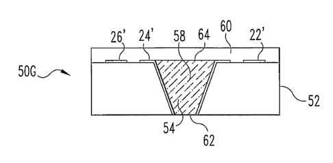
structures in FIG. 1. Device 50A comprises substrate 52, made of a material
that is
not permeable to blood. Cavity 54, shown in FIG. 3B, is formed in substrate 52
by
extraction to create device 50B. As shown in FIG. 3C, device 50C comprises the
device 50B, with the addition of a conductive layer 56 that extends into
recess 54
and along the top surface of device 50C.
Conductor layer 56 is patterned to form device 50D, shown in FIG. 3D.
Conductor traces 22', 24', and 26' correspond generally to conductors 22, 24,
and
26 in FIG. 1. Reagent composition 58 is deposited on top of device 50D, at
least
sufficient to fill recess 54, to form device 50E, shown in FIG. 3E.
The excess reagent 58 (that above the upper surface of substrate 52) is then
removed to yield device 50F as shown in FIG. 3F. This removal may, again, be
performed by squeegee, CMP, or other suitable process that would occur to one
skilled in the art. A layer of encapsulating material 60 is overlaid on device
50F to
form device 50G, as shown in FIG. 3G. In use, bodily fluid directly contacts
CA 02541616 2006-03-28
WO 2005/032362 PCT/US2004/032293
11
conductive reagent 58 at surface opening 62. Oxygen or another substance used
for the detection is transported through layer 60 and into reagent 58 through
surfacelopening 64.
In one preferred form of this embodiment, a glucose sensor, the substrate 52
is
polyimide, and the encapsulation layer 60 is silicone. The conductor layer 56
(and,
therefore, conductor traces 22', 24', and 26') is gold. The reagent 58
comprises
carbon particles in a porous conductive matrix that functions not only as an
immobilizing and stabilizing matrix for the enzyme, but also as an active
electrode
element. The conductive matrix in recess 54 contacts conductor trace 24',
which
forms a conductive path from the working electrode to the connector area of
the
sensor (such as contact 18 in FIG. 1) for connection to a meter or other
circuitry.
In other embodiments, the connectors are another metal, or are caxbon traces
printed or otherwise deposited on a surface of the substrate. In still other
embodiments, the conductor is deposited axound the circumference within recess
54, is deposited on one wall of recess 54, is deposited over the reagent 58,
or is
otherwise in contact with the electrode matrix. In various embodiments, the
reagent mixture (including the conductive matrix) fills at least about 20% of
recess
54, preferably at least about 80% of recess 54, and most preferably
substantially all
of recess 54. The remainder of recess 54 contains either air (in an unused
sensor),
fluid (in a sensor being used), or other material, as would occur to one
skilled in
the art. In yet other embodiments, recess 54 is at least half as deep as it is
wide at
the shortest distance across the opening 64, through which the sample enters
the
electrode; and preferably recess 54 is at least as deep as it is wide at the
shortest
distance across opening 64.
In the preferred embodiment of a glucose sensor, the catalyst in reagent 58 is
preferably manganese dioxide, which reduces the required potential for
hydrogen
peroxide oxidation on the carbon electrode. Other suitable materials for this
catalyst can be found in EP 0 603 154, which is hereby incorporated by
reference.
In other sensors for in vivo measurement, metallic electrodes of platinum or
palladium are used to detect H2O2. With such electrodes, the potential
difference
required for reasonably accurate measurement is about 600-800 mV vs. Ag/AgCI,
while with MnO~, as the catalyst, the required potential is reduced to 300-400
mV.
CA 02541616 2006-03-28
WO 2005/032362 PCT/US2004/032293
12
The designs of many embodiments of the invention enable the efficient
conversion of analyte throughout the volume in recess 54, which is efficiently
electrically connected to conductor 24'. In the exemplary embodiment described
above, the enzyme, glucose oxidase, is entrapped in the polymeric binder
matrix
and adsorbed onto the surface of the carbon particles. This solid-phase
adsorption
increases the stability of the enzyme and allows storage in undesiccated
environments, increasing the convenience of manufacturing and storing the
sensor.
The hydrophobic environment of the polymeric binder matrix is also thought to
increase the stability of the enzyme.
The reagent for use in a preferred embodiment of the present invention is
prepared by mixing the solvent containing a polymeric binder substance with
carbon particles as a pre-formulated screen printing ink mixture, with the
catalyst,
and any additional solvent required to produce a workable mixture. Once those
components are combined, other additives are sometimes included, such as one
or
more detergents or hydrophilic polymers to improve the wetting
characteristics, or
one or more fluorocarbon polymers to improve the oxygen transport properties
of
the reagent. The enzyme may also be included in the reagent to produce a one-
step
reagent. In another variation, only the catalyst is mixed with the ink
formulation,
and the enzyme and other additives are added to the cured porous electrode
reagent
later from an aqueous solution.
The containers may be filled with the reagent mixture by dispensing the-
reagent mixture from a syringe needle, or by placing an excessive amount of
reagent over and into the recesses, then removing the excess with a blade or
squeegee. Alternatively, the reagent may be screen-printed or otherwise
directly
deposited into the recesses. In some cases where the recesses are formed by
creating holes through the substrate, the reagent can be applied from the side
of the
sensor with the larger opening into the cavity (or either side, if the cavity
is
cylinder-shaped), the recesses being filled by capillary action through
opening 64,
shown in Fig. 3G. In each case, the reagent may be dried in an oven, or under
vacuum, or at room temperature, depending on the requirements of the polymeric
binder present in the reagent.
CA 02541616 2006-03-28
WO 2005/032362 PCT/US2004/032293
13
In further embodiments, the reagent itself may be coated with a polymeric
material to resist protein adsorption and to prevent loss of enzyme over the
use-
lifetime of the sensor. MPC, PELLETHANE, and a plasma-produced glyme
coating are examples of substances suitable for this purpose. Hydrophilic
polyurethane coatings such as those described in US Patent 5,322,063 and US
Patent 6,509,148 are also especially advantageous. In addition, the coating
material may be designed or selected to resist interference from compounds
such
as ascorbic acid, uric acid, and acetaminophen. Negatively charged coatings,
such
as NAFION (sold by DuPont) and PVC-malonate are particularly suitable for this
purpose. Alternatively, positively charged coatings, such as those discussed
in
EP 0 603 154, may be used. In the case of the sensor construction with holes
formed all the way through the substrate, the backside of the recess, which
would
normally not come into contact with the sample being tested, may be coated
with
an impermeable material or, preferably, with a material that is permeable to
any
cofactor required by the reagent but not water- or analyte-permeable. A
material
such as a silicone polymer (for example, SYLGARD 184 from the Dow Corning
Corporation) is suitable for this use when the reagent comprises an oxidase.
Alternatively or in addition, to improve the oxygen tolerance of the sensor
when the reagent comprises an oxidase, a material to improve the oxygen
transport
may be incorporated into the reagent itself. Fluorocarbon polymers such as
NAFION are suitable for this purpose.
The reference electrode 28 in various embodiments of the present invention
can be any solid state reference, as will be understood by those skilled in
the art.
One such reference electrode material is a silver-silver chloride (Ag/AgCl)
ink that
is applied to a patterned gold area in a similar fashion to the reagent
material
discussed above. The counter electrode 29 is prepared from a carbon paste, a
noble metal ink, a bare metal surface, or other material as would occur to one
skilled in the art.
Once fabricated, the sensor is cut from the substrate by any of various
methods
known to those skilled in the art. A preferred method is a wet-etch process
that
creates cuts around the periphery of the sensor and leaves smooth, rounded
edges.
The outline of the sensor is preferably created prior to the patterning of
electrodes
CA 02541616 2006-03-28
WO 2005/032362 PCT/US2004/032293
14
and reagent deposition. In other embodiments, the outline may be formed at the
same time as recesses for the electrodes are formed. Bridges are preferably
left to
retain the sensor in a fixed position relative to the substrate sheet to make
subsequent processing steps easier. After fabrication, the bridges may be cut
or
punched, and the sensor is removed from the sheet. The sensors may then be
inserted into hollow-fiber membranes to provide additional bio-compatibility
and
isolation of the sensor from cellular materials and large proteins that are
often
present in the subcutaneous environment.
In various other embodiments, recesses for the reagents are formed using
lithographic techniques. Cylindrical electrode locations may be fabricated by
laminating a photo-imageable coverlay such as PYRALUX or VACREL onto the
patterned substrate, then exposing and developing the coverlay to form a hole
(for
example, having a diameter between 100 ~,m and 1000 ~,m, and being about 10-
125 ~,m thick). Alternatively, the recesses may be etched into the polyimide
substrate by a wet-etch process, or drilled by a laser, or created by other
mechanical processes such as imprinting.
In still other embodiments, the recesses are filled with reagent mixture by
placing excess reagent over and into the recesses, then removing the excess
with a
blade or squeegee as described in connection with FIGS. 2A-2G and 3A-3G above.
In yet other embodiments, the reagent is dispensed or screen-printed into the
recesses. In further embodiments where the recesses axe formed in a polyimide
substrate, the reagent is applied from the opposite side of the substrate,
filling the
recesses by capillary action.
FIG. 4 highlights an alternative cavity configuration according to another
embodiment of the present invention. In this example embodiment, the cavity
has
the shape of a truncated cone, the top of which is a larger circle, and the
bottom of
which is a smaller circle. Reagent fills at least about 80°l0 of the
cavity. The
sample containing the analyte enters the cavity through the smaller circle. In
some
variations of this embodiment, the larger circle is adjacent to a layer that
is
permeable to any cofactors, such as oxygen, that may participate in the
reaction in
the cavity. The cross-sections of the cavity, taken parallel to the smaller
circle,
have monotonically increasing area as they get farther from the smaller
circle.
CA 02541616 2006-03-28
WO 2005/032362 PCT/US2004/032293
Elementary geometry indicates that, for a truncated, right, circular cone (a
"conical
frustum"), and given smaller circle radius ro, larger circle radius rl, and
height h,
the area of the smaller circle is A=~ro2, and the total volume of the cavity
is
V = 3 (ro + rorl + rl2 )
The ratio of the cavity volume to the area of the sample
_V__h 1+r+r~2
z
5 opening is thus '~ 3 ro ro It is noted that, if we define R to be the ratio
YllYp Of the larger (bottom) radius to the smaller (top) radius, then R> 1 and
the
_V _ h (1+R+Rz)> h
volume to entry-area ratio is A 3 . In some preferred
embodiments, h is at least about as long as the diameter 2ro of the smaller
circle, so
in such embodiments this V/A ratio is at least about twice the smaller (top)
radius
10 ro. In other embodiments, h is at least about twice as long as the diameter
2ro of
the smaller circle, so in such embodiments this V/A ratio is at least about
four
times the smaller (top) radius ro.
FIG. 5 shows an alternative cavity configuration according to yet another
embodiment of the present invention. In this embodiment, the cavity has the
shape
15 of a truncated pyramid, the top and bottom of which are substantially
square.
Again, the sample enters the cavity through the smaller square opening (at the
top).
This cavity, for example, is substantially full of the conductive reagent
matrix
discussed in the embodiments shown above. Again, cross-sections of the cavity,
taken parallel to the smaller square, have monotonically increasing area as
they get
farther from the smaller square opening. Given this truncated, right, square
pyramid (a "pyramidal frustum") with small square opening side length so,
large
square opening side length sl, and height h, the area of the small square is
A=soz,
and the volume of the cavity is V = 3 (so + sos, + si ) . The ratio of cavity
volume
to the area of the sample opening is thus V _ la 1+ sl + SZ Again, if R is
A 3 so so
defined to be the ratio the side length of the larger opening to the side
length of the
smaller opening (i.e., sllso), then A = 3 (1 + R + R 2 ) > h again. In some
preferred
CA 02541616 2006-03-28
WO 2005/032362 PCT/US2004/032293
16
embodiments, this ratio is at least about the same as the side length so of
the
smaller (top) square opening.
FIG. 6 shows another alternative cavity configuration according to still
another embodiment of the present invention. In this embodiment, the cavity is
cylindrical and at least about 20 percent full of conductive reagent matrix.
The
cross-section of the cylinder is substantially the same from one end of the
cavity to
the other. With a cylindrical cavity of radius r according to this embodiment,
the
area of the sample opening is, again, A=~Y2, and the total volume of the
cavity is
v =~''2h . The ratio of the cavity volume to the area of the sample opening is
thus
A = la . In some preferred embodiments, this ratio is at least about 2r, or at
least
about the diameter of the sample opening.
Subcutaneous sensors of the current art use membranes to cover the sensor
active surface that are directly in contact with the body fluid. These
membranes
serve the purpose of restricting the diffusion of analyte to the sensor active
surface
in order to improve the sensor measurement range or linearity. They also serve
to
hinder access to the sensor surface of material or substances from the
external fluid
that might impact the sensor performance, such as by fouling the sensor active
surface. These membranes generally become fouled with biological material with
time, and the diffusion of analyte through them becomes restricted. In this
circumstance, the sensitivity of the sensor changes, and the sensor must be
recalibrated, or it will deliver inaccurate results.
Other problems with membranes may also occur. For example, the
membranes may swell through absorption of the bodily fluid, increasing
permeability to the analyte, or the membranes may be degraded by contact with
the
bodily fluid. Components in the body, such as enzymes, or cellular activity,
such
as from macrophages, may increase permeability to the analyte. Any change in
the
permeability of the membrane of such a sensor leads to inaccuracy or the need
for
recalibration.
Current subcutaneous sensors are made to be resistant to the effects of
contact
with the in vivo environment by covering them with a membrane that reduces the
adhesion of protein or cellular material. These membranes are also frequently
CA 02541616 2006-03-28
WO 2005/032362 PCT/US2004/032293
17
formulated to limit the diffusion of the analyte through the membrane. This
diffusion limitation may be required to achieve sensitivity to that analyte
over the
required measurement range. These membranes cover the sensitive area of the
sensor and adhere tightly to the surface to fulfill both required functions.
Subcutaneous glucose sensors, for example, typically incorporate a membrane
to provide an interface to the tissue in which they are implanted. Such
membranes
typically allow the diffusion of glucose and other small molecules to the
sensor
surface, but prevent the passage of larger molecules such as proteins, and
intact
cells. The membranes may combine multiple functions, such as providing the
biological interface, encouraging vascularization, reducing diffusion of
glucose to
the sensor, enhancing oxygen delivery to the sensor, etc. However, these
membranes are subject to fouling, swelling, or degradation over the lifetime
of the
sensor, altering the rate at which glucose can diffuse to the sensor, causing
a
change in the effective sensitivity of the sensor and creating errors in the
measurement values, or the need for recalibration.
The foregoing issues have been addressed in a variety of ways. Membranes
that reduce and resist fouling to greater or lesser extents have been
developed and
applied. Measurement methods which are more independent of the membrane
permeability have been developed. The most widely pursued alternative approach
is the use of microdialysis or microperfusion to collect a liquid sample in
which the
analyte has equilibrated with the in vivo tissue, and to remove the sample to
a
sensor system for analysis. These methods remove the sensor from the
subcutaneous environment. Microperfusion has the advantages of microdialysis,
and claims improved resistance to membrane fouling through the use of large
holes
in the catheter, which cannot be blocked by protein adsorption.
However, membrane fouling and sensor drift are still significant issues with
subcutaneous glucose sensors that have improved membranes and materials.
Microdialysis methods have greatly increased complexity of the measurement
device, and suffer from time lags due to the requirement to move liquid within
the
system. This also yields a very long response time for the analytical system,
as the
fluid must be pumped to the remote sensor at a slow rate to ensure consistent
recovery of analyte from the tissue.
CA 02541616 2006-03-28
WO 2005/032362 PCT/US2004/032293
18
Some embodiments of the present invention provide a subcutaneous sensor
that does not exhibit significant changes in sensitivity, leading to erroneous
results
or requiring recalibration. The solution of the current invention maintains
the
advantages of the microdialysis solution, but avoids the increased complexity.
Various forms of the present invention provides a subcutaneous sensor and
associated systems and methods that provide distinct advantages over certain
prior
art approaches. In general, some embodiments of the present invention provide
a
sensor system that includes a biosensor and an encapsulating membrane that is
spaced from the biosensor to provide an internal volume of fluid in contact
with
the biosensor. The membrane allows for desired equilibrium between the
external
body fluid and the internal volume of fluid, and therefore allows for accurate
analyte reading by the biosensor. In various embodiments, the spacing is fixed
(using spacers between the biosensor and the membrane) or variable (such as
where the biosensor is not secured in a rigid spatial relationship with the
membrane). In some embodiments, the distance h between the membrane and the
active area might be defined as the distance between nearest points; an
average
distance from each point on the active surface, taken perpendicular to that
surface;
or the closest membrane point to the active area measured perpendicular to the
surface of the active area. The size of the internal volume is preferably
controlled
in relation to the active area of the sensor. In preferred embodiments, given
a
sensor active area of s, the internal volume is at least about s3~a/10, or
s3~2, or lOs3~z.
The sensor system is distinguished from at least some prior art in that a
separate membrane is included which is spaced from the biosensor, rather than
being located directly on the active area of the biosensor. This allows the
surface
area of the membrane to be much larger compared to the active area of the
biosensor. This further provides a reservoir of fluid, i.e., the internal
volume of
fluid, that is in fluid communication with both the active area of the
biosensor and,
through the membrane, the body fluid. Moreover, the interior volume is
characterized in that the diffusion coefficient of the analyte in the interior
volume
is about the same as, or greater than, the diffusion coefficient of the
analyte in the
membrane.
CA 02541616 2006-03-28
WO 2005/032362 PCT/US2004/032293
19
These forms of the invention, therefore, provide a sensor system in which the
active area is removed from the interfacial membrane, and has a much smaller
active area than the area of the interfacial membrane that contacts the tissue
in
which the sensor is implanted. Due to the large surface area of the membrane,
the
internal, equilibration volume maintains an analyte concentration that is very
nearly identical to that of the tissue in which it is immersed, even when
diffusion
of analyte across the membrane is hindered or reduced. The biosensor, on the
other hand, consumes small amounts of analyte due to its relatively small
contact
area with the equilibrium volume. Thus, the analyte concentration that the
sensor
measures remains very nearly identical to that in the surrounding tissue, even
in the
presence of hindered diffusion across the membrane surface. Further, the
relatively larger area of the interfacial membrane means that it will take
longer for
fouling to occur, as opposed to the situation where the membrane is comparably
sized to the active area of the biosensor. This yields a longer useful life
for the
sensor system.
It will be appreciated that the advantages of the present invention are
obtained
in a variety of configurations for a biosensor and encapsulating membrane. For
example, in one approach the biosensor has a portion of its surface that is
the
active area, and the encapsulating membrane only extends over, and is spaced
from, the active area of the biosensor. In another approach, the entire
biosensor,
including one or more inactive areas, is surrounded by the membrane. In a
particularly preferred embodiment, the biosensor is received within a membrane
structure that is in the form of a cylinder or other convenient shape. The
sensor
membrane, for example, can be planar as shown in Fig. 7, cylindrical as shown
in
Fig. 9, or another shape. The shape of the interior volume will be largely
determined by the shape of the sensor membrane and the sensing area of the
biosensor. These varieties of configurations are all intended to be
encompassed
herein by reference to an "encapsulating" membrane.
The present invention finds utility with a great variety of biosensors. The
operative concept behind some embodiments of the invention is a sensor system
having a relatively large encapsulating membrane compared to the active area
of
the biosensor, together with an internal volume within the membrane that is
CA 02541616 2006-03-28
WO 2005/032362 PCT/US2004/032293
generally in equilibrium with the external fluid on the outside of the
membrane,
and in communication with the sensing area of the biosensor. The nature of the
biosensor is therefore not critical to the operation of the present invention,
and any
biosensor type that alters the concentration or amount of an analyte as it
operates to
5 detect an analyte in a fluid is useful with the invention. In preferred
embodiments,
the biosensor is an electrochemical sensor, and a particular example of a
sensor
system is one in which the biosensor is useful for the detection of glucose as
described above or in EP 0 603 154. It will be appreciated, however, that the
scope
of the present invention is not so limited, and these only represent examples
of the
10 many other biosensors and analytes with which the present invention has
utility.
It is further noted that the biosensor may separately include various
configurations to provide communication with the internal volume of fluid. For
example, the biosensor may have an exterior surface that directly contacts the
internal fluid, or it may include surface layers or membranes that further
impact the
15 diffusion of the analyte to the sensing area. As used herein, the term
"sensing
area" is intended to encompass such a wide variety of biosensor
configurations.
The sensing area is the effective area in which actual sensing, e.g.,
electrochemical
reacting, occurs.
The choice of encapsulating membrane may similarly vary widely. Forms of
20 the present invention are useful for a wide variety of analytes, and the
membranes
may accordingly be chosen to correlate to the type of analyte and type of
biosensor
that is employed. The membranes may combine multiple functions, such as
providing the biological interface, encouraging vascularization, reducing
diffusion
of analyte to the sensor, enhancing oxygen delivery to the sensor, etc. Such
membranes are well known in the art for use directly on the sensing area of
biosensors, and by way of example, these same membranes may be used in the
present invention as the encapsulating membrane. Selection of appropriate
biosensors and associated membranes for use in the present invention for the
detection of various analytes is therefore well within the skill in the art.
It will be appreciated that the size of the internal volume will have an
impact
on the sensitivity and other operating characteristics of the sensor system.
It will
take longer for a large interior volume to reach equilibrium when there is a
change
CA 02541616 2006-03-28
WO 2005/032362 PCT/US2004/032293
21
in the external body fluid, due to the lag time for analyte to diffuse through
the
membrane. On the other hand, a relatively larger internal volume assists in
other
respects, such as reducing the effect of fouling of the membrane over time.
The practical limitation on the relative dimensions of the equilibrium volume
and the biosensor is the response time of the system to changes in analyte
concentration in the external environment. This presents the opportunity of
trading
off the increase in diffusion resistance for increase in response time. For
example,
the lag time can be "tuned" to the desired application by selecting the shape
and
dimensions of the active area of the biosensor in relation to the size,
position, and
shape of the membrane. Proper selection of such parameters will yield more
stable
results, and a sensor system that can be calibrated less frequently and has a
longer
use lifetime.
The size of the encapsulating membrane, and therefore of the interior volume,
may be selected and optimized for particular sensor systems. This will depend
on
the nature of the biosensor, analyte, body fluid, membrane, and other factors.
The
selection of parameters for such systems is within the skill in the art
without undue
experimentation, and further discussion herein is therefore unnecessary.
Referring to Figs. 7-10, there are shown several alternative embodiments of a
sensor system of the present invention. The system in Fig. 7 has a biosensor
including Sensor Active Surface 71. This portion of the biosensor is sensitive
to
the analyte of interest, and for example converts the analyte into a
measurable
signal. Such surface may be, for example, an electrochemical enzymatic sensor.
The Sensor Active Surface 71 is in fluid contact with the Sensor Interior
Volume
72, and produces a signal that is related to the amount or concentration of an
analyte in the Sensor Interior Volume 71. The Sensor Interior Volume 72 is
separated from the External Volume 74 by the Sensor Membrane 73. The Sensor
Membrane 73 separates the Sensor Interior Volume 72 from the External Volume
74. Analyte is able to penetrate the Sensor Membrane 73 to reach the Sensor
Interior Volume 72. However, some components of the External Volume 74 are
hindered or prevented by the Sensor Membrane 73 from entering the Sensor
Interior Volume 72. The Sensor Membrane 73 may be for example a microdialysis
membrane made of polyamid or polysulfone.
CA 02541616 2006-03-28
WO 2005/032362 PCT/US2004/032293
22
The area of the encapsulating membrane 73 is significantly larger than the
area
of the Sensor Active Surface 71, for example about 2 times, 4 times, or 10
times
larger, up to about 100 times larger. As the membrane 73 begins to be fouled
by
material from the External Volume 74, the maximum possible rate of diffusion
of
analyte across the membrane 73 decreases. The amount of analyte crossing the
membrane 73 is the product of the net rate per unit area and the area of the
membrane 73. The small sensor consumes analyte at a rate proportional to its
concentration in the Sensor Interior Volume 72 and the area of the Sensor
Active
Surface 71. Thus, the larger the area of the Sensor Membrane 73 relative to
the
Sensor Active Surface 71, the less the sensor signal will change in response
to a
change in the maximum rate of analyte diffusion across the membrane 73.
Described in the preceding materials are embodiments of biosensors suitable
for in vivo use. It has been found that additional approaches may be useful to
enhance the biocompatibility of the such sensors - both for the preceding
designs
and more generally. To exemplify this, the following presents a discussion of
the
use of a biocompatible phospholipid coating (MPC) orland a semipermeable
hollow fiber membrane. The following presents one embodiment demonstrating
the configuration of an in vivo device in this manner, and it will be
appreciated that
modifications to these embodiments, as well as other designs of in vivo
sensors,
can be readily accomplished in accordance with the concepts discussed herein.
Sufficient biocompatibility is a prerequisite for use of any sensor in humans
regarding safety and efficacy. To improve the biocompatibility of the sensor
and
to enhance the in vivo lifetime, the sensor is covered by a biocompatible
phospholipid coating (MPC) orland with a semipermeable hollow fiber membrane.
Both the MPC coating and the hollow fiber membrane exclude large proteins and
cells and should avoid electrode fouling processes. Moreover, the diffusion of
potential toxic components into the subcutaneous space should be slowed or
even
avoided.
After implantation of a biosensor, the organism starts a wound healing process
with different phases. Wound healing is a very complex process and is still
unclear
in some detailed aspects. One of these phases - the fibrous reaction (FBR) -
is
accompanied with an increase of more loosely or densely fibrous tissue. The
CA 02541616 2006-03-28
WO 2005/032362 PCT/US2004/032293
23
fibroblasts begin to produce collagen and after several days, up to weeks, the
foreign material (here, the biosensor) will be encapsulated in a collagen(ous)
bag.
The thickness of such a collagen(ous) bag depends on the biocompatibility of
the
foreign material (e.g., the biosensor). At least the diffusion time of the
analyte to
be measured depends on the thickness of this capsule.
One of the reasons for this tissue reaction (tissue damage, inflammation,
insufficient wound healing, encapsulating with fibrous tissue, infiltration of
different inflammation cells, mediators, cytokines and so on) after
implantation of
a biosensor into the subcutaneous space is in the case of reagent based
sensors
(e.g., glucose oxidase) caused by the diffusion of (cell) toxic compounds
(e.g.,
hydrogen peroxide) within the tissue, especially around the active area 71 of
the
sensor.
Since an organism can serve itself with many natural defense mechanisms
(e.g., redox systems, enzymes such as catalase (in the case of hydrogen
peroxide))
this local tissue reaction depends on the local concentration of the toxic
compounds.
With the use of the present membrane system, these compounds could react
with other reactive agents in the tissue fluid within the artificial
compartment
between the surface 71 of the sensor and the membrane 73. Moreover, these
active
substances could diffuse over the entire membrane surface so that the total
amount
will be dispersed. So there will be no more local accumulation of the toxic
compounds around the sensing surface area 71 of the sensor, and these
compounds
will diffuse over the entire surface of membrane 73 so that the amount-per-
area is
less a factor. That is, a particular rate of accumulation per unit area will
affect the
overall device less in a system using a membrane with a larger surface area
than
one using only a membrane directly adjacent to the sensor active area.
Another reason for using such a membrane system is the possibility for the
analyte to diffuse over the entire membrane surface to the active area in case
of
partial closing of membrane pores (e.g., by cell adhesion or protein
adsorption).
Here, fouling of a membrane that results in the partial closing of the pores
has less
of an impact on sensor performance, since more membrane surface area is
CA 02541616 2006-03-28
WO 2005/032362 PCT/US2004/032293
24
available for diffusion of the analyte from the external fluid volume to the
internal
volume, to the active area of the sensor.
Figs. 8 and 9 illustrate yet another in vivo sensor according to the present
invention. In Fig. 8, sensor 80 includes membrane 83 around a biosensing
active
area. External volume 84 of fluid containing one or more analytes of interest
is in
contact with membrane 83, through which the analyte moves to reach the active
area itself. Lead 85 extends out of membrane 83 to a control device (not
shown),
which operates the electrochemical sensor and acquires the output data, as
will be
understood by one of ordinary skill in the art without undue experimentation.
Fig. 9 shows a cross-section of sensor 80, as indicated in Fig. 8. The analyte
in external volume 84 moves through membrane 83 to inner volume 82. Active
area 81 of the sensor includes a reagent and electrical leads to drive and
monitor
the electrochemical sensing reaction. In this embodiment, membrane 83
surrounds
the active sensor area 81 and the substrate that the active area 81 is in or
on. This
provides a very large surface axea for membrane 83, with the resulting
advantages
discussed herein.
Some embodiments of the present invention, including the embodiment shown
in Fig. 10, provide a subcutaneous sensor 90 for i~ vivo testing of the
concentration
or presence of an analyte comprising a sensor head that can be implanted into
the
subcutaneous space 94 with a sensor active volume 91 that is sensitive for an
analyte, and a membrane 98 that encapsulates at least a part of the sensor's
active
volume 91, whereby the membrane 98 is spaced from the surface to provide an
internal volume (or internal compartment) of fluid 92 between the sensor's
active
volume 91 and the membrane 98 when the sensor 90 is implanted into the
subcutaneous tissue. The subcutaneous sensor 90 may further comprise a
chemical
reagent in active volume 91, and the internal volume 92 may be filled with
solution, e.g., ringer solution, for avoiding air bubbles.
The sensor membrane may be connected with the sensor head by any
appropriate means, such as a biocompatible glue. The sensor in one embodiment
is
coated with a biocompatible polymer that is permeable for the analyte, for
example
MPC. The hydrophilic polyurethane coatings of US 5,322,063 or US 6,509,148
may also be advantageously used. The embodiment illustrated in Fig. 10
includes
CA 02541616 2006-03-28
WO 2005/032362 PCT/US2004/032293
a conductive matrix in active volume 91 that includes a carbon paste, Mn02,
and
GOD. Cover layer 96 protects conductive trace 95 from interaction with the
fluid
in inner space 92 (and protects the fluid from the conductor as well), and
includes a
silicone membrane 93 over one end of active volume 91. Thus, in this glucose-
5 sensing example, glucose and oxygen enter active volume 91 through port 99,
and
oxygen enters through silicone membrane 93. The glucose oxidation reaction
occurs in active volume 91, and generates an electrical signal on conductor
95,
which electrically connects the sides of active volume 91 to the sensor output
lead(s). In other sensors, different membranes, electrode structures, and
10 component shapes may be used, as will occur to those of ordinary skill in
the art
without undue experimentation.
The membrane may be a semi-permeable dialysis hollow fiber, or may be
made of polyamide or another material (e.g., polymer) with an appropriate
cutoff
(e.g., in case of glucose sensors, between 10-20 kD).
15 For representative embodiments, cytotoxicity was tested according to ISO
10993-5 using material itself and extracts according to ISO 10993-12,
inhibition of
cell growth and damage was evaluated. The absence of effects on cell growth
and
its morphology under working conditions (U=370 mV) of the sensor indicates
appropriate fixing and caging of the electrode chemistry. The moderate
20 cytotoxicity under non-working conditions may be caused by H2O2 generated
by
GOD-mediated glucose oxidation.
Histomorphological response to the working electrode (WE) without and with
membranes was investigated in male Sprague Dawley rates after an implantation
period of 10 days. The test material was inserted subcutaneously. The base
foil of
25 the sensor was used as control. Foreign body reaction (FBR) and
vascularization
were determined.
Severe FBR occurred using the sensor without any membrane. Both MPC and
polyamide membranes reduced FBR. The sensor covered by both MPC and
polyamide membranes resulted in a FBR comparable to controls. The results
indicated biocompatibility of the sensors tested even under the worst
conditions
(e.g., missing H202 consumption in case of power failure).
CA 02541616 2006-03-28
WO 2005/032362 PCT/US2004/032293
26
These investigations demonstrate that MPC coating and covering by hollow
fiber membrane is effective in avoiding cytotoxicity and in improving
biocompatibility. Reduction of FBR and enhancement of neovascularization
provide good sensor performance in vavo.
All publications, prior applications, and other documents cited herein are
hereby incorporated by reference in their entirety as if each had been
individually
incorporated by reference and fully set forth herein.
While the invention has been illustrated and described in detail in the
drawings
and foregoing description, the same is to be considered as illustrative and
not
restrictive in character, it being understood that only the preferred
embodiments
have been shown and described and that all changes and modifications that
would
occur to one skilled in the relevant art are desired to be protected.