Note: Descriptions are shown in the official language in which they were submitted.
CA 02542890 2006-04-19
WO 2005/039442 PCT/US2004/035179
ENDOLUMINAL PROSTHESIS ENDOLEAK MANAGEMENT
BACKGROUND OF THE INVENTION
[0001] The present invention relates to systems and methods for the treatment
of disorders of the vasculature. More specifically, the present invention is
related to
management of endoluminal prosthesis endoleaks.
[0002] For indications such as abdominal aortic aneurysms (AAA) and
thoracic aortic aneurysms (TAA), traditional open surgery is still the
conventional and most
widely-utilized treatment when the aneurysm's size has grown to the point that
the risk of
aneurysm rapture outweighs the drawbacks of surgery. Surgical repair involves
replacement
of the section of the vessel where the aneurysm has formed with a graft. It is
effective in
preventing death from aneurysm rupture, and its long-term efficacy is well
known. An
example of a surgical procedure is described by Cooley in Surgical Treatment
of Aortic
Aneurysms, 1986 (W.B. Saunders Company),
[0003] Despite its advantages, however, open surgery is fraught with
1 S relatively high morbidity and mortality rates, primarily because of the
invasive and complex
nature of the procedure. Complications associated with surgery include, for
example, the
possibility of aneurysm rupture, loss of function related to extended periods
of restricted
blood flow to the extremities, blood loss, myocardial infarction, congestive
heart failure,
arrhytlunia, and complications associated with the use of general anesthesia
and mechanical
ventilation systems. In addition, the typical patient in need of aneurysm
repair is older and in
poor health, facts that significantly increase the likelihood of
complications.
[0004] Due to the rislcs.and complexities of surgical intervention, various
attempts have been made to develop alternative methods for treating such
disorders. One
such method that has enj oyed some degree of success for abdominal aortic
aneurysms is the
catheter-based delivery of a bifurcated stmt-graft via the femoral arteries to
exclude the
aneurysm from within the aorta. Endovascular repair of thoracic aortic
aneurysms is also
gaining favor as an acceptable mode of treatment.
[0005] Endovascular repair of aortic and thoracic aneurysms represents a
promising and attractive alternative to conventional surgical repair
techniques. The risk of
medical complications is significantly reduced due to the less-invasive nature
of the
procedure. Recovery times are significantly reduced as well, which
concomitantly
diminishes the length and expense of hospital stays. For example, open surgery
to repair an
CA 02542890 2006-04-19
WO 2005/039442 PCT/US2004/035179
abdominal aortic aneurysm requires an average nine-day hospital stay and two
days in the
intensive care unit. In contrast, endovascular repair typically requires a two-
to-three day
hospital stay. Once out of the hospital, patients benefiting from endovascular
repair may
fully recover in two weeks, while surgical patients require at least six to
eight weeks.
[0006] Despite these and other significant advantages, however, endovascular-
based systems have a number of shortcomings. For example, it is estimated that
at least
twenty percent of all endovascular AAA repairs experience a Type I or Type II
endoleak. A
Type I AAA leak refers to blood flow into the aneurysm sac that is caused by
the incomplete
sealing of the proximal and/or distal ends of the endovascular graft against
the aorta or iliac
arteries. A Type II AAA endoleak refers to perfusion of the aneurysm sac via
retrograde
flow through a branch or collateral artery, such as the inferior mesenteric
artery (IMA) or the
lumbar arteries. When endoleaks occur, there is a continued, persistent flow
of blood into the
aneurysm sac that pressurizes the sac and leaves the patient at rislc of
aneurysm nipture.
[0007] Methods of treating Type I and Type II AAA endolealcs include
therapies such as the introduction of coils (as described in, e.g., U.S.
Patent Nos. 4,994,069 to
Ritchart, et al. and 6,117,157 to Telculve), particles, or a liquid embolic
material into the
aneurysm sac. An illustrative example of a liquid embolic material is ethylene
vinyl alcohol
copolymer (EVOH) dissolved in a solvent such as a dimethyl sulfoxide (DMSO),
such as that
manufactured and sold under the trademark OnyxTM by Micro Therapeutics, Inc.
of Irvine,
California and described in U.S. Patent No. 6,203,779 to Ricci et al. Coiling
of the sac
branch vessels can be time consuming, costly, and may require extensive
fluoroscopy time
(and its concomitant undesirable radiation exposure). One problem with
treating endolealcs is
the possibility of distal perfusion of the embolic material away from the
aneurysm sac. Such
distal perfusion of the embolic material creates the potential of embolic
complications in the
bowels and peripheral circulation.
[0008] For the above reasons, improvements are needed to effectively manage
endolealcs around an endoluminal prosthesis while minimizing the potential for
undesirable
distal perfusion away from the aneurysm sac.
BRIEF SUMMARY OF THE INVENTION
[0009] The present invention provides methods, embolic materials, systems,
and kits for managing endolealcs around an endovascular graft that is disposed
in a diseased
portion of a body lumen, such as an artery.
CA 02542890 2006-04-19
WO 2005/039442 PCT/US2004/035179
[0010] In one aspect, the present invention provides a method of reducing
blood flow into a perigraft space between an endovascular graft and an artery
wall. The
method comprises accessing the perigraft space with a delivery device and
delivering an
embolic material into the perigraft space with the delivery device. The
embolic material may
comprise polyethylene glycol diacrylate, pentaerthyritol tetra
3(mercaptopropionate), and a
buffer.
[0011] Individual components of the embolic material may be mixed in
vitf°o
or ira vivo to create the embolic material. The buffer may include
glycylglycine and may be
provided in a proportion ranging from about 5 to about 40 percent weight, and
preferably
about 22 to about 27 weight percent. Alternatively, the buffer may comprise N
[2-
hydroxyethyl]piperazine-N°-[2-ethanesulfonic acid] (HEPES).
[0012] The polyethylene glycol diacrylate typically has a molecular weight
between about 700 and about 800 and may be provided in a proportion ranging
from about SO
to about 55 weight percent. The pentaerthyritol tetra 3(mercaptopropionate)
may be provided
in a proportion ranging from about 0.31 to about 0.53 times weight percent of
the
polyethylene glycol diacrylate present. If desired saline or other inert
biocompatible
materials may be added to the three component embolic material.
[0013] Optionally, the method may comprise temporarily reducing a blood
flow through the endovascular graft and delivering an embolic material into
the perigraft
space while the blood flow through the endovascular graft is reduced or
halted. The blood
flow is substantially stopped through the endovascular graft and/or the
perigraft space during
the delivery of the embolic material so as to reduce, and preferably stop, the
amount of distal
perfusion of the embolic material from the perigraft space. The temporarily
quiescent blood
residing in the perigraft space allows for the injection of the embolic
material into the
perigraft space without concern for excessive distal flow of the embolic
material out of the
aneurysm sac. The blood flow may be reduced by positioning an occlusion member
in the
artery upstream of the endovascular graft. The occlusion member may take many
forms but
is typically in the form of an expandable balloon. The blood flow through the
endovascular
graft may be restored after the embolic material has substantially cured by
deflating the
expandable balloon.
[0014] Access to the perigraft space for injection of the embolic material may
be achieved endoluminally or percutaneously translumbar. For example, the
embolic
material may be endovascularly injected into the perigraft space with a
catheter which has its
distal tip positioned between the endovascular graft and the artery wall.
Additionally or
CA 02542890 2006-04-19
WO 2005/039442 PCT/US2004/035179
alternatively, the,embolic material may be percutaneously injected into the
perigraft space
with a delivery device, such as a syringe and a translumbar needle.
[0015] Upon delivery of the embolic material into the perigraft space, the
embolic material may be in contact with an outer surface of the endovascular
graft and an
inner surface of the compromised portion of the artery wall. The embolic
material may be
radiopaque such that the radiopaque embolic material may be fluoroscopically
monitored
during the delivery of the radiopaque embolic material into the perigraft
space. The embolic
material typically has a first viscosity upon delivery into the perigraft
space and a
progressively higher viscosity as the material begins to cure. After the
embolic material has
substantially cured, it typically becomes a solid. The embolic material may
exhibit, for
example, a cure time between about approximately one minute and approximately
ten
minutes.
[0016] Various chemistries, cure times, viscosities, and radiopacities may be
employed for the embolic material to facilitate the procedure and to allow
optimum leak
sealing while lceeping the aortic occlusion time low. Cure times of the
embolic material may
be varied, as can the amount of dwell time of the embolic material prior to
injecting the
embolic material into the perigraft space so as to achieve a desired working
time, while
keeping the aortic occlusion times low.
[0017] If desired, the site of the endolealc and/or a flow pattern of the
embolic
fluid may first be identified before delivering the embolic material into the
perigraft space.
Typically, while the aortic flow is occluded by the occluding member, a
contrast fluid may be
injected into the perigraft space (e.g., aneurysm sac) to confirm the position
of the endolealc
and/or a distribution path of the conhast fluid material in the perigraft
space using
fluoroscopy or a like technique.
[0018] In some methods, the endovascular graft may be deployed in the artery
just prior to the delivery of the embolic material into the perigraft space.
At least a portion of
the endovascular graft may be inflated with an inflation material. The
inflation material may
be used to inflate at least one of an inflatable cuff and an inflatable
channel on the
endovascular graft. The inflatable cuff may include a proximal and a distal
cuff. The
inflation material may be the same composition as the embolic material or it
may be a
different composition as the embolic material. In such methods, delivery of
the embolic
material around the endovascular graft may prevent the formation of endolealcs
and would
not require a separate surgical procedure to deliver the embolic material.
CA 02542890 2006-04-19
WO 2005/039442 PCT/US2004/035179
[0019] In another aspect, embodiments of the present invention provide
systems for delivering an embolic material into a perigraft space. The systems
may include a
delivery device configured to access the perigraft space and configured to
deliver an embolic
material to the perigraft space. An occlusion assembly is config~.tred to
substantially reduce a
blood flow through the endovascular graft during delivery of the embolic
material. The
embolic material may comprise polyethylene glycol diacrylate, pentaerthyritol
tetra
3(mercaptopropionate), and a buffer:
[0020] The delivery device can be in a variety of forms. For example, the
delivery device may comprise a syringe or a catheter. The occlusion assembly
may include
an occlusion member positioned adjacent a distal end of a guidewire. The
occlusion member
may be an expandable balloon.
[0021] The embolic material may be radiopaque. The buffer may be HEPES
or glycylglycine. The glycylglycine may be provided in a proportion ranging
from about S
to about 40 weight percent. The polyethylene glycol diacrylate may have a
molecular weight
between 700 and 800 and may be provided in a proportion ranging from about 50
to about 55
weight percent. The pentaerthyritol tetra 3(mercaptopropionate) may be in a
proportion
ranging from about 0.31 to about 0.53 times the weight percent of the
polyethylene glycol
diacrylate present.
[0022] The embolic material may further comprise saline or other inert
biocompatible materials. The saline may be in a proportion ranging between
about 20 to
about SO percent by volume.
[0023] hz a further aspect, the present invention provides a lcit for
depositing
an embolic material in a perigraft space between an endovascular graft and an
artery wall.
The lcit may comprise a delivery device configured to access the perigraft
space and an
embolic material comprising polyethylene glycol diacrylate, pentaerthyritol
tetra
3(mercaptopropionate), and a buffer.
[0024] The delivery device may be a catheter configured to endovascularly
access the perigraft space or a syringe that is configured to percutaneously
access the
perigraft space.
[0025] The buffer may comprise a glycylglycine buffer, and may be present in
a proportion ranging from about 5 to about 40 weight percent. The polyethylene
glycol
diacrylate typically comprises a molecular weight between 700 and 800 and may
be present
in a propoution ranging from about 50 to about 55 weight percent. The
pentaerthyritol tetra
CA 02542890 2006-04-19
WO 2005/039442 PCT/US2004/035179
6
3(mercaptopropionate) may be present in a proportion ranging from about 0.31
to about 0.53
times the weight percent of the polyethylene glycol diacrylate present.
[0026] The kits may further include instructions for use setting forth any of
the methods described herein. Optionally, the kits may include an occlusion
assembly for
reducing the flow of blood through the deployed endovascular graft during the
embolization
procedure. The occlusion assembly may include an occlusion member that is in
the form of
an inflatable balloon.
[0027] The kits may also include paclcaging suitable for containing the
delivery device, embolic material, and the instructions for use. Exemplary
containers include
pouches, trays, boxes, tubes, and the lilce. The instructions for use may be
provided on a
separate sheet of paper or other medium. Optionally, the instructions may be
printed in
whole or in part on the packaging. Usually, at least the delivery device and
the occlusion
assembly will be provided in a sterilized condition. Other lcit components,
such as a
guidewire or an endovascular graft, may also be included.
[0028] These and other aspects of the invention will become more apparent
from the following detailed description of the invention when taken in
conjunction with the
accompanying exemplary drawings.
BRIEF DESCRIPTION OF THE DRAWINGS
[0029] FIG. 1 schematically illustrates a bifurcated endovascular graft
positioned in an abdominal aortic aneurysm.
[0030] FIG. 2 schematically illustrates a temporary reduction of blood flow
through the endovascular graft of FIG. 1.
[0031] FIG. 3 schematically illustrates delivery of a contrast fluid or dye
into
the perigraft space.
[0032] FIG. 4 illustrates a cured embolic material in the perigraft space.
[0033] FIG. 5 illustrates a system according to an embodiment of the present
invention.
[0034] FIG. 6 illustrates a kit according to an embodiment of the present
invention.
[0035] FIGS. 7 through 9 illustrate various endovascular grafts according to
embodiments of the present invention.
[0036] FIGS. 10 through 12 illustrate various endovascular grafts according to
alternative embodiment of the present invention.
CA 02542890 2006-04-19
WO 2005/039442 PCT/US2004/035179
DETAILED DESCRIPTION OF THE INVENTION
[0037] The present invention provides methods and compositions for sealing
endolealcs in a perigraft space between an endovascular device and a wall of a
body lumen,
such as an artery. For ease of discussion, the remainder of the discussion
focuses on
managing endolealcs associated with endovascular treatment of an abdominal
aortic aneurysm
(AAA) in which the body lumen is an artery; namely, the aorta. It should be
appreciated
however, that the embodiments of the present invention may also be used for
the treatment of
disease or injury that potentially compromises the integrity of other arteries
and other flow
conduits or lumens in the body. For example, embodiments of the present
invention may be
useful in treating indications in the digestive and reproductive systems as
well as other
indications in the cardiovascular system, including thoracic aortic aneurysms,
arterial
dissections (such as those caused by traumatic injury), etc.
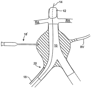
[0038] FIG. 1 schematically illustrates a bifurcated endovascular graft 10
deployed in a diseased aorta. Unless otherwise stated, the term "graft" or
"endovascular
graft" is used herein to broadly refer to a prosthesis capable of repairing
and/or replacing
diseased vessels or portions thereof, including generally tubular and
bifurcated devices and
any components attached or integral thereto.
[0039] For the purposes of this application, with reference to endovascular
graft devices, the term "proximal" describes the end or portion of the graft
that will be
oriented towards the oncoming flow of bodily fluid, typically blood, when the
device is
deployed within a body passageway. The term "distal" therefore describes the
graft end or
portion opposite the proximal end.
[0040] The teen "perigraft space" is used herein to define the space between
an outside surface of the endovascular graft and the inside surface of a body
lumen (e.g., an
artery such as the aorta), typically including the aneurysm sac, from the
proximal end of the
graft to the distal end or ends of the graft.
[0041] Finally, while the drawings in the various figures are accurate
representations of the various embodiments of the present invention, the
proportions of the
various components thereof are not necessarily shown to exact scale within,
among, or
between any given figure(s).
[0042] As shown in FIG.1, endovascular graft 10 may be positioned to
exclude an aneurysm sac AS or an otherwise diseased portion of the aorta from
blood flow.
As illustrated, aneurysm sac AS typically is proximal to the iliac arteries IA
and distal of the
renal arteries RA. In the illustrated embodiment, endovascular graft 10 is
positioned in an
CA 02542890 2006-04-19
WO 2005/039442 PCT/US2004/035179
infrarenal configuration, in which the endovascular graft is deployed below or
distal to the
renal arteries RA. In other embodiments, however, endovascular graft 10 may be
positioned
in a suprarenal configuration, such that the endovascular graft is fixed to
the aorta proximal to
the renal arteries (not shown). This would be the case, for instance, with a
fenestrated graft
that provided holes or fenestrations in the graft body to allow perfusion of
the renal arteries
RA.
[0043] Endovascular graft 10 is designed to exclude the aneurysm sac AS
from blood pressure by redirecting blood flow through its central lumen. But
in some
instances, due to device migration or an aneurysm morphology change, for
instance, blood B
may still flow into aneurysm sac AS via incomplete sealing at the proximal or
distal ends
(i.e., a Type I endolealc), or via branch vessels BV, such as an interior
mesenteric artery
(IMA), lumbar arteries, etc. (i.e., a Type II endolealc).
[0044] FIGS. 2 to 4 illustrate a method of managing endoleal~s in the
perigraft
space according to an embodiment encompassed by the present invention. An
occlusion
member 12 may be advanced through the vasculature in a constrained
configuration (not
shown) to a position that is proximal to endovascular graft 10. Access to the
vasculature may
be achieved via the femoral artery and advancement of occlusion member 12
through the
vasculature may be carried out using conventional catheter or guidewire-based
delivery
methods. The position of occlusion member 12 may be traclced under fluoroscopy
as the
occlusion member is advanced to the desired location. For example, all or a
portion of the
occlusion member and/or guidewire may be radiopaque. Once the occlusion member
12 has
been advanced to the desired location, the occlusion member may be actuated to
temporarily
reduce, and typically substantially stop, the flow of blood from the aorta
into endovascular
graft 10 and aneurysm sac AS.
[0045] As illustrated in FIG. 2, occlusion member 12 is positioned proximal to
the major branch vessels (e.g., renal arteries, celiac arteries, superior
mesenteric arteries
(SMA), etc. and are generically referred to in FIG. 2 as RA) to temporarily
reduce and
preferably stop the blood flow into aneurysm sac AS and endovascular graft 10
via the aouta
A. It is generally desirable that occlusion member 12 be positioned proximal
to the superior
mesenteric arteries SMA (not shown) to prevent perfusion of the aneurysm sac
AS via the
inferior mesenteric arteries IMA (not shown) via systemic blood flow. As may
be
appreciated however, occlusion member 12 may be positioned distal of one or
more of the
major branch vessels, if desired. Such distal positioning may be desirable in
the case, for
CA 02542890 2006-04-19
WO 2005/039442 PCT/US2004/035179
instance, in wluch an inferior mesenteric artery IMA is thrombosed and the
endolealc
originates elsewhere.
[0046] Occlusion member 12 may be in the form of an expandable aortic
balloon that is positioned at or near a distal end of a guidewire 14. The
aortic occlusion
balloon may be delivered through the artery on guidewire 14 in a constrained
configuration
(not shown). Once balloon 12 is positioned in the desired location in the
aorta, balloon 12
may be expanded to an expanded configuration by delivery of an optionally
radiopaque
inflation fluid through an inflation lumen (not shown). Deflation of balloon
12 may be
carried out by removing the inflation fluid from the balloon. The inflation
lumen may be
coupled to guidewire 14 or may be an inner lumen of a hollow catheter.
[0047] As shown in FIG. 3, after the occlusion member 12 is positioned in the
aorta to create temporarily quiescent blood, a "forerunner" contrast fluid 15
may optionally
be injected into the perigraft space via one or more delivery devices 18, 18'
so that the
physician may readily view and confirm a path and distribution pattern of the
embolic fluid
that will be introduced into the perigraft space while the blood flow through
endovascular
graft 10 is stopped. Optionally, delivery devices 18, 18' or other aspiration
devices (not
shown) may be used to aspirate aneurysm sac AS prior to delivery of the
contrast fluid. As
can be appreciated, such aspiration, however, is often mnecessary unless the
endolealc is very
small since introduction of the embolic material may displace fluid that is
present in
aneurysm sac.
[0048] Deflation of the aortic occlusion balloon 12 allows the contrast fluid
to
dissipate from the aneurysm sac by resumed blood flow through the perigraft
space over a
period of time. Dissipation of contrast fluid 15 allows the user to later see
that the embolic
material is adequately distributed within the aneurysm sac AS.
[0049] Once the contrast fluid has substantially dissipated from the perigraft
space, the aortic occlusion balloon 12 may be reinflated to reduce, and
typically substantially
stop, the flow of blood into the endovascular graft (and possibly the
perigraft space). The
halted or otherwise reduced flow into the endovascular graft and/or perigraft
space allows for
the injection and curing of the embolic material in the perigraft space
without the concern of
excessive distal flow of the embolic material.
[0050] The perigraft space may be accessed using a variety of delivery
devices to deposit the contrast fluid into the aneurysm sac. For example, as
shown in FIG. 3,
access to the perigraft space may be achieved endoluminally with a single
lumen or multi-
lumen catheter 18. A distal end 20 of catheter 18 may be guided into a space
between the
CA 02542890 2006-04-19
WO 2005/039442 PCT/US2004/035179
endovascular graft 10 and the arterial wall during or after deployment of the
endovascular
graft. Catheter 18 may be directed between the iliac artery and the
ipsilateral leg 17 of the
graft, the contralateral leg 19 of the graft, or both. While not shown, it may
be possible to
access the perigraft space proximally through the aorta or through the branch
vessels BV, if
desired. Access to the perigraft space via branch vessels BV, when they are
patent, is
generally desirable as such access minimizes the potential for disruption of
the endovascular
graft 10 seal due to passage of catheter 18 between the graft 10 and the
arterial wall.
[0051] Alternatively or additionally, the 'aneurysm sac may be accessed
directly translumbar with one or more delivery devices 18', such as a syringe
and an
10 appropriate needle, so as to percutaneously deliver the contrast fluid
directly into the perigraft
space. As may be appreciated, syringe 18' or another syringe (not shown) may
also be used
to aspirate any blood or other material from the perigraft space.
(0052] As shown in FIG. 4, the single lumen or mufti-lumen catheter 18
and/or syringe 18' may be used to deliver the multiple-component embolic
material of the
present invention into the perigraft space so that the embolic material
contacts an outer
surface of the endovascular graft 10 and a surface of the compromised portion
of the aoutic
wall (e.g., aneurysm sac wall) so as to treat the endoleak(s). Once the
embolic material has
substantially cured, as discussed below, occlusion member 12 may be deflated
and the blood
flow through the endovascular graft may be restored.
[0053] One example of a suitable catheter 18 is an angiographic catheter with
a radiopaque tip. Such a catheter would provide an adequate flow lumen (to
allow manual
injection of embolic material with a syringe) and facilitate location of the
catheter end at the
appropriate site within the aneurysm. Such a catheter could have an outer
diameter up to
about 0.035" or about 0.038", and be guidewire compatible, and are readily
available in
operating rooms, catheterization labs, or radiology suites where endovascular
interventions
are routinely performed. As can be appreciated, however, the present invention
is not limited
to angiographic catheters and many other types of conventional and proprietary
catheters may
be used to deliver the embolic material.
[0054] As may be appreciated, in some embodiments it may be desirable to
use separate catheters or syringes (not shown) to deliver the contrast fluid
and embolic
material to the perigraft space. Alternatively, heparanized saline flush may
be used to clear
contrast fluid from a single-lumen catheter 18 prior to the introduction of
the embolic
material through catheter 18.
CA 02542890 2006-04-19
WO 2005/039442 PCT/US2004/035179
11
[0055] For embolic materials with a longer cure time, the embolic material
may be injected into the perigraft space in a less precise or specific
locations, and the embolic
material may be allowed to flow to the Type I endolealcs on the proximal or
distal ends of the
endovascular graft and/or penetrate into the branch vessels (e.g., for sealing
of Type II
endolealcs), so as to embolize and close off the lealc paths. Depending on the
characteristics
of the embolic material, if a blood flow through the perigraft space and
endovascular graft is
not stopped or substantially reduced, the embolic material may perfuse from
the perigraft
space prior to curing and sealing of the endoleahs and may create potential
embolic
complications in the bowels or peripheral circulation.
[0056] As may be appreciated, while some embodiments of the present
invention reduce, and typically substantially stop the flow of blood through
the endovascular
graft and/or aneurysm sac prior to the sealing of the endolealcs, the
viscosity and curing time
of the embolic material may be chosen such that the occlusion member 12 is not
needed
during the procedure.
[0057] Useful embolic materials generally include those formed by the mixing
of multiple components and that have a cure time ranging from a few minutes or
less to tens
of minutes, preferably from about one to about ten minutes such that the
embolic material is
allowed to penetrate into the targeted branch vessels and/or penetrate into
the endolealc, belt
not beyond. Depending on the composition, the embolic material may be mixed
ifa vivo or ira
vitro. Such a material should be biocompatible, exhibit long-term stability
(preferably but
not necessarily on the order of at least ten years in vivo), and exhibit
adequate mechanical
properties, both pre- and post-cure, suitable for service in the aneurysm sac
of the present
invention in vivo. For instance, such a material should have a relatively low
viscosity before
solidification or curing to facilitate the process of filling the desired
volume. The embolic
material may be radiopaque, both acutely and chronically, although this is not
necessary.
[0058] One class of suitable materials for embolization is the family of
Michael addition polymers formed by reaction of an acrylate monomer and a
mufti-thiol.
These materials can be delivered in liquid or semi-liquid form, and thereafter
crosslinlc in situ
to form a solid polyner gel. Details of the Michael addition polyner class of
compositions
suitable for use as an embolic material are described in U.S. Patent
Application Serial No.
09/496,231 to Hubbell et al., filed Febmary l, 2000 and entitled "Biomaterials
Formed by
Nucleophilic Addition Reaction to Conjugated Unsaturated Groups" and U.S.
Patent
Application Serial No. 09/586,937 to Hubbell et al., filed June 2, 2000 and
entitled
"Conjugate Addition Reactions for the Controlled Delivery of Pharmaceutically
Active
CA 02542890 2006-04-19
WO 2005/039442 PCT/US2004/035179
12
Compounds". The entirety of each of these patent applications are hereby
incorporated
herein by reference.
[0059] One Michael addition material suitable for endolealc management
applications is a polymer formed by mixing polyethylene glycol diacrylate
(PEGDA) with
pentaerythrithritol tetra (3-mercaptopropionate) (QT). A buffer such as
glycylglycine or
other suitable compound may be added to adjust the solidification time and/or
the viscosity of
the liquid components prior to curing as described below in greater detail.
[0060] A radiopaque agent may also be added to facilitate visualization of the
embolization material under fluoroscopy and/or on follow-up imaging modalities
such as
computed tomography (CT). Suitable radiopaque agents include relatively
insoluble
materials such as barium sulfate and tantalum, and soluble materials such as
iodinated
contrast agents. Tantalum is a particularly useful agent in this regard as it
reduces the
potential for late dissipation of radiopacity due to its low solubility
compared to barium
sulfate and its potential for promoting thrombosis.
[0061] In general, we have found that the PEGDA/QT ratio may vary for a
given PEGDA molecular weight, but preferably this ratio should vary in a
defined range. For
instance, for a PEGDA molecular weight of 742, we have found that PEGDA
present in a
proportion ranging from about 1.9 to about 3.2 times the amount of QT present,
by weight, is
useful. Another useful formulation of this PEGDA/QT/buffer material may
comprise:
(1) PEGDA having a molecular weight of between about 700 and 800; preferably
between about 740 and 760; more preferably about 750, present in a proportion
ranging from about 50 to about 55 weight percent; specifically in an overall
proportion of about 53 weight percent,
(2) QT, present in a proportion ranging from about 0.31 to about .53 times the
weight
percent of the PEGDA present; specifically in an overall proportion of about
22
weight percent, and
(3) glycylglycine buffer, having a concentration of between about 100
millimole and
about 500 millimole; preferably about 400 millimole, present in a proportion
ranging
from about 5 to about 40 weight percent; specifically in an overall proportion
of
about 25 weight percent.
[0062] Variations of these components and other fonnttlations as described in
copending U.S. Patent Application Serial Nos. 09/496,231 and 09/586,937, both
to Hubbell et
al., may be used as appropriate. The entirety of each of these patent
applications are hereby
CA 02542890 2006-04-19
WO 2005/039442 PCT/US2004/035179
13
incorporated herein by reference. In addition, PEGDA having a molecular weight
ranging
from about 350 to about 850 may be useful; PEGDA having a molecular weight
ranging from
about 440 to about 750 are also particularly useful.
[0063] Other biological buffers, such as N [2-hydroxyethyl]piperazine-N'-[2-
ethanesulfonic acid] (HEPES), may be used instead of glycylglycine.
[0064] The strength of the buffer (as measured by its molarity) controls the
pH
of this embolic material, which in tum exclusively governs the material's cure
time.
Moreover, as the buffer typically is the least viscous of the three components
described
above, the volume of buffer present most efficiently affects the viscosity of
the material
before it cures. The influence of the buffer on the embolic material viscosity
and cure time
may be therefore be effected by controlling the buffer quantity and strength.
We have fotmd
that when using glycylglycine in quantities ranging from between about 5 and
about 40
weight percent as described above, and preferably about 25 weight percent, a
concentration
of approximately 400 millimole achieves a useful balance between the desired
cure time and
pre-cure viscosity.
[0065] It is within the scope of the present invention to adjust the strength
and
quantity of buffer in this tluee-component material to achieve the desired
combination of
properties (such as viscosity and cure time) for a given indication and
delivery system. For
instance, when managing endolealcs as described herein, it is generally
desirable to increase
the viscosity of the uncured material and thereby facilitate controlled
placement of the
material iya vivo without the tmintended perfusion of peripheral or secondary
vascular beds.
Viscosity may be increased for this and other embolic materials described
herein by
decreasing the buffer volume and increasing the buffer molarity. Bulling or
thixotropic
agents such as silica gel may be additionally or alternatively added in any
combination as
well.
[0066] A polymer formed by mixing ethoxylated trimethylolpropane
triacrylate (ETMPTA) with QT may also be used as an effective embolic
material. A buffer
and/or a radiopaque agent may be used with this system. Another specific
example material
that may be used in the present invention is a polymer formed by mixing
polypropylene oxide
diacrylate (PPODA) with QT. A buffer and/or a radiopaque agent may also be
used with this
system.
[0067] An alternative to these three-component systems is a gel made via
polymer precipitation from biocompatible solvents. Examples of such suitable
polymers
include ethylene vinyl alcohol and cellulose acetate. Examples of such
suitable
CA 02542890 2006-04-19
WO 2005/039442 PCT/US2004/035179
14
biocompatible solvents include dimethylsulfoxide (DMSO), n-methyl pyrrolidone
(NMP) and
others. Such polymers and solvents may be used in various combinations as
appropriate.
Other materials such as cyanoacrylates (such as TRUFILL from Cordis
Corporation, Miami
Lakes, FL) may be used as well.
[0068] Alternatively, various siloxanes may be used as an embolic material.
Examples include hydrophilic siloxanes and polyvinyl siloxanes (such as STAR-
VPS from
Danville Materials of San Racoon, California and various silicone products
such as those
manufactured by NuSil, Inc. of Santa Barbara, California).
[0069] Other gel systems useful as an embolic material for the embodiments
of the present invention include phase change systems that gel upon heating or
cooling from
their initial liquid or thixotropic state. For example, materials such as n-
isopropyl-
polyacrylimide (NIPAM) are suitable.
[0070] Effective gels may also comprise thixotropic materials that undergo
sufficient shear-thimling so that they may be readily injected through a
conduit such as a
delivery catheter or syringe but yet still are able to become substantially
gel-lilce at zero or
low shear rates.
[0071] Cure times may be tailored by adjusting the formulations, mixing
protocol, and other variables according to the requirements of the clinical
setting..
[0072] In the various embodiments of the present invention, it is desirable
that
the embolic material be visible through the use of techniques such as
fluoroscopy during the
time of delivery in which the perigraft space is being Flled with the embolic
material. Such
visibility allows the clinician to monitor and verify that the aneurysm sac,
endolealcs, and/or
branch vessels are filling correctly and to adjust the delivery procedure if
they are not. It also
provides an opportunity to detect any lealcage or otherwise undesirable flow
of the embolic
material out of the perigraft space so that the injection may be stopped,
thereby minmizing
the amount of distal perfusion of the embolic material.
[0073] It is also desirable that the cured embolic material be visible through
the use of follow-up imaging teclmiques such as computed tomography (CT) and
the like.
[0074] While the above embolic materials are examples of preferred materials
that may be used with the methods of the present invention, it may be
appreciated that other
conventional and proprietary embolic materials may be used with the methods of
the present
invention to seal the endolealcs.
[0075] FIG. 5 illustrates a system 30 for managing endolealcs according to an
embodiment of the present invention. System 30 includes a delivery device 32
for accessing
CA 02542890 2006-04-19
WO 2005/039442 PCT/US2004/035179
the perigraft space. Delivery device 32 may include one or more of a catheter
18, a syringe
and needle 18', or other conventional devices that may be used to access a
perigraft space.
System 30 also includes an embolic material 34 that is deliverable by delivery
device 18 into
the perigraft space. The embolic material may be a three-component mixture,
such as a
5 mixture of polyethylene glycol diacrylate, pentaerthyritol tetra
3(mercaptopropionate), and a
buffer. lit the illustrated embodiment, each of the separate components of the
embolic
material are stored in separate containers 35, 37, 39 and are mixed together
just prior to
delivery. As can be appreciated, embolic material 34 may be composed of any of
the other
materials described herein.
10 [0076] System 30 may optionally include an occlusion assembly 36 that is
configured to substantially reduce blood flow through a deployed endovascular
graft and/or
perigraft space. As described above in relation to FIGS. 2 to 4, one
embodiment of occlusion
assembly 36 is an inflatable occlusion member 12 coupled to a distal end of a
catheter 14.
[0077] FIG. 6 illustrates one lcit 40 according to an embodiment of the
present
15 invention. I~it 40 may include a combination of system 30, instructions for
use 42, and one
or more packages 44. Delivery device 32 will generally be as described above,
and the
instniction for use (IFL~ 42 will set forth any of the methods described
above. Package 44
may be any conventional medical device packaging, including pouches, trays,
boxes, tubes,
or the like. The instructions for use 42 will usually be printed on a separate
piece of paper,
but may also be printed in whole or in part on a portion of the paclcage 44.
Optionally, lcit 40
may include a guidewire (not shown) for assisting in the positioning of the
catheter 18, an
endovascular graft 10, and/or a delivery system for delivering the
endovascular graft (not
ShOWll).
[0078] FIGS. 7 to 9 illustrate some examples of an endovascular graft 10 that
may be used with the methods and systems of the present invention to isolate a
diseased
portion (e.g., aneurysm) of a body lumen, such as the aorta, from blood flow.
The
embodiments of FIGS. 7 and 8 are tubular, and the embodiment of FIG. 9 is
bifurcated.
[0079] As shown in FIGS. 7 and 8, graft 10 has a proximal end 54 and a distal
end 52 and includes a generally tubular structure or graft body section 53
comprised of one or
more layers of fusible material, such as expanded polytetrafluoroethylene
(ePTFE). A
proximal inflatable cuff 56 is disposed at or near a proximal end 54 of graft
body section 53
and all optional distal inflatable cuff 57 is disposed at or near a graft body
section distal end
55. Graft body section 53 forms a longitudinal lumen 62 configured to confine
a flow of
CA 02542890 2006-04-19
WO 2005/039442 PCT/US2004/035179
16
fluid therethrough and may range in length from about 5 cm to about 30 cm;
specifically from
about 10 cm to about 20 cm.
[0080] A proximal connector member 66 may be embedded within multiple
layers of graft body section 53 in the vicinity of graft body section proximal
portion 54. In
the embodiment of FIG. 7, the corrector member is a serpentine ring. Other
embodiments of
connector member 66 may take different configurations. As shown in FIG. 8, a
distal
connector member 67 may also be embedded within multiple layers of graft body
section 53
in the vicinity of graft body section distal portion 55.
[0081] One or more expandable members or stems 51, 61 may be coupled or
affixed to either or both proximal connector member 66 and distal connector
member 67 via
one or more comlector member connector elements 68. Such expandable members or
stems
may serve to anchor the endovascular graft 10 within the aorta and resist
longitudinal or axial
forces imposed on the endovascular graft 10 by the pressure and flow of fluids
tluough the
graft 10. In this embodiment, comiector elements 68 of the proximal and distal
connector
members 66 and 67 extend longitudinally outside proximal end 52 and distal end
54 of
endovascular graft 10, respectively.
[0082] FIG. 9 illustrates a bifurcated graft according to an embodiment of the
present invention. A bifurcated device such as endovascular graft 10 may be
utilized to
repair a diseased lumen at or near a bifurcation within the vessel, such as,
for example, in the
case of an abdominal aortic aneurysm in which the aneurysm to be treated may
extend into
the anatomical bifurcation or even into one or both of the iliac arteries
distal to the
bifurcation. In the following discussion, the various features of the graft
embodiments
previously discussed may be used as necessary in the bifurcated graft 10
embodiment unless
specifically mentioned otherwise.
[0083] Graft 10 comprises a first bifurcated portion 70, a second bifurcated
portion 72 and main body portion 74. The size and angular orientation of the
bifurcated
portions 70 and 72, respectively, may vary - even between portion 70 and 72 -
to
accommodate graft delivery system requirements and various clinical demands.
For instance,
each bifurcated portion or leg is shown in FIG. 9 to have a different length,
but this is not
necessary. First and second bifurcated portions 70 and 72 are generally
configured to have an
outer inflated diameter that is compatible with the imler diameter of a
patient's iliac arteries.
First and second bifurcated portions 70 and 72 may also be formed in a curved
shape to better
accommodate curved and even tortuous anatomies in some applications. A
proximal
inflatable cuff 56 is disposed at or near a proximal end 54 of main body
section 74 and
CA 02542890 2006-04-19
WO 2005/039442 PCT/US2004/035179
17
optional distal inflatable cuffs 57 may be disposed at or near one or both of
the distal end of
the first bifurcated portion 70 and the second bifurcated portion 72.
[0084] Similar to the embodiments of FIGS. 7 and 8, a proximal connector
member 66 may be embedded within multiple layers of main body portion 74 and
optionally,
distal connector members 67 may be embedded within multiple layers of
bifurcated portions
70, 72. One or more expandable members or stems 51 may be coupled or affixed
to proximal
corrector member 66 and/or distal connector members 67 via one or more
connector member
connector elements 68.
[0085] As shown in FIGS. 7 to 9, and as will be described in greater detail
below, inflation of cuffs 56, 57, in free space (i.e. when graft 10 is not
disposed in a vessel or
other body lumen) will cause them to assume a generally annular or torodial
shape (especially
when the graft body is in an unconstrained state) with a somewhat circular
longitudinal cross-
section. Inflatable cuffs 56, 57 will generally, however, conform to the shape
of the vessel
within which it is deployed. When fully inflated, cuffs 56, 57 may have an
outside diameter
ranging from about 10 mm to about 45 mm; specifically from about 16 mm to
about 32 mm.
[0086] Referring now to FIG. 7, at least one inflatable chamiel 58 may be
disposed between and in fluid communication with proximal inflatable cuff 56
and distal
inflatable cuff 57. The inflatable chamlels 58 (and inflatable cuffs 56, 57)
maybe integrally
formed in the body section 53 by seams formed in the body section 53. The
networlc of
inflatable cuffs 56, 57, and channel 58 may be inflated, most usefully in
vivo, by introduction
or injection of an inflation material or medium through an injection port 63
that is in fluid
communication with cuff 57 and the associated cuff/channel network.
[0087] As shown in FIG. 8, some embodiments may include a longitudinal
inflatable channel 60 that cormnunicates with the inflatable channel 58 and
inflatable cuffs
56, 57. Inflatable channel 58 provides structural support to graft body
section 53 when
inflated to contain an inflation medium. Inflatable channel 58 further
prevents kinlcing and
twisting of the tubular structure or graft body section when it is deployed
within angled or
tortuous anatomies as well as during remodeling of body passageways (such as
the aorta and
iliac arteries) within which graft 10 is deployed. Channels 58 may talce on a
variety of forms
but are typically in a parallel, linear or helically configuration. Together
with proximal and
distal cuffs 56 and 57, inflatable channel 58 founs a network of inflatable
cuffs and channels
in fluid communication with one other.
[0088] Referring again to FIG. 9, first and second bifurcated portions 70 and
72 may also comprise a network of inflatable cuffs and channels, including
inflatable
CA 02542890 2006-04-19
WO 2005/039442 PCT/US2004/035179
18
chamlels. Channels comprise one or more optional inflatable longitudinal
channels 60 (e.g., a
spine) in fluid communication with one or more approximately parallel
inflatable
circumferential channels 58, all of wluch are in fluid communication with
optional distal
inflatable cuffs 57. Channels 58 may tale on a variety of forms but are
typically in a parallel,
linear configuration. Channels 58 may take the form of a helix, for example,
which would
combine the functions of the parallel circumferential channels 58 and
longitudinal channels
60.
[0089] In the embodiment of FIG. 9, channel 58 forms a continuous cuff and
charmel network extending from first bifurcated portion 70 to main body
portion 74 to second
bifurcated portion 72. Accordingly, inflatable channel 58 fluidly connects
into a network
with proximal inflatable cuff 56, optional distal inflatable cuffs 57. Note
that spine or
longitudinal channels 60 extend proximally along main body portion 74 to be in
fluid
communication with cuffs 56 and 57.
[0090] The network of inflatable cuffs 56, 57, and channel 58 may be inflated,
most usefully ira vivo, by introduction or injection of an inflation material
or medium through
an injection port 63 that is in fluid communication with cuff 57 and the
associated
cuff/channel network. The inflation material may comprise one or more of a
solid, fluid (gas
and/or liquid), gel or other medium. The inflation material may contain a
contrast medium
that facilitates imaging the device while it is being deployed within a
patient's body. For
example, radiopaque materials containing elements such as bismuth, barium,
gold, iodine,
platinum, tantahun or the like may be used in particulate, liquid, powder or
other suitable
for111 aS part of the inflation medium. Liquid iodinated contrast agents are a
particularly
suitable material to facilitate such imaging. Radiopaque marlcers may also be
disposed on or
integrally formed into or on any portion of graft 10 for the same purpose, and
may be made
from any combination of biocompatible radiopaque materials.
[0091] In one embodiment, the inflation material is the same material that is
used as the embolic material, such as those described herein. In other
embodiments, the
inflation material may be a different material than the embolic material. In
such
embodiments, the inflation material and embolic material may be configured to
provide the
mechanical characteristics that are desirable for their specific purpose. For
example, in the
proximal and distal cuffs 56, 57 of the various embodiments of the present
invention, the
inflation material serves as a conformable sealing medium to provide a seal
against the lumen
wall. Desirable mechanical characteristics for the inflation medium in the
proximal and distal
cuffs would therefore include a low shear strength so to enable the cuffs 56,
57 to deform
CA 02542890 2006-04-19
WO 2005/039442 PCT/US2004/035179
19
around any luminal irregularities (such as calcified plaque asperities) and to
conform to the
luminal profile, as well as a high volumetric compressibility to allow the
embolic material to
expand the cuffs as needed to accommodate any late lumen dilatation and
maintain a seal.
[0092] In the channel or channels 58, 60 by contrast, the inflation medium
serves primarily to provide structural support to the lumen within which the
graft is placed
and lcinc resistance to the graft. Desirable mechanical characteristics for
the inflation
medium in the chamlel or channels therefore includes a high shear strength, to
prevent
inelastic deformation of a channel or channel segment due to external
compression forces
from the vessel or lumen (due, for example, to neointimal hyperproliferation)
and low
volumetric compressibility to provide stable support for adjacent channels or
channel
segments that may be in compressive contact with each other, thereby providing
link
resistance to the graft.
[0093] Finally, in the perigraft space, it is desired that the embolic
material
cure time be controlled, typically by ensuring it cures relatively quiclcly
(from times ranging
from about one minute or less to tens of minutes) after introduction into the
perigraft space,
so as to reduce the possibility that the embolic material migrates into
undesirable portions of
the vasculatu re. Desirable mechanical characteristics for the embolic
material in the perigraft
space include high volumetric and chemical stability, given that the embolic
material
typically is in direct contact with either or both tissue and blood.
[0094] Given these contrasting requirements, it may be desirable to have
different inflation materials fill different portions of the graft, such as
one inflation medium
for the proximal and distal cuffs and a second in the channel or channels and
a different
embolic material to manage the endolealcs.
[0095] In some methods of the present invention, it may be desirable to fill
the
perigraft space before the endolealcs are even formed. In such embodiments,
the embolic
material may be delivered into the perigraft space immediately after the
endovascular graft 10
is deployed in the AAA or other diseased portion of the aorta. Such methods
generally
follow similar method steps described above.
[0096] Some alternative configurations of grafts suitable for the present
invention are illustrated schematically in FIGS. 10-12. The alternative
config~.irations
comprise an inflatable graft, such as the ones described and referred to
herein in conjunction
with FIGS. 7-9. W the embodiments of FIGS. 10-12, a separate lumen, charnel,
or network
of lumens or channels 80 may be incorporated into the graft to deliver the
embolic material to
the perigraft space.
CA 02542890 2006-04-19
WO 2005/039442 PCT/US2004/035179
[0097] The embolic material may be delivered into the perigraft space via the
embolic material delivery chamlels or lumen 80 in a variety of ways. For
instance, the
embolic material may be delivered to channels 80 via an injection port 84
(which may be
similar to (FIG. 11) or the same as (FIG. 10) injection port 63). The embolic
material may
5 travel through channel 80 and exit channel 80 into the perigraft space
through one or more
abluminal apeutures or openings 82 in the channels. Some useful aperture
configuration are
shown in FIGS. 10-12. The examples show that the one or more apertures 82 are
disposed
(1) near the proximal cuff 56 of the graft, (2) in the mid-graft region (and
preferably
configured to be oriented towards the aneurysm sac AS upon deployment to
facilitate filling
10 of the perigraft space), and/or (3) in a region of the graft near the
distal cuff 57.
[0098] If desired, apertures 82 may be longitudinally symmetrically
distributed over the graft to ensure that all parts of the perigraft space is
filled at a
substantially equal rate. In other configurations, apertures 82 may be
positioned
asymmetrically over the graft. Alternatively or in addition to the above, one
or more embolic
15 material delivery chamlels may have an open distal end or terminus through
which the
embolic material may enter the perigraft space. It should be appreciated,
however, that any
number of apertures may be used as needed in a variety of locations and
configurations, and
the present invention is not limited to the illustrated examples of FIGS. 10-
12.
[0099] Channels 80 may be the same size, larger or smaller than inflatable
20 lumen chamlels 58. Channels 80 may be positioned anywhere on the graft
body, but typically
overlap inflatable hunen channels and/or are interspersed between inflatable
lumen channels
58. Apertures) 82 may have any shape and size, but are typically round and
have a diameter
between about 0.5 mil and about 2.0 mils.
[0100] Delivery of embolic material in conjunction with the various inflatable
grafts described herein may take place prior to, simultaneous with, or after
inflation of the
networlc of cuffs and channels in the graft. Desirably, the embolic material
is delivered after
the graft is filled so to aid in controlling distal perfusion.
[0101] Various embodiments of grafts and stmt-grafts, methods of
manufacturing the grafts, and methods of delivering the grafts are described
in co-pending
and commonly owned U.S. Patent Application Ser. No. 10/029,557, entitled
"Method and
Apparatus for Manufacturing an Endovascular Graft Section", U.S. Patent
Application Ser.
No. 10/029,570, entitled "Method and Apparatus for Shape Forming Endovascular
Graft
Material", U.S. Patent Application Ser. No. 10/029,584, entitled "Endovascular
Graft Joint
and Method of Manufacture", by Chobotov et al., all of which were filed
December 20, 2001,
CA 02542890 2006-04-19
WO 2005/039442 PCT/US2004/035179
21
U.S. Patent Application Ser. No. 10/327,711, entitled "Advanced Endovascular
Graft", by
Chobotov et al., filed December 20, 2002, PCT Application No. PCT/US02/40997,
entitled
"Method and Apparatus for Manufacturing an Endovascular Graft," by Chobotov et
al., filed
December 20, 2002, U.S. Patent Application Ser. No. 09/774,733, entitled
"Delivery System
and Method for Expandable Intracorporeal Device," by Chobotov et al, filed
January 31,
2002 and U.S. Patent Application Ser. No. 10/122,474, entitled "Delivery
System and
Method for Bifurcated Endovascular Graft," by Chobotov et al., filed April 1
l, 2002, the
entirety of each of which are incorporated herein by reference. Other
embodiments of
devices incorporating features and methods described herein are disclosed in
U.S. Patent No.
6,395,019 (May 28, 2002) to Chobotov, the entirety of which is incorporated
herein by
reference.
[0102] As may be appreciated, a variety of endovascular grafts may be used
with the methods and embolic materials of the present invention, and the
present invention is
not limited to use with the endovascular stem-grafts described herein. For
example, the
embodiments of the present invention may be used with a stmt, tubular graft,
bifurcated
graft, coated stent, covered stem, other configurations of unitary or modular
stmt-grafts, and
the like, such as those sold by Medtronic, W c. (Minneapolis, MN), W.L. Gore ~
Associates,
Inc. (Newarlc, DE), Coolc Group, Inc. (Bloomington, IN), etc.
[0103] While particular forms of the invention have been illustrated and
described, it will be apparent that various modifications can be made without
departing from
the spirit and scope of the invention.