Note: Descriptions are shown in the official language in which they were submitted.
CA 02542953 2006-04-18
WO 2005/060560 PCT/US2004/040436
1
SUCTION SLEEVE AND INTERVENTIONAL
DEVICES HAVING SUCH A SUCTION SLEEVE
BACKGROUND OF THE INVENTION
Field of the Invention
The present invention relates to the field of electrosurgery. In particular,
the present
invention relates to suction sleeves for electrosurgical devices for the
evacuation of hot
gasses, bodily fluids and other aspirates from an electrosurgical site.
Description of the Related Information
In use, electrosurgical instruments generate a great deal of heat at and
around the RF
cutting element of the instrument. All of the RF energy applied to the device
is typically
concentrated at the distal cutting element of the device, which consequently
experiences a
high current density. This high current density creates an arc between the
targeted tissue and
the cutting element of the device, which arc cuts the targeted tissue by
vaporization of the
cells that come into contact with the arc. This arc also creates very high
temperatures. As the
cells are vaporized, hot gasses (such as steam and smoke, for example) are
created.
Moreover, when arterial blood or other fluids fill the cavity around the
cutting element of the
electrosurgical element, these fluids are rapidly heated.
The presence of such hot gasses has several adverse consequences. First among
these
adverse consequences is thermal damage to the otherwise viable and healthy
tissue
surrounding the electrosurgical site. Second, the presence of heated fluids
may also adversely
affect the operation of the RF device itself. As the fluids come into contact
with the RF
cutting element of the electrosurgical device, the arc generated within the
gap between the
targeted tissue and the distal RF tip of the device may be lost. In turn, this
loss of arc results
in a decrease in the current density at the cutting element of the device,
which current is then
redistributed over the comparatively greater surface area of the distal region
of the RF device.
Indeed, instead of the RF energy being concentrated in the very small area of
the cutting
element (e.g., cutting blade or tip) of the device (which leaves adjacent
areas relatively
unaffected by the great temperatures generated at the arc), the applied RF
energy is spread
out over the greater surface of the distal region of the RF device, thereby
heating the entire
cavity. This heating, in addition to causing unintended thermal damage to
adjacent tissue and
structures, may also damage the biopsy specimen, destroying the architecture
of the severed
tissue and hampering histopathological examination thereof. Moreover, the heat
generated at
CA 02542953 2006-04-18
WO 2005/060560 PCT/US2004/040436
2
the cutting element of the device may also transfer to the shaft of the
device, even during a
procedure of relatively short duration.
To reduce the unintended thermal damage to adjacent tissues, it is necessary
to
evacuate the hot gasses and fluids from the electrosurgical site. Doing so in
an efficient
manner reduces the internal temperature of the cavity within which the RF
procedure is being
carried out, and reduces thermal damage to adjacent tissues. Moreover,
efficient evacuation
of gasses, fluids and smoke facilitates the re-initiation of the RF arc by re-
creating the gap
between the targeted tissue and the RF tip.
From the foregoing, it is apparent that evacuation of hot gasses and fluids is
essential
to prevent unintended thermal damage to adj acent tissue structures and to
insure the
maintenance of the RF arc at the distal tip of the electrosurgical device.
What are needed,
therefore, are devices for evacuation of heated gasses and fluids from an RF
electrosurgery
site. Such devices should efficiently remove both heated gasses and fluids
without, however,
unduly increasing the size of the device near the distal tip of the device.
Such a device,
moreover, should not hamper the physician as he or she manipulates (e.g.,
rotates) the
electrosurgical device during the procedure. Ideally, such device should also
be configured
such that tissue coming into contact with it does not block the evacuation of
the heated gasses
and fluids.
SUMMARY OF THE INVENTION
The present invention, according to an embodiment thereof, is a soft tissue
interventional device, including a handle; a shaft defining a first end and a
second end, the
first end being coupled to the handle; a work element coupled to the second
end of the shaft,
and a suction sleeve disposed coaxially around the shaft between the first end
of the shaft and
the work element, the suction sleeve defining a suction port and a plurality
of openings near
the work element, the suction sleeve being configured to enable suction in
through the
plurality of openings and out through the suction port.
The suction sleeve may define a circumference around the shaft and the
plurality of
openings may be defined around the circumference of the suction sleeve. The
plurality of
openings may overlap around the circumference of the suction sleeve. The
suction sleeve
may define a first external surface and a second external surface disposed at
a non-zero angle
relative to first external surface, and at least one of the plurality of
openings may be defined
within the first external surface and at least one of the plurality of
openings may be defined
within the second external surface. At least one of the plurality of openings
defined within the
CA 02542953 2006-04-18
WO 2005/060560 PCT/US2004/040436
3
first external surface may overlap in extent with at least one of the
plurality of openings
defined within the second external surface. One or more of the plurality of
openings may
define a generally cloverleaf shape. The suction port may be defined within
the suction sleeve
adjacent the first end of the shaft. The suction sleeve may be configured to
be freely rotatable
about the shaft. The sleeve may include an internal surface that may define an
interior sleeve
lumen dimensioned so as to allow the shaft to freely rotate therethrough. The
plurality of
openings may be open to the interior sleeve lumen. The interior sleeve lumen
may be
dimensioned so as to allow free passage of aspirates through the plurality of
openings and out
the suction port. The work element may be configured to cut soft tissue. The
work element
may be energizable with RF energy. The suction sleeve may be configured to be
removable
from the shaft. The suction sleeve may be configured to be positioned on the
shaft without
decoupling the shaft from the handle. The suction sleeve may be at least
partially transparent.
The sleeve may comprise a first portion and a second portion, the second
portion being
configured to slide coaxially relative to the first portion to assume a first
position in which the
sleeve has a first length and second positions in which the length of the
sleeve is greater than
the first length. The second portion may telescope (e.g., slide axially)
relative to the first
portion.
Another embodiment of the present invention is a suction sleeve for a soft
tissue
interventional device that includes a shaft that includes a tapered portion
defining a
predetermined extent, the tapered portion defining a first external surface
and a first internal
surface, the first internal surface defining an internal axial lumen spanning
the predetermined
extent, the internal axial lumen being configured to receive the shaft, the
first external surface
defining a suction port and a plurality of openings that open to the internal
axial lumen.
The tapered portion may define a second external surface disposed at a non-
zero angle
relative to the first external surface and both the first and second external
surfaces may define
openings that open to the internal axial lumen. The openings defined within
the first external
surface may overlap with the openings defined within the second external
surface. The
plurality of openings may be shaped and dimensioned so as to enable free
passage of
aspirates (smoke, heated fluids, gasses, for example) from a cavity within
soft tissue through
the internal axial lumen and out through the suction port. One or more of the
plurality of
openings may define a generally cloverleaf shape. The suction sleeve may be at
least partially
transparent. The tapered portion may include a first sleeve half and a second
sleeve half, the
first sleeve half being configured to mate with the second sleeve half. The
suction sleeve may
CA 02542953 2006-04-18
WO 2005/060560 PCT/US2004/040436
4
further include at least one integral hinge bridging the first sleeve half to
the second sleeve
half. The tapered portion may include at least one sleeve mating assembly. The
at least one
sleeve mating assembly may be configured to removably mate the first sleeve
half to the
second sleeve half. The sleeve may include a first portion and a second
portion, the second
portion being configured to slide coaxially with the first portion to assume a
first position in
which the sleeve has a first length and second positions in which the length
of the sleeve is
greater than the first length. The second portion telescopes (e.g., slide
axially) within or
relative to the first portion.
The present invention, according to yet another embodiment thereof, is a
method for
cutting a specimen of soft tissue, comprising the steps of providing a device
including a
handle, a shaft coupled to the handle and defining a first end and a second
end, a cutting
element coupled to the second end of the shaft, and a suction sleeve disposed
coaxially
around the shaft between the first end of the shaft and the cutting element,
the suction sleeve
defining a suction port and a plurality of openings near the work element, the
suction sleeve
being configured to enable suction in through the plurality of openings and
out through the
suction port; inserting the device in the soft tissue; cutting the tissue
specimen using the
cutting element, and applying suction to the suction port during the cutting
step.
The cutting element may be energizable with RF energy and the cutting step may
include a step of applying RF energy to the cutting element.
BRIEF DESCRIPTION OF THE DRAWINGS
Fig. lA is a perspective view of a suction sleeve according to an embodiment
of the
present invention, coupled to an exemplary excisional device.
Fig. 1B is a perspective view of the suction sleeve of Fig. 1, coupled to
another
exemplary excisional device.
Fig. 2 is a detail view of the distal end of the RF device of Fig. 1B, to
illustrate an
exemplary configuration of the distal openings of the suction sleeve,
according to an
embodiment of the present invention.
Fig. 3 is a perspective view of the distal end of the suction sleeve of Figs.
lA-S,
illustrating the overlapping nature of the openings therein.
Fig. 4 is a front view of the distal end of the suction sleeve of Figs. lA-6.
Fig. 5 is a perspective cutaway view of the distal end of the suction sleeve
of the
embodiment of the suction sleeve of Figs. lA-4, illustrating the manner in
which smoke,
fluids andlor other aspirates may be suctioned into the present suction
sleeve.
CA 02542953 2006-04-18
WO 2005/060560 PCT/US2004/040436
Fig. 6 is an exploded side view of the suction sleeve of Figs. lA.
Fig. 7 is a cross-sectional side view of a portion of the suction sleeve shown
in Fig. 3.
Fig. 8 shows an interventional device to which a suction sleeve according to
an
embodiment of the present invention is coupled, illustrating the manner in
which smoke,
5 fluids and/or other aspirates may be evacuated from a target site in the
body during a biopsy
or other procedure.
Fig. 9 is a perspective view of a combination introducer and suction sleeve,
according
to another embodiment of the present invention.
Fig. 10 is a side cross-sectional view of the combination introducer and
suction sleeve
of Fig. 9.
Fig. 11 is a perspective view of the combination introducer and suction sleeve
of Fig.
9, with a trocar inserted therein.
Fig. 12 is a side cross-sectional view of the combination introducer and
suction sleeve
of Fig. 9, illustrating exemplary structure with which the suction sleeve may
attach to the
interventional device.
Fig. 13 is a perspective cross-sectional view of the combination introducer
and
suction sleeve, attached to an exemplary interventional device.
Fig. 14 is a perspective view of another embodiment of a suction sleeve
according to
the present invention, coupled to an exemplary interventional device.
Fig. 15 is a perspective view of another embodiment of a suction sleeve
according to
the present invention, coupled to another exemplary interventional device.
Fig. 16 shows a perspective view of another embodiment of the suction
sleeve/introducer according to another embodiment of the present invention.
Fig. 17 is an exploded perspective view of the embodiment of the present
suction
sleeve/introducer shown in Fig. 16.
Fig. 18 is a perspective view of yet another embodiment of the suction
sleeve/introducer according to the present invention.
Fig. 19 is a perspective view of a suction sleeve according to a still further
embodiment of the present invention, shown in a first configuration.
Fig. 20 is a perspective view of a suction sleeve according to a still further
embodiment of the present invention, shown in a second configuration.
Fig. 21 is a side cross-sectional view of the suction sleeve of Figs. 19 and
20.
CA 02542953 2006-04-18
WO 2005/060560 PCT/US2004/040436
6
Fig. 22 is a perspective cross-sectional view of the suction sleeve of Figs.
19 and 20,
in the first configuration.
Fig. 23 is a perspective cross-sectional view of the suction sleeve of Figs.
19 and 20,
in a second (extended) configuration.
Fig. 24 shows the suction sleeve of Figs. 19 and 20, coupled to an exemplary
electrosurgical device.
Fig. 25 shows the suction sleeve of Figs. 19 and 20, coupled to another
exemplary
electrosurgical device.
Fig. 26 shows an interventional device to which the suction sleeve of Figs. 19
and 20
has been coupled, in use.
Fig. 27 shows the interventional device of Fig. 26 in use, and illustrates
further
aspects thereof.
Fig. 28A is a front view of the distal end of a suction sleeve according to
another
embodiment of the present invention.
1 S Fig. 28B shows the embodiment of Fig. 28A, with an excisional device
inserted
therein, to illustrate the manner in which the distal opening allows suction.
Fig. 29 is a partial side view of the embodiment of the suction sleeve shown
in Figs.
28A and 28B.
Fig. 30 is a side cross sectional view of the embodiment of the present
suction sleeve
shown in Fig. 29.
DETAILED DESCRIPTION
Fig. lA is a perspective view of a suction sleeve 110 according to an
embodiment of
the present invention, coupled to an exemplary excisional device. For
simplicity of
illustration, only the distal portion of the excisional device is shown. The
handle of the
excisional device is shown at 102. A shaft 104 extends from the handle 102 and
defines a
distal tip 106. When energized by an RF source, an arc develops between the
distal tip 106
and the targeted tissue. Such a device is also called a Bovie pencil. When RF
energy is
applied, an arc develops at the tip 106, and cutting of the tissue occurs by
vaporization of the
tissue that comes into contact with the RF arc. Fig. 1B shows the suction
sleeve 110 coupled
to another electrosurgical device. In this case, the electrosurgical cutting
element 108
includes a wire loop that is configured to bow and extend away from the shaft
104 and to
retract back toward the shaft 104. As detailed above, hot gasses and fluids
are frequently
present at the electrosurgical site. To evacuate such heated gasses and fluids
(including
CA 02542953 2006-04-18
WO 2005/060560 PCT/US2004/040436
7
smoke, blood and intercellular fluids, for example), the excisional devices of
Figs. lA and 1B
(as well as other types of electrosurgical devices) may be equipped with a
suction sleeve
according to one of the embodiments of the present invention.
The suction sleeve 110 is disposed coaxially around the shaft 104 between the
proximal end of the shaft 104 (i.e., closest to the handle 102 of the device)
and the cutting
element (in this case, the distal tip 106 or the loop 108 of the shaft 104).
As shown, the
suction sleeve 110 may surround at least a portion of the shaft 104 and define
a first external
surface 118 and a first internal surface 122 (best shown in Fig. 7). The first
internal surface
122 faces the shaft 104 and defines an internal lumen 124 through which the
shaft 104 is or
may be inserted.
To enable the evacuation of hot gasses, fluids and/or other aspirates, the
suction
sleeve 110 defines a plurality of openings 112 at and/or near the distal end
of the suction
sleeve 110 (i.e., that end of the suction sleeve 110 that is closest to the RF
cutting element),
as well as a suction port 114 at or near the proximal end of the sleeve (i.e.,
that end of the
sleeve 110 that is closest to the handle 102). When suction is applied to the
suction port 114,
the suction sleeve 110 enables suction of gasses, fluids and/or aspirates in
through the
plurality of openings 112 and out through the suction port 114. The suction
sleeve 110 may
be fixedly attached to the electrosurgical device in such a manner that it
rotates along with the
shaft 104. Alternatively, the suction sleeve 110 may be attached so as to
enable its free
rotation about and independent of the shaft 104. That is, the shaft 104 may be
rotated within a
stationary suction sleeve 110 or the suction sleeve may be manipulated so as
to rotate it about
a stationary shaft 104. When the suction sleeve 110 is coupled to the
electrosurgical device in
such a manner as to allow its free rotation about the shaft 104, the physician
is free to
manipulate and rotate the RF device during a procedure as needed without
causing a
corresponding rotation in the suction sleeve and the vacuum line 116 attached
to the suction
port 114.
Fig. 2 is a detail view of the distal end of the RF device of Fig. 1B, to
illustrate an
exemplary configuration of the distal openings of the suction sleeve,
according to an
embodiment of the present invention. As shown, the suction sleeve 110 may
define a first
external surface 118 and a second external surface 120 disposed at a non-zero
angle relative
to first external surface 118. As shown in Fig. 2, the first and second
external surfaces 118
and 120 together may give the distal end of the suction sleeve 110 a tapered
appearance, to
facilitate entry thereof into soft tissue.
CA 02542953 2006-04-18
WO 2005/060560 PCT/US2004/040436
8
To enable evacuation of hot gasses and fluids, the first and/or second
surfaces 118,
120 may define a plurality of openings 112. Referring now collectively to
Figs. 3, 4 and 5, at
least one of the plurality of openings 112 may be defined within the first
external surface 118
and at least one of the plurality of openings 112 may be defined within the
second external
surface 120. In the embodiment of Fig. 2, a plurality of openings 112 to the
internal lumen
(reference numeral 124, see Fig. 7) of the sleeve 110 may be defined both in
the first external
surface 118 and in the second external surface 120. Also as shown, one or more
of the
openings 112 defined within the first external surface 118 may overlap in
extent with one or
more of the openings 112 defined within the second external surface 120.
Defining openings
112 in both of the first external surface 118 and the second external surface
120 and the
overlapping nature of the openings defined within the two surfaces 118, 120
decreases the
likelihood that suction will be blocked by tissue drawn to the suction sleeve
110 by the
applied vacuum and seal all of the openings. As best shown in the cutaway view
Fig. 5,
providing openings 112 in both surfaces 118 and 120 provides for an efficient
flow of heated
gasses and fluids by drawing them from both the radial direction relative to
the shaft 104
(through openings 112 defined within the first surface 11 ~ of the suction
sleeve 110) and
from the axial direction relative to the shaft 104 (through openings 112
defined within the
second surface 120 of the suction sleeve 110).
Fig. 6 is an exploded side view of the suction sleeve of Figs. lA-B. As shown,
according to an embodiment of the present invention, the suction sleeve 110
may include a
proximal portion 128 and a distal portion 126. The proximal and distal
portions 128, 126 may
be configured to couple to one another, using an interference or snap-fit, for
example. The
proximal portion 128 may be configured to couple onto the handle 102 of the RF
device, as
shown in Figs. lA and 1B.
Fig. 7 is a side cross-sectional view of the distal portion 126 of the suction
sleeve 110,
according to an embodiment of the present invention. As shown, the second
portion 126 may
define a tapered profile to facilitate its travel within soft tissue (such as,
for example, breast
tissue). Fig. 7 also shows a portion of the internal lumen 124 of the suction
sleeve 110, which
internal lumen 124 communicates with the openings 112 defined within the first
and/or
second external surfaces 118, 120. In turn, the suction port 114 opens to the
internal lumen
124, enabling suction applied thereto to draw gasses, fluids and/or other
aspirates through the
openings 112 and out through the suction port 114. Preferably, a balance
should be struck in
selecting the diameter of the internal lumen 124. The internal lumen 124
should be large
CA 02542953 2006-04-18
WO 2005/060560 PCT/US2004/040436
9
enough to allow sufficient space between the shaft 104 to enable efficient
suction of gasses
and fluids, but not so large as to present to a cross-sectional area that
would impede the
advancement of the sleeve 110 within the tissue. Note that the suction port
114, the internal
lumen 124 and the openings 112 may also be used to deliver gasses and/or
fluids (e.g.,
lidocaine) to the excision site.
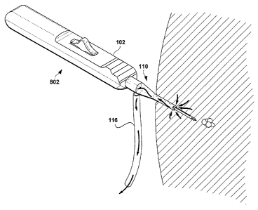
Fig. 8 shows an interventional device 802 to which a suction sleeve 110
according to
an embodiment of the present invention is coupled, illustrating the manner in
which smoke,
fluids and/or other aspirates (collectively referenced by the curved arrows)
may be evacuated
from a target site in the body during a biopsy or other procedure. The suction
applied to the
suction port 114 through the vacuum line 116 may be turned on and off at will
during the
procedure.
Fig. 9 is a perspective view of a combination introducer and suction sleeve
902,
according to another embodiment of the present invention. Fig. 10 is a side
cross-sectional
view thereof. Considering now Figs. 9 and 10 collectively, the first external
surface 904 of
the combination introducer and suction sleeve 902 may have a generally tapered
or funnel
shape, in that it defines a relatively narrow diameter distal end and a
relatively wider
proximal end. Such a generally funnel or tapered shape eases the introduction
of the device
802 within tissue. As with the suction sleeve of Figs. lA - 8, the combination
introducer and
suction sleeve 902 includes a suction port 908 that opens to an internal lumen
916 defined by
the internal surface 918. The combination introducer and suction sleeve 902
also includes a
second external surface 912 that defines a tapered appearance. Defined within
the first and/or
second external surfaces 904, 912 are a plurality of openings 906 that open to
the internal
lumen 916. In Figs. 9-11, only the first external surface 904 defines such
openings 906,
although the openings are not limited to this surface. The suction port 908 is
configured to
couple with a vacuum line, as shown at 116. The combination introducer and
suction sleeve
902 may further include structures to couple to one or more devices, such as a
trocar or an RF
device such as, for example, a Bovie device or the device shown, for example,
at reference
numeral 802 in Fig. 8. Such coupling structures) may include, for example, a
snap or
interference fitting 914 and/or one or more O-rings, such as shown at 910.
Fig. 11 is a perspective view of the combination introducer and suction sleeve
of Fig.
9, with a trocar 1102 inserted therein. According to an embodiment of the
present invention,
the trocar 1102 may be inserted into the combination introducer and suction
sleeve 902 and
CA 02542953 2006-04-18
WO 2005/060560 PCT/US2004/040436
the assembly may be packaged as a (preferably single use) unit. According to
another
embodiment of the present invention, a physician may utilize the assembly as
follows:
1. An incision into tissue is made with a blade;
2. The physician then inserts the assembly including
the trocar 1102 into the
5 tissue and pushes the combination introducer and
suction sleeve 902 into the
tissue through the incision into position under or
near the lesion or targeted
site within the tissue. The pointed andlor sharp
distal tip 1104 of the trocar
1102 and the tapered profile of the combination introducer
and suction sleeve
902 aid the assembly's advancement within the tissue;
10 3. The trocar 1102 may then be removed from the combination
introducer and
suction sleeve 902 and a desired (excisional RF,
for example) device may then
be inserted therethrough, with the shaft thereof
disposed within and protruding
from the internal lumen 916;
4. The combination introducer and suction sleeve 902
may then be pulled back
until it contacts, snaps andlor otherwise locks onto
the device, as shown at
Figs 12 and 13. In Figs. 12 and 13, only the handle
102 of the device is shown,
and the shaft 104 thereof is omitted for clarity
of illustration. Examples of
devices coupled to the combination introducer and
suction sleeve 902 coupled
thereto are shown in Figs. 14 and 15;
5. A vacuum line, such as shown at 116, may then be
attached to the suction port
908;
6. If needed, the device with the combination introducer
and suction sleeve 902
attached thereto may then be repositioned at, near,
under or within the target
lesion, as desired. This repositioning may be carried
out under ultrasound
guidance, for example. The openings 906 may aid with
the ultrasound
visualization. The combination may include other
features and/or markings to
increase the visibility thereof under various imaging
modalities, and
7. The physician may then continue with the intended procedure as per the
instructions for use of the device utilized.
Alternatively, the trocar 1102 may be removed from the combination introducer
and
suction sleeve 902 and the desired RF device introduced and locked therein.
The distal tip of
the desired RF device protruding from the distal end of the combination
introducer and
suction sleeve 902 may then be used to reach the intended biopsy site.
Alternately still, a stopcock may be attached to the suction port 908 instead
of the
suction line 116 and one or more beneficial agents (e.g., antibiotics, fibrin,
lidocaine) may be
delivered to a target site through the openings 906.
The present combination vacuum sleeve and suction sleeve 902 may aid in
positioning a biopsy or other interventional device where it is needed. For
example,
interventional devices that include a rather bulky or high-drag distal end may
be readily
positioned at the intended site by means of the introducer functionality of
the combination
CA 02542953 2006-04-18
WO 2005/060560 PCT/US2004/040436
11
902. While the combination 902 is advantageous before the biopsy or other
interventional
procedure is started by easing the positioning of the biopsy instrument at or
near the target
site, it is also useful during the procedure itself, as it is effective in
evacuating hot gasses and
fluids from the biopsy cavity, thereby decreasing collateral tissue thermal
damage. The same
combination may then also be used to treat the cavity post-procedure by, for
example,
providing a ready-made pathway for the introduction of beneficial agents,
compositions
and/or cavity treatment devices to the cavity or lesion site.
Fig. 16 shows a perspective view and Fig. 17 shows an exploded perspective
view of
another embodiment of the combination introducer and suction sleeve 1602
according to
another embodiment of the present invention. Considering now Figs. 16 and 17
collectively,
the combination introducer and suction sleeve 1602 is similar to the
embodiment of Figs. 9-
15, but for the added feature of being formed of two halves 1702, 1703. As
shown, the
suction port of the combination introducer and suction sleeve 1602 is also
split into two
halves 908a and 908b. As shown in Fig. 17, the two halves 1702, 1703 of the
combination
introducer and suction sleeve 1602 may be coupled to one another via snap fit
male and
female features such as shown at 1704 and 1706, respectively. Alternatively,
the two halves
1702, 1703 may mate to one another by means of an interference fit or other
suitable
mechanism. For example, Fig. 18 shows another embodiment of the present
invention, in
which the two halves of the combination introducer and suction sleeve 1802 are
coupled to
one another by integral hinges, such as shown at reference numerals 1804. For
the
embodiments shown in Figs. 16-18, it may be expedient to locate the O-ring or
other vacuum
sealing structures on the handle 102 of the RF device. The embodiments of the
suction sleeve
shown in Figs. 1-8 may be configured to include two mating suction sleeve
halves, in the
manner shown and described relative to Figs. 16-18 or by means of other
suitable
mechanisms.
Fig. 19 is a perspective view of a suction sleeve 1902 according to a still
further
embodiment of the present invention, shown in a first configuration. Fig. 20
is a perspective
view of the suction sleeve of Fig. 19, shown in a second configuration. As
shown, the suction
sleeve 1902 includes a first portion 1904, a center housing 1905 and a second
portion 1906.
According to this embodiment, the length of the suction sleeve 1902 may be
varied from a
first position in which the length of the suction sleeve 1902 is at a minimum,
and selected
second positions in which the length of the suction sleeve 1902 is greater
than the minimum
length.
CA 02542953 2006-04-18
WO 2005/060560 PCT/US2004/040436
12
As shown in Figs. 19 and 20, the first external surface 1912 of the first
portion 1904
may have a tapered shape to ease entry of the suction sleeve 1902 within
tissue. The first
external surface 1912 may define a plurality of openings 1908 near the distal
end thereof. The
first internal surface 1914 of the suction sleeve 1902 may define an internal
lumen 1916
through which the shaft 104 of an RF device may be introduced. A suction port
1910 may be
defined within the first portion or the center housing 1905 (as shown in Figs.
19-21 ). A
suction line (not shown) may be coupled to the suction port 1910. A second
portion 1906
may be configured to slide (coaxially, for example) relative to the first
portion 1904 (and/or
the center housing 1905) to assume a first position in which the sleeve 1902
defines its
minimum length and selected second positions (one of which is shown in Fig.
20) in which
the length of the sleeve 1902 is greater than the aforementioned minimum
length.
Alternatively, the first portion 1904 could be configured to slide relative to
the second portion
1906 and/or relative to the center housing 1905.
As shown in the side cross-sectional view of Fig. 21, an extension 1918 of the
second
portion 1906 of the suction sleeve 1902 may be configured to axially slide
within the center
housing 1905 and within the internal lumen 1916 of the first portion 1904. An
opening may
be defined in the second portion 1906 to enable gasses, fluids and other
aspirates to be sucked
into the suction port 1910. Alternatively, the sleeve 1902 may be configured
so as to enable
suction only when the sleeve 1902 is in its second, extended configuration as
shown in Fig.
23. Figs, 22 and 23 show the embodiment of Figs. 19-21 in the first
configuration in which
the length of the sleeve 1902 is at its minimum and in the second
configuration in which the
length of the sleeve 1902 is at its maximum, respectively.
Figs. 24 and 25 show the suction sleeve 1902 coupled to two exemplary RF
devices.
The suction sleeve 1902 of Fig. 24 is coupled to an RF device similar to a
Bovie pencil,
whereas the suction sleeve of Fig. 25 is coupled to an RF excisional device
that includes a
bowing RF blade 108, such as available from the present assignee Rubicor
Medical, Inc. of
Redwood City CA. In both cases, the distal end of the shaft 104 is inserted
within and
protrudes from the internal lumen 1916 of the suction sleeve 1902. The second
portion 1906
may (but need not) be coupled to the handle 102 of the RF device. For example,
the second
portion 1906 may be coupled to the handle by a snap fitting, an interference
fit or by other
suitable mechanisms.
Figs. 26 and 27 show the assembly of Fig. 25 in use. The suction sleeve 1902
is
coupled to the handle 102 of an RF device 802. As shown in Fig. 26, the shaft
104 may be
CA 02542953 2006-04-18
WO 2005/060560 PCT/US2004/040436
13
inserted into the tissue through an incision, with the suction sleeve 1902 in
its first
configuration. In such a configuration, the first portion 1904 of the suction
sleeve 1902 may
be maintained outside of the tissue, thereby easing the initial entry of the
RF device through
the tissue. After the distal portion of the shaft has been positioned within
the tissue to the
physician's satisfaction (adj acent a target lesion, for example), the second
portion 1906 being
otherwise stationary, the physician may slide the first portion 1904 (by
manually grasping the
center housing 1905, for example) into the tissue such that the distal end
thereof is adjacent
the RF device's work element (in this case, the bowing RF blade). In this
configuration, hot
gasses and fluids generated incident to the RF cutting and coagulation action
of the RF
device's work element may be evacuated through the openings 1908, through the
internal
lumen 1916, through the suction port 1910 and out through the suction line 116
coupled to
the suction port 1910.
Figs. 28A, 28B, 29 and 30 illustrate aspects of another embodiment of the
present
suction sleeve. Whereas the distal opening of the suction sleeve of Figs. 1-27
is circular in
shape, the present inventions are not so limited. Indeed, the distal end of
the embodiment of
Figs. 28A-30 includes a surface or surfaces 2820 that define an opening 2820
having a
generally cloverleaf shape, shown at reference 2810. This opening may be
described as a
circular shape having a number of side lobe openings. Such a shape enables the
excisional
device 104 to be securely held and centered within the suction sleeve, yet
allows suction to
occur not only at the sides of the suction sleeve, but also at the distal tip
thereof, through the
side lobe openings 2812. As shown in Figs. 29 and 30, the suction sleeve rnay
also include a
number of openings 2812 defined within the exterior surface 118, to further
promote suction
and evacuation of aspirates within the cavity. It is to be understood that the
openings defined
at or near the distal end of the present suction sleeve may have other shapes
than those shown
and described herein. Other variations may occur to those of skill in this
art, and all such
variations or modifications are deemed to fall within the spirit and scope of
the inventions
shown, described and claimed herein.
While the foregoing detailed description has described preferred embodiments
of the
present invention, it is to be understood that the above description is
illustrative only and not
limiting of the disclosed invention. Thus, the present invention should be
limited only by the
claims as set forth below.