Note: Descriptions are shown in the official language in which they were submitted.
CA 02542993 2006-04-19
WO 2005/037110 PCT/US2004/034334
DYNAMIZABLE ORTHOPEDIC IMPLANTS AND
THEIR USE IN TREATING BONE DEFECTS
BACKGROUND OF THE INVENTION
The present invention relates generally to orthopedic devices for promoting
bone
fusion and methods for treating orthopedic defects using the orthopedic
devices.
The spine is composed of both rigid and flexible elements, forming a complex
structure that can readily accommodate a wide range of motions and adjust to a
wide range
of loads. Unfortunately, the spine, like any complex physiological structure,
is also
vulnerable to disease, injury, and congenital deficiencies, all of which can
cause defects to
the spine and, in particular, to the vertebral body and intervertebral discs.
Spinal disease,
injury, and deformity may have a disastrous impact on patient well being,
ranging from
acute pain to chronic debilitating pain, and, in the most severe cases,
partial or complete
paralysis.
Some of the most common pathologies of spinal defects include fractured,
diseased, or decayed vertebral bodies, torn or stretched ligaments, and
damaged or
diseased intervertebral discs.
Common treatments for defective vertebrae include joining or fusing fractured
bone segments or portions together to stabilize the affected parts and
removing the
affected vertebrae, either in part or in whole. Classically, the damaged disc
is excised, the
adjacent vertebrae are mechanically joined together, and oftentimes bone is
grafted into
the region particularly in the disc space between the two vertebrae to promote
fission of the
adjacent vertebrae. The vertebrae can be mechanically joined using a
prosthetic device
such as a bone plate that is attached to the adjacent vertebrae with bone
screws. The bone
plate eliminates disparate motion between the two bone portions to allow
arthrodesis.
It is particularly important that the prosthetic device not stress shield the
new bone
growth and permit a weakened juncture or pseudoarthrodesis between the bone
portions or
adjacent vertebrae to be. fused. It is known that for load bearing bone
members, stronger,
denser bone tissue results when the new bone growth occurs under pressure. The
problem
arises when and how to determine the amount of pressure or force desirable to
develop a
strong junction between the bone portions. The bone portions should be secured
and
CA 02542993 2006-04-19
WO 2005/037110 PCT/US2004/034334
2
supported during bone growth. However, the optinmm support necessary for
desired bone
growth may vary over time as the bony juncture or bridge develops between the
bone
portions.
Similarly, torn and/or structural ligaments can be treated by initially
securing/immobilizing the ligaments. This can be accomplished using either or
both
internal and external prosthetic devices to augment or replace the stability
lost as a result
of the damaged ligaments. Further, the treated ligaments can be susceptible to
repeated
injury. Consequently, it may be desirable to augment the treated ligament by
implanting a
prosthesis or device that allows limited movement of the affected ligaments,
i.e.,
stretching and rotation of the ligaments. Current treatment methods do not
allow for an
implanted device to initially secure/innnobilize the ligaments and then allow
limited
movement of the same without a subsequent surgical revisitation.
In light of the above, there is a continuing need for devices and treatments
that
stabilize and support damaged bone tissue and bony strucW res and connecting
tissue,
provide variable loads to growing bone, as well as a measure of flexible
support to injury
or disease prone bones and connecting tissue. The present invention addresses
this need
and provides other benefits and advantages in a novel and nonobvious manner.
BRIEF SUMMARY OF THE INVENTION
The present invention relates to orthopedic devices, the manufacture and use
thereof. Various aspects of the invention are novel, nonobvious, and provide
various
advantages. While the acW al nature of the invention covered herein can only
be
deteunined with reference to the claims appended hereto, certain forms and
features,
which are characteristic of the preferred embodiments disclosed herein, are
described
briefly as follows.
In one form, the present invention provides an orthopedic device for securing
two
or more bone portions. The device comprises an elongate member configured for
engagement to the two or more bone portions and allowing translational or
rotational
movement for a first one of the two or more bone portions relative to a second
one of the
two or more bone portions; a reinforcing component composed of a biodegradable
material and engaged to the elongate member to inhibit the translational or
rotational
movement for a first one of the two or more bone portions relative to a second
one of the
CA 02542993 2006-04-19
WO 2005/037110 PCT/US2004/034334
3
two or more bone portions; and at least one bone fastener for fixedly securing
the elongate
member to at least one of the two or more bone portions. The orthopedic device
can be
used to treat a variety of bone defects including but not limited to: bone
fractures,
diseased bone tissue, spinal diseases, diseased/damaged vertebrae, torn or
stretched
ligaments and the like.
In another form, the present invention provides a method for treating a bone
defect. The method comprises providing an orthopedic device including an
elongate
member configured to be deformable if?. vivo, and a reinforcing component
encasing at
least a portion of the elongate member. The reinforcing component comprises a
biodegradable material, which is formulated to inhibit deformation of the
elongate
member. The first end of the elongate member can be secured to first bony
structure and
the second end of the elongate structure can be secured to a second bony
structure. The
secured device can support and effectively immobilize the two bone portions
relative to
each other. In vivo, the reinforcing component can be degraded and be
eliminated either
in whole or in part from the device. This effectively transfers at least a
portion of the
biomechanical load and support to the treated site, in general, and to new
tissue and bone
growth, in particular. Further particularly for articulating joints and if
desired, the treated
site can then be allowed at least a limited amount of movement, i. e.
translation and/or
rotation. The secured devices without the reinforcing component can be allowed
to
remain in place indefinitely.
Further objects, features, aspects, forms, advantages and benefits shall
become
apparent from the description _ and drawings contained herein.
BRIEF DESCRIPTION OF THE DRAWINGS
Fig. 1 is perspective view of one embodiment of a bone fixation device in
accordance with the present invention.
Fig. 2 is plan view of an elongate member for use in the bone fixation device
of
Fig. 1.
Fig. 3 is a plan view of an alternate embodiment of bone fixation device in
accordance with the present invention.
Fig. 4 is a perspective view of an elongate member for use in the bone
fixation
device of Fig. 3.
CA 02542993 2006-04-19
WO 2005/037110 PCT/US2004/034334
4
Fig. 5 is a perspective view of yet another embodiment of a bone fixation
device
having a bendable portion in accordance with the present invention.
Fig. 6 is a perspective view of an elongate member for use in the bone
fixation
device of Fig. 5.
Fig. 7 is a perspective view of one embodiment of an orthopedic rod including
a
rigid biodegradable material supporting a portion of the rod in accordance
with the present
invention.
Fig. 8 is one embodiment of a hollow orthopedic rod with an inner core of
reinforcing material in accordance with the present invention.
Fig. 9 is a perspective view of another emL,odiment of an orthopedic rod with
a
movable reinforcing element for use in accordance with the present invention.
Fig. 10 is a perspective view of the orthopedic rod of Fig. 9 with the movable
reinforcing element positioned to allow the rod to be defornZed.
Fig. 11 is a perspective view of one embodiment of the bone fixation device of
Fig.
1 secured to adjacent vertebrae.
Fig. 12 is a perspective view of the bone fixation device of Fig. 1 absent the
reinforcing component.
DESCRIPTION OF THE PREFERRED EMBODIMENTS
For the purposes of promoting an understanding of the principles of the
invention,
reference will now be made to the embodiments illustrated herein and specific
language
will be used to describe the same. It will nevertheless be understood that no
limitation of
the scope of the invention is thereby intended. Any alterations and further
modifications
in the described devices, systems, and treatment methods, and any further
applications of
the principles of the invention as descz~ibed herein, are contemplated as
would normally
occur to one skilled in the art to which the invention relates.
In prefezTed embodiments, the present invention provides an implantable
orthopedic device or prosthesis to facilitate support and repair of defective
bone sfirzzctures
and/or comlective tissue. The defective bone stntctures can be the result of
damaged,
traumatized, and/or diseased tissue. By use of the terns orthopedic device, it
is intending
to include within its meaning a device that can be used defective, diseased
and/or damaged
tissue of the nniscular/skeletal system(s).
CA 02542993 2006-04-19
WO 2005/037110 PCT/US2004/034334
The devices of the present invention can provide initial support and/or
fixation of
selected bone structures. After a selected period of time or under certain
conditions, the
amount and nature of the support/flxation can vary to facilitate a desirable
treatment. For
example, the variable or dynamizable support develops new, strong bone tissue
minimizing the risk of pseudoarthrodesis.
The devices of the present invention also find advantageous use to treat
connecting
tissue such as ligaments. The devices can augment the connecting tissue. After
a
predeterniined period of time or condition, the device can allow limited
movement, either
translational and/or rotational, of the connection tissue and/or attached bone
strictures as
desired. For example, if the natural coimecting tissue is elastic (i.e.,
cartilage or
ligaments), the device can serve to limit or restrict the overall length ox
amount that the
connecting tissue stretches. This restriction can vary depending upon the
length of time or
preselected conditions that the device has been implanted. The following
description
specifically describes non-limiting, specific embodiments for use with the
present
invention.
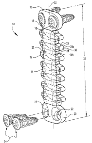
Fig. 1 is a perspective view of one embodiment of a bone fixation device 10 in
accordance with the present invention. Bone ftxation device 10 includes an
elongate
member 12 and a reinforcing component 14. Elongate member 12 can define a
longiW dinal axis 33 and can include a first end 16 that can be configured for
attachment to
one or more bony stn~ctures. In the illustrated embodiment, first end 16
includes first and
second openings 18 and 19, respectively, through which a bone fastener can be
inserted.
Second end 20, opposite first end 16, can be similarly configured to be
secured to bony
structures and can include third and fourth openings 22 and 23. In alternative
embodiments, either or both of first end and second end 20 can be configured
with a single
opening, a plurality of openings, or no openings. In any of the embodiments,
elongate
member 12 can be secured to bony structures using a variety of fasteners.
Examples of
suitable fasteners for use in the present invention include bone nails,
staples, bone
adhesives, bone screws, bone hooks, and the like. In the illustrated
embodiment, elongate
member 12 can be secured to one or more bony structures using one or more bone
screws
24.
Referring additionally to Fig. 2, a first portion or bridge portion 25
introduces
deforniation and/or flexibility into the device 10. This flexibility can be
exhibited by
CA 02542993 2006-04-19
WO 2005/037110 PCT/US2004/034334
6
allowing movement in the longitudinal direction, i.e., translational movement.
In other
embodiments, this flexibility can arise or be derived fron ~ a rotational-
torsional movement.
A related bone plate is disclosed in US Patent No. 6,293,949, which is
incorporated by
reference in its entirety.
Bridge portion 25 is disposed between first end 16 and second end 20. Bridge
portion 25 can be fomned in whole or in part of a metal, polymer, or composite
material
that is flexible. In the illustrated embodiment, bridge portion 25 includes a
plurality of
smicttiral members or an open network. In one embodiment, the open network can
be
provided to include a plurality of trusses 26 spaced from each other by voids
28. Each
individual miss 26a and its neighbor 26b can be connected by a flexible
junction 30. The
length of bridge portion 25 and, consequently, the overall length of device 10
represented
by reference 31 can vary depending upon whether the implant is subjected to
expansive
(tension) or compressive farces. This, in turn, allows the attached bone
portions to move
either closer together or fiirther apart. In addition, or in the alternative,
bridge pouion 25
can twist about its longitudinal axis allowing the attached bone pOrt~OllS to
rotate or twist
relative to each other. It will be understood that in other embodiments, the
network of
voids is not restricted to bridge portion 25 but can be distributed along the
entire length of
elongate member 12.
The flexibility can be accomplished either by specific design configurations
of the
misses 26 interspersed with voids 28 and connected with a variety of flexible
junctions 30.
Alternatively, the flexibility can be accomplished by the choice of material
used to form
the bridge portion. In preferred embodiments, the material selected to provide
the
smictural feaW xes of the bridge portion includes resilient materials such as,
without
limitation, nitonal, titanium, titanium-vanadium-aluminum alloy, cobalt-
chromium alloy,
cobalt-chromium-molybdenum alloy, cobalt-nickel-clwomium-molybdenum alloy,
biocompatible stainless steel, tantalum, niobium, hafnium, tungsten, arid
alloys thereof;
reinforced polymeric materials, poly(ether, ether, ketone) carbon (PEEK), poly
(a.ryl,
ether, ketone) (PAEK), and the like. Consequently, if desired, bridge portion
25 exhibits
an elastic property and preferably performs analogous to a series of leaf
springs stacked on
top of each other.
It should be understood that other configurations can be used which impart the
ability of the elongate member to be flexible both in compression and
elongation as well
CA 02542993 2006-04-19
WO 2005/037110 PCT/US2004/034334
7
as rotational directions. In one embodiment, truss 26 is provided to maintain
rigidity and
support for elongate member 12. Flexible junctures 30 can be formed of a
similar
material, albeit in much thinner dimensions, to allow neighboring truss
portions 26a and
26b to approach one another and thus either elongate or decrease the distance
between first
end 16 and second end 20. Additionally, or in the alternative, flexible
juncture 30 can
allow a rotational movement such that truss 26a pivots about central elongate
axis
represented by reference line 33 while an adjacent truss portion 26b either
remains
stationary or translates rotationally to a lesser extent.
The reinforcing component 14 can be deposited on device 10. In the illustrated
embodiment, reinforcing component 14 is deposited onto and into bridge portion
25.
Consequently, reinforcing component 14 fills voids 28 interspersed between
tntsses 26a
and 26b. The reinforcing component 14 serves to stiffen the bridge portion,
and
consequently, inhibit the translation and/or rotational movement afforded the
device. This
in tuW can inhibit translation and/or rotational movement of the attached bone
portions.
The reinforcing component can be formed or composed of a variety of rigid
materials including, without limitation, resorbable polymeric materials,
resorbable
composite materials, and resorbable ceramic materials:
In one embodiment, reinforcing component 14 can include polymeric materials
formed from oligomers, homopolymers, copolymers, and polymer blends that
include
polymerized monomers derived fiom 1, d, or d/1 lactide (lactic acid);
glycolide (glycolic
acid); ethers; acids; anhydrides; olefins, such as ethylene, propylene, butene-
1, pentene-1,
hexene-1, 4-methylpentene-1, styrene, norbornene and the like; butadiene;
polyfirnctional
monomers such as acrylate, methacrylate, methyl methacrylate; esters, for
example,
caprolactone and hydroxy esters; and mixtures of these monomeric repeating
units.
Use of the term "copolymers" is intended to include within the scope of the
invention polymers formed of two or more unique monomeric repeating units.
Such
copolymers can include random copolymers; graft copolymers; block copolymers;
radial
block, diblock, and triblock copolymers; alternating copolymers; and periodic
copolymers.
Use of the teen "polymer blend" is intended to include polymer alloys, semi-
interpenetrating polymer networks (SIPN), and interpenetrating polymer
networks (IPN).
In a preferred embodiment, the reinforcing component 14 comprises a
biodegradable polymeric material including: poly(amino acids), polyanhydrides,
CA 02542993 2006-04-19
WO 2005/037110 PCT/US2004/034334
polycaprolactones, poly(lactic-glycolic acid), polyhydroxybutyrates,
polyorthoesters, and
polylactic acid, polyglycolic acid, and mixtures thereof. Specific examples of
biodegradable materials for the present invention include poly (d,l-lactide)
(PLDLA).
In other embodiments, the reinforcing component can comprise biodegradable
ceramic materials and ceramic cements. Examples of biodegradable ceramic
materials
include: hydroxy apatite, hydroxyapatite carbonate, corraline, calcium
phosphate, and
tricalcium phosphate. Examples of biodegradable ceramic cements include
calcium
phosphate cement. Such calcium phosphate cements are preferably synthetic
calcium
phosphate materials that include a poorly or low crystalline calcium
phosphate, such as a
low or poorly crystalline apatite, including hydroxyapatite, available from
Etex
Coyoration and as described, for example, in U.S. Patent Nos. 5,783,217;
5,676,976;
5,683,461; and 5,650,176, and PCT International Publication Nos. WO 9S/16268,
WO
9G/39202 and WO 98/16209, all issued to Lee et al. Use of the term "poorly or
low
crystalline" is meant to include a material that is amorphous, having little
or no long range
order and/or a material that is nanocrystalline, exhibiting crystalline
domains on the order
of na ometers or Angstroms.
In other embodiments, the reinforcing component can be formed of composite
materials. Examples of composite materials include as a base material or
matrix, without
limitation: ceramics, resorbable cements, and/or biodegradable polymers listed
above.
Each of the base materials can be impregnated or interspersed with fibers,
platelets, and
particulate reinforcing materials including hydroxy apatite particles (HA)
In one foam, the reinforcing component can comprise a resorbable, moldable
material that can be molded at an elevated temperature and then allowed to set
up into a
hardened material at around body temperature, such as the material sold under
the trade
name BIOGLASSOO discussed in WO 98/40133, which is incorporated by reference
herein.
The reinforcing component of the present invention can be tailored to degrade
at a
predetermined or preselected rate. In preferred embodiments, the reinforcing
component
degrades at a rate comparable to the new bone ingrowth into the bone defect or
bone
fission site. In particularly preferred embodiments, the reinforcing component
has an in
vivo half life of greater than three months, more preferably the ira vivo half
life of the
reinforcing component is greater than six months; still more preferably the
iia vi.vo half life
CA 02542993 2006-04-19
WO 2005/037110 PCT/US2004/034334
9
is greater than one year. By use of the term "half life", it is understood
that the
degradation rate of the reinforcing component is such that the reinforcing
component
loses half of its initial mass isr vivo, presumably due to resorption,
degradation, and/or
elimination.
The reinforcing component of the present invention provides a stabilizing
component for the inventive device. This stabilizing component can provide
rigidity and
support for both the implanted orthopedic fusion device and, consequently, the
attached
bone stllictures. In use, the load supported by the bone fixation device and
supported by
the reinforcing component can vary. This allows the fixation device to become
dynamizable, or change its support characteristics isa vavo. This change in
support
characteristics can be particularly important for developing strong, new bone
tissue at the
bone defection or fusion site. This can prevent stress shielding of the new
bone ingrowth
and can minimize the risk for the development of pseudoarthrodesis.
Fig. 3 is a plan view of yet another embodiment of a bone fixation device 50
in
accordance with the present invention. Bone fixation device 50, similar to
device 10,
includes two basic components, an elongate member 52 and a reinforcing
component 54.
Fig. 4 is an elongated side view of elongate member 52. Elongate member 52
includes a first end 55 and an opposite, second end 56 and a bridge portion 62
therebetween. Both first end 55 and second end 56 are provided with at least
one opening
53 and 60, respectively, through which a bone fastener (not shown) can be
inserted. In a
preferred embodiment, bridge portion 62 is flexible, allowing movement of
first end 55
relative to second end 56. This movement can be translational movement, i.e.,
increasing/decreasing the distance indicated by reference line 64 between
first end 55 and
second end 56, depending upon whether device 50 is subjected to tension or
compressive
force. In other embodiments, bridge portion 62 can allow for rotation or
torsional
movement of first end 55 relative to second end 56. This torsional movement
can occur
by a twisting rotation about the central axis 66 extending along the
longitudinal direction
of elongate member 52. In other embodiments, bridge portion 62 can allow first
end 55 to
bend closer to second end 5G. In this embodiment, bridge portion 62 bends in a
direction
substantially orthogonal to longitudinal axis 66.
In the fixation device 50, prior to implantation, a reinforcing component 54
is
engaged to at least a portion of bridge portion 62. In a preferred embodiment,
the
CA 02542993 2006-04-19
WO 2005/037110 PCT/US2004/034334
reinforcing component 54 envelopes or completely surrounds bridge portion 62.
Consequently, bridge portion 62 is embedded within the reinforcing component.
Reinforcing component 54 can be provided as has been discussed above for
reinforcing
component 14.
5 Fig. 5 illustrates still yet another embodiment of a bone fixation device 80
in
accordance with the present invention. Bone fixation device 80 includes an
elongate
member 82 and a reinforcing component 84. Referring additionally to Fig. 6,
elongate
member 82 is illustrated absent reinforcing component 84. Elongate member 82
includes
a first end 85 and an opposite, second end 86. Each of first end 85 and second
end 86
10 include at least one and preferably a plurality of openings 88 and 90,
respectively, through
which a bone fastener can be inserted (not shown). Elongate member 82 includes
a
flexible or narrowed portion 91. In the illustrated embodiment, portion 91 is
illustrated to
have a substantially reduced cross-sectional area measured transverse to
longitudinal axis
94 than that illustrated in adjacent portions 92 and 93 of the elongate member
82.
Narrowed portion 91 impacts flexibility into fixation device 80. Consequently,
narrowed
portion 91 allows the elongate member 82 to bend substantially orthogonal to
its
longitudinal axis 94. Additionally or in the alternative, narrowed portion 91
allows the
elongate member 82 to rotate or "twist" about the longitudinal axis 94 such
that first end
85 is non planar with second end 86, i.e., fixst end 85 does not lie in the
same plane as
second end 86.
Elongate member 82 is at least partially encased within a reinforcing
component
84. This reinforcing component reduces the flexibility of narrowed portion 91.
This
inhibits movement of first end 85 relative to second end 86. Reinforcing
component 84
can comprise a material as has been described above for reinforcing components
14 and
y 54.
Fig. 7 illustrates still another embodiment of a bone fixation device 120 in
accordance with the present invention. Bone fixation device 120 comprises an
elongate
member 124 illustrated as an elongated tubular member. Elongate member 124 can
be
provided, fox example, as an implantable orthopedic rod such as, fox example,
a spinal rod
or a cross-linking member between adjacent spinal rods. Elongate member 124
includes a
bridge portion 126 represented in the illustration with dashed lines and
disposed internal of
a reinforcing section or component 128. Bridge portion 126 is illustrated as a
rod portion
CA 02542993 2006-04-19
WO 2005/037110 PCT/US2004/034334
11
having a smaller cross-sectional area (radius or diameter) than the adjacent,
non-covered
portion 127. In addition or in the alternative, bridge portion 126 can be
provided with a
plurality of holes or voids selectively sized and spaced to introduce
flexibility into
elongate member 124. Bridge portion 126 impacts a section or portion of
elongate
member 124 that can be more readily or easily bent proximate to this narrowed
or bridge
portion 126. Reinforcing component 128 encases at least a portion of bridge
portion 126.
Reinforcing section 128 can comprise a material substantially as has been
described for
reinforcing components 14 and 54. In this embodiment, it should be understood
that
reinforcing component is illustrated as a cylindrical sleeve that
substantially smTOUnds
bridge portion 126. In alternative embodiments, reinforcing section 128 can be
provided
as a partial sleeve that partially surrounds bridge portion 126. This partial
sleeve can be
perforate or imperforate and can include a variety of slits and other openings
as desired.
Additionally, reinforcing section 128 can be welded, glued, or over molded
onto the
elongate member 124. In other embodiments, reinforcing section 128 can be
provided to
be readily separable from bridge portion 126 and/or elongate member 124. For
example,
reinforcing section 128 can be provided to translate along the longitudinal
axis 130 of
elongate member 124; i.e., slide up the elongate member 124 to reveal the
underlying
bridge portion 12G.
Fig. 8 is still yet another embodiment of a bone fixation device 150 prepared
in
accordance with the present invention. In the illustrated embodiment, bone
fixation device
150 includes an outer cylindrical rod 152 provided as an elongate member 154.
Elongate
member 154 is provided with a hollow interior or lumen into which a
reinforcing
component 156 has been inserted.
In prefeiTed embodiments, elongate member 154 is provided as a flexible
conduit
that can be bent and shaped as desired. The elongate member 154 can be pre-
bent by the
manufacturer or bent by the surgeon either immediately prior to or during
surgery.
Reinforcing component 156 is provided to be disposed in the interior section
160 of
elongate member 154.
The reinforcing component 156 can comprise a material as has been described
for
reinforcing components 14, 84, and 128. Furthermore, reinforcing component 156
can be
separable from elongate member 154. Elongate member 154 and reinforcing
component
156 can be provided to the surgeon as separate components that can be combined
by
CA 02542993 2006-04-19
WO 2005/037110 PCT/US2004/034334
12
sliding elongate member 154 over reinforcing component 156 either prior to or
during
surgery. Alternatively, bone fixation device 150 can be provided to the
surgeon as a one-
piece unit that is ready for implantation or that can be molded, bent, or
deformed as
desired and/or as deemed medically expedient by the orthopedic surgeon.
Furthermore,
reinforcing component 156 inhibits the flexibility of elongate member 154.
Consequently,
when combined together, reinforcing component 158 and elongate member 154
provide a
stiff rod that inhibits both movement, either translational, rotational, or
torsional.
In additional embodiments, elongate member 154 can be secured to one or more
bone portions to induce bone fusion or arthrodesis. This can be accomplished
using a
variety of techniques including gluing, staples, bone screws, hooks, and the
like as known
in the art. Bone fixation devices, elongate members, and reinforcing
components described
in the present invention can be fabricated by a wide variety of techniques,
including
injection molding, extension molding, over molding, blow molding, transfer
molding, and
the like.
Fig. 9 is a perspective view of another embodiment of a bone fixation device
180
for use in accordance with the present invention. Bone fixation device I80
includes an
elongate member 182 and a reinforcing component 196. Elongate member 182 can
be
attached to two or more bone portions. A first end I90 of member 182 can be
attached to
a first bone portion, such as a first vertebra, using a bone fixation device
such as a bone
screw. Similarly, second end 192 of member 182 can be secured to a second
vertebral
body using a bone fastener.
Bone fixation device 180, similar to device 120, includes an elongate member
182,
which is illustrated as an elongate rod. (See also Fig. 10.) Elongate member
182 includes
a narrowed portion 184 that has a diameter that is substantially reduced from
the
remaining portions of elongate member 182. For example, narrowed portion 184
has a
diameter that is substantially smaller than that found in neighboring portions
186 and 188
of elongate member 182. The narrowed portion 184 allows the elongate member
182 to
become flexible, i.e., it can be bent and/or twisted to allow translational
andlor rotational-
torsional movement. For example, narrowed portion 184 can allow a first end
190 of
member 182 to bend toward second end 192 substantially orthogonal to the
longitudinal
axis 194. Additionally, narrowed portion 184 can allow first end 190 and/or
second end
192 to twist about axis 194 to allow rotational-torsional rotation.
CA 02542993 2006-04-19
WO 2005/037110 PCT/US2004/034334
13
In preferred embodiments, elongate member 182 is provided as a spinal rod, a
connecting member between adjacent spinal rods, and/or a spinal rod and a bone
fastener
and/or an orthopedic implant to promote spinal fusion. Reinforcing component
196 is
provided as a movable sleeve 197 about elongate member 182. Movable sleeve I97
can
be provided in a first position illustrated in Fig. 9 wherein sleeve 197 is
disposed adjacent
to or around narrowed portion 184. In this configuration, sleeve 197 inhibits
deformation
of narrowed portion 184 and, consequently, elongate member 182 by preventing
either
bending, i.e., movement substantially orthogonal to longitudinal axis 194
and/or
rotational-torsional movement about axis 194. As seen in Fig. 10, sleeve 197
is slidabIy
disposed about elongate memL~er 182. Consequently, sleeve 197 is provided to
have a
diameter that is larger than the external diameter of elongate member 182.
Alternatively,
elongate member 182 can be provided with at least a portion that has an
external diameter
smaller than the internal diameter of sleeve 197. When thus configured, sleeve
197 can be
slidably disposed about elongate member 182. As shown more fully in Fig. 10,
sleeve 197
can slide either upward or downward on elongate member 182 and expose the
narrowed
portion 184. When thus exposed, narrowed portion 184 can be deformed to allow
the
attached bone portions to have either translational and/or rotational-
torsional movement in
respect to one another.
In use, any of the bone fixation devices 10, 50, 80, 120, 150, and 180 can be
used
to secure and treat bone defects. For example, as illustrated in Fig. 1 l, the
bone fixation
device 10 can be used to treat a spinal defect. In this specific illustration,
the spinal defect
occurs either on the inferior end plate 200 of vertebra 202 and/or the
superior end plate
204 of vertebra 206. The surgeon can perform either a full or partial
discectomy if desired
and if the defect occurs in tile nucleus pulposa and/or spinal disc structure.
The
discectomy can include either replacing the disc with a disc prosthesis and/or
inserting a
spinal spacer between the affected vertebrae, which spinal spacer can induce
bone fusion
or not, as desired. The illustration uses bone fixation device 10 by attaching
its first end
1G to first vertebra 202 and attaching its second end 20 to an adjacent,
second vertebra
206. Device 10 maintains the desired disc space height 208 and maintains
vertebrae 202
and 206 in a rigid confirmation relative to one another.
Referring additionally to Fig. 12, it can be observed that over time or under
selected conditions, the reinforcing component I4 of device 10 has been eroded
or
CA 02542993 2006-04-19
WO 2005/037110 PCT/US2004/034334
14
degraded away, leaving the elongate member 12. In this embodiment, it can be
observed
that a prosthetic disc 210 has been inserted between vertebra 202 and 206.
Consequently,
it is desirable to maintain the relative movement of 202 in relation to
vertebra 206. The
flexibility of elongate member 12 allows limited mobility of the two vertebrae
either by
tl-anslational and/or rotational-torsional movement relative to each other.
In addition or in the alternative, it may be desirable to promote bone fusion
between the adjacent vertebrae or between any bone portions on either side of
a bone
defect. In this embodiment, it may be desirable to include a bone growth
material such as
an osteoinductive or an osteoconductive material. For example, it may be
desirable to
introduce a osteogenic factor such as a bone morphogenic protein (BMP).
Examples of
bone growth materials include an osteoinductive factor, such as an
osteoinductive protein
or a nucleotide or a nucleotide sequence encoding an osteoinductive protein
operably
associated with a promoter (e.g., provided in a vector such as a viral
vector), for example a
bone morphogenetic protein or a gene encoding the same operationally
associated with a
promoter which drives expression of the gene in the animal recipient to
produce an
effective amount of the protein. The bone morphogenic protein (BMP) in
accordance with
this invention is any BMP able to stimulate differentiation and function of
osteoblasts and
osteoclasts. Examples of such BMPs are BMP-2, BMP-4, and BMP-7, more
preferably
rhBMP-2 or rIiBMP-7, most preferably, rhBMP-2. Purified recombinant BMPs are
preferred for use in the inventive compositions for their provision of high
osteoinductive
potentials. BMP gene sequences and methods for producing recombinant and naW
rally-
derived BMPs are lalown in the art, and for additional information on this
subject
reference may be made, for instance, to U.S. Patent Nos. 5,108,753; 5,187,076;
5,366,875;
4,877,864; 5,108,922; 5,116,738; 5,013,649; 5,106,748; and 4,294,753; and
International
Publication Nos. W093/00432; W094/26893; and W094/26892. The osteoinductive
factor may also be LIM mineralization protein (LMP) or a suitable vector
incorporating a
gene encoding the same operably associated with a promotor, as described in
W099/06563 (see also genbanlc accession No. AF095585). When such vectors are
employed as osteogenic factors in accordance with the invention, they are
preferably
delivered in conjunction with cells, for example autologous cells from the
recipient of the
implant. Most preferably the vector is delivered in conjunction with
autologous white
blood cells derived from bone marrow or peripheral blood of the recipient.
CA 02542993 2006-04-19
WO 2005/037110 PCT/US2004/034334
The osteogerlic factor will be incorporated in an amount which is effective to
stimulate the formation of bone within the animal recipient. In more preferred
compositions incorporating protein osteogenic factors, the osteogenic factor
will be
incorporated in a weight ratio of about 1:100 to about 1:1000 relative to the
overall
5 composition, more preferably about 1:100 to about 1:500. As will be
understood, when
the osteogenic factor comprises a nucleotide sequence, sufficient amounts of
the delivery
vehicle (vector) will be incorporated to cause significant transduction of
cells, so as to
cause the generation of sufficient protein at the site to induce bone
formation. The
orthopedic devices of the present invention can be used by themselves or in
conjunction
10 with one or more known orthopedic devices as deemed medically prudent.
Additionally
or in the alternative, the present invention can be used with one or more
devices disclosed
in co-pending US Patent Application Serial No. 10/689,961 filed on October 2I,
2003 and
entitled "Apparatus and Method for Providing Dynamizable Translation to a
Spinal
Construct", Attorney Docket No. 4002-3273, which is hereby incorporated by
reference.
15 The bone growth material may be used singly or in combination with one or
more
spacers, bone plates, screws, fasteners, and the like. In this alternative,
the reinforcing
component of the bone fixation device can be prepared to erode or biodegrade
at a selected
or predetermined rate. The rate of degradation can be selected to allow new
bone growth
to occur under conditions optimal to generate a dense cortical bone bridge
between the
bone portions.
While the invention has been illustrated and described in detail in the
drawings and
foregoing description, the same is considered to be illustrative and not
restrictive in
character, it is understood that only the preferred embodiments have been
shown and
described and that all changes and modifications that come within the spirit
of the
invention are desired to be protected. Any reference to a specific directions,
for example,
references to up, upper, down, lower, and the like, is to be understood for
illustrative
purposes only or to better identify or distinguish various components fiom one
another.
These references are riot to be construed as limiting in any manner to the
orthopedic
device and/or methods for using the orthopedic device as described herein.
All publications, patents, and patent applications cited in this specification
are
herein incorporated by reference as if each individual publication, patent, or
patent
CA 02542993 2006-04-19
WO 2005/037110 PCT/US2004/034334
16
application was specifically and individually indicated to be incorporated by
reference and
set forth in its entirety herein.
Unless specifically identified to the contrary, all terms used herein are used
to
include their nomnal and customary ternZinology. Further, while various
embodiments of
medical devices having specific components and structures are described and
illushated
herein, it is to be understood that any selected embodiment can include one or
more of the
specific components and/or structures described for another embodiment where
possible.
Further, any theory of operation, proof, or fording stated herein is meant to
further
enhance understanding of the present invention and is not intended to make the
scope of
the present invention dependent upon such theory, proof, or fording.