Note: Descriptions are shown in the official language in which they were submitted.
CA 02543568 2006-04-13
SURGICAL CLIP
FIELD OF THE INVENTION
[0001] The present invention relates to surgical instruments, and in
particular to surgical clips
and methods used for ligating vessels, other ducts, and the like.
BACKGROUND OF THE INVENTION
[0002] During many surgical procedures, the surgeon will have to close or
ligate various blood
vessels and other ducts before severing them in order to prevent excessive
bleeding, and reduce
the risk of other complications to the patient. One ligation technique is to
tie a suture about the
vessel to close the vessel. Alternatively, a surgeon can place a clip having a
pair of legs
connected at their proximal ends about the vessel, and urge or squeeze the
legs together to close
the vessel.
[0003] One drawback associated with some current clips used for ligating
vessels is that the legs
of the clip may tend to separate to some extent following release from a clip
applier. This
phenomenon is called duck-billing. Duck-billing can result in insufficient
ligation of a vessel,
thus leading to excessive blood loss and/or unnecessary damage to the vessel.
Further, some
known ligation clips are often difficult to preload into a clip applier
because of resistance
between the tissue disposed between the jaws and the gripping features on the
clip legs.
[0004] Accordingly, there remains a need for an improved surgical instrument
and method, and
in particular for surgical clips used for ligating blood vessels, other ducts,
and the like.
SUMMARY OF THE INVENTION
[0005] The present invention provides various methods and devices for ligating
tissue, such as
vessels, other ducts, and the like. In one aspect, a surgical clip is provided
that includes a pair of
opposed first and second leg members with a knee portion formed therebetween.
While the apex
can have a variety of configurations, in one embodiment, the apex can have
opposed ends joining
the proximal ends of said first and second leg members. Moreover, the apex can
include a notch
formed on an inner surface thereof.
1
CA 02543568 2006-04-13
[0006] The clip can have a variety of features that help provide a more secure
ligation of the
vessel. In one exemplary embodiment, the first and second leg members can
include an inner
surface having at least one tissue-grasping element formed thereon. The tissue-
grasping
elements can have a variety of configurations, such as a longitudinal tongue
formed on the first
leg member, and a longitudinal groove formed on the second leg member. The
tongue and
groove can be complementary and disposed opposite to each other. Moreover, the
tongue and
groove can extend along the entire length of the inner surface of each leg
member, or a portion
thereof. The tissue-grasping elements of the first and second leg members can
also include at
least one channel oriented at an angle with respect to the longitudinal axis
of the first and second
leg members.
[0007] In another exemplary embodiment, the first and second leg members can
include an outer
surface having at least one raised portion formed thereon. The raised portion
can be a pad
disposed on an outer surface of each of the first and second leg members
located proximal to a
point approximately midway between the apex and the knee portion of each leg
member. In one
embodiment, the raised area can be approximately one-third of the way between
the apex and the
knee, and closer to the apex.
[0008] In another aspect, a device for ligating tissue is disclosed having
first and second leg
members, with a knee portion formed therebetween. An apex can join the
proximal ends of the
first and second leg members, such that the first leg member and the second
leg member are
opposed from one another. While the apex can have a variety of configurations,
in one
exemplary embodiment, the apex includes a notch formed in an inner surface
thereof.
[0009] In another aspect, a surgical clip is disclosed being in the form of a
substantially U-
shaped member that includes an apex that joins first and second leg members.
The apex can
further include a notch formed therein. In one exemplary embodiment, the leg
members can
include at least one tissue-grasping element formed on an inner surface
thereof, and a knee
portion formed between the proximal and distal ends thereof. Moreover, each
leg member can
have a width of less than about 0.05 inch, and a yield strength greater than
about 28 ksi. In
another exemplary embodiment, the clip can include a raised area disposed on
an outer surface of
each of the first and second leg members proximal to a point between the apex
and the knee
2
CA 02543568 2013-10-03
portion of each leg member. The raised area can be approximately one-third of
the way between
the apex and the knee, and closer to the apex.
[0010] In another aspect, a device for ligating tissue is provided having
first and second opposed
leg members with proximal and distal ends, and a knee portion formed between
the proximal
ends of each of the leg members. An apex having opposed ends joins the
proximal and distal
ends of the opposed leg members. The leg members further include inner and
outer surfaces, the
outer surface having at least one raised area on a portion thereof. In one
embodiment, the raised
area is located approximately one-third of the way between the apex and the
knee portion, closer
to the apex. In other embodiments, the device can further include at least one
tissue-grasping
feature formed on the inner surface of the opposed leg members, as well as a
notch formed on
the inner surface of the apex.
[0011] In another aspect, a ligation clip is provided having pair of opposed
legs joined together
at a proximal end by an apex. The opposed legs each can have a distal end and
a knee portion
disposed distal of the apex, and a raised area formed on an outer surface of
each leg between the
apex and the knee. The raised area is effective to share with the knee
portions a load applied by
a closing force such that the knee portions are subjected to less plastic
deformation and retain
some elasticity, wherein upon release of the closing force the distal ends of
the clip remain in
contact with one another.
[0012] A method for ligating vessels is also provided where a closing force is
applied to each leg
member such that in a partially closed position the knee portions of each leg
member are
substantially parallel to one another when the distal ends of each leg member
are in contact with
one another. As the closing force is continued to be applied to the clip, the
raised areas and the
knee portions share a load applied by the closing force such that the knee
portions are subjected
to less plastic deformation and retain some elasticity, wherein upon release
of the closing force
the distal ends of the clip remain in contact with one another.
3
CA 02543568 2013-10-03
[0012a] In another aspect, there is provided a surgical clip comprising:
a pair of opposed first and second leg members having proximal and distal ends
with a knee portion formed therebetween, the first and second leg members
further
including an inner surface having at least one tissue-grasping element formed
thereon and
a generally planar outer surface; an apex having opposed ends joining the
proximal ends,
of said first and second leg members, the apex further including at least one
notch formed
on an inner surface thereof; wherein the outer surface has at least one raised
area on a
portion thereof,
wherein the raised area is raised with respect to the outer surface;
and the raised area is disposed distal to the apex and proximal to the knee
portion.
[0012b] In another aspect, there is provided a surgical clip, comprising:
a pair of opposed first and second leg members having proximal and distal ends
with a knee portion formed therebetween, the first and second leg members
further
including an inner surface having at least one tissue-grasping element formed
thereon and
a generally planar outer surface having at least one raised area on a portion
thereof,
wherein the raised area is raised with respect to the outer surface and has a
top surface
having a width that is less than a width of the outer surface; and
an apex having opposed ends joining the proximal ends of said first and second
leg
members, the apex further including at least one notch formed on an inner
surface thereof,
such that the notch has a width that is less than a width of the inner surface
of the apex;
wherein the raised area is disposed distal to the apex and proximal to the
knee
portion and the top surface of the raised area is disassociated from the apex.
BRIEF DESCRIPTION OF THE DRAWINGS
[0013] The invention will be more fully understood from the following detailed
description
take in conjunction with the accompanying drawings, in which:
3a
CA 02543568 2006-04-13
[0014] FIG. 1 is a perspective view of one embodiment of a surgical clip
disclosed herein;
[0015] FIG. 2A is a side perspective view of a clip according to another
embodiment of the
invention;
[0016] FIG. 2B is a side perspective view of a portion of the distal end of a
leg member of the
clip of FIG. 2A;
[0017] FIG. 2C is a plan view of the clip of FIG. 2A;
[0018] FIG. 2D is a sectional view of the clip of FIG. 2C along the lines 2D-
2D;
[0019] FIG. 2E is a sectional view of the clip of FIG. 2C along lines 2E-2E;
[0020] FIG. 3 is another perspective view of a clip according to the
invention;
[0021] FIG. 4A is a perspective view of a clip according to the invention;
[0022] FIG. 4B is a top plan view of an inner portion of the apex of the clip
of FIG. 4A;
[0023] FIG. 4C is a side perspective view of an inner portion of the apex of
the clip of FIG. 4A;
[0024] FIG. 5A is another side perspective view of a clip according to the
invention in an open
position;
[0025] FIG. 5B is a side perspective view of the clip of FIG. 5A in a first
state of partial closure;
[0026] FIG. 5C is a side perspective view of the clip of FIG. 5A in a state of
almost full closure;
[0027] FIG. 5D is a side perspective view of the clip of FIG. 5A fully closed;
and
[0028] FIG. 5E is a side perspective view of the clip of FIG. 5A following
release by a clip
applier.
DETAILED DESCRIPTION OF THE INVENTION
[0029] Certain exemplary embodiments will now be described to provide an
overall
understanding of the principles of the structure, function, manufacture, and
use of the devices
4
CA 02543568 2006-04-13
and methods disclosed herein. One or more examples of these embodiments are
illustrated in the
accompanying drawings. Those skilled in the art will understand that the
devices and methods
specifically described herein and illustrated in the accompanying drawings are
non-limiting
exemplary embodiments and that the scope of the present invention is defined
solely by the
claims. The features illustrated or described in connection with one exemplary
embodiment may
be combined with the features of other embodiments. Such modifications and
variations are
intended to be included within the scope of the present invention.
[0030] The present invention provides various devices for ligating tissue,
such as vessels, other
tubular ducts, and the like. FIGS. 1-4C illustrate exemplary embodiments of a
clip disclosed
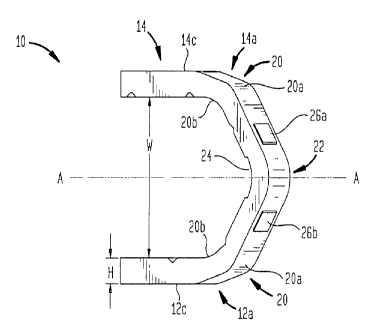
herein in an open position. Referring generally to FIG. 1, the clip 10 in its
open position is
generally U-shaped having opposed leg members 12, 14 joined at an apex 22.
Each leg member
12, 14 has a knee portion 20 disposed distally of the apex 22. Moreover, each
leg member 12, 14
has an inner tissue-contacting surface 12d, 14d and an opposed outer surface
12c, 14c, both of
which may have features to provide a more secure ligation of the vessel or
duct. For example,
the inner surface(s) 12d, 14d can include various tissue-grasping elements
formed therein
(discussed in more detail below). The outer surface(s) 12c, 14c can have at
least one raised area
26 (shown in FIG. 3) formed thereon between the knee portion 20 and the apex
22. While the
clip 10 of the invention is described herein in the context of a device to
ligate vessels, one skilled
in the art will appreciate that the surgical clip 10 of the present invention
can be used to ligate a
variety of other body tissues, including but not limited to, veins, arteries,
ducts, or any other
tubular member within a patient for which ligation is desired. Moreover, the
clip 10 can be used
in a variety of clip appliers, thereby effecting a wide range of surgical
procedures. Although the
clip 10 is described herein with respect to ligation, it is understood that a
variety of other
applications are possible as well.
[0031] The clip 10 can have any shape in its open configuration that allows it
to effectively
ligate a vessel, such as a substantially U-shaped or a substantially V-shaped
design. As noted
above, in an exemplary embodiment, the clip 10 is substantially U-shaped. That
is, proximal
portions 12a, 14a of the leg members 12, 14 of the clip 10 are oriented at an
acute angle with
respect to the central axis A of the clip 10, and transition at a knee portion
20, to an orientation
CA 02543568 2006-04-13
where distal portions 12b, 14b of the leg members 12, 14 are parallel with
respect to one another
and to central axis A.
[0032] One skilled in the art will appreciate that the size of the clip 10 can
vary depending upon
its particular application. In an exemplary embodiment, the clip 10 can have a
length / in the
range of about 5 mm to 15 mm, and more preferably in the range of about 7.5 mm
to 8.5 mm. In
its open configuration, the clip 10 can have a width Was shown in FIG. 3
measured between
opposed inner surfaces 12d, 14d of the leg members 12, 14 in the range of
about 2 mm to 8 mm,
and more preferably in the range of about 3 mm to 4 mm. The size of the leg
members 12, 14
can also vary depending upon the particular application, however in one
embodiment, each leg
member 12, 14 can have a width w, shown in FIGS. 2D and 2E, less than 0.050
inch, more
preferably in the range of about 0.025 inch to about 0.040 inch, most
preferably less than about
0.035 inch. Moreover, each leg member 12, 14 can have a height H (shown in
FIG. 3) in the
range of about 0.015 inch to 0.030 inch, and more preferably in the range of
about 0.018 inch to
0.025 inch, and most preferably in the range of about 0.019 inch to 0.020
inch.
[0033] The clip can also have physical properties, such as yield strength,
that are appropriate for
a desired application. In an exemplary embodiment, the yield strength is
greater than about 28
ksi and less than about 60 ksi, and more preferably in the range of about 30
ksi to 50 ksi. In
general, the clip 10 constructed according to the present invention has a
yield strength that is
equivalent to or greater than clips having larger dimensions.
[0034] The clip 10 of the present invention is further designed so that, upon
closure, a vessel, for
example, is completely encased between the leg members 12, 14 of the clip 10.
This is done by
urging the leg members 12, 14 of the clip 10 together, typically with the
assistance of an applier,
to surround the vessel.
[0035] Referring now to FIGS. 2A-2E, the clip 10 has opposed first and second
leg members 12,
14 each having proximal and distal ends 12a, 14a, 12b, 14b. The proximal and
distal ends 12a,
14a, 12b, 14b have opposed inner tissue-contacting surfaces 12d, 14d and outer
compression-
receiving surfaces 12c, 14c that are connected by superior and inferior sides
12e, 14e, 12f, 14f.
One skilled in the art will appreciate that the leg members 12, 14 can have
any cross-sectional
shape that allows them to effectively close and engage tissue, such as a
vessel. Exemplary cross-
6
CA 02543568 2006-04-13
sectional shapes include, but are not limited to, triangular, rectangular,
trapezoidal, and
pentagonal. As shown, however, the leg members 12, 14 are substantially
rectangular. The
substantially rectangular leg shape is believed to provide an optimized design
that includes a
greater bending resistance for a given clip leg space envelope.
[0036] The leg members 12, 14 can also have a variety of features formed
therein or thereon to
assist with the ligation of a vessel or duct. For example, the inner surface
12d, 14d of each leg
member 12, 14 can include tissue-grasping elements, and the outer surface 12c,
14c of each leg
member 12, 14 can include a knee portion 20 as well as at least one raised
area 26. Optionally,
one or more grooves may be formed on the outer surface 12c, 14c as well.
[0037] As shown in FIGS. 2A-2E, the tissue-grasping elements formed on an
inner surface 12d,
14d of each leg member 12, 14 can include both primary 16, 17 and secondary 18
tissue-grasping
elements. The primary tissue-grasping elements 16, 17 can have any
configuration that allows
them to effectively hold a vessel or duct. In one embodiment, the primary
tissue-grasping
elements can include at least one tongue 17 formed on the inner surface 14d of
the second leg
member 14 and at least one groove 16 formed on the inner surface 12d the first
leg member 12.
The groove 16 and tongue 17 can extend continuously along the inner surface
12d, 14d of each
leg member 12, 14. Alternatively, the inner surface 12d, 14d can include
multiple groove 16 and
tongue 17 segments formed therein.
[0038] The groove 16 and tongue 17 can be formed in a variety of locations on
each of the first
and second leg members 12, 14. In one embodiment, the groove 16 and tongue 17
can extend
longitudinally along the entire length or along at least a portion of the
length of the inner surface
12d, 14d of each respective leg member 12, 14. Alternatively, the groove 16
and tongue 17 can
extend from the distal end 12b, 14b of each leg member 12, 14 to just distal
from the apex 22, or
from the distal end 12b, 14b of each leg member 12, 14 to just distal to the
knee portion 20.
Moreover, the groove 16 and tongue 17 can extend distally from the apex 22 to
a position just
distal to the knee portion 20.
[0039] By way of non-limiting example, FIG. 1 illustrates a longitudinal
groove 16 and a
longitudinal tongue 17 that extend through the knee portion 20 and terminate
just distal to the
notch 24 in the apex 22. Alternatively, FIG. 2A illustrates a longitudinal
groove 16 and a
7
CA 02543568 2006-04-13
longitudinal tongue 17 that extend from the distal end 12b, 14b of each leg
member 12, 14 to a
position just distal to the knee portion 20. A second longitudinal groove 16'
and longitudinal
tongue 17' combination is then formed just distal to the knee portion 20,
extending just distal to
the apex 22. Moreover, FIG. 4A illustrates a longitudinal groove 16 and a
longitudinal tongue
17 that are formed along the entire inner surface 12d, 14d of each of the
first and second leg
members 12, 14. The groove 16 and tongue 17 combination shown in FIG. 4A
terminates in the
notch 24 of the apex 22, as will be discussed in more detail below.
[0040] The tongue 17 and groove 16 can be disposed so as to be complementary
to one another.
Alternatively, the tongue 17 and groove 16 can be located at different
locations along each
respective leg member 12, 14. In an exemplary embodiment, the tongue 17 are
groove 16 are
complementary and disposed opposite one another, such that once the clip 10 is
applied to a
vessel the tongue 17 will urge the tissue of the walls of blood vessel into
the corresponding
juxtaposed groove 16. This cooperation between the tongue 17 and the groove 16
inhibits
longitudinal and angled dislocation of the clip 10 relative to the vessel, and
it also effectively
reduces the gap between the inner (tissue contacting) surfaces of each
respective leg member 12,
14.
[0041] One skilled in the art will appreciate that the groove 16 can have a
variety of shapes. In
an exemplary embodiment, the groove 16 is complementary in shape to the tongue
17 and can be
hemispherical, rectangular, triangular, trapezoidal, or oblong. As shown in
FIG. 2B, an
exemplary embodiment uses a groove 16 that is somewhat triangular, having
opposed sidewalls
16a, 16b connected by a base portion 16c. The sidewalls 16a, 16b can be
oriented at various
angles with respect to the inner surface 12d, 14d of the leg members 12, 14.
In one embodiment,
the sidewalls 16a, 16b are oriented at an angle less than 120 degrees relative
to the inner surface
12d, 14d of the leg members 12, 14, and more preferably at an angle less than
110 degrees
relative to the inner surface 12d, 14d of the leg members 12, 14.
[0042] One skilled in the art will appreciate that the base portion 16c can
have a variety of
configurations. For example, the base portion 16c can be planar or slightly
rounded. In an
exemplary embodiment, however, the base portion 16c is slightly rounded.
8
CA 02543568 2006-04-13
[0043] One skilled in the art will appreciate that the groove 16 should be of
dimensions that are
effective to ligate tissue. For example, the groove 16 can have depths in the
range of about
0.0015 inch to 0.007 inch, more preferably, in the range of about 0.0025 inch
to 0.004 inch. In
one exemplary embodiment, the groove 16 can have a depth of about 0.0025 inch.
Further,
groove 16 can have a width in the range of about 0.004 inch to 0.020 inch,
more preferably in the
range of about 0.006 inch to 0.013 inch. Moreover, the width of the groove 16
can be uniform
throughout the length of the groove 16, or it can decrease in the proximal or
distal direction. In
an exemplary embodiment, the groove 16 has a uniform width.
[0044] One skilled in the art will also appreciate that the tongue 17 can also
have a variety of
configurations. However, in an exemplary embodiment, the tongue 17 is
complementary in
shape and size to the groove 16. Thus, the tongue 17 can be hemispherical,
rectangular,
triangular, trapezoidal, or oblong. In an exemplary embodiment, the tongue 17
is substantially
rectangular or trapezoidal.
[0045] The tongue 17 can also vary in size, however in an exemplary
embodiment, the tongue 17
has a size that is complementary to the size of the groove 16, with a height
and a width no
greater than, and preferably slightly less than, the dimensions of the groove
16. This provides
room for the vessel tissue and minimizes shearing action and locally excessive
pressures on the
vessel tissue during clip forming. That is, the tongue 17 can have a height in
the range of about
0.0015 inch to 0.007 inch, more preferably in the range of about 0.0025 inch
to 0.004 inch. In
one exemplary embodiment, the tongue 17 can have a height of about 0.0025
inch. The tongue
17 can also have a width in the range of about 0.004 inch to 0.020 inch, more
preferably in the
range from about 0.006 inch to 0.013 inch. Moreover, and also similar to the
groove 16 above,
the tongue 17 can have a uniform width or a width that decreases in the
proximal or distal
direction. In an exemplary embodiment, the tongue 17 has a uniform width.
[0046] In addition to primary tissue-grasping elements 16, 17, the inner
surfaces 12d, 14d of
each of the first and second leg members 12, 14 can have at least one
secondary tissue-grasping
element 18, as shown in FIG. 2B. While in one embodiment the secondary tissue-
grasping
elements 18 are formed on the inner surfaces 12d, 14d of both the first and
second leg members
12, 14, the secondary tissue-grasping element 18 can optionally be formed on
the inner surface
9
CA 02543568 2006-04-13
12d, 14d of only one of the first and second leg members 12, 14. One skilled
in the art will
appreciate that the inner surfaces 12d, 14d of the first and second leg
members 12, 14 can have
any number of secondary tissue-grasping elements 18. In the exemplary
embodiment, the inner
surface 12d, 14d has at least four secondary tissue-grasping elements 18.
[0047] The secondary tissue-grasping elements 18 can have any configuration
that allows them
to grasp tissue following application of the clip 10 to the vessel or duct. As
shown in FIG. 2B,
exemplary secondary tissue-grasping elements 18 are in the form of channels
having opposed
first and second walls 18a, 18b connected by base wall 18c. The channels are
generally saw-
toothed in shape, however can also be undercut. In an exemplary embodiment,
the first wall 18a
is formed at an acute angle relative to the inner surface 12d, 14d of each leg
member. In an
exemplary embodiment the angle is in the range of about 40 degrees to 90
degrees, and more
preferably the angle is about 75 degrees. The second wall 18b is likewise
oriented at an acute
angle relative to the inner surface 12d, 14d of each leg member. The acute
angle of the second
wall 18b, which is generally shallower than the angle of the first wall 18a,
can be in the range of
about 15 degrees to about 75 degrees, and more preferably it is about 45
degrees. One skilled in
the art will appreciate that the walls 18a, 18b, 18c can be straight or
arcuate, but in the exemplary
embodiment the walls 18a, 18b, 18c are slightly arcuate to facilitate
grasping.
[0048] As shown in FIGS. 2D-2E, the secondary tissue-grasping elements 18
extend across the
width w of the first and second leg members 12, 14 at an angle (e.g., about 45
degrees) relative to
a longitudinal axis of the leg members 12, 14. In an exemplary embodiment, one
segment of the
secondary tissue-grasping element 18 is located on one side of the tongue 16
or groove 17 on the
first leg member 12, and a second segment 18 continues at the same angle on
the other side of
the tongue 16 or groove 17. The secondary tissue-grasping elements 18 are
similarly constructed
on the second leg member 14, however they are angled at an orientation
opposite that of the first
leg member 12. Thus, when the leg members 12, 14 close around a vessel or
duct, they form a
superimposed "x," as shown in FIG. 2E. This configuration allows for a greater
percentage of
the tissue to be grasped by the secondary tissue-grasping elements 18, thereby
resulting in more
effective ligation.
CA 02543568 2006-04-13
[0049] The leg members 12, 14 can have any number of secondary tissue-grasping
elements 18
formed thereon. In the exemplary embodiment, however each leg member 12, 14
has three
secondary tissue-grasping elements 18 formed thereon. One skilled in the art
will appreciate that
the secondary tissue-grasping elements 18 can be uniformly or non-uniformly
spaced apart from
one another. In an exemplary embodiment, the secondary tissue-grasping
elements 18 are
uniformly spaced apart from one another at a distance in the range of about
0.050 inch to 0.080
inch. Moreover, the secondary tissue-grasping elements 18 can have any size
and depth that is
effective to engage and maintain contact with tissue. However, in an exemplary
embodiment,
the secondary tissue-grasping elements 18 are sized in the range of about
0.008 inches to 0.012
inches wide by about 0.0015 inches to 0.0035 inches deep.
[0050] One skilled in the art will appreciate that the leg members 12, 14 of
the exemplary clip
10, as shown in FIGS. 1-4C, can include any combination of primary tissue-
grasping elements
16, 17 and secondary tissue-grasping elements 18. An exemplary clip 10,
however, includes
both primary and secondary tissue-grasping elements 16, 17, 18. In another
exemplary
embodiment (not shown), the inner surface 12d, 14d of the leg members 12, 14
can be smooth
and free of primary and secondary tissue-grasping elements. The structure and
closing properties
of the clip 10, as discussed herein, allow adequate tissue ligation without
the need for any type of
tissue-grasping elements formed on the inner surface 12d, 14d of the leg
members 12, 14.
[0051] As shown, for example, in FIG. 3, the outer surface 12c, 14c of each
leg member 12, 14
can include a bend or knee portion 20. The knee portion 20 allows the leg
members 12, 14 to
transition from being acutely angled relative to the central axis A of the
clip 10 to being
substantially parallel relative to one another and to the central axis A of
the clip 10. The angled
knee portions 20 of the leg members 12, 14 can be formed at a variety of
angles relative to the
central axis A of the clip 10, however in an exemplary embodiment the angle
can be in the range
of about 45 degrees to about 65 degrees. In one embodiment, the knee portion
20 is designed so
as to be parallel to the force applying jaws of a clip applier during a part
of the clip closing
process as shown in FIG. 5B. This construction is believed to enhance clip
retention by the clip
applier during deployment.
11
CA 02543568 2006-04-13
[0052] The knee portion 20 can have a variety of configurations to effect the
transition of the leg
members 12, 14, however an exemplary knee portion 20 has a beveled or
flattened outer surface
20a and an arcuate inner surface 20b. The bevel on the outer surface 20a can
extend over any
length sufficient to effect the transition, however in an exemplary embodiment
the bevel is in the
range of about 0.030 inch to 0.050 inch. The outer surface 20a of the knee
portion 20 can
optionally include a groove (not shown) formed therein to facilitate formation
of a raised tongue
17 on the inner surface 12d, 14d of the leg members 12, 14. The groove can be
similar in shape
and size to the longitudinal groove 16, discussed herein with respect to FIGS.
2A-2E. The inner
surface 20b of the knee portion 20 can also optionally include features to
assist with the ligation
of the vessel, duct, or tissue. For example, the inner surface 20b can include
primary and/or
secondary tissue-grasping elements 16, 17, 18 similar to those discussed above
with respect to
FIGS. 2B-2D.
[0053] As noted above, the outer surface 12c, 14c of each leg member 12, 14
can have features
to help provide a more secure occlusion and clip performance. In one
embodiment, shown in
FIG. 3, a raised area 26 extends over a portion of the width of the leg
members 12, 14 that is
slightly proximal to the knee portion 20. In an exemplary embodiment, the
raised area 26 is
located approximately one-third of the way between the apex 22 and the knee
portion 20, closer
to the apex 22. The raised portion 26 is believed to help to reduce
overbending of the knee 20 as
well as to help maintain the legs 12, 14 of the clip 10 together after the
clip 10 is fully closed.
While FIG. 3 shows the raised area 26 formed on both the first and second leg
members 12, 14,
in alternate embodiments, the raised area 26 can be formed on either the first
leg member 12 or
the second leg member 14. Moreover, the outer surface 12c, 14c of each leg
member 12, 14 can
have any number of raised areas 26. In the exemplary embodiment, the outer
surface 12c, 14c of
each leg member 12, 14 has one raised area 26a, 26b.
[0054] The raised area 26a, 26b can have any shape that allows the effective
application of
compressive force to the apex 22 such that the apex 22 is crimped to a greater
degree than the
knee portion 20. That is, the raised area 26a, 26b is believed to allow the
region of the leg
member 12, 14 between the apex 22 and the knee 20 to be more elastic, enabling
the knee
portion 20 to spring back to a small degree while maintaining adequate contact
between the
distal ends 12b, 14b of the leg members 12, 14. In an exemplary embodiment,
the raised area
12
CA 02543568 2006-04-13
26a, 26b is a pad having a shape that is complementary to the shape of the leg
member 12, 14.
Thus, the raised area 26a, 26b can be triangular, rectangular, trapezoidal,
pentagonal, etc., but in
an exemplary embodiment, the raised area 26a, 26b is substantially
rectangular.
[0055] One skilled in the art will appreciate that the raised area 26a, 26b
can have a variety of
sizes, depending upon whether full closure or partial closure of the clip is
desired. By way of
non-limiting example, if full closure of the clip is desired, the height of
the raised area 26a, 26b
should be able to maintain the preload at the distal tips of the leg members
12, 14. In an
exemplary embodiment, the raised area 26a, 26b has a height in the range of
about 0.0005 inch to
0.0025 inch, and more preferably is about 0.001 inch. The raised area 26a, 26b
can also have a
length that is large enough so that it can adequately sustain the applied
pressure from a clip
applier. In an exemplary embodiment, the raised area 26a, 26b can have a
length of about 0.020
inch, and a width of about 0.010 inch. If partial closure of the clip is
desired, the height of the
raised area 26a, 26b can be increased.
[0056] As noted above, the proximal ends of each of the leg members 12a, 14a
are connected to
one another by an apex 22. While the apex 22 can have a variety of shapes, as
shown in FIGS.
4A-4C, the apex 22 is substantially U-shaped or substantially V-shaped, and
has opposed inner
(tissue-contacting) 22d and outer (non-tissue contacting) faces 22c that are
connected by superior
and inferior surfaces (not shown).
[0057] The inner surface 22d of the apex 22 can have a variety of
configurations in order to
assist with ligation, for example, at least one notch 24 can be formed
therein. While the inner
surface 22d can have any number of notches formed therein, an exemplary
embodiment utilizes
one notch 24. One skilled in the art will appreciate that the notch 24 can
have any configuration
that allows for the ligation of tissue. In an exemplary embodiment, the notch
24 is formed in a
U-shaped channel that extends through the inner surface 22d of the apex 22.
The U-shaped
channel may join the tongue 16 and groove 17 that extend along at least a
portion of length of the
inner surface 12d, 14d of the leg members 12, 14.
[0058] The notch 24 can further have a variety of shapes to optimize its
mechanical properties
and make it stiff and strong for the amount of material in it, yet leaving
open space for the
material in compression on the inner side of the clip 10 to flow into during
the plastic
13
CA 02543568 2006-04-13
deformation that occurs during clip formation. In an exemplary embodiment, as
shown herein,
the notch 24 is substantially trapezoidal. That is, as shown in FIGS. 4B-4C,
the notch 24 has
opposed first and second walls 24a, 24b connected by opposed third and fourth
walls 24c, 24d
with a base portion 24e extending therebetween. While the walls 24a, 24b, 24c,
24d can have a
variety of configurations, in an exemplary embodiment the walls 24a, 24b, 24c,
24d are formed
at an acute angle relative to the inner surface 22d of the apex 22. The angle
can be any acute
angle, but it is preferably in the range of about 75 degrees. One skilled in
the art will appreciate
that the walls 24a, 24b, 24c, 24d, 24e can have also have any shape that
provides an area into
which deformed tissue can flow. As shown, the walls and the base portion 24a,
24b, 24c, 24d,
24e are rounded or slightly contoured.
[0059] The notch 24 can have a variety of sizes and depths, perhaps best
described in
relationship to the thickness and width of the clip leg members 12, 14. The
width of notch 24
should be such that the webs of material at apex surface 22d are in the range
of about 0.005 inch
to 0.010 inch wide. The depth of notch 24 should be in the range of about 30
percent to 60
percent of the distance between apex surfaces 22c and 22d, with an exemplary
range of about 30
percent to 40 percent of the distance between surfaces 22c and 22d. The length
of notch 24
should be in the range of about 1 times to 2 times the thickness of the clip
leg members 12, 14,
with an exemplary length in the range of about 1.1 times to 1.4 times the
thickness of the clip leg
members 12, 14. In the case of larger, wider clips, optimum results might
require the use of two
or more notches in order to maintain the webs of material at surface 22d in
the range of about
0.005 inch to 0.010 inch. Other aspects of multiple notches would be expected
to follow the
guidelines listed above.
[0060] The outer face 22c of the apex 22 can also have a variety of
configurations in order to
assist with ligation. In an exemplary embodiment, the outer face of the apex
22c has two
opposed beveled surfaces that meet in a rounded tip. The outer face 22c of the
apex 22 is not
sharply formed, but rather has a fabrication-induced radius, thereby allowing
for a more secure
ligation.
[0061] The clip 10 disclosed herein can be made from a variety of surgically-
appropriate
materials including metals and polymers. Moreover, the material can be a
bioabsorbable
14
CA 02543568 2006-04-13
material or a non- bio absorbable material. In one embodiment, the clip 10 can
be made of a
metal or a metal alloy having a relatively high annealed state yield strength
and a relatively high
strain hardening rate, in comparison to existing ligation clips. Suitable
metals include tantalum,
titanium, stainless steel, or alloys thereof. By way of non-limiting example,
the clip 10 can be
made from commercially pure titanium or ASTM grade CP1 titanium. This
material, when
compared with conventional materials, is able to be strain hardened to a
greater extent without
causing excessive gaps in the formed clip 10.
[0062] Moreover, a small amount of interstitial elements, such as oxygen or
nitrogen, can be
added to the clip material to maintain the formability of the clip 10. In an
exemplary
embodiment, oxygen can be incorporated within the clip material. Other
interstitial elements can
include nitrogen, carbon, and iron. The clip 10 can also optionally be coated
with an
antimicrobial or antibiotic material in order to increase the effectiveness of
the clip against a
broad range of infectious agents or pathogens.
[0063] FIGS. 5A-5E sequentially illustrate selected steps of clip closure, for
example to ligate a
vessel. As shown in FIG. 5A, an open clip 10 is presented, and it can be
placed around a desired
vessel. A closing force is then applied to the outer surface 12c, 14c of the
leg members 12, 14
by, for example, the force-applying jaws 100 of a clip applier. As clip
closure begins, as shown
in FIG. 5B, the knee portion 20 and the apex 22 are deformed such that the
distal ends 12b, 14b
of the leg members 12, 14 are moved inward towards one another. In the
position shown in FIG.
5B, the clip features at the knees 20 have become predominately parallel to
each other and to the
clip applying jaws 100, helping to stabilize the clip 10 in the jaws 100 of
the applier.
[0064] As the application of closing force to the clip 10 continues and the
distal ends 12b, 14b of
the leg members 12, 14 move closer to one another, the raised area 26 begins
to share the clip
radial closure forces with the knee portion 20. As a result of this reduction
in pressure, the knee
20 is deformed to a lesser extent, as shown in FIG. 5C. FIG. 5D illustrates a
condition of full
clip closure, with the closing force still applied to the clip 10 by the
closing jaws 100. At the
final stages of crimping, the raised area 26a, 26b takes some load off of the
knee portion 20,
thereby reducing the amount of plastic deformation of the knee portion 20. The
raised area 26
thus allows the knee portion 20 to have increased elasticity, such that, for
example, the knee
CA 02543568 2013-10-03
portion 20 can bend inward slightly when forming loads are released,
preloading the tips of the
clip 10. This is particularly advantageous in that when the applier is removed
from the clip 10 as
shown in FIG. 5E, the raised area 26 allows the leg members 12, 14 to remain
together from the
knee portion 20 to the distal ends 12b, 14b thereof, thereby lessening the
duck-billing of the clip
10.
[0065] One advantage provided by the clip 10 of the present invention is that
it tends to be more
resistant to "duck-billing," a condition in which the distal tips of the leg
members 12, 14 of the
clip 10 tend to separate after the closing force is removed. Some previously
known clips tend to
duckbill as a result of residual elasticity within the apex. The clip 10 of
the present invention is
believed to overcome the tendency to duckbill because the apex 22 is able to
crimp to a greater
extent and thus minimize the effect of any springback. At the same time,
increased elasticity
between the apex 22 and the knee portion 20 enables any springback at the knee
portion 20 to
direct the distal ends 12b, 14b of the leg members 12, 14 toward each other.
An additional
advantage of the above-mentioned characteristics of the clip 10, is that
tissue is able to be
captured at any location within the clip 10, including near the apex 22 or
near the distal ends 12b,
14b of the leg members 12, 14, and still be effectively ligated. As a result,
a surgeon can
securely ligate vessels having a variety of sizes.
[0066] One skilled in the art will appreciate further features and advantages
based on the
above-described embodiments.
16