Note: Descriptions are shown in the official language in which they were submitted.
CA 02543977 2006-04-27
USE OF PARTICULATE LABELS IN BIOANALYTE DETECTION METHODS
[0001]
Technical Field
[0002] The invention relates to methods that utilize submicron sized
particulate labels
containing signal-generating moieties whose characteristic hue can be adjusted
over a range
of distinguishable types, thus permitting a variety of multiplexed assays. The
invention
also relates to assays for which multiplexed particles provide a preferred but
not exclusive
embodiment. The invention also relates to the field of clinical investigations
of new drugs
and assessment of responses of individual subjects to treatment protocols.
Background Art
100031 It is often desirable to test a sample for reactivity against a
multiplicity of
reagents. For example, in cytokine secretion profiling, the ability of the
secreted proteins
from a particular tissue to immunoreact with a panel of antibodies raised with
respect to the
family of antigens is required, with tens to hundreds of family members of
potential
interest. In this instance, detection of the presence of the antibody-antigen
complex
normally requires that either a label be attached to the antibody in the
complex or that a
second antibody be bound to the first antibody where the second antibody has a
label
attached to it. Then, detection of the label confirms the presence of the
antibody-antigen
complex in the sample.
100041 Commonly, the second antibody is a biotinylated goat anti-primary IgG
that will
react with avidin-horse radish peroxidase and, in the presence of a redox
sensitive color
indicator and substrate (hydrogen peroxide), result in a change in color, on a
filter for
example, indicating the presence of the antibody-antigen complex.
Alternatively, instead
of biotinylating the secondary antibody, an 125I-labeled secondary antibody
can be used. If
an 1251-label is used, exposure of the filter to X-ray film will allow for the
detection of the
antibody-antigen complex.
1
CA 02543977 2006-04-27
WO 2005/045396 PCT/US2004/037077
[0005] Often the signal emitted from these labels is not strong enough to be
detected
due to the low expression level of the protein of interest or limited supply
of specimen. In
addition, although it is possible to detect the presence of several different
antigens in a
sample by using an antibody directed towards each antigen, a common readout
for all
antibodies makes it impossible to clearly distinguish one antigen-antibody
complex from
another without prior fractionation (e.g., by gel electrophoresis) or parallel
assay of
specimen aliquots (including capture on discrete locations on a chip or a
combinatorially
colored particle set). To address the sensitivity issue, it would be
advantageous to have a
label that emits a signal strong enough to allow detection of antigens in a
sample even if the
antigen is present at low amounts. It would further be advantageous to
determine the
presence of multiple antigens in a complex mixture by virtue of a family of
such labels with
distinguishable signals. For example, serological tissue typing for HLA
antigens probes ¨6
genetic loci for dozens to hundreds of allelic variants at each locus. Any
given individual
will at most express 2 alleles for each locus, but hundreds of separate assays
are needed to
accomplish the typing. With multiplexing, all the assays can be run on one
specimen,
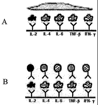
providing a more efficient system. For an application such as this, it is
important that the
staining reagents themselves be multiplexed, as contrasted to multiplexed
binding surfaces
to which a single staining reagent binds.
[0006] An even more compelling need for sensitive, highly multiplexed
detection is
readily apparent in the case of cytokine secretion assays in which the
proteins secreted from
a single cell are captured on the underlying surface and then analyzed in
situ. Such assays
have heretofore only been described at the 2-plex level, with the vast
majority of work at
the 1-plex level to avoid the increased assay complexity inherent in
previously available
multiplexing approaches. Increasing the multiplexing capacity enables
identification of
novel T-cell subtypes that would require immense effort to discover by looking
only at
pairwise combinations of the two dozen or more cytokines. More generally,
normal cell to
cell variation makes it difficult to identify novel cell types based on
multivariate properties
when only two or three properties are measured per cell.
[0007] It is also desirable to multiplex DNA sequencing. Originally,
sequencing by the
chain-termination method involves the synthesis of a DNA strand by a DNA
polymerase
using a single stranded template. Synthesis initiated at the site where an
oligonucleotide
primer anneals to the template was terminated by the incorporation of a low
level of
2
CA 02543977 2006-04-27
WO 2005/045396 PCT/US2004/037077
radio-labeled nucleotide analog (ddNTP) into the elongation reagent cocktail.
When proper
mixtures of dNTP's and one of the four ddNTP's are used, polymerization
terminates
randomly at each possible site allowing for the sequence of the DNA to be read
following
size separation by gel or capillary electrophoresis. Four parallel reactions
are fractionated
in parallel lanes to identify the base at each termination length. More
recently, the four
reactions have each been terminated using a ddNTP conjugated to a different
color of
fluorescent dye, and all four reaction product mixtures fractionated together
in one lane of a
gel or one capillary electrophoresis channel. Thus, what was originally a 1-
plex assay
became a 4-plex assay. With the additional multiplexing capacity of the
present invention,
more than one DNA template could be sequenced in a single lane or capillary.
[0008] High detection sensitivity is also important for multiplexed DNA
sequencing to
avoid overloading the gel or capillary which can perturb the migration of the
DNA
molecules. The signal emitted from conventional radioactive or fluorescent dye-
based
labels is often not strong enough to be detected without extensive
amplification of the
DNA; a more sensitive label enables decreasing assay complexity and the
associated
potential for artifactual results. And, with the extra data channels provided
by sensitive
multiplexing, an internal sizing ladder can be included in every lane.
[0009] One embodiment of suitable multiplexing technology has been previously
described in detail in U.S. patents 6,642,062 and 6,492,125. Briefly, in a
preferred
embodiment latex (polystyrene) particles are impregnated with organic dye
fluors in
varying ratios to generate a combinatorially coded set of labels.
Alternatively, fluorescent
labels comprising nanocrystalline semiconductor structures of various types,
commonly
called quantum dots, may be employed. Such particles can also be coupled to
biorecognition molecules, or can be used in other particulates in varied
ratios as substitutes
for fluorescent dyes. Han, M.Y., et al., Nat. Biotechnol. (2001) 19:631-635
describes
polystyrene particles embedded with multicolor CdSe quantum dots at various
color and
intensity combinations. Based on entirely different principles, Nicewamer-
Pena, S.R., et
al., Science (2001) 294:137-141 have reported a metallic nanobarcoding
technology for
multiplexed bioassays.
[0010] The use of submicron particles that are bright enough for single
particle
detection enables a variety of assay formats not accessible to conventional
signal
generating labels for which integrated intensity of a population of labels is
measured. In
3
CA 02543977 2006-04-27
WO 2005/045396 PCT/US2004/037077
addition to latex microspheres and quantum dots, for which fluorescence is the
signal, other
possible signals include phosphorescence, NMR spectra and Raman spectra, and
modifiable reflectance properties.
[00111 The use of particulate labels to investigate spatial relationships
among
individual cellular components is described in U.S. patent 6,642,062,
incorporated herein
by reference. As described in this patent, individual particles coupled to
reagents specific
for various cellular components can be prepared in a multiplicity of
distinguishable "hues"
which are detectable by microscopy and can provide a picture of the spatial
arrangement of
intracellular components and organelles. Further, as described in U.S.
6,673,554, the
changes in spatial arrangement of these components in response to stimuli may
be used to
evaluate the toxicity of compounds and to identify treatment protocols for
disease
conditions. The present invention, in one embodiment, relates specifically to
the
application of these techniques to clinical biopsy samples using these and
additional
techniques which rely on the sensitivity and multivariant nature of
particulate labeling.
Certain improvements in particulate labels themselves are also described.
[0012] The identity of the particulate labels can be assessed, e.g., for
fluorescent labels
with a suitable excitation light source and emission filters able to detect
wavelengths from
the blue to the near infrared, microscopically to determine the position or
presence of a
single particulate label. Therefore, multiple antigen-antibody complexes, or
other
biospecific pairs, can be distinguished in a sample by the unique emission
properties of
each particulate label, enabling multiple parallel assays to be run at the
same time in the
same physical chamber. In addition, due to the high detectability of
particulate labels, as
compared to conventional dye molecules, sequencing reactions and the like can
be run even
when available sample size is too small for conventional analysis. Thus the
present
invention provides for a more sensitive and more versatile approach to detect
the presence
of proteins, nucleic acids and the like in a sample or samples. Specific
innovative uses of
particulate labels are disclosed relating to particular types of multiplexed
biospecific
interactions. Certain of these assays, whose development was prompted by the
availability
of convenient particulate labels, are novel in their own right. Although
particulate labels
provide a preferred embodiment, the use of particulate labels is not a strict
requirement for
such assays.
4
CA 02543977 2012-11-05
CA 2543977
[0013] Assays of clinical interest are also provided. It is well
understood that the cost of
bringing a new drug to market is now of the order of $800,000,000, a number
driven by the high
failure rate of drug candidates. Most of this failure rate is attributable to
the reliance by the
industry on animal studies in preclinical trials; the transition from results
in animals to results in
humans is not marked by a one-to-one correspondence. It would therefore
represent a step forward
to utilize biopsied human tissue samples to assess disease conditions and
efficacy of drugs. The
present invention facilitates the use of such samples.
[0013A] Various embodiments of this invention provide a method to identify
cells that
secrete an immunoglobulin of desired specificity and affinity which method
comprises: a)
providing a multiplicity of cell samples on a surface at a multiplicity of
locations; b) probing the
surface with a multiplicity of epitopes and antigens each labeled with a
distinguishable particulate
label; c) selecting any location of the surface to which desired epitopes and
antigens are bound, but
undesired epitopes and antigens are not bound; and d) correlating the location
of the surface thus
identified with the location of a cell sample on the surface.
10013B1 Various embodiments of this invention provide a method to identify
cells that
secrete an immunoglobulin of desired specificity and affinity which method
comprises: a)
providing a footprint of immunoglobulins for each of a multiplicity of cell
samples on a surface; b)
probing the surface with a multiplicity of epitopes and antigens each labeled
with a particulate
label; c) selecting a location on the surface of a footprint which contains a
particulate label bound to
said desired epitope or antigen, but does not contain particulate labels bound
to undesired epitopes
and antigens; and d) correlating the location of the footprint on the surface
thus selected with the
cell sample that provided the selected footprint on the surface. The cell
samples may be provided
on a membrane that overlays the surface with the membrane being permeable to
selected
immunoglobulins but not to the cell samples.
[0013C] Various embodiments of this invention provide a method to identify and
recover
cells contained within a cell sample that secrete an immunoglobulin of desired
specificity and
affinity, which method comprises: (a) providing onto a sample surface a
multiplicity of cell
samples, each sample containing cells that secrete immunoglobulins onto the
sample surface,
wherein the samples are dispensed onto specified locations on the sample
surface; (b) probing said
locations with a multiplicity of epitopes, antigens or both, each labeled with
a distinguishable
particulate label, wherein said multiplicity includes desired epitopes,
antigens or both, and
undesired epitopes, antigens or both, to which the immunoglobulins may bind;
(c) selecting by
CA 02543977 2012-11-05
CA 2543977
microscopic observation of said particulate labels wherein independent
particulate labels can be
distinguished as individual particulate labels by said microscopic
observation, any location of the
surface which comprises immunoglobulins that bind to desired epitopes,
antigens or both, but not to
undesired epitopes or antigens; (d) identifying the cell sample at the
selected location; and (e)
recovering the identified cell sample.
[0013D] In the aforementioned embodiments, the surface may comprise a capture
reagent for
immunoglobulins. The methods may further comprise immortalizing cells
identified by the method
and/or cloning a nucleic acid encoding the immunoglobulins secreted by cells
identified by the
method.
Disclosure of the Invention
[0014] The invention provides improved methods to interrogate single
cells using
microscopy. By employing the methods of the present invention, profiles of the
compositions of
individual cells may readily be obtained and, in some embodiments, compared to
those of other
cells. Particular applications of such interrogation include obtaining results
in clinical situations
and in evaluating protocols for treatment. The methods also permit
identification of single cells
with desired characteristics, wherein the cells may be further manipulated
such as altering their
genetic component or immortalizing them. In the improved methods of the
invention, it is
generally possible to maintain viability of the single cell being interrogated
and to retrieve it for
further manipulation if desired.
[0015] In one aspect, the invention is directed to a method to obtain a
sample that may be
used in characterizing a subset of components of an individual cell, which
method comprises
supporting said cell on a permeable membrane support, said support having been
overlaid on a
sample surface, and allowing cellular components to penetrate the membrane and
be deposited on
the sample surface.
[0016] The membrane supporting the cell (or supporting the residue
thereof which does not
permeate the membrane) may then be removed from the sample surface and
retained. Typically, in
this method, a multiplicity of single cells is supported in a pattern on the
membrane which has been
overlaid on at least one said sample surface. When the membrane is lifted
containing the cell or
remainder of the cellular components that do not permeate, the arrangement on
the membrane
corresponds to that of the sample surfaces so that the results from an
individual sample surface may
be correlated to result from
5a
CA 02543977 2006-04-27
WO 2005/045396 PCT/US2004/037077
particular single cells. The sample surfaces are examined microscopically to
determine
their contents and characteristics and the data associated with each cell
noted or recorded.
[0017] Depending on the information sought to be gained, the cell or cells may
simply
be permitted to secrete proteins and the composition of the mixture of
secreted proteins
determined. Alternatively, the cell or cells may be disrupted and their
intracellular contents
included in the sample surface to be tested. A particularly useful application
of the former
embodiment is to provide information as to the nature of immunoglobulins
secreted by the
cell.
[0018] The foregoing embodiment may also be employed to test the effects of a
drug or
protocol on cells as measured by changes in the profile of components measured
in the
assay.
[0019] In another aspect, the invention is directed to a method to obtain a
multiplexed
characterization of single cell components which method comprises providing a
sample of
such components with a multiplicity of particulate labels, each displaying a
different hue
and further comprising a reagent which is a specific binding partner for a
particular cellular
component. As the particulate labels can be identified individually using a
wide field or
confocal microscope, numbers of individual particulate labels associated with
each
component can be determined. The resultant profile may be represented
computationally
by representing a multiplicity of data points ¨ one data point for each of n
components
evaluated plotted in n-dimensional space ¨ thus providing a vector in such
space for each
cell. The vector for each cell is then projected in three-dimensional space to
provide a
visible/comprehensive characteristic position. In this aspect, as well, the
assay may be
adapted to demonstrate the influence of a drug, protocol or other external
stimulus, such as
a toxin on the profile of cellular components.
[0020] In still another aspect, the invention is directed to an improved
method to obtain
immortalized cells that secrete desired immunoglobulins. In this method,
because of the
sensitivity obtainable, individual B-cells isolated directly from spleen,
peripheral blood or
lymph nodes, including mucosal-associated lymphatic tissue, can be tested
individually for
secretion of desired antibodies. Because the assay can be multiplexed, high
throughput
assays are practical to retrieve cells not only that provide desired antigen
recognition, but
also that bind specifically to one or more of a variety of epitopes of a
single desired
antigen, and/or to retrieve a multiplicity of cells that bind a multiplicity
of antigens. In this
6
CA 02543977 2006-04-27
WO 2005/045396 PCT/US2004/037077
method, a particulate label which is immunoreactive with antibodies in general
may be
used as a control if desired and the number of particulate labels that bind to
these generic
antibodies compared to the number of labels that bind antibodies
immunoreactive with the
desired antigen or epitope. In general, cells may be screened for antigen
independent
properties such as isotype. In this way, the affinity of the antibodies
secreted by particular
cells can be rank ordered. This is a significant advance over conventional
assays in which
weak affinity at high secretion rate is not distinguishable from strong
affinity at low
secretion rate.
[0021] Many of the foregoing methods are applicable in clinical contexts, and
in assays
to evaluate therapeutic protocols, and the invention includes these
applications of the
invention methods.
[0022] In an additional specific aspect, the invention is directed to a method
of
evaluating drug performance by evaluating the profile of the T-cell
population. Other
indicators can also be used to characterize disease states, including
translocation patterns,
cell surface antigen staining, and the like. Thus, T-cells and tumor cells,
for example, can
be characterized by their surface antigen pattern which pattern may be
elucidated by the use
of the particulate labels of the invention. Intracellular translocation may
also be used as an
indicator.
[0023] In another aspect, the invention is directed to a particular type of
particulate
label wherein a relatively large particle assigned a particular hue by a
combination of
signal-generating entities is coated with a defmed number of smaller particles
each with its
own distinctive hue. Because microscopy can distinguish sizes, such tandem
particles
permit a great expansion of multiplexing as will be described below.
[0024] In still other aspects, the invention is directed to methods to examine
tissues
histologically using the multiplexed labels of the invention, and to determine
the nature of
growth factors that influence embryonic development. In still other aspects,
the invention
is directed to methods that are advantageous to analyze soluble biological
samples that
have been subjected to chromatography or electrophoresis. Because of the
multiplexed
nature of the assays, multiplicities of components can be determined in a
single migration
lane. For example, sizing ladders may be included within the same lane in
which
components of biological samples are to be tested.
7
CA 02543977 2006-04-27
WO 2005/045396 PCT/US2004/037077
[0025] In another aspect, the invention is directed to an improved apparatus
and
method for removing a membrane from the sample surface, whereby an aperture
adjacent
to the surface prevents suction from creating turbulence in the upper portion
of the
membrane.
Brief Description of the Drawings
[0026] Figures 1A-1C are diagrammatic representations of the expanded
multiplexing
offered by association of tandem particles of different sizes.
[0027] Figure 2 is a photomicrograph of one such tandem particle.
[0028] Figure 3 is a schematic illustration of bioconjugation methods for
linking a
particulate label with a biomolecule.
[0029] Figure 4 shows a comparison of (a) the excitation and (b) the emission
profiles
of the organic dye rhodamine 6G and CdSe particulate labels. Similar filter
sets are useable
to measure either.
[0030] Figures 5A and 5B show a schematic of the multiplexed assay employing
the
multiplexed labels of the invention. In step 1 shown in Figure 5A, multiple
cytokines
secreted from a single T-cell are captured on an underlying surface by a
mixture of specific
capture antibodies; in step 2 shown in Figure 5B, after removing the cells the
captured
cytokines are detected using a mixture of specific detection antibodies each
coupled to a
distinguishable particulate label.
[0031] Figure 6 is a micrograph obtained using a wide field microscope image
of IL-2
secreted from a single cell and labeled with the particulate labels of the
invention. Each
individual particle bound to FL-2 is detectable by virtue of its label.
[0032] Figure 7 shows the intensity of each of two color channels for pure
particulate
labels in a multiplicity of hues. Such multiplicity is useful and sometimes
mandatory in the
methods of the invention.
[0033] Figure 8 is a bar graph showing representative 5-cytokine profiles for
each of
six cells. Decoding of the particulate labels is based on the reference label
intensities in
two color channels shown in Figure 7.
[0034] Figure 9 illustrates the secreted cytokine profiles from a multiplicity
of single
cells of two murine strains. Each point is a representation of a 5-cytokine
profile for a
single cell, projected onto the first three principal components of 5-
dimensional space.
8
CA 02543977 2006-04-27
WO 2005/045396 PCT/US2004/037077
Although both strains show a spectrum of types, the distribution is offset
between the two
strains with one strain represented by gray and the other by black dots.
[0035] Figure 10 shows the pattern of bound particulate labels obtained from
an
individual hybridoma cell resting on a membrane, with the secreted IgG
captured on the
underlying surface. This "footprint" has been probed with two distinguishable
particulate
labels. One particulate label (black) is conjugated to an anti-Ig reagent and
is used to
quantify the secretion level from the cell. A second particulate label (gray)
is conjugated to
an antigen and defines antigen specificity. With additional labels, multiple
antigen
specificities can be probed simultaneously.
[0036] Figure 11 shows one embodiment of an improved method for lifting a
membrane from a sample surface. As shown, an aperture directly above the
sample surface
prevents suction from creating turbulence at the upper surface of the membrane
to be
removed.
Modes of Carrying Out the Invention
[0037] Some of the assays of the invention utilize particulate labels and
microscopic
observation techniques to phenotype individual cells based on the cell's
constellation of
surface antigens, intracellular antigens, and/or secreted proteins. Such
assays include:
characterization of the multivariate cytokine secretion profile of single
cells under various
conditions; characterization of the specificity and other properties (such as
isotype) of
secreted immunoglobulins against one another or a panel of antigens;
characterization of
the tissue milieu associated with patterned tissue growth, including
angiogenesis and
regeneration; automated pathology in which multiple cell types are
individually
recognizable by virtue of binding cell type specific antibodies coupled to
distinguishable
particulate labels or by physical entrapment of the labels (tissue paints);
characterization of
subcellular localization of proteins; characterization of mRNA expression at
single cell
level; characterization of protein, nucleic acid, carbohydrate or lipid
content of disrupted
cells, in situ or spotted in a micro array or following chromatographic or
electrophoretic
fractionation; characterization of the affinity of a biospecific interaction
for a test pair as
compared to internal positive controls.
[0038] Although certain of these assays are known in the literature, and have
been
subjected to multiplexed analysis by other means, the utility of conducting
such assays
9
CA 02543977 2006-04-27
WO 2005/045396 PCT/US2004/037077
using particulate labels has not been previously described. The use of
submicron
particulate labels requires a sophisticated image analysis system.
Surprisingly, the number
of particles needed to give a reliable result in the assay is small enough to
be
accommodated by the field of view of a microscope able to image individual
particles. The
experiments described herein establish for the first time the feasibility of
conducting such
assays. For example, in the footprint of a cell secreting an antibody, a
typical particle count
for one field of view is 500 particles (300 urn diameter) with a background of
10-50
particles and a maximum of ¨3,000 particles that can still be resolved,
providing one and a
half log units of dynamic range. With particles of 5 p.m diameter, the
footprint of a single
cell could only accommodate 1 particle which is not sufficient for these
assays. As particle
size (and therefore intensity) goes down, however, the feasibility of
detecting individual
particles in order to define their hue becomes more difficult. In short, the
availability of
previously described multiplexing reagents, useful for bulk fluorescence
measurements or
measurements over a large field of view (e.g., a DNA chip position measuring
100 p.m or
more on a side), does not permit assays on the spatial scale of the present
invention
(comparable to the diameter of single biological cells).
[0039] For some of these assays, e.g., assay of secreted proteins, it is
advantageous to
interpose a porous membrane between the cells and the assay surface. The cells
can
thereby be removed and the "footprint" analyzed. Cells of interest, such as B-
lymphocytes
secreting a desired antibody, can be identified by their footprints and then
recovered from
the corresponding location on the membrane. This aspect of the invention
represents a
departure from traditional membrane based assays (filter lifts, Western and
Southern blots
and the like), and is independent of the use of particulate labels for the
analysis of the
footprint.
[0040] This aspect may further be improved by measures that secure the cells
to their
defined location on the membrane but do not disturb viability of the cells.
Thus, in one
embodiment, the cells are deposited on the upper surface of the membrane in
the presence
of a matrix-forming material, such as methylcellulose. When downward pressure
is exerted
on the membrane, such as through centrifugation, the cells migrate through the
semisolid
matrix and are held in place at the surface of the membrane. While
methylcellulose is
exemplified, any polymer whose behavior is similar may be substituted ¨ i.e.,
polymers that
become more rigid, e.g., on cooling, pH change or when a moderate downward
pressure is
CA 02543977 2006-04-27
WO 2005/045396 PCT/US2004/037077
exerted. Further, the integrity of the accumulation of cellular components on
the sample
surface may be improved by adding a binding partner for the relevant component
to the
matrix material either simply by mixing or by coupling the binding partner
covalently to
the material that forms the matrix. For example, if the component to be
assessed on the
sample surface is an immunoglobulin, protein A may be used as the binding
partner. In a
modification of this approach, an additional matrix containing binding
partner, such as a
dextran matrix, may be superimposed on the upper surface of the membrane
containing the
sample cells. This feature reduces the background of the assay.
[0041] In this assay, further improvements can be realized by providing an
apparatus
which is constructed so as to prevent turbulence from occurring at the upper
surface of the
membrane when it is removed from the sample surface. This can be accomplished
by
employing an aperture adjacent the sample surface to prevent suction from
creating this
turbulence. This is illustrated in Figure 11.
[0042] In general, assays which examine cellular parameters, such as secreted
proteins,
intracellular components, cell surface markers, and the like, which take
advantage of the
multiplexing capability of the particulate labels and the ability to measure
these parameters
in a single cell, can be performed either using the membrane-based technology
described
above or can be conducted simply on a surface in which the cell is embedded or
otherwise
associated. By using microscopic observation and the particulate labels of the
invention,
multiple cellular components can be enumerated and identified in a single
assay. ThiS is
particularly useful in determining the effects of an external stimulus, such
as a drug, on the
interrogated cell. In such assays, a control cell that has not been subjected
to the external
stimulus is interrogated and the profile of components compared to that
obtained from a
cell that has been treated with the external stimulus. The comparison of the
profiles
elucidates the nature of the cellular response to the stimulus. In one
embodiment, where
the stimulus is a drug, dose response curves may be obtained by exposing
individual cells
to various levels of drug and comparing each level to the control. Such assays
may be
performed on a variety of animal tissues, including human tissues where
evaluating
individual response to particular drugs or protocols is important.
[0043] The present invention is also directed to methods of using particulate
labels
coupled to biospecific probes in bioanalyte detection. The particulate labels
are
individually detectable and distinguishable and can be located in spatial
relationship to each
11
CA 02543977 2006-04-27
WO 2005/045396 PCT/US2004/037077
other, if desired. Particulate labels can be used as nonradioactive biolabels
by linking a
biorecognition moiety, such as proteins, antibodies, ddNTP's, primers, or
markers, etc., to
the particulate label. The paradigmatic particulate label is a latex particle
impregnated with
organic dye fluors, as described in U.S. patent 6,492,125 incorporated herein
by reference;
other particulate labels, e.g., those that employ quantum dots, may also be
used as further
described below. Particulate labels can also be constructed using detectable
properties
other than fluorescence, such as phosphorescence, electrochemical
luminescence, as well as
NMR or Raman signals. Further, self-assembled dendrimers can create in situ an
object
equivalent to such pre-assembled particulate labels. As described below, the
particulate
label may also be comprised of a relatively large particle to which a
multiplicity of smaller
particles are bound to the surface, wherein each of the smaller particles has,
itself, a
distinct hue.
[0044] The optical properties of particulate labels are ideal for
multiplexing, as two or
more individual signal generating moieties may be used at various ratios to
generate a
multiplicity of hues. The number of signal generating moieties may be expanded
as
desired. For example, ten intensity levels at each of six colors could
theoretically code for
one million nucleic acid or protein sequences. Particulate labels that are in
the size range
of 10-300 nm have dimensional similarity with biological macromolecules (e.g.,
nucleic
acids, proteins, and protein complexes such as ribosomes or viruses). This
similarity
allows improved integration of the nanomaterials with biological molecules, as
compared
to the much larger combinatorially colored particles (5-50 gm) known in the
literature,
although these larger particles may also be used in some aspects of the
invention. The
smaller particles have advantages in immunoassays, medical diagnostics,
targeted
therapeutics, and high-throughput drug screening when used as tags for
diffusible reagents
whose final location provides the assay readout. This is in contrast to assays
described, for
example, in U.S. patent 6,492,125 where particles with characteristic labels
are used to
identify particular analytes and the quantity of the analyte is determined by
an independent
label attached to the analyte itself. While some applications of the invention
method have
been previously described using multiplexed labels, the use of particulate
labels in assays
based on counting individual particles, as contrasted to integrating intensity
from a
population, is permitted by the ability to detect individual particles
microscopically.
12
CA 02543977 2006-04-27
WO 2005/045396 PCT/US2004/037077
[0045] Further enhancement of multiplexing can be obtained by virtue of the
ability of
microscopic techniques to discern size in addition to hue. A particularly
advantageous
embodiment of this concept is illustrated in Figures 1A-C. As shown, by way of
illustration, a set of four fluorophores may conveniently be used in various
combinations to
generate 20 hues on particles of relatively small size, e.g., 10-100 nm,
typically 50-100 nm.
Thus, 20 such particles provide 20 distinct labels. Larger particles, for
example,
200-500 nm in diameter can similarly be coded with, if desired, the same four
fluorophores
to generate another set of particles with 20 hues, as shown in Figure 1B. If
the particles of
Figure 1A and Figure 1B are used together in an assay, a total of 40
distinguishable
particles will be available ¨20 hues in two sizes. By adsorbing or covalently
coupling the
smaller particles to the surface of the larger, as shown in Figure 1C, a
multiplicity of 20 x
20 or 400 "tandem" particles with distinct hues is provided. If an assay
readout requires
proximity of two such particles, 400 x 400 = 160,000 distinguishable assays
can be
conducted (e.g., suitable for genomic scale DNA assays).
[0046] The smaller particles may be adhered to the larger ones through a
variety of
linking techniques known in the art, for example, by providing each particle
with a reactive
group and supplying a bifunctional linker. Alternatively, the smaller
particles may be
provided with a substituent containing an amino group while the larger
particles comprise
substituents with carboxyl groups; formation of an amide linkage would then
result in the
desired coupling. Alternatively, the small particle might be provided a
covalently linked
biotin and the larger particle a covalently linked avidin wherein the
biotin/avidin interaction
provides the desired linkage. A wide variety of methods to link these
particles is available
in the art.
[0047] Particles of this type have been successfully prepared. Figure 2 shows
a
photomicrograph of the embodiment shown in Figure 1C.
[0048] The tandem particles can be made more complex by increasing the number
of
sizes associated with a single particulate label. For example, a large
particle of, for
example, 1-50 Am, preferably about 10 pun, might have attached to its surface
or embedded
within it intermediate size particles of, e.g., 500 nm ¨ 800 nm and
additional, even smaller
particles of about 100-200 nm. By providing each of the smaller and middle
size particles
with a distinctive hue, the multiplexing of the tandem particle is further
increased.
13
CA 02543977 2006-04-27
WO 2005/045396 PCT/US2004/037077
[0049] Such tandem particles may be prepared by a variety of means including
inkjet
printing wherein the smaller particles are suspended in a polymer that is
expanded to create
the larger host particle. Alternatively, the largest particle may be assembled
to about half
its final size and then the smaller particles added in a manner that causes
them to stick to
the surface followed by resumption of polymerization. Still a third way to
prepare these
complex tandem particles is to swell the largest particle in an organic
solvent so that the
smaller ones can diffuse inside. In still another approach, a "smart" polymer
may be used ¨
i.e., one that undergoes a sharp phase transition as a function of conditions
such as
temperature and pH to alter the permeability of the largest particle so as to
admit the
smaller ones.
[0050] In one application, the invention applies to profiling a multiplicity
of cellular
components, whether intracellular, surface displayed, or secreted. In these
aspects, the
invention is directed to labeling the desired multiplicity of cellular
components with the
particulate labels described above having a biorecognition moiety coupled to
the
distinguishable particle, the biorecognition moieties selected for the desired
components.
The selected proteins can be the cytokine secretion pattern of single T-cells,
for example.
The method may also be used to identify single B-cells or hybridoma colonies
that secrete
immunoglobulins of the desired specificity and affinity. In another specific
application, the
biorecognition moieties are selected so as to interact with HLA proteins
displayed on the
surface of the cell. Other specific applications include the determination of
a number of
reporter genes which can be expressed as secreted proteins or determined upon
lysis of the
cell. A multiplicity of effects of factors that regulate gene expression can
thus be
determined simultaneously. Similarly, the profiling methods of the invention
are
applicable to all types of cells, including yeast, bacteria, plant cells and
the like.
[0051] In still another application, the methods of the invention may be used
to "paint"
histological samples with a granular label, i.e., the multihued particulate
labels useful in the
invention. A granular label is more readily visualized than standard
histological dyes.
Imaging of this type is also useful in vivo.
[0052] The particulate labels may also be used in capillary electrophoretic
analysis,
such as DNA sequencing reactions, and in mutation-detection analysis methods,
such as
single-strand conformation polymorphism (SSCP), allowing for the detection of
nucleic
acids and amino acid mutations with increased sensitivity, because of the
enhanced
14
CA 02543977 2006-04-27
WO 2005/045396 PCT/US2004/037077
sensitivity in the ability to detect single particles microscopically. Thus,
the presence of an
analyte in a sample may be detected using chromatography in conjunction with
Western
(antibody), Southern (DNA) and Northern (mRNA) analysis, optionally employing
transfer
to a filter which is then probed. In such applications, the multiplexing
capacity of
particulate labels allows for multiple analytes present in a substance to be
analyzed at the
same time, in the same lane of the sorbent. The presence of at least one
analyte in a
solubilized biological sample can be detected by separating components of said
solubilized
biological sample by chromatography or electrophoresis, said components being
labeled
before or after separation with a particulate label; and detecting a signal
such as fluorescent
emission using a microscope from the particulate label indicating the presence
of the
analyte. The soluble components can also be analyzed following diffusion
through a
membrane and capture on an underlying surface as described above. The
footprint of
individual cells, either intact or disrupted, can be analyzed; likewise, the
insoluble
components that are retained by the membrane can also be analyzed.
[0053] The following terms may be defined to clarify the invention: "A", "an"
and
"any" are each intended to include both the singular and plural forms.
100541 The invention employs, as means to associate the particulate labels
with
individual analytes, "specific binding partners" for the analytes. Common
forms of specific
binding partners are antibodies or immunoglobulins. The classical "antibody"
specific for
a particular antigen is obtained through immunization of a suitable vertebrate
and recovery
of polyclonal antibodies from the plasma or serum or recovery of monoclonal
antibodies
through hybridoma or recombinant technology. It is well known that antibodies
as
obtained in this way are not required in their entirety for binding
specificity; only the
variable regions need be present. Thus, included within the term "antibodies"
as used
herein, are fragments of the antibodies such as Fab, and Fab, fragments. In
addition, the
availability of recombinant techniques makes possible altered and species-
adapted forms of
specifically binding regions, such as single-chain F, "antibodies." "Specific
binding
partners" also include proteins in general that can contain a variable binding
region. A
solvent-exposed loop in such proteins can be altered using mutagenic
techniques to afford a
wide variety of binding specificities (Napolitano, E.W., et al., Chem. & Biol.
(1996)
3:359-367). Also included in this category of specific binding agents are
miniproteins,
such as the variants of avian pancreatic peptide described by Schepartz and
colleagues
CA 02543977 2006-04-27
WO 2005/045396 PCT/US2004/037077
(WO 01/81375). In addition to proteins, oligonucleotides of differing binding
specificities
can be generated, e.g., by the Selex technique (U.S. patent No. 5,567,588).
Other specific
binding partners include ligands and their corresponding receptors,
avidin/biotin, and any
other moiety that can interact specifically with an opposite member.
[0055] "Fluorescence" is the emission of light resulting from the absorption
of
radiation at one wavelength (excitation) followed by nearly immediate re-
radiation usually
at a different wavelength (emission). Organic fluorescent dyes are typically
used in this
context. "Luminescence" refers to the process of emitting electromagnetic
radiation (light)
from an object. Luminescence results from a system which is "relaxing" from an
excited
state to a lower state with a corresponding release of energy in the form of a
photon. These
states can be electronic, vibronic, rotational, or any combination of the
three. The
transition responsible for luminescence can be stimulated through the release
of energy
stored in the system chemically or added to the system from an external
source. The
external source of energy can be of a variety of types including chemical,
thermal,
electrical, magnetic, electromagnetic, physical or any other type capable of
causing a
system to be excited into a state higher than the ground state. For example, a
system can be
excited by absorbing a photon of light, by being placed in an electrical
field, or through a
chemical oxidation-reduction reaction. The energy of the photons emitted
during
luminescence can be in a range from low-energy microwave radiation to high-
energy X-ray
radiation. Typically, luminescence refers to photons in the range from UV to
IR. radiation.
[0056] A "biological sample" refers to cellular components, such as DNA or
RNA, a
sample of isolated cells, tissue or fluid, including but not limited to, for
example, plasma,
serum, spinal fluid, semen, lymph fluid, the external sections of the skin,
respiratory,
intestinal, and genitourinary tracts, tears, saliva, milk, blood cells,
tumors, organs, and also
samples of in vitro cell culture constituents (including but not limited to
conditioned
medium resulting from the growth of cells in cell culture medium, putatively
virally
infected cells, recombinant cells, and cell components).
[0057] The word "footprint" as used in the context of the assays herein has a
specific
meaning. It is the collection of moieties on a sample surface that has
accumulated a
complement of components associated with a single cell or defined clone, so as
to provide a
profile of the components of the cell (including secreted components) or of a
subset
thereof. In a preferred embodiment, the complement of components has been
filtered
16
CA 02543977 2006-04-27
WO 2005/045396
PCT/US2004/037077
through a permeable membrane from the single cell or defined clone. Thus, in
one method
of the invention, cells are supported individually or in defined clones on a
permeable
membrane and a footprint collected beneath them on the sample surface.
Mixtures of cells
may also be used to generate a footprint which then characterizes the mixture
rather than a
single cell.
[0058] "Particulate label" refers to a particle of typically nanometer
dimensions, e.g.,
5-800 urn, 10-500 rim or about 50-200 rim to which may be bound a
biorecognition moiety
(i.e., a specific binding partner for an analyte) and which is detectable
through emission of
signal, typically, but not necessarily, emission of light. A preferred size is
¨300 rim. The
particulate label will have a "hue" which is defined as its characteristic
signal. As
described herein, its characteristic signal or "hue" is, in some cases,
generated by a
multiplicity, typically 2-4 or more signal generating moieties present on the
particle at
specific ratios. Fluorophores are typically used to generate signals. Quantum
dots may
also be used. When quantum dots are used, because they have very sharp
emission peaks, a
single quantum dot may be sufficient to constitute the particulate label
optionally bound to
a biorecognition moiety, which then has the characteristic hue of that quantum
dot.
= [0059] Figure 3 shows a comparison of (a) the excitation and (b) the
emission profiles
between a previously used organic dye rhodarnine 6G and CdSe quantum dots. The
= quantum dot emission spectrum is nearly symmetric and much narrower in
peak width. Its
excitation profile is broad and continuous. By contrast, the organic dye
rhodamine 6G has
a broad and asymmetric emission peak and is excited only in a narrow
wavelength range.
By varying the size and composition of quantum dots, the emission wavelength
can be
tuned from the blue to the near infrared. Despite these differences, the same
filter set can
be used to read either fluor. In principle, the narrower emission profile of
the CdSe label
should allow making a family of such labels, each a slightly different size,
and thus
emitting a slightly different wavelength. In practice, the ability to achieve
tight size
distribution during manufacture limits the diversity of types.
[0060] Alternatively, particles with more complex hues may be constructed
using
quantum dots as the signaling means. In these methods, a larger particle,
typically on the
order of 1-5 gm, is associated with a specific ratio of 2, 3 or 4 quantum
dots, thus
enhancing the number of alternative hues available. These larger particles are
in some
contexts troublesome because of their size; the resolution required in most
biological
17
CA 02543977 2006-04-27
WO 2005/045396 PCT/US2004/037077
samples is sufficiently fine-grained that in some methods a 5 inn particle is
disadvantageous.
[0061] For very small particulate labels in a particular set it may also be
advantageous
to have a uniform "clamped value" signaling moiety that is of the same energy
and same
intensity for all members of the set of particulate labels. This allows the
number of
particles associated with a particular detection space to be determined even
if the resolving
power is not sufficient to limit the detection space to the dimensions of a
single particle.
The nature of these "clamped value" parameters is further described in PCT
publication
US 2003/031818, incorporated herein by reference.
[0062] A "hue characteristic of a labeled component" refers to the hue of the
particulate
label to which that component is attached. Thus, when a multiplicity of
components is
labeled with a multiplicity of particulate labels, by pairing a particular
component with a
particular particulate label, the component assumes the characteristic hue
associated with
the particulate label.
[0063] Thus, the term "particulate label" may be used in two contexts: in one
case, the
label is the particle itself that emits a characteristic signal or collection
of signals to give a
characteristic hue where the label itself is said to be attached to a reagent
or biorecognition
moiety. In the other context, the "particulate label" further includes the
biorecognition
moiety as it is used to label an additional component to which the
biorecognition moiety is
specifically bound.
[0064] The particulate labels useful in the invention can be of some variety.
In one
embodiment, these are nanoscale latex spheres wherein the hue can be adjusted
by varying
the ratio of impregnated organic dye fluors. Latex and similar particles
coupled to
combinations of signal generating moieties, especially fluorophores to
generate a
multiplicity of hues and coupling of these particles to biorecognition
molecules is described
in detail in U.S. patents 6,642,062 and 6,492,125 cited above. A very large
number of
possible hues can be generated by using the types of particulate labels
described above,
wherein a multiplicity of smaller particles each with a distinctive hue is
distributed on the
surface of a larger particulate with its own hue.
[0065] In other embodiments, luminescent quantum dot labels may be used
wherein the
emission of the label can be adjusted according to the size and material
composition of the
quantum dot. Semiconductor quantum dots may be coupled to biorecognition
molecules.
18
CA 02543977 2006-04-27
WO 2005/045396 PCT/US2004/037077
Although these nanometer-sized conjugates are generally not water-soluble,
they can be
suitably modified to improve solubility and biocompatibility. Metal and
semiconductor
nanoparticles are available in the 1-10 rim size range, and may be linked to
biomolecules
such as peptides, proteins and DNA. Analogous particulate labels where a
signal
generating moiety emits distinguishable signals other than fluorescence,
including
phosphorescence, reflectance, electrochemical luminescence, as well as NMR or
Raman
signals may be used. Self-assembled dendrimers that create in situ an. object
equivalent to
particulate labels may be used rather than, e.g., latex particles.
[0066] Particulate labels may be smaller than the minimum wavelength of
visible light
(i.e., their detection depends on the signals they emit, not on imaging in the
conventional
passive sense, including phase contrast or absorption), must be "bright"
enough (i.e.,
detectable enough) for single particles to be counted microscopically, as
contrasted with
integrating total intensity arising from a population of particles, and
should, in some
applications, be available in at least 5, and preferably at least 10
distinguishable types.
[0067] With respect to embodiments that employ quantum dots, these substances
are
typically composed of atoms from periodic table groups II and VI (or III and
V), and are
defmed as particles with physical dimensions smaller than the exciton Bohr
radius. For
spherical CdSe particles, this occurs when the particle diameter is less than
¨10 rim. Use of
group IV atoms, such as silicon, has enabled a simpler chemistry for
conjugation to carbon
containing moieties; further, oxidation of carbon containing moieties to yield
charged
moieties simplifies creation of water soluble forms. Both group II-VI (e.g.,
CdSe, CdTe,
CdS, and ZnSe) and group III-V (e.g., InP and InAs) nanocrystals have been
synthesized
and studied extensively.
[0068] Morphologically, quantum dots are not smooth spherical particles, but
are
faceted with many planes and edges. Reduced aggregation and precipitation of
the
solubilized quantum dots by using chemically modified proteins to coat and
"passivate" the
surface has been accomplished. The protein layer provides multiple functional
groups
(amines, carboxylic acids, and cysteine residues) for covalent conjugation
with biospecific
probes.
[0069] Conjugation of biospecific probes to quantum dots can be accomplished
by
numerous methods that are well known in the art of biospecific binding assays,
schematically illustrated in Figure 4. Reactive functional groups include
primary amines,
19
CA 02543977 2006-04-27
WO 2005/045396 PCT/US2004/037077
carboxylic acids, alcohols, and thiols. Water soluble quantum dots carrying
one of these
moieties may be derivatized by known methods. Because the surface area of a
single
quantum dot is large, two to five protein molecules and fifty or more small
molecules (such
as oligonucleotides or peptides) may be conjugated to a single, e.g., 4 nm,
quantum dot.
Examples of bioconjugation methods for linking a quantum dot with a
biomolecule include:
(a) use of a bifunctional ligand such as mercdptoacetic acid (Chan, W.C.W., et
al., Science
(1998) 281:2016-2018); (b) TOPO-capped quantum dots bound to a modified
acrylic acid
polymer by hydrophobic forces; (c) use of a mercaptosilane compound (Bruchez,
M., Jr., et
al., Science (1998) 281:2013-2015); (d) linking positively charged
biomolecules to
negatively charged quantum dots by electrostatic attraction (Mattoussi, H., et
al., J. Am.
Chem. Soc. (2000) 122:12142-12150); and (e) incorporation of quantum dots in
microparticles and nanoparticles which have reactive groups (Han, M.Y., et
al., Nat.
Biotechnol. (2001) 19:631-635).
[0070] Although, a single quantum dot is ¨20 times as bright as a single
organic dye
molecule, such as rhodamine or fluorescein, quantum dots are 5-10 times the
size of a dye
molecule, and the increased signal can be mimicked by larger dyes. Bawendi and
coworkers (Murray, C.B., et al., J. Am. Chem. Soc. (1993) 115:8706-8715 and
Dabbousi, B.O., et al., J. Phys. Chem. B. (1997) 101:9463-9475) estimated that
the molar
extinction coefficients of CdSe quantum dots are similar to the absorption
cross-sections of
phycoerythrin, a multichromophore fluorescent protein. In practice, the
previously
disclosed latex particles impregnated with dyes are of comparable brightness,
and allow
creation of even larger numbers of distinguishable objects than quantum dot
technology
affords using readily available manufacturing techniques.
[0071] Signal generating moieties that emit light, whether organic dyes,
luminescent
materials, or quantum dots can be detected by standard detectors which can
sense the
intensity of light of particular wavelengths or wavelength ranges, including
photomultiplier
tubes or photographic film. That is, the emitted light from quantum dots is
unremarkable
as compared to light emitted from standard fluors. Preferred commercially
available
detectors are charge coupled devices (CCD's) equipped with standard color
filters, as used
in wide field or confocal fluorescence microscopes, instruments which provide
high
resolution in three spatial dimensions, multiple color dimensions, and which
can be adapted
to provide high time resolution as well. Commercial suppliers include Applied
Precision
CA 02543977 2006-04-27
WO 2005/045396 PCT/US2004/037077
(Seattle, Washington) with many pioneering publications authored by a group
headed by
John W. Sedat. These publications include Urata, Y., et al., J. Cell MicrobioL
(1995)
131:279-295; Paddy, M.R., et al., J Cell Sci. (1996) 109:591-607; Chen, H., et
aL, J.
Structural Biol. (1996) 116:56-60; and Kam, Z., et al., BioImaging (1997) 5:40-
49. In
addition, a summary of these techniques is provided in a chapter by Swedlow,
J.R., et al.,
in "Deconvolution of Images in Spectra", Second Edition, (1997) Academic
Press,
pages 286-307.
[0072] The assay modalities set forth below illustrate a number of the
invention
features. The first four modalities illustrate various applications of the
ability to
characterize the components of single cells or defined mixtures of cells.
Applications of
such an ability include clinical studies, for example, by analyzing cytokine
profiles of
T-cells, improvements in obtaining suitable cells for the preparation of
monoclonal
antibodies, analysis of cell surface markers, and use of these assays to test
the effectiveness
of protocols or to identify cells that have unusual phenotypic
characteristics. While
obtaining cytokine profiles and testing for secretion of antibodies of
particular specificities
are illustrated, the assays of the invention are also useful for a variety of
cellular
components, including reporter proteins that may most conveniently be
expressed as
secreted proteins in response to regulators of gene expression. Thus, the
influence of a
multiplicity of expression regulators may be examined at the same time. In
addition, the
methods are applicable not only to vertebrate cells, but any cells, including
bacteria, fungi,
plants and the like. For bacteria that have periplasmic space, cell walls may
be ruptured
during the assay and the contents of the periplasmic space examined according
to the
method of the invention. The method is also applicable to determining the
variety of
components expressed using phage display.
[0073] Because of the multiplexed nature of the assays, profiles containing
data with
respect to three or more cellular components may be obtained simultaneously.
Preferably,
four, five, ten or more components can be determined in this way.
[0074] Some of the assays employ the features of the invention whereby a
sample
surface footprint is correlated with a cell or combination of cells on a
superimposed
membrane; other embodiments simply employ cells immobilized on surfaces
individually.
Some of the individual features of these assays, such as the use of
corresponding locations
on membranes and sample surfaces are applicable regardless of the nature of
the label.
21
CA 02543977 2006-04-27
WO 2005/045396 PCT/US2004/037077
However, in many instances, the assays are made practical by use of the
particulate labels
of the invention. This is the case, for example, in the context of assay
modalities 5-7 where
the properties of the particulate labels make possible extremely small samples
or permit
modifications to assays that would not otherwise be obtainable.
[0075] Since the particulate labels used in the invention are small, bright
and diverse,
they are useful in assays that are inherently microscopic in scale, such as
single cell
analysis, and in assays for which specimen material is limiting or heavily
contaminated
with numerous interfering analyte types, such as shed tumor cell detection.
Assay Modality 1. Cytokine Profiling of Single T-cells.
[0076] In one illustrative assay, particulate labels are used to characterize
the proteins
secreted from a single cell. The general assay format for secreted proteins is
known as
ELISPOT (ELISA on a spot). Although the basic technique has been known for
decades,
some improvements on the ELISPOT assay are described in U.S. patent 6,410,252
(primarily use of a membrane as capture surface for secreted proteins).
ELISPOT assays
have previously been designed to determine one or two secreted proteins. First
a capture
surface is prepared which includes antibodies against the secreted proteins of
interest or the
surface itself is used as a non-specific capture medium. Since the eventual
multiplexing to
follow is virtually unlimited when using particulate labels as detection
reagents, as many
capture antibodies or other specific binding partners as there are secreted
proteins of
interest in the experiment may be deposited on the well surface. A cell
suspension is then
deposited in the microplate well. Specific cell stimulants, if needed, may
then be added to
the well to elicit the cell response of interest. After an incubation period,
the wells are
washed removing the cells and leaving behind the secreted proteins of interest
bound to
their respective antibodies on the well surface (the "secretion footprint" of
the cell). In the
conventional ELISPOT assay, antibody conjugated to a high amplification
detection tag,
such as alkaline phosphatase or horseradish peroxidase, is used to probe the
captured
proteins, with subsequent generation of signal by adding enzyme substrate.
[0077] The use of particulate labels simplifies and considerably extends this
assay. To
identify the secreted proteins in the cell surround or footprint, a suspension
of particles
labeled with a second set of antibodies is added to the well, in a classic
"sandwich" assay
format (one antigen but two non-overlapping epitopes ¨ a capture antibody
recognition site
22
CA 02543977 2006-04-27
WO 2005/045396 PCT/US2004/037077
and a detection antibody recognition site). Alternatively, only the
particulate labels are
coupled to specific binding partners for the antigen, relying on general
adhesion properties
of the sample surface to retain the antigen/cytokine. Each conjugated
characteristically
hued particle binds to its respective secreted protein, thus identifying its
presence in the
secretion footprint of the specific cell that was previously located at the
site. Numerous
cells within a well can be characterized in an automated way and grouped
according to
their multiplexed secretion profiles, thus providing a powerful and
qualitatively new
capability in immunological investigations.
[0078] This is illustrated in Figure 5. In step 1 (Figure 5A), a single cell
is permitted to
reside above a collection of antibodies absorbed or otherwise coupled to a
solid surface,
such as a microtitre plate well. The secreted proteins are bound to their
respective
antibodies on the surface. After removal of the cell by washing (or if the
cell is contained
on a membrane, by lifting the membrane), the surface is probed in step 2
(Figure 5B) with
sandwich forming antibodies, each specific for a different cytokine and each
labeled with a
particulate with a distinguishable hue characteristic of the antibody to which
it is bound.
The surface is then profiled for the identification of the number and type of
individual
particles.
[0079] Figure 6 shows a photomicrograph of a single cell profiled for its
secretion of
1L-2. In obtaining this photograph, antibodies immunospecific for IL-2 were
first coated
on a well surface, a sample sufficiently diluted to provide views of
individual cells was
added to the well and the well incubated under conditions where 1L-2 would be
secreted.
After washing away the cells, the well was treated with a counterpart anti-IL-
2 antibody
coupled to a fluorescing particle. As shown, the individual particles are
discernable as they
radiate from the actual location of the cell itself. At low particle
concentration, density of
the particles in the center zone is low enough that individual particles can
be recognized.
With this resolution, three or more cytokines can readily be measured.
[0080] As shown diagrammatically in Figures 7 and 8, five secreted cytokines
are
detected simultaneously: 1L-2, IF'N-gamma, and TNF-beta (TH1 subtype canonical
cytokines), and 1L-4 and IL-5 (TH2 subtype canonical cytokines). The multihued
particles
useful in this assay can readily be distinguished as illustrated in Figure 7.
As shown, the
hues are created by combination of green and red fluorescence and each type of
particle
distinguished by the predetermined ratio. The reference hue chart in Figure 7
shows
23
CA 02543977 2006-04-27
WO 2005/045396 PCT/US2004/037077
intensity in each of two color channels for pure particles of each type. More
than 5 particle
types are fully distinguishable (>98% accuracy). The same protocols that have
been used
to accomplish this 5-plex assay are readily extendable to higher degrees of
multiplexing.
[0081] Actual results are shown in Figure 8. Spleen cells were deposited in a
microplate well that had been prepared with a capture surface containing
antibodies to each
of the 5 cytokines above. The heterogeneous spleen cell mixture was stimulated
with a
combination of anti-CD3 and anti-CD28 thus eliciting a cytokine secretion
response. As
shown in Figure 8, the pattern of cytokine secretion differs among the six
cells tested.
= [0082] Figure 9 illustrates the cytokine profiles for T-cells from
spleens of two murine
strains, C57/Black and Balb/C. The cells were stimulated using PMA /
ionomycin,
following standard immunological protocols, and plated onto a polystyrene
surface
previously coated with capture antibodies for MN-gamma, IL-2, IL-4, IL-5 and
IL-6, a
suite that has been described as containing representatives of either Thl (IFN-
g, IL-2) or
Th2 (IL-4, IL-5, IL-6) T-cell subtypes. After incubation overnight, the cells
were washed
off and the polystyrene surface exposed to detection antibodies for the 5
cytokines, each
conjugated to a different particle type. After washing, the areas of high
particle density
were identified at low magnification and the particles themselves imaged at
high
magnification in a wide field fluorescent microscope. The number of each
particle hue in
the "footprint" of each cell was recorded.
[0083] For each murine strain, ¨1,000 cell footprints were analyzed. The
profile of
each cell (i.e., the number of particles for each of the 5 cytokine detector
particle types)
was plotted as a point in a 5-dimensional (5-D) space. The number of particles
detecting
IL-2 were considered as plotted on the x-axis, the number for the IL-4 type on
the y-axis
and so forth. Since 5-D space cannot be readily visualized, the resulting data
were
projected onto the most informative three dimensions, which were determined by
applying
principal components analysis to the 5-dimensional data set. This is a
standard tool of
multivariate analysis.
[0084] Figure 9, then, shows the 5-D cell profiles in the reduced 3-
dimensional (3-D)
space for the two strains (black and gray points respectively). Two
conclusions are
supported by this analysis. First, the cell phenotypes show a spectrum of
types, not two
clumps corresponding to Thl and Th2, contrary to the widely held description
of T-cells as
falling into two types, Thl and Th2. Second, the Thl vs. Th2 paradigm is not
entirely
24
CA 02543977 2006-04-27
WO 2005/045396 PCT/US2004/037077
false, however. The two murine strains are known to differ in their bias
towards Thl or
Th2 response, and indeed the distribution of the cell profiles for the two
strains are offset
reflecting this bias.
[0085] For some applications, it is important to recover the secreting cells
following the
analysis, not just to document their phenotypes. This property is achieved by
plating the
colonies onto a membrane and capturing the secreted protein after it passes
through the
membrane. Surprisingly, the footprint expansion introduced by diffusion
through the
membrane is tolerable, allowing the captured protein to be related back to the
overlying
cells. This aspect of the technology provides an opportunity to investigate
the effect of a
drug on the cytokine secretion profile. A population of T-cells is plated on a
membrane
and the secreted proteins are captured on a plate underneath the membrane. The
membrane
is then moved to a new plate, after which the cells are exposed to the drug,
and the secreted
proteins again captured on the plate. The frequency and character of the
changes induced
are then recorded. Other perturbants can also be analyzed in this manner,
e.g., hormones,
other cells, toxins. For example, the perturbant could be a cosmetic whose
skin irritancy
properties are under investigation, with the T-cell population drawn from test
subjects as an
alternative to animal testing.
[0086] Any array of secreted proteins can be identified and analyzed in this
way, not
simply cytokines secreted by T-cells. Thus, a variety of paracrine and
autocrine factors can
be determined by appropriate selection of antibodies or other specific binding
partners as
biorecognition molecules coupled to the particulate labels. The technique is
not limited to
mammalian cells, but can be used to investigate the phenotype, including
transformed
phenotypes of prokaryotic plant, and animal cells in general. It is further
not limited to
secreted components, but may be applied to other components, according to the
treatment
of the individual cells.
Assay Modality 2. IgG Profiling of Single B-Cells or Hybridoma Colonies.
[0087] IgG secreted from a single B-lymphocyte or hybridoma may also be
analyzed
using the invention methods. A population of, or individual, B-cell(s) may be
screened
against many antigens concurrently, allowing selection for specificity as well
as affinity.
To be useful in antibody isolation, it is important to recover the secreting
cells following
the analysis, not just to document their phenotypes. This property is achieved
by
CA 02543977 2006-04-27
WO 2005/045396 PCT/US2004/037077
supporting the cells on a membrane and capturing the secreted proteins after
they pass
through the membrane.
[0088] As a method to isolate antibodies of interest, this aspect of the
invention
provides substantial advantages. Antibody preparations ranging from crude
antisera to
highly purified recombinant polypeptides have been used in bioanalytical
assays for the
past half century. Natural antisera typically include extremely useful
antibody species, but
only a small amount of such antigen-specific antibody is present within a
large excess of
non-specific antibody. Techniques have been developed to isolate the specific
antibodies
through affinity purification on a sorbent coated with the antigen. Key to
this process was
identification of conditions for recovery of active antibody following
elution, e.g., with a
low pH buffer that gently disrupts the antibody-antigen complex.
[0089] Although affinity purification yields monospecific reagents (binding to
one
antigen), the underlying antibody population is polyclonal, and is therefore
difficult to
standardize for industrial scale assays or to manufacture on a scale suitable
for use as a
therapeutic. The development of monoclonal antibodies represented a major step
forward
in use of antibodies as assay reagents, delivering antibodies that recognize a
single epitope
on the antigen at a defined affinity. The standard approach to generate
monoclonal
antibodies, hybridoma technology, is time-consuming and labor-intensive, and
can only be
applied to a small subset of B-cells from an immunized host. The process
involves
isolating spleen cells from an immunized animal (generally a mouse) and fusing
them to
myeloma cells. For poorly understood reasons, antigen stimulated B-cells are
preferentially represented in the resulting hybridomas. Still, the work load
involved in
growing, screening, and purifying hybridomas means that only a very small
fraction of the
underlying polyclonal response is effectively sampled in the hybridoma
process, typically
well under 1%. Rare clones, which may be the most useful, are thus lost.
[0090] One aspect of the invention is a screening methodology that enables
superior
identification of desired hybridomas. Immunization and cell fusion are
conducted in the
normal manner. Fused cells are distributed at high density into large culture
wells with a
membrane on the bottom. The membranes are designed to retain cells but to
allow secreted
proteins to pass through freely. Useful pore sizes range from 0.1 to 3
microns. The
membrane chamber is positioned on a larger solid support of high protein-
binding capacity
such as polystyrene, or other suitable protein adsorbing material. The solid
support may be
26
CA 02543977 2006-04-27
WO 2005/045396 PCT/US2004/037077
pre-coated with an Ig capture reagent, e.g., antibody raised against
immunoglobulin of the
host species (goat anti-mouse, for example). After a sufficient level of
secreted antibody
has been captured, the top chamber containing the cells is gently removed from
the support,
preserving the living cells. The underlying support is then probed using a
panel of binding
reagents, each of which is labeled with a distinguishable particulate label.
[0091] The panel of binding reagents can include full-length antigen, antigen
fragments
containing specific epitopes, potentially cross-reactive molecules, and
optionally
anti-immunoglobulin (for quantifying the amount of captured antibody), as well
as
isotyping reagents (to distinguish the more useful IgG secreting cells from
those secreting
other isotypes). When a sample surface is found with a desirable binding
profile
(characterized by the set of labels that it has and has not bound), the
physical coordinates of
the spot are recorded, preferably by automated microscopy. The hybridoma
residing at that
coordinate is then recovered from the membrane bottomed culture plate. Both
the number
and type of particulate labels present are informative.
[0092] Given the high sensitivity of the particulate label based detection
system, it has
proven possible to probe the secreted Ig from a single cell as illustrated in
Figure 10. The
number of captured particulate labels is thus more directly related to
intrinsic affinity of the
interaction than is true for hybridoma screening for which the different
growth rate and
different secretion rate of particular clones are confounding variables when
analyzing
supernatants from individual wells. By adjusting the buffer conditions, a
threshold of
affinity can be imposed for a cell's secreted Ig to be scored as positive.
[0093] To illustrate this, two hybridoma lines were obtained from ATCC. One
secretes
antibody against myc peptide, the other against PSA. The myc peptide was
conjugated to
BSA which was then conjugated to particle type 1 (green); PSA was directly
conjugated to
particle type 2 (red). In addition, goat antibody against mouse IgG was
conjugated to
particle type 3 (pink). In all cases, the particle surface included aldehyde
groups and the
proteins were covalently attached by reductive amination. The anti-myc
secreting cell line
was spiked into the anti-PSA line as varying dilutions.
[0094] Illustrated in Figure 10 are the cells on the membrane (gray circle,
upper left
corner), imaged using bright field microscopy. The underlying "footprint" for
one cell is
shown as the large circle with black and gray specks (corresponding to green
and pink
particles) whose density diminishes to background level outside that circle.
In the inset at
. 27
CA 02543977 2006-04-27
WO 2005/045396 PCT/US2004/037077
higher magnification, the individual black and gray objects correspond to the
green and
pink particles.
[0095] In detail, hybridoma cells were deposited onto a membrane with 0.4 gm
pores
that was in contact with an underlying polystyrene surface coated with Protein
A. (Upper
circle in Figure 10.) Cells were suspended in 2% methylcellulose. To assure
that all cells
were firmly settled onto the membrane, the plate was briefly centrifuged.
After
centrifugation, the methylcellulose serves to hold the cells into a fixed
position.
[0096] As secreted IgG diffused out of the hybridoma cells, a portion went
through the
membrane and was captured on the coated polystyrene surface. For these
experiments, the
membrane was supported using a plastic holder that allows the membrane to be
gently
removed from the polystyrene. The Transwelr from Costar was adapted for this
purpose
by lowering the holder so that the membrane actually makes contact with the
underlying
polystyrene. To reduce noise from IgG that diffused laterally before going
through the
membrane, the methylcellulose matrix included Protein A conjugated to dextran.
[0097] After incubation for 4 hours, the membrane was removed and incubated in
fresh
growth media. The underlying polystyrene surface was incubated with the
detection
particles (green, red, pink). After washing, the surface was scanned at low
magnification
(macro lens, 1.5x zoom) using a Kramer M2 digital microscope. (Middle circle
in
Figure 10.) Areas with high concentration of particles were readily identified
by automated
software. After switching to higher magnification (10x lens, 4x zoom), those
spots were
imaged in two color channels. (Lowest circle in Figure 10.) Software
identified each
particle type as green, red, or pink and counted the number of particles of
each type. A cell
was deemed to be secreting anti-myc antibody if the green particles were
approximately
equal in number to the pink particles, with red particles at least 10 to 100-
fold lower
(background noise).
[0098] In this experiment, the anti-myc cells could be readily identified when
spiked
into the hybridoma mix at 1 in 10,000 cells. After identifying the location of
the anti-myc
secreting cells, several were recovered from the corresponding location on the
membrane
by a computer controlled micropipette. Alignment of the imaged polystyrene and
the cells
on the membrane was readily achieved by matching the geometric relations of
the cells.
After growth of the selected cells, the specificity for the myc antigen was
confirmed by
conventional Elisa methods.
28
CA 02543977 2006-04-27
WO 2005/045396 PCT/US2004/037077
[0099] Since the particulate label detection is so sensitive, the density of
plated cells
can be quite high. That is, non-secreting, or weak affinity, cells will be
invisible in the
primary assay, enabling the entire spleen population of B-cells to be
screened. This feature
represents a major advance over conventional hybridoma technology, eliminating
the major
limitation of hybridomas as compared to polyclonal sera. To screen these large
numbers of
cells, a pre-screen without the membrane can be used to identify wells with
positive clones,
which are then replated at lower density on membranes to facilitate single
cell retrieval.
Further, the background in the trans-membrane footprint can be reduced by
including a sink
for Ig (e.g., Protein A dextran) above the membrane; Ig that diffuses
laterally before
diffusing through the membrane is thereby minimized in favor of Ig that
diffused directly
through the membrane under the cell. Since B-cells are non-adherent cells, it
is useful to
embed the cells in a viscous medium, preferably one that is semisolid after
cells have been
deposited upon the membrane. When the membrane is moved, the cells thus retain
their
spatial location. Methylcellulose has proven to be an effective agent for this
purpose.
Thus, the cells are applied to the membrane as a dilute suspension of cells in
methylcellulose, followed by centrifugation to deposit the cells on the
membrane. The
methylcellulose is semisolid and retains the cells in position. Recovery of
cells by use of a
micromanipulator is not impeded by this agent. Alternatively, laser capture
microdissection techniques can be used to recover the desired cells. Both
methods are
suitable for automation.
[0100] In order to focus the resulting footprint and prevent overlap of
footprints from
adjacent cells, a capture reagent that is non-specific can be included in the
methylcellulose
matrix or deposited on top of it. If immunoglobulins are to be assayed on the
sample
surface, Protein A is an appropriate reagent for this purpose. The Protein A
may also be
coupled covalently to the matrix.
[0101] The types of particulate label present directly address the specificity
of the
antibody. As noted above, one of the attractive features of polyclonal sera is
that highly
specific antibody species are often present. Recovering those species in a
standard
hybridoma screen is difficult due to the low sampling rate, with specificity a
secondary
screening criterion. Further, the present method simplifies preparation of a
family of
antibodies with low intra-family cross-reactivity, since the specificity
aspect is readily
incorporated into the primary screen.
29
CA 02543977 2006-04-27
WO 2005/045396 PCT/US2004/037077
[0102] Still further efficiency improvements are attainable by immunizing a
single
mouse with 10 or more different antigens. The specific clones of interest are
sufficiently
rare that each can be identified among the population without creating an
inordinate burden
on the screening process or compromising the quality of clones ultimately
generated
against each antigen. Moreover, specificity can be evaluated early in the
screening process.
In one experiment, 7 peptides from a single protein were used as co-
immunogens. Only 2
of the peptides elicited a strong enough response to yield detectable serum
antibody in
standard assays, yet cells secreting antibodies that recognized each of the 7
were found in
the primary spleen population from the immunized mice. Since immunization with
intact
protein results in presentation of its fragments in vivo, this approach is
able to capture rare
specificities.
[0103] Because fusion to a myeloma cell is not necessary in order to get
enough Ig to
assay the specificity, alternative methods of immortalizing the recovered
cells of interest
are possible. One approach is to fuse individual B-cells to a myeloma using
lasers, rather
than bulk hybridoma formation using PEG or Sendai virus. Another approach is
to clone
the encoding DNA. Similarly, the entire process can be applied to proteins
secreted from,
e.g., microorganisms, or to Ig-like molecules captured from lysed cells (after
first preparing
a replicate plate of live cells). The recombinant antibodies in this aspect of
the invention
can be a product of the B-cell or hybridoma screen, or can be a library of
random
specificities as a primary source.
[0104] Non-Ig families of proteins prepared recombinantly can similarly be
assessed
for their ability to bind partners specifically. They can be analyzed as such
or fused to a
carrier. A preferred carrier is the Fc portion of Ig. This carrier is useful
for attaining high
secretion levels when the proteins are prepared recombinantly and for easy
capture of the
secreted protein on the underlying surface. As an example of a useful non-Ig
protein
family, we have used the avian pancreatic peptide described by Schepartz and
colleagues
(WO 01/81375). This 36-amino acid peptide spontaneously folds into a very
stable tertiary
structure, with a melting temperature of 65 C. Solvent exposed residues can be
replaced
with randomized amino acids without grossly affecting stability of the
miniprotein. Fusion
to Fc enables screening of recombinantly produced muteins by methods analogous
to
screening of hybridomas.
CA 02543977 2006-04-27
WO 2005/045396 PCT/US2004/037077
[0105] Employing cells on a membrane, with trans-membrane footprints analyzed
after
membrane is removed is thus useful to identify cells of interest which can
then be
recovered from the removed membrane. The use of this method is not dependent
on the
use of particulate labels, although particulate labels provide a preferred
embodiment.
When the desired components of the footprints comprise imrnunoglobulins, the
footprint is
improved by including protein A above the membrane.
Assay Modality 3. Cell Surface Antigen Constellation Analysis.
[0106] Cytokine secretion is a phenotype of multiple different types of T-cell
(naïve,
memory, killer) as well as other immune system cell types, such as dendritic
cells. Surface
antigenic markers have been found that demarcate these various cell types.
Accordingly, it
is useful to correlate the cytokine secretion profile with cell surface
markers. Since the
secretion profile can be conducted on cells plated on a membrane, the cells
remain
available for further analysis.
[0107] A specific application of cell surface staining is HLA typing. In this
context, a
particular cell will only bind two types of particulate label per locus
(maternal and paternal
alleles). Since some loci have hundreds of alleles, however, it is useful to
have high
multiplexing capacity.
[0108] A similar need for high multiplexing capacity in cell surface antigens
is
detection of shed tumor cells in blood (or urine, stool, lung lavage, etc.).
Many tumors
have distinctive combinations of normal cell surface antigens, as well as
mutant forms of
particular antigens. Thus, staining for a single cell surface marker is not
sufficient to
identify a cell as a tumor cell. However, if a blood sample contains 20 cells
that have the
same abnormal constellation of antigens, it is very likely that they are
clonal descendents
from a single tumor progenitor. With sufficient multiplexing capacity, normal
cell types
are all identifiable, making recognition of the abnormal cells more reliable.
[0109] Among the normal cell antigens of interest for characterizing tumors
are those
relevant to selecting therapy. For example, the MDR pump, if expressed at high
levels in a
tumor cell, is correlated with poor prognosis for drugs that are substrates
for the pump.
Similarly, if the estrogen receptor is expressed, the cells are more likely to
respond to drugs
= such as tamoxifen. Thus, multiplexed cell staining can be used to define
normal cell types,
to define abnormal cell types, and to characterize functionally relevant
antigens.
31
CA 02543977 2006-04-27
WO 2005/045396 PCT/US2004/037077
[0110] Thus, single cells or arrays of individual cells on membranes may be
immersed
in a complex mixture of specific binding partners for these markers, each
specific binding
partner coupled to a particulate label with a different hue. In this manner, a
complex
complement of receptors available on a single cell can be ascertained, and
multiple cell
samples from particular tissues can be sorted for heterogeneity.
Assay Modality 4: Characterization of Clinical Samples
[0111] Disease conditions and their response to treatment by therapeutic
protocols are
characterized by a multiplicity of parameters, including translocation of
proteins
intracellularly, changes in surface antigen profiles and changes in secretion
profiles. Cells
characteristic of disease states may display such typical patterns ¨ for
example, tumor cells
for any particular type of tumor will have a characteristic pattern of surface
antigens; the
progress of treatment can be monitored by the fate of cells with this pattern.
The following
examples are illustrative of the type of characterization made possible by the
methods of
the invention as applied to clinical samples.
[0112] Disease conditions can be characterized by changes in the T-cell
population in
an affected subject. Such a subject may be any mammal; the method is of
particular
interest when applied to humans but is equally applicable in veterinary
contexts for
domestic and farm animals, in particular. In one embodiment of characterizing
a disease
state, the T-cell profile of the subject is obtained. The profile can be
monitored as a
function of treatment protocols, and the data thus generated are useful both
in informing the
practitioner of the progress and effectiveness of treatment and, on a more
industrial scale,
in evaluating candidate drugs and protocols for future clinical use.
[0113] Because the cells can be recovered and then treated using various
protocols, the
effects of a single drug or a combination of drugs on the cells can be
investigated.
Alternatively, aliquots of the human specimen can be treated in parallel
experiments to
explore different conditions. For example, to investigate the effect of a drug
on the
cytokine secretion profile, a population of T-cells is plated, after which the
cells are
exposed to the drug, and the secreted proteins captured on the plate. The
frequency and
character of the changes induced are then recorded. Other perturbants can also
be analyzed
in this manner, e.g., hormones, other cells, toxins. For example, the
perturbant could be a
32
CA 02543977 2006-04-27
WO 2005/045396 PCT/US2004/037077
cosmetic whose skin irritancy properties are under investigation, with the T-
cell population
drawn from test subjects as an alternative to animal testing.
[0114] For clinical studies, typically, profiles of cytokine secretion are
obtained for a
representative sample of the T-cell population. Generally, sample sizes of 50,
preferably 100, and more preferably 1,000 individual T-cells should be
profiled in order to
ascertain a characteristic picture of the T-cell population. By the term
"characterizing the
T-cell population of a subject" refers to obtaining profiles of cytokine
secretion of a
representative sample of said T-cell population.
[0115] As noted above, the impact of various treatment protocols, including
individual
drugs and drug combinations can be tested for the effects on the T-call
population profile in
a subject. In addition, other indicators of clinical effectiveness can be
used. For example,
intracellular translocation of proteins, the distribution and type of surface
displayed
proteins, and secretion profiles in general may characterize particular
disease states and the
effect of drugs on these disease states may be tested as described in U.S.
6,673,554.
Assay Modality 5. Tissue Paints.
[0116] The utility of multiplexed staining is not limited to defining cell
types in blood
, specimens, but is more broadly applicable to any histological context. For
demarcating cell
types in order to simplify automated pathology, for example, any antigen is
useable, not
just cell surface antigens. Tissue slices, for instance, can be stained with
particulate label
conjugated antibody and observed microscopically. Various intracellular
antigens can be
demarcated, including nuclear envelope antigens, Golgi apparatus, and
microtubules. Just
as tumor cells can display normal antigens in abnormal constellations, so too
can
intermediate stage stem cells (indeed, some tumors are thought to be the
result of
disregulation of this type of stem cell). By identifying normal cells with a
multivariate
staining protocol, such unusual cells are more readily discovered.
[0117] Tissue paints are also useful in elucidating the interaction of cell
types in tissue
level processes such as angiogenesis or regeneration. These processes involve
multiple cell
types, each secreting factors that influence the surrounding cells.
[0118] For purposes of tissue painting, mRNA can be used as markers instead of
protein antigens, with a complementary probe on a particulate label. Single
mRNA
molecules have been imaged in cells, by self-assembling a complex of fluors at
the site to
33
CA 02543977 2006-04-27
WO 2005/045396 PCT/US2004/037077
yield a highly localized concentration of fluors analogous to the fluor
concentration within
a particulate label. The technique was used to demonstrate that mRNA from
different
genes have different spatial localizations within the cell (U.S. patent No.
5,641,675).
Extension to multiplexed labels and to tissue painting are not disclosed in
this patent.
[0119] Particulate labels can also be used as retrograde transport labels. In
this
technique, particulate labels are taken up into a nerve cell by endocytosis,
and transported
back up the axon to the cell body. In conventional retrograde labeling to map
out neuronal
circuits, the efficiency of endocytotic uptake is deliberately kept low so as
to label only a
few cells per experiment. With particulate labels available in a rainbow of
hues, a field of
nerve endings can be exposed to a rainbow spread of particulate labels, and
the full
circuitry analyzed in a single animal, which is more accurate than collating
data from '
separate animals. Similarly, particulate labels can be injected into a cell
body. If the
particulate label is coated with a protein that promotes transport along the
microtubules,
then the circuitry can be mapped in an anterogade fashion as well. The
observation of
single cells using microscopy permits the observer to note the locations of
multiple cellular
components.
[0120] Similarly, each cell of an early stage embryo can be loaded with a
different
particulate label type, and the process of morphogenesis tracked. The number
of progeny
cells that can be identified is limited by the partitioning of the particulate
labels, but as only
a few particulate labels are needed in a given cell to identify the hue
accurately, several
generations can be observed. Alternatively, enhancer trap constructs have
previously been
used to label cells for elucidating embryogenesis. In this technique, a
reporter gene such as
luciferase or beta-galactosidase, is cloned at random into a germ line
chromosomal site. If
the reporter comes under control of a transcription promoter that is specific
to a particular
lineage, then the cells of that lineage will be labeled. With the added
multiplexing
provided by particulate labels, hundreds of distinguishable antigens can be
used as
reporters. Similarly, the reporters can be engineered using a secreted protein
as the carrier
for a series of epitope tags (distinguishable short peptides). Applied to
tissue culture cells,
this approach to multiplexed gene reporter assays enables many pathways to be
interrogated concurrently, for study of pathway interactions. As noted above
with regard to
screening miniproteins such as avian pancreatic peptide, Pc portion of
antibody provides a
suitable carrier for the epitope tags.
34
CA 02543977 2006-04-27
WO 2005/045396 PCT/US2004/037077
[0121] As still another example, the early embryo of Drosophila uses ¨30 genes
to
create the basic segmentation plan. The gene products have been studied by
immunohistochemical staining, three at a time, in order to visualize the
process.
Considerable effort is required to align the maps created in this fashion.
With the same 30
antibodies each attached to a different particulate label, the entire map can
be constructed
in one embryo, providing greater insight into the effects of mutations.
Assay Modality 6. Growth Factor Discovery.
[0122] It is believed that more than 5,000 genes encode secreted proteins.
Only a small
fraction have been well characterized. Their roles in autocrine and paracrine
cell signaling
can be studied by the methods discussed above under "Tissue Paints," but only
after some
preliminary identification of what factors are relevant to what tissues. One
property to
examine is trophic or growth stimulatory effects. An efficient approach to
studying the
effect of a putative trophic factor on many different cell types is to expose
embryoid bodies
to the factor. As described in U.S. patent No. 5,914,268, dissociation of a
mammalian
embryo at the 8-cell stage into media with the appropriate protein factors
leads to mitotic
proliferation as undifferentiated stem cells. Upon withdrawal of a necessary
factor, the
cells form clumps, typically solid or hollow spheres of ¨500 cells, and
proceed to
differentiate. Numerous cell types are formed, but in a disorganized fashion.
A single
96-well microplate well can hold hundreds of these embryoid bodies. For
purposes of
discovering growth factors, the mouse (or man) has effectively been shrunk
down to a
single microplate well. In order to exploit this miniaturization, a highly
sensitive means is
needed for quantifying the dozens to hundreds of cell types. With particulate
labels as
labels, it is possible to examine all the cell types for which specific
antibodies are available.
Antigens can be cell surface or intracellular. Cell surface antigens also
facilitate recovery
of the cells for further analysis.
[0123] Using the antibody isolation technology described above, hundreds or
even
thousands of antigens can be screened concurrently, with secondary screening
on embryoid
bodies providing a means of finding suitable tissue paints for this purpose
and for
automated pathology in general. Further, as a tertiary screen, antibodies
attached to
particulate labels can be probed in a multiplexed fashion against tissue
arrays (samples of
all tissues from a mouse or man). Such a screen will reveal intermediate stage
stem cells.
CA 02543977 2006-04-27
WO 2005/045396 PCT/US2004/037077
Growth factors active in the embryoid body context can then be tested for
their impact on
the growth of the putative intermediate stage stem cells. Such growth factors
have
potential utility for inducing regeneration or wound healing. Conversely,
neutralizing
antibodies (or receptor domains) can be used to reduce activity, as a
treatment for
proliferative disorders, including cancer.
[0124] Thus, an embryoid body is labeled with a multiplicity of
distinguishable
particulate labels each having a specific binding partner for a marker for an
individual type
of cell contained in the embryoid body. This "control" embryoid body is
compared to a
test embryoid body which has been treated with a candidate compound and then
labeled in
a manner similar to that of the "control." Comparison is then made with regard
to the
number and type of each cell that has been labeled in the "control" as
compared to the test
embryoid body. A candidate compound which results in the expansion or
proliferation of
at least one cell type in the test antibody as compared to the "control" is
then identified as a
growth factor for that cell type. In this way, a multiplicity of different
cell types can be
tested simultaneously for a response to a single candidate compound.
Assay Modality 7. Multiplexed Detection of Fractionated Biomaterials.
[0125] Fractionation methods entail a diverse group of techniques used to
separate
mixtures of substances based on differences in their intrinsic molecular
properties,
including size, charge, and relative affinities of the substances for a mobile
phase (a
moving gas or fluid) and a stationary phase (sorbent, including a:porous solid
or gel or a
liquid coated on a solid support). In the latter cases, the rate at which each
substance in
carried along by the mobile phase depends on its solubility (in a liquid
mobile phase) or
vapor pressure (in a gas mobile phase) and on its affinity for the sorbent.
Related
techniques based on other physical properties such as charge or size include,
but are not
limited to ID gels, 2D gels, agarose gels, and capillary electrophoresis.
[0126] Perhaps the simplest fractionation method is separation of soluble and
insoluble
proteins following cell disruption. Applied to cells obtained by laser capture
microdissection (U.S. 2204/0053326 Al), this method allows multiple signaling
pathway
proteins to be analyzed on minute quantities of material. If the cells are
disrupted in situ on
a capture surface, directly or after diffusion through a membrane, a
multiplexed ELISA-
style assay can be conducted at the single cell level.
36
CA 02543977 2006-04-27
WO 2005/045396 PCT/US2004/037077
[0127] Electrophoresis is used to separate complex mixtures of proteins (e.g.,
from
cells, subcellular fractions, column fractions, or immunoprecipitates), to
investigate subunit
compositions, and to verify homogeneity of protein samples. In polyacrylamide
gel
electrophoresis, analytes migrate in response to an electrical field through
pores in the gel
matrix; pore size decreases with higher acrylamide concentrations. The
combination of gel
pore size and protein charge, size, and shape determines the migration rate of
the protein.
Variations known in the art include ultrathin gels, multiple single-
concentration gels,
gradient gels, and multiple gradient gels and minigels; gels can also be run
in 2 dimensions,
with a first separation based on isoelectric point, for example, followed by a
second
dimension based on size, for example. Analytes separated on gels can be
subsequently
analyzed in situ by autoradiography or phosphor imaging, or staining with
dyes.
[0128] Greater flexibility in analysis is provided by blotting (transfer from
the gel to a
membrane, either by diffusion, wicking, or migration under the influence of an
electric
field). In the case of DNA, it is referred to as southern blotting; in the
case of mRNA, as
northern blotting, and in the case of proteins, as western blotting. Suitable
membranes
include nitrocellulose, PVDF, or nylon. The transferred analytes are bound to
the surface
of the membrane, providing access for reaction with detection reagents. All
remaining
binding sites are blocked by immersing the membrane in a solution containing
either a
protein or detergent blocking agent. Once immobilized on a membrane, analytes
can be
probed with biospecific binding agents conjugated to particulate labels.
[0129] Multiplexed staining is useful for correlating different properties on
a single
sample. Instead of running replicate samples in adjacent lanes, each stained
for a different
property, the adjacent lanes can be used to compare samples whose source cells
had been
isolated before and after treatment with a drug or other perturbant, or to
compare normal
and diseased tissue and the like. Properties of interest to examine on the
same specimen,
typically with antibodies to the feature, include, but are not limited to:
phosphotyrosine and
phosphoserine; sequence "motifs" including zinc finger or leucine zipper
motifs; 5H2
domains and other protein interaction domains; engineered tags such as c-myc,
His-tag,
FLAG epitope; and specific carbohydrate moieties (using lectins in place of
antibodies, for
example). The art of analyte detection on blots is extensive. In all cases,
the use of
multiplexed particulate labels allows more information to be gained from the
same sample,
37
CA 02543977 2006-04-27
WO 2005/045396 PCT/US2004/037077
and reduction in apparatus size. In particular, advances in microfabrication
are leading to
drastic reduction in size of gel electrophoresis, from tens of centimeters to
millimeters.
[0130] Although multiplexed staining has been described, the identification of
analytes
in gel fractionated and blotted specimens by cataloging individual particulate
labels has
not. One application employs one of the numerous protein size ladders
available from
commercial sources. There are critical to accurate determination of analyte
molecular
weight. A kit from Invitrogen, for example, consists of 10 proteins for a
sizing ladder
ranging in apparent molecular weight from ¨10 to 190 kDa. In standard
practice, the sizing
ladder is run as a separate lane, and stained non-specifically for protein.
With particulate
labels, the 10 proteins can all be modified to bind a particular particulate
label, pre or post
separation as appropriate. Thus, the sizing ladder can be included in each and
every lane,
providing far more precise alignment with the analytes. The same benefit
accrues to DNA
sizing ladders.
[0131] Capillary electrophoresis (CE) is another fractionation technique in
widespread
use, especially for DNA sequencing (Carrilho, F., Electrophoresis (2000) 21:55-
65) and
fragment-size analysis (Butler, J.M., Methods MoL Biol. (1998) 98:279-289).
For example,
sequencing by the chain-termination method involves the synthesis of a DNA
strand by a
DNA polymerase using a single stranded template and a specific primer. The
synthesis
reaction ends upon incorporation of a nucleotide analog (ddNTP) that
terminates
elongation. When proper mixtures of dNTP's and one of the four dc1NTP's are
used,
polymerization will be terminated randomly at each possible site. In current
commercial
sequencers (e.g., the ABI 373 from Applied Biosystems), the chain terminators
are also
labeled with one of four distinguishable organic dye fiuors. Use of quantum
dots as the
fluors increases sensitivity.
[0132] With eight distinguishable quantum dots, two DNA strands can be
sequenced in
a single capillary. Much higher degrees of multiplexing are available in a
post-separation
labeling mode. The separated DNA is deposited on a membrane, either by
dripping out of
the end of the capillary onto a moving drum, or by the older technique of gel
electrophoresis separation followed by southern blotting, the efficiency of
which is
improved by using very thin gels; thin gels have lower capacity, but since
detection is so
sensitive, that is acceptable. If DNA sample #1 is initiated with a primer
that includes a
unique sequence (18 bases is sufficient in the human genome), then the
complementary
38
CA 02543977 2006-04-27
WO 2005/045396 PCT/US2004/037077
sequence can be attached to particulate label #1. With hundreds of
distinguishable
particulate labels, hundreds of DNA fragments can be distinguished in a single
lane,
representing a radical increase in throughput. Since the particulate labels
are bright enough
that detection of analyte approaches the single molecule level, this technique
is particularly
useful for DNA that is only available in trace amounts, or to avoid cumbersome
amplification of the DNA by PCR or cloning prior to sequencing. As a post-
separation
labeling approach, use of particulate labels as tags also avoids artifacts
associated with the
fluorescent dye terminators altering migration rates of the DNA's.
[0133] Performance of capillary electrophoresis in micro-fabricated devices
has been
shown to decrease the electrophoresis time without any significant loss in
resolution (see
Medintz, I.L., et al., Electrophoresis (2001) 22:3845-3856 and Jin, L.J., et
al.,
Biotechniques (2001) 31:1332-1340, 1342 for recent reviews). DNA sequencing
with read
lengths of approximately 500 bp can be performed in less than 30 min (reviewed
in
Medintz, I.L., et al., Electrophoresis (2001) 22:3845-3856). The high
sensitivity provided
by particulate labels is of particular value in this context, as
miniaturization reduces the
loading capacity for the DNA.
101341 In addition, peptides and other oligomers with a wide variety of
specificities can
be constructed using combinatorial techniques to obtain panels of paralogs
with widely
differing binding specificities (U.S. patent No. 5,340,474). In general,
multiplexed ELISA
style assays, shrunk down to microscopic scale, are useful for measuring
blood, urine, etc.,
biomarkers. The Growth Factor Discovery modality should yield numerous
proteins whose
elevated level in blood is an early warning signal for cancer.
39