Note: Descriptions are shown in the official language in which they were submitted.
CA 02544183 2006-04-28
WO 2005/044098 PCT/US2004/035799
DIGITAL DOCUMENTING OPHTHALMOSCOPE
CROSS-REFERENCE TO RELATED APPLICATIONS
[0001] This application claims priority to and the benefit of co-pending U.S.
Provisional Patent Application Serial No. 60/515,136, filed October 28, 2003,
which
application is incorporated herein by reference in its entirety. This
application is related to
U.S. Patent Application Serial No. 09/862,636 entitled "Eye Viewing Device
Comprising
Eyepiece and Video Capture Optics " filed May 22, 2001, which is a
Continuation-in-part of
U.S. Patent Application Serial No. 09/783,481 entitled "Eye Viewing Device for
Retinal
Viewing Through Undilated Pupil" filed Feb. 14, 2001, which is a Continuation-
in-part of
U.S. Patent Application Serial No. 09/444,161 entitled "Eye Viewing Device for
Retinal
Viewing Through Undilated Pupil" filed Nov. 22, 1999, which is a Continuation-
in-part of
U.S. Patent Application Serial No. 09/198,545 entitled "Ophthalmoscope
Comprising
Defocused Light Source" filed Nov. 24, 1998, which issued May 23, 2000 as Pat.
No.
6,065,837. Each of the above-identified applications is incorporated herein by
reference in its
entirety.
FIELD OF THE INVENTION
[0002] The invention relates generally to medical diagnostic instruments, and
specifically to an eye viewing device for use in retinal viewing.
1
CA 02544183 2006-04-28
WO 2005/044098 PCT/US2004/035799
BACKGROUND OF THE INVENTION
[0003] Commercially available eye viewing devices for use in retinal viewing
have
been observed to exhibit numerous limitations.
[0004] According to an ophthalmoscope design, a beam splitter is provided in
the
optical viewing path which directs illumination light rays into an eye, and
simultaneously
allows receive imaging light rays to pass therethrough. The substantial light
losses inherent
with this design requires that a large, high powered light source be
incorporated in the device
for the device to satisfactorily illuminate a retina. High powered light
sources, in general, are
difficult to package, consume excessive amounts of electrical input power, and
produce large
amounts of heat and unwanted light such as glare. High powered light sources
also have large
filaments, typically larger than the diameter of an undilated pupil. This
makes indirect
ophthalmoscopes especially susceptible to glare problems attributable to
incident light rays
being reflected from outer eye structures such as the iris, cornea and sclera.
Additionally,
because there is a limit to the level of illumination which is safe to
introduce into they eye,
high powered illumination systems never fully compensate for the losses
introduced by a
beamsplitter.
[0005] Cameras for use in retinal viewing, such as fundus cameras, provide
high
quality imaging. However, retinal viewing cameras, in general, are expensive,
typically
require pupil dilation for retinal viewing, and typically require operation by
a highly skilled
and trained camera operator and these cameras are also large, bulky, and
consume excessive
space. Because present retinal viewing cameras are fixed position cameras,
they require that a
2
CA 02544183 2006-04-28
WO 2005/044098 PCT/US2004/035799
a patient move into a certain position relative to the camera for an operative
position to be
achieved. Further, they frequently illuminate with infrared illumination only
during "aiming"
which makes the views during aiming unsuitable for diagnosis.
[0006] There is a need for a compact, lower input power eye hand-held viewing
device which provides appropriate retinal illumination, which facilitates wide
field retinal
viewing without requiring pupil dilation, and which can be adapted for use in
producing both
a suitable view for diagnosis and the capability of capturing images
corresponding to eye
structures.
SUMMARY OF THE INVENTION
[0007] According to its major aspects and broadly stated, the present
invention is a
low input power, law cost eye viewing device for use in viewing a retina and
for obtaining
electronic images thereof.
[0008] The digital documenting ophthalmoscope comprises an illumination module
for providing continuous, convergent illumination; an optical module
configured to direct at
least a portion of the illumination to an eye and to communicate return
illumination from the
eye through an undilated pupil of the eye, the at least a portion of the
illumination directed
toward the eye having an intensity below a safety limit, the optical module
comprising a
Ma~wellian view system; a viewing module having an eyepiece configured to
provide a true
color live view to an operator of at least a portion of the eye using the
return illumination; an
electronic imager module having an imager for capturing an image of at least a
portion of the
eye using the return illumination; and a mirror having a first state to
provide the true color
3
CA 02544183 2006-04-28
WO 2005/044098 PCT/US2004/035799
live view of at least a portion of the eye and a second state to provide the
image of at least a
portion of the eye.
[0009] In one embodiment, the digital documenting ophthalmoscope further
comprises an illumination control apparatus configured to direct the return
illumination from
the eye in part to the viewing module and in part to the electronic imager
module. In one
embodiment, the illumination control apparatus is configured to control in
serial temporal
fashion the return illumination directed in part to the viewing module and in
part to the
electronic imager module, such that direct viewing occurs during a first time
interval and
electronic imaging occurs during a second time interval, wherein the first and
second time
intervals do not substantially overlap. In one embodiment, the illumination
control apparatus
is a selected one of a mirror and a shutter. 111 one embodiment, the mirror is
a selected one of
a movable mirror and an electronically controllable mirror. In one embodiment,
an
integration time of the electronic imager is adjustable. Ln one embodiment,
the integration
time interval of the electronic imager is adjusted to be different than that
of a viewing time
interval.
[0010] In one embodiment, the digital documenting ophthalmoscope further
comprises a dot plate glare removal apparatus. In one embodiment, the digital
documenting
ophthalmoscope further comprises glare removal apparatus comprising a
polarizer and a dot
plate.
[0011] In one embodiment, a field of view of at least 10 degrees is accessible
for a
selected one of a true color live view and an electronic image. In one
embodiment, at least
CA 02544183 2006-04-28
WO 2005/044098 PCT/US2004/035799
one of the illumination module and the optical module comprise a
reconfigurable illumination
system wherein an illumination angle is adjustable.
[0012] In another aspect, the invention features a method of obtaining
information
about at least a portion of an eye of a patient. The method comprises the
steps of providing a
hand held digital documenting ophthalmoscope. The hand held digital
documenting
ophthalmoscope comprises an illumination module for providing continuous,
convergent
illumination; an optical module configured to direct at least a portion of the
illumination to an
eye and to communicate return illumination from the eye through an undilated
pupil of the
eye, the at least the portion of the illumination directed toward the eye
having an intensity
below a safety limit, the optical module comprising a Maawellian view system;
a viewing
module having an eyepiece configured to provide a live view by an operator of
at least a
portion of the eye using the return illumination; an electronic imager module
having an
imager for capturing an image of at least a portion of the eye using the
return illumination;
and a mirror having a first state to provide the live view of at least a
portion of the eye and a
second state to provide the image of at least a portion of the eye. The live
view is a true color
live view suitable for diagnosis. The method also includes the steps of
illuminating at least a
portion of the eye with illumination from the illumination module, the
illumination passing
through the optical module in at least one direction; controlling the state of
the mirror; and
depending on the state of the mirror, providing a selected one of a true color
live view of at
least a portion of the eye and an image of at least a portion of the eye;
whereby information
about at least a portion of the eye is obtained.
CA 02544183 2006-04-28
WO 2005/044098 PCT/US2004/035799
[0013] In one embodiment, the method further comprises the step of directing
the
return illumination from the eye in part to the viewing module and in part to
the electronic
imager module. In one embodiment, the step of directing the return
illumination from the eye
in part to the viewing module and in part to the electronic imager module
comprises
providing a direct view during a first time interval and providing electronic
imaging during a
second time interval, wherein the first and second time intervals do not
substantially overlap.
[0014] In one embodiment, the method further comprises a step of adjusting an
integration time of the electronic imager. In one embodiment, the method
further comprises
the step of removing glare from a selected one of the true color live view of
the portion of the
eye and the image of the portion of the eye.
[0015] In yet another aspect, the invention relates to a method of assessing a
condition
of an eye in a single interrogation of the eye. The method comprises the steps
of viewing
the eye in a true color live view by an operator; and capturing an image of
the eye in an
imager.
[0016] The foregoing and other objects, aspects, features, and advantages of
the
invention will become more apparent from the following description and from
the claims.
BRIEF DESCRIPTION OF THE DRAWINGS
[0017] The objects and features of the invention can be better understood with
reference to the drawings described below, and the claims. The drawings are
not necessarily
to scale, emphasis instead generally being placed upon illustrating the
principles of the
invention. In the drawings, like numerals are used to indicate like parts
throughout the
6
CA 02544183 2006-04-28
WO 2005/044098 PCT/US2004/035799
various views. One or more embodiments of the invention will now be described
by way of
example only, with reference to the accompanying figures, wherein:
[0018] FIG. 1A is a functional schematic diagram of an eye viewing device
according
to the invention showing illumination light rays for illustrating operation of
an illumination
system according to the invention;
[0019] FIG. 1B is a functional schematic diagram of an eye viewing device
according
to the invention showing receive optical light rays which illustrate operation
of the device's
imaging system;
[0020] FIG. 1C is a functional schematic diagram of an eye viewing device
according
to the invention showing incident illumination light rays when the device is
at a distance away
from an operative position;
[0021] FIG. 1D is a functional schematic diagram of the eye viewing device of
FIG.
1C showing receive optical light rays when the device is at a distance away
from an operative
position;
[0022] FIG. 1E is a functional diagram of an eye viewing device according to
the
invention showing incident light rays reflected from an objective lens;
[0023] FIG. 2A is a functional schematic diagram showing incident light rays
of an
illumination system which may be incorporated in embodiments of the invention;
[0024] FIG. 2B is a schematic diagram illustrating a specific embodiment of
the
invention;
[0025] FIG. 2C is an exploded view of a section of the specific embodiment
shown in
FIG. 2A;
7
CA 02544183 2006-04-28
WO 2005/044098 PCT/US2004/035799
[0026] FIG. 3A is a functional schematic diagram of an embodiment of the
invention
showing light rays from an on-axis object illustrating operation of an imaging
system having a
defocused mirror;
[0027] FIG. 3B is a functional schematic diagram of an embodiment of the
invention
showing light rays from an off axis object illustrating operation of an
imaging system having
a defocused mirror;
[0028] FIG. 3C is a functional schematic diagram of an embodiment of the
invention
showing illumination light rays which illustrate operation of an illumination
system having an
on-axis light source;
[0029] FIG. 4 is a functional schematic diagram of another embodiment of the
invention having a defocused light source;
[0030] FIG. 5 is functional schematic diagram of the invention configured for
binocular viewing;
[0031] FIGS. 6A-6I~ are physical schematic diagrams illustrating various
features
which may be incorporated in certain specific embodiments of the invention;
[0032] FIG. 7A is a schematic diagram of another embodiment of the digital
documenting ophthalmoscope according to principles of the invention;
[0033] FIG. 7B is a schematic diagram of a further embodiment of the digital
documenting ophthalmoscope according to principles of the invention;
[0034] FIG. 8A is a schematic diagram that shows how the elements of a
plurality of
embodiments of digital documenting ophthalmoscopes are specified and how they
cooperate,
according to principles of the invention;
8
CA 02544183 2006-04-28
WO 2005/044098 PCT/US2004/035799
[0035] FIG. 8B is a schematic diagram showing alternative embodiments of a
boost
component, according to principles of the invention;
[0036] FIG. 8C is a schematic diagram showing alternative embodiments of a
corneal
glare control component, according to principles of the invention;
[0037] FIG. 8D is a schematic diagram showing alternative embodiments of
optical
trains that can be employed in instruments designed using principles of the
invention; and
[0038] FIG. 8E is a schematic diagram showing additional items of hardware
that can
be employed in instruments designed using principles of the invention.
DETAILED DESCRIPTION OF THE INVENTION
[0039] An exemplary embodiment of an eye viewing device according to the
invention is described with reference to FIGS. lA-lE. Eye viewing device 10
includes an
illumination system, the operation of which is described mainly with reference
to FIG. 1A,
and an imaging system, the operation of which is described mainly with
reference to FIG. 1B.
[0040] The device of FIGS. lA-lE is especially well suited for use in viewing
a retina
through an undilated pupil. Small diameter undilated pupils present numerous
challenges to
viewing retinal images. Small diameter undilated pupils tend to inhibit the
transmission of
both incident light directed toward a retina and reflected light corresponding
to a retinal
image. Furthermore, light that is directed into a pupil and that is blocked
from entry into a
pupil by highly reflective surfaces of outer eye structures such as the iris
and sclera tends to
be reflected into a viewing system as glare. As will be explained herein
below, the device of
FIGS. lA-IE includes features which operate in combination to overcome the
numerous
9
CA 02544183 2006-04-28
WO 2005/044098 PCT/US2004/035799
challenges to viewing a retinal image through an undilated pupil. 1n one
aspect, the device of
FIGS. lA-lE includes the combination of a converging light source illumination
system and
an aperture stop. The converging light source illumination system operates to
direct a
substantial amount of light through a small diameter opening while the
aperture stop operates
to block glare attributable to light rays being reflected from outer eye
structures.
[0041] As best seen by FIG. 1A, the illumination system operates to generate
illumination light rays that converge at an apex 34 and diverge therea$er. An
eye viewing
device having a converging light ray illumination system is positioned in an
operative
position relative to a patient when substantially a maximum amount of incident
light enters
eye 11 through pupil 12. In the device of FIG. lA-1E, an operative position is
achieved when
apex 34 of the cone of light generated by the illumination system is
positioned at about a
pupil 12 of a patient. With a converging light ray illumination system, a
substantial amount
of illumination light enters a pupil of small diameter and at the same time
illuminates a wide
retinal field. A converging light ray illumination system can be provided by
the combination
of a light source 14 and objective lens 16 positioned forward of the light
source 14 far
converging light rays emanating from the source 14. With a converging light
source
illumination system, a much higher percentage of incident light rays enter the
pupil 12 to
illuminate the retina 19 than are reflected off outer eye structures 17 and
21. Because there is
little wasted incident light, a converging light ray illumination system
reduces the electrical
input power consumption of the illumination system. Because a relatively
smaller amount of
incident light reflects off outer eye structures such as iris 17 and sclera
21, there is less
unwanted light received by the imaging system.
CA 02544183 2006-04-28
WO 2005/044098 PCT/US2004/035799
[0042] Light source 14 can be a light generating light source, such as a
filament-based
lamp, an arc lamp, a fiber optic light source or a solid state light source.
However, with
presently available technology, light generating light sources are
sufficiently large that they
introduce packaging problems. Therefore, a preferred light source for the eye
viewing device
is the light source described with reference to FIG. 2A. In the embodiment of
FIG. 2A, light
source 14 is provided by a reflective element such as a mirror, which operates
in association
with a light-generating light source 18, such as a lamp, and a condenser lens
20 Chart
converges light from light source 18 onto mirror 14.
[0043] Aspects of the imaging system of the device will now be described with
reference mainly to FIG. 1B. The imaging system of the device includes
objective lens 16,
imaging lens 22, and an eyepiece lens 24. A retinal image focal plane 26 is
produced
intermediate objective lens 16 and imaging lens 22, while an eyepiece focal
plane 28 is
produced intermediate imaging lens 22 and eyepiece lens 24. The imaging system
further
includes an imaging axis 30 on which lenses 16, 22, and 24 are substantially
centered. In all
references herein, the term "lens" can refer to a single optical element or a
plurality of optical
elements functioning together, while an operative position has been defined
herein as the
position at which substantially a maximum amount of incident light rays enter
eye 11 through
pupil 12. An operative position can also be defined as the position at which a
patient's pupil
is conjugate to aperture stop 32.
[0044] The retinal image light rays crossing retinal focal plane 26 consist of
light rays
that enter eye 11 through pupil 12 and which are reflected from retina 19
through pupil 12.
Since small undilated pupils tend to inhibit the transmission of both incident
light into an eye
11
CA 02544183 2006-04-28
WO 2005/044098 PCT/US2004/035799
and reflected retinal image light out of the eye, retinal images viewed
through undilated
pupils are readily obscured by glare (which is especially prevalent when
retinas are viewed
through undilated pupils since incident light is more likely to be reflected
from highly
reflective outer eye structures). In addition to glare attributable to light
being reflected from
outer eye structures, retinal images can be obscured by glare attributable to
other sources such
as light that is reflected from a patient's cornea (corneal glare) and light
that is reflected from
a component of the eye viewing device such as a lens of the device (internal
glare).
[0045] To the end that the device is well adapted for viewing retinal images
through
an undilated pupil, device 10 preferably includes features which operate to
reduce such glare,
and in so doing reduce the percentage of received light rays not corresponding
to a retinal
image relative to the percentage of received light rays corresponding to a
retinal image.
[0046] One feature, which operates to reduce the percentage of light rays not
corresponding to the retinal image, is the feature of converging light
illumination, described
above. In a converging light illumination system, a relatively high percentage
of light enters
eye 11 through pupil 12, and a relatively low percentage of light is reflected
from outer eye
structures 17 and 21 as seen in F1G. 1A. Other features which may be
incorporated to
increase the percentage of retinal image forming received light relative to
unwanted light are
described hereinbelow.
[0047] In the device of F1G. 1B, an aperture stop 32 is positioned forward of
imaging
lens 22 to bloclc unwanted light. Aperture stop 32 should be positioned
substantially
coaxially with imaging axis 30 and substantially conjugate to a patient's
pupil 12 when in an
operative position in relation to device 10. Positioning of aperture stop 32
substantially
12
CA 02544183 2006-04-28
WO 2005/044098 PCT/US2004/035799
coaxial with imaging axis 30 encourages substantially a maximum amount of
useful received
(or returned) imaging light to be admitted through imaging lens 22 without
also admitting
glare light that originates radially outside the patient's pupil 12. By
positioning aperture stop
32 so that it is substantially conjugate to a pupil, aperture stop 32 operates
to block light
reflected from outer eye structures 17 and 21. Because the apex 34 of the cone
of light
generated by illumination system is substantially conjugate to a patient's
pupil for positioning
the device in an operative position, and because the preferred position of
aperture stop is also
one that is conjugate to the pupil, then the preferred position of aperture
stop 32 in a device
made in accordance with FIGS. lA-lE can be described as one that is
substantially conjugate
to the apex of the cone of light generated by the illumination system.
[0048] For optimal blocking of unwanted received light, aperture 33 of
aperture stop
32 should be sized in accordance with the diameter of the pupil through which
a retina is
viewed. The diameter of an undilated pupil is about 2 mm. Accordingly, for
optimally
configuring device 10 for viewing a retina through an undilated pupil,
aperture 33 should be
sized to correspond to a patient pupil diameter of about 2 mm. The resulting
diameter of
aperture 33 is determined by multiplying the pupil diameter by the
magnification of the pupil
in the plane of the aperture stop 32. This same principle can be applied to
optimize the
instrument design for other pupil sizes, larger and smaller.
[0049] In addition to reducing glare and improving image quality when device
10 is in
an operative position, aperture stop 32 reduces glare and improves image
quality prior to the
device being moved into an operative position. FIGS. 1C and 1D illustrate
illumination light
rays exiting the device and reflecting off the eye as they are received in a
viewing system of
13
CA 02544183 2006-04-28
WO 2005/044098 PCT/US2004/035799
device 10 during entry of the device into an eye (during the process of moving
the device into
an operative position). FIG. 1C illustrates incident light rays generated by
device 10 when the
device is at a distance away from an operative position, while FIG. 1D
illustrates received
reflected light rays of a device positioned at the same distance away from an
operative
position as is shown in FIG. 1C. It is seen that when the device is away from
an operative
position, then light rays generated by the illumination system strike eye 11
in a diverged state
(apex 34 of the cone of light is positioned forward of pupil 12). Thus, a
relatively small
percentage of incident rays enter an eye through pupil 12 and a relatively
high percentage
light rays are reflected from the highly reflective outer surfaces of eye
structures such as iris
17 and sclera 21. Light rays reflected from outer eye structures 17 and 21
tend to be reflected
at an angle with respect to imaging axis 30. The curved surface of eye 11
assures that
reflected light rays are reflected at an angle with respect to axis 30. When
device 10 is a
substantial distance away from an operative position many light rays reflected
from eye 11
during entry of the device are reflected out of the viewing system entirely as
is indicated by
rays 36. The majority of light rays that are received in the viewing system
are blocked by
aperture stop 32 as is indicated by rays 36. ~nly a small percentage of light
rays such as rays
37 pass through aperture 33. Light rays that pass through aperture 33 consist
of rays that
originated as incident light rays directed substantially along axis 30 and
that passed through
pupil 12 to retina 19. Thus, during entry of device 10 into eye 11, it can be
seen that aperture
stop 32 tends to block unwanted light and to pass light corresponding to a
retinal image.
[0050] It will be seen that without aperture stop 32, a substantial majority
of light rays
transmitted to eyepiece focal plane 28 during entry would be light rays
reflected from outer
14
CA 02544183 2006-04-28
WO 2005/044098 PCT/US2004/035799
eye structures 17 and 21. Thus, the image received at eyepiece focal plane 28
would be
heavily obscured by glare. With aperture stop 32 the substantial majority of
light rays
received at eyepiece focal plane correspond to retina 19. During entry into
the eye, the user
will see a small field image of the retina, known as the "red reflex" which
helps an operator
move the device into an operative position without significant glare. An
operative position
can easily be achieved by maintaining the retinal image spot near the center
of eyepiece focal
plane 28 and moving the device toward an eye 11.
[0051] Additional glare or unwanted light reducing features may be
incorporated in
the device. As is shown in FIGS. lA-lE, light source 14 may be positioned just
forward of
aperture stop 32 outside of the boundary between received and blocked light
and off axis with
respect to imaging axis 30 of device 10. Positioning light source forward of
aperture stop 32,
outside of the boundary between received and blocked light defined by aperture
33, assures
that light source 14 has no obscuring effect on the viewed image and assures
maximum image
brightness in the user's eye. Positioning light source 14 off axis also
reduces both internal
and corneal glare. By positioning light source off axis, incident light that
is reflected off of
lens 16 or off of cornea 15 is directed at an angle with respect to axis 30
and, therefore, away
from the optical receive path.
[0052] Glare may be further reduced by shaping the first surface 23 of
objective lens
16 so that first surface 23 is curved and substantially concentric with the
center of aperture 33
as seen by the embodiment of FIG. 1E. This assures that light that is
reflected from surface
23 is reflected to a point equal to and opposite light source 14 with respect
to imaging axis 30.
If light source 14 is positioned outside of the boundary dividing blocked and
received light
CA 02544183 2006-04-28
WO 2005/044098 PCT/US2004/035799
light defined by aperture 33, the concentric curved first surface 23 assures
that internal glare
resulting from light being reflected from surface 23 is blocked by aperture
stop 32.
[0053] In addition to the above features reducing unwanted received light,
glare can
be reduced by disposing linear polarizers in the imaging and illumination
paths in a crossed
configuration.
[0054] A specific embodiment of an eye viewing device described generally with
reference to FIGS. lA-2A is described with reference to the physical layout
diagram of FIG.
2B. This embodiment is advantageous compared to that in FIG. 2A because fewer
lenses are
used and because the non-eyepiece lenses are made from inexpensive molded
plastic. The
surfaces of the various elements of the illumination system of the eye viewing
device of FIG.
2B are numbered surfaces 100 through 113. The elements containing these
surfaces are
briefly described hereinbelow.
[0055] Referring to elements of the embodiment of FIG. 2B in greater detail,
lamp
filament 102 provides the source of illumination for the illumination system.
In the
embodiment of FIG. 2B, light source 102 preferably comprises a filament having
a length of
about 0.025 to 0.030 inches, a diameter of between about 0.0123 and 0.0136
inches, a number
of turns of between 6.5 to 7.5, and a power rating of between approximately
3.25 and 3.33
watts. Lamp filament 102 is preferably oriented horizontally and rotated about
90 degrees
from the viewing axis.
[0056] Device 10 may have an aperture window 104 that lies in plane X. In the
case
that device 10 includes an aperture window that lies in plane X, the aperture
window should
be formed at a position that is conjugate to a patient's retina. A smaller
aperture provides
16
CA 02544183 2006-04-28
WO 2005/044098 PCT/US2004/035799
easier view of a patient's retina through small pupils and cataracts. A larger
aperture may be
used for dilated pupils and for general examination of the central and
peripheral retina.
[0057] Device 10 further includes an aperture wheel 106 comprising a plurality
of
optical elements which may be rotated into a position forward of filament 102
in the
illumination optical path. Aperture wheel 106, for example, may carry an
apertured glass
108. Apertured glass 108 may comprise plate glass having a lithography-formed
slit or a
machined slit in a metal substrate. The slit is helpful in determining various
levels of retinal
lesions, particularly tumors and edematous optic discs.
[0058] Apertured glass 108 may further comprise light filtering material.
Preferably,
apertured glass 108 filters red light and blue light. The red-free filter
excludes red retinal rays
for easy identification of veins, arteries, and nerve fibers. The blue filter
is used in
conjunction with fluorescein drops applied to the eye to detect corneal
abrasions and other
anterior and posterior segment lesions. Spacing apertured glass 108 a distance
away from
plane X minimizes the imaging of surface imperfections onto a retina. The
illumination
system shown in FIG. 2B further includes wide band hot mirror 110 that limits
infrared and
UV energy from entering a patient's eye.
[0059] Referring to further components of the illumination system of FIG. 2B,
the
illumination system includes condenser lens 20, which as described previously
collects light
from filament 102 and operates in combination with objective lens 16 to
project an image of
filament 102 onto or near a patient's cornea.
[0060] The illumination system shown in FIG. 2B further includes linear
polarizer
112. As will be described further herein, linear polarizer 112 operates in
combination with
17
CA 02544183 2006-04-28
WO 2005/044098 PCT/US2004/035799
linear polarizes 202 of the imaging system to reduce corneal glare and glare
that originates
from the objective lens.
[0061] In the specific embodiment of the invention shown in FIG. 2B light
source 14
is reflected by mirror 114. The magnification of filament 102 onto mirror 114
is about 1.5 in
the embodiment shown. Mirror 114 is mounted at an angle, a, of 3.8 degrees
from imaging
axis 30 relative to objective lens 16. The orientation of the filament matches
the geometric
shape of the mirror, thus minimizing the mirror size.
[0062] Objective lens 16 operates in combination with condenser lens 20 to
project an
image of filament 102 onto a patient's cornea 15. Objective lens 16 and cornea
15 also form
part of the imaging system.
[0063] Referring now to elements of the imaging system, retinal image light
rays pass
through cornea 15 in a collimated formation. Objective lens 16 focuses the
parallel light from
the patient's eye to a retinal image focal plane 26 between the objective lens
and aperture stop
32, FIG. 2C.
[0064] Aperture stop 32 operates to block light that originates outside a 2 mm
diameter circle located about 25 mm from the objective lens. This is the
location of a
patient's pupil when the instrument is in its nominal operating position.
[0065] Linear polarizes 202, as alluded to previously, operates in combination
with
linear polarizes 112 of the illumination system to reduce internal and
external glare, especially
internal glare from the objective lens and external glare attributable to
corneal reflections.
Linear polarizes 112 of the illumination system and linear polarizes 202 of
the imaging
system are disposed in a cross-polarized configuration.
18
CA 02544183 2006-04-28
WO 2005/044098 PCT/US2004/035799
[0066] Imaging lens 22 in the embodiment of FIG. 2B includes two lens
elements, a
first lens element 22A and second lens element 22B. The lens elements forming
the imaging
lens are separated by an air gap. Imaging lens 22 images the retinal image
focal plane 26 of
the objective lens 16 to the eyepiece focal plane 28.
[0067] A field stop (not shown) sized to correspond to the field of view may
be
disposed at eyepiece plane 28. Retinal image focal plane 26 and eyepiece focal
plane 28 are
conjugate to the patient's and viewer's retinas, respectively. Two internal
image planes are
required for proper orientation of the user's view of the patient's retina
eyepiece lens 24 not
labeled in FIG. 2b.
[0068] Eyepiece lens 24 comprises two lens elements 24A and 24B. The eyepiece
assembly in the embodiment of FIG. 2B has an approximately +/-18 diopter
focusing range.
An apparatus for use in moving eyepiece lens elements 24A and 24B is described
in
commonly assigned copending U.S. Patent Application Serial No. 09/774,726
entitled
"Focusing Mechanism" filed Jan. 31, 2001 and incorporated herein by reference.
[0069] In developing guidelines for the manufacture of alternative embodiments
of
the eye viewing device having the general configuration shown in FIGS, lA-2B,
the inventors
have found that it is advantageous to maintain certain dimensions of the
system and
relationships between certain components of the system within certain ranges.
Specifically,
with respect to the embodiment shown in FIGS. 2B and 2C, relationships
described
hereinbelow apply.
19
CA 02544183 2006-04-28
WO 2005/044098 PCT/US2004/035799
[0070] Referring to features of the illumination system, the inventors have
found it
advantageous to maintain the focal length of the condenser lens 20 between
about 8 mm and
15 mm, and to maintain the magnification of the filament onto mirror between
about 1 and 2.
As has been explained with reference to FIG. 1E, internal glare is reduced by
shaping the
concave surface of objective lens 16 so that the concave surface is
substantially centered
about the center of aperture stop 32. The inventors have found the glare-
reducing benefits of
such a configuration are substantially yielded if the radius of the concave
surface and the
distance from the center of the aperture stop to the concave lens surface
differ by
approximately less than 10 percent. The length of imaging lens 22 should be
maintained
between about 10 mm and 20 mm. The inventors have also found that imaging lens
22
preferably operates in a reduction mode with a magnification of between about
0.5 and about
0.9.
[0071] The optical elements described with reference to FIG. 2B herein may be
housed in a housing such as a housing shown in one of the commonly assigned
Design patent
application Serial Nos. 29/137,181; 29/137,172; and 29/137,182 all entitled
"Eye Viewing
Device" and filed February 14, 2001 and incorporated herein by reference.
[0072] An alternative embodiment of the invention is described with reference
to
FIGS. 3A-3C. In the embodiment shown in FIGS. 3A-3C, light source 14 is
disposed directly
in the field of view in a highly defocused position in relation to focal
planes 26 and 28. By
disposing light source 14 on imaging axis 30, light source 14 provides for
maximally efficient
illumination of a retina 19. Positioning the light source off axis as is shown
by light source
CA 02544183 2006-04-28
WO 2005/044098 PCT/US2004/035799
14' results in less-than-maximally efficient retinal illumination, but also
reduces glare for
reasons that have been discussed herein.
[0073] Light source 14 in the embodiment of FIGS. 3A-3C should be positioned
in a
highly defocused position in relation to any image plane of the eye viewing
device conjugate
to a patient's retina 19 in an operative position in relation to be positioned
in a highly
defocused position in relation to any image plane of the eye viewing device
conjugate to a
patient's retina 19 in an operative position in relation to device 10. As
shown in the imaging
system diagrams of FIGS. 3A-3C, a highly defocused position for source 14 in
relation to an
image focal plane conjugate to a retina is provided by disposing source 14
intermediate retinal
focal plane 26 and imaging lens 22. In general, source 14 becomes less in
focus at any plane
conjugate to and including eyepiece focal plane 28 as the source is moved
toward imaging
lens 22 and away from retinal focal plane 26. Preferably, source 14 is
positioned as close as
is physically possible to lens 22. In some embodiments, the imaging device
allows a field of
view of at least 10 degrees to be illuminated, viewed and imaged.
[0074] Corneal glare can be reduced in the embodiment of FIGS. 3A-3C if source
14
is disposed in device 10 in a position that is conjugate to the surface of a
cornea when the
device is in an operative position in relation to a patient. If light source
14 is positioned
conjugate to cornea 15, many light rays which do happen to be reflected from
cornea 15 are
imaged directly onto light source 14. If light source 14 is provided by a
reflective element as
shown, these light rays correspond to a cornea image and are blocked before
reaching
eyepiece focal plane 28, thereby reducing corneal glare.
21
CA 02544183 2006-04-28
WO 2005/044098 PCT/US2004/035799
[0075] In other specific examples of eye viewing devices designed according to
the
general configuration described with reference to FIGS. lA-lE and 3A-3C, the
objective lens
16 may be provided by a lens system having a focal length of about 25 mm, and
a back focal
length of about one-half the focal length. The eye viewing device may be
configured so that
the lens surface closest to the patient in the objective lens system is
positioned about 25 mm
from a patient's cornea when in an operative position. The objective lens
system accepts
parallel or nearly parallel light from a patient's eye and focuses the light
to an internal image
located at or near the back focal plane 26 of the objective. The objective
lens system may
have a diameter of about 25 mm. Imaging lens 22, meanwhile, may be provided by
a lens
system having a focal length of about 25 mm, a back focal length of about 18
mm and a cleax
aperture of about 20 mm. The imaging lens may project an internal image from
the objective
focal plane 26 to eyepiece focal plane 28 at a magnification of about
0.6×. Eyepiece
focal plane 28 may have an aperture of about 8 mm in diameter, corresponding
to the focal
plane diameter of a typical 20x eyepiece. The axial length from objective lens
16 to eyepiece
focal plane 28 may be about 90 to 10 mm. In the illumination system described
with
reference to FIG. 3C, condenser lens 20 may be provided by a condenser system
having a
numerical aperture of about 0.2 to 0.4, working at a magnification of about lx
to 2x, with a
focal length of about 9 mm. In the embodiment of FIGS. lA-lE, aperture stop 32
may be
positioned substantially normal to axis 30 and approximately halfway between
the most
rearward point of light source 14 and the most forward point of imaging lens
22. Aperture
stop 32 may have an aperture diameter of about 4.6 mm.
22
CA 02544183 2006-04-28
WO 2005/044098 PCT/US2004/035799
[0076] An alternative optical configuration for the eye viewing device of
FIGS. 3A-
3C having a defocused light source is described with reference to FIG. 4. In
the eye viewing
device of FIG. 4, light source 14 is disposed forward of objective lens 16 and
imaging lens 22
is deleted. Light source 14 is disposed in a highly defocused position in
relation to retinal
focal plane 26 by disposing light source 14 in proximity with objective lens
16. In the
embodiment of FIG. 4, objective lens 16 does not form part of the optical
illumination
system. Instead, illumination light rays which converge at a cornea 15 and
diverge toward a
retina 19 are formed by disposing condenser lens 20 in relationship with light
source mirror
14 such that light rays reflected from the mirror converge after being
reflected. Further with
reference to the embodiment of FIG. 4, eyepiece lens 24 may optionally be
removed and
replaced with image sensor 52, such as a CCD image sensor, which is positioned
on retinal
focal plane 26. A processor system (not shown) in communication with sensor
52, can be
configured to capture image signals generated by sensor 52, process such
signals, and if
desirable, electronically reverse or magnify any captured images to
accomplish~the function
provided optically by imaging lens 22 of the eye viewing device of FIGS. lA-
3C.
[0077] The conventional lenses in the systems described hereinabove can be
replaced
with similarly functioning optical elements such as diffractive lenses, binary
gratings, phase
filters, holographic optical elements (HOE), gradient-index lenses, and hybrid
optical
elements.
[0078] It is believed that the invention can be adapted to provide binocular
viewing as
is illustrated by the embodiments of FIG. 5. As seen in FIG. 5, a binocular
eye viewing
device according to the invention typically includes a collimating optical
element 70 for
23
CA 02544183 2006-04-28
WO 2005/044098 PCT/US2004/035799
collimating light rays of the imaging path, and separating optics 72 for
splitting light rays
transmitted by collimating optics 70 into two separate imaging paths 74A and
74B.
Separating optics 72 typically include a combination of such optical elements
as prisms
and/or mirrors. Continuing with reference to FIG. 5, binocular eye viewing
device 10" may
further include orientation optics 76 disposed in each binocular imaging path
74A, 74B for
setting the orientation of images transmitted by separating optics as is
necessary. Orientation
optics 76 may include such optical elements as prism and/or mirror optical
elements.
Binocular eye viewing device 10" may further include decollimation optics 78
and eyepiece
optics 80 disposed in each imaging path 74A and 74B. Each eyepiece optics 80
collimates
light so that images can be perceived by a viewer. The eye tubes (not shown)
of eyepiece
optics 80 may be arranged in an orientation slightly diverging toward a
viewer's eyes to
approximate the direct viewing condition of a target by a pair of eyes.
[0079] Several functional aspects of the invention have been described.
Certain
additional features which may be incorporated in physical embodiments of the
invention will
now be described in detail.
[0080] Shown in FIG. 6A is a physical schematic diagram of an embodiment of
the
invention which can be reconfigured for optimizing various functional aspects
of the eye
viewing device. In the embodiment of FIG. 6A, primary housing 44 of eye
viewing device 10
includes lens holders 60, 61, 62 and 66 and replaceable lens modules 40, 41,
42 and 46
replaceably received in their respective holders. As will be explained
hereinbelow, replacing
a certain lens module or a grouping of lens modules changes functional aspects
of the eye
viewing device enabling the ophthalmoscope to be optimized for a specific
intended use. For
2~
CA 02544183 2006-04-28
WO 2005/044098 PCT/US2004/035799
example, with reference to FIGS. lA-lE, and 3A-3C, it is seen that the area of
retina 19 that
is illuminated by the illumination system depends on the diameter and optical
power of
objective lens 16 and on the magnification selected for the lens at the
operative position of the
eye viewing device. This area corresponds to the angle a as shown in FIGS. 1A
and 3C. The
field of view of the imaging system, meanwhile, also depends on the diameter
and optical
power of objective lens 16 and on the magnification of the lens at the
operative position of the
eye viewing device.
[0081] It is desirable that eye viewing device 10 images a wide field of view.
While a
wide field of view and illumination angle, a, are highly desirable for an
accurate and efficient
diagnosis of various problems, a smaller field of view and illumination angle
are desirable for
ease of use. As the angle of illumination, a, becomes less steep, illumination
light rays are
more easily directed into an eye through a pupil, so that entry into an eye is
easier. This is
because as the illumination angle, a, becomes less steep, light rays from
source 14 can be
directed through pupil 12 over a greater range of cornea-to-lens distances.
Accordingly, in
view of the above, it would be beneficial to provide an eye viewing device
which could be
configured either for optimized field of view or optimized ease of use.
[0082] In a preferred embodiment, the imaging system of device 10 images a
field that
contains the area of a retina that is illuminated by the illumination system.
Most preferably
the area of the retina that is imaged by the imaging system is about 15
percent to 30 percent
larger than the area that is illuminated. This feature provides improved
orientation of a
viewed field and reduces alignment considerations between illumination and
viewing.
CA 02544183 2006-04-28
WO 2005/044098 PCT/US2004/035799
[0083] A possible embodiment of reconfigurable eye viewing device according to
the
invention is described with reference to the physical schematic diagram of
FIG. 6A. This
particular physical layout diagram includes first and second lens modules 40
and 41. First
lens module 40 includes objective lens 16, while second lens module 41
includes imaging
lens 22. While the field of view and illumination angle depend mainly on the
sizing, optical
power, and magnification selected for objective lens 16, imaging lens 22 will
normally be
replaced along with lens 16, since the sizing and optical power of lens 16 are
coordinated
with those of lens 22. The housing 44 and lens modules 40, 41 are
complementarily designed
so that the modular lens modules can be manually removed and replaced from
housing 44
while maintaining a common eyepiece focal plane 28. In a reconfigurable eye
viewing
device, a first set of lens modules can be provided to configure the eye
viewing device for
imaging a wide field of view, while a second set of modules can provide a
reduced field of
view (but with increased magnification), malting the instrument easier to
maneuver into an
operative position. Such a device can be made easier to use simply by
replacing the first set
of lens modules with the second set of lens modules.
[0084] To complement the change in field of view accomplished by changing the
first
and second lens modules, the illumination condenser system may also be changed
in a
modular fashion to optimize the illumination characteristics to suit the
user's needs. In all
condenser systems with a given condenser size, the ability to collect the
light from a light
generating light source is balanced with the angle at which the light can be
transmitted and
the magnification at which the image of the light generating light source is
projected. The
26
CA 02544183 2006-04-28
WO 2005/044098 PCT/US2004/035799
lenses inside the illumination lens module 42 can be selected such that the
illumination
system matches the illumination numerical aperture of the given objective
module 40.
[0085] In a further alternate embodiment,the invention can be adapted to
capture
electronic images representing an imaged retina. One such embodiment is
described with
reference to FIG. 6A. In FIG. 6A, an eye viewing device 10 is shown that can
be
reconfigured for electronic image capture. FIG. 6A shows an eye viewing device
adapted so
that eyepiece module 46 can be replaced with a video (or electronic imagery
module 50. It is
seen that eye viewing device 10 normally includes an eyepiece module 46 having
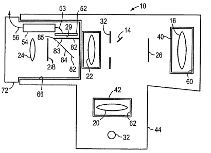
an eyepiece
lens 24 which collimates imaging light rays so that a retinal image can be
viewed by a user.
Eyepiece 46 can be replaced with video module 50 which includes certain
components that
configure the eye viewing device for video capture. In particular, a video
module 50 may
contain an image sensor 52, such as a CCD or CMOS image sensor, which is in an
operative
position in relation to the imaging system when the video module is installed
in holder 66.
The image sensor 52 is in electrical communication with a processor system 54,
typically
including a microprocessor and associated memory, which may be programmed to
control
image sensor 52 and to capture and, possibly, to store image data generated by
and received
from image sensor 52. While processor system 54 is shown as being disposed in
video
module 50, it is understood that processor system 54 could be disposed
external to video
module 50. The video module 50 may further be in communication with display
screen
external to housing 44 and module 50 and/or a processing system external to
housing 44 and
to module 50 via a combination of communication link components which
comprises cable
56 and associated inputloutput interfaces, for example, so that video image
information
27
CA 02544183 2006-04-28
WO 2005/044098 PCT/US2004/035799
corresponding to image signals generated by image sensor 52 can be displayed
or otherwise
output, and possibly archived. The communication link including cable 56 can
be replaced
with a combination of communication link components which comprises a wireless
transmitter-receiver combination. Image information corresponding to image
signals
generated by image sensor 52 can also be communicated to electronic components
external to
module 50 and housing 44 with use of a combination of communication link
components
including transportable memory structure such as a computer disk, a compact
disk or a
memory stick. An encoder for encoding such a memory structure may be located
in a module
as described herein or external to a module in housing 44.
[0086] Video module 50 can be designed so that image sensor 52 lies on
eyepiece
focal plane 28 when module 50 is in an operative position in holder 66. It is
seen that an eye
viewing device of the invention can be configured for video capture by
replacing eyepiece
module 46 with a video module 50 without adding or replacing additional lenses
of the
imaging system. Alternative sized image sensors may also be used, with the
addition of
image resizing lenses. Such a configuration shifts the location of focal plane
28.
[0087] Eye viewing devices having a viewing module holder for receiving
various
alternative types of viewing modules are shown in FIGS. 6B-6I. Viewing module
46 of FIG.
6B is an alternative version of eyepiece viewing module 46 shown in FIG. 6A.
Viewing
module 50 of FIG. 6C is an alternative version of video viewing module 50
shown in FIG.
6A.
[0088] FIG. 6D shows a viewing module 70 adapted to provide both optical
viewing
and video capture. Viewing module 70 includes a beam sputter 80 for splitting
the retinal
28
CA 02544183 2006-04-28
WO 2005/044098 PCT/US2004/035799
image and generating a pair of retinal image focal planes, a first, eyepiece
focal plane 28, and
a second retinal image focal plane 29 at which image sensor 52 is disposed.
Viewing module
70, like viewing module 50, includes processor system 54 in communication with
image
sensor 52 via lead 53 for controlling image sensor 52 and capturing and
possibly storing
image data corresponding to image signals generated by image sensor 52.
Processor system
54 may be programmed to electronically generate a mirror image of the image
formed at
image sensor 52. Video module 70 further includes lead 56 for providing
communication of
video images and data with external displays and/or external processing
systems.
[0089] Shown as being located inside module 70, processor system 54 could in
the
alternative be positioned at a position external to the module but inside
housing 44 as is
indicated by processor system 54' of FIG. 6C or at a location external to both
module 70 and
housing 44. If the processor system associated with any one of the viewing
modules
described herein having an image sensor 52 is located external to the module
but inside
housing as is indicated by the embodiment of FIG. 6C, then the processor
system 54' and
image sensor 52 should be arranged so that an electrical connection is made
between the
processor system 54' and image sensor 52 when the viewing module having the
image sensor
is fitted into the viewing module holder 66 of the eye viewing device 10. Such
an electrical
connection can be provided by positioning complementarily mounted mating
connectors in
the viewing module and primary device housing 44, respectively, such as mating
connectors
85 shown in FIG. 6I.
[0090] Mating connectors such as connectors 85 may also serve to facilitate
linkage
between an electrical component of any one of the viewing modules described
and a power
29
CA 02544183 2006-04-28
WO 2005/044098 PCT/US2004/035799
supply of a device. For example, mating connectors 85 in the embodiment of
FIG. 6I may be
adapted so that processor system 54 is electrically linked to a battery supply
power source in
proximity with light source 32 when connectors of mating connectors 85 are
mated together.
[0091] Further, it will be understood that the processor system receiving
image signals
from image sensor 52 in any one of the embodiments described hexein need not
be located
within a viewing module ox within housing 44. The processor system receiving
image signals
from image sensor may be located externally relative to both housing 44, and
the viewing
module and may be provided, for example, by a processor system of a personal
computer. If
an eye viewing device according to the invention includes an image information
processing
processor system located a substantial distance away from an image signals
generating image
sensor, it is useful to configure the processor system and image sensor so
that the image
sensor and processor system communicate with one another via a high speed
communication
technology, such as Universal Serial Bus communication technology or Firewire
technology.
[0092] An embodiment of a viewing module similar to the viewing module 70 of
FIG.
6D is shown in FIG. 6E. The viewing module of FIG. 6E includes all of the
elements of
viewing module 70 of FIG. 6D except that viewing module 72 includes a two-
position mirror
82 in place of beam sputter 80 FIG. 6D. Two-position mirror 82 is moveable
between two
positions 83, 84. In a first position, indicated by solid line 83 mirror is in
a position such that
a retinal image is formed at eyepiece focal plane 28. In a second position,
indicated by
dashed line 84, mirror 82 is in a position such that a retinal image is formed
at image sensor
52. Mirror 82 may be mounted using a hinge within viewing module 72 as is
indicated by
pivot point 85. Mirror 82 may be adapted to be manually moveable between the
first and
CA 02544183 2006-04-28
WO 2005/044098 PCT/US2004/035799
second positions or else mirror 82 may be adapted to be movable by means of
motor motion.
Mirror 82 can be understood to operate in the same manner that the mirror in a
single lens
reflex (SLR) camera operates, alternatively passing light to a viewfinder in
one position, and
in the second position, passing light to a recording medium such as
photographic film, or to
an electronic imaging device. Again, similar to a single lens reflex camera,
in which a
mechanical or electronic shutter is provided in order to control an exposure
(or integration)
time, devices according to the invention can comprise a shutter for the
purposes of controlling
duration of illumination. With an electronic imaging device, one can
additionally control the
integration time. The integration time of the electronic imager (or second
time interval) is
adjusted to be different than that of the viewing time duration (or first time
interval). In an
alternative embodiment, mirror 82 rests in position 84, and is electronically
controllable to
become more reflective, thereby passing light to image sensor 52, or to become
less reflective
(more transparent) thereby passing light to the eyepiece.
[0093] As should be clear from the above description, some systems, such as
beamsplitter systems, provide a first fraction of illumination to one receiver
(such as the
eyepiece) and another fraction of the illumination to a second receiver (such
as an imagery at
substantially contemporaneous and overlapping periods of time. It should be
equally clear
that other systems, such as systems similar to SLR camera systems, provide a
first fraction of
illumination to one receiver (such as the eyepiece) and another fraction of
the illumination to
a second receiver (such as an imagery at substantially non-overlapping,
sequential or serial,
periods of time.
31
CA 02544183 2006-04-28
WO 2005/044098 PCT/US2004/035799
[0094] In FIG. 6F, a viewing module received in a viewing module holder 66 is
shown that contains a built-in display 58. In viewing module 74, image sensor
52 is
positioned at the position of eyepiece focal plane 28 when the module is
properly received in
holder 66. Image sensor 52 is in communication with processor system 54
programmed to
control and capture image data corresponding to image signals generated by
image sensor 52.
In addition to being in communication with image sensor 52, processor system
54 is in
communication via lead 55 with a display 58 which is built directly into
module 74. Display
58 may be provided, for example, by a light weight LCD display as is well
known. Display
58 is conveniently located at the face portion 74f of viewing module 74 as is
indicated by
FIG. 6F. Viewing module 74 may include, in addition, a lead 56 for providing
external
communication of video images andlor other data with an external display or
processing
system located externally with respect to the viewing module and housing 44.
[0095] The viewing module 75 of FIG. 6G is similar to the viewing module of
FIG.
6F except that externally mounted display 58 is replaced with an interior
mounted display 59
mounted at an interior of module 75. Display 59 is preferably a miniature LCD
display.
Viewing module 75 may include an eyepiece lens 24 for collimating light rays
generated by
display 59.
[0096] Alternative embodiments of eye viewing devices having built-in or
attachable
displays are shown in FIGS. 6H and 6I. In the embodiment of FIG. 6H, viewing
module 76
includes a display 58 mounted to a top surface 76t of an externally extending
portion of
module 76. In the embodiment of FIG. 6I, a display 58 is fixedly mounted to a
top surface
44t of primary device housing 44. Display 58 could in the alternative be
detachably mounted
32
CA 02544183 2006-04-28
WO 2005/044098 PCT/US2004/035799
to housing 44 or pivotally attached to housing 44. In the embodiment of FIG.
6I, viewing
module 77 includes lead 55A that matingly connects to lead 55B in
communication with
display 58 when module 77 is received in holder 66. The mating connection
between leads
55A and 55B may be provided by complementarily mounted mating connectors 85.
[0097] The viewing modules 46, 50, 70, 72, 74, 75, 76 and 77 preferably have
similarly sized outer housings so that each may be fitted into a single
viewing module holder
which is adapted to receive one viewing module at a time. One or more of the
above viewing
modules may be sold or made available in a system wherein viewing modules can
be
interchanged for optimization of an eye viewing device for a particular
application. A
viewing module according to the invention is adapted to be held in place in a
complementarily formed holder by friction forces or other known retaining
means.
[0098] Of course, the elements incorporated in the above-described removably
installable viewing modules 46, 50, 70, 72, 74, 75, 76 and 77 can be
permanently mounted in
an eye viewing device that does not contain a viewing module holder.
[0099] As indicated above, viewing modules having a processor system 54 for
processing images may include a lead 56 for providing communication between
the processor
system and an external display device or processor system external to module
and housing 44.
One type of external display which may be in electrical communication with
viewing module
having a video processor system is a head mounted display assembly 57
including a display
59 as shown in FIGS. 6J and 6K. Head mounted displays are useful in enhancing
the mobility
of a viewer. In the embodiment of FIG. 6J, an eye viewing device 10 includes a
head
mounted display assembly 57, voice activated control, an audio feedback means,
and a
33
CA 02544183 2006-04-28
WO 2005/044098 PCT/US2004/035799
personal computer 63. From the embodiment of FIG. 6J it is seen that the
elements of an eye
viewing device can be spread out over several physically separate components
including
primary device housing 44, a viewing module, a personal computer 63 and a
video assembly
57.
[0l 00] It will be understood that the image sensor referred to in any one of
the above
viewing modules having an image sensor may be any commercially available image
sensor.
For example the image sensor may be a visible light image sensor or an image
sensor that is
selectively responsive to light in a specific band, such as an infrared or
ultraviolet image
sensor. The image sensor may also be a spectral imaging type image sensor
which makes
available spectral profile data characterizing the spectrum of light incident
at each pixel of the
image sensor. In addition, processor system 54 and image sensor 52 can be
incorporated in a
single piece of silicon. For example, image sensor 52 and processor system 54
can readily be
integrated in a single piece of silicon utilizing CMOS fabrication methods.
[0101] Further, it will be understood that any one of the electrically
conductive lines
described herein, e.g. lines 53, 55, 55a, 55b and 56 could be replaced with a
wireless data
communication link such as an IR link or an RF link, for example an RF link
utilizing the
"Blue Tooth" communication protocol.
[0102] Figs. 7A and 7B are schematic diagrams of other embodiments of the
digital
documenting ophthalmoscope according to principles of the invention. The
digital
documenting ophthalmoscope, also referred to as a digital fundus imager,
provides some
important advantages as compared to conventional ophthalmoscopes. The
invention also
provides a method of assessing a condition of an eye in a single interrogation
of the eye,
34
CA 02544183 2006-04-28
WO 2005/044098 PCT/US2004/035799
comprising viewing the eye in a true color live view by an operator, and
capturing an image
of the eye in an imager.
[0103] Ophthalmoscopes are among the most commonly used medical devices. They
are used for a variety of examination and diagnostic procedures in the eye. In
the field of
optometry, fundus (or retinal) cameras are used to document the condition of
the retina as
viewed with an ophthalmoscope (or other diagnostic instrument). When two
separate
instruments are used (one to diagnose, one to document, often by two different
individuals), it
is readily recognized that the images that are captured will not always
completely reflect the
conditions that the practitioner wishes to document. Combining these two
instruments
requires finding an optimal tradeoff between image quality, image field of
view and small
pupil performance, since these parameters are all related to the amount of
light in the system.
Traditional fundus cameras solve this tradeoff by using IR/flash illumination.
Flash
illumination is unacceptable for use as a live-view diagnosing instrument
because the
practitioner cannot examine the fundus in a meaningful manner during the short
duration of
the flash. "Live" viewing generally is best performed using substantially
continuous
illumination, or illumination lasting at least for a duration sufficient for
easy observation by a
human operator (e.g., some seconds or longer, rather than milliseconds).
Another option
would be to use an extended series of pulses of illumination that appear to be
substantially
continuous to a human observer. IR illumination is unacceptable fox use as a
live-view
diagnosing instrument because the practitioner cannot examine the fundus using
IR
illumination, which is not detected by the human eye. The digital documenting
ophthalmoscope provides both a live, diagnosable view by a practitioner and a
captured
CA 02544183 2006-04-28
WO 2005/044098 PCT/US2004/035799
documenting image in a single instrument in real time. In addition to this
unique two-in one
functionality, some additional benefits that the digital documenting
ophthalmoscope provides
include: true non-mydriatic optics that enable a field of view of up to 25
degrees through a
pupil as small as 2 millimeters without use of a flash; the ability to
capture, store, and print
images; the ability to use the images for such activities as patient
education, ophthalmic
practice management including record-keeping and documentation for purposes of
reimbursement, and use of images for "diagnosis at a distance," (or
telemedicine) for example
by communicating one or more captured images over a communication medium such
as a
telephonic connection or over a network such as the Internet, a LAN, or a WAN,
for viewing
by a practitioner for consultation or diagnosis in substantially real time,
even when the patient
and the practitioner are in physically remote locations one from the other;
and use of stored
images for archival purposes, such as following the condition of an eye of a
patient over time.
The captured images can include or have associated therewith a time and/or
date stamp, an
identifier for the patient, an identifier as to whether the image is one of
the right or left eye
(e.g., an "R" or an "L" can be added to the image electronically, for example
in a corner
thereof), an identifier of the practitioner, and such information as notes or
other information
of significance. The digital documenting ophthalmoscope can be optionally
mounted on a
cart, for use as a mobile device in an office or hospital setting, or it
optionally can be a small,
easily portable, handheld unit suitable for use in the office or in the field,
for example in an
ambulance. Yet another benefit is the possibility of providing the digital
documenting
ophthalmoscope at a competitive price, especially as compared to the price of
two distinct
instruments.
36
CA 02544183 2006-04-28
WO 2005/044098 PCT/US2004/035799
[0104] Turning now to Fig. 7A, a first schematic diagram depicts one
embodiment of
the digital documenting ophthalmoscope 700, which comprises a number of
modules. An
illumination module 710 is provided as a component of the digital documenting
ophthalmoscope 700. The illumination module 710 provides illumination for both
the true
color live view of the eye of the patient by the operator of the instrument,
as well as the
illumination for the digital imaging operation of the instrument. The
illumination module 710
comprises a panoptic lamp 711, a panoptic Wide Band Hot Mirror (WBHM) 712, one
or
more panoptic condensing lenses 713, one or more panoptic aperture plates or
filters 714, and
a polarizes 715 that linearly polarizes the illumination beam before it exits
the illumination
module 710. The illumination module 710 provides light having controlled
intensity and
spectral characteristics. The panoptic lamp 711 is controlled by a lamp
controller 755, which
is in electrical communication with the lamp 711.
[0105] The digital documenting ophthalmoscope 700 further comprises an optical
module 720 that handles the transmission of illumination from the illumination
module 710
to an eye 770 of a patient and handles the collection of reflected light from
the eye 770 for
provision of the reflected light to a viewing module 730 for ultimate delivery
to an eye 735 of
a human operator for a "true color live view" and to an images module 740 fox
delivery to an
images 742 for capture of a digital image. The digital image can be a color
image, a black
and white image, or a grayscale or false color image, as may be useful.
[0106] The optical module 720 comprises a mirror 721 or equivalent structure
to
steer and project the illumination beam from the illumination module 710
toward the eye 770
of the patient. The illumination passes through one or more objective lenses
722 as it
37
CA 02544183 2006-04-28
WO 2005/044098 PCT/US2004/035799
propagates toward the eye 770 of the patient. The one or more objective lenses
722 focus and
direct the illumination. Optionally, an eye cup 772 is provided between the
eye 770 of the
patient and an extremity of the instrument 700. Light that is reflected from
the eye 770 of the
patient is collected by the one or more objective lenses 772, through an
appropriate shape
aperture stop 723 , and through one or more relay lenses 724. The reflected
illumination
beam passes through a transparent portion of a dot plate 726, and the internal
glare reflections
from one or more surfaces of the objective lens are intercepted by an opaque
portion of the
dot plate 726, as is described in more detail below. The portion of the
reflected illumination
than passes through the transparent portion of the dot plate 726 passes
through one or more
focus lenses 725 before exiting the optical module 720.
[0107] The reflected illumination exiting the optical module 720 is directed
into either
or both of the viewing module 730 and the imager module 740. In one
embodiment, a mirror
731, similar to the mirror in a single lens reflex (SLR) camera, is provided.
The SLR mirror
731 is movable between a first position (shown in Fig. 7A as a solid line) and
a second
position (shown in Fig. 7A as a dotted line 731'). In the first position,
mirror 731 causes
illumination exiting from the eye 770 to reflect toward mirror 732, and to
reflect therefrom
toward focusable eyepiece 733, which comprises one or more lenses, and to pass
through
polarizer 734, and then to the eye 735 of an operator, such as a doctor, to
provide a "true color
live view" of the eye 770 of a patient, or of a portion thereof, such as the
fundus of eye 770.
In some embodiments, alternatives to the mirror 731 are employed, which
alternatives permit
simultaneous illumination to both the eye 735 of the operator and to the
imager module 740,
as are described in greater detail below.
38
CA 02544183 2006-04-28
WO 2005/044098 PCT/US2004/035799
[0108] When mirror 731 is situated in position 731', the illumination that
exits from
the eye 770 is not intercepted by mirror 73 l, but passes into the imager
module 740. The
imager module 740 comprises an IR filter 743, an imager 742 such as a CCD
imager having a
two-dimensional array of photosensitive elements or pixels, and an image
controller 741 in
electrical communication with the irnager 742. The IR filter 743 passes
illumination having
wavelengths in one or more pass bands, and absorbs illumination having
wavelengths in at
least one absorption band, the wavelengths of the pass band and the
wavelengths of the
absorption band being mutually exclusive. The illumination that reaches the
imager 742 is
converted to electrical signals on a pixel by pixel basis, as is understood in
the light sensing
arts, and the electrical signals are extracted from the imager 742 under the
control of the
imager controller 741, using conventional control procedures for extracting
signals from two-
dimensional imaging chips.
[0109] The digital documenting ophthalmoscope 700 further comprises a control
module 750 that controls and synchronizes the operation of the various modules
and
components of the digital documenting ophthalmoscope 700. Control module 750
comprises
a computer interface controller and power supply 751 that is a microprocessor-
based
programmable controller, and a power supply such as a battery (in a portable
unit) or a
conventional power supply fed from a wall plug. In some embodiments, a machine-
readable
memory is provided with the computer interface controller and power supply 751
for
purposes of holding data extracted from the imager 742, and/or for holding one
or more
commands for operating the computer interface controller and power supply 751.
The
computer control interface and power supply 751 is in electrical communication
with the
39
CA 02544183 2006-04-28
WO 2005/044098 PCT/US2004/035799
images controller 741, both for controlling the images controller 741 and for
receiving from
the images controller 741 data corresponding to an image of the eye 770 of the
patient.
Computer interface controller and power supply 751 is also in electrical
communication with
a trigger controller 752, and in communication by wireless connection (or
alternatively by a
wire connection) with an external computing element 760, such as a
conventional
commercially available laptop or desktop computer. The trigger controller 752
and the
computer interface controller and power supply 751 communicate to exchange
commands and
timing signals, such as a trigger signal indicating activation of trigger 753
in electrical
communication with trigger controller 752, or signals from computer interface
controller and
power supply 751 to trigger controller 752 relating to illumination levels to
be achieved by
panoptic lamp 71 l, or timing signals for moving SLR mirror 731 to position
731' to
accommodate increased illumination levels produced by panoptic lamp 711, as
will be
explained in greater detail below. Trigger controller 752 is in electrical
communication with
lamp controller 755 that controls panoptic lamp 711, and with solenoid 754
that controls the
motion of mirror 731.
[0110] A second embodiment of the digital documenting ophthalmoscope 700 is
shown in Fig. 7B. Because there are several differences between the two
embodiments, the
instrument of Fig. 7B is denoted by numeral 702. The digital documenting
ophthalmoscope
702 lacks the dot plate 726 present in optical module 720 of digital
documenting
ophthalmoscope 700. The digital documenting ophthalmoscope 702 comprises a
polarizes
744 in images module 740, which polarizes is lacking in digital documenting
ophthalmoscope
700.
CA 02544183 2006-04-28
WO 2005/044098 PCT/US2004/035799
[0111] Polarizers 715, 734 and 744 are all linear polarizers having a
polarization axis.
Polarizers 715 and 734, and when polarizes 744 is present, polarizers 715 and
744, are
adjustable so that the polarization axis of polarizers 734 and 744 can be
oriented relative to
the polarization axis of polarizes 715 so that the polarization axes can be
positioned at any
angular relationship from parallel (e.g., no diminution by polarization of
propagating light) to
perpendicular (e.g., perfect extinction of propagating light). In the
embodiment of Fig. 7B,
internal glare reflection is eliminated by use of the polarizers 734 and 744.
[0112] Instruments according to principles of the invention provide both a
live,
diagnosable view and a captured documenting image in a single instrument,
which may be
hand-held in some embodiments. The basic architecture of this instrument is
similar to the
Welch-Allyn Pan-Optic ophthalmoscope. The instrument according to principles
of the
present invention includes improvements to facilitate the electronic capture
of images
gathered by the instrument, which electronic capture of images is not provided
in the Welch-
Allyn Pan-Optic ophthalmoscope. The polarized image embodiment of Fig. 7B
provides a
polarized view to both the images and the viewfinder. Polarizes 734 (P2) in
the live view is
crossed with illumination polarizes 715 (P1), while polarizes 744 (P3) is
crossed with
polarizes 715 (P1) in the images view. For images 742, which in some
embodiments is very
sensitive in the near 1R region, an additional IR filter 743 is added in the
images view to
remove residual 1R before the illumination reaches the images 742. The
polarizers 715, 734
and 744 provide a considerable amount of glare reduction, which facilitates
aiming of the
instrument. Use of crossed polarizers can cause a lack of edge definition and
tissue surface
reflectivity in the image of the retina. In addition, the crossed polarizers
reduce the image
41
CA 02544183 2006-04-28
WO 2005/044098 PCT/US2004/035799
irradiance by about 50%, although this problem can be mitigated by the use of
a lamp boost
circuit, described in greater detail below.
[0113] The 'Black Dot' embodiment depicted in Fig. 7A eliminates polarizer 744
(P3), thus providing an unpolarized view to the imager, while retaining the
polarizer 734 of
the viewfinder (eyepiece). Glare reduction in the imagex path is provided in
part by a dot
plate 726, which is an optical surface with a small black dot in the center to
block internal
reflections of the illumination system. Glare external to the instrument
(primarily corneal and
scleral glare) is not filtered by this approach, which is one reason why the
viewfinder is still
polarized. This external glare is minimized by proper alignment of the
instrument 700 to the
patient's eye 770. The optical viewfinder is used to properly align the
instrument 700 and
then capture the unpolarized image. It is possible to incorporate a glare
sensor (not shown in
Fig. 7A) that senses excessive glare into the imager path. Excessive glare
degrades the
captured image. The glare sensor provides a feedback signal that causes a
status signal to be
displayed to the operator of the instrument, such as with a red LED for 'image
outside
acceptable parameters' and a green LED for 'OK to capture'. The embodiment of
Fig. 7A has
the advantage of an improvement of edge definition and tissue surface
reflectivity in the
image of the retina, compared to a polarized image. In addition, the
irradiance of the
unpolarized image is about 50% higher than the polarized image.
[0114] In both embodiments, a movable mirror 731 is provided to direct all of
the
returning light rays to either the eyepiece or the imager. This ensures that
each path receives
sufficient light. In embodiments where sufficient light is available through
the return path, a
42
CA 02544183 2006-04-28
WO 2005/044098 PCT/US2004/035799
beam sputter can be used in place of the mirror 731, to provide a simultaneous
view to both
the eyepiece and the imager.
[0115] In some embodiments, a 'boost' circuit is provided to increase image
irradiance. When the operator wishes to capture an image, the trigger 753 is
activated. The
default position of the mirror 731 is such that illumination passes to the
eyepiece 733.
Operation of the trigger 753 causes the solenoid 754 to move the mirror to
position 731' to
send the light rays to the imager 742 and to prevent illumination from
reaching the operator's
eye 735. In parallel, the trigger 753 activates a 'boost' circuit, which
momentarily increases
the output of the lamp 711, while the imager 742 is exposed. The lamp 711 is
returned to its
previous 'normal' output level when the mirror 731 is in position to direct
illumination to the
eye 735 of the operator. This gives the captured image additional
"brightness", but without
the drawbacks of a conventional flash lamp illumination system.
[0116] Some additional features that can be incorporated in various
embodiments of
the digital documenting ophthalmoscope according to the invention include the
following.
[0117] An external fixation target, such as a target on a LED display
presented to an
eye 770 of a patient, may be provided at an optimal gazing angle for corneal
glare control. By
holding the eye in a particular gazing angle, the glare reflection from an
illumination source
can be reduced or eliminated. This fixation target can be any convenient
target, such as a
picture, a "bulls-eye," an "X" or a visible LED. The fixation target causes
the eye to avoid
moving, accordingly easing the process of capturing high quality digital
images.
[0l 18] A mechanical stabilizer, such as a forehead rest, a browrest, a
chinrest, or a
device that holds a facial surface in a particular location, can be provided
to minimize
43
CA 02544183 2006-04-28
WO 2005/044098 PCT/US2004/035799
motions of the head and eye 770 of a patient, and thereby improve control of
blur caused by
motion.
[0119] The apparatus can include a magnifier over a live view image projected
on a
screen or mirror for control of accommodation of the eye of the operator of
the instrument.
One can reduce instrument myopia, or tendency toward accommodation of an out-
of focus
image by a human viewer, by providing a screen, such as a partially frosted
glass or other
transparent material or a mirror surface and a magnifier with which the
operator can view an
image that falls on the partially frosted or mirror surface. In another
embodiment, one can
present to the eye of an operator of the instrument an image that represents
the image that is
presented to the imaging device, so that the operator can judge whether proper
focus has been
achieved. In other embodiments, an autofocus system can be provided to
automatically bring
the image presented to the imager into focus.
[0120] In another embodiment, the apparatus can include a SLR mechanism with
dual
mirror/window function for real-time observation of a patient's eye 770 by the
imager and
corneal glare/alignment assessment through a feedback loop from the imager to
the live view
observed by the instrument operator. The window can provide optical signals to
the imager
even when much of the light passes through the window and reaches the
operator's eye 735.
When proper alignment, focus and/or glare reduction is attained, the operator
can receive a
signal, such as an image of an LED, when one sees visual cues as to the
camera's operational
state when looking through the viewfinder of a SLR camera.
[0121] In some embodiments, the ophthahnoscope can comprise an alignment
control
using IR light projected onto the patient's cornea and a feedback loop from
the imager to the
44
CA 02544183 2006-04-28
WO 2005/044098 PCT/US2004/035799
true color live view seen by the operator. The images detects the IR light
reflection, and the
apparatus can deduce from the size and/or arrangement of the reflected light
image (for
example, by observing the number of illuminated pixels and their relative
locations) whether
the image is in focus and/or a distance to a surface of a patient's eye 770. A
signal can be
provided to the operator to inform the operator of the status of focus.
[0122] In some embodiments, the ophthalmoscope can additionally comprise a
feedback loop between the images and the live view provided to the operator to
inform the
operator about such features as alignment and glare via LED indicators.
[0123] In some embodiments, the ophthalmoscope can include an optional
capability
of boosting the illumination output with maintenance of a constant spectral
envelope.
Various options that are possible include using multiple lamps or LED arrays
and changing
the number of operating units or their intensity, use of an electro-optic
shutter to control the
amount of illumination impinging on the eye, and boosting the operating point
of a single
source with constant spectral envelope in the normal and boosted illumination
modes.
[0124] In some embodiments, the ophthalmoscope includes
parfocality/accommodation control. This can be accomplished by any of several
methods,
such as use of a negative one diopter ("-1D") calibration offset, use of an
autofocus images
mechanism, or use of two-step focus setting.
[0125] As has been alluded to earlier, the ophthalmoscope can comprise a
polarizes in
the image path, which polarizes can be adjusted to have an angular
relationship with the
illumination polarizes varying from parallelism (zero degree difference
between the optical
CA 02544183 2006-04-28
WO 2005/044098 PCT/US2004/035799
axes of the polarizers) to crossed polarizers (e.g., 90 degree difference
between the optical
axes of the polarizers), or any intermediate value.
[0126] While the "true color live view" of the eye 770 of the patient is the
"gold
standard" today for examination, in the future it may be reasonable to have a
display, such as
a CRT, flat panel display, or LCD , which display is provided for use by the
operator in
viewing the eye 770 of the patient. One benefit of such a display may be
elimination of
accommodation by the eye of the operator.
[0127] In some embodiments, different options for corneal glare control can be
included and used in ophthalmoscopes of the invention. The options include use
of a large
working distance (approx. 30 mm) combined with reduced field of view (FOV)
(e.g., 16 to 17
deg.); the use of multiple field stops; the use of a "capture many" mode; the
use of a polarizer
switch in the imager path; the use of an external fixation target, such as one
on a swinging
arm; the use of a feedbaclc loop from the imager and the live-view; and the
use of digital
image processing for removal of glare based on sets of images recorded at
different
polarization vectors.
[0128] FIG. 8A is a schematic diagram 800 that shows how the elements of a
plurality
of embodiments of digital documenting ophthalmoscopes are specified and how
they
cooperate. The schematic diagram 800 can be understood as a flow chart for
selecting
components for use in an apparatus according to the invention, and can also be
understood as
outlining the operation of such an apparatus as regards the cooperation of the
various included
components. As indicated at box 802, an apparatus according to the invention
will provide at
least two capabilities, including diagnostic capability and image capture
capability. As
46
CA 02544183 2006-04-28
WO 2005/044098 PCT/US2004/035799
As indicated in boy 804, any of several configurations can be employed,
individually or in
combination, including configurations that operate by malting observations
through an
undilated pupil, configurations that employ continuous low level illumination,
configurations
that provide true color observations, configurations that provide handheld
operation,
configurations that employ convergent illumination, and configurations that
provide a
Maxwellian image using a Maxwellian view system. In various embodiments, at
least one of
the illumination and viewing systems of the device are configured to produce a
true color
view of the retina suitable for diagnosis. At boxes 806, 808, and 810, one of
three viewing
regimes is selected as a particular viewing configuration for a particular
embodiment of an
instrument of the invention. The three alternative viewing regimes are a
beamsplitter
configuration as indicated at box 806, a single lens reflex ("SLR")
configuration as indicated
at box 808, and a direct view configuration, for example using a camera and
display, as
indicated at box 810.
[0129] The configurations of instruments according to principles of the
invention can
further include optical elements as indicated in boxes 812, 814, 816, 818, 820
and 822. In
some embodiments, an external illumination source is used, as indicated at box
812. In some
embodiments, a PanOptic configuration is used, as indicated at box 814. In the
instance that a
PanOptic configuration is used, in some embodiments a black dot rotated
polarizer is also
used, as indicated by box 820. In some embodiment, the instrument comprises an
internal
illumination source, as indicated at box 816. In each of the internal
illumination 816, external
illumination 812, and PanOptic 814 configurations, the instrument can further
comprise a
boost module 818, and can comprise a battery 822 for provision of electrical
power under
47
CA 02544183 2006-04-28
WO 2005/044098 PCT/US2004/035799
handheld and/or untethered operation, such as an internal battery, with or
without a
recharging capability.
[0130] FIG. 8B is a schematic diagram 801 showing alternative embodiments of a
boost module 818 or component. The boost module 818 is provided to increase an
illumination capability so as to permit appropriate operation in shorter time
periods than
would be possible without the boost module 818, as has been described
hereinabove. In any
instance, due care to assure the safety of a patient and of an operator of the
apparatus is
exercised, by any or all of provision of limits on the boost module as to
power and duration,
and provision of limits on exposure of an operator to undesirable illumination
andlor power
levels. Limits can be implemented in electrical design, for example through
use of control
circuits, and through mechanical design, such as use of shielding as may be
necessary.
Embodiments of the boost module 818 and its implementation include increasing
an
operating voltage of a single source of illumination (whether one or more
sources are present)
as illustrated by box 824, provision and use of multiple illumination sources
such as lamps as
indicated by box 826, provision and use of an light emitting diode ("LED")
array as indicated
by box 828, and use of an electro-optic shutter to control amount and timing
of illumination
as indicated by box 830. In some instances, combinations of the various
embodiments of
boost components can be combined, for example using multiple lamps 826 with an
electro-
optic shutter 830.
[0131] FIG. 8C is a schematic diagram 803 showing alternative embodiments of a
corneal glare control component 840. It is recognized that the features of
glare control and
image quality may at times provide mutual constraints, so that improvement in
one feature
48
CA 02544183 2006-04-28
WO 2005/044098 PCT/US2004/035799
may cause a degradation or a limitation of the ultimately attainable extent of
the other feature.
Glare control can be implemented using any of a plurality of approaches and
apparatus
providing suitable characteristics therefor. In FIG. 8C, there are at least
ten different
approaches indicated. These include: the provision and use of multiple field
stops as
indicated at box 841; the use of a large working distance and a reduced field
of view ("FOV")
as indicated at box 842; the provision of ring-type internal illumination, for
example with a
ring-shaped internal light source, as indicated at box 843; the use of a small
beam that
expands (or "opens up") within the eye under observation, using a suitably
focused light
source, as indicated at box 844; the provision and use of a polarizes that is
rotated relative to a
surface or another polarizes, as indicated at box 845; the use of an external
fixation device to
cause the eye under test to be fixated at a preferred, and in some
circumstances, an optimal
angle to avoid glare, as indicated in box 646, Some of the enumerated
techniques and
associated apparatus are amenable to being performed (and provided) in
alternative ways. For
example, the ring-type internal illuminator 843 that provides ring-type
internal illumination
can be accomplished using either a ring having fixed diameter, or a ring
having variable
diameter, as indicated respectively at boxes 851 and 852. The selection and
variation of a
diameter of an illumination can be accomplished using any of lenses, irises,
and optical stops,
for example. Different types of rotated polarizers 845 can be provided with
fixed angle
polarizers 853, variable angle polarizers 854, and one or more polarizes
switches 855, for
example.
[0132] FIG. 8D is a schematic diagram 805 showing alternative embodiments of
optical trains that can be employed in instruments that use principles of the
invention. As
49
CA 02544183 2006-04-28
WO 2005/044098 PCT/US2004/035799
indicated at box 860, the apparatus can use and SLR and a beamsplitter. At box
862, the
apparatus is indicated to provide parfocality, that is, that the object being
viewed is in focus
when either of two or more objective lenses are interchanged. Here, the SLR
and
beamsplitter systems have two separate focusing systems so that correct focus
for each can be
maintained. In the boxes 864 and 866, respectively, autofocus apparatus and
projected image
apparatus axe indicated.
[0133] FIG. 8E is a schematic diagram 807 showing additional items of hardware
that
can be employed in instruments designed using principles of the invention. Box
870 indicates
generally that other items of hardware can be provided to enable instruments
of the invention
to be operated in handheld configuration. One item, indicated at box 872, is a
control for a
trigger for operating the instrument to eliminate "twitch," or an
inappropriate trigger
response. Another feature, indicated at box 874, is mechanical balance of the
apparatus, so
that a user can operate the instrument in handheld mode without undue effort,
or without
requiring undue strength to maintain the apparatus in a desired orientation.
Yet a third feature
is stability control, which generally relates to maintaining the instrument in
a stable position
and orientation with respect to an eye of a patient, such as the provision of
an eyecup, a chin
rest, a forehead rest, and other mechanical parts that assist in maintaining
the correct position
and orientation relative to an eye.
[0134] Many functions of electrical and electronic apparatus can be
implemented in
hardware (for example, hard-wired logic), in software (for example, logic
encoded in a
program operating on a general purpose processor), and in firmware (for
example, logic
encoded in a non-volatile memory that is invoked for operation on a processor
as required).
CA 02544183 2006-04-28
WO 2005/044098 PCT/US2004/035799
The present invention contemplates the substitution of one implementation of
hardware,
firmware and software for another implementation of the equivalent
functionality using a
different one of hardware, firmware and software. To the extent that an
implementation can
be represented mathematically by a transfer function, that is, a specified
response is generated
at an output terminal for a specific excitation applied to an input terminal
of a "black box"
exhibiting the transfer function, any implementation of the transfer function,
including any
combination of hardware, firmware and software implementations of portions or
segments of
the transfer function, is contemplated herein.
[0135] While the present invention has been particularly shown and described
with
reference to the structure and methods disclosed herein and as illustrated in
the drawings, it is
not confined to the details set forth and this invention is intended to cover
any modifications
and changes as may come within the scope and spirit of the following claims.
51