Note: Descriptions are shown in the official language in which they were submitted.
CA 02544296 2011-05-24
WO 2005/046529 PCT/US2004/034478
IMPLANTABLE VALVULAR PROSTHESIS
FIELD OF THE INVENTION
The present invention relates to a medical device, and
more particularly to a frame based unidirectional flow
prosthetic valve, and the method for fabricating such valve.
BACKGROUND OF RELATED ART
The human body has numerous biological valves that
control fluid flow through body lumens and vessels. For
example the circulatory system has various heart valves that
allow the heart to act as a pump by controlling the flow of
blood through the heart chambers, veins, and aorta. In
addition, the venous system has numerous venous valves that
help control the flow of blood back to the heart,
particularly from the lower extremities.
These valves can become incompetent or damaged by
disease, for example, phlebitis, injury, or the result of an
inherited malformation. Heart valves are subject to
disorders, such as mitral stenosis, mitral regurgitation,
aortic stenosis, aortic regurgitation, mitral valve prolapse
and tricuspid stenosis. These disorder are potentially life
threatening. Similarly, incompetent or damaged venous
1
CA 02544296 2006-05-01
WO 2005/046529 PCT/US2004/034478
valves usually leak, allowing the blood to improperly flow
back down through veins away from the heart (regurgitation
ref lux or retrograde blood flow). Blood can then stagnate
in sections of certain veins, and in particular, the veins
in the lower extremities. This stagnation of blood raises
blood pressure and dilates the veins and venous valves. The
dilation of one vein may in turn disrupt the proper function
of other venous valves in a cascading manner, leading to
chronic venous insufficiency.
Numerous therapies have been advanced to treat symptoms
and to correct incompetent valves. Less invasive procedures
include compression, elevation and wound care. However,
these treatments tend to be somewhat expensive and are not
curative. Other procedures involve surgical intervention to
repair, reconstruct or replace the incompetent or damaged
valves, particularly heart valves.
Surgical procedures for incompetent or damaged venous
valves include valvuloplasty, transplantation, and
transposition of veins. However, these surgical procedures
provide somewhat limited results. The leaflets of some
venous valves are generally thin, and once the valve becomes
incompetent or destroyed, any repair provides only marginal
relief.
2
CA 02544296 2006-05-01
WO 2005/046529 PCT/US2004/034478
As an alternative to surgical intervention, drug
therapy to correct valvular incompetence has been utilized.
Currently, however, there are no effective drug therapies
available.
Other means and methods for treating and/or correcting
damaged or 'incompetent valves include utilizing xenograft
valve transplantation (monocusp bovine pericardium),
prosthetic/bioprosthetic heart valves and vascular grafts,
and artificial venous valves. These means have all had
somewhat limited results.
What is needed is an artificial endovascular
(endoluminal) valve for the replacement of incompetent
biological human valves, particularly heart and venous
valves. These valves may also find use in artificial hearts
and artificial heart assist pumps used in conjunction with
heart transplants.
SUMMARY OF THE INVENTION
The present 'invention relates to a medical device, and
in particular, to a stent-based valve. A prosthetic valve
comprises a radially expandable structural frame defining a
longitudinal axis. The structural frame includes an anchor
structure having a first and a second open end, a connecting
3
CA 02544296 2011-05-24
member having a first and a second end, and a cantilever valve strut having a
first
and a second end. The first end of the connecting member is attached to the
second
end of the anchor structure. The first end of the cantilever valve strut is
cooperatively
associated with the second end of the connecting member. The prosthetic valve
further includes a biocompatible membrane assembly having a substantially
tubular
configuration disposed longitudinally about the structural frame. The membrane
assembly has a first end having a first diameter and a second end having a
second
diameter, wherein the first diameter is greater than the second diameter. The
first end
of the membrane assembly is attached along the second end of the cantilever
valve
strut.
More particularly, there is provided a prosthetic valve comprising:
a radially expandable structural frame defining a longitudinal axis, including
an anchor structure having first and second open ends, a connecting member
having
first and second ends, the first end of the connecting member being attached
to the
second end of the anchor structure, and a cantilever valve strut having first
and a
second ends, the first end of the cantilever valve strut being cooperatively
associated
with the second end of the connecting member; and
a biocompatible membrane assembly having a substantially tubular
configuration disposed longitudinally about the structural frame, the membrane
assembly including a first end having a first diameter and a second end having
a
second diameter, wherein the first diameter is greater than the second
diameter, the
first end of the membrane assembly being attached along the second end of the
cantilever valve strut, wherein the structural frame comprises a proximal
collar
attached to the second end of the connecting member and first end of the
cantilever
valve strut.
4
CA 02544296 2011-05-24
In another embodiment of the invention, the prosthetic valve comprises a
radially expandable anchor structure formed from a lattice of interconnected
elements. The anchor has a substantially cylindrical configuration with a
first and a
second open end and a longitudinal axis defining a longitudinal direction
extending
there between. A connecting member and a cantilever valve strut, each having
first
and second ends, are also provided. The first end of the connecting member is
attached to the second end of the anchor. The first end of the cantilever
valve strut is
4a
CA 02544296 2006-05-01
WO 2005/046529 PCT/US2004/034478
cooperatively associated with the second end of the
connecting member. The prosthetic valve further includes a
biocompatible membrane assembly having a substantially
tubular configuration disposed longitudinally about at least
a portion of the connecting member. The membrane assembly
has a first end having a first diameter and a second end
having a second diameter, wherein the first diameter is
greater than the second diameter. The first end of the
membrane assembly is attached along the second end of the
cantilever valve strut.
In still another embodiment of the present invention,
the prosthetic valve comprises a radially expandable anchor
structure formed from a lattice of interconnected elements.
The anchor structure has a substantially cylindrical
configuration with a first and a second open end and a
longitudinal axis defining a longitudinal direction
extending there between. A collar is provided and located
proximal to the radially expandable anchor. At least one
connecting member having a first and a second end is
provided such that the first end of the connecting member is
attached to the second end of the anchor and the second end
of the connecting member is attached to the proximal collar.
A cantilever valve strut having a first and a second end is
5
CA 02544296 2006-05-01
WO 2005/046529 PCT/US2004/034478
also provided. The first end of the cantilever valve strut
is attached to the proximal collar and extends in a distal
direction substantially parallel to the longitudinal axis.
The prosthetic valve further includes a biocompatible
membrane assembly having a substantially tubular
configuration disposed longitudinally about at least a
portion of the connecting member. The membrane assembly has
a first end having a first diameter and a second end having
a second diameter, wherein the first diameter is greater
than the second diameter. The first end of the membrane
assembly is attached along the second end of the cantilever
valve strut.
BRIEF DESCRIPTION OF THE DRAWINGS
Figure 1A shows a perspective view of a prosthetic
venous valve in the deployed state according to one
embodiment of the present invention.
Figure 2A shows a perspective view of the prosthetic
venous valve structural frame in the deployed state
according to one embodiment of the present invention.
Figure 2B shows a perspective view of the prosthetic
venous valve structural frame having helical connecting
6
CA 02544296 2006-05-01
WO 2005/046529 PCT/US2004/034478
members according to one embodiment of the present
invention.
Figure 2C shows a perspective view of the prosthetic
venous valve structural frame having a sinusoidal cantilever
valve strut assembly according to one embodiment of the
present invention.
Figure 2D shows a perspective. view of the prosthetic
venous valve structural frame having a helical valve strut
assembly according to one embodiment of the present
invention.
Figure 2E shows a perspective view of the prosthetic
venous valve structural frame having a proximal centering
mechanism in the deployed state according to one embodiment
of the present invention.
Figure 2F shows a perspective view of the prosthetic
venous valve structural frame having distal and proximal
anchor mechanisms according to one embodiment of the present
invention.
Figure 3A shows a perspective view of the distal stent
anchor having a plurality of hoop structures according to
one embodiment of the present invention.
7
CA 02544296 2006-05-01
WO 2005/046529 PCT/US2004/034478
Figure 3B shows a close-up perspective view of a loop
member from the anchor having inner and outer radii
according to one embodiment of the present invention.
Figure 3C illustrates a single hoop anchor having three
connecting members connected to the proximal end of the
distal anchor at the outer radii of the inflection point of
the loop members.
Figure 3D illustrates a single hoop anchor having three
connecting members connected to the proximal end of the
distal anchor at the inner radii of the inflection point of
the loop members.
Figure 3E illustrates a single hoop anchor having three
connecting members connected to the proximal end of the
distal anchor along the strut members connecting the loop
members.
Figure 4A is a perspective view illustrating one
embodiment of the deployed prosthetic venous valve assembly
in the open position.
Figure 4B is a section view illustrating one embodiment
of the deployed prosthetic venous valve assembly in the open
position.
8
CA 02544296 2006-05-01
WO 2005/046529 PCT/US2004/034478
Figure 5A is a perspective view illustrating one
embodiment of the deployed prosthetic venous valve assembly
in the closed position.
Figure 5B is a section view illustrating one embodiment
of the deployed prosthetic venous valve assembly in the
closed position.
Figure 6A is a perspective view illustrating a membrane
limiting means according to one embodiment of the present
invention.
Figure 6B is a perspective view illustrating a membrane
limiting means according to one embodiment of the present
invention.
Figure 6C is a perspective view illustrating a membrane
limiting means according to one embodiment of the present
invention.
DETAILED DESCRIPTION OF THE PREFERRED EMBODIMENTS
The stent-based valves of the present invention provide
a method for overcoming the difficulties associated with the
treatment of valve insufficiency. Although stent based
venous valves are disclosed to illustrate one embodiment of
the present invention, one of ordinary skill in the art
would understand that the disclosed invention can be equally
9
CA 02544296 2006-05-01
WO 2005/046529 PCT/US2004/034478
applied to other locations and lumens in the body, such as,
for example, coronary, vascular, non-vascular and peripheral
vessels, ducts, and the like, including but not limited to
cardiac valves, venous valves, valves in the esophagus and
at the stomach, valves in the ureter and/or the vesica,
valves in the biliary passages, valves in the lymphatic
system and valves in the intestines.
In accordance with one aspect of the present invention,
the prosthetic valve is designed to be percutaneously
delivered through a body lumen to a target site by a
delivery catheter. The target site may be, for
example, a location in the venous system adjacent to an
insufficient venous valve. Once deployed the prosthetic
venous valve functions to assist or replace the incompetent
or damaged natural valve by allowing normal blood flow
(antegrade blood flow) and preventing or reducing backflow
(retrograde blood flow).
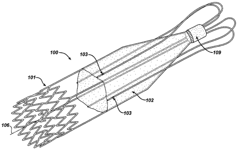
A perspective view of a prosthetic venous valve in the
deployed state according to one embodiment of the present
invention is shown in Figure 1. The prosthetic venous valve
100 comprises a structural frame 101 and a biocompatible
membrane assembly 102. The membrane assembly 102 is a thin-
walled biocompatible material formed into a tube with a
CA 02544296 2006-05-01
WO 2005/046529 PCT/US2004/034478
closed end. Exemplary configurations of a closed end tube
would include a tubular cup or cone shape, however one of
skill in the art would understand that other configurations
could also be used.
Alternatively, the cup or cone end of membrane assembly
102 may also be partially open, having a cross-sectional
area that is substantially smaller than the open end of the
membrane assembly. This reduced cross-sectional area must
be sized to effectively minimize or reduce fluid flow past
the prosthetic valve 100, substantially occluding the
vessel, when the valve 100 is in the closed (expanded)
position. The partially open-end configuration will allow
fluid to pass through the tube (membrane assembly 102)
during antegrade blood flow, preventing or reducing fluid
stagnation within the tube. In applications where the
prosthetic valve 100 is placed in the bloodstream, this
reduced stagnation or pooling may decrease the risk of
clotting.
For clarity, a perspective view of the prosthetic
venous valve 100 structural frame 101 according to one
embodiment of the present invention is shown in Figure 2A.
The structural frame 101 consists of an anchor structure 104
connected by at least one connecting member 105 to a
11
CA 02544296 2006-05-01
WO 2005/046529 PCT/US2004/034478
proximal collar 108. In a preferred embodiment, at least
three connecting members 105 are utilized. By way of
example, the embodiment illustrated in Figure 2A shows four
connecting members 105.
One or more cantilever valve struts 107 extend from the
proximal collar 108 in a proximal direction (upstream)
before looping back in a distal (downstream) direction
substantially parallel to the structural frame 101
longitudinal axis 106. This configuration allows the
cantilever valve strut 107 to be longer, increasing the
flexibility of the struts 107 and helping to reduce the
strains imposed in the structural frame 101 and/or membrane
assembly 102. The cantilever valve struts 107 are attached
to the biocompatible membrane assembly 102 (not shown in
Figure 2A) and further support the assembly in the open and
closed positions. The proximal collar 108 serves as a
connection point between the one or move valve strut members
107 and the one or more connecting members 105.
Each of the cantilever valve struts 107 illustrated in
Figure 2A have a loop end 112 incorporated into the proximal
end and a single branch distal end 113. The loop end 112 of
the valve strut 107 is attached directly to the proximal end
of the proximal collar 108, and has a semi-circular
12
CA 02544296 2006-05-01
WO 2005/046529 PCT/US2004/034478
configuration, substantially symmetric about its center.
This configuration allows the loop end 112 to effectively
reverse the direction of the cantilever valve strut 107 from
a proximal direction, where it attaches to the proximal end
of proximal collar 108, to a distal direction.
In a preferred embodiment, at least three cantilever
valve struts 107 are utilized. In the embodiment
illustrated in Figures 2A four cantilever valve struts 107
are shown.
The number of cantilever valve struts 107 and
connecting members 105 illustrated are not meant to limit
the scope of the invention. One of skill in the art would
understand that other quantities and combinations of valve
struts 107 and connecting members 105 could be used and
still accomplish the general intent of the invention.
In addition, the structural frame 101, particularly the
connecting members 105 and/or cantilever valve struts 107
may include radiopaque markers or marker bands attached or
integrated thereto. The radiopaque markers are opaque to
radiation, especially to X rays and MRI, allowing the
position of the structural frame 101 or its components to be
viewed "in vivo". Figure 1 illustrates marker bands 103
along the cantilever valve strut 107 members.
13
CA 02544296 2006-05-01
WO 2005/046529 PCT/US2004/034478
It should be noted that the terms proximal and distal
are typically used to connote a direction or position
relative to a human body. For example, the proximal end of
a bone may be used to reference the end of the bone that is
closer to the center of the body. Conversely, the term
distal can be used to refer to the end of the bone farthest
from the body. In the vasculature, proximal and distal are
sometimes used to refer to the flow of blood to the heart,
or away from the heart, respectively. Since the prosthetic
valves described in this invention can be used in many
different body lumens, including both the arterial and
venous system, the use of the terms proximal and distal in
this application are used to describe relative position in
relation to the direction of fluid flow. As used herein,
the terms upstream and downstream are relative to the normal
direction of fluid flow (antegrade flow). By way of
example, for venous valves, downstream connotes a direction
of blood flow toward the heart. Accordingly, the use of the
term proximal in the present application describes an
upstream member, section or relative position, regardless of
its orientation relative to the body. The use of the term
distal is used to describe a downstream member, section or
relative position regardless of its orientation relative to
14
CA 02544296 2006-05-01
WO 2005/046529 PCT/US2004/034478
the body. Similarly, the use of the terms proximal and
distal to connote a direction describe upstream (retrograde)
or downstream (antegrade) respectively.
In the embodiment illustrated in Figures 2A, the
connecting members 105 are substantially linear members,
connecting the stent based distal anchor 104 and the
proximal collar 108. Alternatively, the connecting members
105 may be twisted in a helical fashion as they extend
between the proximal collar 108 and the distal anchor 104.
This alternate embodiment is illustrated in Figure 2B.
Specifically, the connection points between the connecting
members 105 and the distal anchor 104, and the connecting
members 105 and the proximal collar 108, are rotationally
phased 180 degrees from each other to provide the helical
design.
Similarly, the distal end 113 of the cantilever valve
struts 107 are illustrated as substantially straight
members, but may take on other configurations. By way of
example, Figure 2C shows a structural frame 101 having
sinusoidal cantilever valve struts 107 along the distal end
113, while Figure 2D shows a structural frame 101 having
helical cantilever valve struts 107 along the distal end
113. These various configurations may be used to change the
CA 02544296 2006-05-01
WO 2005/046529 PCT/US2004/034478
properties of the structural frame, for example, by
providing more flexibility in a particular plane or
direction. Still other configurations are possible as would
be understood by one of skill in the art.
The structural frame 101 could also include a secondary
mechanism to center the proximal end of the frame in the
body vessel or lumen. This mechanism may also provide
additional anchoring to the vessel wall to further stabilize
the prosthetic valve 100.
Figure 2E shows a centering mechanism 205 incorporated
into the proximal end of the structural frame 101 according
to one embodiment of the present invention. The centering
mechanism 205 is comprised of one or more legs 210 that
extend in a substantially radial direction from the
longitudinal centerline 106 to the vessel wall (not shown).
In the illustrated embodiment, 4 legs 210 are shown for the
purpose of example. The legs 210 terminate with a blunt
end, such as the curved bend illustrated, to reduce the
possibility of the leg end perforating the vessel wall. The
opposite end of the leg 210 is attached to the structural
frame at or near the proximal collar 108. In the embodiment
illustrated in Figure 2E, the centering legs 210 are cut
from the same tube as the remainder of the structural frame
16
CA 02544296 2006-05-01
WO 2005/046529 PCT/US2004/034478
101 such that the structural frame 101, including legs 210,
is a one piece unit. Alternatively, the centering legs 210
may be separate wire units and crimped or suitably attached
to the structural frame 101 at the proximal collar 108. The
leg 210 may include barbs 215 on or along the end portion to
further anchor the structural frame 101 to the vessel wall.
The structural frame 101 may also include a second
anchor mechanism' 203, similar to anchor 104, as shown in
Figure 2F. Aside from providing additional support and
anchoring for the proximal end of the structural frame 101,
the proximal anchor 203 may also act as a centering
mechanism to center the proximal end of the structural frame
101 in the vessel or lumen (not shown). The proximal anchor
203 may be attached directly to the structural frame 101 at
the proximal collar 108, or may be attached to the proximal
collar by connecting members 206 as shown in Figure 2F. As
disclosed above, the proximal anchor 203 and connecting
members 206 may be cut from the same tube as the remainder
of the structural frame 101 such that the structural frame
101, including the anchor 203 and connecting members 206, is
a one piece unit. Alternatively, the anchor 203 and
connecting members 206 may be separate units crimped or
17
CA 02544296 2006-05-01
WO 2005/046529 PCT/US2004/034478
suitably attached to the structural frame 101 at the
proximal collar 108.
The materials for the structural frame 101 should
exhibit excellent corrosion resistance and biocompatibility.
In addition, the material comprising the structural frame
101 should be sufficiently radiopaque and create minimal
artifacts during MRI.
The present invention contemplates deployment of the
prosthetic venous valve 100 by both assisted (mechanical)
expansion, i.e. balloon expansion, and self-expansion means.
In embodiments where the prosthetic venous valve 100 is
deployed by mechanical (balloon) expansion, the structural
frames 101 is made from materials that can be plastically
deformed through the expansion of a mechanical assist
device, such as by the inflation of a catheter based
balloon. When the balloon is deflated, the frame 101
remains substantially in the expanded shape. Accordingly,
the ideal material has a low yield stress (to make the frame
101 deformable at manageable balloon pressures), high
elastic modulus (for minimal recoil), and is work hardened
through expansion for high strength. The most widely used
material for balloon expandable structures 101 is stainless
steel, particularly 316L stainless steel. This material is
18
CA 02544296 2006-05-01
WO 2005/046529 PCT/US2004/034478
particularly corrosion resistant with a low carbon content
and additions of molybdenum and niobium. Fully annealed,
stainless steel is easily deformable.
Alternative materials for mechanically expandable
structural frames 101 that maintain similar characteristics
to stainless steel include tantalum, platinum alloys,
niobium alloys, and cobalt alloys. In addition other
materials, such as polymers and bioabsorbable polymers may
be used for the structural frames 101.
Where the prosthetic venous valve 100 is self-
expanding, the materials comprising the structural frame 101
should exhibit large elastic strains. A suitable material
possessing this characteristic is Nitinol, a Nickel-Titanium
alloy that can recover elastic deformations of up to 10
percent. This unusually large elastic range is commonly
known as superelasticity.
The disclosure of various materials comprising the
structural frame should not be construed as limiting the
scope of the invention. One of ordinary skill in the art
would understand that other material possessing similar
characteristics may also be used in the construction of the
prosthetic venous valve 100. For example, bioabsorbable
polymers, such as polydioxanone may also be used.
19
CA 02544296 2006-05-01
WO 2005/046529 PCT/US2004/034478
Bioabsorbable materials absorb into the body after a period
of time. The period of time for the structural frame 101 to
absorb may vary, but is typically sufficient to allow
adequate tissue growth at the implant location to adhere to
and anchor the biocompatible membrane 102.
The structural frame 101 may be fabricated using
several different methods. Typically, the structural frame
101 is constructed from sheet, wire (round or flat) or
tubing, but the method of fabrication generally depends on
the raw material form used.
The structural frame 101 can be formed from wire using
convention wire forming techniques, such as coiling,
braiding, or knitting. By welding the wire at specific
locations a closed-cell structure may be created. This
allows for continuous production, i.e. the components of the
structural frame 101, such as the anchors, to be cut to
length from a long wire mesh tube. The connecting members
(i.e. 206, 105) may then be attached to the proximal and
distal anchors (i.e. 203, 104 respectively), by welding or
other suitable connecting means. When this fabrication
method is used, the proximal collar 108 may also be crimped
over the wire frame ends (i.e. connecting members,
cantilever struts, and/or centering legs) to connect the
CA 02544296 2006-05-01
WO 2005/046529 PCT/US2004/034478
individual members together. Alternatively, the wire ends
may be attached to the proximal collar 108 by welding or
other suitable connecting means.
Alternatively, some or all of the complete structural
frame 101 may be cut from a solid wall tube or sheet of
material. Laser cutting, water-jet cutting and
photochemical etching are all methods that can be employed
.to form the structural frame 101 from sheet and tube stock
as are known in the art.
Referring to Figure 2A for example, the structural
frame 101 (including the distal anchor 104, connecting
members 105, cantilever valve struts 107 and proximal collar
108) may all be cut from a solid tube eliminating the need
for welding or mechanically attaching individual components
together. In this embodiment, the proximal collar 108 shown
is the actual pre-cut solid wall tube (and remains in the
pre-cut, pre-expansion size), while the remainder of the
components comprising the structural frame 101 are shown in
the expanded (deployed) position. As one of skill in the
art would understand, the proximal collar 108 serves as a
common termination point for the cantilever valve struts 107
and connecting members 105.
21
CA 02544296 2006-05-01
WO 2005/046529 PCT/US2004/034478
In other embodiments, the proximal anchor 203 or
centering legs 210 may similarly be cut from the same solid
wall tube as the remainder of the structural frame 101.
Alternatively, the connecting members 105 and
cantilever valve struts 107 may be separate loose
components, and tied to each other by the proximal collar-
108. In this configuration, the proximal collar 108 acts as
a connection point to connect or crimp down and hold the
loose members in place. In other embodiments disclosed
above, the centering legs 210, connecting members 206 and/or
proximal anchor 203 may also be fabricated separate from the
other structural frame 101 components, and similarly
attached or crimped in place at the proximal collar 108.
As discussed above, the disclosure of various methods
for constructing the structural frame 101 should not be
construed as limiting the scope of the invention. One of
ordinary skill in the art would understand that other
construction methods may be employed to form the structural
frame 101 of the prosthetic venous valve 100.
In one embodiment of the invention, the anchor 104 (and
in other particular embodiments, proximal anchor 203) are
stent-based structures. This configuration facilitates the
percutaneous delivery of the prosthetic venous valve 100
22
CA 02544296 2006-05-01
WO 2005/046529 PCT/US2004/034478
through the vascular system in a compressed state. Once
properly located, the stent-based venous valve 100 may be
deployed to the expanded state.
A perspective views of a typical stent-based anchor in
the expanded (deployed) state is shown in Figures 3A.
Although stent anchor 104 incorporating a plurality of hoop
structures (306A through 306D) is shown in the illustrated
embodiment, each stent anchor may utilize a single hoop
structure.
The distal stent anchor 104 (and in some embodiments
proximal stent anchor 203) is comprised of a tubular
configuration of structural elements having proximal and
distal open ends and defining the longitudinal axis 106
extending therebetween. The stent anchor 104 has a first
diameter (not shown) for insertion into a patient and
navigation through the vessels, and a second diameter D2 for
deployment into the target area of a vessel, with the second
diameter being greater than the first diameter. The stent
anchor 104, and thus the stent based venous valve 100, may
be either a mechanical (balloon) or self-expanding stent
based structure.
The stent anchor 104 comprises at least one hoop
structure 306 (306A through 306D are shown) extending
23
CA 02544296 2006-05-01
WO 2005/046529 PCT/US2004/034478
between the proximal and distal ends. The hoop structure
306 includes. a plurality of longitudinally arranged strut
members 308 and a plurality of loop members 310 connecting
adjacent struts 308. Adjacent struts 308 are connected at
opposite ends in a substantially S or Z shaped pattern so as
to form a plurality of cells. The plurality of loops 310
have a substantially semi-circular configuration, having an
inter radii 312 and outer radii 314, and are substantially
symmetric about their centers. The inner and outer radii
312, 314 respectively, are shown in a close-up perspective
view illustrated in Figure 3B.
In the illustrated embodiment, the distal stent anchor
104 comprises a plurality of bridge members 314 that connect
adjacent hoops 306A through 306D. Each bridge member 314
comprises two ends 316A, 316B. One end 316A, '316B of each
bridge 314 is attached to one loop on one hoop. Using hoop
sections 3060 and 306D for example, each bridge member 314
is connected at end 316A to loop 310 on hoop section 3060 at
a point 320. Similarly, the opposite end 316B of each
bridge member 314 is connected to loop 310 on hoop sections
306D at a point 321.
As described earlier, although a Z or S shaped pattern
stent anchor is shown for the purpose of example, the
24
CA 02544296 2006-05-01
WO 2005/046529 PCT/US2004/034478
illustration is not to be construed as limiting the scope of
the invention. One of ordinary skill in the art would
understand that other stent geometries may be used.
The connecting member 105 may be connected to the
distal anchor 104 at various points along the structure. As
illustrated in Figure 3A, the connecting members 105 are
connected to the proximal end of the distal anchor 104 at
the inflection point of the loop members 310, particularly
at the outer radii 314 of the inflection point of loop
members 310. Similarly, Figure 3C illustrates a single hoop
anchor 104 having three connecting members 105 connected to
the proximal end of the distal anchor 104 at the outer radii
314 of the inflection point of loop members 310.
Preferably the connecting members 105 are connected to
the inflection point of loop members 310 at evenly spaced
intervals along the circumference of the tubular anchor 104.
This configuration facilitates the radial expansion of the
prosthetic valve from the collapsed (delivered) state to the
expanded (deployed) state, and provides a substantially
symmetrical valve configuration.
Alternatively, the connecting members 105 may be
connected to the proximal end of the distal anchor 104 at
the inner radii 312 of the inflection point of loop member
CA 02544296 2006-05-01
WO 2005/046529 PCT/US2004/034478
310. This configuration is illustrated in Figure 3D.
Figure 3D also illustrates a partial perspective view of the
structural frame 101 having a single hoop structure 306 and
three connecting members.
In still a further embodiment, the connecting members
105 may be connected along the strut members 308 of the
distal anchor 104 as shown in Figure 3E.
In any of the above described configurations, the
connections between the connecting members 105 and the
anchor 104 may be made at every inflection point around the
circumference of the structure; or alternatively, at a
subset of the inflection points around the circumference of
the structure. In other words, connected inflection points
alternate with unconnected inflection points in some defined
pattern.
The distal anchor 104 secures the prosthetic valve 100
to the inside wall of a body vessel such as a vein, and
provide anchor points for the connecting members 105. Once
deployed in the desired location, the anchor 104 will expand
to an outside diameter slightly larger that the inside
diameter of the native vessel (not shown) and remain
substantially rigid in place, anchoring the valve assembly
to the vessel. The connecting members 105 preferably have
26
CA 02544296 2006-05-01
WO 2005/046529 PCT/US2004/034478
an inferior radial stiffness, and will conform much more
closely to the native diameter of the vessel, facilitating
,the operation and stability of the prosthetic valve 100.
The stent anchor may also have spurs or barbs (not
shown) protruding from its proximal or distal end to further
assist anchoring the prosthetic valve.
The membrane assembly 102 is formed from a flexible
membrane-like biocompatible material shaped into a tubular
structure with a closed or substantially closed end.
Exemplary embodiments would include a cup or cone shaped
tube. The flexible membrane may be elastic, semi-elastic or
display little or no elasticity. One of skill in the art
would appreciate that there are many different methods, some
known in the art, which may be employed to manufacture the
membrane assembly 102 from this material.
The biocompatible material may be a biological
material, such as a vein or small intestine submucosa (SIS)
formed into a cup or pocket, but is preferably a synthetic
material such as a polymer, for example an elastic or
elastomeric polymer, including a fluoropolymer,
fluoroelastomer, or a bioabsorbable material, such as a
bioabsorbable polymer or bioabsorbable elastomer.
Bioabsorbable materials may allow cells to grow and form a
27
CA 02544296 2006-05-01
WO 2005/046529 PCT/US2004/034478
tissue membrane over the bioabsorbable membrane. The
bioabsorbable membrane then absorbs into the body, leaving
the tissue membrane in place to act as a new natural tissue
valve.
The membrane material may also be made from other
synthetics, such as thin metallic materials or membranes.
The membrane must be strong enough to resist tearing
under normal use, yet thin enough to provide the necessary
flexibility that allows the biocompatible membrane assembly
102 to open and close satisfactorily. To achieve the
necessary flexibility and strength of the membrane assembly
102, the synthetic material may be, for example, reinforced
with a fiber, such as an electro-statically spun (ESS)
fiber, or formed from a porous foam, such as ePTFE, or a
mesh.
Particular ESS fibers suitable for the spinning process
include fluoropolymers, such as a crystalline fluoropolymer
with an 85/15% (weight/weight ratio) of vinylidene
fluoride/hexafluoropropylene (VDF/HFP). Solvay Solef 21508
and Kynarflex 2750-01 are two such examples. However, one
of skill in the art would understand that any material
possessing the desired characteristics may be used,
including, for example: bioabsorbable polymers, such as
28
CA 02544296 2006-05-01
WO 2005/046529 PCT/US2004/034478
polyglycolic acid, polylactic acid, poly (paradioxanone),
polycaprolactone, poly (trimethylenecarbonate) and their
copolymers; and semicrystalline bioelastomers, such as
60/40%(weight/weight ratio) of polylactic acid /
polycaprolactone (PLA/PCL), 65/35 (weight/weight ratio) of
polyglycolic acid/polycaprolactone (PGA/PCL), or
nonabsorbable siliconized polyurethane, non-siliconized
polyurethanes, siliconized polyureaurethane, including
siliconized polyureaurethane end capped with silicone or
fluorine end groups, or natural, polymers in combination
thereof. it should be noted that
poly(trimethylenecarbonate) can not be spun as a
homopolymer.
The ESS formed membrane assembly 102 may also be coated
with a polymer solution, such as fluoroelastomer. The
coating process may take place before the membrane assembly
is attached to the cantilever valve struts 107 or connecting
members 105, or after the membrane assembly 102 and
structural frame 101 are assembled.
The coating process may act to encapsulate and attach
at least a portion of the spun ESS reinforcement fiber to
the structural frame, in particular the cantilever valve
strut 107 assembly or connecting members 105. It should be
29
CA 02544296 2006-05-01
WO 2005/046529 PCT/US2004/034478
noted that in some embodiments of the invention, some
movement between the membrane assembly 102 and the
structural frame 101 is desired. Accordingly, not all of
the ESS fiber spun structural frame 101 may be coated.
The coating process may also remove some porosity of
the membrane material. However, it may be desirable to
maintain some porosity in particular embodiments to promote
biological cell grown on and within the membrane tubular
structure.
The coating solution preferably comprises a polymer put
into solution with a solvent. As the solvent evaporates,
the polymer comes out of solution forming the coating layer.
Accordingly, for the process to work properly, the solvent
used in the coating solution should not dissolve or alter
the ESS fibers being coated. By way of example, a coating
solution of 60/40% VDF/HFP in methanol (methanol being the
solvent) has been found to be a suitable solution for
coating an ESS fiber comprised of 85/15% VDF/HFP.
In one embodiment of the invention, the polymer
comprising the coating is Daikin's Dai-El G701BP, which is a
60/40% VDF/HFP. In addition, Daikin's Dai-El T630, a
thermoplastic elastomer based on vinylidene
fluoride/hexafluoropropylene/tetrafluoroethylene.
CA 02544296 2006-05-01
WO 2005/046529 PCT/US2004/034478
,(VDF/HFP/TFE) can also be used. Again, one of ordinary
skill in the art would understand that other materials
having suitable characteristics may be used for the coating,
for example, other polymers, such as siliconized
polyurethane, including Polymer Technology Group's Pursil,
Carbosil, Purspan and Purspan F.
In another embodiment the membrane assembly is made
from a micro-cellular foam or porous material, such as, for
example an ePTFE membrane.
In this embodiment, the membrane assembly 102 is
fabricated from a polymer material that can be processed
such that it exhibits an expanded cellular structure,
preferably expanded Polytetrafluoroethylene (ePTFE). The
ePTFE tubing is made by expanding Polytetrafluoroethylene
(PTFE) tubing, under controlled conditions, as is well known
in the art. This process alters the physical properties
that make it satisfactory for use in medical devices.
However, one of ordinary skill in the art would understand
that other materials that possess the necessary
characteristics could also be used.
The micro-cellular foam or porous material (preferably
expanded Polytetrafluoroethylene (ePTFE)) may be coated with
a polymer. The polymer can be coated on the inside or
31
CA 02544296 2006-05-01
WO 2005/046529 PCT/US2004/034478
outside surface of the ePTFE tube. Alternatively, the
polymer may be coated on the inside and outside of the ePTFE
tube.
In a preferred embodiment of the invention, the polymer
comprising the coating includes. Daikin's Dai-El T630, a
thermoplastic elastomer based on vinylidene
fluoride/hexafluoropropylene/tetrafluoroethylene
(VDF/HFP/TFE) and blends thereof. Again, one of ordinary
skill in the art would understand that other materials
having suitable characteristics may be used for the coating,
for example, other polymers, such as siliconized
polyurethanes and blends thereof, including Polymer
Technology Group's Pursil, Carbosil, Purspan and Purspan F.
The membrane assembly 102 formed from the micro-
cellular foam or porous membrane may also be coated with a
fluoroelastomer. In one embodiment of the invention, the
coating is Daikin G701BP, which is a 60/40% VDF/HFP. Again,
one of ordinary skill in the art would understand that other
materials having suitable characteristics might be used for
the coating, for example, other polymers, such as
siliconized polyurethane.
As previously described, the coating process may take
place before the membrane assembly is attached to the
32
CA 02544296 2006-05-01
WO 2005/046529 PCT/US2004/034478
structural frame 101, or after the membrane assembly 102 and
structural frame 101 are assembled. The coating process may
act to encapsulate and attach at least a portion of the
micro-cellular foam or porous membrane tube to the
structural frame 101.
Some post processing of the membrane assembly 102 may
also take place to achieve particular desired
characteristics or configurations. This may includes
creating-the final closed or substantially closed cup or
cone shape of the membrane assembly 102 if needed. In
addition, post processing may change the characteristics of
the membrane assembly 102 by thickening or thinning the
membrane in particular locations. Thickening the membrane
may add rigidity and reinforcement to a particular area.
Thinning the membrane may make the membrane more pliable,
which is a desirable characteristic. Still other post
processing procedures may change the physical shape of the
membrane assembly 102, for example, by forming loop collars
(such as loop collars 605 in Figures 6A through6C) along the
distal edge of membrane assembly 102.
The thickness of the synthetic valve membrane assembly
102 is dependent on the size, type and location of the
prosthetic valve. For venous valves applications a
33
CA 02544296 2006-05-01
WO 2005/046529 PCT/US2004/034478
polymeric membrane assembly 102 having a thickness of
between 12 m and 100pm and preferably between 25pm and 50pm
has been found to be acceptable.
The membrane assembly 102 is placed or formed over the
structural frame 101, similar to a graft. In particular,
the membrane assembly 102 is formed into a closed end or
substantially closed end tube over at least a portion of the
connecting members 105. The cantilever valve struts 107 are
then placed over the outer surface of the membrane assembly
102. The connecting members 105 and the cantilever valve
struts 107 act to support the membrane assembly in a
substantially tubular configuration.
The membrane assembly 102 may be formed into the
tubular configuration separately, and then placed over the
structural frame 101. Alternatively, the membrane assembly
102 may be. formed into the tubular configuration directly
over the structural frame 101, such as by an electrostatic
spinning process that spins the ESS fiber directly over the
structural frame. This process is disclosed in a co pending
patent application, serial number 10/402,048 entitled METHOD
OF FORMING A TUBULAR MEMBRANE ON A STRUCTURAL FRAME, filed
on March 28, 2004, and is hereby incorporated by reference.
34
CA 02544296 2006-05-01
WO 2005/046529 PCT/US2004/034478
Figures 4A and 4B are perspective and section views,
respectively, illustrating one embodiment of the expanded
(deployed) prosthetic venous valve assembly 100 in the open
position. In this embodiment, the term open means that the
prosthetic venous valve 100 is configured to allow antegrade
.blood flow 400 to pass through the valve. To accomplish
this, the membrane assembly 102 is in a substantially
collapsed position.
The embodiment illustrated in Figures 4A and 4B has
three connecting members 105 and three cantilever valve
struts 107. The membrane assembly 102 is placed over a
portion of the structural frame 101, particularly over the
connecting members 105, proximal collar 108 and at least a
portion of the loop end 112 of the cantilever valve struts
107. A compression ring 109 may be used to fix the membrane
assembly 102 to the proximal collar 108. The ring. 109
should be sized to apply a radially compressive force on the
membrane assembly 102, effectively fixing the membrane
assembly 102 against the proximal collar 108.
The flexible membrane assembly illustrated in Figure
4A is formed into a tubular cone having a first (distal) and
second (proximal) ends 401, 402 respectively. The. first end
401 of the membrane assembly 102 is located at the distal
CA 02544296 2006-05-01
WO 2005/046529 PCT/US2004/034478
end of the cantilever valve struts 107, near the proximal
end of the distal anchor 104, and is capable of opening to
substantially the full diameter of the native vessel. In
one embodiment of the invention, the membrane assembly 102
is fixedly attached along the distal end of the cantilever
valve struts 107 and connecting members 105. Alternatively
the membrane assembly 102 may be slidably attached to the
connecting members 105. This configuration may assist the
membrane assembly 102 when opening and closing.
The membrane assembly extends in a proximal direction
along the connecting members 105 and terminates at the
second end 402. The second (proximal) end 402 of the
membrane assembly 102 is fixedly or slideably attached along
the loop end 112 of the cantilever valve struts 107. The
proximal end 402 of the membrane assembly 102 has an open
end with a substantially reduced cross-sectional area. As
previously disclosed, the proximal end 402 may alternatively
terminate with a closed cup or cone end.
In an alternative embodiment, the proximal end 402 may
terminate at the proximal collar 108 with a closed or open
end.
The illustrated embodiment shows a valve assembly 100
having a single cone or cup, and may be considered a
36
CA 02544296 2006-05-01
WO 2005/046529 PCT/US2004/034478
monocusp design. However, other configurations using more
than a single cup or cone are also contemplated by the
present invention.
During retrograde flow, blood passes the leading edge
along the first end 401 of the membrane assembly 102 and
enters the interior (i.e. "cup") portion of membrane
.assembly 102. The membrane assembly 102 quickly fills with
the retrograde flowing blood, expanding and opening the
membrane assembly 102. As the membrane assembly 102 opens,
the first end 401 is forced out toward vessel wall,
substantially occluding the vessel and thus reducing
retrograde flow through the valve. In a preferred
embodiment, the membrane assembly 102 will expand to a
sufficient diameter to substantially seal against the inner
vessel wall.
As previously described, the membrane assembly 102 may
have a closed or substantially closed proximal end 402. In
embodiments where the membrane assembly 102 proximal end 402
is substantially closed, the proximal opening must be of a
sufficiently reduced cross-sectional area to substantially
reduce or prevent the flow of fluid through the proximal end
402 of the membrane assembly 102.
37
CA 02544296 2006-05-01
WO 2005/046529 PCT/US2004/034478
In. the embodiment illustrated in Figure 4A, the
proximal end 402 of the membrane assembly 102 is a
substantially closed end tube (open but having a reduced
cross-sectional area) disposed about the proximal loop end
112 of the cantilever valve struts 107. In particular, the
proximal end 402 of the membrane assembly 102 is disposed
about the cantilever valve strut 107 in close proximity to
the interface between the cantilever valve strut 107 and
proximal collar 108. The membrane assembly 102 and
cantilever valve strut 107 are configured such that when the
valve is in the open position (collapsed to allow blood flow
to pass through the valve), the proximal loop ends 112 of
the cantilever valve struts 107 are separated and allow the
proximal end 402 of the membrane assembly to remain in an
open tubular position. When the valve closes during
retrograde blood flow, the proximal loop ends .112 of the
cantilever valve struts 107 move closer together, urging the
proximal end 402 of the membrane assembly 102 together.
This movement substantially or completely closes the
proximal end 402 of the membrane assembly 102, allowing the
membrane assembly to substantially or completely occlude the
vessel.
38
CA 02544296 2006-05-01
WO 2005/046529 PCT/US2004/034478
Figures 5A and 5B show perspective and section views,
respectively,' illustrating one embodiment of the expanded
(deployed) prosthetic venous valve assembly 100 in the
closed position. As the term is used herein, closed means
that the prosthetic venous valve 100 is configured to
substantially prohibit retrograde blood flow 410 to pass
through the valve. To accomplish this, the membrane
assembly 102 is in an expanded position, substantially
occluding the vessel.
In a preferred embodiment of the invention, the
membrane assembly 102 is normally configured in the open
position (membrane assembly 102 substantially collapsed),
and only moves to the closed position (membrane assembly 102
substantially expanded) upon retrograde blood flow. This
configuration minimizes interference with blood flow
(minimized occlusion) and reduces turbulence at and through
the valve. The cantilever valve struts 107 in this
embodiment have an inferior radial stiffness, and provide a
natural bias against the movement of the membrane assembly
102 to the closed position. This bias assists the valve
membrane assembly 102 when returning to the open position.
Depending on the application, it may also be desirable
for the bias towards opening the prosthetic valve 100
39
CA 02544296 2006-05-01
WO 2005/046529 PCT/US2004/034478
(collapsing the membrane assembly 102) be sufficiently high
to commence collapsing the membrane assembly 102 before
antegrade blood flow begins, i.e. during a point in time
when the blood flow is stagnant (there is neither antegrade
nor retrograde blood flow), or when minimal retrograde flow
is experienced.
In other applications, it may be desirable to have the
valve assembly 100 normally configured in the closed
position (membrane assembly 102 in the expanded position),
biased closed, and only open upon antegrade flow.
As earlier described, the membrane assembly 102 is made
from a flexible membrane-like biocompatible material. The
membrane assembly 102 can be woven, non-woven (such as
electrostatic spinning), mesh, knitted, film or porous film
(such as foam).
The membrane assembly 102 may be fixedly attached to
the structural frame 101 (particularly cantilever valve
struts 107 and/or connecting members 105) by many different
methods, including attachment by means of a binder, heat, or
chemical bond, and/or attachment by mechanical means, such
as welding or suturing. In one embodiment, some of the
membrane assembly 102, such as distal end 401, is slideably
attached to the connecting member 105. Allowing the distal
CA 02544296 2006-05-01
WO 2005/046529 PCT/US2004/034478
end 401 to slide along the connecting member 105 107 may
allow or improve the opening and closing of the membrane
assembly 102. The sliding movement may also assist the
membrane assembly 102 cup when filling and emptying.
In some applications, excessive sliding movement of the
membrane assembly 102 is undesirable. In, these embodiments,
a limiting means may be integrated into the prosthetic valve
100 to limit the sliding movement of the membrane assembly
102. Examples of limiting means are shown in Figures 6A to
6C. In each embodiment a stop 600 (illustrated as stop
600A, 600B, and 600C in Figures 6A to 6C respectively) is
integrated into the connecting member 105. The membrane
assembly 102 is wrapped around the connecting member 105 and
bonded to itself to form a loop collar 605. The loop collar
605 must be sized to inhibit the distal end 401 of the
membrane assembly 102 from, sliding past the stop 600. In
Figure 6A, the connecting member 105 has a thickened or
"bulbous" section forming stop 600A. Figure 6B illustrates
an undulating stop 600B configuration. Similarly, Figure 6C
shows the stop 600C configured as a double bulbous section.
It should be noted that the various configurations
illustrated in Figures 6A through 6C are exemplary. One of
41
CA 02544296 2006-05-01
WO 2005/046529 PCT/US2004/034478
ordinary skill in the art would understand that other
configurations of stops may used.
It is important to note that the local delivery of
drug/drug combinations may be utilized to treat a wide
variety of conditions utilizing any number of medical
devices, or to enhance the function and/or life of the
device. Medical devices that may benefit from this
treatment include, for example, the frame based
unidirectional flow prosthetic implant subject of the
present invention.
Accordingly, in addition to the embodiments described
above, therapeutic or pharmaceutic agents may be added to
any component of the device during fabrication, including,
for example, the ESS fiber, polymer or coating solution,
membrane tube, structural frame or inner and outer membrane,
to treat any. number of conditions. In addition, therapeutic
or pharmaceutic agents may be applied to the device, such as
in the form of a drug or drug eluting layer, or surface
treatment after the device has been formed. In a preferred
embodiment, the therapeutic and pharmaceutic agents may
include any one or more of the following:
antiproliferative/antimitotic agents including natural
products such as vinca alkaloids (i.e. vinblastine,
42
CA 02544296 2006-05-01
WO 2005/046529 PCT/US2004/034478
vincristine, and vinorelbine), paclitaxel,
epidipodophyllotoxins (i.e. etoposide, teniposide),
antibiotics (dactinomycin (actinomycin D) daunorubicin,
doxorubicin and idarubicin), anthracyclines, mitoxantrone,
bleomycins, plicamycin (mithramycin) and mitomycin, enzymes
(L-asparaginase which systemically metabolizes L-asparagine
and deprives cells which do not have the capacity to
synthesize their own asparagine) ; antiplatelet agents such
as G(GP) llb/llla inhibitors and vitronectin receptor
antagonists; antiproliferative/antimitotic alkylating agents
such as nitrogen mustards (mechlorethamine,
cyclophosphamide and analogs, melphalan, chlorambucil),
ethylenimines and methylmelamines (hexamethylmelamine and
thiotepa), alkyl sulfonates-busulfan, nirtosoureas
(carmustine (BCNU) and analogs, streptozocin), trazenes
dacarbazinine (DTIC); antiproliferative/antimitotic
antimetabolites such as folic acid analogs (methotrexate),
pyrimidine analogs (fluorouracil, floxuridine, and
cytarabine), purine analogs and related inhibitors
.20 (mercaptopurine, thioguanine, pentostatin and 2-
chlorodeoxyadenosine {cladribine}); platinum coordination
complexes (cisplatin, carbo'platin), procarbazine,
hydroxyurea, mitotane, aminoglutethimide; hormones (i.e.
43
CA 02544296 2006-05-01
WO 2005/046529 PCT/US2004/034478
estrogen); anticoagulants (heparin, synthetic heparin salts
and other inhibitors of thrombin); fibrinolytic agents (such
as tissue plasminogen activator, streptokinase and
urokinase), aspirin, dipyridamole, ticlopidine, clopidogrel,
abciximab; antimigratory; antisecretory (breveldin); anti-
inflammatory: such as adrenocortical steroids (cortisol,
cortisone, fludrocortisone, prednisone, prednisolone, 6(G-
methylprednisolone, triamcinolone, betamethasone, and
dexamethasone), non-steroidal agents (salicylic acid
derivatives i.e. aspirin; para-aminophenol derivatives i.e.
acetominophen; indole and indene acetic acids (indomethacin,
sulindac, and etodalac) , heteroaryl acetic acids (tolmetin,
diclofenac, and ketorolac), arylpropionic acids (ibuprofen
and derivatives), anthranilic acids (mefenamic acid, and
meclofenamic acid), enolic acids (piroxicam, tenoxicam,
phenylbutazone, and oxyphenthatrazone), nabumetone, gold.
compounds (auranofin, aurothioglucose, gold sodium
thiomalate); immunosuppressives: (cyclosporine, tacrolimus
(FK-506), sirolimus (rapamycin), azathioprine, mycophenolate
mofetil); angiogenic agents: vascular endothelial .growth
factor (VEGF), fibroblast growth factor (FGF); angiotensin
receptor blockers; nitric oxide donors; anti-sense
oligionucleotides and combinations thereof; cell cycle
44
CA 02544296 2006-05-01
WO 2005/046529 PCT/US2004/034478
inhibitors, mTOR inhibitors, and growth factor receptor
signal transduction kinase inhibitors; retenoids; cyclin/CDK
inhibitors; HMG co-enzyme reductase inhibitors (statins);
and protease inhibitors.
While a number of variations of the invention have been
shown and described in detail, other modifications and
methods of use contemplated within the scope of this
invention will be readily apparent to those of skill in the
art based upon this disclosure. It is contemplated that
various combinations or subcombinations of the specific
embodiments may be made and still fall within the scope of
the invention. For example, the embodiments variously shown
to be prosthetic "venous valves" may be modified to instead
incorporate prosthetic "heart valves" and are also
contemplated. Moreover, all assemblies described are
believed useful when modified to treat other vessels or
lumens in the body, in particular other regions of the body
where fluid flow in a body vessel or lumen needs to be
controlled or regulated. This may include, for example, the
coronary, vascular, non-vascular and peripheral vessels and
ducts. Accordingly, it should be understood that various
applications, modifications and substitutions may be made of
CA 02544296 2006-05-01
WO 2005/046529 PCT/US2004/034478
equivalents without departing from the spirit of the
invention or the scope of the following claims.
The following claims are provided to illustrate
examples of some beneficial aspects of the subject matter
disclosed herein which are within the scope of the present
invention.
46