Note: Descriptions are shown in the official language in which they were submitted.
CA 02544376 2006-05-O1
WO 2005/044328 PCT/IB2003/005003
1
DESCRIPTION
"METHOD FOR PREPARING DRUG ELUTING MEDICAL DEVICES AND
DEVICE OBTAINED THEREFROM"
[0001]. The present invention relates to a method for
preparing drug eluting medical devices and devices
obtained therefrom. In particular, the invention relates
to a method for preparing a vascular stent covered with
one or more drugs for treating and/or preventing re
stenosis.
[0002]. In angioplasty, the use of stems in treating
coronary occlusions is currently well known and widely
accepted and practised. Stems are reticular metal
prostheses positioned in the stenotic portion of the
vessel which remain at the site of the lesion after the
elution system and the balloon have been withdrawn. In
this way, the stmt compresses the plaque and provides
the vessel wall with a mechanical support in order to
maintain the diameter of the vessel re-established by
expanding the balloon, and prevent collapse of the
vessel.
[0003]. However, the long-term effectiveness of using
intercoronary stents still presents the major problem of
post-angioplasty coronary re-stenosis, that is the
phenomenon of reocclusion of the coronary vessel. In
fact, this phenomenon of re-stenosis occurs in 15-30% of
CA 02544376 2006-05-O1
WO 2005/044328 PCT/IB2003/005003
2
pat Tents undergoing angioplasty with stents, as described
for example in Williams DO, Holubkov R, Yeh W et al.
"Percutaneous coronary interventions in the current era
are compared with 1985-1986: The National Heart, Lung and
Blood Institute Registries", Circulation 2000; 102:2945-
2951 .
f00 04~. Stenosis caused by insertion of the stent is
due to the hyperplasia of the newly formed intima. In
particular, the mechanical damage to the artery wall
caused by the stent and the foreign body reaction caused
by the presence of the stent produce a chronic
inflammatory process in the vessel. This phenomenon gives
rise in turn to the elution of cytokins and growth
factors which promote activation of proliferation and
migration of the smooth muscle cells (SMC). The growth of
the se cells together with the production of an
ext racellular matrix produce enlargement of the section
of the vessel occupied by neointima and therefore the
process of reduction in the opening of the vessel, giving
rise to the above-mentioned re-stenosis.
(00 05]. To prevent this problem, various methods have
been developed including one which provides for covering
the stent directly with a drug or with a coating of the
polymer type capable of incorporating the drug and
eluting it locally by a controlled mechanism. A typical
CA 02544376 2006-05-O1
WO 2005/044328 PCT/IB2003/005003
3
example of a coated stent capable of eluting drugs (DES,
drug eluting stent) is described in the paper by Takeshi
Suzuki and collaborators "Stent-Based Delivery of
Sirolimus Reduces Neointimal Formation in a Porcine
Coronary Model", Circulation 2001; 104: 1188-1193. The
materials used are generally polymers, either degradable
or non-degradable which must have characteristics of
adhesion to the metal substrate (stent), the ability to
regulate the rate of elution of the drug, an absence of
toxicity phenomena and favourable interaction with the
surrounding tissue.
[0006. In particular, as far as the last
characteristic is concerned, the interactions of the
material with the surrounding tissue are to a large
extent controlled by the surface properties of the
material. The materials used in medical devices in
general do not present optimum surface characteristics as
far as interaction with the host tissue is concerned.
This circumstance manifests itself from a clinical point
of view with the onset of foreign body reaction phenomena
and, in particular for materials in contact with the
blood, with the formation of thrombi and/or emboli. The
extent of the phenomenon is such that the thrombogenicity
of synthetic materials is the most serious obstacle to
the development of small-sized artificial vessels.
CA 02544376 2006-05-O1
WO 2005/044328 PCT/IB2003/005003
4
[0007]_ To attempt to overcome these disadvantages,
procedures have been developed which, by means of
chemical reactions, provide for the covering of the
thrombogenic material with natural non-thrombogenic
molecules. The anticoagulant heparin is a typical
example. These procedures provide for a first step in
which chemical groups suitable for binding heparin,
hialuronic acid or other biomolecules are introduced onto
the surface of the stmt (or of the medical device in
general), and a second step consisting in chemical
bonding of the heparin, hyaluronic acid or other
biomolacules with chemical groups introduced by means of
the previous step .
[0008]_ Consequently, the polymers used for drug
delivery are not capable as they stand of directly
binding biomolecules but require the above step of
introducing functional groups and subsequently
immobilising said biomolecules.
[0009]_ There are polymers which of themselves contain
functional groups such as amino groups or from which
amino groups can be generated. These polymers can be
applied to the surface of the stents using conventional
technol ogy .
[OOlO]_ However, it has been found that these polymers
suffer from the serious disadvantage of being hydrophilic
CA 02544376 2006-05-O1
WO 2005/044328 PCT/IB2003/005003
and, since the step of bonding with heparin or other
biomolecules generally takes place in a solvent and in
particular for heparin in an aqueous environment, there
is a major risk of losing at least part of the drug
5 during preparation of the stmt precisely because of the
solubility of the polymer in water; moreover, precisely
because of the hydrophilic nature of the polymer, the
ability to control drug elution is limited and it is
entirely unsuitable for controlling elution of drugs
which in their turn are hydrophilic.
[0011]. Moreover, the drug eluted into the solution
containing heparin and the functional groups may
interfere with the immobilisation reaction, jeopardizing
a successful outcome.
[0012]. The problem addressed by the present invention
is therefore that of making available a method of
preparing a drug eluting vascular stent capable of
overcoming the disadvantages mentioned above.
(0013]. These problems are solved by a method for
preparing a drug eluting medical device which simplifies
the production procedure and at the same time avoids loss
of the drug or other compounds which may jeopardize the
preparation of the stent.
[0014]. A first object of the invention is therefore to
make available a method for preparing a medical device as
CA 02544376 2006-05-O1
WO 2005/044328 PCT/IB2003/005003
6
outlined in the appended main claim.
[0015] . A second object of the invention is that of
providing a drug eluting medical device obtainable
according to the above-mentioned method.
[0016]. By the term "drug eluting medical device" is
meant a device to be inserted in the human or animal
body, internally or subcutaneously, intended to remain in
said human or animal body for a defined period of time or
permanently, and which is capable of eluting a
pharmaceutically effective dose of one or more drugs for
at least part of the time during which it resides in the
human or animal body. This medical device may be a
vascular device, prosthesis, probe, catheter, dental
implant or similar. More preferably, this device will be
a vascular stent.
[0017]. Other characteristics and advantages of the
present invention will become clear from the following
description of an embodiment provided by way of non-
limiting example, in which:
- Figure 1 shows the elution curve for a hydrophilic drug
from a stent covered with polymer according to the state
of the art compared with the elution curve for a
hydrophilic drug from a stent covered with polymer
according to the invention;
- Figure 2 shows the elution curve for a hydrophobic drug
CA 02544376 2006-05-O1
WO 2005/044328 PCT/IB2003/005003
7
from a stent covered with polymer according to the state
of the art compared with the elution curve for a
hydrophobic drug from a stent covered with polymer
according to the invention.
[0018]. Following numerous experiments, it was
surprisingly found that if polymers having functional
groups such as amino groups were applied to the surface
of the medical device in a single step using a cold
plasma method, coverage of the stmt was obtained in the
form of a. hydrophobic film, adhering well and with active
and stable functional groups capable of rapid binding of
heparin, hialuronic acid or other biomolecule.
[0019]. The following description will relate to a
vascular stmt, but could also be applied to any other
medical device of the invention.
[0020]. In particular, it has been observed that
polymers with amino functional groups deposited on the
metal surface of vascular stems by cold plasma assume
characteristics of hydrophobicity, excellent adhesion to
the stent, a high degree of cross-linking so as to
operate as a barrier slowing the diffusion of a drug and
the ability to bind heparin and other biomolecules by
means of said amino groups.
[0021]. The method for preparing a drug eluting
vascular stmt as disclosed in the invention therefore
CA 02544376 2006-05-O1
WO 2005/044328 PCT/IB2003/005003
8
comprises application to the surface of said stmt of a
polymer having stable reactive functional groups, such as
for example amino, carboxyl and sulphhydryl groups, in
which this application takes place in a single step by
means of cold plasma methods.
[0022]. According to a first form of embodiment, the
polymers are deposited in the form of a film. In
particular, said polymers have functional groups capable
of forming a covalent bond with said biological
molecules, preferably chosen from among heparin,
hyaluronic acid or anti-thrombotic substances in general.
More particularly, said polymers are chosen from the
group constituted by polymers containing amino, carboxyl
and sulphhydryl groups. Preferably, the polymers with
amino groups are derived from precursors or monomers
chosen from among allylamine, heptylamine, aliphatic or
aromatic amines; polymers with carboxyl groups are
derived from precursors or monomers chosen from between
acrylic acid and methacrylic acid. Polymers with
sulphhydriyl groups are derived from precursors or
monomers chosen from among volatile mercaptans.
(0023. The method disclosed by the invention may also
provide for further polymer layers to be deposited
depending on the degree or type of mechanisms for elution
of the drug which it is wished to obtain. These latter
CA 02544376 2006-05-O1
WO 2005/044328 PCT/IB2003/005003
9
deposits are produced according to methods known in the
art such as immersion in a suitable solution or spraying
with a pneumatic spray gun or using the above-mentioned
cold plasma method. It should be noted that in any case
the outermos t layer must be deposited according to the
cold plasma method using the above-mentioned polymers
having funct Tonal groups.
[0024]. The plasma used according to the invention is a
cold plasma, that is the temperature of the total mass of
gas in the plasma phase is of the same order as the
ambient temperature. Said plasma is generated in a
conventional reactor of the type comprising a treatment
chamber insi de which there is a support for the material
to be treated, with a discharge source located nearby to
produce the plasma.
[0025]. The cold plasma may be produced under vacuum or
at atmospheric pressure and may be generated using
various electromagnetic sources, that is sources of
various frequencies and various geometries, such as for
example radiofrequency generators or microwave
generators, with electrodes of the inductive or
capacitive type .
[0026]. In. general, when the vacuum method is used, the
cold plasma is produced in a chamber with a pressure
which may vary between 0.01 and 10 mbar.
CA 02544376 2006-05-O1
WO 2005/044328 PCT/IB2003/005003
[0027]. As far as the conditions of treatment are
concerned, these depend on the electrical power which may
vary from 1 to 500 W, on the geometry of the source which
produces the plasma which may be inductive or capacitive
5 and on the frequencies of the electromagnetic radiation
used to produce the plasma which may be in the microwave
or radiofrequency range.
[0028]. Moreover, the cold plasma which is generated is
characterised by a charged species density of between 108
10 and 1012 cm 3, a condition of substantial neutrality of
charges (quasi-neutral, ion density ~ electron density),
electron energies from 0.1 to 10 eV or mean electrical
energy calculated as (ekBT/m)1/2 (e=1.9 10-19 C, kB=1.38
10-23 J/K , m= 9.1 10-31 kg, T = absolute temperature in
Kelvin), while the ions and the neutral particles are at
temperatures of the order of ambient temperature .
(0029]. The treatment time in a cold plasma is
generally not more than 30 minutes, is preferably between
0.1 and 20 minutes and still more preferably between 1
and 10 minutes.
(0030]. Preferably, the plasma treatment under vacuum
takes place according to a discontinuous or continuous
method. Said method will not be described in detail here
since it is widely known in the art.
L0031]. The cold plasma used may preferably be
CA 02544376 2006-05-O1
WO 2005/044328 PCT/IB2003/005003
11
generated at a pressure of less than atmospheric
pressure. The precursor or monomer which will be
polymerized in the plasma phase is introduced into the
reactor in the form of gas or vapour, with flow rates
which vary from 0.1 to 200 sccm (cubic centimetres in
standard conditions per minute). At this point, the
plasma is initiated and the treatment is carried out.
[0032]. A preferably conventional type of reactor, not
shown, according to the invention is represented by a
radiofrequency plasma reactor, with parallel flat plate
electrodes, comprising a treatment chamber of steel,
aluminium or glass, connected to a vacuum pump. The
precursor or monomer is introduced in the form of gas or
vapour inside the chamber by means of a suitable feed
system, and a potential difference is applied between the
electrodes. In this way, the flow of gas or vapour is
ionized, triggering the series of reactions which leads
to its being deposited according to the methods typical
of plasma polymerisation. The precursor or monomer which
gave the best results was allylamine since the presence
of the double bond substantially increases the speed of
deposition and therefore the speed with which the optimum
thicknesses for use are reached. In particular, the
thicknesses which are generally used for a drug eluting
polymer are in fact between 0.01 micron and 10 microns.
CA 02544376 2006-05-O1
WO 2005/044328 PCT/IB2003/005003
12
Preferably, as far as allylamine is concerned, the
thicknesses vary from 0.1 to 10 microns.
[0033]. According to a variant embodiment of the
invention, the method for preparing a vascular stent also
comprises, before the polymer comprising functional
groups is deposited by cold plasma, a step of applying at
least one layer of drug incorporated where appropriate in
a polymer capable of eluting said drug. This step is
carried out using conventional methods such as immersion
or spraying and using conventional polymers.
[0034]. The nature of the polymers normally used for
this step is substantially dictated by the elution
mechanism envisaged for the drug and, in any case, within
the scope of a person skilled in the art. For example, in
the case of coronary stents for which elution times of
the order of months are required, it will be essential to
use polymers which produce a slow elution mechanism. In
the case of hydrophilic drugs, such as imatinib mesilate
(sold under the name of Glivec~ by the Novartis company),
it will be preferable to use hydrophobic hydrocarbon
polymers such as polystyrene, polyethylene, polybutadiene
and polyisoprene. Polybutadiene, because of its
elastomeric nature, the absence of toxic effects and its
availability is the preferred polymer. In the case of
hydrophobic drugs, such as taxol, tacrolimus and similar
CA 02544376 2006-05-O1
WO 2005/044328 PCT/IB2003/005003
13
or dexamethasone, more hydrophilic polymers may be used,
such as hydrophilic polyamides, polyurethanes,
polyacrylates or polymethacrylates. Polyhydroxy-
butylmethacrylate and polyhydroxyethylmethacrylate
applied alone or with the hydrophobic component
polybutadiene, so as to regulate the elution mechanism
more finely, are the preferred polymers.
[0035]. As described previously, these polymers will
preferably be applied in the form of a solution in
organic solvents by immersion or spraying. In particular,
the technique of spraying by means of an airbrush or
similar air-operated systems, or the technique of
spraying using ultrasound nozzles may be used.
L0036]. The thickness of the layer deposited depends on
the nature of the drug, the polymer and the elution
mechanism desired. In any case, indicative values for a
person skilled in the art are between 0.5 and 20 microns,
preferably between 1 and 10 microns. Adjustments on the
basis of what has been stated are in any case part of the
2 0 state of the art .
[0037] . As far as the drug to be eluted is concerned,
in general al l drugs known f or the purpose may be used .
In particular, anti-inflammatory, anti-proliferative,
anti-migratory drugs or immunosuppressive agents may be
used. Preferably, imatinib mesilate may be used, that is
CA 02544376 2006-05-O1
WO 2005/044328 PCT/IB2003/005003
14
4- [ (4-methyl-1-piperazinyl)methyl] -N- [4-methyl-3- [ [4- (3-
pyridinyl)-2-pyrimidinyl]amino]-phenyl]benzamide
methanesulphonate, marketed under the name Gliveco by the
Novartis company.
[0038. The quant zty of drug to be combined with the
polymer varies according to the class of drug. For
example, when the drug is an anti-inflammatory, it is
usually present in quantities of between 0.001 mg and 10
mg per device. When the drug is an anti-proliferative, it
is present in quant.zties of between 0.0001 and 10 mg per
device. when the drug has an anti-migratory action it may
be present in quantities of 0.0001 mg to 10 mg per
device. When the drug is an immunosuppressant, it is
present in quantities of 0.0001 mg to 10 mg by weight per
device. When the drug is imatinib mesilate (Glivec~) it
is present in quanti ties of 0.001 mg to 10 mg per device.
[0039]. The method for preparing a medical device
according to the invention also comprises a step of
binding/immobilising anti-thrombotic substances on the
surface of the polymer bearing the functional groups. In
particular, this deposit consists in chemically bonding
the heparin or hyaluronic acid, for example, to amino
groups of the polymer which is deposited in turn on the
stent using the cold plasma technique.
[0040]. Preferably, the anti-thrombotic substance is
CA 02544376 2006-05-O1
WO 2005/044328 PCT/IB2003/005003
deposited by immersing the stmt covered with polymer by
the cold plasma method with functional groups in an
aqueous solution for example of heparin or hyaluronic
acid. The aqueous solution generally used comprises from
5 0.01 % to 1% by weight of heparin or hyaluronic acid.
This solution is generally prepared by dissolving 0.01 g
to 1 g of heparin, for example, in 100 cc of a buffer,
such as a phosphate buffer, for example, and adding 0.001
g to 1 g of a substance with an oxidising action, such as
10 sodium periodate. After a period of time of between 6 and
hours remaining in solution, from 20 to 200 cc of a
buffer solution such as a 0.001-O. la acetic acid-sodium
acetate solution are added. From 1 to 10 cc are then
taken from said solution and placed in a suitable
15 receptacle such as a Petri dish. The stmt is then
immersed in the dish and 0.001 to 0.01 g of a substance
with a reducing action, such as sodium cyanoborohydride,
is added. After a period of time of not more than 30
minutes, preferably between 15 and 30 minutes, the stent
20 is removed and washed with water. It is then dried in an
oven.
[0041]. According to a further variant embodiment of
the invention, further biodegradable layers may be
applied, with or without a drug, over the layer of
heparin, hyaluronic acid or other immobilised molecules
CA 02544376 2006-05-O1
WO 2005/044328 PCT/IB2003/005003
16
which as a result of their normal process of degradation
expose the heparin, hyaluronic acid or said other
immobilised biomolecules.
[0042] . The method according to the invention may also
comprise a preliminary step of cleaning and/or washing
the surface of the stmt so as to prepare it for the
above-mentioned steps of deposition. Generally, the
cleaning/washing step consists in treating with
degreasing solutions, such as organic solvents or
water/isopropyl alcohol mixtures, or treating with cold
plasma of air or argon_
[0043]. This preliminary step may in addition be
followed by at least one pretreatment step to promote
adhesion of the drug, where appropriate bound to an
elution polymer, or of subsequent layers. In general, the
pretreatment step may include treatment with cold plasma
of air or oxygen, or the deposition by plasma of organic
layers which function as adhesion promoters between the
stmt and the material to be deposited.
[0044]. From what has been described so far, it is
clear that the method for preparing a medical device
according to the present invention eliminates the step of
treatment of the drug eluting polymer required to insert
on its surface functional groups that are such as to
allow bonding with biomolecules. In fact, this step is
CA 02544376 2006-05-O1
WO 2005/044328 PCT/IB2003/005003
17
eliminated because of the deposition of a particular
class of polymers selected precisely for their
characteristics of already possessing such groups when
deposited using cold plasma technology. Moreover,
combining it with the use of the cold plasma method
advantageously enables the polymer to be deposited
without damaging the characteristics of its functional
groups.
(0045] . In addition to the above-mentioned examples of
the method for preparing the medical device, the polymers
selected and deposited by cold plasma promote bonding
with biomolecules such as heparin and ensure that they
are held in situ, preventing dispersion in the aqueous
environment during preparation of the device.
(0046]. It has also been observed that with cold plasma
deposition of the polymers having functional groups as
described above, the relevant drug is eluted more slowly,
thus producing a barrier effect. Consequently, this
effect permits a more lasting anti-stenotic action on the
part of the drug.
(0047] . A second obj ect of the present invention is to
make available a drug eluting medical device obtainable
according to the method described previously.
(0048] . In particular, said medical device may for
example comprise a device structure, at least one first
CA 02544376 2006-05-O1
WO 2005/044328 PCT/IB2003/005003
18
layer covering the surface of said structure comprising a
drug, at least one second layer covering said at least
one first layer comprising a polymer having stable
reactive functional groups and a biological molecule
layer applied to said at least one second layer by means
of bonding with said functional groups, in which said at
least one second layer of polymer having functional
groups is deposited on said at least one first layer of
drug by means of the cold plasma method.
[0049]. Preferably, said at least one first layer of
drug comprises a drug eluting polymer as described
previously. The drug may be chosen from among the drugs
listed with reference to the method for preparing the
stmt .
[0050]. Said at least one second layer of polymer
having functional groups may be selected from among the
polymers mentioned previously and may be deposited
according to the cold plasma method referred to above.
[0051]. Also, as regards the biomolecule applied to the
outer surface of the stent, this may preferably be
represented by though not limited to any one of the
substances described previously.
[0052]. The use of polymers having functional groups
for covering vascular stents by means of cold plasma
methods is also an object of the present invention.
CA 02544376 2006-05-O1
WO 2005/044328 PCT/IB2003/005003
19
Preferably, said polymers are the polymers specified
previously.
(0053] . From what has been stated so far, the medical
devices prepared according to the above-mentioned method
are seen to be particularly advantageous compared with
the devices criticised in the introductory part of the
present description, particularly where the drug elution
mechanism is concerned. Ire fact, it has been observed
that the stems disclosed in the invention allow more
controlled elution of the drug because of the particular
layer of polymer with functional groups which in some way
acts as a far more active barrier compared with the
polymers of the state of the art.
(0054]. In addition, the polymers deposited by plasma
have excellent adhesion to the vascular stmt and at the
same time have proved completely free of toxic phenomena.
(0055]. Below, some embodiments of the invention are
described purely by way of non-limiting example.
EXAMPLE 1
Comparison between the elution mechanism of a hydrophilic
drug from a stent covered with a polymer according to the
state of the art and the mechanism from a stent covered
with polymer according to the invention
(0056] . From capsules of the drug Glivec~ 10 mg of the
active principle imatinib mesilate were extracted by
CA 02544376 2006-05-O1
WO 2005/044328 PCT/IB2003/005003
dissolving in water, filtering to remove the insoluble
excipients using Albet 400 filter paper (43-38 micron)
and evaporating the water using a Rotavapor (Heidolph) so
as to recover the active principle in powder form. Two
5 stainless steel stems 11 mm in length produced by the
INVATEC company were coated using an Artis I airbrush
(Efbe, Germany) in the following manner.
[0057]. Firstly, 1 ec of a 0.250 o solution in
cyclohexane of polybutadiene sold by the Aldrich company
10 having a mean molecular weight of 420,000 was applied.
Following this, 1 cc of a solution obtained by dissolving
10 mg of Imatinib Mesilate ( IM) in 1 cc of methanol was
applied. Then 1 cc of a 0.5o solution of polybutadiene in
cyclohexane, as specif zed above, was applied. Finally, 1
15 cc of a 0.5o solution in cyclohexane of polybutadiene
with a molecular weight of between 1,000,000 and
4,000,000 was applied.
[0058]. At this point, one of the two stents was placed
in a EUROPLASMA reactor and underwent a cycle of plasma
20 deposition of allylamine (introduced as vapour from an
external receptacle which contained it as a liquid) for 8
minutes with the reactor switched to a power of 200 W at
a pressure of 0.2 mbar_
[0059]. Next, the stems were immersed in test tubes
containing 1 cc of physiological solution and the rate of
CA 02544376 2006-05-O1
WO 2005/044328 PCT/IB2003/005003
21
elution of the drug was measured by acquiring the visible
UV spectrum using a Unicam 8700 spectrophotometer and
reading off the absorbance at 261 nm. The correlation
between absorbance and concentration was established by
measuring the absorbance of solutions of known
concentration (calibration curve). The drug elution
measurements were carried out at fixed time intervals and
the physiological solution was changed at each
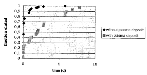
measurement. The elution curves shown in figure 1 were
obtained.
[0060]. In particular, figure 1 shows that deposition
of the polymer by cold plasma significantly delays the
elution of the hydrophilic drug compared with the elution
deriving from application of a polymer according to the
state of the art.
ExAMPLE 2
Comparison between the elution mechanism of a hydrophobic
drug from a stmt covered with a polymer according to the
state of the art and the mechanism from a stmt covered
with polymer according to the invention
[0061]. The same procedure described in Example 1 was
repeated here with the difference that a hydrophobic
drug, dexamethasone, was used.
[0062]. 10 mg of dexamethasone were dissolved in 1 cc
of ethanol and applied as described previously. The
CA 02544376 2006-05-O1
WO 2005/044328 PCT/IB2003/005003
22
elution curves were again measured as described in
example 1 and the absorbance at 264.4 nm was read off.
The results shown in figure 2 were obtained.
[0063] . It should be noted that in this case, too, the
polymer of allylamine deposited by cold plasma provides a
notable reduction in the mechanism of elution of the
drug.
EXAMPLE 3
Comparison of the degree of hydrophilicity between a
metal stmt treated with heparin and a metal substrate
without heparin
[0064]. A stent prepared according to example 1 with
allylamine deposited by cold plasma underwent a process
of bonding with heparin in the following manner.
[0065]. 0.5 g of heparin (Bioiberica) was dissolved in
100 cc of phosphate buffer and 0.016 g of sodium
periodate (Sigma-Aldrich) was added. After 16 hours of
remaining in solution, 100 cc of 0.05% acetic acid-sodium
acetate solution were added. 5 cc of this solution were
taken and placed in a Petri dish. The stmt was then
immersed in the dish and 0.01 g of sodium
cyanoborohydride (Sigma-Aldrich) were added. After 30
minutes, the stmt was removed and washed with water. It
was then dried in an oven. At this point, the stent was
far more hydrophilic compared with a non-heparinized
CA 02544376 2006-05-O1
WO 2005/044328 PCT/IB2003/005003
23
stmt precisely because of the presence of heparin bonded
onto its surface.
[0066]. To provide an analytical base, the same
treatment as j ust described was carried out on plates of
ASI 316 L steel of side 1 cm, that is the material of
which the stent was constituted. A heparinized plate was
compared with a non-heparinized plate by a comparison
using X-ray Photoelectron Spectroscopy (XPS) analysis to
supply the chemical composition of the surface layer. The
XPS analysis was carried using a Perkin Elmer PHI 5500
ESCA System instrument. The result of the analysis
expressed in atomic % is given in table 1 below.
Table 1
Specimen. C O N S Si Other
(<1%)
Non- 78.4 10.7 9.4 - 1.3 Na, P
heparinized
plate
Heparinized 69.2 21.9 2.4 3.2 1.9 Mg,
plate Cl, Na
[0067]. Compared with the untreated specimen, the
specimen treated with heparin shows an increase in the
O/C ratio and in S concentration expected in the
heparinization processes.
CA 02544376 2006-05-O1
WO 2005/044328 PCT/IB2003/005003
24
EXAMPLE 4
Comparison of the degree of hydrophilicity between a
metal stent treated with h~raluronic acid and a metal
stmt without hyaluronic acid
[006]. A stent prepared according to example 1 with
allylamine deposited by cold plasma underwent a process
of bonding with hyaluronic acid in the following manner.
(0069. 0.5 g of hyaluronic acid (Lifecore) was
dissolved in 100 cc of deionized water. 5 CC Of said
solution were taken and placed in. a Petri dish. The stent
was then immersed in the dish and 0.03 g of N-hydroxy
succinimide and 0.04 of dimethyl carbodiimide (EDC) (both
Sigma-Aldrich) were added. After 30 minutes, the stmt
was removed and washed with water. It was then dried in
an oven. At this point, the stent was far more
hydrophilic compared with a s tent not covered with
hyaluronic acid precisely becau se of the presence of
hyaluronic acid bound onto its surface.
EXAMPLE 5
Production of a stmt covered with olymer according to
the invention, with immobilisation of hvaluronic acid and
further covering with a biodegradable hyaluronic acid
derivative-based layer
(0070. From capsules of the drug Glivec~ 10 mg of
active principle imatinib mesilate were extracted by
CA 02544376 2006-05-O1
WO 2005/044328 PCT/IB2003/005003
dissolving in water, filtering to remove the insoluble
excipients and evaporating the water as described in
example 1. Two stainless steel stems 11 mm in length
produced by the INVATEC company were coated using an
5 Artis I airbrush (Efbe, Germany) in the following manner.
(0071]. Firstly, 1 cc of a 0.250 o solution in
cyclohexane of polybutadiene (Aldrich, mean molecular
weight 420,000) was applied. Following this, 1 cc of
solution obtained by dissolving 10 mg of Imatinib
10 Mesilate (IM) in 1 cc of methanol was applied. Then 1 cc
of 0.5o solution of polybut adiene (details as previously)
in cyclohexane was applied. Finally, 1 cc of a 0.5%
solution in cyclohexane of polybutadiene with a molecular
weight of between 1,000,000 and 4,000,000 was applied.
15 [0072]. At this point, one of the two stents was placed
in a EUROPLASMA reactor for the plasma treatment and
underwent a cycle of plasma deposition of allylamine
(introduced as vapour from an external receptacle which
contained it as a liquid) for 8 minutes with the reactor
20 switched to a power of 200 W at a pressure of 0.2 mbar.
[0073]. Next, 0.5 g of hyaluronic acid (Lifecore) was
dissolved in 100 cc of deionized water. 5 cc of said
solution were taken and placed in a Petri dish. The stmt
was then immersed in the dish and 0.03 g of N-hydroxy
25 succinimide and 0.04 of dimethyl carbodiimide (EDC) (both
CA 02544376 2006-05-O1
WO 2005/044328 PCT/IB2003/005003
26
Sigma-Aldrich) were added. After 3 0 minutes, the stent
was removed and washed with water and dried. At this
point, a layer was applied of a hyaluronic acid
derivative total
insoluble
in water
and degradable,
the
ben~yl ester HYAFF 11) (Fidia Advanced Biopolymers, Abano
Terme, Italy). This drug
material, together
with the
imatinib applied from a solution of 0.2%
mesilate,
was
HYAFF and 1% IM in hexafluoroi sopropanol using
an
airbrush.
[0074]. In this way, a stmt is obtained which elutes
the drug from the surface layer of HYAFF and from the
underlying layer, in which the surface layer will degrade
in situ leaving exposed the surface on which the
hyaluronic acid is bonded to the harrier and functional
layer deposited by plasma.