Note: Descriptions are shown in the official language in which they were submitted.
CA 02544431 2006-05-O1
WO 2005/045787 PCT/US2004/035070
TITLE
Breast Mode~1 Teaching Aid and Method
CROSS R'BFERFS°NCE APPLICATIONS
This application is a non-provisional application
claiming the benefits of.provisional application no.
60/423,270 filed November 02, 2002.
FIELD OF INVENTION
The present invention relates to a breast cancer lump
detection simulator and training aid.
BACKGROUND OF TI3$ INVENTION
Fast growing breast cancers can double in size every
three days. It can take as little as three months for a
single breast cancer cell. to double 30 times and produce a
one centimeter size cancerous tumor. Therefore, a breast
cancer can grow to a four centimeter diameter (stage III to
'stage IV) in a nine month period, between annual clinical
exams. About one in eight women in the United States will
develop breast cancer in their life.
A need exists to disseminate to women and medical
practitioners worldwide a method to detect with one's hands
a small lump in the breast.
CA 02544431 2006-05-O1
WO 2005/045787 PCT/US2004/035070
The present invention provides a life size model of a
human breast with a chosen sized lump imbedded in it. The
student must push down on the lump in a proper direction, as
taught by medical professionals, in order to trigger an
electronic alarm.
SUb,~ARY OF THE IN"V'3~NTION
The main aspect of the present invention is to provide
a life like training model of a human breast with a hidden
lump tied to~a microswitch ~, thereby enabling a student to
probe for the lump~.in a medically proficient manner.
Another aspect of the present invention is to provide a
variety of models including ~a small lump, a large lump, no
lump, and two models with various combinations of lumps.
Other aspects of this invention will appear from the
following description and appended claims, reference being
made to the accompanying drawings forming a part of this
specification wherein like reference characters designate
corresponding parts ix~ the several views.
At least two breast self exam (BSE) techniques may be
used on the models. First, the Pat and Rub technique uses
the three middle fingers applying pressure and patting and
rubbing in a circular motion completely around the breast
from _the stem (outer breast) to the nipple. Second, the
Spoke Wheel Method uses the ,three middle fingers with the
2
CA 02544431 2006-05-O1
WO 2005/045787 PCT/US2004/035070
fingers placed in a straight line from the stem to the
nipple. Fingers are~moved until the entire breast has been
felt. Tn~hen the individual finds a lump and the technique is
done correct~..y, a light will come on to indicate the
technique has been performed correctly.
Finding a lump and being reinforced when~the light goes
on provides motivation to use the model and enables showing
others how to practice BSE's with the model. Using the
model might provide intrinsic reinforcement (e. g. internal
good feelings). When a lump is found arid the Light goes on,
an individual feels. successful. The model and the training
techniques that can be developed around it may serve to
lessen anxiety, and they may~provide reinforcement,
motivation and ~each~BSE techniques for early detection of
breast cancer.
SYMPTOMS OF BREAST CANCER
The most important physical symptom of breast cancer is
a painless mass or lump. Up to 10% of patients have breast
pain and no mass. Less~common symptoms include persistent
changes to the breast (thickening, swelling, skin irritation
or distortion) and nipple symptoms.(spontaneous discharge,
erosion, inversion, or tenderness). Early breast cancer,
when it is most treatable, usually does not produce any
symptoms. It is therefore, very important for women to
3
CA 02544431 2006-05-O1
WO 2005/045787 PCT/US2004/035070
follow recommended guidelines to find breast cancer before
symptoms develop (mammography, clinical breast examination
(CBE), and breast self examination (BSE)). Because a small
percentage of cancers may be missed by mammography, it is
important for women aged 40 and.older, to also perform a
.monthly BSE and have an annual CBE.
BREAST. SELF-EXAMINATION (BSE)
A woman performs a BSE in the same way that a health
care professional performs a clinical examination. Using
the pads of the .fingers~gently feel the breasts, giving
special attention to their shape and texture, location of
any lumps, 'and whether such~lumps are attached to the skin
or to deeper t~.ssues. A woman should do a BSE monthly to
T5 become~familiar with both the appearance~and feel of her
breasts so she is aware of any change. Lumps are not
necessarily abnormal, they come anc~ go with a woman's
menstrual cycle. Tn~hen Lumps are detected and tested, the
majority are found to be noncancerous.
According to the World Organization of Family
Practitioners, 9 out of 10 breast cancers are discovered by
women themselves. Several different breast self-exam
. techniques are available. However, none of these deals
directly with the emotional aspects of performing breast
self-exams. Many women could be frightened away from
4
CA 02544431 2006-05-O1
WO 2005/045787 PCT/US2004/035070
performing breast self-exams if they have inadequate
.education related to the breasts, and lack knowledge of
normal hormonal fluctuations and sensations such~as pain,
swelling and tenderness. The female breast and its
underlying structure are complex to evaluate for changes.
For example, a lump may be found with a woman in one
position and concealed when she changes positions. A woman
is given the responsibility of screening for breast cancer
and sometimes not given adequate tools to provide an
accurate screening.
If women and t~.eir health care professionals are
provided with an educational tool, especially the present
invention, instructed in the signs and symptoms of breast
cancer, early diagnosis of breast cancer could be
15~ facilitated. The model teaching aid will'help women, health
professions, and educators learn more about breast cancer
self-exams and early detection of breast cancer.
DEVELOPMENT OF THE CHECK-IT MODEL TEACHING AID
The model could be used for reinforcement of breast
self-exam. ~BSE) A model may be made of silicone, oil and a
catalyst loured into a mold. An uncured silicone mixed with
oil gives a more lifelike texture. ,A catalyst may be added
to cure the silicone. A small amount of silicone may be set
aside, and a darker colored catalyst may be added. The
darker silicone mixture may be poured carefully into the
5
CA 02544431 2006-05-O1
WO 2005/045787 PCT/US2004/035070
mold area of the nipple to simulate the areola. The
silicone may then be allowed to set. Then the rest of the
silicone may be poured slowly in a circular manner into the
mold..
A switch with a wooden bead attached (to simulate a
lump) may be placed in the silicone substance. The switch
may be either free flowing or attached by a plastic strip,
which are two examples. The switch may have two wires
attached. One wire may gc~ to the light and. the other to an
energy source (battery). The switch could be one of three
submini lever~switches, roller lever switch or momentary
push button switch, as but a few examples. Each switch
might have its own use indifferent positions and~allow for
different functions. A light, blue LED, e.g, might be
illuminated when pressure is applied to the switch
(completes a circuit).
The light could be replaced with a vibrator that could
be placed in the silicone and produce vibrations ox waves
could also be replaced with a buzzer or a lever that makes a
clicking sound that would make noise when pressure is
applied to the switch and the.circuit is completed. The
switch could be attached to a tape recorder that would tell
you that you found a lump in the breast and if other--lumps
are to be found. Different types of sensory devices could
6
CA 02544431 2006-05-O1
WO 2005/045787 PCT/US2004/035070
be placed in the model before pouring silicone, so that they
would be next to the surface, and different amounts of
pressure would light different colored lights.. To show the
amount of pressure needed to detect a lump, a gape recording
could also be used that would tell you if more or less
pressure is needed.
Sensory switches could be placed in areas that have
high incidence of cancer or lower incidences. The light or
a recorded voice could tell if you were in the correct place
or not. This will allow for more effective detection, not
only for the model, but to carry into testing of self-
awareness. The silicone could be different colors to
represent areas of the breast. that are the most common
places cancer ~.s found. The silicone could be colorless to
allow the internal parts~of the breast model to~be seen as
well as locations of lumps. Each internal part of~the
breast could be a different color with muscles at the back
of the breast poured at different times as well as in
different directions allowing for realization. The top half
of the breast model could be clear silicone. The bottom
half could be colored to give an idea of the location o~ the
internal parts in relation to the. outside of the breast.
Silicone could be of different grades and cured at
different temperatures or with different-catalyst added to
change the texture of the silicone. Perhaps each internal
7
CA 02544431 2006-05-O1
WO 2005/045787 PCT/US2004/035070
part would need to have a separate mold. Then the parts
could be placed inside the breast model with clear silicone
poured into the mold to fill and keep the~internal parts in
place. The internal parts might not need to be made of
silicone, but other materials could suffice as we~.l.
Different sizes of breasts for different stages of life
(ages) as well as shapes could be made. Lumps of silicone
might be added~to show swelling in different areas of the
breast. The lumps could be placed next to the outside of
the mold. Then a layer of. silicone poured over the lump
could simulate what a lump.might look or feel like.
Silicone, acting as the outer skin, could have color to
look like skin cancer, red with black or a dark center,
irregular edges (pinpoint , "zit-like"). The mold itself may
need to be made differently. Tt may need to have the
pigment added to some areas before other silicone is~poured.
The nipple may have to be hollowed out so that it could be
inverted and extended as a normal nipple would be. A latex
product inside the silicone nipple could allow flexibility
2Q and the nipple to be inverted and extended a number of times
so that it wouldn't wear out prematurely.
The nipple could have micxo holes put into it to allow
a fluid to escape with a light squeeze and or palpitation of
the nipple and~areola. A-sack of fluid could be-placed
behind the nipple. An extended sack with a duct that would
8
CA 02544431 2006-05-O1
WO 2005/045787 PCT/US2004/035070
lead to the nipple so that~with pressure, a small amount of
fluid would be produced at.the end of the nipple, might also
be provided. The fluid could be a white, cloudy substance.
This could be replenished with a~needle and syringe. The
sack could be self-sealing~with a back flow preventor to the
nipple so fluid would flow in only one direction.
When cancer is in an advanced stage, the body may give
off an odor. This might be reproduced by a sack into the
back of the breast that can be refilled by a needle and
syringe. The system would be a self-sealing sack and the
odor would be~emitted with palpitation of the swollen area.
This method would give reinforcement on how to check and
what to check for in the breast.
Lymph nodes typically enlarge when a woman has breast
cancer. Lymph nodes are different~from breast cancer tumor.
The lymphatic system forms lumps that are not as dense or
fluid as a fibrous c~rst. Tn the model, lymph nodes would be
placed into the muscle level of the breast, pectoralis major
and minor, at approximately the location where they would
normally be found. Apical lymph nodes, central lymph nodes,
anterior axillary or pectoral lymph nodes and internal
thoracic lymph nodes may be placed at each site. Two normal
nodes and two enlarged nodes could be placed in a breast
model. The normal node~could be attached to one colored
light while the enlarged node could be attached to a
9
CA 02544431 2006-05-O1
WO 2005/045787 PCT/US2004/035070
different colored°light so that a.comparison could be made.
The lymph nodes also typically change in size and density
with age.
Along with the model, a CD or VHS tape describing the
procedure of breast self-examination could be included. An
individual could practice breast self-exam along with the
tape or video, using, e.g., the pat and rub or the spoke
method.
As can be easily understood from the foregoing, the
. basic concepts of the present invention may be embodied in a
variety of ways. It involves both training techniques- as
well as devices to accomplish the appropriate training. In
this application, the training techniques are disclosed as
part of the results shown to be achieved by the various
devices described and as steps which are inherent to~
utilization. They are simply the natural result of
utilizing the devices as intended and described. In
addition, while some devices are disclosed, it should be
understood that these not only accomplish certain methods
but also can be varied in a number of ways. Importantly, as
to all of the foregoing, all of these facets. should be
understood to be encompassed by this disclosure.
10
CA 02544431 2006-05-O1
WO 2005/045787 PCT/US2004/035070
BRIEF DESCRIPTION OF THE DRAWINGS
FIG. 1 is a side perspective view of a preferred microswitch
with its circuit schematic.
FIG. 2 is a top perspective view.of a mold for forming a
silicone implant having the microswitch.
FIG. 3 is a top perspective view showing the silicone
pouring into the mold shown in FTG. 2.
FIG. 4 is a side perspective view of the finished silicone
lump implant ready for placement into the human
breast model.
FIGS. 5,6 show the use of a rubber glove as the mold for the
silicone lump implant, wherein a tube is shown in
dots to provide a channel for the microswitch and
wires.
FIG. 7 is a side perspective view of a completed silicone
lump implant made from the method taught in FIGS.
5,6.
FIG. 8 is a top perspective view of a mold for a human
breast model.
FIG. 9 is a cross sectional view of the model~shown in FIG.
8 taken along line 4-9 showing a first pouring of
the nipple material into the mold.
FIG. 10 is the same view as FIG. 9 showing the already
poured outside latex skin for the model plus two
11
CA 02544431 2006-05-O1
WO 2005/045787 PCT/US2004/035070
silicone lump implants positioned for their
permanent placement in. the model.
FIG. 11 is the same view as FIG. 10 showing.the pouring of
the silicone filler in the model.
FIG. 12 is the same view as FIG. 1.2 showing the final
pouring of the latex back of the model.
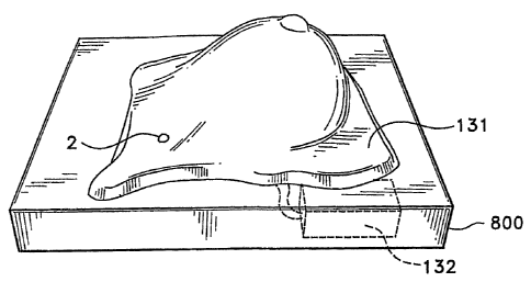
FIG. 13 is an exploded view of a base~of the model receiving
the silicone breast.
FIG. 14 is a top perspective view of the completed model of
FIG. 13.
FIG. 15 is a top perspective view of a student practicing
the three finger pat and rub technique.
FIG. 16 is the same view. as FIG. 15 showing the student
using tl~e spoke wheel technique.
15' FIG. 17 is a schematic diagram of the pat and rub technique.
FIG. Z8 is a schematic diagram of the spoke wheel technic~u.e.
FIG. 19 is an exploded view of an inflamed lymph node model.
FIG. 20 is an exploded view of a model with an inflamed
lymph node switch and a normal lymph node without a
switch in the model.
FTG. 21 is a top perspective view of a simulated normal
lymph node.
1z
CA 02544431 2006-05-O1
WO 2005/045787 PCT/US2004/035070
' Before explaining the disclosed embodiment of the
present invention in detail, it is to be understood that the
invention~is not limited in its application to the details
of the particular arrangement shown, since the invention is
capable of other.embodiments. Also, the terminology used .
herein is for the purpose of description and not of ,.
limitation.
DETAILED DESCRIPTION OF THE DRAWINGS
Referring first to FIG. l~a schematic shows a power
supply, preferably~a battery 1 connected in series with a
bulb 2 and a switch 4. The bulb 2 is connected in parallel
with an optional teaching device, preferably a stored
message device with a speaker; such as a tape recorder 3.
The momentary pushbutton switch 4 could be a Radioshack~
SPST miniswitch, part no. 275-1547. It could also be~a
variable rheostat switch that increases the current flow the
further the action end is depressed. A dab of glue 5 holds
a simulated lump 6 to the action end 7 of the switch 4.
Clearly a sideways force Fl will not activate the, switch 4,
whereas a downward force F2 will. aotivate the switch 4. The
simulated lump 6 could~be a wooden sphere selected at a size
to approximate a cancerous lump.
13
CA 02544431 2006-05-O1
WO 2005/045787 PCT/US2004/035070
The essence of the present invention is to suspend this
switch arrangement or an equivalent thereof in a soft,
pliable model of a human breast. A student can use
medically defined probing methods to locate the simulated
lump 6 and move.the action~end 7 to light the bulb 2. A
variety of equivalents exist for, the bulb 2 including noise
makers and vibration alarms.
Referring next to FIGS..2,3,4 the preferred method to
encapsulate the switch/lump assembly 64 is shown. A mold 23
consists of a bottom.22 and halves 20, 21.~ A hole 230
allows the assembly 64 to protrude therethrough. Wires 8,9
are threaded through the hole 230 up and out cavity 24
before a silicone or like substance 31 is poured in via
container 30.
25 The insert 40 consists of a silicone~body having a
cylindrical shape with wires 8,9 extending from an end and
the assembly 64 extending from a side. This insert 40 can
be placed in any orientation desired in a model of a human
breast. Multiple inserts 40 can also be used in the human
breast model as.shown in F1G. 12. . .
FTGS: 5,6,7 show an alternative method.of forming a
similar insert 70. A rubber glove~50 has fingers
51,52,53,54 that are used as molds for.the insert 70. A
hole 61 is drilled into the finished mold to allow the wires
8,9 to be inserted as shown. Thus, the methods disclosed
14
CA 02544431 2006-05-O1
WO 2005/045787 PCT/US2004/035070
herein allow anyone to create their own model all over the
world to promote early breast cancer detection.
Referring next to FIG. 8 a breast mold 80 has a cavity
81 which has a flat base portion 82, a breast portion 83 and
a nipple portion.84. .
In FIGS. 9-14 nipple portion 84 has been filled with a
nipple colored silicone 90. Then skin layer 100 is poured.
Next an insert (s) 40 is placed in the cavity 81. Next the
silicone 110 is poured. Finally base layer 120 is poured.
The wires are connected in FIG.~~13 to a battery 132 and bulb
2. A hole 130 in the Completed model 131 allows the bulb 2
to protrude. A base 800 supports the model 131 and the
battery 132 and any other optional training aids such as a
voice chip/recorder/speaker.
An uncured silicone mixed with an oil to give'a more
lifelike texture than a catalyst i~s added for curing the
silicone 110. A small amount 90 has been set aside and a
different color is added. This is poured carefully into the
mold area of the nipple and areola giving a different color.
The silicone 90 is allowed to set then the rest of the
silicone 110 is poured in.a circular manner.into the mold
forming the skin layer 7.00. Inserts 40 with a substance
that is harder than the surrounding silicone attached to it
110 are placed in the cavity 81, then.the silicone is
poured. The switch 4 has two wires 8, 9 attached. FIG. 1
CA 02544431 2006-05-O1
WO 2005/045787 PCT/US2004/035070
and FIG. 19 show the microswitches that could be used in the
model. FIG. 1 has one point of contact whereas FIG. 19 has
points of contact. FIG. 1 simulates location~of a lump
while FIG. 19 illustrates simulation of.finding a swollen
5 lymph node. A lump encompasses~a smaller surface. area then
the surface area of a swollen lymph node: One wire goes to
the light and the other to an energy source (battery). The
light, a blue LED,is illuminated with pressure applied to
the switch (completes a circuit). This can be replaced with
a vibrator that would be placed in the silicone and produce
waves throughout the~silicone. The light could also be
replaced with a buzzer or a lever, that would make a buzzing
or clicking noise with pressure to the switch and the
circuit is completed. The switch could be attached to a
tape recorder that would tell you that you found a lump in
the breast and if other lumps are.to be found. Different
types of sensors could be placed in the model before pouring
silicone so different amounts of pressure would light
different colored lights. The amount of pressure depends on
placement of sensors and distance from the surface. To show
the amount of pressure needed to detect a lump, a tape
recording could also be used that would tell you if more or
less pressure is needed. These sensory switches would be
placed in areas that have high incidence of cancer or lower
incidences than the light or a record would tell if you are
I6
CA 02544431 2006-05-O1
WO 2005/045787 PCT/US2004/035070
.in the correct place or not. This will allow for more
effective detection not only for the model but to carry into
testing of self awareness.
Silicone can be different colors to represent areas of
the breast that. are the most common places cancer is found.
Silicone could be colorless to allow the.internal parts of
the breast model to be seen as well as locations of lumps.
Each internal part of the breast could be a different color
with muscles at the back of the breast poured at different
times as well as in different directions allowing for an
anatomically correct model. The top half of the breast
model could be clear silicone. The bottom half could be
colored to give an idea of the location of internal parts in
relation to the,outside of the breast. Silicone could be of
different grades and cured at different temperatures or with
different catalyst added to change the texture of the
silicone. Each internal part would have to have a separate
mold. Then these parts would be placed inside the breast
model with clear silicone poured into the mold to fill and
keep the internal parts in place: The internal part might
not need to be made of silicone but other materials.
Different sizes of breast for different stages of life
(ages) as well as shapes could be made. Lumps of silicone
might be added to show swelling in different areas of the
breast. These would need to~be placed next to the outside
17
CA 02544431 2006-05-O1
WO 2005/045787 PCT/US2004/035070
of the mold. Then they would have a layer of silicone poured.
over the lump showing what a lump might look like or feel
like. Silicone acting as the outer skin can have color to
look like skin cancer, red with black or a dark center,
irregular edges .(pinpoint, zit like). The mold itself may
need to be made differently. It may need to have the
pigment added to these areas before the silicone is poured.
The nipple may have to be hollowed out so that it could be
inverted and then extended as a normal nipple would be.
This may need a latex product inside the silicone nipple to
allow flexibility so it could be inverted and extended a
number of times, so that it wouldn't wear out'.
The nipple could have micro holes put into it to allow
a fluid to escape with a light squeeze and or palpitation of
the nipple and areo~.a. 'A sack of fluid would be placed
behind the nipple. An extended sack with a duct that would
lead to the nipple so that with pressure, a small amount of
fluid would be produced at the end of the nipple. The fluid
would be a white, cloudy substance.. This could~be
replenished with a needle and syringe. The sack would be
self sealing with a back flow preventor to the nipple so
fluid would flow in only one direction. When cancer has
advanced to a stage it will give off an odor. This might be
reproduced by a sack into the back of the breast that can be
refilled by a needle and syringe. This would be a self
~..s
CA 02544431 2006-05-O1
WO 2005/045787 PCT/US2004/035070
sealing sack the odor would only be emitted with palpitation
of the swollen area. This method would give reinforcement
on how to check and what to check for in the breast.
Lymphatic systems, different than lumps used for cancer
not as density and not. fluid as fibrous cyst. These would
be placed in muscle level of the breast, pectoralis major
and minor at approximately the location where they would
normally be found. Apical lymph nodes, central lymph nodes,
anterior axillary or pectoral lymph nodes and internal
thoracic lymph nodes are to be placed at each site. Two
normal nodes and two enlarged nodes could be placed in the
breast model. The normal node could be attached to one
colored light while the enlarged node could be attached to a
different colored light so that a comparison could be made.
These also change in size and density with. age.
Along with these models, a DVD or VHS with a 3-5 minute
long message describing the procedure of breast self-
examination would be given so that a person can practice
along with .the tape, the pat and rub or the spoke method.
Tn FIGS. 15, 17, the student 15Q is using the pat and
rub technique using three middle fingers to apply pressure.
Pat and rub in a circular motion moving completely around
the breast in one direction c (clockwise) or cc
(counterclockwise) direction starting from the stem (outer
breast) to the nipple 90.
19
CA 02544431 2006-05-O1
WO 2005/045787 PCT/US2004/035070
In FTGS. 16, 18 the spoke wheel technique is used with
the three middle fingers applying pressure in a straight
line (arrows w) from the stem to the nipple 90. Fingers are
moved until the entire breast has been examined. The
movements of the. fingers look like the spokes of a wheel.
Lymph nodes typically enlarge when a women has breast
cancer. Lymph nodes are different from breast cancer tumor.
The lymphatic system forms lumps that are not as dense or
fluid as a fibrous cyst. In the model, lymph nodes would be
placed in the muscle level of the breast, pectoralis major
and minor, at approximately the location where they would
normally be found. Apical lymph nodes, central lymph nodes,
anterior axillary lymph nodes.or pectoral lymph nodes and
internal thoracyc lymph nodes may be placed at each site.
Two normal nodes and two enlarged nodes could be placed in a
breast model. The normal node could be attached to one
colored light while the enlarged node could be attached to a
different colored light so that a comparison could be made..
The lymph nodes also typically change in size and density
with age.
The artificial lumps~may be made from wood, rubber,
plastic, metal or virtually any hard material. Generally
the diameter of the spherical lump ranges from .5 cm. to 3
millimeters. Non-spherical lumps such as to simulate a
fibrous lump could also be used. Switches such as a wall
CA 02544431 2006-05-O1
WO 2005/045787 PCT/US2004/035070
mounted rheostat, but with a~linear actuator could be used
to teach a student a proper pressure level for his
palpitations.
Referring next to~FTGS. l9, 20, 21 a lymph node
training breast 1310 has installed therein a normal
simulated lymph node lump 2100, and a larger inflamed
simulated lymph node 2200. Each are preferably made of a
harder plastic like the nipple 90. The toggle switch 2207
has an armature 2207. with a top 2206. The simulated lymph
node 2200 is connected to the top.2206 as well as to arms
2202, 2203, 2204, 2205. The student can activate the light
by depressing any of the items 2206, 2202, 2203., 2204, 2205.
Although the present invention has been described with
reference to preferred embodiments; numerous modifications
and variations can be made and still the result will come
within the scope of the invention. No limitation with
respect to the specific embodiments disclosed herein is
intended or should be inferred. Each apparatus embodiment
described herein has numerous equivalents.
21