Note: Descriptions are shown in the official language in which they were submitted.
CA 02544766 2006-05-04
WO 2005/046794
PCT/US2004/036918
- 1 -
IMPLANTABLE RADIOTHERAPY/BRACHYTHERAPY RADIATION
DETECTING APPARATUS AND METHODS
BACKGROUND OF THE INVENTION
The invention relates generally to apparatus and methods for use in treating
proliferative tissue disorders, and more particularly to apparatus and methods
for the
treatment of such disorders by delivering radiation with a brachytherapy
device that also
measures treatment characteristics.
Malignant tumors are often treated by surgical resection of the tumor to
remove
as much of the tumor as possible. Infiltration of the tumor cells into normal
tissue
surrounding the tumor, however, can limit the therapeutic value of surgical
resection
because the infiltration can be difficult or impossible to treat surgically.
Radiation
therapy can be used to supplement surgical resection by targeting the residual
malignant
cells after resection, with the goal of sterilizing them, reducing the rate of
recurrence or
delaying the time to recurrence. Radiation therapy can be administered through
one of
several methods, or a combination of methods, including permanent or temporary
interstitial brachytherapy, and external-beam radiation.
Brachytherapy refers to radiation therapy delivered by a spatially confined
source of therapeutic rays inserted into the body at or near a tumor or other
proliferative
tissue disease site. For example, brachytherapy can be performed by implanting
radiation sources directly into the tissue to be treated. Brachytherapy is
most
appropriate where 1) malignant tumor regrowth occurs locally, within 2 or 3 cm
of the
original boundary of the primary tumor site; 2) radiation therapy is a proven
treatment
for controlling the growth of the malignant tumor; and 3) there is a radiation
dose-response relationship for the malignant tumor, but the dose that can be
given safely
with conventional external beam radiotherapy is limited by the tolerance or
normal
tissue. In brachytherapy, radiation doses are highest in close proximity to
the
radiotherapeutic source, providing a high tumor dose while sparing surrounding
normal
tissue. Brachytherapy is useful for treating malignant brain and breast
tumors, among
others.
CA 02544766 2006-05-04
WO 2005/046794
PCT/US2004/036918
- 2
Prior art brachytherapy devices have provided a number of advancements in the
delivery of radiation to target tissue. For example, Winkler U.S. patent no.
6,413,204
describes a brachytherapy method and apparatus for treating tissue surrounding
a
surgically excised tumor with radioactive emissions to kill cancer cells that
may be
present in the tissue surrounding the excised tumor. The radiation is
delivered in a
predetermined dose range defined as being between a minimum prescribed
absorbed
dose for delivering therapeutic effects to tissue that may include cancer
cells, and a
maximum prescribed absorbed dose above which healthy tissue necrosis may
result.
The resulting treatment helps to prevent over-exposure to tissue at or near
the
brachytherapy device, while still delivering the minimum prescribed dose at
the
maximum prescribed distance from the device.
While such advancements have improved the treatment of proliferative tissue
diseases, some challenges remain. Currently, the desired radiation dose is
calculated
based on the characteristics of the brachytherapy applicator (device), the
radiation
source and the surrounding tissue, yet the actual dose delivered is not tested
to assure
that over and/or under treatment do not occur. For example, if the radiation
source is a
radioactive seed positioned in the center of an expanded balloon, the
calculated dose is
based on the central positioning of the radiation source. If for some reason
the
radioactive seed was positioned off center, prior art brachytherapy devices do
not have
the means to determine that this harmful situation has or is occurring. Prior
art
brachytherapy devices also lack the ability to directly sense the surrounding
tissue and
determine the effectiveness of the proliferative tissue disorder treatment.
SUMMARY OF THE INVENTION
The present invention provides brachytherapy apparatus and methods for
delivering and monitoring radioactive emissions to an internal body location.
The
device includes a catheter body member having a proximal end, a distal end,
and an
outer spatial volume disposed proximate to the distal end of the body member.
A
radiation source is preferably positioned in the outer spatial volume, and a
treatment
feedback sensor is disposed on the device.
CA 02544766 2006-05-04
WO 2005/046794 PCT/US2004/036918
- 3 -
In one embodiment, the treatment feedback sensor is a radiation sensor which
can detect radiation emitted by the radiation source. The radiation sensor
preferably
produces data useful for determining if the delivered radiation dose was
within the
prescribed range. The data can also preferably be used to determine if the
desired
radiation profile was delivered to the surrounding tissue. In another aspect
of the
invention, the treatment feedback sensor is capable of detecting tissue
temperature,
oxygenation, pH, treatment agent concentration, cytokine concentration, or
other
characteristics related to radiation treatment.
In one embodiment, the treatment feedback sensor is positioned within the
catheter body member. Other locations where the sensor may preferably be
located
include disposed on an expandable surface member which defines the outer
spatial
volume, or outside the device.
In another embodiment, the present invention includes a radiation therapy
apparatus for delivering and monitoring radioactive emissions to a resected
tumor
cavity. The apparatus includes a catheter body member with proximal and distal
ends,
an expandable surface member disposed proximate to the distal end of the
catheter body,
a treatment feedback sensor, and an external radiation source positioned
outside the
tissue cavity for delivering radiation to target tissue surrounding the tissue
cavity. The
expandable surface member can be positioned within a resected tissue cavity
and
expanded to position the surrounding tissue such that the delivery of a
radiation beam
from the external radiation source is accurately delivered and measured by the
treatment
feedback sensor positioned within the tissue cavity.
In another embodiment, the invention includes the method of delivering and
monitoring radioactive emissions to an internal body location. The method
includes
inserting a brachytherapy device into a resected cavity, the brachytherapy
device
including a catheter body member with proximal and distal ends, and an
expandable
surface member disposed proximate to the distal end of the catheter body
member. A
radiation source is preferably disposed within the expandable surface member.
The
method further includes inserting a radiation sensor into the resected cavity
and
delivering a minimum prescribed absorbed radiation dose to a target tissue,
the target
tissue being defined between the expandable surface member and a minimum
distance
outward from the expandable surface member. The radiation sensor senses the
delivered
CA 02544766 2012-08-28
- 4 -
radiation dose and data output from the sensor confirms that the brachytherapy
device
delivers the minimum prescribed dose.
In another embodiment, the present invention includes a brachytherapy
apparatus
for delivering and monitoring radioactive emissions to an internal body
location. The
apparatus includes a catheter body member having a proximal end, a distal end,
and an
outer spatial volume disposed proximate to the distal end of the body member.
A
radiation source is disposed in the outer spatial volume and a treatment
feedback sensor
is provided on the device. The treatment feedback sensor can be used to
evaluate the
treatment of proliferative tissue disorders.
In one aspect, the present invention resides in a brachytherapy apparatus for
delivering and monitoring radioactive emissions to an internal body location,
comprising: a catheter body member having a proximal end, a distal end, and an
outer
spatial volume disposed proximate to the distal end of the body member; a
radiation
source disposed in the outer spatial volume; a radiation sensor provided on
the device;
and an additional radiation sensor is positioned outside the brachytherapy
device;
wherein the radiation sensors measures the radiation delivered from the
radiation source.
In another aspect, the present invention resides in a brachytherapy apparatus
for
delivering radioactive emissions to an internal body location, comprising: a
catheter
body member having a proximal end, a distal end, and an outer spatial volume
disposed
proximate to the distal end of the body member; a radiation source disposed in
the outer
spatial volume; a treatment feedback sensor provided on the device; and an
additional
treatment feedback sensor is positioned outside the brachytherapy apparatus;
wherein the
treatment feedback sensors can be used to evaluate the treatment of
proliferative tissue
disorders.
BRIEF DESCRIPTION OF THE DRAWINGS
The invention will be more fully understood from the following detailed
description taken in conjunction with the accompanying drawings:
FIG. 1 illustrates the device of the present invention including a sectioned
view
of the outer spatial volume showing radiation sensors positioned therein;
CA 02544766 2012-08-28
- 4a -
FIG. 2 illustrates another embodiment of the brachytherapy device of the
present
invention shown in perspective;
FIG. 3 illustrates another embodiment of the brachytherapy device of the
present
FIG. 3A illustrates a cross sectional view of the device pictured in FIG. 3;
FIG. 4 illustrates another embodiment of the brachytherapy device of the
present
FIG. 5 illustrates another embodiment of the brachytherapy device of the
present
invention shown in full view.
CA 02544766 2006-05-04
WO 2005/046794 PCT/US2004/036918
- 5 -
DETAILED DESCRIPTION OF THE INVENTION
The present invention provides interstitial brachytherapy devices that can
deliver
radioactive emissions used to treat proliferative tissue disorders and detect
treatment
characteristics to monitor the treatment regimen. The devices include a
catheter body
member with a proximal end, a distal end, and a inner lumen. An outer spatial
volume is
disposed proximate to the distal end of the body member with a radiation
source
preferably disposed therein. A treatment feedback sensor is disposed on the
device.
Brachytherapy devices treat proliferative tissue disorders, such as cancerous
tumors, by delivering radiation to the target area which contains both
cancerous cells
and healthy tissue. The radiation destroys the more radiosensitive cells, e.g.
cancer
cells, while hopefully minimizing damage to the surrounding healthy tissue.
The most
effective treatment delivers a dose above a minimum radiation dose necessary
to destroy
the proliferative tissue and below a maximum radiation dose to limit damage to
healthy
tissue. In addition to delivering a radiation dose within the proper range,
brachytherapy
devices may also deliver the radiation in a desired pattern. For example, it
may be
desirable to deliver radiation in a uniform three dimensional profile.
In use, the desired radiation dose is calculated based on factors such as the
position of the radiation source, the type of radiation used, and the
characteristics of the
tissue and brachytherapy device. The brachytherapy device is then positioned
within a
tissue cavity and the dose is delivered. Unfortunately, variations in the
brachytherapy
device, in the surrounding tissue, or in the positioning of the radiation
source can effect
the delivered dose. For example, in some cases the radiation source is loaded
into the
brachytherapy device after the device has been positioned within the tissue
cavity, but if
the radiation source is improperly positioned during the loading process the
surrounding
tissue may not receive the desired treatment. The present invention overcomes
these
difficulties by positioning a treatment feedback sensor on the brachytherapy
device. In
one embodiment, the treatment feedback sensor is a radiation sensor that can
monitor the
delivered dose and assure that the prescribed radiation dose is delivered to
the correct
tissue. In addition, data from the radiation sensor allows the dose to be
modified based
on feedback from an initial radiotherapy/brachytherapy fraction.
=
CA 02544766 2006-05-04
WO 2005/046794
PCT/US2004/036918
- 6 -
In addition to detecting radiation, or as an alternative, the treatment
feedback
sensor can detect other characteristics related to treatment of proliferative
tissue
disorders. For example, the treatment feedback sensor could detect changes in
tissue
caused by radiation treatment including changes in tissue temperature,
oxygenation, pH,
and cytokine concentration. By monitoring such characteristics, the
effectiveness of the
treatment can be analyzed. In addition, radiation treatment can be combined
with other
supplemental treatments such tissue heating and/or delivery of a treatment
agent (e.g., a
chemotherapy drug). The treatment feedback sensor can be used to monitor
supplemental treatment regimens. For example, the sensors can be used to
detect the
delivery of a treatment agent, e.g., the flux of a chemotherapy drug being
delivered to
surrounding tissue, or to detect changes in tissue caused by the supplemental
treatment,
e.g., changes in tissue temperature.
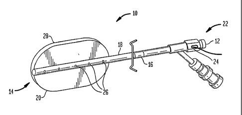
FIG. 1 depicts one embodiment of brachytherapy device 10 of the present
invention including catheter body member 16 having proximal end 12, distal end
14, and
inner lumen 18. Outer spatial volume 20 is preferably disposed on the distal
portion of
catheter body member 16. The proximal end of catheter body 16 preferably
includes a
handle portion 22 for manipulating the device, and a port 24 which opens to
inner lumen
18. At least one treatment feedback sensor 26 is positioned on the device, and
may be
disposed within catheter body member 16 as shown in FIG. 1. In addition a
radiation
source (not shown) is preferably positioned within outer spatial volume 20.
Outer spatial volume 20 is preferably defined by an expandable surface member
28 which can be used to position tissue, provide spacing between the radiation
source
and the adjacent tissue, and/or supply containment for radiation source
materials. In
addition, sensor 26 (or sensors 26) can be positioned on expandable surface
member 28
as shown in FIG. 2. A person of skill in the art will appreciate that
positioning sensor 26
on the expandable surface member could include positioning on the inner or
outer
surface of expandable surface member 28, as well as, positioning the sensor
within the
wall of expandable surface member 28.
In one embodiment, the treatment feedback sensor positioned on the expandable
surface member can sense radiation ("radiation sensor"). A radiation sensor
may be
preferable for providing an accurate view of the strength of the radiation as
it leaves the
,
device. Other types of sensors can also be placed on the expandable surface
member to
CA 02544766 2012-08-28
- 7 -
detect the effect of radiation on surrounding tissue. The sensor may also have
the ability
to sense other treatment characteristics of the tissue in contact with the
expandable
surface member.
A variety of expandable surface members can be used with the present
invention,
and in one embodiment expandable surface member 28 is an inflatable balloon.
It will be
understood that the term "balloon" is intended to include distensible devices
which can
be, but need not be, constructed of elastic material. Exemplary balloons
include the
variety of distensible devices designed for use with surgical catheters. In
use, the balloon
can be expanded by injecting an inflation material though catheter body member
16 and into the balloon by way of an inflation port 34 in the catheter body
member.
In one embodiment, the balloon is constructed of a solid material that is
substantially impermeable to active components of a treatment fluid (e.g.
radiation
source material) with which it can be filled, and is also impermeable to body
fluids, e.g.,
blood, cerebrospinal fluid, and the like. An impermeable balloon is useful in
conjunction
with a radioactive treatment fluid to prevent the radioactive material from
escaping the
treatment device and contaminating the surgical field or tissues of the
patient.
In another embodiment, the balloon is permeable to a treatment agent, and
permits a treatment agent to pass out of device 10 and into a body lumen, body
cavity, or
the anatomical site of the device location. A permeable balloon is useful when
the
treatment agent is a drug such as for example, a chemotherapeutic drug which
must
contact tissue to be effective. U.S. Patent Nos.: 6,537,194 to Winkler and
5,931,774 to
Williams et al. disclose exemplary permeable balloons and treatment
substances. The
treatment feedback sensor can be used to monitor the passage of treatment
agent out of
the permeable balloon. For example, treatment feedback sensor 26 could be
positioned
on the balloon to measure treatment agent concentration. An additional sensor
could also
be positioned in or on tissue surrounding device 10 to detect the
concentration of
treatment agent.
By positioning treatment feedback sensors on the device, a user can monitor
the
delivery of a treatment material from the device to surrounding tissue. The
sensor could
be used to find information on the rate of delivery, the extent of delivery,
the uniformity
of delivery, and other dosing information. Such a sensor can be particularly
CA 02544766 2012-08-28
- 8 -
advantageous because they can overcome the difficulty of determining how much
treatment agent is being delivered and to where it is being delivered.
Presently, the
delivery rate of treatment agent is determined indirectly by measuring factors
such as the
pressure applied to the fluid and/or the change in volume of the fluid. The
treatment
feedback sensors of the present invention allow for direct measurement of the
treatment
agent as it leaves the device.
A treatment agent can also be delivered from the surface of the balloon to the
surrounding tissue. U.S. Patent No. 7,524,275 entitled DRUG ELUTING
BRACHYTHERAPY METHODS AND APPARATUS discloses such devices. A
treatment feedback sensor can be used to detect the delivery of a treatment
agent from
the surface of the balloon to the surrounding tissue. A sensor positioned in a
layer of
treatment material positioned on the outer surface can preferably sense how
much of the
treatment agent has been delivered and/or how much remains. Sensor are useful
for
determining when the treatment agent is fully delivered. Where multiple
treatment
agents are layered on the outer surface of the balloon, the sensor is also
useful for
sensing which treatment agent is being delivered. The sensor could also be
useful for
detecting the level of treatment agent in the adjacent tissue.
The invention also contemplates the use of multiple balloons, e.g., a double-
walled structure as shown in FIG. 3. Such a balloon can comprise, for example,
an inner
balloon 30 containing an inner spatial volume 32 being positioned within an
outer
balloon 28, and the outer balloon defining the outer spatial volume 20 as the
space
between the inner wall and the outer wall. Outer spatial volume 20 is
preferably in fluid
communication with first inner lumen 18 through first inflation port 34, while
inner
spatial volume 32 is preferably in fluid communication with a second inner
lumen 36 via
second inflation port 38. First and second inner lumens 18, 36 are shown by
the cross
sectional view of catheter body member 16 in FIG. 3 A.
A double walled balloon (or even a higher order balloon, e.g. triple walled)
provides more options for controlling and direction radiation dosing. For
example, a
double walled balloon can provide spacing between a radiation source and
adjacent
tissue so that more powerful radiation sources can be used. (See, e.g., U.S.
Pat. Nos.
5,913,813 to Williams et al. and 6,413,204 to Winkler et al.)
CA 02544766 2012-08-28
- 9 -
While "hotter" radiation sources allow delivery of absorbed doses deeper into
the target
tissue and reduce the risk of healthy tissue necrosis, proper spacing and
positioning of
the radiation source are important. Sensor 26 of the present invention can
therefore
provide useful feedback to assure the balloons walls and the radiation source
are
appropriately configured. In particular, a radiation-sensing treatment
feedback sensor
can be used to detect radiation levels and determine if any area is receiving
too much or
too little radiation. Other sensors could also be used to indirectly determine
proper
spacing and positioning by monitoring characteristics such as tissue
temperature.
In some applications, the brachytherapy device 10 is designed to provide a
dosing profile consistent with the shape of the outer spatial volume. That is,
the absorbed
dose within the target tissue at points equidistant from the surface of the
outer spatial
volume should be substantially uniform in substantially every direction
creating three
dimensional isodose profiles substantially similar in shape to the outer
spatial volume. In
addition, the expandable surface member of the outer spatial volume may be
sufficiently
firm so as to force the target tissue to take on the shape of the expandable
surface
member. With the tissue thus shaped, the surrounding tissue receives a uniform
dose of
radiation.
Treatment feedback sensors positioned on the device of the present invention
can
be used to confirm that a three dimensional isodose profile is generated and
delivered to
the surrounding tissue. The sensors may be particularly useful where the
expanded
surface member is used to shape the tissue cavity walls. Although imaging
techniques
can confirm the relative position of the tissue cavity and the brachytherapy
device,
treatment feedback sensors can provide actual dosing information to assure the
tissue
and the device do not shift during the procedure. Although the sensors can be
positioned
anywhere on the device, in may be desirable to position radiation-sensing
treatment
feedback sensors on the expandable surface member as shown in FIG. 3 to
directly test
the radiation levels leaving the surface of the device (e.g. all the radiation
sensor
readings should be roughly equal).
In an alternative embodiment, it may be desirable to deliver an asymmetric
radiation dose to protect radiation-sensitive tissue. Two possible
arrangements for
delivering an asymmetric dose include radiation shielding and/or positioning
the
CA 02544766 2012-08-28
- 10 -
radiation source in an asymmetric configuration as described in U.S. Patent
No.
6,482,142 to Winkler etal. For example, shielding can be accomplished when all
or a
portion of the expandable surface member is formed from, or coated with, a
radio-
opaque material. An asymmetric isodose profile can also be created by the
relative
position of the radiation source or sources to one another and to the outer
spatial volume.
Delivering an asymmetric dose provides an additional challenge because the
radiation dose is focused on one region and shielded from another. If the
device is
improperly positioned within the tissue cavity or if the radiation profile has
an
unexpected shape, sensitive tissue could be damaged. Treatment feedback
sensors can
therefore help to protect radiation-sensitive tissue by confirming proper
shielding and
proper positioning of device 10. Although sensors may be located anywhere on
the
device to provide dosing information, sensors positioned toward the exterior
or outside
of device 10 can provide valuable data regarding the amount of radiation
reaching
sensitive tissue. In one embodiment, a radiation-sensing treatment feedback
sensor can
be positioned on the wall of a tissue cavity in the area which needs
shielding.
The radiation source of the present invention preferably includes any
radiation
source which can deliver radiation to treat proliferative tissue disorders.
Exemplary
radiation sources include high dose brachytherapy radiation, medium dose
brachytherapy radiation, low dose brachytherapy radiation, pulsed dose rate
brachytherapy radiation, external beam radiation, and combinations thereof.
Although
the device of the present invention is described with reference to radiation
sources
positioned within the device, possible radiation sources can include external
radiation
sources positioned outside the device or patient's body such as IMTR, 3-D
conformal
therapy, orthovolatage, stereotactic radiation, and combinations thereof
In one embodiment, device 10 treats proliferative tissue disorders by using
the
expandable surface member to position and/or stabilize tissue surrounding the
tissue
cavity and then deliver radiation from a source external to the tissue cavity.
U.S. Patent
No. 7,524,274 entitled TISSUE POSITIONING SYSTEMS AND METHODS FOR
USE WITH RADIATION THERAPY discloses such devices. Treatment feedback
sensors positioned
CA 02544766 2006-05-04
WO 2005/046794
PCT/US2004/036918
- 11 -
on the device can provide feedback to ensure the prescribed dose is delivered
to the
positioned tissue.
The radiation source can also be positioned within brachytherapy device 10,
and
even more preferably, can be positioned within outer spatial volume 20. In
particular,
the radiation source may be disposed within inner spatial volume 32 inside
outer spatial
volume 20, e.g. inside an inner balloon as shown in FIG. 3. The radiation
source can
include a predetermined radionuclide, for example, 1-125, 1-131, Yb-169 or
other
sources of radiation, such as radionuclides that emit photons, beta particles,
gamma
radiation, or other therapeutic rays including x-ray radiation (e.g., manmade
radiation
sources such as miniature x-ray generators or linear accelerators). The
radioactive
material contained within the outer spatial volume can be a fluid made from
any solution
of radionuclides(s), e.g., a solution of 1-125 or 1-131. A radioactive fluid
can also be
produced using a slurry of suitable fluid containing small particles of solid
radionuclides, such as Au-198, Y-90. Moreover, the radionuclides(ds) can be
embodied
in a gel. A person of skill in the art will appreciate that various radiation
sources can be
used with the brachytherapy device of the present invention.
In another embodiment, the radiation source may be a solid spherical radiation
emitting material 40 positioned within catheter body member 16 as shown in
FIG. 4.
For example, radioactive micro spheres of the type available from the 3M
Company of
St. Paul, Minn., may be used. This radioactive source can either be preloaded
into the
catheter body member at the time of manufacture or loaded into the device
after it has
been implanted into the space formerly occupied by the excised tumor. The
solid
radiation emitting material 40 can be inserted through catheter 16 on a wire
42, for
example, using an afterloader (not shown). Such a solid radioactive core
configuration
offers an advantage in that it allows a wider range of radionuclides than if
one is limited
to liquids. Solid radionuclides that could be used with the device of the
present
invention are currently generally available as brachytherapy radiation
sources.
Treatment feedback sensors can provide valuable information regarding the
characteristics of the radiation source. For example, a radiation-sensing
sensor can be
used to provide data on the location of the radiation source material after it
is loaded into
the device. This data can then be used to calculate the radiation dose and/or
to alert
users if the radiation source material is improperly loaded. Useful radiation
sensors can
CA 02544766 2006-05-04
WO 2005/046794
PCT/US2004/036918
- 12 -
be positioned anywhere on the device including on catheter body member 16 and
expandable surface member 28. In one embodiment, a radiation sensor (or
sensors) is
positioned on the catheter body to detect when the radiation source material
is properly
positioned. For example, as shown in FIG. 4, sensors may be positioned at the
point on
the catheter body member where it is desired to position the radiation
emitting material
40. When the sensor reaches its maximum radiation reading the user will know
the
radiation emitting material is properly positioned.
Catheter body member 16 of device 10 provides a means for positioning outer
spatial volume 20 within the resected tissue cavity and presents a path for
delivering
radiation source material and inflation material (if used). Although the
exemplary
catheter body members illustrated in the FIGS. have a tubular construction,
one of skill
in the art will appreciate that catheter body member 16 can have a variety of
shapes and
sizes. Catheter body members suitable for use in the invention can include
catheters
which are known in the art. Although catheter body member 16 can be
constructed from
a variety of materials, in one embodiment the catheter body member material is
silicone,
preferably a silicone that is at least partially radio-opaque, thus
facilitating x-ray
localization of catheter body member 16 after insertion of device 10. Catheter
body
member 16 can also include conventional adapters for attachment to a treatment
fluid
receptacle and the balloon, as well as devices, e.g., right-angle devices, for
conforming
catheter body member 16 to contours of the patient's body.
As shown in FIG. 1, treatment feedback sensors 26 can be positioned within
catheter body member 16 to provide information on the proliferative tissue
disorder
treatment being delivered to the patient. The sensor or sensors can be
positioned inside
any inner lumen (e.g. first inner lumen 18) of catheter body member 16.
Alternatively,
sensor(s) 26 could be positioned within the wall of catheter body member 16 or
on the
outside of the catheter body member. Multiple sensors 26, as shown in FIG. 1,
may be
preferable to improve accuracy and provide a more detailed picture of the
therapy. In
one embodiment, a string of spaced apart radiation-sensing sensors 26 can
provide data
from points along the whole length of brachytherapy device 10. Preferably,
sensors 26
are positioned at intervals of about 1 cm.
CA 02544766 2006-05-04
WO 2005/046794 PCT/US2004/036918
- 13 -
Treatment feedback sensor 26 used with the device of the present invention
preferably includes radiation sensors capable of detecting and/or measuring
radiation
delivered by brachytherapy devices or external beam radiotherapy. Exemplary
radiation
includes penetrating emissions such as gamma rays, x-rays, and non-penetrating
emissions such as beta particles (negative and positive), alpha particles,
protons and
combinations thereof. Preferably, the radiation sensors are capable of
measuring
radiation over a range of about 1.0 Gy to 400 Gy. An exemplary radiation
sensor can
include MOSFET, diode dosimeters, ionization chambers, thermoluminescent
dosimeters and combinations thereof. The radiation sensor should preferably be
small
enough to be positioned on the brachytherapy device. For example, the sensor
may be in
the range of about 0.01 mm to 3.0 cm in the longest dimension. Preferred sizes
for
individual sensors is 1 mm by 1 mm by 3 mm or smaller in any or all of those
dimensions.
In addition to the ability to sense radiation, or as an alternative, the
treatment
feedback sensor 26 can preferably detect other physical properties such as
temperature,
oxygenation of tissue, pH, drug concentration, cytokine concentration, and/or
other
tissue properties.
Treatment feedback sensor 26 can be disposed on brachytherapy device 10 in a
variety of ways including fixing the sensor to the device. By mating the
sensor with the
device, the location of the device is know and the sensor and brachytherapy
device can
be inserted in one step. A person of skill in the art will appreciate that the
sensor can be
fixed to the device in a variety of ways, including but not limited to,
adhesion,
embedding within the brachytherapy device, insert molding, surface deposition,
ultrasonic welding and beading.
In an alternative embodiment treatment feedback sensor 26 can be
nonpermanently disposed on brachytherapy device 10. For example, the sensor
can be
in contact with the device, but not mated thereto. Nonmating contact allows
sensor 26 to
be inserted into a tissue cavity separately from brachytherapy device 10. In
one
embodiment, the sensor can be inserted into a tissue cavity and then the
brachytherapy
device can be inserted and expanded. Expansion of the brachytherapy device can
then
hold the sensor in position during therapy. Alternatively, sensor 26 can be
disposed
CA 02544766 2006-05-04
WO 2005/046794
PCT/US2004/036918
- 14 -
within device 10 by insertion into catheter body member 16 before or after
insertion of
the brachytherapy device into the tissue cavity.
In yet a further embodiment, an additional sensor(s) can be positioned around
the
outside of device 10. FIG. 5 shows sensors 26 positioned in tissue 50
surrounding the
resected tissue cavity. By positioning sensors outside the device, as well as
on the
device, the characteristics of the tissue a distance from the device can be
determined.
For example, it is expected that the radiation level drops as a function of
distance from
the radiation source, and sensors positioned in the surrounding tissue area
can confirm
that the radiation drops to the expected level. In addition, sensors
positioned in tissue
beyond the target tissue area can be used to confirm a minimal dose (or no
dose) is
delivered to healthy tissue. Such sensors may also have the capability to
sense other
treatment characteristics such as temperature and treatment agent
concentration.
Along with data received from treatment feedback sensor 26, additional
information such as the sensor's location helps to determine the profile of
the radiation
dose. While sensor 26 may be positioned in a predetermined location, the
location can
also be determined in vivo. For example, the treatment feedback sensor may
preferably
be visible to a medical imaging modality such as, radiotherapy (e.g. x-rays,
fluoroscopy), computed tomography, magnetic resonance imaging, and ultrasoUnd.
In
one embodiment, the brachytherapy device is inserted into the tissue cavity
and then the
device and/or sensor is imaged to determine the location of the device and the
sensor.
Other means for determining the position of the device, the sensor and/or
target tissue
include fiducial markers. Fiducial markers may be markers positioned on the
device,
and may include anatomical landmarks in the body, and/or implanted foreign
bodies
such as radio-opaque markers or surgical clips.
The treatment feedback sensor preferably communicates with an external device
that displays, processes, and/or records the radiation dose. Communication
between the
sensor and the external device can be made via direct physical connection (via
wires or
fiber that transmit the signals) or via wireless interface that communicates
the signal
without benefit of cabling.
The present invention also includes the method of using the brachytherapy
device to treat target tissue and sense radioactive emissions. The
interstitial
brachytherapy apparatus of the invention can be used in the treatment of a
variety of
CA 02544766 2006-05-04
WO 2005/046794 PCT/US2004/036918
- 15 -
malignant tumors, and is especially useful in the treatment of brain and
breast tumors.
Treatment feedback sensor 26 can monitor the treatment and help to ensure the
prescribed successful treatment of the surrounding tissue.
Many breast cancer patients are candidates for breast conservation surgery,
also
known as lumpectomy, a procedure that is generally performed on early stage,
smaller
tumors. Breast conservation surgery is typically followed by postoperative
radiation
therapy. Studies report that 80% of breast cancer recurrences after
conservation surgery
occur near the original tumor site, strongly suggesting that a tumor bed
"boost" of local
radiation to administer a strong direct dose may be effective in killing any
remaining
cancer and preventing recurrence at the original site. Numerous studies and
clinical
I trials have established equivalence of survival for appropriate
patients treated with
conservation surgery plus radiation therapy compared to mastectomy.
Surgery and radiation therapy are the standard treatments for malignant solid
brain tumors. The goal of surgery is to remove as much of the tumor as
possible without
damaging vital brain tissue. The ability to remove the entire malignant tumor
is limited
by its tendency to infiltrate adjacent normal tissue. Partial removal reduces
the amount
of tumor to be treated by radiation therapy and, under some circumstances,
helps to
relieve symptoms by reducing pressure on the brain.
A method according to the invention for treating these and other malignancies
begins by surgical resection of a tumor site to remove at least a portion of
the cancerous
tumor and creation of a resection cavity. Following tumor resection the
surgeon places
an interstitial brachytherapy device, having an outer spatial volume as
described above,
into the tissue cavity. The outer spatial volume, preferably being defined by
an
expandable surface member, is expanded and the prescribed dose of radiotherapy
is
delivered. This treatment may be repeated over the course of a treatment
regimen.
In one embodiment, treatment feedback sensors positioned on the device sense
the radiation delivered by the radiation source(s) during radiation dosing.
The radiation
sensors can deliver data during irradiation (e.g., real-time measurements),
after each
fraction of radiotherapy/brachytherapy, and/or upon completion of the entire
course of
radiotherapy/brachytherapy. The data is preferably collected by a computer and
can be
used to verify the delivered radiation dose. The verification step confirms
that the
radiation dose is delivered to the correct area and/or is within the
prescribed limits.
CA 02544766 2012-08-28
- 16 -
A feedback step can also be useful for modifying future radiation doses (or
fractions) to improve the distributed radiation profile. In particular, sensor
26 can detect
the radioactive emissions and delivers data regarding the radiation levels
detected. In one
embodiment, a radiation source emits a first dose of radiation which is
detected by the
sensor positioned on the device. The first dose can be any dose smaller than
the full
prescribed dose. Data collected from the first dose is then used to evaluate
the dosing
profile and dosing intensity, and any errors can be fixed prior to delivering
the full dose.
In another embodiment, the treatment feedback sensors can be used to evaluate
the treatment procedure. The sensors can collect data used to determine if the
residual
malignant cells are being destroyed and to evaluate damage to healthy tissue.
By sensing
physical characteristics of the surrounding tissue such as, for example
oxygenation, pH,
temperature, and cytokine concentration, a user can determine how the
delivered dose
effected the surrounding tissue. In some cases, different regions of tissue or
different
patients may required different doses of radiation. By using treatment
feedback sensors
to directly sense the surrounding tissue, the device of the present invention
can help to
ensure effective treatment.
In yet another embodiment, the treatment feedback sensor can sense the
delivery
of a supplemental treatment such as tissue heating or the delivery of a
treatment agent.
Sensors positioned on the device can be used monitor the supplemental
treatment and
determine its effectiveness.
A person skilled in the art will appreciate that the foregoing is only
illustrative of
the principles of the invention, and that various modifications can be made by
those
skilled in the art as described herein. The scope of the claims should not be
limited by
the preferred embodiments set forth in the examples, but should be given the
broadest
interpretation consistent with the description as a whole.