Note: Descriptions are shown in the official language in which they were submitted.
t t
CA 02545167 2006-05-05
- 1 -
DESCRIPTION
ASPIRATION CATHETER
Technical Field
The present invention relates to a catheter
percutaneously and transluminally introduced into the body
to remove by aspiration a substance present in the body, and
in particular, the invention relates to an aspiration
catheter for removing by aspiration thrombi formed in the
internal blood vessels and debris, such as atheromas,
released in the blood vessels, by applying a negative
pressure from the proximal end of the catheter.
Background Art
Conventionally, when stenosis or occlusion occurs in
vessels, such as blood vessels, and when blood vessels are
blocked by thrombi, angioplasties (e. g., PTA: Percutaneous
Transluminal Angioplasty and PTCA: Percutaneous Transluminal
Coronary Angioplasty) are commonly performed in order to
dilate narrowed areas or reopen occluded areas of blood
vessels so that blood flow in the peripheries of blood
vessels is improved. Many angioplasties have been performed
in many medical institutions. Furthermore, in recent years,
stents have been used to maintain the dilated state of
narrowed areas in many cases.
CA 02545167 2006-05-05
- 2 -
A balloon catheter for PTA or PTCA is used together
with a guiding catheter and a guidewire mainly for the
purpose of dilating a narrowed area or occluded area of a
blood vessel. In an angioplasty for the coronary artery
using the balloon catheter, first, the guiding catheter is
inserted into the femoral artery and advanced through the
aorta, and the guiding catheter is positioned in the opening
of the coronary artery. Then, the guidewire passing through
the balloon catheter is advanced beyond the narrowed area or
occluded area of the blood vessel. The balloon is inflated
while being positioned at the narrowed area or occluded area
so that the narrowed area or occluded area is dilated. The
balloon is then deflated and removed from the body. The
application of the balloon catheter is not limited to
treatment of narrowed areas or occluded areas of blood
vessels, and the balloon catheter is also useful for many
other medical applications, such as insertion into blood
vessels and insertion into various body cavities and tubular
tissue structures.
However, when occlusion is caused by thrombi in the
blood vessel, if the occluded area is dilated by the balloon
catheter, there may be a possibility that the thrombi are
detached from the inner wall of the blood vessel to occlude
peripheral vessels downstream. In the case of the narrowed
area of the blood vessel in which the lesion contains many
CA 02545167 2006-05-05
r J . n
- 3 -
athero-plaques, there may be a possibility that dilation by
the balloon catheter leads to scattering of the athero-
plaques (atheromas) to occlude peripheral vessels. When
peripheral vessels are blocked as described above, even if
the occluded area or narrowed area is dilated, blood is
prevented from flowing into the peripheries, resulting in
slow-flow or no-reflow.
When such a situation arises, in the coronary artery or
the like, it is general practice to wait and see if the
blood flow is recovered, but a long recovery time is
required. According to circumstances, a vasodilator, such
as nitroglycerin, may be administered to recover the blood
flow, or a thrombolytic agent, such as urokinase, may be
locally administered to dissolve the obstruction. In either
case, a long recovery time is still required. When
peripheral vessels are heavily occluded to produce poor
hemodynamics, an auxiliary procedure, such as intra-aortic
balloon pumping (IABP), may be used.
Besides the thrombolytic therapy, a method has been
attempted in which thrombi are mechanically fragmented and a
negative pressure is simultaneously applied from the
proximal end of the catheter to remove the thrombi from the
body.
However, in order to fragment a thrombus at the
catheter tip, it is of course necessary to efficiently
t
CA 02545167 2006-05-05
- 4 -
transmit the mechanical power applied from the proximal end
of the catheter to the distal end of the catheter.
Consequently, in order to enhance the transmission of power
in the catheter shaft, the entire catheter shaft must be
composed of a relatively hard material, often resulting in
difficulty in advancing the catheter to the target site in
the blood vessel. Furthermore, since a negative pressure
must be applied from the proximal end of the catheter
simultaneously with the application of mechanical power, a
large-scale device is required, and thus this method has not
become widely used.
On the other hand, the effect of a catheter having a
simple structure in which thrombi are removed by aspiration
from the body by the application of a negative pressure from
the proximal end has been being clinically confirmed.
However, the cross-sectional area of the aspiration lumen
for aspiration is not sufficiently secured, and only
catheters having low aspiration capability are available.
The reason for this is that the catheter is advanced over
the guidewire to the target site in the blood vessel.
Namely, since a guidewire lumen tracking the guidewire is
provided in the aspiration lumen, it is not possible to
secure a sufficient aspiration lumen.
On the other hand, in a structure in which a guidewire
lumen is provided outside an aspiration lumen, the outer
z
CA 02545167 2006-05-05
- 5 -
diameter of the aspiration catheter inevitably increases.
Consequently, the outer diameter of the guiding catheter
used together increases so that a sufficient inner diameter
is secured, resulting in an enormous burden to the patient.
In addition, since any of the guidewire lumens
described above usually has a length of about 30 cm from the
tip of the aspiration catheter, the entire catheter shaft
lacks flexibility, resulting in poor insertability into
tortuous blood vessels.
Patent Document 1 discloses a catheter that is
insertable into a blood vessel without a guidewire. The
catheter includes a passage for injecting a drug solution,
an imaging agent, or the like, disposed therein; a hub
disposed at the proximal end thereof; and a superelastic
wire provided with a detachable hub. In order to increase
the rate of injection of a drug solution, an imaging agent,
or the like, from the hub, the superelastic wire is designed
to be withdrawn from the catheter so that the effective
lumen of the internal injection passage is increased.
However, when a catheter having such a structure is used as
an aspiration catheter in a conventional PTCA procedure, it
is not possible to advance the catheter to an affected site
over a guidewire, and low operationality has been pointed
out as a problem.
CA 02545167 2006-05-05
r s
- 6 -
[Patent Document 1] Japanese Examined Patent
Application Publication No. 3-74590
Disclosure of Invention
In order to overcome the problems described above, it
is an object of the present invention to provide an
aspiration catheter which secures a largest possible
aspiration lumen, which is sufficiently flexible to be
advanced to a target site following a guidewire and to
satisfactorily track tortuous blood vessels, and in which
the possibility of kinking of the catheter shaft is
decreased when the aspiration catheter is inserted into a
guiding catheter from outside the body, thus achieving good
operationality.
As a result of intensive research conducted by the
present inventors, it has been found that the problems can
be overcome by an aspiration catheter having the following
structure, and thus the present invention has been completed.
Namely, an aspiration catheter for removing by
aspiration a substance from a living body includes a main
shaft including a distal shaft and a proximal shaft, the
main shaft having an aspiration lumen disposed therein, the
aspiration lumen being used for removing the substance by
aspiration; a guidewire shaft disposed at the distal region
of the distal shaft, the guidewire shaft having a guidewire
CA 02545167 2006-05-05
1 f
lumen into which a guidewire is insertable, the guidewire
lumen being disposed in the guidewire; a hub provided at the
proximal end of the proximal shaft, the aspiration lumen
extending to the hub; and a detachable core wire disposed in
the aspiration lumen.
The present invention also relates to the aspiration
catheter, in which a connector is fixed on the proximal end
of the core wire, and the connector is mounted to the
proximal end of the hub in a detachable manner.
The present invention also relates to the aspiration
catheter, in which the interior of the aspiration lumen can
be flushed through the connector with the connector being
mounted in a detachable manner.
The present invention also relates to the aspiration
catheter, in which the distal end of the core wire recedes
from the distal end of the aspiration lumen in the proximal
direction.
The present invention also relates to the aspiration
catheter, in which the relationship 0.3 s R1/R2 s 0.9 is
satisfied, and more preferably, the relationship 0.4 s R1/R2
s 0.7 is satisfied, wherein R1 is the maximum outer diameter
of the core wire, and R2 is the minimum inner diameter of
the aspiration lumen located on the distal side of the hub.
The present invention also relates to the aspiration
catheter, in which the core wire is a spring wire made of
r
CA 02545167 2006-05-05
coiled metal wire.
The present invention also relates to the aspiration
catheter, in which at least a portion of the core wire has a
tapered shape in which the outer diameter becomes larger
toward the proximal end.
The present invention also relates to the aspiration
catheter, in which at least a portion of the core wire has
flexibility which becomes higher toward the distal end.
The present invention also relates to the aspiration
catheter, in which the core wire is composed of stainless
steel, a Co-Cr alloy, an Ni-Ti alloy, an Ni-Ti-Fe alloy, an
Ni-Ti-Cu alloy, an Ni-Ti-Cr alloy, an Ni-Ti-V alloy, an Ni-
Ti-Co alloy, an Ni-Ti-Nb alloy, an Ni-Ti-Pd alloy, an Ni-Ti-
Cu-Cr alloy, or a composite thereof.
The present invention also relates to the aspiration
catheter, in which the tip of the distal shaft is obliquely
cut, the distal end of the guidewire shaft is positioned at
the obliquely cut distal end of the distal shaft or
protrudes from the distal end of the distal shaft in the
distal direction, and the relationship 0.5 s L2/L1 is
satisfied, wherein L1 is the length of the obliquely cut
portion of the distal shaft in the longitudinal direction of
the catheter, and L2 is the length from the proximal end of
the guidewire shaft to the distal end of the distal shaft.
The present invention also relates to the aspiration
r
CA 02545167 2006-05-05
- 9 -
catheter, in which the relationship 2 mm s L1 s 10 mm is
satisfied.
The present invention also relates to the aspiration
catheter, in which the guidewire shaft is provided with a
radiopague marker.
The present invention also relates to the aspiration
catheter, in which the proximal shaft is composed of a
polyimide.
The present invention also relates to the aspiration
catheter, in which the proximal shaft is composed of a
braided tube in which a metal braid and a polymer material
are combined.
The present invention also relates to the aspiration
catheter, in which the braided tube includes an inner layer
defining the aspiration lumen, a metal braid disposed on the
outer surface of the inner layer, and an outer layer
disposed on the outer surface of the metal braid.
The present invention also relates to the aspiration
catheter, in which at least a proximal portion of the
proximal shaft has a flexural modulus of 1 GPa or more.
The present invention also relates to the aspiration
catheter, in which at least a portion of the distal shaft is
applied with a hydrophilic coating that exhibits a
lubricating property in a wet environment.
Furthermore, the present invention relates to a method
s
CA 02545167 2006-05-05
1~ -
for using the aspiration catheter including the steps of
inserting the aspiration catheter into a living body with
the core wire being present in the aspiration lumen, then
withdrawing the core wire, and applying a negative pressure
to the aspiration lumen to remove by aspiration a substance
from the living body.
Brief Description of the Drawings
Fig. 1 is a cross-sectional view showing an aspiration
catheter in an embodiment of the present invention.
Fig. 2 is a cross-sectional view showing an aspiration
catheter in another embodiment of the present invention.
Fig. 3 is a cross-sectional view showing the aspiration
catheter shown in Fig. 1 from which a core wire is withdrawn.
Fig. 4 is a cross-sectional view showing the aspiration
catheter shown in Fig. 2 from which a core wire is withdrawn.
Fig. 5 is a cross-sectional view showing an example of
core wire used in the aspiration catheter of the present
invention.
Fig. 6 is a cross-sectional view showing the other
example of core wire used in the aspiration catheter of the
present invention.
Fig. 7 is a cross-sectional view showing the other
example of core wire used in the aspiration catheter of the
present invention.
CA 02545167 2006-05-05
- 11 -
Fig. 8 is a schematic diagram showing a method for
evaluating kinking resistance and passability through a bent
portion with respect to aspiration catheters of the present
invention.
Fig. 9 is an enlarged view of a plate including a bent
portion shown in Fig. 8.
Fig. 10 is an example of L1 and L2.
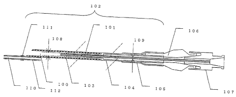
In the drawings, reference numeral 100 represents an
aspiration lumen, 101 a core wire, 102 a main shaft, 103 a
distal shaft, 104 a proximal shaft, 105 a strain relief, 106
a hub, 107 a connector, 108 a minimum inner diameter of the
aspiration lumen, 109 a maximum outer diameter of the core
wire, 110 a guidewire lumen, 111 a radiopaque marker, and
112 a guidewire shaft.
Furthermore, in the drawings, reference numeral 113
represents a tank, 114 a plate including a bent portion, 115
a simulated aorta, 116 a guiding catheter, and 117 a
hemostasis valve. Furthermore, reference numeral 118
represents a polyethylene tube, 119 a bent portion, 120 a
linear portion, 121 an outer diameter of the polyethylene
tube, 122 an inner diameter of the polyethylene tube, and
123 a guidewire.
Best Mode for Carrying Out the Invention
The embodiments of the aspiration catheter of the
CA 02545167 2006-05-05
- 12 -
present invention will be described in detail with reference
to the drawings. However, it is to be understood that the
present invention is not limited thereto.
As shown in the embodiment of Fig. 1 or 2, an
aspiration catheter of the present invention includes a main
shaft 102 including a distal shaft 103 and a proximal shaft
104, the main shaft 102 having an aspiration lumen 100
disposed therein, the aspiration lumen 100 being used for
removing a substance by aspiration; a guidewire shaft 112
disposed at the distal region of the distal shaft 103, the
guidewire shaft 112 having a guidewire lumen 110 into which
a guidewire is insertable, the guidewire lumen 110 being
disposed in the guidewire shaft 112; a hub 106 provided at
the proximal end of the proximal shaft 104, the aspiration
lumen 100 extending to the hub 106; and a detachable core
wire 101 disposed in the aspiration lumen 100. By providing
the core wire 101 in the aspiration lumen 100, the
possibility of kinking of the catheter shaft is effectively
decreased when the aspiration catheter is inserted into a
guiding catheter from outside the body, thus achieving good
operationality. Furthermore, since the guidewire lumen 110
is provided, the aspiration catheter can be easily advanced
to tortuous sites over a guidewire.
If the core wire 101 is provided in the aspiration
lumen 100, the cross-sectional area of the aspiration lumen
CA 02545167 2006-05-05
- 13 -
100 decreases, and thus it is not possible to achieve a
sufficient amount of aspiration. However, in the present
invention, since the core wire 101 is provided in a
detachable manner, it is possible to remove the core wire
101 during aspiration as shown in the embodiment of Fig. 3
or 4. Therefore, a sufficient amount of aspiration can be
easily achieved. In an aspiration catheter in which the
core wire 101 is fixed, an increase in the cross-sectional
area of the aspiration lumen 100 is the only way to achieve
the same amount of aspiration as in the aspiration catheter
of the present invention, resulting in an increase in the
outer diameter of the catheter shaft. If the outer diameter
of the catheter shaft is increased, the size of a guiding
catheter or a sheath used for insertion of the aspiration
catheter must be increased, thus increasing the burden on a
patient undergoing aspiration treatment, which is
undesirable.
The aspiration catheter of the present invention is
characterized by the inclusion of the detachable core wire
101 as described above. The mechanism for allowing the core
wire 101 to be detachable is not particularly limited.
However, in consideration of operationality during the
detachment of the core wire 101, preferably, a connector 107
is fixed on the proximal end of the core wire 101, and the
connector 107 is mounted to the proximal end of the hub 106
CA 02545167 2006-05-05
- 14 -
in a detachable manner. The method for fixing between the
proximal end of the core wire 101 and the connector 107 does
not restrict the advantageous effects of the present
invention at all, and fixing may be performed using an
adhesive or the like. In such a case, the type of adhesive
used is not particularly limited. The method for connecting
the connector 107 to the proximal end of the hub 106 is not
limited as long as the connector 107 is detachable. In one
preferred embodiment, the distal end of the hub 106 is
formed as a female Luer adaptor and the connector 107 is
formed as a male Luer adaptor. Thereby, the core wire 101
can be reliably and easily detached. Furthermore, by
forming the proximal end of the hub 106 as a female Luer
adaptor, it is also possible to simply apply a negative
pressure to the aspiration lumen 100 using a syringe or the
like.
As described above, when the connector 107 is fixed on
the proximal end of the core wire 101 and the connector 107
is mounted to the proximal end of the hub 106 in a
detachable manner, it is possible to achieve a structure in
which the aspiration lumen 100 is flushed through the
connector 107. When the aspiration catheter of the present
invention is used, it is necessary to flush the aspiration
lumen 100 with a suitable solution, such as a solution of
physiological heparinized saline, before insertion into the
CA 02545167 2006-05-05
- 15 -
body. Flushing prevents thrombus formation when the
aspiration catheter is inserted into the body, in particular,
the blood vessels. Flushing is usually performed using a
syringe. Consequently, by forming the proximal end of the
connector 107 as a female Luer adaptor, it is possible to
perform flushing with the core wire 101 being mounted, and
it is possible to insert the aspiration catheter into the
body promptly after flushing to start treatment.
The positional relationship between the guidewire lumen
110 and the aspiration lumen 100 does not restrict the
advantageous effects of the present invention at all. As
shown in Fig. 1, the guidewire lumen 110 and the aspiration
lumen 100 may be disposed independently of each other. As
shown in Fig. 2, the guidewire lumen 110 may be partially
disposed inside the aspiration lumen 100. Alternatively,
the guidewire lumen 110 may be entirely disposed inside the
aspiration lumen 100. However, when the guidewire lumen 110
is partially or entirely disposed inside the aspiration
lumen 100, the cross-sectional area of the aspiration lumen
100 is smaller compared with the case in which the guidewire
lumen 110 and the aspiration lumen 100 are disposed
independently of each other. In particular, an increase in
the length in the longitudinal direction of the portion of
the guidewire lumen 110 disposed inside the aspiration lumen
100 leads to a decrease in the amount of aspiration.
CA 02545167 2006-05-05
- 16 -
Therefore, the length of the portion of the guidewire lumen
110 disposed inside the aspiration lumen 100 is preferably
as small as possible. On the other hand, when the guidewire
lumen 110 and the aspiration lumen 100 are disposed
independently of each other, there is an increased risk that
the guidewire shaft 112 will be separated from the distal
shaft 103 when the aspiration catheter is inserted or
withdrawn along a guidewire. It is also possible to
reinforce the joint between the guidewire shaft 112 and the
distal shaft 103 using another component. In such a case,
however, the outer diameter of the joint significantly
increases. As described above, the aspiration ability and
safety of the catheter greatly depend on the positional
relationship between the guidewire lumen 110 and the
aspiration lumen 100. Therefore, it is obvious to those
skilled in the art that the aspiration catheter can be
appropriately designed in consideration of the target site
to be treated, method for use, required amount of aspiration,
substance subjected to aspiration, etc.
With respect to the material for the guidewire shaft
112, in order to secure good slidability with a guidewire,
at least the inner surface thereof is preferably composed of
a polyolefin, in particular, a polyethylene.
The method for bonding between the distal shaft 103 and
the guidewire shaft 112 does not restrict the advantageous
CA 02545167 2006-05-05
- 17 -
effects of the present invention at all. Namely, if the
distal shaft 103 and the guidewire shaft 112 are composed of
materials that can be welded to each other, bonding can be
performed by welding. Alternatively, if the distal shaft
103 and the guidewire shaft 112 are composed of materials
that cannot exhibit sufficient bonding strength when welded,
bonding may be performed using an adhesive. In such a case,
the chemical species in the adhesive used is not
particularly limited. For example, a cyanoacrylate,
urethane, epoxy, or silicone adhesive is preferably used.
The curing mechanism of the adhesive is also not
particularly limited. For example, a moisture-curing, two-
part curing, or photo-curable adhesive is suitably used. If
the distal shaft 103 and the guidewire shaft 112 are
composed of materials having poor adhesion properties,
surface treatment may be performed, for example, by oxygen
plasma or corona discharge, or using a silane coupling agent,
before bonding.
Preferably, the distal end of the core wire 101 recedes
from the distal end of the aspiration lumen 100 in the
proximal direction. If the distal end of the core wire 101
protrudes from the distal end of the aspiration lumen 100,
there is a high risk of internal injuries during insertion.
Furthermore, after aspiration treatment is performed with
the core wire 101 being dismounted, when it becomes
CA 02545167 2006-05-05
- 18 -
necessary to move the aspiration catheter to treat another
site and the core wire 101 is mounted inside the aspiration
lumen 100, internal injuries due to the core wire 101 are
highly likely to occur.
As long as the distal end of the core wire 101 recedes
from the distal end of the aspiration lumen 100 in the
proximal direction, the advantageous effects of the present
invention are not restricted at all. The position of the
distal end of the core wire 101 can be determined in
consideration of kinking of the catheter shaft during
insertion, operationality when the aspiration catheter is
inserted or moved over a guidewire, rigidity balance of the
entire aspiration catheter, etc.
Preferably, the relationship 0.3 s R1/R2 s 0.9 is
satisfied, wherein R1 is a maximum outer diameter 109 of the
core wire 101, and R2 is a minimum inner diameter 108 of the
aspiration lumen 100. If Rl/R2 < 0.3, the core wire 101 is
too thin relative to the aspiration lumen 100, and therefore
the effect of preventing folding by the core wire 101 during
insertion is not shown sufficiently. If R1/R2 > 0.9, the
entire aspiration catheter becomes rigid, and it becomes
extremely difficult to pass the aspiration catheter through
the tortuous site. More preferably, the relationship 0.4 s
R1/R2 s 0.7 is satisfied.
The structure and shape of the core wire 101 do not
CA 02545167 2006-05-05
- 19 -
restrict the advantageous effects of the present invention
at all. A typical example of the core wire 101 is one with
a straight shape as shown in Fig. 5. From the standpoint of
further improving the passability through tortuous sites, as
shown in Fig. 6, preferably, the core wire 101 is a spring
wire made of coiled metal wire. In such a case, the outer
diameter, pitch, etc., of the wire constituting the spring
wire are not particularly limited. The pitch of the spring
wire may be changed continuously or stepwise so that the
flexibility of the core wire 101 becomes higher toward the
distal end. Furthermore, although not shown in Fig. 6, a
core wire may be disposed inside the spring.
Although the straight shape is shown in Fig. 5 as the
typical example, a tapered core wire 101 as shown in Fig. 7
may also be used suitably: When such a tapered wire is used,
by controlling the tapered shape, it is possible to control
the flexibility of the aspiration catheter.
In the core wire 101, preferably, the flexibility
becomes higher toward the distal end. By increasing the
flexibility of the core wire 101, passability can be
enhanced in the case, for example, in which the site to be
treated is tortuous, or the aspiration catheter must pass
through a tortuous site to reach the site to be treated.
Examples of the means for imparting flexibility include use
of a spring wire or a tapered wire as the core wire 101 as
CA 02545167 2006-05-05
- 20 -
described above. Further examples include a combination of
a spring wire and a tapered shape, and provision of various
cuts to the surface of a wire.
The core wire 101 is preferably composed of a metal in
consideration of prevention of kinking of the aspiration
catheter. In view of corrosion resistance,
antithrombogenicity, etc., the core wire 101 is preferably
composed of stainless steel or a Co-Cr alloy. Furthermore,
a superelastic alloy may be used to prevent kinking of the
core wire 101 itself. Examples of the superelastic alloy
suitable for use include Ni-Ti alloys, Ni-Ti-Fe alloys, Ni-
Ti-Cu alloys, Ni-Ti-Cr alloys, Ni-Ti-V alloys, Ni-Ti-Co
alloys, Ni-Ti-Nb alloys, Ni-Ti-Pd alloys, and Ni-Ti-Cu-Cr
alloys.
Preferably, the tip of the distal shaft 103 is
obliquely cut. By cutting obliquely, a wide entrance of the
aspiration lumen 100 can be secured, and thereby the
aspiration efficiency can be increased. Preferably, the
relationship 0.5 s L2/Ll is satisfied, wherein L1 is the
length of the obliquely cut portion of the distal shaft in
the longitudinal direction of the catheter, and L2 is the
length from the proximal end of the guidewire shaft 112 to
the distal end of the distal shaft 103. If L2/L1 < 0.5, the
area of the joint between the guidewire shaft 112 and the
distal shaft 103 decreases, and the guidewire shaft 112 is
CA 02545167 2006-05-05
s . ~ '
- 21 -
highly likely to be separated from the distal shaft 103.
Preferably, the relationship 2 mm s L1 s 10 mm is
satisfied. If L1 is less than 2 mm, it is difficult to
remove debris by aspiration efficiently. If L1 exceeds 10
mm, there is an increased risk that the inner wall of the
blood vessel will be damaged by the obliquely cut portion
during the advancement of the aspiration catheter through
the body, in particular, the tortuous blood vessel. In
order to prevent internal injuries during insertion into the
body or during aspiration treatment, the obliquely cut
portion may be subjected to chamfering so that the edges are
smoothened. Examples of the chamfering method which may be
used include, but are not limited to, a method in which
edges are melted by heating, and a mechanical polishing
method.
In the aspiration catheter of the present invention,
preferably, the guidewire shaft 112 is provided with a
radiopaque marker 111. In such a case, more preferably, the
radiopaque marker 111 is disposed at a position from which
the position of the distal end of the aspiration lumen 100
can be confirmed. The radiopaque marker 111 makes it
possible to confirm the position of the distal end of the
aspiration lumen 100 during insertion of the aspiration
catheter or during aspiration treatment, and therefore, the
risk of internal injuries due to the obliquely cut tip of
CA 02545167 2006-05-05
- 22 -
the distal shaft 103 is decreased.
The radiopaque marker 111 may be composed of any
material that has sufficient radiopacity. Preferably, the
radiopaque marker 111 is composed of a metal material, and
examples thereof include gold, silver, platinum, tantalum,
iridium, tungsten, and alloys of these metals. Furthermore,
the structure of the radiopaque marker 111 does not restrict
the advantageous effects of the present invention at all.
The radiopaque marker 111 may be ring-shaped or braid-shaped,
or may have a structure other than this. The method for
fixing the radiopaque marker 111 is not particularly limited.
The proximal shaft 104 is preferably composed of a
polyimide or a braided tube in which a metal wire and a
polymer material are combined. With respect to the
polyimide, because of its excellent tensile strength,
tensile yield strength, and compressive strength, the
thickness of the shaft can be decreased. With respect to
the braided tube, by selecting the shape of the wire, number
of wires, pitch, or type of polymer material used, the
thickness of the shaft can be decreased as in the polyimide.
By decreasing the thickness of the shaft, the diameter of
the aspiration lumen 100 can be increased, resulting in
significant improvement in the aspiration ability.
Preferably, the braided tube includes an inner layer
defining the aspiration lumen 100, a metal braid disposed on
CA 02545167 2006-05-05
- 23 -
the outer surface of the inner layer, and an outer layer
disposed on the outer surface of the metal braid. Such a
double layer structure allows the physical properties of the
braided tube to be more finely controlled. For example, by
using, as the inner layer, a fluorocarbon resin, such as
polptetrafluoroethylene (PTFE), a
tetrafluoroethylene~perfluoroalkylvinylether copolymer (PFA),
a tetrafluoroethylene~hexafluoropropylene copolymer (FEP), a
tetrafluoroethylene~ethylene copolymer (ETFE),
poly(vinylidene fluoride) (PVDF), or
polychlorotrifluoroethylene (PCTFE); high-density
polyethylene; or the like, thrombi and atheromas are
prevented from adhering to the interior of the aspiration
lumen 100, and aspiration can be performed efficiently. By
using, as the outer layer, an elastomer, such as a polyamide
elastomer, a polyester elastomer, or a polyolefin elastomer,
the strength and flexibility of the braided tube can be
controlled.
The material and structure of the metal braid
constituting the braided tube do not restrict the
advantageous effects of the present invention at all, and
various materials and structures may be used. Namely, a
metal braid may be formed using one or a plurality of metal
wires per group, each metal wire being obtained by
processing stainless steel, e.g., SUS304 or SUS316, spring
CA 02545167 2006-05-05
- 24 -
steel, piano wire, oil tempered wire, a Co-Cr alloy, an Ni-
Ti alloy, or the like, so as to have a cross-section that is
circular, oval, square, or the like. The number of metal
wires per braid is not particularly limited.
Preferably, at least a proximal portion of the proximal
shaft 104 is composed of high-modulus material with a
flexural modulus of 1 GPa or more. By using the shaft
composed of such a high-modulus material, power applied by
the operator in operating the aspiration catheter can be
fully transmitted to the tip of the catheter. Namely, in
addition to the pushing force and the pulling force, the
rotating force can be easily transmitted to the tip.
Examples of the high-modulus material which may be suitably
used include metal materials, such as stainless steel, Co-Cr
alloys, and Ni-Ti alloys; and resin materials, such as
polyimides, polyether ether ketones, and polyamide-imides.
Composites of these materials may be used.
The distal shaft 103 is preferably composed of a
material having a lower modulus compared with the proximal
shaft 104 so that rigidity continuously changes in the
longitudinal direction of the aspiration catheter. Examples
of the material which may be suitably used for the distal
shaft 103 include polyolefins (e. g. polyethylene),
polyamides, polyesters, polyurethanes, polyolefin elastomers,
polyamide elastomers, polyester elastomers, and polyurethane
CA 02545167 2006-05-05
- 25 -
elastomers. The method for bonding the distal shaft 103 to
the proximal shaft 104 is not particularly limited, and a
known method, such as welding or adhesion, may be used.
The materials for a strain relief 105, which is used to
reduce the difference in rigidity between the proximal shaft
104 and the hub 106, and the hub 106 do not restrict the
advantageous effects of the present invention at all. In
view of moldability, resin materials are preferably used for
the strain relief 105 and the hub 106.
Preferably, at least a portion of the distal shaft 103
is applied with a hydrophilic coating that exhibits a
lubricating property in a wet environment. In particular,
in an aspiration catheter provided with the guidewire shaft
112, if the size of the aspiration lumen 100 is increased as
much as possible, the outer diameter of the distal shaft 103
is increased. Consequently, when the aspiration catheter is
inserted into a blood vessel in particular, there may be a
possibility that the sliding friction of the aspiration
catheter with the inner wall of the blood vessel increases
because of the distal shaft 103. Therefore, at least a
portion of the distal shaft 103 is preferably applied with a
hydrophilic coating to reduce sliding friction. Of course,
the distal shaft 103 may be entirely applied with a
hydrophilic coating, or the proximal shaft 104 may be
partially or entirely applied with a hydrophilic coating.
CA 02545167 2006-05-05
i
- 26 -
The advantageous effects of the present invention are
not particularly restricted by the method for applying the
hydrophilic coating and the material for the hydrophilic
coating, and the method and the material may be
appropriately selected depending on the materials of the
distal shaft 103, the proximal shaft 104, the guidewire
shaft 112, etc. For example, a hydrophilic polymer, such as
poly(2-hydroxyethyl methacrylate), polyacrylamide, or
polyvinylpyrrolidone, may be used. Furthermore, by
adjusting the thickness of and the material for the
hydrophilic coating in the longitudinal direction of each
shaft, the sliding friction may be controlled so as to
gradually increase or decrease.
A method for using the aspiration catheter according to
the present invention includes the steps of inserting the
aspiration catheter into a living body with the core wire
101 being present in the aspiration lumen 100, then
withdrawing the core wire 101, and applying a negative
pressure to the aspiration lumen 100 to remove by aspiration
a substance from the living body. In this method, the
method for applying a negative pressure to the aspiration
lumen 100 is not particularly limited. For example, a
negative pressure may be applied manually using a syringe
equipped with a lock, or automatically using a pump or the
like.
CA 02545167 2006-05-05
- 27 -
[Examples]
Examples and comparative examples of the present
invention will be described in detail below.
(Example 1)
As a proximal shaft, a polyimide tube with an outer
diameter of 1.30 mm, an inner diameter of 1.10 mm, and a
length of 1,100 mm was formed by dip forming using a
polyamic acid varnish. As a distal shaft, a tube with an
outer diameter of 1.30 mm, an inner diameter of 1.00 mm, and
a length of 300 mm was formed by extrusion molding of low-
density polyethylene (LF480M, Japan Polychem Corporation).
The diameter of one end of the proximal shaft was reduced by
thermal drawing. The portion in which the diameter was
reduced was inserted into the distal shaft and fixed by
bonding using a two-part curing urethane adhesive (Nipporan
4235/Coronate 4403, Nippon Polyurethane Industry Co., Ltd.),
and a main shaft was thereby obtained. Since the distal
shaft was composed of a material with poor adhesion
properties, oxygen plasma treatment was performed before
bonding.
The tip of the distal shaft was obliquely cut so that
the length L1 in the longitudinal direction of the catheter
was 2 mm. A hub produced by injection molding of
polycarbonate (Makrolon 2658, Bayer AG) and a strain relief
CA 02545167 2006-05-05
- 28 -
produced by injection molding of a polyamide elastomer
(PEBAX5533SA01, Elf Atochem, Inc.) were fixed on the
proximal end of the proximal shaft by bonding using a two-
part curing urethane adhesive (Nipporan 4235/Coronate 4403,
Nippon Polyurethane Industry Co., Ltd.).
A tube with an outer diameter of 0.60 mm, an inner
diameter of 0.42 mm, and a length of 10 mm was formed by
extrusion molding of a high-density polyethylene (HY540,
Japan Polychem Corporation), and a radiopaque marker
composed of a platinum-tungsten alloy (tungsten content 8
wt~) with an outer diameter of 0.72 mm, an inner diameter of
0.65 mm, and a length of 1 mm was fixed by swaging on the
center of the tube. A guidewire shaft was thereby produced.
The guidewire shaft and the distal shaft were placed so that
the length L2 was 1 mm and the guidewire shaft protrudes
from the distal shaft in the distal direction, and bonded to
each other by heat welding. During bonding, in order to
secure a guidewire lumen and an aspiration lumen, mandrels
were inserted into both shafts.
A straight wire composed of SUS304 alloy steel with an
outer diameter of 0.605 mm and a length of 1,300 mm was used
as a core wire. A connector produced by injection molding
polycarbonate (Makrolon 2658, Bayer AG) was bonded to one
end of the core wire using a two-part curing urethane
adhesive (Nipporan 4235/Coronate 4403, Nippon Polyurethane
CA 02545167 2006-05-05
- 29 -
Industry Co., Ltd.). The core wire was inserted, from the
end not provided with the connector, through the hub, and
the hub and the connector were fastened to each other. An
aspiration. catheter was thereby produced.
(Example 2)
An aspiration catheter was produced as in Example 1
except that a straight wire composed of SUS304 alloy steel
with an outer diameter of 0.715 mm was used as a core wire.
(Example 3)
An aspiration catheter was produced as in Example 1
except that the guidewire shaft was placed inside the distal
shaft and that a straight wire composed of an Ni-Ti alloy
with an outer diameter of 0.495 mm was used as a core wire.
(Example 4)
An aspiration catheter was produced as in Example 3
except that a straight wire composed of SUS304 alloy steel
with an outer diameter of 0.385 mm was used as a core wire.
(Example 5)
An aspiration catheter was produced as in Example 3
except that a straight wire composed of an Ni-Ti alloy with
an outer diameter of 0.880 mm was used as a core wire.
(Example 6)
An aspiration catheter was produced as in Example 1
except that a tapered wire composed of SUS304 alloy steel
with an outer diameter at the proximal end of 0.605 mm, an
CA 02545167 2006-05-05
- 30 -
outer diameter at the distal end of 0.385 mm, and a length
of the tapered portion of 600 mm was used as a core wire.
(Example 7)
An aspiration catheter was produced as in Example 1
except that a spring wire composed of SUS304 alloy steel
with an outer diameter of 0.605 mm (0.150 mm wire closely
coiled around 0.300 mm core wire) was used as a core wire.
(Example 8)
An aspiration catheter was produced as.in Example 1
except that a braided tube with an outer diameter of 1.30 mm,
an inner diameter of 1.10 mm, and a length of 1,100 mm was
used as a proximal shaft, the braided tube including a metal
braid formed using metal wires composed of SUS304 alloy
steel with a size of 0.10 mm x 0.03 mm (one metal wire per
group, 16 groups), an inner layer composed of
polytetrafluoroethylene (POLYFLON F-207, Daikin Industries,
Ltd.), and an outer layer composed of a polyamide elastomer
(PEBAX7233SA01, Elf Atochem, Inc.); and that a tube with an
outer diameter of 0.60 mm, and inner diameter of 0.42 mm,
and a length of 10 mm was formed by extrusion molding of a
polyamide elastomer (PEBAX7233SA01, Elf Atochem, Inc.), and
a radiopaque marker composed of a platinum-tungsten alloy
(tungsten content 8 wt~) with an outer diameter of 0.72 mm,
an inner diameter of 0.65 mm, and a length of 1 mm was fixed
by swaging on the center of the tube to produce a guidewire
CA 02545167 2006-05-05
- 31 -
shaft .
(Example 9)
An aspiration catheter was produced as in Example 1
except that a straight wire composed of SUS304 alloy steel
with an outer diameter of 0.275 mm was used as a core wire.
(Example 10)
An aspiration catheter was produced as in Example 1
except that a straight wire composed of SUS304 alloy steel
with an outer diameter of 1.05 mm was used as a core wire.
(Comparative Example 1)
An aspiration catheter was produced as in Example 1
except that no core wire was used.
(Evaluation of kinking resistance during insertion and
passability through bent portion)
As shown in Fig. 8, a simulated aorta 115 and a guiding
catheter 116 were disposed in a tank 113 filled with a
physiological saline solution kept at 37°C, and a hemostasis
valve 117 was fixed to the guiding catheter 116. The tip of
the guiding catheter 116 was connected to a plate 114
provided with a simulated coronary artery, and a guidewire
123 with an outer diameter of 0.014 inch was preliminarily
passed through the guiding catheter. As shown in Fig. 9, a
polyethylene tube 118 serving as a simulated coronary artery
was disposed in the plate 114, and the polyethylene tube 118
included a bent portion 119 and a linear portion 120. The
CA 02545167 2006-05-05
- 32 -
bent portion 119 had a radius of curvature of 15 mm, and the
linear portion 120 had a length of 80 mm. The polyethylene
tube 118 had an outer diameter 121 of 5 mm and an inner
diameter 122 of 3 mm. Each of the aspiration catheters in
the examples and comparative examples was inserted into the
guiding catheter 116 over the guidewire 123 from the
hemostasis valve 117, and occurrence of kinking and
passability through the bent portion 119 were evaluated.
The results are shown in Table 1.
In Table 1, evaluation criteria were as follows:
Kinking resistance
O: Excellent
0: Fair
x: Poor
Passability
O: Excellent
0: Fair
x: Poor
CA 02545167 2006-05-05
a
0 0 0 0 x 0 0 0 0 a a
a
U
C
C
X 0 0 0 a 0 0 0 0 a 0
Yw
L
N
L(7IfWC~L(7O ~ ~ lf7LL7l()
y f~C~~ M 00lf~~ InN Q7,
O O O O O O O O O O
E ~ ~ ~ tf~O ~
E O - 07OOOOO O O I~O
COf~~ M 00CflCOGON
O O O O O O O O O
O
w- ~ ~ O ~ O ~'~'~ d'~f'
~
M - O O = O - O O O O O
.
M ~ M M laM (aM M M M M
~
_
N
L
~ cncnz cnz cncncncncn
~
4-. ~ _ _ _ _ L _ _ _
~ L L ~ t t ~ ~ t t L
O
L
_ ~
~ L
'~O '~-~-~i
~
_ N
~i- N N N N O O O ~ O N O
O
~
~ E E E E ~EE ~E~ E E E
-
_ _ _ _ _ _ _
.__ _ ~ _
.a~ - ?'?'_ - , _>,_~
E
_ O O O O O O O '-O O O
d ~
L
r-N M 'cTL(7Cflt'0007~ .
>
_N_N_NN O ~ _O_ON O
E E E E E E E E E ~ aE
I L LLL l11LLtlxJLLLL~ U
J J l j LL
E-~
CA 02545167 2006-05-05
- 34 -
In each of Examples 1 to 8 according to the present
invention, kinking did not substantially occur in the
aspiration catheter, and relatively good passability through
the bent portion was shown.
In each of Examples 1 to 3 and 6 to 8, no kinking
occurred in the aspiration catheter, and also passability
through the bent portion was excellent.
On the other hand, in Comparative Example 1, the
aspiration catheter did not show sufficient performance.
Industrial Applicability
As described above, in accordance with the present
invention, it is possible to easily provide an aspiration
catheter for removing by aspiration a substance from a
living body, the catheter including an aspiration lumen for
removing by aspiration a substance, the aspiration lumen
extending to a hub provided at the proximal end of the
catheter; and a detachable core wire disposed in the
aspiration lumen. The aspiration catheter is sufficiently
flexible to satisfactorily track tortuous blood vessels, and
the possibility of kinking of the catheter shaft is
decreased when the aspiration catheter is inserted into a
guiding catheter from outside of the body, and thus good
operationality is achieved.