Note: Descriptions are shown in the official language in which they were submitted.
CA 02545330 2006-05-09
WO 2005/051542 PCT/GB2004/004843
Laminated Device
Field of Inyention
The invention is concerned with a microfluidic device and a method of making
such a
device. In particular but not exclusively the invention concerns a diagnostic
device
suitable for the measurement of analytes in or for the measurement of the
properties
of a fluid sample, such as a sample of bodily fluid.
Back rg ound
Simple disposable diagnostic devices suitable for the measurement of analytes
in
sample of bodily fluids are known and typically comprise a measurement or
reaction
chamber, a suitable vent and a fluid conduit for delivery of the sample fluid
to the
chamber, see for example EP537761. The dimensions of the fluidic pathway will
typically have at least one capillary dimension such that fluid may proceed
along the
fluid pathway by capillary action, requiring no or minimal external forces.
The
internal fluid volume space may be achieved by providing a sidewall spacer
between
upper and lower laminae. The height of the sidewall spacer will effectively
define the
height of the fluidic channel. The side wall spacer may be formed for example
by
printing a glue spacer onto an internal surface of one or both the laminae or
the spacer
may itself be formed of a solid material. Such devices may be individually
produced
or mass-produced using large sheets of substrate materials that may be
laminated
using either a batch or continuous process and subsequently cut to produce the
desired
devices.
Such devices are typically small and designed to be suitable for use with
fluid samples
of volumes typically between 1-50uL.
Outline of the Invention
The present inventors have realised that one of the problems that arises when
dosing
reagent into a reaction chamber is one of containment of the reagent. Liquids
that are
dosed into the reagent chamber have a tendency to move or be drawn into the
fluid
conduit that connects the chamber. This occurs due to capillary forces that
exist at the
CA 02545330 2006-05-09
WO 2005/051542 PCT/GB2004/004843
2
chamber/conduit interface and can lead to the fluid conduit being partially or
completely blocked by reagent, resulting in inferior fluid flow
characteristics within
the device. This is particularly a problem when dosing into a very small
chamber, as it
is difficult to ensure that the liquid comprising or containing the reagent
does not
contact the sides of the chamber and/or the fluid conduit/chamber interface
leading to
capillary movement of the reagent into the fluid conduit. The problems of
accurate
dosing are also exacerbated when manufacturing devices on a large scale using
automated equipment. One possible way around the problem is to design the
chamber
to be of a larger cross-sectional area. However, this results in a larger
chamber
volume requiring a larger volume of fluid test sample. Another solution is to
create a
chamber having a depth greater than minimum depth required for capillary flow,
i.e. a
depth greater than a capillary dimension. This however has drawbacks in that
it also
results in a larger chamber thus requiring a larger volume of fluid sample.
Furthermore it is difficult to move sample fluid from a region of high
capillarity
within the fluid conduit to one of low capillarity in the fluid chamber.
It is a desirable feature of a diagnostic testing device that the volume of
fluid sample
required be as low as possible. This is particularly so for collection of
samples of
bodily fluid, such as samples of capillary blood from a finger stick or
lancet, as
collection of large volumes prove to be painful for the patient. A further
drawback of
providing larger fluid chambers is that the device becomes structurally weaker
due to
the presence of less substrate per unit area. This effect becomes greater for
devices
having multiple chambers and/or fluid conduits.
The invention provides for a microfluidic diagnostic device and relatively
simple, low
cost method of manufacture. The invention is particularly suited to devices
having
more than one chamber and/or fluid conduit.
In one aspect the invention provides a microfluidic device comprising at least
four
laminae, the laminae including two outer laminae and at least two further
laminae
disposed between the outer laminae and defining a microfluidic pathway wherein
inner surfaces of the outer laminae define the upper and lower surfaces of a
fluid
pathway, a one of said further laminae defining at least a first fluidic
element, and at
CA 02545330 2006-05-09
WO 2005/051542 PCT/GB2004/004843
3
least a second of said further laminae defining at least a second fluidic
element,
wherein the first and second fluidic elements are fluidically coupled.
The device may have at least three further laminae wherein each further
laminae
defines at least a respective one microfluidic element.
A reagent may be provided in one or more of the microfluidic elements.
In a second aspect the invention provides a method of constructing a
microfluidic
device comprising providing at least a first lamina or lamina assembly which
serves to
define at least a first microfluidic element or area; providing at least a
second lamina
or lamina assembly which serves to define at least a second microfluidic
element or
area; and connecting the at least first and second laminae or laminate sub-
assemblies
such that the first and second microfluidic elements become fluidically
coupled.
Prior to connection of the first and second laminae or lamina assemblies, a
reagent
may be dispensed into one of the microfluidic elements.
The reagent may be disposed in the particular microfluidic element or elements
in a
liquid form or dispersed or dissolved in a suitable liquid carrier or liquid
solvent.
In one embodiment, one of the microfluidic elements is of a higher capillarity
than the
other. Reagent may be disposed in the microfluidic element having a lower
capillarity.
The microfluidic element having a lower capillarity may be a fluid chamber and
the
microfluidic element having a higher capillarity a fluid conduit.
In another embodiment, the microfluidic elements are of the same capillarity.
Reagents may be dispensed into both of the or respectively each microfluidic
element.
The invention also relates to a microfluidic device prepared according to the
second
aspect, and to a diagnostic assay device prepared according to the second
aspect
According to another aspect, the invention provides for a microfluidic
diagnostic
assay device and method of construction thereof comprising the steps of:
CA 02545330 2006-05-09
WO 2005/051542 PCT/GB2004/004843
4
(a) provision of first and second laminae or lamina sub-assemblies which
respectively serve to contain or define first and second microfluidic elements
;
(b) dosing of a reagent into the first and/or second microfluidic elements;
(c) assembling the first and second laminae or lamina sub-assemblies such that
the respective microfluidic elements become fluidically coupled.
According to a further aspect, the microfluidic element may be defined by one
portion
of a lamina or lamina sub-assembly and a further microfluidic element may be
defined by another portion of the same lamina or lamina sub-assembly. The
laminated
structure may then be folded so as to fluidically couple the microfluidic
elements. The
invention need not necessarily be limited to one fold, and more folds could be
provided if desired. The substrate could for example be folded in half upon
itself to
provide two laminae, or the substrate could be partially folded such that the
lower
lamina were bigger in cross-sectional area than the upper lamina or vice-
versa. Thus
in principle the entire diagnostic device could be provided from one
substrate.
Alternatively, the folded lamina could be used to produce a sub-lamina
assembly to
which further laminae or lamina sub-assemblies could be attached.
The reagent would be applied to one of the microfluidic elements prior to the
folding
of the substrate and fluidic coupling of the microfluidic elements.
An advantage provided by this folding method is that all of the microfluidic
elements
may be provided on the one lamina or lamina sub-assembly if desired. A further
advantage is that it removes the need for precise location of the individual
laminae.
This is especially so for structures having very small microfluidic elements,
wherein
precise location is essential to ensure effectively fluidic communication from
one
microfluidic element to another.
The term microfluidic element is intended to refer to any microfluidic
structure
through which fluid sample may flow through or into and includes but is not
restricted
to, a vent for the venting of gases, chamber, channel, conduit, filter, time
gate and so
on. The microfluidic element may be of regular or irregular dimensions.
CA 02545330 2006-05-09
WO 2005/051542 PCT/GB2004/004843
Preferably the microfluidic elements will have at least one capillary
dimension such
that fluid is able to flow along and pass from one microfluidic element to
another
under the influence of capillary action. Alternatively or additionally the
fluid sample
may be caused to flow along or between microfluidic elements under the
influence of
5 other forces such as gravity, electro-kinetic or electro-osmotic pumping and
so on.
Typical dimensions of the microfluidic elements are those having a cross-
sectional
dimension, such as a cross-sectional diameter, of between 0.1 and 500um, more
typically having a cross-sectional dimension of between 1 and 100um.
A key aspect of the invention is the provision of a microfluidic device and
method of
manufacture thereof wherein key microfluidic elements that make up the
microfluidic
only become fluidically coupled upon assembly of the device, namely upon
attachment of the various sections, laminae or lamina sub-assemblies that
define the
respective microfluidic elements.
By separation of the key microfluidic elements, reagent that is deposited into
a
particular microfluidic element is not able to flow into a second microfluidic
element.
This serves to contain the reagent in the particular microfluidic element.
This is
particularly advantageous in cases where it is necessary to contain the
reagent in a
particular microfluidic element or where flow of reagent from one microfluidic
element into another or flow of reagent into the interface between two
rnicrofluidic
elements may cause the microfluidic flow path to be impeded or blocked. This
is
especially true of microfluidic elements which have at least a capillary
dimension,
such that reagent is able to flow from one to another by capillary action.
This effect
may be magnified where for example reagent is dosed into a microfluidic
element
having a particular capillary force such as a chamber that is fluidically
coupled and
adj acent to a microfluidic element having a greater capillary force, such as
a fluid
conduit having a smaller cross-sectional dimension. Thus according to a
further
aspect, the invention provides for a microfluidic device and method of
construction
thereof wherein reagent provided in a first microfluidic element is
subsequently
fluidically coupled to a second microfluidic element wherein the second
microfluidic
element has a higher capillarity than the first microfluidic element or
wherein the
CA 02545330 2006-05-09
WO 2005/051542 PCT/GB2004/004843
6
second microfluidic element has a region of higher capillarity which is
positioned
adjacent a first microfluidic element having a region of lower capillarity.
By separation of the key microfluidic elements, it removes the need to dose
reagent
into a microfluidic element so accurately, for example it removes the need to
dose
reagent such that it would not have a tendency to flow (for example by
ensuring that it
did not touch the side walls of the element). This is especially advantageous
when
dosing reagent into elements of very small volume which may be of the order of
100n1 or greater.
The term key microfluidic elements is intended to refer to those microfluidic
elements
in which it is desirable to keep separate during assembly. Thus for example,
there
may not be a need to necessarily separate particular microfluidic elements
from one
another, in particular microfluidic elements which are not situated adjacent
to
microfluidic elements into which reagent will be dosed. For example, one
lamina may
be provided with a fluid application element that is fluidically coupled to a
fluid
conduit element. A second lamina could then be provided with a chamber element
into which reagent is dosed, followed by assembly of the first and second
laminae
such that the fluid chamber becomes fluidically coupled and situated adjacent
to the
fluid conduit and be fluidically coupled and situated remote from the fluid
application
element.
The invention is not necessarily restricted to two laminae or lamina sub-
assemblies
and the various microfluidic elements may be provided in any number of
laminae.
The individual laminae may comprise more than one microfluidic element if
desired
which may or may not be in fluidic connection with each other.
The reagents may specifically or non-specifically react or bind with an
analyte of
interest or serve to modify a property of the fluid sample such as viscosity
or pH.
Non-limiting examples of reagents that could be employed are specific binding
partners to the analyte of interest such as antibodies, binding proteins,
antigens and
the like, enzymes, reagents that serve to influence a property of hemostasis
of a fluid
sample such as thromboplastin, or reagents that serve to interact with the
fluid sample
CA 02545330 2006-05-09
WO 2005/051542 PCT/GB2004/004843
7
or analytes contained therein in any way or to enable the measurement to talce
place
such as magnetic or magnetisable particles.
The diagnostic assay device according to the invention is suitable for the
measurement of the amount or presence of analyte in or for the measurement of
the
properties of a fluid sample, such as a sample of bodily fluid.
A particular microfluidic element may also serve to hold the fluid sample such
that
the property of the sample or analytes contained therein might be determined.
Typically this might be carried out in a fluid chamber element. The manner by
which
the particular parameter of interest could be determined or measured could be
for
example by optical means, electrochemical means, magnetic means, by the use of
a
piezoelectric crystal and so on. The chamber would be arranged to contain or
cooperate with suitable transduction means, such as suitably positioned optics
or
electrodes.
Examples of analytes include hormones, drugs, bacteria, toxins, organic
compounds,
proteins, peptides, micro organisms, bacteria, viruses, amino acids, nucleic
acids,
carbohydrates, hormones, steroids, vitamins, pollutants, pesticides, and
metabolites of
or antibodies to any of the above substances and so on.
The fluid sample for use in the diagnostic assay device can be derived from
any
source, such as a physiological fluid, including blood, serum, plasma, saliva,
interstitial fluid, ocular lens fluid, sweat, urine, and the like. Besides
physiological
fluids, other samples can be used such as water, food products, soil extracts,
and the
like for the performance of industrial, environmental, or food production
assays as
well as diagnostic assays. In addition, a solid material suspected of
containing the
analyte can be used as the test sample once it is modified to form a liquid
medium or
to release the analyte.
Dosing of reagents into the reaction chambers may be achieved using a number
of
techniques known in the art such as screen-printing, pen plotting or
airbrushing. The
reagent may be applied in a liquid, semi-liquid, gel or semi-solid form and/or
be
dispersed or dissolved in a suitable liquid carrier or solvent as required.
More than
CA 02545330 2006-05-09
WO 2005/051542 PCT/GB2004/004843
8
one reagent may be added. For example, in the case of a diagnostic assay for
the
measurement of coagulation time, thromboplastin may be added along with
magnetic
particles.
If desired the solvent or carrier may be removed or partially removed to yield
a dry
reagent or a reagent that is substantially immobile before the device is
assembled and
the key microfluidic elements are fluidically coupled.
The microfluidic structures may be created for example by embossing methods
such
as stamping a particular substrate, lamina or lamina sub-assembly. This method
removes the need to provide upper and lower laminated which serve to seal and
the
microfluidic elements. Alternatively, the microfluidic elements may be defined
by
cutting entirely through the thickness of the particular laminae. In this
case, upper and
lower laminae may be provided in order to seal the microfluidic elements, as
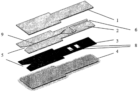
exemplified by laminae (1) and (4) in Figure 1.
The laminae making up the device may be chosen to be of any suitable material
such
as polycarbonate. The laminae may be treated where necessary to increase their
hydrophilicity, for example by the provision of a suitable surface coating.
The
dimensions of the lamina, namely thickness, length or width may be chosen to
be any
suitable. The individual dimensions and materials of the laminae may be chosen
to be
the same or chosen to be different. The thickness of the individual laminae
might
typically range from around 50uM to around 2000uM although in principle any
thickness could be contemplated.
A suitable method to attach the laminae to each other is to provide an
adhesive on one
or both surfaces of the respective laminae. The adhesive may also be
hydrophilic
which may serve to provide enhanced flow characteristics of the fluid sample.
The
individual laminae may be provided for example with an adhesive coating and
with a
further backing lamina. Removal of this backing lamina thus reveals the
adhesive
coating.
According to one embodiment the diagnostic device is constructed from four
laminae
comprising upper and lower laminae which serve to define the upper and lower
CA 02545330 2006-05-09
WO 2005/051542 PCT/GB2004/004843
9
surfaces of the fluid pathway, a first intermediate lamina comprising at least
one
channel and a second intermediate lamina comprising at least a chamber. The
intermediate laminae may be provided in any order.
Brief description of the drawings
Examples incorporating the invention will now be described with reference to
the
accompanying drawings, in which:
Fig 1 shows a perspective exploded view of a device embodying the invention;
Fig 2 shows a perspective exploded view of a second device embodying the
invention;
Figure 3 shows a cross-sectional view of a laminated device embodying the
invention;
Figure 4 shows a cross-sectional view of a second laminated device
embodying the invention;
Fig 5 shows a perspective exploded view of a third device embodying the
invention;
Fig 6 shows a perspective exploded view of a fourth device embodying the
invention;
Fig 7 shows a substrate defining different structures;
Fig 8 shows a first cut-out lamina;
Fig 9 shows a second cut-out lamina; and
Fig 10 shows a partial view of a detection system.
A four member laminated device is shown in Figure 1. Upper and lower members
(1)
and (4) sandwich a first intermediate member (2) with channels (9) and a
second
intermediate member (3) with chamber areas (8). Each member is a thin sheet,
and is
hereinafter referred to as a lamina.
As may be seen from Figure 1, intermediate laminae (2) and (3) have
microfluidic
features cut to the full depth of the material. Lamina (2) is provided with an
adhesive
coating on its top surface whilst lamina (3) is provided with adhesive
coatings on both
sides (not shown).
CA 02545330 2006-05-09
WO 2005/051542 PCT/GB2004/004843
Lamina (2) further contains a sample application feature (5), channelling (9)
to
transport the fluid sample as well as venting means (6) which serves to vent
gases
from the chambers (~). Lamina (3) also contains a sample application feature
(5).
The venting means may be any suitable. Figure 1 shows a venting means wherein
fluid is prevented from escaping from the device by provision of a capillary
stop
feature.
The device may be constructed by attaching lamina (3) to lower lamina (4) thus
providing a reagent chamber having sidewalls as well as a lower surface. If
required,
reagent may then be dosed into one or more chambers and allowed to dry. Lamina
(2)
10 may then be subsequently attached to lamina (3). Upper lamina (1) may then
be
attached to lamina (2) to seal the device and close off the channels.
It should be recognized that a key aspect of the invention is to ensure that
particular
microfluidic elements are separated from one another during construction and
that
alternative assembly steps could be carned out to those described above. Thus
for
example according to the device of Figure 1, construction could be carried out
as
follows: Attachment of lamina (1) to lamina (2) to form a first lamina sub-
assembly
A. Attachment of lamina (3) to lamina (4) to form a second lamina sub-assembly
B,
followed by subsequent attachment of lamina assemblies A and B.
Using this approach, the chamber is separated from the channels whilst dosing
the
reagent into the chambers thus ensuring that the reagent cannot flow into the
channels.
Once dried, the reagent is unable to flow into the channels when the device is
connected together such that the chamber and channels become fluidically
coupled.
In a second embodiment a laminated device was prepared comprising upper and
lower
laminae, an intermediate lamina with channels cut out, a middle lamina with
through
holes to fluidically connect the intermediate lamina with channels to the
intermediate
lamina with reaction chambers and a further intermediate lamina with reaction
areas
cut out.
CA 02545330 2006-05-09
WO 2005/051542 PCT/GB2004/004843
11
A device prepared from five laminae is shown in Figure 2. Upper (1) and lower
(5)
laminae were provided with an intermediate lamina (2) with channel and vent
areas, a
second intermediate lamina (4) with chamber areas (4) and a middle lamina (3)
with
through holes (6).
Construction of the device can be achieved fox example using a web-based
manufacturing process whereby laminae or lamina sub-assemblies can be
assembled
in independent steps.
The laminae or lamina sub-assemblies were bonded together. This bonding was
achieved through the use of glue laminae on various respective sides of the
lamina
components. In a preferred embodiment the glue laminae are in the position as
shown
in Figure 3.
Figure 3 shows a cross-sectional view of a four lamina structure device
showing the
positions of the adhesive layers.~Lamina (1) has a top surface (5) that is
hydrophobic
and a bottom surface (6) that is hydrophilic. Lamina (2) has a top surface
that is
coated with an adhesive layer (7) and a bottom surface (8) that is
hydrophilic. Lamina
(3) has top and bottom surfaces that are coated with hydrophilic glue layers
(9) and
(10). Lamina (4) has a top surface (11) that is hydrophilic and a bottom
surface (12)
that is hydrophobic.
Figure 4 shows a cross-sectional view of an alternative embodiment of a four
lamina
structure showing the positions of the adhesive layers. Lamina (1) has a top
surface
(5) that is hydrophobic and a bottom surface that is coated with a hydrophilic
glue
layer (6). Lamina (2) has a top surface (7) that is hydrophilic and a bottom
surface (8)
that is also hydrophilic. Lamina (3) has top and bottom surfaces that are
coated with
hydrophilic glue layers (9) and (10). Lamina (4) has a top surface (11) that
is
hydrophilic and a bottom surface (12) that is hydrophobic.
A key aspect of the lamination process is in the provision of a laminated
structure
wherein certain microfluidic elements of the device are provided which are
free from
adhesive. A further key aspect is in the provision of adhesives which are
either
CA 02545330 2006-05-09
WO 2005/051542 PCT/GB2004/004843
12
hydrophobic or hydrophilic. Yet a further aspect of the invention is in the
provision of
laminae having particular hydrophilic or hydrophobic surfaces.
For example in Figure 3, the chamber element that is defined by laminae (3)
and (4)
does not have adhesive on the walls defining it. This ensures that reagent or
reagents
contained within the chamber are not affected by the presence of glue, for
example
the mobility of particles provided within the chamber. In this particular
case, it is
preferable that the uncoated surfaces (8) and (11) are made hydrophilic to
assist in
filling of the chamber element:
Furthermore, as shown in Figure 3, the upper surfaces of lamina (2) that serve
to
define a fluid channel are provided with a hydrophilic adhesive. This is to
assist the
flow of fluid along the channel. In contrast the upper and lower surfaces of
lamina (3)
are provided with a hydrophobic adhesive which has better adhesive properties,
since
this particular region does not come into contact with fluid sample.
In general it is desirable to provide hydrophilic inside surfaces that serve
to define the
microfluidic elements. Furthermore, it is desirable to provide hydrophobic
upper
and/or lower exterior surfaces, as indicated by (5) and (12) of Figure 4 as
well as a
device having hydrophobic sides. This ensures that fluid sample applied to the
device
is encouraged to flow into the sample application feature.
The invention provides for a low cost manufacturing method and design of
devices
that are suitable for the measurement of analytes in or the measurement of the
properties of low volumes (ranging from less than 1uL to around 50uL) of a
fluid
sample. The invention is particularly suitable for devices having multiple
microfluidic
elements, especially multiple microfluidic chambers.
Figure 5 shows an alternative embodiment whereby the chambers are provided on
separate laminae (4) and (2).
Figure 7 shows a substrate having various microfluidic structures on one
substrate.
The substrate may be folded about an axis (a-a) so as to align the respective
CA 02545330 2006-05-09
WO 2005/051542 PCT/GB2004/004843
13
microfluidic elements. Once the devices have been assembled, the laminated
structure
may then be cut to provide multiple devices.
Figure 5 shows a further embodiment wherein the chambers areas (6) and (7) are
provided respectively within separate laminae (4) and (2). Furthermore the
chamber
(6) is situated directly over chamber (7). The two chambers are separated by
lamina
(3) containing a feed channel (8) and a venting channel (9). In this
embodiment when
sample is applied to the assembled structure the sample migrates down the feed
channel and then moves into chamber (6) and chamber (7). A device wherein the
microfluidic elements, in this case detection chambers, are arranged to be
situated
substantially directly over one another, has the advantage that a single
transduction
element, such as a solenoid may be arranged such as to cooperate with both
detection
chambers and removes the need for one transduction element per chamber.
In another embodiment as shown by Figure 6, a five layer laminated structure
(1-5) is
provided wherein chamber (6) is provided directly over a chamber (7). The two
chambers are separated by lamina (3) containing a feed channel (10) and
venting
channels (11). Fluidic connection is made from lamina (3) with the chamber (6)
in
lamina (2) by fluidic overlaps, one leading off the chamber (8) and one
leading off the
channel (9). Fluidic overlaps are also created to connect the feed channel
(10) to
chamber (7) and from the chambers (6 and 7) to the venting channels (11). By
provision of fluidic overlaps or designing the individual microfluidic
elements such
that they overlap when assembled it reduces the need for exacting tolerances
when
assembling the device. Provision of such fluidic overlaps ensures that the
individual
microfluidic elements are fluidically coupled upon assembly of the device. The
invention is not restricted to the fluidic overlaps as indicated by Figure 6
and other
designs could be envisaged, for example provision of a conduit having a
circular or
wider section at one end.
CA 02545330 2006-05-09
WO 2005/051542 PCT/GB2004/004843
14
Example 1
Preparation of laminae
As exemplified in Figure 3, a sheet (1) having a hydrophobic upper surface (5)
(contact angle greater than 60°) and a hydrophilic lower surface
(contact angle less
than 30°) was sourced (Tape Specialities Ltd., Hertfordshire, UK). This
sheet had a
thickness of 175 gum.
A sheet (2) 175 ~,m thick was cut with a laser (C10, Alltec UK Ltd, Rotherham,
UK)
in the design as shown in Figure 8. This sheet had a notch (1) to aid sample
filling
when the device was constructed, two through holes that create chambers (2)
when
the device is constructed and a capillary break area (3). This sheet had a
25,um thick
hydrophobic glue layer on the upper surface.
A sheet (3) 175 ~.m thick was cut with a laser (C10, Alltec UK Ltd, Rotherham,
UK)
in the design shown in Figure 9. This sheet had a notch (1) to aid sample
filling when
the device was constructed, channelling (2) and a capillary break area (3).
This sheet
had 25 ~,m hydrophilic glue layers on both the upper and lower surfaces.
A sheet (4) 175 ~,m thick having a hydrophilic upper surface (contact angle
less than
30°) and a hydrophobic lower surface (contact angle greater than
60°) was sourced
(Tape Specialities Ltd., Hertfordshire, UK). Laminae (1) and (4) were the same
material and differed only in the orientation in which they were used.
Assembly of lamina sub-assemblies
The liner covering the upper surface of lamina (2) was removed and the
hydrophilic
side of lamina (1) was bonded to the exposed glue lamina of lamina (2) by
pressing
the two materials together. This created a lamina sub-assembly (A).
The liner covering the lower surface of lamina (3) was removed and the
hydrophilic
side of lamina (4) was bonded to the exposed glue lamina of lamina (3) by
pressing
the two materials together. This created a lamina sub-assembly (B).
CA 02545330 2006-05-09
WO 2005/051542 PCT/GB2004/004843
Deposition of rea ents
The lamina sub-assembly (B) was placed such that the two well chambers were
uppermost and approx. 50n1 of rabbit brain thromboplastin (prepared by
techniques
known in the art [for example, US4416812]) was sprayed using an in-house built
air
5 brush system across the chambers. This was carried out at a spray rate of
0.16 ,ul/mm
at 0.6 bar spray pressure. Subassembly (B) was subsequently placed on an air-
drying
machine (Hedinair Ltd., Romford, Essex, LTK) and the reagents dried by heating
to
55° C (setting 4) for 6 minutes 20 seconds. Following the deposition of
thromboplastin reagent, an aqueous solution of 6 % (wlv) in 60 % sucrose of
10 superparamagnetic particles having a diameter of approximately 5 ~Cm
(Liquids
Research Ltd, Bangor, Wales) was sprayed into the chambers at a rate of 0.5
~l/rnm.
Following this second spraying step, the subassembly was again placed on the
air
dryer and the reagents dried by heating to 55° C for 6 minutes and 20
seconds to
remove the solvent.
15 Assembly of lamina
As shown in Figure 3, the top liner on subassembly B (9) was removed to reveal
the
glue layer and this was bonded to the bottom of subassembly (A) by pressing
the two
materials together. Assembled devices containing dried reagents were stored at
4° C
by sealing in aluminium foil pouches containing a silica desiccant.
Detection of blood coagulation
The foil pouch containing the assembled lamina was removed from 4° C
and allowed
to equilibrate to room temperature for 5 minutes. The foil pouch was opened
and the
lamina removed and placed between two electromagnets such that the poles of
the
magnets were in contact with the side edge of the assembled lamina. Figure 10
shows
a top view of a schematic of the layout of the detection system. The laminated
device
(1) was oriented such that the chamber (2) was placed in close contact with
two
electromagnets (3 and 4). An optical assembly consisting of an LED (Everlight
Electronics Co, Ltd., Catalogue number 11-21SURC/5530-A3/TR8 with a 632 nrn
peak emission) and a detector (Everlight Electronics Co, Ltd., Catalogue
number
CA 02545330 2006-05-09
WO 2005/051542 PCT/GB2004/004843
16
PD15-22C/TR8) was placed such that light from the LED interrogated part of a
region
(5) of the chamber (2). The electromagnets were driven by a simple electrical
circuit
that passed current at 60 mA into one electromagnet for 250 ms and then
switched the
60 mA current into a second electromagnet for 250 ms, this produced a magnetic
field
with a strength of approx. 40 mT (at the pole). The current was switched
between the
electromagnets a number of times. Fresh capillary whole blood was added to the
front of the lamina. Blood moved by capillary action through the laminated
device
and entered into the reaction chambers. Upon reaching the reaction chambers
the
thromboplastin and magnetic particle reagents were resuspended by the blood
and the
particles began to move backwards and forwards in the reaction chamber due to
the
force imparted on them by the electromagnets. The LED was illuminated by
applying
18 mA and the signal from the detector was collected.