Note: Descriptions are shown in the official language in which they were submitted.
CA 02545930 2006-05-11
WO 2004/044590 PCT/GB2003/004950
- 1 -
Tumour Marker Proteins and Uses Thereof
Field of the invention
The invention relates to tumour marker proteins
and their preparation from fluids from one or more
cancer patients, wherein said fluids are those which
collect in a body cavity or space which is naturally
occurring or which is the result of cancer or medical
intervention for cancer. Exemplary fluids are
l0 ascites, pleural effusion, seroma, hydrocoele and
wound drainage fluid. The invention also relates to
preparation of tumour marker proteins from excretions
taken from patients with cancer.
The said tumour marker proteins are useful in
cancer detection methods which involve detecting or
quantitatively measuring autoantibodies to circulating
tumour markers or markers expressed on or in tumour
cells and in various research applications. The
invention is also directed to such uses.
Bac~round to the invention
The development and progression of cancer in a
patient is generally found to be associated with the
presence of markers in the bodily fluid of the
patient, these "tumour markers" reflecting different
aspects of the biology of the cancer (see Fateh-
Maghadam, A. & Steilber, P. (1993) Sensible use of
tumour markers. Published by Verlag GMBH, ISBN 3-
926725-07-9). Tumour markers are often found to be
altered forms of wild-type proteins expressed by
"normal" cells, in which case the alteration may be a
change in primary amino acid sequence, a change in
secondary, tertiary or quaternary structure or a
change in post-translational modification, for
example, abnormal gly~.osylation. In addition, wild-
type proteins which are up-regulated or over-expressed
in tumour cells, possibly as a result of gene
amplification or abnormal~~transcriptional regulation,
may also be tumour markers.
CA 02545930 2006-05-11
WO 2004/044590 PCT/GB2003/004950
- 2 -
Established assays for tumour markers present in
bodily fluids tend to focus on the detection of tumour
markers which reflect tumour'bulk and as such. are of
value late in the disease process, for example in the
diagnosis of metastatic disease. The most widely used
of these markers include carcinoembryonic antigen
(CEA) and the glycoprotein termed CA 15.3, both of
which have been useful mainly as indicators of
i0 systemic disease burden and of relapse following
therapy (Molina, R., Zanon, G., Filella, X. et al. Use
of serial carcinoembryonic antigen and CA 15.3 assays
in detecting relapses in breast cancer patients.
(1995) Breast Cancer Res Treat 36: 41-48). These
markers are of limited use earlier in the course of
the disease, for example in early detection or in the
screening of asymptomatic patients. Thus, in the
search for tumour markers present in bodily fluid that
are of use in assisting diagnosis earlier in the
disease process the present inventors have sought to
identify markers which do not depend on tumour bulk
per se.
Differences between a wild type protein expressed
by "normal" cells and a corresponding tumour marker
protein may, in some instances, lead to the tumour
marker protein being recognised by an individual's
immune system as "non-self" and thus eliciting an.
immune response in that individual. This may be a
humoral (i.e B cell-mediated) immune response leading
to the production of autoantibodies immunologically
specific to the tumour marker protein. Autoantibodies
are naturally occurring antibodies directed to an
antigen which an individual's immune system recognises
as foreign even though that antigen actually
originated in .the individual. They may be present in
the circulation as circulating free autoantibodies or
in the form of circulating immune complexes consisting
of autoantibodies bound to their target tumour marker
protein.
CA 02545930 2006-05-11
WO 2004/044590 PCT/GB2003/004950
- 3 -
As an alternative to the direct measurement or
detection of tumour marker protein~in bodily fluids,
assays may be developed to measure the immune response
of the individual to the presence of tumour marker
protein in terms of autoantibody production. Such
assays essentially constitute indirect detection of
the presence of tumour marker protein. Because of the
nature of the immune response, it is likely that
autoantibodies can be elicited by a very small amount
of circulating tumour marker protein and indirect
methods which rely on detecting the immune response to
tumour markers will consequently be more sensitive
than methods for the direct measurement of tumour
markers in bodily fluids. Assay methods based on the
detection of autoantibodies may therefore be of
particular value early in the disease process and
possibly also in relation to screening of asymptomatic
patients, for example in screening to identify
individuals "at risk" of developing disease amongst a
population of asymptomatic individuals. Furthermore,
they may be useful for earlier detection of recurrent
disease.
Tumour marker proteins observed to elicit serum
autoantibodies include a particular class of mutant
p53 protein, described in US Patent No. 5,652,115,
which can be defined by its ability to bind to the 70
kd heat shock protein (hsp70). p53 autoantibodies can
be detected in patients with a number of different
benign and malignant conditions (described in US
5,652,115) but are in each case present in only a
subset of patients. For example, one study utilizing
an ELISA assay for detection of autoantibodies
directed against the p53 protein in the serum of
breast cancer patients reported that p53
autoantibodies were produced by 26% of patients and
1.3% of control subjects (Mudenda, B., Green, J. A.,
Green, B. et a1. The relationship between serum p53
autoantibodies and characteristics of human breast
CA 02545930 2006-05-11
WO 2004/044590 PCT/GB2003/004950
- 4 -
cancer, (1994) Br J Cancer 69: 4445-4449). A second
tumour marker protein known to elicit serum
autoantibodies is the epithelial mucin MUC1 (Hinoda,
Y. et a1. (1993) Immunol Left. 35: 163--168; Kotera,
Y. et a1. (1994) Cancex Res. 54: 2856-2860).
WO 99/58978 describes methods for use in the
detection/diagnosis of cancer which are based on
evaluating the immune response of an individual to two
or more distinct tumour markers. These methods
generally involve contacting a sample of bodily fluid
taken from the individual with a panel of two or more
distinct tumour marker antigens, each derived from a
separate tumour marker protein, and detecting the
formation of complexes of the tumour marker antigens
bound to circulating autoantibodies immunologically
specific for the tumour marker proteins. The presence
of such circulating autoantibodies is taken as an
indication of the presence of cancer.
Cancer detection methods based on detection of
circulating autoantibodies are frequently immunoassays
utilizing an "immunoassay reagent" reactive with the
circulating autoantibodies. Typically, the "reagents"
used in such assays comprise recombinant tumour marker
proteins (expressed in bacterial, insect, yeast or
mammalian cells) or chemically synthesised tumour
marker antigens, which may comprise substantially
whole tumour marker proteins, or fragments thereof,
such as short~peptide antigens. Other potential
sources of tumour-associated proteins for use as the
basis of immunoassay reagents for the detection of
anti-tumour auto-antibodies include cultured tumour
Cells (and the spent media used for their growth),
tumour tissue, and serum from individuals with
neoplasia. The majority of these sources have
significant drawbacks, as discussed below.
With cultured tumour cells (and their spent
media) the amount of expressed protein can vary
CA 02545930 2006-05-11
WO 2004/044590 PCT/GB2003/004950
- 5 -
depending on growth phase at the time of harvest,
leading to variations in quality and quantity. In
addition, the desired protein is generally present at
low concentration, therefore'it is time-consuming to
purify sufficient quantities of protein. Furthermore,
the cell stock will be clonal, unlike cell stock in a
tumour which is likely to have become heterogeneous in
nature during the growth of the neoplasm, therefore
producing variations in protein (especially in the
degree of glycosylation).
Recombinant proteins expressed in bacterial cells
are not glycosylated, and thus significantly different
from naturally glycosylated proteins. In addition,
refolding of recombinantly expressed proteins may not
be appropriate, thus giving an incorrect conformation
for auto-antibody recognition.
Tumour tissue is usually only available in small
quantities and the purification of proteins therefrom
is laborious and time consuming.
Serum samples are usually available only in small
quantities, therefore it is difficult to purify
sufficient quantities of protein.
The present inventors have now determined that
significant advantages can be gained by the use of
tumour marker antigens purified from bodily fluids
derived from a body cavity or space in which a tumour
is present or with which it is or was associated, such
as ascites fluid, pleural effusion, seroma, hydrocoele
or wound drainage fluid, or from excretions, as the
"reagent" in auto-antibody immunoassays. In
particular, the inventors have observed that use of
reagents comprising tumou.~' marker antigens purified
from bodily fluids derived from the above defined body
cavities or spaces results in increased sensitivity
(as compared to the use of reagents derived from a
~ "normal" body fluid) and produces a more "clinically
CA 02545930 2006-05-11
WO 2004/044590 PCT/GB2003/004950
- 6 -
relevant" result. There are also significant
practical advantages to be gained from the use of such
fluids as a source of assay reagent.
Summary of the invention
In a first aspect the invention relates to a
method of detecting cancer-associated anti-tumour
autoantibodies, which method is an immunoassay
comprising contacting a sample to be tested for the
l0 presence of such autoantibodies with an immunoassay
reagent and detecting the presence of complexes formed
by specific binding of the immunoassay reagent to any
cancer-associated anti-tumour autoantibodies present
in the sample, wherein the immunoassay reagent
comprises tumour marker protein prepared from bodily
fluid derived from a body cavity or space within which
a tumour is or was present or with which a tumour is
or was associated, from one or more cancer patients
and/or tumour marker protein prepared from an
excretion from one or more cancer patients, wherein
said tumour marker protein exhibits selective
reactivity with cancer-associated anti-tumour
autoantibodies.
In a second aspect the invention relates to use
of tumour marker protein prepared from bodily fluid
derived from a body cavity or space within which a
tumour is or was present or with which a tumour is or
was associated, of one or more cancer patients and/or
tumour marker protein derived from an excretion of one
or more cancer patients in the manufacture of an
immunoassay reagent exhibiting selective reactivity '
with cancer-associated anti-tumour autoantibodies.
In a third aspect, the invention relates to a
method of preparing a tumour marker protein which
method comprises isolating said tumour marker protein
from bodily fluid wherein said fluid is:
(i) collected from a body cavity or space in
which a tumour is or was present or with '
CA 02545930 2006-05-11
WO 2004/044590 PCT/GB2003/004950
-
which a tumour is or was associated, and
(ii) said fluid represents the pooled fluid
samples from two or more cancer patients.
In a fourth aspect, the invention relates to a
method of preparing a tumour marker protein which
method comprises isolating said tumour marker protein
from an excretion wherein:
(i) said excretion or any component thereof has
been in contact with a tumour or tumour
cells, and
(ii) said excretion represents pooled excretion
samples from two or more cancer patients.
In a fifth aspect the invention relates to tumour
marker protein preparations prepared using the methods
described above which are substantially immunoglobulin
free and to kits and reagents comprising said
preparations.
Detailed description of the invention
In the first aspect, the invention relates to a
method of detecting "cancer-associated" anti-tumour
autoantibodies.
The term "cancer-associated" anti-tumour
autoantibodies refers to autoantibodies which are
characteristic of the cancer disease scat e; and which
are directed against epitopes present on forms of
tumour marker proteins which are preferentially
expressed in the cancer disease state.
The method of the invention comprises an
immunoassay to detect and/or quantitatively measure
. autoantibodies immunologically specific for one or
more tumour marker proteins, and is characterised in
that the "immunoassay reagent" used in the immunoassay
comprises tumour marker protein prepared from bodily
fluid derived from a body cavity or space in which a
tumour is or was present or with which a tumour is or
CA 02545930 2006-05-11
WO 2004/044590 PCT/GB2003/004950
_ g _
was associated, from one or more cancer patients
and/or tumour marker protein prepared from an
excretion of one or more cancer patients. Generally,
the excretion will have passed through an organ in
which cancer is present wherein the excretion is in
contact with said cancer, or the excretion will
include one or more components which have been in
contact with cancer elsewhere in the body. A
particular example is bile which may be in contact
20 with cancer in the gall bladder but will appear in the
faeces.
The immunoassay reagent exhibits "selective
reactivity" with cancer-associated anti-tumour
autoantibodies. As used herein "selective reactivity"
means a tumour marker protein has a greater affinity
for autoantibodies to the tumour-associated antigen
than it does for any~antibody or autoantibody made to
the same antigen which exists in the normal i.e. non-
tumour possessing state.
The term "body cavity or space" includes any body
cavity or space, whether it be a natural cavity or a
space or cavity arising as a result of diseases or
medical intervention including collapsed or former
Cavities. The fluid is derived from such a cavity or
space in which a tumour is or was present or with
which a tumour is or was associated. Preferably the
"bodily fluid derived from a body cavity" will be a
tumour-induced body fluid, meaning a body fluid which
is produced during the disease process, for example in
response to or as a consequence of the presence of a
tumour cells. Exemplary body fluids are ascites,
pleural effusion, seroma, hydrocoele and wound
drainage f 1 iud .
For the avoidance of doubt "bodily fluids derived
from a body cavity or space" do not include fluids
derived from the systemic circulation, such as whole
4o blood or serum.
CA 02545930 2006-05-11
WO 2004/044590 PCT/GB2003/004950
_ g _
The term "excretion" includes, inter alza, urine,
faeces, and seminal fluid.
The general features of immunoassays, for example
ELISA, radioimmunoassays and the like, are well known
to those skilled in the art (see Immunoassay, E.
Diamandis and T. Christopoulus, Academic Press, Inc.,
San Diego, CA, 1996). Immunoassays for the detection
of antibodies having a particular immunological
specificity (e. g. autoantibodies having immunological
reactivity with a given tumour marker protein)
generally require the use of a reagent that exhibits
specific immunological reactivity with the antibody
under test. Depending on the format of the assay this
reagent may be immobilised on a solid support. A
sample to be tested for the presence of the antibody
is brought into contact with the reagent and if
antibodies of the required immunological reactivity
are present in the sample they will immunologically
react with the reagent to form autoantibody-reagent
complexes which may then be detected or quantitatively
measured.
Suitable samples of tumour marker protein for use
as the basis of the "immunoassay reagent" may be
isolated from bodily fluids derived from a body cavity
or space from one or more cancer patients and/or from
excretions from one or more cancer patients using
standard protein purification techniques, such as are
generally known in the art. For example, tumour
marker proteins may be isolated by affinity
chromatography using a suitable antibody (or antibody
fragment) immunologically specific for the tumour
marker protein. The inventors have shown in the
accompanying examples that se.~reral different tumour
marker proteins may be purified. using purification
methods based on affinity chromatography. It would be
apparent to the skilled reader that analogous
purification methods used for any other tumour marker
CA 02545930 2006-05-11
WO 2004/044590 PCT/GB2003/004950
- 10 -
proteins, with the use of a suitable antibody or
antibody fragment.
The starting material of bodily fluids derived
from a body cavity and/or excretions is/are taken from
one or more cancer patients. In this context the term
"cancer patient" includes an individual previously
diagnosed as having cancer. The fluid/excretion may
be taken from a single patient or samples from two or
to more patients may be pooled together. Samples may be
pooled from two or more patients having the same or
different stages of the same or different types of
cancers. Samples may also be pooled from different
types of bodily fluids or excretions from a single or
multiple patients. Advantageously, an immunoassay
reagent prepared from fluid and/or excretion taken
from cancer patients) with a particular type of
cancer may be used to assist in the diagnosis of the
same types of cancers in other individuals.
In one embodiment the "cancer patient" from which
the fluid/excretion is taken may be the same patient
which it is later intended to test using the assay
reagent. For example, a stock of reagent prepared
from a patient diagnosed with cancer may be used at a
later date to assess the immune status of the same
patient, for example to monitor disease progression
and/or to assess the effectiveness of a course of
anti-cancer treatment in that patient.
The "immunoassay reagent" or "tumour marker
preparation" may comprise substantially whole tumour
marker protein, for example tumour marker protein
substantially in the form in which it is isolated from
the fluid/excretion, or it may comprise a fragment of
the tumour marker protein. To be effective as an
immunoassay reagent any such "fragment" must retain
immunological reactivity with the (auto)antibodies for
which it is desired to test using the reagent.
Suitable fragments might, for example, be prepared'by
CA 02545930 2006-05-11
WO 2004/044590 PCT/GB2003/004950
- 11 -
chemical or enzymatic cleavage of the isolated tumour
marker protein.
Depending on the precise nature of the
immunoassay in which it will be used, the "reagent" or
"tumour marker protein preparation" may comprise a
tumour marker protein, or fragment thereof, linked to
one or more further molecules which impart some
desirable characteristic not naturally present in the
tumour marker protein. For example, the tumour marker
protein may be conjugated to a revealing label, such
as a fluorescent label, coloured label, luminescent
label, radiolabel or heavy metal such as colloidal
gold.
The tumour marker protein as prepared by the
method described herein can also be immobilized for
use on a solid support such as a bead or surface of a
well of a multiwell plate. The immobilization may be
2o by absorption or by covalent attachment.
The tumour marker protein (or assay reagent
comprising such protein) is preferably substantially
immunoglobulin free by virtue of the fact that
following isolation, for example, by affinity
chromatography, the protein preparation is treated to
specifically remove contaminating immunoglobulins.
The use of an immunoassay reagent comprising a
tumour marker protein (or fragment thereof) isolated
from body cavity fluids and/or excretions taken from
one or more cancer patients provides significant
advantages aver the use of other reagents, such as
recombinantly expressed or chemically synthesised
polypeptides, in the clinical detection of cancer
(including diagnosis, monitoring of disease recurrence
or disease progression, etc).
It might be expected that the precise
characteristics of tumour marker proteins isolated
CA 02545930 2006-05-11
WO 2004/044590 PCT/GB2003/004950
- 12 -
from cancer patients could vary depending upon the
source material ~e.g. tissue or fluid) from which the
tumour marker protein is isolated. For example, the
characteristics of proteins isolated from urine may be
different to those isolated from whole blood or serum,
which may be different again to those isolated from
ascites or pleural effusion. This may in turn affect
the utility of the tumour marker protein as an assay
reagent.
In fact, the inventors have surprisingly observed
that reagents prepared from tumour marker proteins
isolated from body cavity-derived fluids or excretions
from cancer patients, particularly ascites fluid,
pleural effusion, seroma or wound drainage fluid are
generally more specific for cancer-associated
autoantibodies than reagents based on the equivalent
proteins isolated from "normal" individuals. This
increased specificity for cancer-associated
autoantibodies means that immunoassays based on the
use of reagents prepared from body cavity-derived
fluids or excretions from cancer patients produce
results that are more "clinically relevant" in the
detection of an immune response to cancer.
Prior to the present invention, it was not clear
how reagents comprising antigens prepared from body
cavity-derived fluids or excretions from cancer
patients would perform as reagents for immunological
detection of autoantibodies. In particular, it was
not known. whether such antigens would exhibit higher
specificity for cancer-associated autoantibodies. It
could not be predicted whether antigens from such
sources would perform similarly to or better than
antigen prepared from blood or serum, in terms of
their ability to detect cancer-associated
autoantibodies. Whilst it was known that tumour
marker proteins may be present in fluids derived from
body cavities and spaces, there is generally more
potential for the antigens in these body cavities and
CA 02545930 2006-05-11
WO 2004/044590 PCT/GB2003/004950
- 13 -
spaces to be broken down, This in turn would mean
that they might not detect autoantibodies as well as
serum-derived antigens. Furthermore, it could not be
concluded with certainty that antigens derived from
cavity-derived fluids and excretions are
immunologically similar to antigens derived from
serum. Accordingly, it was surprising to observe that
antigens prepared from cavity-derived fluids and
excretions of cancer patients perform well as
l0 immunoassay reagents.
The inventors postulate that the improved
specificity observed with the use of reagents prepared
from fluids derived from body cavities of cancer
patients, such as ascites, pleural effusion, seroma or
wound drainage fluid, is due to the origin of such
fluids within the body cavities or spaces of cancer
patients. It is postulated that fluids originating in
body cavities or spaces due to the presence of a
tumour in contact with the major organs may pick up
more "cancer-associated" forms of the tumour marker
protein, which are actually relevant to the cancer
disease state, and contain less of the corresponding
"normal" proteins. Since it~is generally differences
between "tumour" marker proteins and their "normal"
counterparts which trigger the development of an
immune response (i.e. autoantibody production), the
inventors hypothesise that reagents based on the use
of tumour markers isolated from cancer patients will
be more specific for cancer autoantibodies than the
equivalent "normal" proteins. This is indeed the case
with tumour marker antigens isolated from ascites,
pleural effusion or seroma, as shown in the
accompanying Examples.
There are. further practical advantages associated
with the use of ascites fluid, pleural effusion,
seroma, hydrocoele or wound drainage fluid, as a
.source of tumour marker proteins. These fluids may be
readily removed from patients in relatively large
CA 02545930 2006-05-11
WO 2004/044590 PCT/GB2003/004950
- 14 -
volumes as part of the therapeutic strategy. This
material, which would otherwise be discarded, is a
valuable source of useful assay reagent.
Given that fluids such as ascites fluid, pleural
effusion, seroma, hydrocoele or wound drainage fluid
are produced in large volumes, there was doubt as to
whether the concentration of tumour marker proteins in
such fluids would be high enough to enable such fluids
to be used as a practical source of antigens. One
might reasonably expect the concentration of tumour
marker proteins to be more dilute in such fluids as
compared to blood or serum. Surprisingly, the
inventors observed that the concentrations of tumour
marker proteins ixi such fluids are in fact
significantly higher than in serum. Accordingly,
there are substantial benefits to be gained in terms
of yield in recovering tumour marker proteins from
such fluids.
Furthermore, it has also been observed by the
inventors that additional significant advantages can
be secured by pooling body cavity fluid samples or
excretions from two or more patients. Apart from
increasing protein yield, the product secures at least
as good a detection rate as marker protein from an
individual sample while, at the same time, being more
consistent in its characteristics from batch to batch.
Thus, adequate affinity of the antigen can be relied
upon every time.
2n particular embodiments the methods of the
invention may comprise immunoassays to
(simultaneously) detect two or more types of
autoantibodies, each having specificity for different
tumour marker proteins or for different epitopes on
the same tumour marker proteins. These methods will
typically involve use of a panel of two or more assay
reagents, each reagent comprising a different tumour
marker protein. These methods, which may be
CA 02545930 2006-05-11
WO 2004/044590 PCT/GB2003/004950
- 15 -
hereinafter referred to as "panel assays", utilise a
panel of two or more reagents to monitor the overall
immune response of an individual to a tumour or other
carcinogenic/neoplastic change. These methods thus
detect a "profile" of the immune response in a given
individual, indicating which tumour markers elicit an
immune response resulting in autoantibody production.
The use of a panel of two or more reagents to monitor
production of autoantibodies against two or more
different tumour markers is generally more sensitive
than the detection of autoantibodies to single markers
and gives a much lower frequency of false negative
results.
The methods of the invention are preferred for
the detection of circulating free autoantibodies, but
may be adapted for detection of autoantibodies present
in immune complexes, as would be appreciated by the
skilled reader, for example by the competitive use of
labelled tumour marker.
In preferred applications the method of the
invention will be used to detect the presence of
cancer-associated anti-tumour autoantibodies in human
subjects or patients, and will most preferably take
the form of an in vitro immunoassay, performed on
samples of bodily fluid taken from the
subject/patient. Such in vitro immunoassays are non-
invasive and can be repeated as often as is thought
necessary to build up a profile of autoantibody
production in a patient, either prior to the onset of
disease, as in the screening of "at risk" individuals,
or throughout the course of disease (further discussed
below in relation to preferred applications of the
method). As used herein the term "bodily fluid", when
referring to the material to be tested for the
presence of autoantibodies by immunoassay, includes
inter alia plasma, serum, whole blood, urine, sweat,
lymph, faeces, cerebrospinal fluid, ascites, pleural
effusion, seminal fluid, sputum or nipple aspirate.
CA 02545930 2006-05-11
WO 2004/044590 PCT/GB2003/004950
- 16 -
The type of bodily fluid used may vary. depending upon
the type of cancer involved and the clinical situation
in which the assay is used. In general, it is
preferred to perform the assays on samples of serum or
plasma.
As aforesaid, the "immunoassay" used to
detect/quantitate cancer-associated autoantibodies may
be carried out according to standard techniques known
in the art. In a most preferred embodiment the
immunoassay may be an ELISA. ELISAs are generally
well known in the art. In a typical "sandwich" ELhSA
a reagent having specificity for the autoantibodies
under test is immobilised on a solid surface (e.g. the
wells of a standard microtiter assay plate, or the
surface of a microbead) and a sample of body fluid to
be tested for the presence of autoantibodies is
brought into contact with the immobilised reagent.
Any autoantibodies of the desired specificity present
in the sample will bind to the immobilised reagent.
The bound autoantibody/reagent complexes may then be
detected using any suitable method. In one preferred
embodiment a labelled secondary anti-human
immunoglobulin antibody, which specifically recognises
an epitope common to one or more classes of human
immunoglobulins, is used to detect the
autoantibody/reagent complexes. Typically the
secondary antibody will be anti-IgG or anti-IgM. The
secondary antibody is usually labelled with a
detectable marker, typically an enzyme marker such as,
for example, peroxidase or alkaline phosphatase,
allowing quantitative detection by the addition of a
substrate for the enzyme which generates a detectable
product, for example a coloured, chemiluminescent or
fluorescent product. Other types of detectable labels
known in the art may be used with equivalent effect.
ELISA's may be performed in a qualitative format,
in which the objective is merely to determine the
presence or absence~of autoantibodies in the sample,
CA 02545930 2006-05-11
WO 2004/044590 PCT/GB2003/004950
1.7 _
or in a quantitative format, which provides a
measurement of the quantity of autoantibodies present
in the sample. For quantitative assays, a standard
curve may be generated by measuring the signal
obtained (using the same detection reaction as will be
used for the assay) from a series of standard samples
containing known concentrations of antibodies having
similar specificity as the autoantibodies under test.
The quantity of autoantibodies present in the sample
under test may then be interpolated from the standard
curve.
Panel assays may be performed in a mufti-well
format in which each one of the two or more assay
reagents is placed in a separate well of a mufti-well
assay plate or, alternatively, in a single-pot format
in which the two or more assay reagents are placed in
a single container.
The method of the invention may be adapted for
use in the detection of autoantibodies to essentially
any tumour marker protein for which a suitable "assay
reagent" may be prepared from bodily fluid derived
from a body cavity and/or from an excretion from a
cancer patient. Tn particular, the method may be
adapted to detect/measure autoantibodies to the
epidermal growth factor receptor-related protein c-
erbB2 (Dsouza, B. et al. (1993) Oncogene. 8: 1797-
1806), the glycoprotein MUC1 (Batra, S. K. et al.
(1992) Int. J. Paracreatology. 12: 271-283) and the
signal transduction/cell cycle regulatory proteins Myc
(Blackwood, E. M. et al. (1994) Molecular Biology of
the Cell 5: 597-609), p53 (Matlashewski, G. et al.
(1984) EMBO J. 3: 3257-3262; Wolf, D. et al. (1985)
Mol. Cell. Biol. 5: 1887-1893) and ras (or Ras)
(Capella,-G. et al. (1991) Environ Health
Perspectives. 93: 125-131), and also BRCA1 (Scully, R.
et al. (1997) PNAS 94: 5605-10), BRCA2 (Sharan, S. K.
et a1. (1997). Nature. 386: 804-810) , APC (Su, L. K. et
al. (1993) Cancer Res. 53: 2728-2731; Munemitsu, S.
CA 02545930 2006-05-11
WO 2004/044590 PCT/GB2003/004950
- 18 -
et a1. {1995) PNAS 92: 3046-50), CA125 (Nouwen, E. J.
et a1. (1990) Differentiation. 45: 192-8), PSA '
{Rosenberg, R. S. et a1. {1998) Biochem Biophys .l2es
Commun. 248: 935-939), carcinoembryonic antigen CEA
{Duffy, M.J. (2001) Clin Chem, Apr 47 (4) :624-30) , and
CA19.9 {Haga, Y. et al {1989) Clin Biochem {1989) Oct
22(5): 363-8). However, the invention is not intended
to be limited to the detection of autoantibodies to
these particular tumour markers.
The assay method of the invention may be employed
in a variety of different clinical situations. In
particular, the method may be used in the detection or
diagnosis of cancer, in monitoring the progress of
cancer or other neoplastic disease in a patient, in
detecting early neoplastic or early carcinogenic
change in an asymptomatic human subject, in screening
a population of asymptomatic human subjects in order
to identify those subjects who are at increased risk
of developing cancer, in monitoring the response of a
cancer patient to anti-cancer treatment, in the
detection of recurrent disease in a patient previously
diagnosed as having cancer who has undergone anti-
cancer treatment to reduce the amount of cancer
present, or in the selection of an anti-cancer vaccine
for use in a particular patient.
The inventors have generally observed that levels
of cancer-associated autoantibodies show a positive
correlation with disease state (see also WO 99/58979,
the contents of which are incorporated herein by
reference). Hence, when the method of the invention
is used in clinical applications increased levels of
anti-tumour marker autoantibodies, as compared to
suitable controls, are generally taken as an
indication of the cancer disease state.
For example, when the immunoassays are used in
the diagnosis of cancer, the presence of an elevated
level of autoantibodies, as compared to "normal"
CA 02545930 2006-05-11
WO 2004/044590 PCT/GB2003/004950
- 19 -
control individuals, is taken as an indication that
the individual has cancer. The "normal" control
individuals will preferably be age-matched controls
not having any diagnosis of'cancer based on clinical,
imaging and/or biochemical criteria.
When the immunoassays are used in monitoring the
progress of cancer or other neoplastic disease in a
patient, the presence of an elevated level of
autoantibodies, as compared to a "normal control", is
taken as an indication of the presence of cancer in
the patient. The "normal control" may be levels of
autoantibodies present in control individuals,
preferably age-matched, not having any diagnosis of
cancer based on clinical, imaging and/or biochemical
criteria. Alternatively, the "normal control" may be
a "base-line" level established for the particular
patient under test. The "base-line" level may be, for
example, the level of autoantibodies present when
either a first diagnosis of cancer or a diagnosis of
recurrent cancer was made. Any increase above the
base-line level would be taken as an indication that
the amount of cancer present in the patient has
increased, whereas any decrease below the base-line
would be taken as an indication that the amount of
cancer present in the patient has decreased. The
"base-line" value may also be, for example, the level
before a new treatment is commenced. A change in the '
level of autoantibodies would be taken as an
indication of the effectiveness of the therapy. The
direction of the "change" (i.e. increase vs decrease)
indicating a positive response to treatment will be
dependent upon the precise nature of the treatment.
For~any given treatment the direction of the "change"
in autoantibody levels indicating a positive result
may be readily determined,,. for example by monitoring
autoantibody levels in comparison to other clinical or
biochemical indicators of response to the treatment.
~ When the immunoassays are used in screening a
CA 02545930 2006-05-11
WO 2004/044590 PCT/GB2003/004950
- 20 -
population of asymptomatic human subjects to identify
those subjects who are at increased risk of developing
cancer, individuals having an elevated level of
autoantibodies, as compared~to "normal" control
individuals, are identified as being "at risk" of
developing cancer. The "normal" control individuals
will preferably be age-matched controls not identified
as having any predisposition to developing cancer or
any significant elevated risk of developing cancer.
An exception to this may be where age itself is a
major risk factor.
When the immunoassays are used in monitoring the
response of a cancer patient to anti-cancer treatment,
the presence of a decreased level of autoantibodies
after treatment is taken as an indication that the
patient has responded positively to the treatment. A
base-line level of autoantibodies taken before
treatment is commenced may be used for comparison
purposes in order to determine whether treatment
results in a "decrease" in autoantibody levels.
When the immunoassays are used in detection of
recurrent disease, the presence of an increased level
of autoantibodies in the patient, as compared to a
"normal control", is taken as an indication that
disease has recurred. The "normal control" may be
levels of autoantibodies present in control
individuals, preferably age-matched not having any
diagnosis of cancer based on clinical, imaging and/or
biochemical criteria. Alternatively, the "normal
control" may be a "base-line" level established for
the particular patient under test. The "base-line"
level may be, for example, the level of autoantibodies
present during a period of remission from disease
based on clinical, imaging and/or biochemical
criteria.
The assay method of the invention may be applied
in the detection of many different types of cancer, of
CA 02545930 2006-05-11
WO 2004/044590 PCT/GB2003/004950
- 21 -
which examples are breast, bladder, colorectal,
prostate and ovarian cancers. The assays may
complement existing methods of screening and
surveillance. For example, ~in the case of primary
breast cancer immunoassays for autoantibodies could be
used to alert clinicians to biopsy small lesions on
mammograms which radiographically do not appear
suspicious or to carry out breast imaging or to repeat
imaging earlier than planned. In the clinic, the
l0 assay methods of the invention are expected to be more
objective and reproducible compared to current imaging
techniques (i.e. mammography and ultrasound), the
success of which can be operator-dependent.
"Panel assays" may be tailored having regard to
the particular clinical application. A panel of
reagents for detection of autoantibodies to at least
p53 and c-erbB2 is particularly useful for many types
of cancer and can optionally be supplemented with
other markers having a known association with the
particular cancer, or a stage of the particular
cancer, to be detected. For example for breast cancer
the panel might include MUC 1 and /or c-myc and/or
BRCA1 and/or BRCA2 and/or PSA whereas bladder cancer
the panel might optionally include MUC 1 and/or c-myc,
for colorectal cancer ras and/or APC, for prostate
cancer PSA and/or BRCA 1 and/or BRCA2 or for ovarian
cancer BRCA1 and/or BRCA2 and/or CA125. There are
other preferred embodiments in which p53 or c-erbB2
are not necessarily essential. For example, in the
case of breast cancer suitable panels could be
selected from the following:
p53 and MUC 1 with optional c-erbB2 and/or c-myc,
and/or BRCAl and/or BRCA2 and/or PSA;
p53 and c-myc .with optional c-erbB2 and/or MUC1 and/or
BRCA1 and/or BRCA2 and/or PSA;
p53 and BRCA1 with optional c-erB2 and/or MUC 1 and/or
c-myc and/or BRCA2 and/or PSA;
p53 and BRCA2 with optional c-erbB2 and/or MUC 1
CA 02545930 2006-05-11
WO 2004/044590 PCT/GB2003/004950
- 22 -
and/or c-myc and/or BRCAl and/or PSA;
c-erbB2 and MUC l with optional p53 and/or c-myc,
and/or BRCA1 and/or BRCA2 and/or PSA;
c-erbB2 and c-myc with optional p53 and/or MUC1 and/or
BRCA1 and/or BRCA2 and/or PSA;
c-erbB2 and BRCAl with optional p53 and/or MUC 1
and/or c-myc and/or BRCA2 and/or PSA;
c-erbB2 and BRCA2 with optional p53 and/or MUC 1
and/or c-myc and/or BRCAl and/or PSA.
In the case of colorectal cancer suitable panels
could be selected for example from the following:
p53 and ras with optional c-erbB2 and/or APC;
p53 and APC with optional c-erbB2 and/or Ras;
Ras and APC with optional p53 and/or c-erbB2
Such panels might also include CEA or CA19-9.
In the case of prostate cancer suitable panels
could be selected for example from the following:
p53 and PSA with optional BRCA1 and/or BRCA2 and/or c-
erbB2;
c-erbB2 and PSA with optional p53 and/or BRCA1 and/or
BRCA2.
In the case of ovarian cancer suitable panels
could be selected for example from the following:
p53 and CA125 with optional c-erbB2 and/or BRCA1
and/or BRCA2;
c-erbB2 and CA125 with optional p53 and/or BRCA1
and/or BRCA2.
In a further embodiment, the immunoassay method
of the invention may be used in the selection of an
anti-cancer vaccine for use in a particular patient.
In this embodiment a sample of bodily fluid taken from
the patient is tested using a panel of two or more
immunoassay reagents, each corresponding to a
different tumour marker protein, in order to determine
the relative strength of the patient's. immur~.e response
to each of the different tumour marker proteins. The
CA 02545930 2006-05-11
WO 2004/044590 PCT/GB2003/004950
- 23 -
"strength of immune response" to a given tumour marker
protein or proteins is indicated by the presence
and/or the amount of cancer-associated autoantibodies
specific to that tumour marker protein detected using
the immunoassay; where autoantibodies are quantified,
the greater the level of cancer-associated auto-
antibodies, the stronger the immune response. The
tumour marker protein or proteins identified as
eliciting the strongest immune response or responses
in the patient (i.e. the highest level of
autoantibodies) is or are then selected to form the
basis of an anti-cancer vaccine for use in the
patient.
In a further embodiment, the invention provides a
method of monitoring whether vaccination of a subject
with an anti-cancer vaccine based on a particular
tumour marker protein has been successful in eliciting
a humoral immune response (i.e. antibodies against the
said tumour marker protein). This method is based on
the same immunoassay methodology used to measure
cancer-associated anti-tumour autoantibodies (i.e. use
of an immunoassay reagent based on tumour marker
protein purified from a body cavity fluid or an
excretion taken from a cancer patient), the only
difference being what is measured in the assay is an
antibody response rather than an autoantibody
response.
In this embodiment a sample of bodily fluid taken
from a patient previously treated with the anti-cancer
vaccine (e. g. an immunogenic preparation comprising
the relevant tumour marker protein, or an antigenic
fragment thereof or a vaccine comprising a nucleic
acid encoding said relevant tumour marker protein) is
contacted with an immunoassay reagent and complexes H
formed by specific binding of the immunoassay reagent
to cancer-associated antibodies present in the sample
are detected. The immunoassay reagent again comprises
a sample of the said'tumour~marker protein prepared
CA 02545930 2006-05-11
WO 2004/044590 PCT/GB2003/004950
_ 2~ _
from bodily fluid derived from a body cavity or space
as defined herein from one or more cancer patient s
and/or tumour marker protein prepared from an
excretion from one or more cancer patients.
In addition to clinical applications in the
detection of cancer, etc., the method of the invention
may be used in any application where it is desired to
test for the presence of cancer-associated anti-tumour
l0 autoantibodies. For example, the method of the
invention may have applications in the laboratory as a
research tool.
The tumour marker protein preparations provided
by the invention are advantageously used as
(components of) immunoassay reagents for use in the
assay methods of the invention. However, the utility
of the tumour marker protein preparations is not
limited to such use. For example, they too may have
applications in the laboratory as research tools.
Moreover, it is possible for tumour marker proteins to
have utility as therapeutic agents. The availability
of large quantities of protein as provided by the
bodily fluids defined herein allows pre-clinical and
clinical testing, either in vi tro or in vivo in humans
or non-human animals, to determine efficacy of
particular tumour marker proteins as therapeutic
agents. Such testing methods would be applicable to
each or all of the various tumour marker proteins
described herein.
Another utility for tumour marker preparations of
the invention is as a calibration material to be used
in conjunction with the development of diagnostic
tests for the presence of cancer or risk of cancer,
which tests are based upon determination of the
presence and/or level of any particular tumour marker
protein in a clinical sample from a patient. The
tumour marker protein preparations of the invention
can be used to construct calibration curves for such
CA 02545930 2006-05-11
WO 2004/044590 PCT/GB2003/004950
tests. In particular this aspect of the invention
includes:
A method of calibrating an assay for measurement
or detection of a given tumour marker protein in a
Clinical sample which method comprises the steps of:
a) preparing at least two samples of a tumour
marker protein prepared according to the method of the
invention, each of which comprises said given tumour
marker protein and each of which has a different
tumour marker protein concentration to each of the
other said samples:
b) carrying out a quantitative measurement of the
concentration of said tumour marker protein in each of
said samples using:
i) a spectrometric or spectrophotometric
method and/or,
ii) an antibody reagent to said tumour
marker protein, and
c) constructing a standard curve for tumour
marker protein concentration based on the measurements
obtained in step (b) .
Such standard curves may be constructed fox any
or all of the specific tumour marker proteins
described herein.
The invention will be further understood with
reference to the following experimental Examples,
together with the accompanying Figures in which:
Figure 1 shows a post~Ig disruption gel
filtration chromatogram of a preparation of MUC16
(CA125) from ascites;
Figure 2 shows a silver stained gel of_c-myc
purification from ascitic fluid, post immunoaffinity
chromatography;
Figure 3 shows an immunoprobed blot, c-myc
CA 02545930 2006-05-11
WO 2004/044590 PCT/GB2003/004950
- 26 -
purification from ascitic fluid, post immunoaffinity
chromatography;
Figure 4 shows a comparison of patient serum
(patients with no evidence of breast cancer themselves
but with a family history of breast cancer and those
with primary breast cancer) auto-antibody reactivity
against MUC1 isolated from various body fluids: urine
(from "normal" individuals), pleural effusion from a
cancer patient and serum from advanced breast cancer
patients (ABC serum);
Figure 5 shows autoantibody reactivity in serum
from normal individuals against MUC1 from various body
fluids: urinary MUC1 (normal), pleural effusion from a
cancer patient and from advanced breast cancer
patients (ABC serum);
Figure_6 shows the autoantibody reactivity in
serum samples from pre-operative patients with ovarian
masses against normal MUC16 (CA125) and against
tumour-associated MUC16 from ascites;
Figure 7 shows the cancer-associated MUC1
concentration in sera, pleural effusion and ascitic
fluid;
Figure 8 shows the cancer-associated MUC1
concentration in serum, wound drainage fluid and in
seroma;
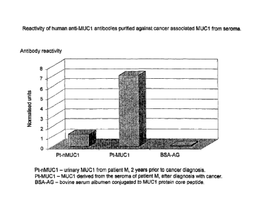
Figure 9 shows the reactivity of purified
autoantibodies from seroma of patient M with cancer
against purified urinary MUC1 from patient M taken two
years prior to cancer diagnosis, MUC1 derived from the
seroma of patient M, after diagnosis with cancer and
bovine serum albumen conjugated to MUCl protein core
peptide;
Figure 10 shows serum autoantibody reactivity
CA 02545930 2006-05-11
WO 2004/044590 PCT/GB2003/004950
_ 27 _
against MUC1 purified from pooled ascites fluid and
against MUC1 purified from individual ascites samples
from cancer patients;
Figure 11 shows serum autoantibody reactivity
against MUC1 purified from pooled pleural effusions
and against MUC1 purified from individuals pleural
effusion samples from cancer patients; and
Figure 12 shows a calibration curve prepared from
MUC1 from a pleural effusion.
Example 1-General protocol for purification of MUC1
antigen
Monoclonal anti-MUC1 antibody B55 (also known as NCRC
11, Xoma Corporation) is conjugated to CNBr-sepharose
beads. Other anti-MUC1 monoclonal antibodies may be
substituted for B55.
Tumour-induced body fluids (e. g. pleural effusion,
ascites, seroma or wound drainage fluid) are diluted
1/10 with phosphate buffered saline (PBS) and filtered
to 0.45 ,um.
Diluted body fluids are incubated with the anti-MUC1
sepharose beads (25 ml diluted fluid to 1 ml packed
volume of beads) overnight at 4 °C with rolling
("batch" method) or re-circulated overnight through a
packed column containing anti-MUC1 sepharose beads
("column" method).
"Batch" method:-
Beads are packed by centrifugation and the supernatant
removed;
Beads re-suspended in 5-10 ml PBS and rolled for 10
mins then packed by centrifugation. and the supernatant
removed; repeat 5 times ~(or until A~BO nm ~0) ;
CA 02545930 2006-05-11
WO 2004/044590 PCT/GB2003/004950
- 28 -
Beads re-suspended in 5 ml 100 mM DEA pH 11, and
rolled at room temperature for 10 mins;
Beads packed by centrifugation and the supernatant
removed, pH adjusted to 7 by the addition of pH 7 Tris
buffer, dialysed against PBS for 24 hours minimum (100
DEA fraction) ;
l0 Beads re-suspended in 5 ml PBS and rolled for 10 mins
then packed by centrifugation and the supernatant
removed, pH adjusted to 7 by the addition of pH 7 Tris
buffer, dialysed against PBS for 24 hours minimum
(post-DEA fraction);
MUC1 content of each fraction confirmed by ELISA
using, for instance, the monoclonal anti-MUC1 antibody
C595 (available from Cancer Research Campaign
Laboratories, UK) (see example 5 for details) or B55,
prior to pooling of the two fractions and storage at -
20°C.
"Column" method:-
Column washed with 5 column volumes of PBS, or until
eluate reads ~0 at A28o nmi
1 column volume of 1trb mM DEA pHl1 applied, followed
by 5 column volumes of PBS;
Eluate fractions (2m1) collected from the time of DEA
application through the application of PBS;
Fractions dialysed overnight against PBS;
Fractions assayed for MUC1 content by ELISA using, for
instance, the monoclonal anti-MUC1 antibody C595 or
B55, prior to pooling MUC1 positive fractions and
storage at -20°C.
CA 02545930 2006-05-11
WO 2004/044590 PCT/GB2003/004950
- 29 -
In order to remove contaminating immunoglobulins, MUC1
pooled fractions are incubated with dithiothreitol
(DTT) to 50 mM for 30 rains, then iodoacetamide (to 75
mM) before being subjected to gel filtration on an
5300 column.
Resulting fractions (5m1) are assayed for MUC1 and
human immunoglobulin (Ig) content by ELISA.
MUC1 containing fractions (uncontaminated with human
Ig) are pooled and stored at -20°C.
Example 2a-General protocol for purification of MUC16
antigen (previously known as CA125)
One volume (e. g. 50 ml) of saturated ammonium sulphate
was added to one volume (e. g. 50 ml) of tumour-induced
body fluid (e.g. pleural effusion, ascites, seroma or
wound drainage fluid) and incubated overnight at 4°C.
The resultant precipitate is collected by
centrifugation (3500 rpm for 30 min in a standard
benchtop centrifuge) and resuspended in ~ volume PBS.
This resuspension is subjected to gel filtration
chromatography through an 5300 column (2.5 x. 100 cm)
using PBS as the eluting buffer.
Fractions (5 or 10 ml) are collected and assayed by
ELISA for MUC16, using for instance anti-CA125 from
ICN or the anti-MUC16 antibody VK8 (Memorial Sloane
Kettering, New York), prior to pooling MUC16 positive
fractions and storage at -20°C.
In order to remove contaminating immunoglobulins,
MUC16 pools are incubated with NaSCN (to 1.5M) for 10
minx, DTT (to 50mM) for 30 rains, then iodoacetamide
(to 75mM) for 30 rains before being subjected toNgel
filtration on, for instance, an 5300 or a SuperdexT"" 75
column.
CA 02545930 2006-05-11
WO 2004/044590 PCT/GB2003/004950
- 30 -
Resulting fractions (5m1) are assayed for MUC16 and
human immunoglobulin (Ig) content by ELISA.
MUC16 containing fractions (uncontaminated with human
Ig) are pooled and stored at -20°C.
Example 2b-Post Ig disruption gel filtration
chromatography
For a sample prepared in the manner described above,
fractions from a post-Ig disruption gel filtration
were assayed for MUC16 using anti-MUC16 antibody VK8
and for human Ig using an anti-human Ig. The results
are shown in Figure 1. As is clearly demonstrated,
two substantially immunoglobulin free MUC16 peaks are
eluted.
Example 3-purification of c-myc antigen
Methodology as per purification of MUC1 (Example 1),
except that:
Monoclonal anti-c-myc antibody 9E10 (ATCC) is used (or
equivalent anti-c-myc antibody).
Gel filtration is performed on a SuperdexT"" 75 column.
Electrophoresis and Western blotting
Purity of MUC1, MUC16 and c-myc fractions are
assessed by denaturing polyacrylamide gel
electrophoresis and Western blotting, performed
according to standard protocols using BioRadT"' Mini
Protean IIIT"" system and BioRadT"' DryBlotT"" system.
Protein patterns were revealed on gels for c-myc by
silver staining (Figure 2). Western blots of c-myc
were immuno-probed using monoclonal antibodies 9E10
(Figure 3). In each case, c-myc as well as
immunoglobulin heavy and light chains are identified.
CA 02545930 2006-05-11
WO 2004/044590 PCT/GB2003/004950
- 31 -
Example 4-standard auto-antibody assay
Tumour antigen (e. g. MUC1, MUC16 or c-myc prepared
according to Examples 1-3) diluted appropriately in
PBS is plated out at 50 ~.1 per well in a standard 96
well microtiter plate and left to air dry overnight;
to Plate washed once with PBS/TweenT"" to remove residual
salt crystals;
Plate blocked for 60 mins with 0.1% casein or 1% BSA
in PBS;
Plate washed x3 with PBS/TweenT"';
Serum (diluted 1/100 in PBS/0.1% casein) plated out in
triplicate (50 ~,1 per well), also monoclonal antibody
controls;
Incubate for 60 mins at room temperature with shaking;
30
Wash plate x4 with PBS/TweenT"';
Add horseradish peroxidase (HRP)-conjugated anti-Ig
antibody (Dako) to each well (50 ~.1 per well) at
1/8000 dilution for anti-human and 1/1000 for anti-
mouse;
Incubate for 60 mins at room temperature with shaking;
Wash plate x4 with PBS/TweenT"";
Add 50.1 TMB (tetramethylbenzadine) per well and read
kinetically over a 10 min period at.~A6so nm.
Experimental Data
Using the method as described in Example 4, cancer-
associated autoantibodies to MUC1 and MUC16 were
CA 02545930 2006-05-11
WO 2004/044590 PCT/GB2003/004950
- 32 -
measured in a variety of sera using MUCl and MUC16
isolated from the various sources as described herein.
Results generated are shown in Figures 4 to 6.
Figure 4 shows a comparison of patient serum auto-
antibody reactivity against MUC1 isolated from various
body fluids: urine (from "normal" individuals),
pleural effusion from a cancer patient and serum from
advanced breast cancer patients (ABC serum). T'he
patient serum tested was from either individuals~with
no evidence of breast cancer themselves but with a
family history of breast cancer (i.e. one or more
relatives who had breast cancer at a young age) or
individuals with primary breast cancer.
Standard auto-antibody ELISAs were performed as
described above, utilising MUC2 isolated from urine
(normal), pleural. effusion or ABC serum as antigen.
Data was normalised to an internal control reaction
using the DF3 anti-MUC1 monoclonal antibody (as
opposed to a serum sample) against each of the MUC1
antigens.
As can be seen from the Figure, MUC1 derived from
normal urine (nMUCl) was consistently lower in its
reactivity than MUC1 derived from either pleural
effusion (PE) or ABC serum. Furthermore, MUCl derived
from PE was of similar reactivity to cancer-associated
MUC1 autoantibodies as MUCl isolated from the serum of
patients with ABC and therefore of equal diagnostic
value.
Figure 5 shows the results of an identical exercise to
Figure 4 except that all serum samples tested were for
normal individuals (no breast cancer or family history
of breast cancer). As can be seen, there is no
significant difference in the reactivity of the serum
to the three different antigens.
Figure 6 shows reactivity of MUC16 cancer-associated
CA 02545930 2006-05-11
WO 2004/044590 PCT/GB2003/004950
- 33 -
autoantibodies from serum of patients with ovarian
masses (pre-operative) against MUC16 (CA125) isolated
from the serum of normal individuals and from ascites
fluid in a patient with breast cancer. Antigens were
prepared as in Example 2 and autoantibodies detected
using ELISA assay as described in Example 4.
As can be seen, greatly enhanced reactivity of the
cancer-associated MUC16 autoantibodies is seen with
the MUC16 antigen from ascites fluid as compared to
the "normal" MUC16. This experimental result
therefore confirms the usefulness of ascites fluid as
an antigen source for detection of cancer-associated
autoantibodies.
Example 5-Measurement of cancer-associated MUC1 levels
in ascites fluid, pleural effusion, seroma and wound
drainage fluid
MUCl levels found in the serum of a patient with
cancer were compared with the levels found in ascites
fluid, pleural effusion, wound drainage fluid or
seroma, in each case in the same patient from whom the
serum sample was taken. MUC1 in the samples was
quantified according to the following protocol:
Capture MUC1 ELISA Protocol
Aliquot 50.1 per well antibody solution into
triplicate wells of a microtitre plate (usually l~.g
ml-z C595(IgG) and appropriate negative control) and
incubate at RT with shaking for 1hr for the protein to
adsorb to the plate.
Wash the plate x4 with PBS/Tween using 2501 per well.
Block the plate using 1% BSA 100.1 per well and
incubate at RT with shaking for lhr.
Wash the plate x4 with PBS/Tween using 250,u1 per well.
CA 02545930 2006-05-11
WO 2004/044590 PCT/GB2003/004950
- 34 -
Apply 50.1 per well of fluid being tested, diluted
1/10 in PBS and incubate at RT with shaking for lhr.
Wash the plate x4 with PBS/Tween using 250.1 per well.
Add 50.1 per well biotinylated C595 (l~,g/ml) and
incubate at RT with shaking for lhr.
l0 Wash the plate x4 with PBS/Tween using 250.1 per well.
Add 50J~1 per well extra-avidin peroxidise at 2/1000
dilution and incubate at RT with shaking for lhr.
Wash the plate x4 with PBS/Tween using 250.1 per well.
Add 50,1 per well TMB substrate and read kinetically
at 650nm for 10 minutes .
2o The results are shown in Figures 7 and 8.
As will be readily apparent from the data serum levels
of the cancer-associated MUCl antigen are
significantly lower than the level found in either
ascites fluid, pleural effusion, seroma or wound
drainage fluid. Accordingly, there are substantial
benefits to be gained in terms of yield in recovering
tumour markex'antigen from those body cavity fluids.
Example 6-Reactivity of human anti-MUC1 antibodies
purified against cancer-associated MUC1 from seroma
Human antibodies from seroma from patient M were
purified by immunoaffinity chromatography against MUC1
derived from seroma fluid from the same cancer patient
M. Purified antibodies were then tested against BSA
conjugated protein core peptide to MUC1 and MUCl
derived from:- patient M's urine taken two years prior
to cancer diagnosis; patient M's seroma taken after
4o cancer diagnosis. The antibody purification from
CA 02545930 2006-05-11
WO 2004/044590 PCT/GB2003/004950
- 35 -
seroma was carried out according to the following
protocol:
Human anti-MUC1 antibody purification
Purification of human anti-MUC1 auto-antibodies was by
affinity chromatography.
Seroma fluid, diluted 10 fold in PBS pH 7.6, was
applied at 0.5m1/min by overnight re-circulation at
4°C, to an affinity matrix in column format,
consisting of CNBr sepharose (Pharmacia) coupled
(following the manufacturers instructions) to Pt-MUC1.
After seroma fluid application, the column was washed
with 15m1 of PBS (ensuring return of AzBOnm reading to
zero) prior to elution of antibody using 10m1 of 3M
NaSCN, at lm/min.
Fractions of lml were collected throughout, desalted
by dialysis against PBS and tested by ELISA for the
presence of antibody.
Positive fractions were pooled, purity of antibody
verified (by PAGE) and antibody concentration
determined.
Assay of the purified antibodies against the three
MUCl. antigens identified above was carried out
according to the following protocol:
MUC1 ELISA Protocol
Aliquot 50.1 per well of the MUC1 antigen solution
into triplicate wells of a microtitre plate and dry
down at RT overnight.
Wash the plate x2 with PBS/Tween using 250.1 per well.
Block the plate with 1% BSA using 100,1 per well and
CA 02545930 2006-05-11
WO 2004/044590 PCT/GB2003/004950
- 36 -
incubate at RT with shaking for lhr.
Wash the plate x2 with PBS/Tween using 250,u1 per well.
Add 501 per well purified antibody solution at l~Cg/ml
and incubate at RT with shaking for lhr.
Wash the plate x4 with PBS/Tween using 250,1 per well.
l0 Add 50.1 per well a-human Ig HRP (DAKO), freshly
diluted as per manufacturers instructions, and
incubate at RT with. shaking for lhr.
Wash the plate x4 with PBS/Tween using 250.1 per well.
Add 50,1 per well TMB substrate and read kinetically
at 650nm for 10 minutes .
The results are shown in Figure 9.
Reactivity of the antibodies against MUC1 peptide was
negligible. Reactivity of antibodies against normal
MUC1 was considerably lower than that seen towards
patient M's seroma derived MUC1. It can be inferred
from this result that normal MUC1 molecule is
substantially different with regard to its immune
recognition, to that found in seroma fluid from an
individual with cancer.
Example 7-Serum reactivity against MUC1 purified from
pooled ascitic fluid and pleural effusions
MUC1 was purified from pooled ascitic fluid and from
pooled pleural effusion from patients with advanced
breast cancer using the protocol described in Example
1 and its reactivity against serum from patients with
primary breast cancer measured as described in Example
4. The antigen from the pooled fluids was compared in
each case with antigen isolated from 3 individual
samples of ascitic fluid or pleural effusion
CA 02545930 2006-05-11
WO 2004/044590 PCT/GB2003/004950
- 37 -
respectively from patients with ABC. The results are
shown in Figures 10 and 11.
In the case of both ascitic'fluid and pleural effusion
the reactivity of the MUC1 from pooled fluid is as
good as that isolated from individual samples.
Furthermore, while there is great scope for
variability of reactivity using samples from
individuals, pooled samples provide greater
IO consistency of product so that one would not expect
the reactivity to significantly vary between batches
from pooled samples.
Example 8-Calibration Curve using MUC1
zs
Serial dilutions of MUC1 which had been isolated from
pleural effusion were prepared. Their MUC1
concentrations were measured by the method as shown in
example 4 except that no human sera were used.
20 Detection was by mouse B55 antibody followed by Dako
anti-mouse HRP using an end-point rather than a
kinetic reading.
The results are shown in Figure 12 and confirm the
25 utility of the tumour marker proteins prepared in
accordance with the invention as a calibration
material.
30 Sources of antibodies to tumour marker proteins
The following lists sources of antibodies which
may be used in the purification of tumour marker
proteins by affinity chromatography. Affinity
35 chromatography may be performed following the general
methodology described in Example 1 (in relation to
MUC1), with appropriate modification. It will be
appreciated that other antibodies specific for the
relevant marker protein may also be used.
CA 02545930 2006-05-11
WO 2004/044590 PCT/GB2003/004950
- 38 -
Carcinoembryonic antigen (CEA):
1116NS-3d, ATCC number CRL-8019, B lymphocyte
hybridoma producing monoclonal antibody against CEA;
T84.66A3.1A.1F2, ATCC number HB-8747, B lymphocyte
hybridoma producing monoclonal antibody against CEA.
P53
Rabbit anti-human p53 polyclonal, commercially
available from from Serotec Ltd, Kidlington, Oxford
OX5 1JE, United Kingdom.
Monoclonal anti-p53, clone BP53-12, commercially
available from Sigma.
CA19-9:
Mouse anti-human CA19-9 monoclonal, type clone 1116-
NS-19-9, IgGl, commercially available from from
Serotec Ltd, Kidlington, Oxford OX5 1JE, United
Kingdom.
H-ras p21:
Rabbit polyclonal IgG, commercially available from
Santa Cruz Biotechnology, Inc., Santa Cruz,
California, USA.
BRCA1:
Rabbit polyclonal IgG, commercially available from
Santa Cruz Biotechnology, Inc., Santa Cruz,
California, USA."
BRCA2:
Goat polyclonal IgG, commercially available from Santa
Cruz Biotechnology, Inc., Santa Cruz, California, USA.
APC:
Rabbit polyclonal TgG, commercially available from
Santa Cruz Biotechnology, Inc., Santa Cruz,
California, USA.
PSA
Mouse monoclonal IgG, commercially available from
CA 02545930 2006-05-11
WO 2004/044590 PCT/GB2003/004950
- 39 -
Santa Cruz Biotechnology, Inc., Santa Cruz,
California, USA.