Note: Descriptions are shown in the official language in which they were submitted.
CA 02546054 2012701-19
WO 2005/059106 PCT/US2004/041777
INTERFERON ALPHA ANTIBODIES AND THEIR USES
Background of the Invention
Type I interferons (TN) OFN-a, IFN-f3, ]FN-co, IFN-T) are a family of
structurally related cytokines having antiviral, antitumor and
immunomodulatory effects
(Hardy et al. (2001) Blood 97:473; Cutrone and Langer (2001) J. Biol. Chem.
276:17140).
The human IFNa locus includes two subfamilies. The first subfamily consists of
at least 14
non allelic genes and 4 pseudogenes having at least 75% homology. The second
subfamily,
all or omega (c)), contains 5 pseudogenes and 1 functional gene which exhibits
70%
homology with the IFNa genes. The subtypes of IFNa have different specific
activities but
they possess the same biological spectrum (Streuli et al. (1981) Proc. Natl.
Acad. Sci. USA
78:2848) and have the same cellular receptor (Agnet M. et al. (1983) in
"Interferon 5" Ed. I.
Grosser p. 1-22, Academic Press, London).
All human type I interferons bind to a cell surface receptor (IFN alpha
receptor, PFNAR) consisting of two transmembrane proteins, IFNAR-1 and IFNAR-2
(Uze
et. al. (1990) Cell 60:225; Novick etal. (1994) Cell 77:391; Pestka et al.
(1987) Annu Rev.
Biochem. 56:727; Mogen.sen et al. (1999)J. Interferon Cytokine Res. 19:1069).
IFNAR-1 is
essential for high affinity binding and differential specificity of the IFNAR
complex (Cutrone
(2001) supra). While functional differences for each of the type I MN subtypes
have not
been identified it is thought that each may exhibit different interactions
with the IFNAR
receptor components leading to potentially diverse signaling outcomes (Cook et
al. (1996)J.
Biol. Chem. 271:13448). In particular, studies utilizing mutant forms of
IFNAR1 and
IFNAR2 suggested that alpha and beta interferons signal differently through
the receptor by
interacting differentially with respective chains (Lewerenz et al. (1998) J.
Mol. Biol.
282:585).
Early functional studies of type I1FNs focused on innate defense against viral
infections (Haller et al. (1981)J Exp. Med. 154:199; Lindenmarm et al. (1981)
Methods
CA 02546054 2006-05-12
WO 2005/059106
PCT/US2004/041777
2
Enzymol. 78:181). More recent studies, however, implicate type I IFNs as
potent
immunoregulatory cytokines in the adaptive immune response. Specifically, type
I IFNs
have been shown to facilitate differentiation of naïve T cells along the Thl
pathway
(Brinkmann et al. (1993)1 Exp. Med. 178:1655), to enhance antibody production
(Finkelman et al. (1991)1 Exp. Med. 174:1179) and to support the functional
activity and
survival of memory T cells (Santini, et al. (2000) J. Exp. Med. 191:1777;
Tough et al. (1996)
Science 272:1947).
Recent work by a number of groups suggests that IFN-a may enhance the
maturation or activation of dendritic cells (DCs) (Santini, et al. (2000) J.
Exp. Med.
191:1777; Luft et al. (1998) J. Immunol. 161:1947; Luft et al. (2002) Int.
Immunol. 14:367;
Radvanyi et al. (1999) Scand. I Immunol. 50:499; Paquette et al. (1998)1
Leukoc. Biol.
64:358). Furthermore, increased expression of type I interferons has been
described in
numerous autoimmune diseases (Foulis et al. (1987) Lancet 2:1423; Hooks et al.
(1982)
Arthritis Rheum 25:396; Hertzog et al. (1988) Clin. Immunol. Immunopathol.
48:192;
Hopkins and Meager (1988) Clin. Exp. Immunol. 73:88; Arvin and Miller (1984)
Arthritis
Rheum. 27:582). The most studied examples of this are insulin-dependent
diabetes mellitus
(FDDM) (Foulis (1987) supra), systemic lupus erythematosus (SLE) (Hooks (1982)
supra;
Blanco et al. (2001) Science 294:1540; Ytterberg and Schnitzer (1982)
Arthritis Rheum.
25:401; Batteux et al. (1999) Eur. Cytokine Netw. _:509), and autoimmune
thyroiditis
(Prununel and Laurberg (2003) Thyroid 13:547; Mazziotti et al. (2002)1
Endocrinol. Invest.
25:624; You et al. (1999) Chin. Med. J. 112:61; Koh et al. (1997) Thyroid
7:891),which are
all associated with elevated levels of IFN a, and rheumatoid arthritis (RA)
(Hertzog (1988),
Hopkins and Meager (1988), Arvin and Miller (1984), supra) in which IFN-P may
play a
more significant role.
Moreover, administration of interferon a has been reported to exacerbate
underlying disease in patients with psoriasis, autoimmune thyroiditis and
multiple sclerosis
and to induce an SLE like syndrome in patients without a previous history of
autoimmune
disease. Interferon a has also been shown to induce glomerulonephritis in
normal mice and to
accelerate the onset of the spontaneous autoimmune disease of NZB/W mice.
Further, IFN-a
therapy has been shown in some cases to lead to undesired side effects,
including fever and
neurological disorders. Hence, there are pathological situations in which
inhibition of TN-
CA 02546054 2006-05-12
WO 2005/059106
PCT/US2004/041777
3
a activity may be beneficial to the patient and a need exists for agents
effective in inhibiting
IFN-a activity.
Summary of the Invention
The present invention provides isolated monoclonal antibodies that bind to
IFN alpha and inhibit the biological activity of multiple IFN alpha subtypes,
but not
substantially inhibit the biological activity of IFN alpha subtype 21, or of
IFN beta or IFN
omega. In preferred embodiments, the antibodies of the invention are capable
of inhibiting
surface expression of cell markers induced by IFN alpha, inhibiting IP-10
expression induced
by IFN alpha and/or inhibiting dendritic cell development mediated by plasma
from patients
with systemic lupus erythematosus (SLE). These antibodies can be used for
therapeutic,
including prophylactic, purposes, for example in situations where the
production or
expression of interferon alpha is associated with pathological symptoms. Such
antibodies can
also be used for the diagnosis of various diseases or for the study of the
evolution of such
diseases.
In one embodiment, the present invention includes an antibody or antibody
fragment that binds to IFN alpha, preferably human IFN alpha (e.g., human IFN
alpha 2a,
human IFN alpha 2b), and inhibits the biological activity of multiple lFN
alpha subtypes, but
does not substantially inhibit the biological activity of IFN alpha subtype
21, or IFN beta or
IFN omega. In addition, in various embodiments, the antibodies of the
invention are capable of
inhibiting surface expression of cell markers induced by IFN alpha, inhibiting
IP-10 expression
induced by IFN alpha and/or inhibiting dendritic cell development mediated by
plasma from
patients with systemic lupus erythematosus (SLE). The antibody or antibody
fragment
preferably is a human antibody or antibody fragment, or alternatively can be a
murine, chimeric
or humanized antibody. In certain embodiments, an antibody of the invention
functions by a
non-competitive mechanism of action. For example, in preferred embodiments,
the antibody:
(i) does not inhibit the binding of an IFN alpha, such as IFN alpha 2a, to
cells expressing
interferon alpha receptor (IFNAR) and (ii) binds to cells expressing IFNAR in
the presence of
an IFN alpha, such as IFN alpha 2a.
In one aspect, the invention pertains to isolated antibodies, or antigen
binding
portions thereof, wherein the antibodies:
CA 02546054 2006-05-12
WO 2005/059106
PCT/US2004/041777
4
(a) comprise a heavy chain variable region of a human VII 1-18 or 4-61
gene;
(b) comprise a light chain variable region of a human A27 gene; and
(c) inhibit the biological activity of interferon alpha (e.g., inhibits the
biological activity of at least one IFN alpha subtype).
In another aspect, the invention pertains to isolated monoclonal antibodies,
or
antigen binding portions thereof, comprising a heavy chain variable region
comprising
CDR1, CDR2, and CDR3 sequences and a light chain variable region comprising
CDR1,
CDR2, and CDR3 sequences, wherein:
(a) the heavy chain variable region CDR3 sequence comprises the amino
acid sequence of SEQ ID NO: 7,8, or 9, or conservative modifications thereof;
(b) the light chain variable region CDR3 sequence comprises the amino
acid sequence of SEQ ID NO: 16, 17, or 18, or conservative modifications
thereof;
(c) the antibody inhibits the biological activity of multiple IFN alpha
subtypes but does not substantially inhibit the biological activity of IFN
alpha 21; and
(d) the antibody exhibits at least one of the following properties:
(i) the antibody does not substantially inhibit the biological
activity of IFN beta or IFN omega;
(ii) the antibody inhibits IFN-induced surface expression of CD38 or
MHC Class I on peripheral blood mononuclear cells;
(iii) the antibody inhibits IFN-induced expression of IP-10 by
peripheral blood mononuclear cells;
(iv) the antibody inhibits dendritic cell development mediated by
systemic lupus erythematosus (SLE) plasma.
In such antibodies, the heavy chain variable region CDR2 sequence can
comprise the amino acid sequence of SEQ ID NO: 4, 5, or 6, or conservative
modifications
thereof; and the light chain variable region CDR2 sequence can comprise the
amino acid
sequence of SEQ ID NO: 13, 14, or 15, or conservative modifications thereof.
Furthermore,
in such antibodies, the heavy chain variable region CDR1 sequence can comprise
the amino
acid sequence of SEQ ID NO: 1, 2, or 3, or conservative modifications thereof;
and the light
CA 02546054 2006-05-12
WO 2005/059106 PCT/US2004/041777
chain variable region CDR1 sequence can comprise the amino acid sequence of
SEQ ID NO:
10, 11, or 12, or conservative modifications thereof.
In another aspect, the invention pertains to isolated monoclonal antibodies,
or
antigen binding portions thereof, comprising a heavy chain variable region and
a light chain
5 variable region, wherein:
(a) the heavy chain variable region comprises an amino acid sequence that
is at least 80% homologous to SEQ ID NO: 19, 20, or 21;
(b) the light chain variable region comprises an amino acid sequence that
is at least 80% homologous to SEQ ID NO: 22, 23, or 24;
(c) the antibody inhibits the biological activity of multiple IFN alpha
subtypes but does not substantially inhibit the biological activity of IFN
alpha 21; and
(d) the antibody exhibits at least one of the following properties:
(i) the antibody does not substantially inhibit the
biological
activity of IFN beta or IFN omega;
(ii) the antibody inhibits IFN-induced surface expression of CD38 or
MHC Class I on peripheral blood mononuclear cells;
(iii) the antibody inhibits IFN-induced expression of IP-10 by
peripheral blood mononuclear cells;
(iv) the antibody inhibits dendritic cell development mediated by
systemic lupus erythematosus (SLE) plasma.
In another aspect, the invention pertains to isolated monoclonal antibodies,
or
antigen binding portions thereof, comprising a heavy chain variable region and
a light chain
variable region, wherein:
(a) the heavy chain variable region comprises an amino acid sequence
(b) the light chain variable region comprises an amino acid comprising the
amino acid sequence of SEQ ID NO: 22, 23, or 24;
wherein the antibody inhibits the biological activity of interferon alpha
(e.g.,
inhibits the biological activity of at least one IFN alpha subtype).
CA 02546054 2006-05-12
WO 2005/059106
PCT/US2004/041777
6
In yet another aspect, the invention pertains to mutated variants of SEQ ID
NO: 19 having increased stability. Preferred embodiments include an isolated
monoclonal
antibody, or antigen binding portion thereof comprising:
(a) a heavy chain variable region comprising an amino acid sequence selected
from the group consisting of SEQ ID NOs: 34, 35, 36 and 37; and
(b) a light chain variable region comprising the amino acid sequence of SEQ ID
NO: 22;
wherein the antibody inhibits the biological activity of at least one
interferon alpha
subtype.
In yet another aspect, the invention pertains to an isolated monoclonal
antibody, or antigen binding portion thereof comprising:
(a) a heavy chain variable region CDR1 comprising an amino acid sequence
selected from the group consisting of SEQ ID NOs: 1, 2 and 3;
(b) a heavy chain variable region CDR2 comprising an amino acid sequence
selected from the group consisting of SEQ ID NOs: 4, 5 and 6;
(c) a heavy chain variable region CDR3 comprising an amino acid sequence
selected from the group consisting of SEQ ID NOs: 7, 8 and 9;
(d) a light chain variable region CDR1 comprising an amino acid sequence
selected from the group consisting of SEQ ID NOs: 10, 11 and 12;
(e) a light chain variable region CDR2 comprising an amino acid sequence
selected from the group consisting of SEQ ID NOs: 13, 14 and 15; and
(f) a light chain variable region CDR3 comprising an amino acid sequence
selected from the group consisting of SEQ ID NOs: 16, 17 and 18;
wherein the antibody inhibits the biological activity of interferon alpha
(e.g.,
inhibits the biological activity of at least one MN alpha subtype).
In yet another aspect, the invention pertains to an isolated monoclonal
antibody, or antigen binding portion thereof, that competes for binding to IFN
alpha 2a or
IFN alpha 2b with any of the above mentioned antibodies.
In yet another aspect, the invention pertains to an isolated human antibody,
or
antigen-binding portion thereof, that inhibits the biological activity of
multiple interferon
(IFN) alpha subtypes, wherein the antibody does not inhibit binding of IFN
alpha to
interferon alpha receptor (IFNAR)-expressing cells and wherein the antibody
associates with
IFNAR-expressing cells in the presence, but not the absence, of IFN alpha.
CA 02546054 2012-01-19
= WO
2005/059106 PCT/US2004/041777
7
The invention also encompasses nucleic acid molecules that encode the
antibodies or antigen-binding portions thereof in any of the above mentioned
antibodies.
The antibodies of the invention can be of any isotype. Preferred antibodies
are
of the IgG1 or IgG4 isotype. The antibodies of the invention can be full-
length antibodies
comprising variable and constant regions, or they can be antigen-binding
fragments thereof,
such as a single chain antibody or a Fab fragment.
The invention also encompasses immunoconjugates of the antibodies of the
invention, in which the antibody is linked to a therapeutic agent, such as a
cytotoxin or a
radioactive isotope. The invention also encompasses bispecific molecules
comprising an
antibody of the invention, in which the antibody is linked to a second
functional moiety
having a different binding specificity than the antibody.
Pharmaceutical compositions comprising an antibody, or antigen binding
portion thereof, or immunoconjugate or bispecific molecule thereof, are also
provided. Such
pharmaceutical compositions comprise the active agent and a pharmaceutically
acceptable
carrier.
In another aspect, the present invention includes a method of inhibiting the
biological activity of interferon alpha, either in vivo or in vitro,
comprising contacting
interferon alpha with an anti-]FN alpha antibody of the invention, such that
the biological
activity of interferon alpha is inhibited.
In another aspect, the present invention includes a method of treating an
interferon alpha-mediated disease or disorder in a subject, comprising
administering to the
subject an anti-IFN alpha antibody of the invention, such that the interferon-
alpha mediated
disease in the subject is treated. Examples of diseases that can be treated
include
autoimmime diseases (e.g., systemic lupus erythematosus, multiple sclerosis,
insulin
dependent diabetes mellitus, inflammatory bowel disease, psoriasis,
autoimrnune thyroiditis,
rheumatoid arthritis and glomerulonephritis), transplant rejection and graft
versus host
disease.
Other features and advantages of the instant invention will be apparent from
the following detailed description and examples, which should not be construed
as limiting.
CA 02546054 2006-05-12
WO 2005/059106
PCT/US2004/041777
8
Brief Description of the Drawings
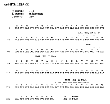
Figure lA shows the nucleotide sequence (SEQ ID NO: 25) and amino acid
sequence (SEQ ID NO: 19) of the heavy chain variable region of the 13H5 human
monoclonal antibody. The CDR1 (SEQ ID NO: 1), CDR2 (SEQ ID NO: 4) and CDR3
(SEQ
ID NO: 7) regions are delineated and the V, D and J germline derivations are
indicated.
Figure 1B shows the nucleotide sequence (SEQ ID NO: 28) and amino acid
sequence (SEQ ID NO: 22) of the light chain variable region of the 13115 human
monoclonal
antibody. The CDR1 (SEQ ID NO: 10), CDR2 (SEQ ID NO: 13) and CDR3 (SEQ ID NO:
16) regions are delineated and the V and J germline derivations are indicated.
Figure 2A shows the nucleotide sequence (SEQ ID NO: 26) and amino acid
sequence (SEQ ID NO: 20) of the heavy chain variable region of the 13117 human
monoclonal antibody. The CDR1 (SEQ ID NO: 2), CDR2 (SEQ ID NO: 5) and CDR3
(SEQ
ID NO: 8) regions are delineated and the V, D and J germline derivations are
indicated.
Figure 2B shows the nucleotide sequence (SEQ ID NO: 29) and amino acid
sequence (SEQ ID NO: 23) of the light chain variable region of the 13117 human
monoclonal
antibody. The CDR1 (SEQ ID NO: 11), CDR2 (SEQ ID NO: 14) and CDR3 (SEQ ID NO:
17) regions are delineated and the V and J germline derivations are indicated.
Figure 3A shows the nucleotide sequence (SEQ ID NO: 27) and amino acid
sequence (SEQ ID NO: 21) of the heavy chain variable region of the 7H9 human
monoclonal
antibody. The CDR1 (SEQ ID NO: 3), CDR2 (SEQ ID NO: 6) and CDR3 (SEQ ID NO: 9)
regions are delineated and the V, D and J germline derivations are indicated.
Figure 3B shows the nucleotide sequence (SEQ ID NO: 30) and amino acid
sequence (SEQ ID NO: 24) of the light chain variable region of the 7H9 human
monoclonal
CA 02546054 2009-07-21
WO 2005/059106
PCT/US2004/041777
9
antibody. The CDR1 (SEQ ID NO: 12), CDR2 (SEQ ID NO: 15) and CDR3 (SEQ ID NO:
18) regions are delineated and the V and J gennline derivations are indicated.
Figure 4 shows the alignment of the amino acid sequence of the heavy
chain variable region of 13H5 (SEQ ID NO:19) and 7H9 (SEQ ID NO:21) with the
human germline VH 1-18 amino acid sequence (SEQ ID NO:31).
Figure 5 shows the alignment of the amino acid sequence of the heavy
chain variable region of 13H7 (SEQ ID NO:20) with the human germline VH 4-61
amino
acid sequence (SEQ ID NO:32).
Figure 6 shows the alignment of the amino acid sequence of the light
chain variable region of 13115 (SEQ ID NO:22), 13117 (SEQ ID NO:23) and 7119
(SEQ
ID NO:24) with the human germline VK A27 amino acid sequence (SEQ ID NO:33).
Figure 7 is a graph showing competition of binding of125I-TFNa 2a to IFNAR-
expressing Daudi cells by unlabeled IFNa 2a (0) versus enhancement of 125I-
IFNcx 2a
binding by mAb 13H5 (V). An isotype control antibody had no effect on binding
(=).
Figure 8 is a graph showing binding of 1251-13H5 to Daudi cells in the
presence of IFNot 2a (0) but not in the absence of IFNa 2a (A). Specific IFNa-
dependent
binding of 13H5 is represented by circles (9).
Figure 9 is a graph showing the results of ADCC assays of Raji cell lysis by
fresh human mononuclear cells in the presence of 13H5 (B), 13H5 + IFNa (A), an
isotype
control antibody + IFNa (V), or a positive control antibody (9). Lysis was
only seen with
the positive control.
Detailed Description of the Invention
CA 02546054 2006-05-12
WO 2005/059106
PCT/US2004/041777
The present invention relates to isolated monoclonal antibodies that bind to
IFN alpha and that are capable of inhibiting the biological activity of
multiple IFN alpha
subtypes, but not the biological activity of IFN alpha subtype 21, or IFN beta
or IFN omega.
5 The antibodies of the invention are capable of inhibiting surface
expression of cell markers
induced by IFN alpha, inhibiting IP-10 expression induced by 1FN alpha and
inhibiting
dendritic cell development mediated by plasma from patients with systemic
lupus
erythematosus (SLE). The invention provides isolated antibodies, methods of
making such
antibodies, immunoconjugates and bispecific molecules comprising such
antibodies and
10 pharmaceutical compositions containing the antibodies, immunconjugates
or bispecific
molecules of the invention. The invention also relates to methods of using the
antibodies to
inhibit TEN alpha activity, for example in the treatment of autoimmune
disorders, or for
inhibiting or preventing transplant rejection or in the treatment of graft
versus host disease.
In order that the present invention may be more readily understood, certain
terms are first defined. Additional definitions are set forth throughout the
detailed
description.
The terms "interferon alpha" and "IFN alpha" are used interchangeably and
intended to refer to IFN alpha proteins encoded by a functional gene of the
interferon alpha
gene locus with 75% or greater sequence identity to IFN alpha 1 (Genbank
number
NP 076918 or protein encoded by Genbank number NM 024013). Examples of IFN
alpha
subtypes include IFN alpha 1, alpha 2a, alpha 2b, alpha 4, alpha 5, alpha 6,
alpha 7, alpha 8,
alpha 10, alpha 13, alpha 14, alpha 16, alpha 17 and alpha 21. The term
"interferon alpha" is
intended to encompass recombinant forms of the various IFN alpha subtypes, as
well as
naturally occurring preparations that comprise IFN alpha proteins, such as
leukocyte IFN and
lymphoblastoid IFN. The term IFN alpha is not intended to encompass IFN omega
alone,
although a composition that comprises both IFN alpha and IFN omega is
encompassed by the
term IFN alpha.
The term "MN alpha receptor" as used herein is intended to refer to members
of the IFN alpha receptor family of molecules that are receptors for the
ligand IFN alpha.
Examples of ITN alpha receptors are IFN alpha receptor 1 and TN alpha receptor
2.
The term "immune response" refers to the action of, for example,
lymphocytes, antigen presenting cells, phagocytic cells, granulocytes, and
soluble
CA 02546054 2006-05-12
WO 2005/059106
PCT/US2004/041777
11
macromolecules produced by the above cells or the liver (including antibodies,
cytokines, and
complement) that results in selective damage to, destruction of, or
elimination from the
human body of invading pathogens, cells or tissues infected with pathogens,
cancerous cells,
or, in cases of autoimmunity or pathological inflammation, normal human cells
or tissues.
A "signal transduction pathway" refers to the biochemical relationship
between a variety of signal transduction molecules that play a role in the
transmission of a
signal from one portion of a cell to another portion of a cell. As used
herein, the phrase "cell
surface receptor" includes, for example, molecules and complexes of molecules
capable of
receiving a signal and the transmission of such a signal across the plasma
membrane of a cell.
An example of a "cell surface receptor" of the present invention is the IFN
alpha receptor 1 or
IFN alpha receptor 2.
The term "antibody" as referred to herein includes whole antibodies and any
antigen binding fragment (i.e., "antigen-binding portion") or single chains
thereof. An
"antibody" refers to a glycoprotein comprising at least two heavy (H) chains
and two light (L)
chains inter-connected by disulfide bonds, or an antigen binding portion
thereof. Each heavy
chain is comprised of a heavy chain variable region (abbreviated herein as VH)
and a heavy
chain constant region. The heavy chain constant region is comprised of three
domains, CH1,
CH2 and CH3. Each light chain is comprised of a light chain variable region
(abbreviated
herein as VL) and a light chain constant region. The light chain constant
region is comprised
of one domain, CL. The VH and VL regions can be further subdivided into
regions of
hypervariability, termed complementarity determining regions (CDR),
interspersed with
regions that are more conserved, termed framework regions (FR). Each VH and VL
is
composed of three CDRs and four FRs, arranged from amino-teirninus to carboxy-
terminus
in the following order: FR1, CDR1, FR2, CDR2, FR3, CDR3, FR4. The variable
regions of
the heavy and light chains contain a binding domain that interacts with an
antigen. The
constant regions of the antibodies may mediate the binding of the
immunoglobulin to host
tissues or factors, including various cells of the immune system (e.g.,
effector cells) and the
first component (Clq) of the classical complement system.
The term "antigen-binding portion" of an antibody (or simply "antibody
portion"), as used herein, refers to one or more fragments of an antibody that
retain the ability
to specifically bind to an antigen (e.g., IFN alpha). It has been shown that
the antigen-
binding function of an antibody can be performed by fragments of a full-length
antibody.
Examples of binding fragments encompassed within the term "antigen-binding
portion" of an
CA 02546054 2006-05-12
WO 2005/059106
PCT/US2004/041777
12
antibody include (i) a Fab fragment, a monovalent fragment consisting of the
VL, VH, CL and
CHI domains; (ii) a F(abt)2 fragment, a bivalent fragment comprising two Fab
fragments
linked by a disulfide bridge at the hinge region; (iii) a Ed fragment
consisting of the VH and
CHI domains; (iv) a Fv fragment consisting of the VL and VH domains of a
single arm of an
antibody, (v) a dAb fragment (Ward et al., (1989) Nature 341:544-546), which
consists of a
VH domain; and (vi) an isolated complementarity determining region (CDR).
Furthermore,
although the two domains of the FIT fragment, VL and VH, are coded for by
separate genes,
they can be joined, using recombinant methods, by a synthetic linker that
enables them to be
made as a single protein chain in which the VL and VH regions pair to form
monovalent
molecules (known as single chain Fv (scFv); see e.g., Bird et al. (1988)
Science 242:423-426;
and Huston et al. (1988) Proc. Natl. Acad. Sci. USA 85:5879-5883). Such single
chain
antibodies are also intended to be encompassed within the term "antigen-
binding portion" of
an antibody. These antibody fragments are obtained using conventional
techniques known to
those with skill in the art, and the fragments are screened for utility in the
same manner as are
intact antibodies.
An "isolated antibody", as used herein, is intended to refer to an antibody
that
is substantially free of other antibodies having different antigenic
specificities (e.g., an
isolated antibody that specifically binds IFN alpha is substantially free of
antibodies that
specifically bind antigens other than IFN alpha). An isolated antibody that
specifically binds
IFN alpha may, however, have cross-reactivity to other antigens, such as IFN
alpha
molecules from other species. Moreover, an isolated antibody may be
substantially free of
other cellular material and/or chemicals.
The terms "monoclonal antibody" or "monoclonal antibody composition" as
used herein refer to a preparation of antibody molecules of single molecular
composition. A
monoclonal antibody composition displays a single binding specificity and
affinity for a
particular epitope.
The term "human antibody", as used herein, is intended to include antibodies
having variable regions in which both the framework and CDR regions are
derived from
human germline immunoglobulin sequences. Furthermore, if the antibody contains
a
constant region, the constant region also is derived from human germline
immunoglobulin
sequences. The human antibodies of the invention may include amino acid
residues not
encoded by human germline immunoglobulin sequences (e.g., mutations introduced
by
random or site-specific mutagenesis in vitro or by somatic mutation in vivo).
However, the
CA 02546054 2006-05-12
WO 2005/059106
PCT/US2004/041777
13
term "human antibody", as used herein, is not intended to include antibodies
in which CDR
sequences derived from the germline of another mammalian species, such as a
mouse, have
been grafted onto human framework sequences.
The term "human monoclonal antibody" refers to antibodies displaying a
single binding specificity which have variable regions in which both the
framework and CDR
regions are derived from human germline immunoglobulin sequences. In one
embodiment,
the human monoclonal antibodies are produced by a hybridoma which includes a B
cell
obtained from a transgenic nonhuman animal, e.g., a transgenic mouse, having a
genome
comprising a human heavy chain transgene and a light chain transgene fused to
an
immortalized cell.
The term "recombinant human antibody", as used herein, includes all human
antibodies that are prepared, expressed, created or isolated by recombinant
means, such as (a)
antibodies isolated from an animal (e.g., a mouse) that is transgenic or
transchromosomal for
human immunoglobulin genes or a hybridoma prepared therefrom (described
further below),
(b) antibodies isolated from a host cell transfouned to express the human
antibody, e.g., from
a transfectoma, (c) antibodies isolated from a recombinant, combinatorial
human antibody
library, and (d) antibodies prepared, expressed, created or isolated by any
other means that
involve splicing of human immunoglobulin gene sequences to other DNA
sequences. Such
recombinant human antibodies have variable regions in which the framework and
CDR
regions are derived from human germline immunoglobulin sequences. In certain
embodiments, however, such recombinant human antibodies can be subjected to in
vitro
mutagenesis (or, when an animal transgenic for human Ig sequences is used, in
vivo somatic
mutagenesis) and thus the amino acid sequences of the VH and VL regions of the
recombinant
antibodies are sequences that, while derived from and related to human
germline VH and VL
sequences, may not naturally exist within the human antibody germline
repertoire in vivo.
As used herein, "isotype" refers to the antibody class (e.g., IgM or IgG1)
that is
encoded by the heavy chain constant region genes.
As used herein, an antibody that "inhibits the biological activity" of an IFN
alpha subtype is intended to refer to an antibody that inhibits the activity
of that subtype by at
least 10%, more preferably at least 20%, 30%, 40%, 50%, 60%, 70% or 80%, as
compared to
the level of activity in the absence of the antibody, for example using a
functional assay such
as those described in the Examples, such as the Daudi cell proliferation
assay. Alternatively,
CA 02546054 2006-05-12
WO 2005/059106
PCT/US2004/041777
14
an antibody that "inhibits the biological activity" of an TN alpha subtype can
refer to an
antibody that inhibits the activity of that subtype with an EC50 of less than
200 nM or less,
more preferably 100 nM or less, even more preferably 50 nM or less and even
more
preferably 10 nM or less.
As used herein, an antibody that "does not substantially inhibit the
biological
activity" of an IFN alpha subtype, or of IFN beta or IFN omega, is intended to
refer to an
antibody that inhibits the activity of that subtype by at less than 10%, more
preferably by less
than 5% and even more preferably by essentially undetectable amounts.
Alternatively, an
antibody that "does not inhibit the biological activity" of an IFN alpha
subtype can refer to an
antibody that inhibits the activity of that subtype with an BCH, of 300 nM or
greater.
As used herein, "specific binding" refers to antibody binding to a
predetermined
antigen. Typically, the antibody binds with a dissociation constant (KD) of 10-
8M or less,
and binds to the predetermined antigen with a KD that is at least two-fold
less than its KD for
binding to a non-specific antigen (e.g., BSA, casein) other than the
predetermined antigen or
a closely-related antigen. The phrases "an antibody recognizing an antigen"
and" an
antibody specific for an antigen" are used interchangeably herein with the
term "an antibody
. which binds specifically to an antigen".
The term "Kassoc" or "Ka", as used herein, is intended to refer to the
association rate of a particular antibody-antigen interaction, whereas the
term "Kdis" or "Kd,"
as used herein, is intended to refer to the dissociation rate of a particular
antibody-antigen
interaction. The term "KD", as used herein, is intended to refer to the
dissociation constant,
which is obtained from the ratio of IQ to Ka KdKa) and is expressed as a
molar
concentration (M). KD values for antibodies can be determined using methods
well
established in the art. A preferred method for determining the KD of an
antibody is by using
surface plasmon resonance, preferably using a biosensor system such as a
Biacoree system.
As used herein, the term "high affinity" for an IgG antibody refers to an
antibody having a KD of 10-8M or less, more preferably 10-9 M or less and even
more
preferably 10-1 M or less. However, "high affinity" binding can vary for
other antibody
isotypes. For example, "high affinity" binding for an IgM isotype refers to an
antibody
having a KID of 10-7 M or less, more preferably 10-8M or less.
As used herein, the term "subject" includes any human or nonhuman animal.
The term "nonhuman animal" includes all vertebrates, e.g., mammals and non-
mammals,
CA 02546054 2006-05-12
WO 2005/059106
PCT/US2004/041777
such as nonhuman primates, sheep, dogs, cats, horses, cows chickens,
amphibians, reptiles,
etc.
Various aspects of the invention are described in further detail in the
following
subsections.
5
Anti-IFN alpha Antibodies
The antibodies of the invention are characterized by particular functional
features or properties of the antibodies. For example, in particular
embodiments, the
antibodies bind specifically to multiple subtypes of IFN alpha, such as FEN
alpha 2a and IFN
10 alpha 2b. Preferably, an antibody of the invention binds to IFN alpha 2a
and/or alpha 2b with
high affinity, for example with a KD of 108 M or less or le M or less or even
1010 M or
less. In a preferred embodiment, the antibody binds to human IFN alpha 2a and
human IFN
alpha 2b. The binding affinity and kinetics of the antibodies of the invention
can be
examined by, for example, Biacore analysis as described in the Examples.
15 Furthermore, in other embodiments, the antibodies of the
invention exhibit
various functional properties. For example, the antibodies may be capable of
inhibiting the
biological activity of multiple IFN alpha subtypes but may not substantially
inhibit the
biological activity of IFN alpha 21. The antibodies also may not substantially
inhibit the
biological activity of IFN beta or IFN omega. The antibodies of the invention
also may be
capable of inhibiting IFN-induced surface expression of cell markers, such as
CD38 or MHC
Class I, on normal human peripheral blood mononuclear cells. The antibodies
also may be
capable of inhibiting IFN-induced expression of IP-10 by normal human
peripheral blood
mononuclear cells. Inhibition of biological activity of IFN alpha subtypes,
IFN beta and/or
LEN omega can be evaluated using functional assays such as those described in
the Examples,
such as a Daudi cell proliferation assay.
Still further, the antibodies may be capable of inhibiting dendritic cell
development mediated
by plasma of patients with systemic lupus erythematosus (SLE). Dendritic cell
development
can be assessed by examining the expression of cell surface markers, such as
CD38, MHC
Class I and/or CD123, as described in the Examples.
In certain preferred embodiments, an antibody of the invention inhibits the
biological activity of IFN alpha by a non-competitive mechanism of action,
i.e., the antibody
does not compete for binding of IFN alpha to IFNAR. Rather, such an antibody
becomes
CA 02546054 2006-05-12
WO 2005/059106
PCT/US2004/041777
16
associated with cell-surface IFNAR in the presence of IFN alpha and inhibits
cell signaling
through IFNAR. In other preferred embodiments, an antibody having these
binding
properties does not exhibit significant ADCC activity. Assays for examining
these functional
properties of the antibody are known in the art, such as the assays described
in Examples 8
and 9. For example, the ability of the antibody to inhibit binding of
radiolabeled IFN alpha to
IFNAR-expressing cells can be examined. The inability of the antibody to
inhibit the binding
of radiolabeled IFN alpha to IFNAR is indicative of a non-competitive
mechanism of action.
To further examine this mechanism of action, the binding of radiolabeled
antibody, in the
presence or absence of IFN alpha, to IFNAR-expressing cells can be assayed.
Binding of the
radiolabeled antibody to IFNAR-expressing cells in the presence, but not the
absence, of IFN
alpha is indicative this mechanism of action.
In a preferred embodiment, antibodies of the invention bind to the IFN alpha ¨
IFNAR complex with a greater affinity (e.g., KD) than to IFN alpha alone (one
or more
subtypes) and/or to IFNAR alone. For example, in certain embodiments,
antibodies of the
invention bind the IFN alpha-IFNAR complex with a KD of 10-8 M or greater
affinity, a KD of
10-9 M or greater affinity, or a KD of 10-10 M or greater affinity.
In another preferred embodiment, antibodies of the invention are bispecific
for
IFN alpha (one or more subtypes) and IFNAR (IFNAR1 and/or IFNAR2), meaning
that the
antibodies associate with both IFN alpha and IFNAR (IFNAR1 and/or IFNAR2).
Accordingly, the present invention includes bispecific molecules comprising at
least one first
binding specificity for TN alpha and a second binding specificity for IFNAR1,
wherein, for
example, the second binding specificity for IFNAR1 can be formed by the
association of the
antibody with IFN alpha. The present invention also includes bispecific
molecules
comprising at least one binding specificity for IFN alpha and a second binding
specificity for
IFNAR2, wherein, for example, the second binding specificity for INFAR2 can be
formed by
association of the antibody with IFN alpha.
Monoclonal Antibodies 13H5, 13H7 and 7H9
Preferred antibodies of the invention are the human monoclonal antibodies
13H5, 13H7, and 7H9, isolated and structurally characterized as described in
the Examples.
The VH amino acid sequences of 13H5, 13H7, and 7119 are shown in SEQ ID NOs:
19, 20,
CA 02546054 2006-05-12
WO 2005/059106
PCT/US2004/041777
17
and 21, respectively. The VL amino acid sequences of 13115, 13117, and 7H9 are
shown in
SEQ ID NOs: 22, 23 and 24, respectively. Given that each of these antibodies
can bind to
TN alpha, the VH and VL sequences can be "mixed and matched" to create other
anti- IFN
alpha binding molecules of the invention. IFN alpha binding or neutralizing
activity of such
"mixed and matched" antibodies can be tested using the binding assays
described above and
in the Examples (e.g., ELISA, Biacore analysis, Daudi cell proliferation
assay). Preferably,
the VH sequences of 13115 and 7H9 are mixed and matched, since these
antibodies use VH
sequences derived from the same germline sequence (VH 1-18) and thus they
exhibit
structural similarity. Additionally or alternatively, the VL sequences of
13115, 13H7 and 7H9
can be mixed and matched, since these antibodies use VL sequences derived from
the same
germline sequence (Vk A27) and thus they exhibit structural similarity.
Accordingly, in one aspect, the invention provides an isolated monoclonal
antibody, or antigen binding portion thereof, comprising:
(a) a heavy chain variable region comprising an amino acid sequence selected
from the group consisting of SEQ ID NOs: 19, 20, and 21; and
(b) a light chain variable region comprising an amino acid sequence selected
from the group consisting of SEQ ID NOs: 22, 23, and 24;
wherein the antibody inhibits the biological activity of interferon alpha.
Preferred heavy and light chain combinations include:
(a) a heavy chain variable region comprising the amino acid sequence of SEQ
ID NO: 19; and (b) a light chain variable region comprising the amino acid
sequence of SEQ
ID NO:22; or
(a) a heavy chain variable region comprising the amino acid sequence of SEQ
ID NO: 20; and (b) a light chain variable region comprising the amino acid
sequence of SEQ
rD NO:23; or
(a) a heavy chain variable region comprising the amino acid sequence of SEQ
ID NO: 21; and (b) a light chain variable region comprising the amino acid
sequence of SEQ
ID NO:24.
In another aspect, the invention provides antibodies that comprise the heavy
chain and light chain CDR1s, CDR2s, and CDR3s of 13115, 13117, and 7119, or
combinations
thereof. The amino acid sequences of the VH CDR1s of 13H5, 13117, and 7H9 are
shown in
CA 02546054 2006-05-12
WO 2005/059106
PCT/US2004/041777
18
SEQ IN NOs: 1, 2, and 3. The amino acid sequences of the VH CDR2s of 13H5,
13H7, and
7H9 are shown in SEQ IN NOs: 4, 5, and 6. The amino acid sequences of the VH
CDR3s of
13H5, 13117, and 7H9 are shown in SEQ IN NOs: 7, 8, and 9. The amino acid
sequences of
the VL CDR1s of 13H5, 13H7, and 7119 are shown in SEQ IN NOs: 10, 11, and 12.
The
amino acid sequences of the VL CDR2s of 13115, 13117, and 7119 are shown in
SEQ IN NOs:
13, 14, and 15. The amino acid sequences of the VL CDR3s of 13H5, 13H7, and
7H9 are
shown in SEQ IN NOs: 16, 17, and 18. The CDR regions are delineated using the
Kabat
system (Kabat. E.A., et al. (1991) Sequences of Proteins of Immunological
Interest, Fifth
Edition, U.S. Department of Health and Human Services, NIH Pulibcation No. 91-
3242).
Given that each of these antibodies was selected based on IFN binding activity
and
that antigen-binding specificity is provided primarily by the CDR1, 2 and 3
regions, the VH
CDR1, 2 and 3 sequences and VL CDR1, 2 and 3 sequences can be "mixed and
matched"
(i.e., CDRs from different antibodies can be mixed and match, although each
antibody must
contain a VH CDR1, 2 and 3 and a VL CDR1, 2 and 3) to create other anti-IFN
alpha
molecules of the invention. IFN alpha binding of such "mixed and matched"
antibodies can
be tested using the binding assays described in the Examples (e.g., ELISA
and/or Biacore).
Preferably, when VH CDR sequences are mixed and matched, the CDR1, CDR2 and/or
CDR3 sequence from a particular VH sequence is replaced with a structurally
similar CDR
sequence(s). Likewise, when VL CDR sequences are mixed and matched, the CDR1,
CDR2
and/or CDR3 sequence from a particular VL sequence preferably is replaced with
a
structurally similar CDR sequence(s). For example, the VH CDR1s of 13115 and
7H9 share
some structural similarity and therefore are amenable to mixing and matching.
It will be
readily apparent to the ordinarily skilled artisan that novel VH and VL
sequences can be
created by substituting one or more VH and/or VL CDR region sequences with
structurally
similar sequences from the CDR sequences disclosed herein for monoclonal
antibodies
antibodies 13115, 13H7 and 7H9.
Accordingly, in another aspect, the invention provides an isolated monoclonal
antibody, or antigen binding portion thereof comprising:
(a) a heavy chain variable region CDR1 comprising an amino acid sequence
selected from the group consisting of SEQ ID NOs: 1, 2, and 3;
(b) a heavy chain variable region CDR2 comprising an amino acid sequence
selected from the group consisting of SEQ ID NOs: 4, 5, and 6;
CA 02546054 2006-05-12
WO 2005/059106
PCT/US2004/041777
19
(c) a heavy chain variable region CDR3 comprising an amino acid sequence
selected from the group consisting of SEQ ID NOs: 7, 8, and 9;
(d) a light chain variable region CDR1 comprising an amino acid sequence
selected from the group consisting of SEQ ID NOs: 10, 11, and 12;
(e) a light chain variable region CDR2 comprising an amino acid sequence
selected from the group consisting of SEQ ID NOs: 13, 14, and 15; and
(f) a light chain variable region CDR3 comprising an amino acid sequence
selected from the group consisting of SEQ ID NOs: 16, 17, and 18;
wherein the antibody the antibody inhibits the biological activity of
interferon
alpha.
In a preferred embodiment, the antibody comprises:
(a) a heavy chain variable region CDR1 comprising SEQ ID NO: 1;
(b) a heavy chain variable region CDR2 comprising SEQ ID NO: 4;
(c) a heavy chain variable region CDR3 comprising SEQ ID NO: 7;
(d) a light chain variable region CDR1 comprising SEQ ID NO: 10;
(e) a light chain variable region CDR2 comprising SEQ ID NO: 13; and
(f) a light chain variable region CDR3 comprising SEQ ID NO: 16.
In another preferred embodiment, the antibody comprises:
(a) a heavy chain variable region CDR1 comprising SEQ NO: 2;
(b) a heavy chain variable region CDR2 comprising SEQ ID NO: 5;
(c) a heavy chain variable region CDR3 comprising SEQ ID NO: 8;
(d) a light chain variable region CDR1 comprising SEQ ID NO: 11;
(e) a light chain variable region CDR2 comprising SEQ ID NO: 14; and
(f) a light chain variable region CDR3 comprising SEQ ID NO: 17.
In another preferred embodiment, the antibody comprises:
(a) a heavy chain variable region CDR1 comprising SEQ ID NO: 3;
(b) a heavy chain variable region CDR2 comprising SEQ ID NO: 6;
CA 02546054 2006-05-12
WO 2005/059106
PCT/US2004/041777
(c) a heavy chain variable region CDR3 comprising SEQ ID NO: 9;
(d) a light chain variable region CDR1 comprising SEQ ID NO: 12;
(e) a light chain variable region CDR2 comprising SEQ ID NO: 15; and
(f) a light chain variable region CDR3 comprising SEQ ID NO: 18.
5
Antibodies Having Particular Germline Sequences
In certain embodiments, an antibody of the invention comprises a heavy chain
variable region from a particular germline heavy chain immunoglobulin gene
and/or a light
10 chain variable region from a particular gennline light chain
immunoglobulin gene.
For example, in a preferred embodiment, the invention provides an isolated
monoclonal antibody, or an antigen-binding portion thereof, therein the
antibody:
(a) comprises a heavy chain variable region of a human VH 1-18 or 4-61 gene;
(b) comprises a light chain variable region of a human Vk A27 gene; and
15 (c) the antibody inhibits the biological activity of
interferon alpha.
In one embodiment, the antibody comprises a heavy chain variable region of a
human VH 1-18 gene. Examples of antibodies having a VH and Vk gene sequence of
VH 1-
18 and Vk A27, respectively, include 13H5 and 7H9. In another embodiment, the
antibody
comprises a heavy chain variable region of a human VH 4-61 gene. An example of
an
20 antibody having a VH and Vk gene sequence of VH 4-61 and Vk A27,
respectively, is 13H7.
As used herein, a human antibody comprises heavy or light chain variable
regions "of' (i.e., the products of) or "derived from" a particular germline
sequence if the
variable regions of the antibody are obtained from a system that uses human
germline
immunoglobulin genes. Such systems include immunizing a transgenic mouse
carrying
human immunoglobulin genes with the antigen of interest or screening a human
immunoglobulin gene library displayed on phage with the antigen of interest. A
human
antibody that is "of' (i.e., the product of) or "derived from" a human
germline
immunoglobulin sequence can be identified as such by comparing the amino acid
sequence of
the human antibody to the amino acid sequences of human germline
immunoglobulins and
selecting the human geimline immunoglobulin sequence that is closest in
sequence (i.e.,
CA 02546054 2006-05-12
WO 2005/059106
PCT/US2004/041777
21
greatest % identity) to the sequence of the human antibody. A human antibody
that is "of'
(i.e., the product of) or "derived from" a particular human germline
immunoglobulin
sequence may contain amino acid differences as compared to the germline
sequence, due to,
for example, naturally-occurring somatic mutations or intentional introduction
of site-directed
mutation. However, a selected human antibody typically is at least 90%
identical in amino
acids sequence to an amino acid sequence encoded by a human germline
immunoglobulin
gene and contains amino acid residues that identify the human antibody as
being human when
compared to the germline immunoglobulin amino acid sequences of other species
(e.g.,
murine germline sequences). In certain cases, a human antibody may be at least
95%, or
even at least 96%, 97%, 98%, or 99% identical in amino acid sequence to the
amino acid
sequence encoded by the germline immunoglobulin gene. Typically, a human
antibody
derived from a particular human germline sequence will display no more than 10
amino acid
differences from the amino acid sequence encoded by the human germline
immunoglobulin
gene. In certain cases, the human antibody may display no more than 5, or even
no more
than 4, 3, 2, or 1 amino acid difference from the amino acid sequence encoded
by the
germline immunoglobulin gene.
Homologous Antibodies
In yet another embodiment, an antibody of the invention comprises heavy and
light chain variable regions comprising amino acid sequences that are
homologous to the
amino acid sequences of the preferred antibodies described herein, and wherein
the
antibodies retain the desired functional properties of the anti-lFN alpha
antibodies of the
invention.
For example, the invention provides an isolated monoclonal antibody, or
antigen binding portion thereof, comprising a heavy chain variable region and
a light chain
variable region, wherein:
(a) the heavy chain variable region comprises an amino acid
sequence that
is at least 80% homologous to an amino acid sequence selected from the group
consisting of
SEQ ID NOs: 19, 20, and 21;
CA 02546054 2006-05-12
WO 2005/059106
PCT/US2004/041777
22
(b) the light chain variable region comprises an amino acid sequence that
is at least 80% homologous to an amino acid sequence selected from the group
consisting of
SEQ 1D NOs: 22, 23, and 24;
(c) the antibody inhibits the biological activity of multiple ITN alpha
subtypes but does not substantially inhibit the biological activity of IFN
alpha
21;
(d) the antibody exhibits at least one of the following properties:
(i) the antibody does not substantially inhibit the
biological
activity of IFN beta or IFN omega;
(ii) the antibody inhibits 1FN-induced surface expression of CD38 or
MHC Class I on peripheral blood mononuclear cells;
(iii) the antibody inhibits IFN-induced expression of IP40 by
peripheral blood mononuclear cells;
(iv) the antibody inhibits dendritic cell development mediated by
systemic lupus erythematosus (SLE) plasma.
In other embodiments, the VH and/or VL amino acid sequences may be 85%,
90%, 95%, 96%, 97%, 98% or 99% homologous to the sequences set forth above. An
antibody having VH and VL regions having high (i.e., 80% or greater) homology
to the VH
and VL regions of SEQ ID NOs: 19, 20, and 21 and 22, 23, and 24, respectively,
can be
obtained by mutagenesis (e.g., site-directed or PCR-mediated mutagenesis) of
nucleic acid
molecules encoding SEQ ID NOs: 19, 20, and 21 and/or 22, 23, and 24, followed
by testing
of the encoded altered antibody for retained function (i.e., the functions set
forth in (c) and
(d) above) using the functional assays described herein.
As used herein, the percent homology between two amino acid sequences is
equivalent to the percent identity between the two sequences. The percent
identity between
the two sequences is a function of the number of identical positions shared by
the sequences
(i.e., % homology = # of identical positions/total # of positions x 100),
taking into account the
number of gaps, and the length of each gap, which need to be introduced for
optimal
alignment of the two sequences. The comparison of sequences and determination
of percent
identity between two sequences can be accomplished using a mathematical
algorithm, as
described in the non-limiting examples below.
CA 02546054 2012-01-19
=
WO 2005/059106
PCT/US2004/041777
23
The percent identity between two amino acid sequences can be determined
using the algorithm of E. Meyers and W. Miller (Comput. Appl. Biosci., 4:11-17
(1988))
which has been incorporated into the ALIGN program (version 2.0), using a
PAM120 weight
residue table, a gap length penalty of 12 and a gap penalty of 4. In addition,
the percept
identity between two amino acid sequences can be determined using the
Needleman and
Wunsch (J. Mol. Biol. 48:444-453 (1970)) algorithm which has been incorporated
into the
GAP program in the GCG software package, using either a Blossum 62 matrix or a
PAM250
matrix, and a gap weight of 16, 14, 12, 10, 8, 6, or 4 and a length weight of
1, 2, 3, 4, 5, or 6.
Additionally or alternatively, the protein sequences of the present invention
can further be used as a "query sequence" to perform a search against public
databases to, for
example, identify related sequences. Such searches can be performed using the
)(BLAST
program (version 2.0) of Altschul, et al. (1990) J. Ma ho!, 215:403-10. BLAST
protein
searches can be performed with the )(BLAST program, score = 50, wordlength 3
to obtain
amino acid sequences homologous to the antibody molecules of the invention. To
obtain
gapped alignments for comparison purposes, Gapped BLAST can be utilized as
described in
Altschul et al., (1997) Nucleic Acids Res. 25(17):3389-3402. When utilizing
BLAST and
Gapped BLAST programs, the default parameters of the respective programs
(e.g., XBLAST
and NB LAST) can be used.
Antibodies with Conservative Modifications
In certain embodiments, an antibody of the invention comprises a heavy chain
variable region comprising CDR1, CDR2 and CDR3 sequences and a light chain
variable
region comprising CDR1, CDR2 and CDR3 sequences, wherein one or more of these
CDR
sequences comprise specified amino acid sequences based on the preferred
antibodies
described herein (e.g., 13H5, 13H7, or 7H9), or conservative modifications
thereof, and
wherein the antibodies retain the desired functional properties of the anti-
IFN alpha
antibodies of the invention. For example, preferred antibodies of the
invention include those
in which the heavy chain variable region CDR3 sequence comprises the amino
acid sequence
of SEQ ID NO: 3, or conservative modifications thereof, and the light chain
variable region
CDR3 sequence comprises the amino acid sequence of SEQ ID NO: 6, or
conservative
CA 02546054 2006-05-12
WO 2005/059106 PCT/US2004/041777
24
modifications thereof. Accordingly, the invention provides an isolated
monoclonal antibody,
or antigen binding portion thereof, comprising a heavy chain variable region
comprising
CDR1, CDR2, and CDR3 sequences and a light chain variable region comprising
CDR1,
CDR2, and CDR3 sequences, wherein:
(a) the heavy chain variable region CDR3 sequence comprises the amino
acid sequence selected from the group consisting of SEQ ID NO: 7, 8, and 9,
and
conservative modifications thereof;
(b) the light chain variable region CDR3 sequence comprises the amino
acid sequence selected from the group consisting of SEQ ID NO: 16, 17, and 18,
and
conservative modifications thereof;
(c) the antibody inhibits the biological activity of multiple IFN alpha
subtypes but does not substantially inhibit the biological activity of IFN
alpha
21;
(d) the antibody exhibits at least one of the following properties:
(i) the antibody does not substantially inhibit the biological
activity of IFN beta or IFN omega;
(ii) the antibody inhibits IFN-induced surface expression of CD38 or
MHC Class I on peripheral blood mononuclear cells;
(iii) the antibody inhibits IFN-induced expression of IP-10 by
peripheral blood mononuclear cells;
(iv) the antibody inhibits dendritic cell development mediated by
systemic lupus erythematosus (SLE) plasma.
In a further embodiment, the heavy chain variable region CDR2 sequence
comprises the amino acid sequence selected from the group consisting of amino
acid
sequences of SEQ ID NO: 4, 5, and 6, and conservative modifications thereof;
and the light
chain variable region CDR2 sequence comprises the amino acid sequence selected
from the
group consisting of amino acid sequences SEQ ID NO: 13, 14, and 15, and
conservative
modifications thereof. In a still further embodiment, the heavy chain variable
region CDR1
sequence comprises the amino acid sequence selected from the group consisting
of amino
acid sequences of SEQ ID NO: 1, 2, and 3, and conservative modifications
thereof; and the
light chain variable region CDR1 sequence comprises the amino acid sequence
selected from
CA 02546054 2006-05-12
WO 2005/059106
PCT/US2004/041777
the group consisting of amino acid sequences of SEQ ID NO: 10, 11, and 12, and
conservative modifications thereof.
As used herein, the term "conservative sequence modifications" is intended to
refer to amino acid modifications that do not significantly affect or alter
the binding
5 characteristics of the antibody containing the amino acid sequence. Such
conservative
modifications include amino acid substitutions, additions and deletions.
Modifications can be
introduced into an antibody of the invention by standard techniques known in
the art, such as
site-directed mutagenesis and PCR-mediated mutagenesis. Conservative amino
acid
substitutions are ones in which the amino acid residue is replaced with an
amino acid residue
10 having a similar side chain. Families of amino acid residues having
similar side chains have
been defined in the art. These families include amino acids with basic side
chains (e.g.,
lysine, arginine, histidine), acidic side chains (e.g., aspartic acid,
glutamic acid), uncharged
polar side chains (e.g., glycine, asparagine, glutamine, senile, threonine,
tyrosine, cysteine,
tryptophan), nonpolar side chains (e.g., alanine, valine, leucine, isoleucine,
proline,
15 phenylalanine, methionine), beta-branched side chains (e.g., threonine,
valine, isoleucine) and
aromatic side chains (e.g., tyrosine, phenylalanine, tryptophan, histidine).
Thus, one or more
amino acid residues within the CDR regions of an antibody of the invention can
be replaced
with other amino acid residues from the same side chain family and the altered
antibody can
be tested for retained function (i.e., the functions set forth in (c) and (d)
above) using the
20 functional assays described herein.
Antibodies that Bind to the Same Epitope as Anti-IFN Alpha Antibodies of the
Invention
In another embodiment, the invention provides antibodies that bind to the
25 same epitope as do the various human IFN alpha antibodies of the
invention provided herein,
such as other human antibodies that bind to the same epitope as the 13115,
13H7, and 7H9
antibodies described herein. Such antibodies can be identified based on their
ability to cross-
compete (e.g., to competitively inhibit the binding of, in a statistically
significant manner)
with other antibodies of the invention, such as 13H5, 13117 or 7119, in
standard IFN alpha
binding assays. For example, as demonstrated in the Examples by Biacore
analysis, 13H5
binds with high affinity to lFN alpha 2a and lFN alpha 2b. Accordingly, in one
embodiment,
the invention provides antibodies, preferably human antibodies, that compete
for binding to
CA 02546054 2006-05-12
WO 2005/059106
PCT/US2004/041777
26
IFN alpha 2a or IFN alpha 2b with another antibody of the invention (e.g.,
13115, 13117 or
7H9). The ability of a test antibody to inhibit the binding of, e.g., 13115,
13117 or 7H9 to IFN
alpha 2a or IFN alpha 2b demonstrates that the test antibody can compete with
that antibody
for binding torFN alpha 2a or IFN alpha 2b; such an antibody may, according to
non-limiting
theory, bind to the same or a related (e.g., a structurally similar or
spatially proximal) epitope
on IFN alpha 2a or IFN alpha 2b as the antibody with which it competes. In a
preferred
embodiment, the antibody that binds to the same epitope on IFN alpha 2a or IFN
alpha 2b as,
e.g., 13H5, 13117, or 7119, is a human monoclonal antibody. Such human
monoclonal
antibodies can be prepared and isolated as described in the Examples.
Engineered and Modified Antibodies
An antibody of the invention further can be prepared using an antibody having
one or more of the VH and/or VL sequences disclosed herein as starting
material to engineer a
modified antibody, which modified antibody may have altered properties from
the starting
antibody. An antibody can be engineered by modifying one or more residues
within one or
both variable regions (i.e., VH and/or VL), for example within one or more CDR
regions
and/or within one or more framework regions. Additionally or alternatively, an
antibody can
be engineered by modifying residues within the constant region(s), for example
to alter the
effector function(s) of the antibody.
One type of variable region engineering that can be performed is CDR
grafting. Antibodies interact with target antigens predominantly through amino
acid residues
that are located in the six heavy and light chain complementarity determining
regions
(CDRs). For this reason, the amino acid sequences within CDRs are more diverse
between
individual antibodies than sequences outside of CDRs. Because CDR sequences
are
responsible for most antibody-antigen interactions, it is possible to express
recombinant
antibodies that mimic the properties of specific naturally occurring
antibodies by constructing
expression vectors that include CDR sequences from the specific naturally
occurring
antibody grafted onto framework sequences from a different antibody with
different
properties (see, e.g., Riechmann, L. et al. (1998) Nature 332:323-327; Jones,
P. et al. (1986)
Nature 321:522-525; Queen, C. et al. (1989) Proc. Natl. Acad. Sci. U.S.A.
86:10029-10033;
CA 02546054 2012-01-19
WO 2005/059106
PCT/US2004/041777
27
U.S. Patent No. 5,225,539 to Winter, and U.S. Patent Nos. 5,530,101;
5,585,089; 5,693,762
and 6,180,370 to Queen et al.)
Accordingly, another embodiment of the invention pertains to an isolated
monoclonal antibody, or antigen binding portion thereof, comprising a heavy
chain variable
region comprising CDR1, CDR2, and CDR3 sequences comprising the amino acid
sequences
selected from the group consisting of SEQ ID NO: 1,2, and 3, SEQ ID NO: 4, 5,
and 6 and
SEQ ID NO: 7, 8, and 9, respectively, and a light chain variable region
comprising CDR1,
CDR2, and CDR3 sequences comprising the amino acid sequences selected from the
group
consisting of SEQ ID NO:10, 11, and 12, SEQ ID NO: 13, 14, and 15 and SEQ ID
NO: 16,
17, and 18, respectively. Thus, such antibodies contain the VH and VL CDR
sequences of
monoclonal antibodies 13115, 13H7 or 7119, yet may contain different framework
sequences
from these antibodies.
Such framework sequences can be obtained from public DNA databases or
published references that include germline antibody gene sequences. For
example, germline
DNA sequences for human heavy and light chain variable region genes can be
found in the
"VBase" human germline sequence database (available on the Internet at
wwvv.mrc-
cpe.cam.ac.uk/vbase), as well as in Kabat, E. A., et al. (1991) Sequences of
Proteins of
Immunological Interest, Fifth Edition, U.S. Department of Health and Human
Services, NIH
Publication No. 91-3242; Tomlinson, I. M., et al. (1992) "The Repertoire of
Human Gennline
VH Sequences Reveals about Fifty Groups of VH Segments with Different
Hypervariable
Loops" J. Mol. Biol. 227:776-798; and Cox, J. P. L. et al. (1994) "A Directory
of Human
Germ-line VH Segments Reveals a Strong Bias in their Usage" Eur. J. Imrnunol.
M:827-836.
Preferred framework sequences for use in the antibodies of the invention are
those that are structurally similar to the framework sequences used by
selected antibodies of
the invention, e.g., similar to the VII 1-18 or 4-61 and VK A27 framework
sequences used by
the preferred monoclonal antibodies of the invention. The VH CDR1, 2 and 3
sequences, and
the VL CDR1, 2 and 3 sequences can be grafted onto framework regions that have
the same
sequence as that found in the germline irmnunoglobulin gene from which the
framework
sequence derive, or the CDR sequences can be grafted onto framework regions
that contain
one or more mutations as compared to the germline sequences. For example, it
has been
found that in certain instances it is beneficial to mutate residues within the
framework regions
CA 02546054 2006-05-12
WO 2005/059106
PCT/US2004/041777
28
to maintain or enhance the antigen binding ability of the antibody (see e.g.,
U.S. Patent Nos.
5,530,101; 5,585,089; 5,693,762 and 6,180,370 to Queen et al).
Another type of variable region modification is to mutate amino acid residues
within the VH and/or VL CDR1, CDR2 and/or CDR3 regions to thereby improve one
or more
binding properties (e.g., affinity) of the antibody of interest. Site-directed
mutagenesis or
PCR-mediated mutagenesis can be performed to introduce the mutation(s) and the
effect on
antibody binding, or other functional property of interest, can be evaluated
in in vitro or in
vivo assays as described herein and provided in the Examples. Preferably
conservative
modifications (as discussed above) are introduced. The mutations may be amino
acid
substitutions, additions or deletions, but are preferably substitutions.
Moreover, typically no
more than one, two, three, four or five residues are altered within a CDR
region are altered.
Accordingly, in another embodiment, the invention provides isolated anti-IFN
alpha monoclonal antibodies, or antigen binding portions thereof, comprising a
heavy chain
variable region comprising: (a) a VH CDR1 region comprising an amino acid
sequence
selected from the group consisting of SEQ ID NOs: 1, 2, and 3, or an amino
acid sequence
having one, two, three, four or five amino acid substitutions, deletions or
additions as
compared to SEQ ID NOs: 1, 2, or 3; (b) a VH CDR2 region comprising an amino
acid
sequence selected from the group consisting of SEQ ID NOs: 4, 5, and 6, or an
amino acid
sequence having one, two, three, four or five amino acid substitutions,
deletions or additions
as compared to SEQ ID NOs: 4, 5, or 6; (c) a VH CDR3 region comprising an
amino acid
sequence selected from the group consisting of SEQ ID NOs: 7, 8, and 9, or an
amino acid
sequence having one, two, three, four or five amino acid substitutions,
deletions or additions
as compared to SEQ ID NOs: 7, 8, or 9; (d) a VL CDR1 region comprising an
amino acid
sequence selected from the group consisting of SEQ ID NOs: 10, 11, and 12, or
an amino
acid sequence having one, two, three, four or five amino acid substitutions,
deletions or
additions as compared to SEQ ID NOs: 10, 11, or 12; (e) a VL CDR2 region
comprising an
amino acid sequence selected from the group consisting of SEQ ID NOs: 13, 14,
and 15, or
an amino acid sequence having one, two, three, four or five amino acid
substitutions,
deletions or additions as compared to SEQ ID NOs: 13, 14, or 15; and (f) a VL
CDR3 region
comprising an amino acid sequence selected from the group consisting of SEQ ID
NOs: 16,
17, and 18, or an amino acid sequence having one, two, three, four or five
amino acid
substitutions, deletions or additions as compared to SEQ ID NOs: 16, 17, or
18.
CA 02546054 2006-05-12
WO 2005/059106
PCT/US2004/041777
29
Engineered antibodies of the invention include those in which modifications
have been made to framework residues within VH and/or VL, e.g. to improve the
properties of
the antibody. Typically such framework modifications are made to decrease the
immunogenicity of the antibody. For example, one approach is to "backmutate"
one or more
framework residues to the corresponding germline sequence. More specifically,
an antibody
that has undergone somatic mutation may contain framework residues that differ
from the
germline sequence from which the antibody is derived. Such residues can be
identified by
comparing the antibody framework sequences to the gennline sequences from
which the
antibody is derived. For example, for 13H5, amino acid residue #81 (within
FR3) of VH is a
leucine whereas this residue in the corresponding VH 1-18 germline sequence is
a methionine
(see Figure 4). To return the framework region sequences to their germline
configuration, the
somatic mutations can be "backmutated" to the germline sequence by, for
example, site-
directed mutagenesis or PCR-mediated mutagenesis (e.g., residue 81 of the VH
of 13H5 can
be "backmutated" from leucine to methionine). Such "backmutated" antibodies
are also
intended to be encompassed by the invention.
Another type of framework modification involves mutating one or more
residues within the framework region, or even within one or more CDR regions,
to remove T
cell epitopes to thereby reduce the potential immunogenicity of the antibody.
This approach
is also referred to as "deimmunization" and is described in futher detail in
U.S. Patent
Publication No. 20030153043 by Can et al.
In addition or alternative to modifications made within the framework or CDR
regions, antibodies of the invention may be engineered to include
modifications within the Fc
region, typically to alter one or more functional properties of the antibody,
such as serum
half-life, complement fixation, Fc receptor binding, and/or antigen-dependent
cellular
cytotoxicity. Furthermore, an antibody of the invention may be chemically
modified (e.g.,
one or more chemical moieties can be attached to the antibody) or be modified
to alter it's
glycosylation, again to alter one or more functional properties of the
antibody. Each of these
embodiments is described in further detail below. The numbering of residues in
the Fc region
is that of the EU index of Kabat.
In one embodiment, the hinge region of CH1 is modified such that the number
of cysteine residues in the hinge region is altered, e.g., increased or
decreased. This approach
is described further in U.S. Patent No. 5,677,425 by Boclmer et al. The number
of cysteine
CA 02546054 2006-05-12
WO 2005/059106
PCT/US2004/041777
residues in the hinge region of CH1 is altered to, for example, facilitate
assembly of the light
and heavy chains or to increase or decrease the stability of the antibody.
In another embodiment, the Fe hinge region of an antibody is mutated to
decrease the biological half life of the antibody. More specifically, one or
more amino acid
5 mutations are introduced into the CH2-CH3 domain interface region of the
Fe-hinge fragment
such that the antibody has impaired Staphylococcyl protein A (SpA) binding
relative to
native Fe-hinge domain SpA binding. This approach is described in further
detail in U.S.
Patent No. 6,165,745 by Ward et al.
In another embodiment, the antibody is modified to increase its biological
half
10 life. Various approaches are possible. For example, one or more of the
following mutations
can be introduced: T252L, T254S, T256F, as described in U.S. Patent No.
6,277,375 to
Ward. Alternatively, to increase the biological half life, the antibody can be
altered within
the CH1 or CL region to contain a salvage receptor binding epitope taken from
two loops of a
CH2 domain of an Fe region of an IgG, as described in U.S. Patent Nos.
5,869,046 and
15 6,121,022 by Presta et al.
In yet other embodiments, the Fe region is altered by replacing at least one
amino acid residue with a different amino acid residue to alter the effector
function(s) of the
antibody. For example, one or more amino acids selected from amino acid
residues 234, 235,
236, 237, 297, 318, 320 and 322 can be replaced with a different amino acid
residue such that
20 the antibody has an altered affinity for an effector ligand but retains
the antigen-binding
ability of the parent antibody. The effector ligand to which affinity is
altered can be, for
example, an Fe receptor or the Cl component of complement. This approach is
described in
further detail in U.S. Patent Nos. 5,624,821 and 5,648,260, both by Winter et
al.
In another example, one or more amino acids selected from amino acid
25 residues 329, 331 and 322 can be replaced with a different amino acid
residue such that the
antibody has altered Clq binding and/or reduced or abolished complement
dependent
cytotoxicity (CDC). This approach is described in further detail in U.S.
Patent Nos.
6,194,551 by Idusogie et al.
In another example, one or more amino acid residues within amino acid
30 positions 231 and 239 are altered to thereby alter the ability of the
antibody to fix
complement. This approach is described further in PCT Publication WO 94/29351
by
Bodmer et cd.
CA 02546054 2006-05-12
WO 2005/059106
PCT/US2004/041777
31
In yet another example, the Fe region is modified to increase the ability of
the
antibody to mediate antibody dependent cellular cytotoxicity (ADCC) and/or to
increase the
affinity of the antibody for an Fey receptor by modifying one or more amino
acids at the
following positions: 238, 239, 248, 249, 252, 254, 255, 256, 258, 265, 267,
268, 269, 270,
272, 276, 278, 280, 283, 285, 286, 289, 290, 292, 293, 294, 295, 296, 298,
301, 303, 305,
307, 309, 312, 315, 320, 322, 324, 326, 327, 329, 330, 331, 333, 334, 335,
337, 338, 340,
360, 373, 376, 378, 382, 388, 389, 398, 414, 416, 419, 430, 434, 435, 437, 438
or 439. This
approach is described further in PCT Publication WO 00/42072 by Presta.
Moreover, the
binding sites on human IgG1 for FcyR1, FcyRII, FcyRIII and FcRn have been
mapped and
variants with improved binding have been described (see Shields, R.L. et al.
(2001) J. Biol.
Chem. 276:6591-6604). Specific mutations at positions 256, 290, 298, 333, 334
and 339
were shown to improve binding to FcyRIII. Additionally, the following
combination mutants
were shown to improve FcyRIII binding: T256A/5298A, S298A/E333A, S298A/K224A
and
S298AJE333A/K334A.
In still another embodiment, the glycosylation of an antibody is modified. For
example, an aglycoslated antibody can be made (i.e., the antibody lacks
glycosylation).
Glycosylation can be altered to, for example, increase the affinity of the
antibody for antigen.
Such carbohydrate modifications can be accomplished by, for example, altering
one or more
sites of glycosylation within the antibody sequence. For example, one or more
amino acid
substitutions can be made that result in elimination of one or more variable
region framework
glycosylation sites to thereby eliminate glycosylation at that site. Such
aglycosylation may
Increase the affinity of the antibody for antigen. Such an approach is
described in further
detail in U.S. Patent Nos. 5,714,350 and 6,350,861 by Co et al.
Additionally or alternatively, an antibody can be made that has an altered
type
of glycosylation, such as a hypofucosylated antibody having reduced amounts of
fucosyl
residues or an antibody having increased bisecting GlcNac structures. Such
altered
glycosylation patterns have been demonstrated to increase the ADCC ability of
antibodies.
Such carbohydrate modifications can be accomplished by, for example,
expressing the
antibody in a host cell with altered glycosylation machinery. Cells with
altered glycosylation
machinery have been described in the art and can be used as host cells in
which to express
recombinant antibodies of the invention to thereby produce an antibody with
altered
glycosylation. For example, EP 1,176,195 by Hanai et al. describes a cell line
with a
functionally disrupted FUT8 gene, which encodes a fucosyl transferase, such
that antibodies
CA 02546054 2006-05-12
WO 2005/059106
PCT/US2004/041777
32
expressed in such a cell line exhibit hypofucosylation. PCT Publication WO
03/035835 by
Presta describes a variant CHO cell line, Lec13 cells, with reduced ability to
attach fucose to
Asn(297)-linked carbohydrates, also resulting in hypofu.cosylation of
antibodies expressed in
that host cell (see also Shields, R.L. et al. (2002) J. Biol. Chem. 277:26733-
26740). PCT
Publication WO 99/54342 by Umana et al. describes cell lines engineered to
express
glycoprotein-modifying glycosyl transferases (e.g., beta(1,4)-N-
acetylglucosaminyltransferase ifi (GnTIII)) such that antibodies expressed in
the engineered
cell lines exhibit increased bisecting GlcNac structures which results in
increased ADCC
activity of the antibodies (see also Umana etal. (1999) Nat. Biotech. 17:176-
180).
Another modification of the antibodies herein that is contemplated by the
invention is pegylation. An antibody can be pegylated to, for example,
increase the
biological (e.g., serum) half life of the antibody. To pegylate an antibody,
the antibody, or
fragment thereof, typically is reacted with polyethylene glycol (PEG), such as
a reactive ester
or aldehyde derivative of PEG, under conditions in which one or more PEG
groups become
attached to the antibody or antibody fragment. Preferably, the pegylation is
carried out via an
acylation reaction or an alkylation reaction with a reactive PEG molecule (or
an analogous
reactive water-soluble polymer). As used herein, the term "polyethylene
glycol" is intended
to encompass any of the forms of PEG that have been used to derivatize other
proteins, such
as mono (CI-CIO) alkoxy- or aryloxy-polyethylene glycol or polyethylene glycol-
maleimide.
In certain embodiments, the antibody to be pegylated is an aglycosylated
antibody. Methods
for pegylating proteins are known in the art and can be applied to the
antibodies of the
invention. See for example, EP 0 154 316 by Nishimura et al. and EP 0 401 384
by Ishikawa
et al.
Modified Antibodies with Increased Stability
In another aspect, the invention provides modified forms of the 13H5 antibody
that exhibit increased stability as compared to wild-type 13H5. As described
in further detail
in Example 10, the 13H5 antibody contains a deamidation site at Asn-55 within
CDR2 of the
VH chain. The amino acid sequence at this site, from positions 55 to 58) is
NGNT (amino
acid residues 55-58 of SEQ ID NO: 19). Accordingly, in certain embodiments,
the amino
acid sequence of the 13H5 VH chain is mutated at position 55 from asparagine
to a different
CA 02546054 2006-05-12
WO 2005/059106
PCT/US2004/041777
33
amino acid. Additionally or alternatively, amino acid positions around Asn-55
that influence
deamidation can be mutated. Preferred amino acid substitutions at position 55
include
aspartic acid and glutamine, with glutamine being more preferred. The amino
acid sequence
of 13115 with a N55D substitution is shown in SEQ ID NO: 34. The amino acid
sequence of
13H5 with a N55Q substitution is shown in SEQ ID NO: 35. In another
embodiment, Asn-57
of the 13H5 VH chain is also mutated, together with mutation of Asn-55. A
preferred amino
acid substitution at position 57 is glutamine. The amino acid sequence of
13115 with N55Q
and N57Q substitutions is shown in SEQ ID NO: 36. These three mutated
antibodies exhibit
increased stability, under forced deamidation conditions, as compared to wild-
type 13H5, as
described further in Example 11.
In another embodiment, the glycine at amino acid position 56 is mutated to an
alanine
(G56A), since it has been determined from model peptides that the rate of
deamidation is
approximately 20-fold less with an alanine adjacent to the asparagine, rather
than a glycine
adjacent to the alanine (see e.g., Ahern, T. and Manning, M.C., eds. Stability
of Protein
Pharmaceuticals, Pharmaceutical Biotechnology, volume 2, chapter 1, pages 1-
30). Thus, the
G56A mutation represents a balance between decreased reactivity and minimal
structural
change to the wild type sequence, thus increasing stability while maintaining
activity. The
amino acid sequence of 13115 with a G56A substitution is shown in SEQ ID NO:
37.
Accordingly, in various embodiments, the invention provides an IFN alpha
antibody of the invention having an amino acid substitution at Asn-55, Gly-56
and/or Asn-57
of the CDR2 of the 13H5 VH chain, the wild type sequence of which is shown set
forth in
SEQ ID NO: 19. Preferred mutated antibodies comprise a heavy chain variable
region
comprising an amino acid sequence selected from the group consisting of SEQ ID
NOs: 34,
35, 36 and 37. Preferably, the antibody VH chain is paired with the VK chain
of 13H5, as set
forth in SEQ ID NO: 22.
Methods of Engineering Antibodies
As discussed above, the anti-IFN alpha antibodies having VH and VL
sequences disclosed herein can be used to create new anti-IFN alpha antibodies
by modifying
the VH and/or VL sequences, or the constant region(s) attached thereto. Thus,
in another
aspect of the invention, the structural features of an anti-IFN alpha antibody
of the invention,
CA 02546054 2009-07-21
WO 2005/059106
PCT/1JS2004/041777
34
e.g. 13115, 13117 or 7H9, are used to create structurally related anti-IFN
alpha antibodies that
retain at least one functional property of the antibodies of the invention,
such as binding to
ITN alpha. For example, one or more CDR regions of 13115, 13117 or 7119, or
mutations
thereof, can be combined recombinantly with known framework regions and/or
other CDRs
to create additional, recombinantly-engineered, anti-IFN alpha antibodies of
the invention, as
discussed above. Other types of modifications include those described in the
previous
section. The starting material for the engineering method is one or more of
the VH and/or VL
sequences provided herein, or one or more CDR regions thereof. To create the
engineered
antibody, it is not necessary to actually prepare (i.e., express as a protein)
an antibody having
one or more of the VH and/or VL sequences provided herein, or one or more CDR
regions
thereof. Rather, the infoiniation contained in the sequence(s) is used as the
starting material
to create a "second generation" sequence(s) derived from the original
sequence(s) and then
the "second generation" sequence(s) is prepared and expressed as a protein.
Accordingly, in another embodiment, the invention provides a method for
preparing an anti-EFN alpha antibody comprising:
(a) providing: (i) a heavy chain variable region antibody sequence comprising
a CDR1 amino acid sequence selected from the group consisting of SEQ ID NOs:
1, 2 and 3;
and/or a CDR2 amino acid sequence selected from the group consisting of SEQ ID
NOs: 4, 5,
and 6; and/or a CDR3 amino acid sequence selected from the group consisting of
SEQ ID
NOs: 7, 8, and 9; and/or (ii) a light chain variable region antibody sequence
comprising a
CDR1 amino acid sequence selected from the group consisting of SEQ ID NOs: 10,
11, and
12; and/or a CDR2 amino acid sequence selected from the group consisting of
SEQ ID NOs:
13, 14, and 15; and/or a CDR3 amino acid sequence selected from the group
consisting of
SEQ NOs: 16, 17 and 18;
(b) altering at least one amino acid residue within the heavy chain variable
region antibody sequence and/or the light chain variable region antibody
sequence to create at
least one altered antibody sequence; and
(c) expressing the altered antibody sequence as a protein.
Standard molecular biology techniques can be used to prepare and express the
altered antibody sequence.
CA 02546054 2006-05-12
WO 2005/059106
PCT/US2004/041777
Preferably, the antibody encoded by the altered antibody sequence(s) is one
that retains one, some or all of the functional properties of the anti-IFN
alpha antibodies
described herein, which functional properties include, but are not limited to:
(i) inhibiting the biological activity of interferon alpha;
5 (ii) inhibiting the biological activity of multiple IFN alpha
subtypes but not
substantially inhibiting the biological activity of 1FN alpha 21;
(iii) not substantially inhibiting the biological activity of ITN beta or
IFN
omega;
(iv) inhibiting IFN-induced surface expression of CD38 or MHC Class I on
10 peripheral blood mononuclear cells;
(v) inhibiting IFN-induced expression of T-10 by peripheral blood
mononuclear cells;
(vi) inhibiting dendritic cell development mediated by systemic lupus
erythematosus (SLE) plasma;
15 (vii) binding to human interferon alpha 2a with high affinity;
(viii) binding to human interferon alpha 2b with high affinity.
The functional properties of the altered antibodies can be assessed using
standard assays available in the art and/or described herein. For example, the
ability of the
20 antibody to bind IFN alpha can be determined using standard binding
assays, such as those
set forth in the Examples (e.g., ELISAs and/or Biacores). The abilityof the
antibody to
inhibit various functional activities of interferon alpha can be determined
using assays such
as those described in the Examples (e.g., Daudi cell proliferation, IFN-
induced cell marker
expression, TN-induced 1P-10 expression etc.)
25 In certain embodiments of the methods of engineering antibodies
of the
invention, mutations can be introduced randomly or selectively along all or
part of an anti-
IFN alpha antibody coding sequence (e.g., 13H5 coding sequence) and the
resulting modified
anti-IFN alpha antibodies can be screened for binding activity and/or other
functional
properties as described herein. Mutational methods have been described in the
art. For
30 example, PCT Publication WO 02/092780 by Short describes methods for
creating and
screening antibody mutations using saturation mutagenesis, synthetic ligation
assembly, or a
combination thereof. Alternatively, PCT Publication WO 03/074679 by Lazar et
al.
CA 02546054 2006-05-12
WO 2005/059106
PCT/US2004/041777
36
describes methods of using computational screening methods to optimize
physiochemical
properties of antibodies.
Nucleic Acid Molecules Encoding Antibodies of the Invention
Another aspect of the invention pertains to nucleic acid molecules that encode
the antibodies of the invention. The nucleic acids may be present in whole
cells, in a cell
lysate, or in a partially purified or substantially pure form. A nucleic acid
is "isolated" or
"rendered substantially pure" when purified away from other cellular
components or other
contaminants, e.g., other cellular nucleic acids or proteins, by standard
techniques, including
alkaline/SDS treatment, CsC1 banding, column chromatography, agarose gel
electrophoresis
and others well known in the art. See, F. Ausubel, et al., ed. (1987) Current
Protocols in
Molecular Biology, Greene Publishing and Wiley Interscience, New York. A
nucleic acid of
the invention can be, for example, DNA or RNA and may or may not contain
intronic
sequences. In a preferred embodiment, the nucleic acid is a cDNA molecule.
Nucleic acids of the invention can be obtained using standard molecular
biology techniques. For antibodies expressed by hybridomas (e.g., hybridomas
prepared
from transgenic mice carrying human immunoglobulin genes as described further
below),
cDNAs encoding the light and heavy chains of the antibody made by the
hybridoma can be
obtained by standard PCR amplification or cDNA cloning techniques. For
antibodies
obtained from an immunoglobulin gene library (e.g., using phage display
techniques), nucleic
acid encoding the antibody can be recovered from the library.
Preferred nucleic acids molecules of the invention are those encoding the VH
and VL sequences of the 13H5, 13H7, or 7H9 monoclonal antibodies. DNA
sequences
encoding the VH sequences of 13H5, 13H7, and 7H9 are shown in SEQ ID NOs: 25,
26, and
27, respectively. DNA sequences encoding the VL sequences of 13H5, 13H7, and
7H9 are
shown in SEQ ID NOs: 28, 29, and 30, respectively.
Once DNA fragments encoding VH and VL segments are obtained, these DNA
fragments can be further manipulated by standard recombinant DNA techniques,
for example
to convert the variable region genes to full-length antibody chain genes, to
Fab fragment
genes or to a scFv gene. In these manipulations, a VI,- or VH-encoding DNA
fragment is
operatively linked to another DNA fragment encoding another protein, such as
an antibody
CA 02546054 2006-05-12
WO 2005/059106
PCT/US2004/041777
37
constant region or a flexible linker. The term "operatively linked", as used
in this context, is
intended to mean that the two DNA fragments are joined such that the amino
acid sequences
encoded by the two DNA fragments remain in-frame.
The isolated DNA encoding the VH region can be converted to a full-length
heavy chain gene by operatively linking the VH-encoding DNA to another DNA
molecule
encoding heavy chain constant regions (di, CH2 and CH3). The sequences of
human
heavy chain constant region genes are known in the art (see e.g., Kabat, E.
A., el al. (1991)
Sequences of Proteins of Immunological interest, Fifth Edition, U.S.
Department of Health
and Human Services, NIH Publication No. 91-3242) and DNA fragments
encompassing these
regions can be obtained by standard PCR amplification. The heavy chain
constant region can
be an IgGl, IgG2, IgG3, IgG4, IgA, IgE, IgM or IgD constant region, but most
preferably is
an IgG1 or IgG4 constant region. For a Fab fragment heavy chain gene, the VH-
encoding
DNA can be operatively linked to another DNA molecule encoding only the heavy
chain
CH1 constant region.
The isolated DNA encoding the VL region can be converted to a full-length
light chain gene (as well as a Fab light chain gene) by operatively linking
the VL-encoding
DNA to another DNA molecule encoding the light chain constant region, CK. The
sequences
of human light chain constant region genes are known in the art (see e.g.,
Kabat, E. A., et al.
(1991) Sequences of Proteins of Immunological Interest, Fifth Edition, U.S.
Department of
Health and Human Services, NIH Publication No. 91-3242) and DNA fragments
encompassing these regions can be obtained by standard PCR amplification. The
light chain
constant region can be a kappa or lambda constant region, but most preferably
is a kappa
constant region.
To create a scFv gene, the VH- and VL-encoding DNA fragments are
operatively linked to another fragment encoding a flexible linker, e.g.,
encoding the amino
acid sequence (Gly4 -Ser)3, such that the VH and VL sequences can be expressed
as a
contiguous single-chain protein, with the VL and VH regions joined by the
flexible linker (see
e.g., Bird et al. (1988) Science 242:423-426; Huston et al. (1988) Proc. Natl.
Acad. Sci. USA
85:5879-5883; McCafferty et al., (1990) Nature 348:552-554).
Production of Monoclonal Antibodies of the Invention
CA 02546054 2006-05-12
WO 2005/059106
PCT/US2004/041777
38
Monoclonal antibodies (mAbs) of the present invention can be produced by a
variety of techniques, including conventional monoclonal antibody methodology
e.g., the
standard somatic cell hybridization technique of Kohler and Milstein (1975)
Nature 256: 495.
Although somatic cell hybridization procedures are preferred, in principle,
other techniques
for producing monoclonal antibody can be employed e.g., viral or oncogenic
transformation
of B lymphocytes.
The preferred animal system for preparing hybridomas is the murine system.
Hybridoma production in the mouse is a very well-established procedure.
Immunization
protocols and techniques for isolation of immunized splenocytes for fusion are
known in the
art. Fusion partners (e.g., murine myeloma cells) and fusion procedures are
also known.
Chimeric or humanized antibodies of the present invention can be prepared
based on the sequence of a murine monoclonal antibody prepared as described
above. DNA
encoding the heavy and light chain immunoglobulins can be obtained from the
murine
hybridoma of interest and engineered to contain non-murine (e.g.,. human)
immunoglobulin
sequences using standard molecular biology techniques. For example, to create
a chimeric
antibody, the murine variable regions can be linked to human constant regions
using methods
known in the art (see e.g., U.S. Patent No. 4,816,567 to Cabilly et al.). To
create a
humanized antibody, the murine CDR regions can be inserted into a human
framework using
methods known in the art (see e.g., U.S. Patent No. 5,225,539 to Winter, and
U.S. Patent
Nos. 5,530,101; 5,585,089; 5,693,762 and 6,180,370 to Queen et al.).
In a preferred embodiment, the antibodies of the invention are human
monoclonal antibodies. Such human monoclonal antibodies directed against IFN
alpha can
be generated using transgenic or transchromosomic mice carrying parts of the
human immune
system rather than the mouse system. These transgenic and transchromosomic
mice include
mice referred to herein as HuMAb mice and KM mice, respectively, and are
collectively
referred to herein as "human Ig mice."
The HuMAb mouse (Medarex, Inc.) contains human immunoglobulin gene
miniloci that encode unrearranged human heavy (p, and y) and K. light chain
immunoglobulin
sequences, together with targeted mutations that inactivate the endogenous
and K chain loci
(see e.g., Lonberg, et al. (1994) Nature 368(6474): 856-859). Accordingly, the
mice exhibit
reduced expression of mouse IgM or -K, and in response to immunization, the
introduced
human heavy and light chain transgenes undergo class switching and somatic
mutation to
CA 02546054 2012-01-19
WO 2005/059106
PCT/US2004/041777
39
generate high affinity human IgGx monoclonal (Lonberg, N. et al. (1994),
supra; reviewed in
Lonberg, N. (1994) Handbook of Experimental Pharmacology 113:49-101; Lonberg,
N. and
Huszar, D. (1995) Intern. Rev. Immunol. 13: 65-93, and Harding, F. and
Lonberg, N. (1995)
Ann. N.Y. Acad. Sci. 764:536-546). The preparation and use of HuMab mice, and
the
genomic modifications carried by such mice, is further described in Taylor, L.
et al. (1992)
Nucleic Acids Research 20:6287-6295; Chen, J. et al. (1993) International
Immunology 5:
647-656; Tuaillon et al. (1993) Proc. Natl. Acad. Sci. USA 90:3720-3724; Choi
et al. (1993)
Nature Genetics 4:117-123; Chen, J. et al. (1993) EMBO J. 12: 821-830;
Tuaillon et al.
(1994) J. Inimunol. 152:2912-2920; Taylor, L. et al. (1994) International
Immunology 6:
579-591; and Fishwild, D. et al. (1996) Nature Biotechnology 14: 845-851. See
further, U.S.
Patent Nos. 5,545,806; 5,569,825; 5.,625,126; 5,633,425; 5,789,650; 5,877,397;
5,661,016;
5,814,318; 5,874,299; and 5,770,429; all to Lonberg and Kay; U.S. Patent No.
5,545,807 to
Surani etal.; PCT Publication Nos. WO 92/03918, WO 93/12227, WO 94/25585, WO
97/13852, WO 98/24884 and WO 99/45962, all to Lonberg and Kay; and PCT
Publication
No. WO 01/14424 to Korman et al.
In another embodiment, human antibodies of the invention can be raised using
a mouse that carries human immunoglobulin sequences on transgenes and
transchomosomes,
such as a mouse that carries a human heavy chain transgene and a human light
chain
transchromosome. Such mice, referred to herein as "KM mice", are described in
detail in
PCT Publication WO 02/43478 to Ishida et al.
Still further, alternative transgenic animal systems expressing human
inununoglobulin genes are available in the art and can be used to raise anti-
IFN alpha
antibodies of the invention. For example, an alternative transgenic system
referred to as the
Xenomouse (Abgenix, Inc.) can be used; such mice are described in, for
example, U.S. Patent
Nos. 5,939,598; 6,075,181; 6,114,598; 6, 150,584 and 6,162,963 to Kucherlapati
etal.
Moreover, alternative transchromosomic animal systems expressing human
immunoglobulin genes are available in the art and can be used to raise anti-
IFN alpha
antibodies of the invention. For example, mice carrying both a human heavy
chain
transchromosome and a human light chain tranchromosome, referred to as "TC
mice" can be
used; such mice are described in Tomizuka et al. (2000) Proc. Natl. Acad. Sci.
USA 97:722-
727. Furthermore, cows carrying human heavy and light chain transchromosomes
have been
CA 02546054 2006-05-12
WO 2005/059106
PCT/US2004/041777
described in the art (Kuroiwa et al. (2002) Nature Biotechnology 20:889-894)
and can be
used to raise anti-IFN alpha antibodies of the invention.
Human monoclonal antibodies of the invention can also be prepared using
phage display methods for screening libraries of human immunoglobulin genes.
Such phage
5 display methods for isolating human antibodies are established in the
art. See for example:
U.S. Patent Nos. 5,223,409; 5,403,484; and 5,571,698 to Ladner et al.; U.S.
Patent Nos.
5,427,908 and 5,580,717 to Dower et al.; U.S. Patent Nos. 5,969,108 and
6,172,197 to
McCafferty et al.; and U.S. Patent Nos. 5,885,793; 6,521,404; 6,544,731;
6,555,313;
6,582,915 and 6,593,081 to Griffiths et al.
10 Human monoclonal antibodies of the invention can also be prepared
using
SCID mice into which human immune cells have been reconstituted such that a
human
antibody response can be generated upon immunization. Such mice are described
in, for
example, U.S. Patent Nos. 5,476,996 and 5,698,767 to Wilson et al.
15 Immunization of Human Ig Mice
When human Ig mice are used to raise human antibodies of the invention, such
mice can be immunized with a purified or recombinant preparation of IFN alpha
antigen, as
described by Lonberg, N. et al. (1994) Nature 368(6474): 856-859; Fishwild, D.
et al. (1996)
20 Nature Biotechnology 14: 845-851; and PCT Publication WO 98/24884 and WO
01/14424.
Preferably, the mice will be 6-16 weeks of age upon the first infusion. For
example, a
purified preparation of lymphoblastoid IFN (25-100 jig), prepared by treating
a
lymphoblastoid cell line with virus such that the cell line produces an IFN
alpha preparation
containing multiple IFN alpha subtypes (but not IFN omega) can be used to
immunize the
25 human Ig mice intraperitoneally. Alternatively, mixtures of recombinant
forms of IFN alpha
subtypes can be used as the immunogen.
Detailed procedures to generate fully human monoclonal antibodies to IFN
alpha are described in Example 1 below. Cumulative experience with various
antigens has
shown that the transgenic mice respond when initially immunized
intraperitoneally (IP) with
30 antigen in complete Freund's adjuvant, followed by every other week IP
immunizations (up to
a total of 6) with antigen in incomplete Freund's adjuvant. However, adjuvants
other than
Freund's are also found to be effective. In addition, whole cells in the
absence of adjuvant are
CA 02546054 2006-05-12
WO 2005/059106
PCT/US2004/041777
41
found to be highly immunogenic. The immune response can be monitored over the
course of
the immunization protocol with plasma samples being obtained by retroorbital
bleeds. The
plasma can be screened by ELISA (as described below), and mice with sufficient
titers of
anti-IFN alpha human immunoglobulin can be used for fusions. Mice can be
boosted
intravenously with antigen 3 days before sacrifice and removal of the spleen.
It is expected
that 2-3 fusions for each immunization may need to be performed. Between 6 and
24 mice
are typically immunized for each antigen. For HuMab mice, usually both HCo7
and HCo12
strains are used. In addition, both HCo7 and HCo12 transgene can be bred
together into a
single mouse having two different human heavy chain transgenes (HCo7/HCo12).
Alternatively or additionally, the KM mouse strain can be used, as described
in Example 2.
Generation of Hybridomas Producing Human Monoclonal Antibodies of the
Invention
To generate hybridomas producing human monoclonal antibodies of the
invention, splenocytes and/or lymph node cells from immunized mice can be
isolated and
fused to an appropriate immortalized cell line, such as a mouse myeloma cell
line. The
resulting hybridomas can be screened for the production of antigen-specific
antibodies. For
example, single cell suspensions of splenic lymphocytes from immunized mice
can be fused
to one-sixth the number of P3X63-Ag8.653 nonsecreting mouse myeloma cells
(ATCC, CRL
1580) with 50% PEG. Cells are plated at approximately 2 x 10⁵ in flat bottom
microtiter
plate, followed by a two week incubation in selective medium containing 20%
fetal Clone
Serum, 18% "653" conditioned media, 5% origen (IGEN), 4 mM L-glutamine, 1 mM
sodium
pyruvate, 5mM HEPES, 0.055 mM 2-mercaptoethanol, 50 units/ml penicillin, 50
mg/ml
streptomycin, 50 mg/ml gentamycin and 1X HAT (Sigma; the HAT is added 24 hours
after
the fusion). After approximately two weeks, cells can be cultured in medium in
which the
HAT is replaced with HT. Individual wells can then be screened by ELISA for
human
monoclonal IgM and IgG antibodies. Once extensive hybridoma growth occurs,
medium can
be observed usually after 10-14 days. The antibody secreting hybridomas can be
replated,
screened again, and if still positive for human IgG, the monoclonal antibodies
can be
subcloned at least twice by limiting dilution. The stable subclones can then
be cultured in
vitro to generate small amounts of antibody in tissue culture medium for
characterization.
CA 02546054 2006-05-12
WO 2005/059106
PCT/US2004/041777
42
To purify human monoclonal antibodies, selected hybridomas can be grown in
two-liter spinner-flasks for monoclonal antibody purification. Supernatants
can be filtered
and concentrated before affinity chromatography with protein A-sepharose
(Pharmacia,
Piscataway, N.J.). Eluted IgG can be checked by gel electrophoresis and high
performance
liquid chromatography to ensure purity. The buffer solution can be exchanged
into PBS, and
the concentration can be determined by 0D280 using 1.43 extinction
coefficient. The
monoclonal antibodies can be aliquoted and stored at -80 C.
Generation of Transfectomas Producing Monoclonal Antibodies of the
Invention
Antibodies of the invention also can be produced in a host cell transfectoma
using, for example, a combination of recombinant DNA techniques and gene
transfection
methods as is well known in the art (e.g., Morrison, S. (1985) Science
229:1202).
For example, to express the antibodies, or antibody fragments thereof, DNAs
encoding partial or full-length light and heavy chains, can be obtained by
standard molecular
biology techniques (e.g., PCR amplification or cDNA cloning using a hybridoma
that
expresses the antibody of interest) and the DNAs can be inserted into
expression vectors such
that the genes are operatively linked to transcriptional and translational
control sequences. In
this context, the term "operatively linked" is intended to mean that an
antibody gene is ligated
into a vector such that transcriptional and translational control sequences
within the vector
serve their intended function of regulating the transcription and translation
of the antibody
gene. The expression vector and expression control sequences are chosen to be
compatible
with the expression host cell used. The antibody light chain gene and the
antibody heavy
chain gene can be inserted into separate vector or, more typically, both genes
are inserted into
the same expression vector. The antibody genes are inserted into the
expression vector by
standard methods (e.g., ligation of complementary restriction sites on the
antibody gene
fragment and vector, or blunt end ligation if no restriction sites are
present). The light and
heavy chain variable regions of the antibodies described herein can be used to
create full-
length antibody genes of any antibody isotype by inserting them into
expression vectors
already encoding heavy chain constant and light chain constant regions of the
desired isotype
such that the VH segment is operatively linked to the CH segment(s) within the
vector and the
CA 02546054 2006-05-12
WO 2005/059106
PCT/US2004/041777
43
VL segment is operatively linked to the CL segment within the vector.
Additionally or
alternatively, the recombinant expression vector can encode a signal peptide
that facilitates
secretion of the antibody chain from a host cell. The antibody chain gene can
be cloned into
the vector such that the signal peptide is linked in-frame to the amino
terminus of the
antibody chain gene. The signal peptide can be an immunoglobulin signal
peptide or a
heterologous signal peptide (i.e., a signal peptide from a non-immunoglobulin
protein).
In addition to the antibody chain genes, the recombinant expression vectors of
the invention carry regulatory sequences that control the expression of the
antibody chain
genes in a host cell. The term "regulatory sequence" is intended to include
promoters,
enhancers and other expression control elements (e.g., polyadenylation
signals) that control
the transcription or translation of the antibody chain genes. Such regulatory
sequences are
described, for example, in Goeddel (Gene Expression Technology. Methods in
Enzymology
185, Academic Press, San Diego, CA (1990)). It will be appreciated by those
skilled in the
art that the design of the expression vector, including the selection of
regulatory sequences,
may depend on such factors as the choice of the host cell to be transformed,
the level of
expression of protein desired, etc. Preferred regulatory sequences for
mammalian host cell
expression include viral elements that direct high levels of protein
expression in mammalian
cells, such as promoters and/or enhancers derived from cytomegalovirus (CMV),
Simian
Virus 40 (SV40), adenovirus, (e.g., the adenovirus major late promoter (AdMLP)
and
polyoma. Alternatively, nonviral regulatory sequences may be used, such as the
ubiquitin
promoter or P-globin promoter. Still further, regulatory elements composed of
sequences
from different sources, such as the SRa promoter system, which contains
sequences from the
SV40 early promoter and the long terminal repeat of human T cell leukemia
virus type 1
(Takebe, Y. et al. (1988) Mot Cell. Biol. 8:466-472).
In addition to the antibody chain genes and regulatory sequences, the
recombinant expression vectors of the invention may carry additional
sequences, such as
sequences that regulate replication of the vector in host cells (e.g., origins
of replication) and
selectable marker genes. The selectable marker gene facilitates selection of
host cells into
which the vector has been introduced (see, e.g., U.S. Pat. Nos. 4,399,216,
4,634,665 and
5,179,017, all by Axel et al.). For example, typically the selectable marker
gene confers
resistance to drugs, such as G418, hygromycin or methotrexate, on a host cell
into which the
vector has been introduced. Preferred selectable marker genes include the
dihydrofolate
CA 02546054 2006-05-12
WO 2005/059106
PCT/US2004/041777
44
reductase (DHFR) gene (for use in dhfr- host cells with methotrexate
selection/amplification)
and the neo gene (for G418 selection).
For expression of the light and heavy chains, the expression vector(s)
encoding the heavy and light chains is transfected into a host cell by
standard techniques.
The various fauns of the term "transfection" are intended to encompass a wide
variety of
techniques commonly used for the introduction of exogenous DNA into a
prokaryotic or
eukaryotic host cell, e.g., electroporation, calcium-phosphate precipitation,
DEAE-dextran
transfection and the like. Although it is theoretically possible to express
the antibodies of the
invention in either prokaryotic or eukaryotic host cells, expression of
antibodies in eukaryotic
cells, and most preferably mammalian host cells, is the most preferred because
such
eukaryotic cells, and in particular mammalian cells, are more likely than
prokaryotic cells to
assemble and secrete a properly folded and immunologically active antibody.
Prokaryotic
expression of antibody genes has been reported to be ineffective for
production of high yields
of active antibody (Boss, M. A. and Wood, C. R. (1985) Immunology Today 6:12-
13).
Preferred mammalian host cells for expressing the recombinant antibodies of
the invention include Chinese Hamster Ovary (CHO cells) (including dhfr- CHO
cells,
described in Urlaub and Chasin, (1980) Proc. Natl. Acad. Sci. USA 77:4216-
4220, used with
a DHFR selectable marker, e.g., as described in R. J. Kaufman and P. A. Sharp
(1982) Mol.
Biol. /59:601-621), NSO myeloma cells, COS cells and SP2 cells. In particular,
for use with
NSO myeloma cells, another preferred expression system is the GS gene
expression system
disclosed in WO 87/04462, WO 89/01036 and EP 338,841. When recombinant
expression
vectors encoding antibody genes are introduced into mammalian host cells, the
antibodies are
produced by culturing the host cells for a period of time sufficient to allow
for expression of
the antibody in the host cells or, more preferably, secretion of the antibody
into the culture
medium in which the host cells are grown. Antibodies can be recovered from the
culture
medium using standard protein purification methods.
Characterization of Antibody Binding to Antigen
Antibodies of the invention can be tested for binding to IFN alpha by, for
example, standard ELISA or by Biacore analysis. Briefly, for ELISAs,
microtiter plates are
coated with TN alpha (e.g., the recombinant form of different lFN alpha
subtypes, or
CA 02546054 2012-01-19
= WO
2005/059106 PCT/US2004/041777
leukocyte or lymphoblastoid IFN) at 0.25 pg/ml in PBS, and then blocked with
5% bovine
serum albumin in PBS. Dilutions of antibody (e.g., dilutions of plasma from
IFN alpha-
immunized mice) are added to each well and incubated for 1-2 hours at 37 C.
The plates are
washed with PBS/TweeTMn and then incubated with secondary reagent (e.g., for
human
5 antibodies, a goat-anti-human IgG Fe-specific polyclonal reagent)
conjugated to alkaline
phosphatase for 1 hour at 37 C. After washing, the plates are developed with
pNPP substrate
(1 mg/m1), and analyzed at OD of 405-650. Preferably, mice which develop the
highest titers
will be used for fusions.
An ELISA assay as described above can also be used to screen for hybridomas
10 that show positive reactivity with IFN alpha immunogen. Hybridomas that
bind with high
avidity to IFN alpha are subcloned and further characterized. One clone from
each
hybridoma, which retains the reactivity of the parent cells (by ELISA), can be
chosen for
making a 5-10 vial cell bank stored at -140 C, and for antibody purification.
To purify anti-IFN alpha antibodies, selected hybridomas can be grown in
15 two-liter spinner-flasks for monoclonal antibody purification.
Supernatants can be filtered
and concentrated before affinity chromatography with protein A-sepharoTMse
(Pharmacia,
Piscataway, NJ). Eluted IgG can be checked by gel electrophoresis and high
performance
liquid chromatography to ensure purity. The buffer solution can be exchanged
into PBS, and
the concentration can be determined by 0D280 using 1.43 extinction
coefficient. The
20 monoclonal antibodies can be aliquoted and stored at -80 C.
To determine if the selected anti-IFN alpha monoclonal antibodies bind to
unique epitopes, each antibody can be biotinylated using commercially
available reagents
(Pierce, Rockford, IL). Competition studies using unlabeled monoclonal
antibodies and
biotinylated monoclonal antibodies can be performed using IFN alpha coated-
ELISA plates
25 as described above. Biotinylated mAb binding can be detected with a
strep-avidin-alkaline
phosphatase probe.
To determine the isotype of purified antibodies, isotype ELISAs can be
performed using reagents specific for antibodies of a particular isotype. For
example, to
determine the isotype of a human monoclonal antibody, wells of microliter
plates can be
30 coated with 1 g/m1 of anti-human immunoglobulin overnight at 4 C.
After blocking with
1% BSA, the plates are reacted with 1 g /ml or less of test monoclonal
antibodies or purified
isotype controls, at ambient temperature for one to two hours. The wells can
then be reacted
CA 02546054 2012-01-19
=
WO 2005/059106 PCT/US2004/041777
46
with either human IgG1 or human IgM-specific alkaline phosphatase-conjugated
probes.
Plates are developed and analyzed as described above.
Anti-rFN alpha human IgGs can be further tested for reactivity with IFN alpha
antigen by Western blotting. Briefly, cell extracts from cells expressing }FN
alpha can be
prepared and subjected to sodium dodecyl sulfate polyacrylamide gel
electrophoresis. After
electrophoresis, the separated antigens are transferred to nitrocellulose
membranes, blocked
with 10% fetal calf serum, and probed with the monoclonal antibodies to be
tested. Human
IgG binding can be detected using anti-human IgG alkaline phosphatase and
developed with
BCIP/NBT substrate tablets (Sigma Chem. Co., St. Louis, Mo.).
Immunoconjugates
In another aspect, the present invention features an. anti-IFN alpha antibody,
or
a fragment thereof, conjugated to a therapeutic moiety, such as a cytotoxin, a
drug (e.g., an
immunosuppressant) or a radiotoxin. Such conjugates are referred to herein as
"immunoconjugates". Immunoconjugates that include one or more cytotoxins are
referred to
as "immunotoxins." A cytotoxin or cytotoxic agent includes any agent that is
detrimental to
TM
(e.g., kills) cells. Examples include taxol, cytochalasin B, gramicidin D,
ethidium bromide,
emetine, mitomycin, etoposide, tenopo side, vincristine, vinblastine,
colchicin, doxorubicin,
daunorubicin, dihydroxy anthracin dione, mitoxantrone, mithramycin,
actinomycin D, 1-
dehydrotestosterone, glucocorticoids, procaine, tetracaine, lidocaine,
propranolol, and
puromycin and analogs or homologs thereof. Therapeutic agents also include,
for example,
antimetabolites (e.g., methotrexate, 6-mercaptopurine, 6-thioguanine,
cytarabine, 5-
fluorouracil decarbazine), alkylating agents (e.g., mechlorethamine, thioepa
chlorambucil,
melphalan, carmustine (BSNU) and lomustine (CCNU), cyclothosphamide, busulfan,
dibromomannitol, streptozotocin, mitomycin C, and cis-dichlorodiamine platinum
(1) (DDP)
cisplatin), anthracyclines (e.g., daunorubicin (formerly daunomycin) and
doxorubicin),
antibiotics (e.g., dactinomycin (formerly actinomycin), bleomycin,
mithramycin, and
anthxamycin (AMC)), and anti-mitotic agents (e.g., vincristine and
vinblastine).
Other preferred examples of therapeutic cytotoxins that can be conjugated to
an antibody of the invention include duocarmycins, calicheamicins, maytansines
and
CA 02546054 2006-05-12
WO 2005/059106
PCT/US2004/041777
47
auristatins, and derivatives thereof. An example of a calicheamicin antibody
conjugate is
commercially available (MylotargTm; Wyeth-Ayerst).
Cytoxins can be conjugated to antibodies of the invention using linker
technology available in the art. Examples of linker types that have been used
to conjugate a
cytotoxin to an antibody include, but are not limited to, hydrazones,
thioethers, esters,
disulfides and peptide-containing linkers. A linker can be chosen that is, for
example,
susceptible to cleavage by low pH within the lysosomal compartment or
susceptible to
cleavage by proteases, such as proteases preferentially expressed in tumor
tissue such as
cathepsins (e.g., cathepsins B, C, D).
For further discussion of types of cytotoxins, linkers and methods for
conjugating therapeutic agents to antibodies, see also Saito, G. et al. (2003)
Adv. Drug Deliv.
Rev. 55:199-215; Trail, P.A. et al. (2003) Cancer Immunol. Immunother. 52:328-
337; Payne,
G. (2003) Cancer Cell 3:207-212; Allen, T.M. (2002) Nat. Rev. Cancer 2:750-
763; Pastan, I.
and Kreitman, R. J. (2002) Curr. Opin. Investig. Drugs 3:1089-1091; Senter,
P.D. and
Springer, C.J. (2001) Adv. Drug Deliv. Rev. 53:247-264.
Antibodies of the present invention also can be conjugated to a radioactive
isotope to generate cytotoxic radiopharmaceuticals, also referred to as
radioimmunoconjugates. Examples of radioactive isotopes that can be conjugated
to
antibodies for use diagnostically or therapeutically include, but are not
limited to, iodine131,
indium111, yttrium90 and lutetium177. Method for preparing
radioimmunconjugates are
established in the art. Examples of radioimmunoconjugates are commercially
available,
including ZevalinTM (DEC Pharmaceuticals) and BexxarTM (Corixa
Pharmaceuticals), and
similar methods can be used to prepare radioimmunoconjugates using the
antibodies of the
invention.
The antibody conjugates of the invention can be used to modify a given
biological response, and the drug moiety is not to be construed as limited to
classical
chemical therapeutic agents. For example, the drug moiety may be a protein or
polypeptide
possessing a desired biological activity. Such proteins may include, for
example, an
enzymatically active toxin, or active fragment thereof, such as abrin, ricin
A, pseudomonas
exotoxin, or diphtheria toxin; a protein such as tumor necrosis factor or
interferon-y; or,
biological response modifiers such as, for example, lymphokines, interleukin-1
("IL-1"), ,
interleukin-2 ("IL-2"), interleukin-6 ("IL-6"), granulocyte macrophage colony
stimulating
CA 02546054 2006-05-12
WO 2005/059106
PCT/US2004/041777
48
factor ("GM-CSF"), granulocyte colony stimulating factor ("G-CSF"), or other
growth
factors.
Techniques for conjugating such therapeutic moiety to antibodies are well
known, see, e.g., Arnon et aL, "Monoclonal Antibodies For Immunotargeting Of
Drugs In
Cancer Therapy", in Monoclonal Antibodies And Cancer Therapy, Reisfeld et al.
(eds.), pp.
243-56 (Alan R. Liss, Inc. 1985); Hellstrom et aL, "Antibodies For Drug
Delivery", in
Controlled Drug Delivery (2nd Ed.), Robinson et al. (eds.), pp. 623-53 (Marcel
Dekker, Inc.
1987); Thorpe, "Antibody Carriers Of Cytotoxic Agents In Cancer Therapy: A
Review", in
Monoclonal Antibodies '84: Biological And Clinical Applications, Pinchera et
al. (eds.), pp.
475-506 (1985); "Analysis, Results, And Future Prospective Of The Therapeutic
Use Of
Radiolabeled Antibody In Cancer Therapy", in Monoclonal Antibodies For Cancer
Detection
And Therapy, Baldwin et al. (eds.), pp. 303-16 (Academic Press 1985), and
Thorpe et al.,
"The Preparation And Cytotoxic Properties Of Antibody-Toxin Conjugates",
Immunol. Rev.,
62:119-58 (1982).
Bispecific Molecules
In another aspect, the present invention features bispecific molecules
comprising an anti-IFN alpha antibody, or a fragment thereof, of the
invention. An antibody
of the invention, or antigen-binding portions thereof, can be derivatized or
linked to another
functional molecule, e.g., another peptide or protein (e.g., another antibody
or ligand for a
receptor) to generate a bispecific molecule that binds to at least two
different binding sites or
target molecules. The antibody of the invention may in fact be derivatized or
linkd to more
than one other functional molecule to generate multispecific molecules that
bind to more than
two different binding sites and/or target molecules; such multispecific
molecules are also
intended to be encompassed by the term "bispecific molecule" as used herein.
To create a
bispecific molecule of the invention, an antibody of the invention can be
functionally linked
(e.g., by chemical coupling, genetic fusion, noncovalent association or
otherwise) to one or
more other binding molecules, such as another antibody, antibody fragment,
peptide or
binding mimetic, such that a bispecific molecule results.
Accordingly, the present invention includes bispecific molecules comprising
at least one first binding specificity for IFN alpha and a second binding
specificity for a
CA 02546054 2012-01-19
WO 2005/059106
PCT/US2004/041777
49
second target epitope. In a particular embodiment of the invention, the second
target epitope
is an Fe receptor, e.g., human FcyRI (CD64) or a human Feu, receptor (CD89).
Therefore,
the invention includes bispecific molecules capable of binding both to FcyR,
Foca or FcER
expressing effector cells (e.g., monocytes, macrophages or polymorphonuclear
cells
(PMNs)), and to target cells expressing IF'N alpha. These bispecific molecules
target TN
alpha expressing cells to effector cell and trigger Fc receptor-mediated
effector cell activities,
such as phagocytosis of an IFN alpha expressing cells, antibody dependent cell-
mediated
cytotoxicity (ADCC), cytolcine release, or generation of superoxide anion.
In an embodiment of the invention in which the bispecific molecule is
multispecific, the molecule can further include a third binding specificity,
in addition to an
anti-Fe binding specificity and an anti-lFN alpha binding specificity. In one
embodiment, the
third binding specificity is an anti-enhancement factor (EF) portion, e.g., a
molecule which
binds to a surface protein involved in cytotoxic activity and thereby
increases the immune
response against the target cell. The "anti-enhancement factor portion" can be
an antibody,
functional antibody fragment or a ligand that binds to a given molecule, e.g.,
an antigen or a
receptor, and thereby results in an enhancement of the effect of the binding
determinants for
the Fe receptor or target cell antigen. The "anti-enhancement factor portion"
can bind an Fe
receptor or a target cell antigen, Alternatively, the anti-enhancement factor
portion can bind
to an entity that is different from the entity to which the first and second
binding specificities
bind. For example, the anti-enhancement factor portion can bind a cytotoxic T-
cell (e.g. via
CD2, CD3, CD8, CD28, CD4, CD40, ICAM-1 or other immune cell that results in an
increased immune response against the target cell).
In one embodiment, the bispecific molecules of the invention comprise as a
binding specificity at least one antibody, or an antibody fragment thereof,
including, e.g., an
Fab, Fab', F(ab')2, Fv, or a single chain Fv. The antibody may also be a light
chain or heavy
chain dimer, or any minimal fragment thereof such as a Fv or a single chain
construct as
described in Ladner et al. U.S. Patent No. 4,946,778.
In one embodiment, the binding specificity for an Fey receptor is provided by
a monoclonal antibody, the binding of which is not blocked by human
immunoglobulin G
(IgG). As used herein, the term "IgG receptor" refers to any of the eight y-
chain genes
located on chromosome 1. These genes encode a total of twelve transmembrane or
soluble
CA 02546054 2012-01-19
=
WO 2005/059106
PCT/US2004/041777
receptor isoforrns which are grouped into three Fey receptor classes: FeyRI
(CD64), Fey
RII(CD32), and FeyRIII (CD16). In one preferred embodiment, the Fey receptor a
human
high affinity FeyRI. The human FcyRI is a 72 kDa molecule, which shows high
affinity for
monomeric IgG (10' - 109M-1).
5
The production and characterization of certain preferred anti-Fey monoclonal
antibodies are
described by Fanger etal. in PCT Publication WO 88/00052 and in U.S. Patent
No. 4,954,617. These
antibodies bind to an epitope of FcyRI, FcyRli or FcyRIII at a site which is
distinct from the
Fey binding site of the receptor and, thus, their binding is not blocked
substantially by
10 physiological levels of IgG. Specific anti-FcyRI antibodies useful in
this invention are mAb
22, mAb 32, mAb 44, mAb 62 and mAb 197. The hybridoma producing mAb 32 is
available
from the American Type Culture Collection, ATCC Accession No. HB9469. In other
embodiments, the anti-Fey receptor antibody is a humanized form of monoclonal
antibody 22
(H22). The production and characterization of the H22 antibody is described in
Graziano,
15 R.F. et al. (1995) J. Inununol 155 (10): 4996-5002 and PCT Publication
WO 94/10332. The
H22 antibody producing cell line was deposited at the American Type Culture
Collection
under the designation HA022CL1 and has the accession no. CRL 11177.
In still other preferred embodiments, the binding specificity for an Fe
receptor
is provided by an antibody that binds to a human IgA receptor, e.g., an Fe-
alpha receptor (Fe
20 aRI (CD89)), the binding of which is preferably not blocked by human
immunoglobulin A
(IgA). The term "IgA receptor" is intended to include the gene product of one
cc-gene (Pea
RI) located on chromosome 19. This gene is known to encode several
alternatively spliced
transmembrane isoforms of 55 to 110 kna. Fecal (CD89) is constitutively
expressed on
monocytes/macrophages, eosinophilic and neutrophilic granulocytes, but not on
non-effector
25 cell populations. FcaRI has medium affinity (k$ 5 x 10⁷M-9 for both IgAl
and IgA2, which
is increased upon exposure to eytokines such as G-CSF or GM-CSF (Morton, H.C.
et al.
(1996) Critical Reviews in Immunology 16:423-440). Four FeocRI-specific
monoclonal
antibodies, identified as A3, A59, A62 and A77, which bind FcaRI outside the
IgA ligand
binding domain, have been described (Monteiro, R.C. et al. (1992) J. Immunol.
148:1764).
30 FcaRI and FcyRI are preferred trigger receptors for use in
the bispecific
molecules of the invention because they are (1) expressed primarily on immune
effector cells,
e.g., monocytes, PMNs, macrophages and dendritic cells; (2) expressed at high
levels (e.g.,
CA 02546054 2006-05-12
WO 2005/059106
PCT/US2004/041777
51
5,000-100,000 per cell); (3) mediators of cytotoxic activities (e.g., ADCC,
phagocytosis); (4)
mediate enhanced antigen presentation of antigens, including self-antigens,
targeted to them.
While human monoclonal antibodies are preferred, other antibodies which can
be employed in the bispecific molecules of the invention are murine, chimeric
and humanized
monoclonal antibodies.
The bispecific molecules of the present invention can be prepared by
conjugating the constituent binding specificities, e.g., the anti-FcR and anti-
IFN alpha
binding specificities, using methods known in the art. For example, each
binding specificity
of the bispecific molecule can be generated separately and then conjugated to
one another.
When the binding specificities are proteins or peptides, a variety of coupling
or cross-linking
agents can be used for covalent conjugation. Examples of cross-linking agents
include
protein A, carbodiimide, N-succinimidyl-S-acetyl-thioacetate (SATA), 5,5'-
dithiobis(2-
nitrobenzoic acid) (DTNB), o-phenylenedimaleimide (oPDM), N-succinimidy1-3-(2-
pyridyldithio)propionate (SPDP), and sulfosuccinimidyl 4-(N-maleimidomethyl)
cyclohaxane-l-carboxylate (sulfo-SMCC) (see e.g., Kaipovsky et al. (1984) 1
Exp. Med.
160:1686; Liu, MA et al. (1985) Proc. Natl. Acad. Sci. USA 82:8648). Other
methods
include those described in Paulus (1985) Behring Ins. Mitt. No. 78, 118-132;
Brennan et al.
(1985) Science 229:81-83), and Glennie et al. (1987) J. Immunol. 139: 2367-
2375). Preferred
conjugating agents are SATA and sulfo-SMCC, both available from Pierce
Chemical Co.
(Rockford, IL).
When the binding specificities are antibodies, they can be conjugated via
sulfhydryl bonding of the C-terminus hinge regions of the two heavy chains. In
a particularly
preferred embodiment, the hinge region is modified to contain an odd number of
sulfhydryl
residues, preferably one, prior to conjugation.
Alternatively, both binding specificities can be encoded in the same vector
and
expressed and assembled in the same host cell. This method is particularly
useful where the
bispecific molecule is a mAb x inAb, mAb x Fab, Fab x F(abl)2 or ligand x Fab
fusion
protein. A bispecific molecule of the invention can be a single chain molecule
comprising
one single chain antibody and a binding determinant, or a single chain
bispecific molecule
comprising two binding determinants. Bispecific molecules may comprise at
least two single
chain molecules. Methods for preparing bispecific molecules are described for
example in
U.S. Patent Number 5,260,203; U.S. Patent Number 5,455,030; U.S. Patent Number
CA 02546054 2012-01-19
= WO
2005/059106 PCT/US2004/041777
52
4,881,175; U.S. Patent Number 5,132,405; U.S. Patent Number 5,091,513; U.S.
Patent
Number 5,476,786; U.S. Patent Number 5,013,653; U.S. Patent Number 5,258,498;
and
U.S. Patent Number 5,482,858.
Binding of the bispecific molecules to their specific targets can be confirmed
by, for example, enzyme-linked immunosorbent assay (ELISA), radioimmunoassay
(RIA),
FACS analysis, bioassay (e.g., growth inhibition), or Western Blot assay. Each
of these
assays generally detects the presence of protein-antibody complexes of
particular interest by
employing a labeled reagent (e.g., an antibody) specific for the complex of
interest. For
example, the FcR-antibody complexes can be detected using e.g., an enzyme-
linked antibody
or antibody fragment which recognizes and specifically binds to the antibody-
FcR
complexes. Alternatively, the complexes can be detected using any of a variety
of other
immunoassays. For example, the antibody can be radioactively labeled and used
in a
radioimmunoassay (RIA) (see, for example, Weintraub, B., Principles of
Radioimmunoassays, Seventh Training Course on Radioligand Assay Techniques,
The
Endocrine Society, March, 1986. The radioactive isotope can be detected by
such means as
the use of a y counter or a scintillation counter or by autoradiography.
Pharmaceutical Compositions
In another aspect, the present invention provides a composition, e.g., a
pharmaceutical composition, containing one or a combination of monoclonal
antibodies, or
antigen-binding portion(s) thereof, of the present invention, formulated
together with a
pharmaceutically acceptable carrier. Such compositions may include one or a
combination of
(e.g., two or more different) antibodies, or immunoconjugates or bispecific
molecules of the
invention. For example, a pharmaceutical composition of the invention can
comprise a
combination of antibodies (or immunoconjugates or bispecifics) that bind to
different
epitopes on the target antigen or that have complementary activities.
Pharmaceutical compositions of the invention also can be administered in
combination therapy, i.e., combined with other agents. For example, the
combination therapy
can include an anti-IFN alpha antibody of the present invention combined with
at least one
other anti-IFN alpha agent (e.g., an immunosuppressing agent).
CA 02546054 2006-05-12
WO 2005/059106
PCT/US2004/041777
53
As used herein, "pharmaceutically acceptable carrier" includes any and all
solvents, dispersion media, coatings, antibacterial and antifungal agents,
isotonic and
absorption delaying agents, and the like that are physiologically compatible.
Preferably, the
carrier is suitable for intravenous, intramuscular, subcutaneous, parenteral,
spinal or
epidermal administration (e.g., by injection or infusion). Depending on the
route of
administration, the active compound, i.e., antibody, immunoconjuage, or
bispecific molecule,
may be coated in a material to protect the compound from the action of acids
and other
natural conditions that may inactivate the compound.
The pharmaceutical compounds of the invention may include one or more
pharmaceutically acceptable salts. A "pharmaceutically acceptable salt" refers
to a salt that
retains the desired biological activity of the parent compound and does not
impart any
undesired toxicological effects (see e.g., Berge, S.M., et al. (1977) 1 Pharm.
Sci. 66:1-19).
Examples of such salts include acid addition salts and base addition salts.
Acid addition salts
include those derived from nontoxic inorganic acids, such as hydrochloric,
nitric, phosphoric,
sulfuric, hydrobromic, hydroiodic, phosphorous and the like, as well as from
nontoxic
organic acids such as aliphatic mono- and dicarboxylic acids, phenyl-
substituted alkanoic
acids, hydroxy alkanoic acids, aromatic acids, aliphatic and aromatic sulfonic
acids and the
like. Base addition salts include those derived from alkaline earth metals,
such as sodium,
potassium, magnesium, calcium and the like, as well as from nontoxic organic
amines, such
as N,N'-dibenzylethylenediamine, N-methylglucamine, chloroprocaine, choline,
diethanolamine, ethylenediamine, procaine and the like.
A pharmaceutical composition of the invention also may include a
pharmaceutically acceptable anti-oxidant. Examples of pharmaceutically
acceptable
antioxidants include: (1) water soluble antioxidants, such as ascorbic acid,
cysteine
hydrochloride, sodium bisulfate, sodium metabisulfite, sodium sulfite and the
like; (2) oil-
soluble antioxidants, such as ascorbyl palmitate, butylated hydroxyanisole
(BHA), butylated
hydroxytoluene (BHT), lecithin, propyl gallate, alpha-tocopherol, and the
like; and (3) metal
chelating agents, such as citric acid, ethylenediamine tetraacetic acid
(EDTA), sorbitol,
tartaric acid, phosphoric acid, and the like.
Examples of suitable aqueous and nonaqueous carriers that may be employed
in the pharmaceutical compositions of the invention include water, ethanol,
polyols (such as
glycerol, propylene glycol, polyethylene glycol, and the like), and suitable
mixtures thereof,
vegetable oils, such as olive oil, and injectable organic esters, such as
ethyl oleate. Proper
CA 02546054 2006-05-12
WO 2005/059106
PCT/US2004/041777
54
fluidity can be maintained, for example, by the use of coating materials, such
as lecithin, by
the maintenance of the required particle size in the case of dispersions, and
by the use of
surfactants.
These compositions may also contain adjuvants such as preservatives, wetting
agents, emulsifying agents and dispersing agents. Prevention of presence of
microorganisms
may be ensured both by sterilization procedures, supra, and by the inclusion
of various
antibacterial and antifungal agents, for example, paraben, chlorobutanol,
phenol sorbic acid,
and the like. It may also be desirable to include isotonic agents, such as
sugars, sodium
chloride, and the like into the compositions. In addition, prolonged
absorption of the
injectable pharmaceutical form may be brought about by the inclusion of agents
which delay
absorption such as aluminum monostearate and gelatin.
Pharmaceutically acceptable carriers include sterile aqueous solutions or
dispersions and sterile powders for the extemporaneous preparation of sterile
injectable
solutions or dispersion. The use of such media and agents for pharmaceutically
active
substances is known in the art. Except insofar as any conventional media or
agent is
incompatible with the active compound, use thereof in the pharmaceutical
compositions of
the invention is contemplated. Supplementary active compounds can also be
incorporated
into the compositions.
Therapeutic compositions typically must be sterile and stable under the
conditions of manufacture and storage. The composition can be formulated as a
solution,
microemulsion, liposome, or other ordered structure suitable to high drug
concentration. The
carrier can be a solvent or dispersion medium containing, for example, water,
ethanol, polyol
(for example, glycerol, propylene glycol, and liquid polyethylene glycol, and
the like), and
suitable mixtures thereof. The proper fluidity can be maintained, for example,
by the use of a
coating such as lecithin, by the maintenance of the required particle size in
the case of
dispersion and by the use of surfactants. In many cases, it will be preferable
to include
isotonic agents, for example, sugars, polyalcohols such as mannitol, sorbitol,
or sodium
chloride in the composition. Prolonged absorption of the injectable
compositions can be
brought about by including in the composition an agent that delays absorption,
for example,
monostearate salts and gelatin.
Sterile injectable solutions can be prepared by incorporating the active
compound in the required amount in an appropriate solvent with one or a
combination of
CA 02546054 2006-05-12
WO 2005/059106
PCT/US2004/041777
ingredients enumerated above, as required, followed by sterilization
microfiltration.
Generally, dispersions are prepared by incorporating the active compound into
a sterile
vehicle that contains a basic dispersion medium and the required other
ingredients from those
enumerated above. In the case of sterile powders for the preparation of
sterile injectable
5 solutions, the preferred methods of preparation are vacuum drying and
freeze-drying
(lyophilization) that yield a powder of the active ingredient plus any
additional desired
ingredient from a previously sterile-filtered solution thereof.
The amount of active ingredient which can be combined with a carrier
material to produce a single dosage form will vary depending upon the subject
being treated,
10 and the particular mode of administration. The amount of active
ingredient which can be
combined with a carrier material to produce a single dosage form will
generally be that
amount of the composition which produces a therapeutic effect. Generally, out
of one
hundred per cent, this amount will range from about 0.01 per cent to about
ninety-nine
percent of active ingredient, preferably from about 0.1 per cent to about 70
per cent, most
15 preferably from about 1 per cent to about 30 per cent of active
ingredient in combination with
a pharmaceutically acceptable carrier.
Dosage regimens are adjusted to provide the optimum desired response (e.g., a
therapeutic response). For example, a single bolus may be administered,
several divided
doses may be administered over time or the dose may be proportionally reduced
or increased
20 as indicated by the exigencies of the therapeutic situation. It is
especially advantageous to
formulate parenteral compositions in dosage unit form for ease of
administration and
uniformity of dosage. Dosage unit form as used herein refers to physically
discrete units
suited as unitary dosages for the subjects to be treated; each unit contains a
predetermined
quantity of active compound calculated to produce the desired therapeutic
effect in
25 association with the required pharmaceutical carrier. The specification
for the dosage unit
forms of the invention are dictated by and directly dependent on (a) the
unique characteristics
of the active compound and the particular therapeutic effect to be achieved,
and (b) the
limitations inherent in the art of compounding such an active compound for the
treatment of
sensitivity in individuals.
30 For administration of the antibody, the dosage ranges from about
0.0001 to
100 mg/kg, and more usually 0.01 to 5 mg/kg, of the host body weight. For
example dosages
can be 0.3 mg/kg body weight, 1 mg/kg body weight, 3 mg/kg body weight, 5
mg/kg body
weight or 10 mg/kg body weight or within the range of 1-10 mg/kg. An exemplary
treatment
CA 02546054 2006-05-12
WO 2005/059106
PCT/US2004/041777
56
regime entails administration once per week, once every two weeks, once every
three weeks,
once every four weeks, once a month, once every 3 months or once every three
to 6 months.
Preferred dosage regimens for an anti-EFN alpha antibody of the invention
include 1 mg/kg
body weight or 3 mg/kg body weight via intravenous administration, with the
antibody being
given using one of the following dosing schedules: (i) every four weeks for
six dosages, then
every three months; (ii) every three weeks; (iii) 3 mg/kg body weight once
followed by 1
mg/kg body weight every three weeks.
In some methods, two or more monoclonal antibodies with different binding
specificities are administered simultaneously, in which case the dosage of
each antibody
administered falls within the ranges indicated. Antibody is usually
administered on multiple
occasions. Intervals between single dosages can be, for example, weekly,
monthly, every
three monthgs or yearly. Intervals can also be irregular as indicated by
measuring blood
levels of antibody to the target antigen in the patient. In some methods,
dosage is adjusted to
achieve a plasma antibody concentration of about 1-1000 pg /ml and in some
methods about
25-300 iu.g /ml.
Alternatively, antibody can be administered as a sustained release
formulation,
' in which case less frequent administration is required. Dosage and
frequency vary depending
on the half-life of the antibody in the patient. In general, human antibodies
show the longest
half life, followed by humanized antibodies, chimeric antibodies, and nonhuman
antibodies.
The dosage and frequency of administration can vary depending on whether the
treatment is
prophylactic or therapeutic. In prophylactic applications, a relatively low
dosage is
administered at relatively infrequent intervals over a long period of time.
Some patients
continue to receive treatment for the rest of their lives. In therapeutic
applications, a
relatively high dosage at relatively short intervals is sometimes required
until progression of
the disease is reduced or terminated, and preferably until the patient shows
partial or
complete amelioration of symptoms of disease. Thereafter, the patient can be
administered a
prophylactic regime.
Actual dosage levels of the active ingredients in the pharmaceutical
compositions of the present invention may be varied so as to obtain an amount
of the active
ingredient which is effective to achieve the desired therapeutic response for
a particular
patient, composition, and mode of administration, without being toxic to the
patient. The
selected dosage level will depend upon a variety of pharmacokinetic factors
including the
activity of the particular compositions of the present invention employed, or
the ester, salt or
CA 02546054 2006-05-12
WO 2005/059106
PCT/US2004/041777
57
amide thereof, the route of administration, the time of administration, the
rate of excretion of
the particular compound being employed, the duration of the treatment, other
drugs,
compounds and/or materials used in combination with the particular
compositions employed,
the age, sex, weight, condition, general health and prior medical history of
the patient being
treated, and like factors well known in the medical arts.
A "therapeutically effective dosage" of an anti-IFN alpha antibody of the
invention
preferably results in a decrease in severity of disease symptoms, an increase
in frequency and
duration of disease symptom-free periods, or a prevention of impairment or
disability due to
the disease affliction. For example, in the case of systemic lupus
erythematosus (SLE), a
therapeutically effective dose preferably prevents further deterioration of
physical symptoms
associated with SLE, such as, for example, pain or fatigue. A therapeutically
effective dose
preferably also prevents or delays onset of SLE, such as may be desired when
early or
preliminary signs of the disease are present. Likewise it includes delaying
chronic
progression associated with SLE. Laboratory tests utilized in the diagnosis of
SLE include
chemistries (including the measurement of IFN alpha levels), hematology,
serology and
radiology. Accordingly, any clinical or biochemical assay that monitors any of
the foregoing
may be used to determine whether a particular treatment is a therapeutically
effective dose for
treating SLE. One of ordinary skill in the art would be able to determine such
amounts based
on such factors as the subject's size, the severity of the subject's symptoms,
and the particular
composition or route of administration selected.
A composition of the present invention can be administered via one or more
routes of administration using one or more of a variety of methods known in
the art. As will
be appreciated by the skilled artisan, the route and/or mode of administration
will vary
depending upon the desired results. Preferred routes of administration for
antibodies of the
invention include intravenous, intramuscular, intradermal, intraperitoneal,
subcutaneous,
spinal or other parenteral routes of administration, for example by injection
or infusion. The
phrase "parenteral administration" as used herein means modes of
administration other than
enteral and topical administration, usually by injection, and includes,
without limitation,
intravenous, intramuscular, intraarterial, intrathecal, intracapsular,
intraorbital, intracardiac,
intradermal, intraperitoneal, transtracheal, subcutaneous, sub cuticular,
intraarticular,
subcapsular, subarachnoid, intraspinal, epidural and intrasternal injection
and infusion.
CA 02546054 2012-01-19
WO 2005/059106
PCT/US2004/041777
58
Alternatively, an antibody of the invention can be administered via a non-
parenteral route, such as a topical, epidermal or mucosal route of
administration, for example,
intranasally, orally, vaginally, rectally, sublingually or topically.
The active compounds can be prepared with carriers that will protect the
compound against rapid release, such as a controlled release formulation,
including implants,
transdermal patches, and microencapsulated delivery systems. Biodegradable,
biocompatible
polymers can be used, such as ethylene vinyl acetate, polyanhydrides,
polyglycolic acid,
collagen, polyorthoesters, and polylactic acid. Many methods for the
preparation of such
formulations are patented or generally known to those skilled in the art. See,
e.g., Sustained
and Controlled Release Drug Delivery Systems, J.R. Robinson, ed., Marcel
Dekker, Inc.,
New York, 1978.
Therapeutic compositions can be administered with medical devices known in
the art. For example, in a preferred embodiment, a therapeutic composition of
the invention
can be administered with a needleless hypodermic injection device, such as the
devices
disclosed in U.S. Patent Nos. 5,399,163; 5,383,851; 5,312,335; 5,064,413;
4,941,880;
4,790,824; or 4,596,556. Examples of well-known implants and modules useful in
the
present invention include: U.S. Patent No. 4,487,603, which discloses an
implantable micro-
infusion pump for dispensing medication at a controlled rate; U.S. Patent No.
4,486,194,
which discloses a therapeutic device for administering medicants through the
skin;
U.S. Patent No. 4,447,233, which discloses a medication infusion pump for
delivering
medication at a precise infusion rate; U.S. Patent No. 4,447,224, which
discloses a variable
flow implantable infusion apparatus for continuous drug delivery; U.S. Patent
No. 4,439,196,
which discloses an osmotic drug delivery system having multi-chamber
compartments; and
U.S. Patent No. 4,475,196, which discloses an osmotic drug delivery system.
Many other such implants, delivery systems, and modules are known to those
skilled in the
art.
In certain embodiments, the human monoclonal antibodies of the invention
can be formulated to ensure proper distribution in vivo. For example, the
blood-brain barrier
(BBB) excludes many highly hydrophilic compounds. To ensure that the
therapeutic
compounds of the invention cross the BBB (if desired), they can be formulated,
for example,
in liposomes. For methods of manufacturing liposomes, see, e.g., U.S. Patents
4,522,811;
5,374,548; and 5,399,331. The liposomes may comprise One or more moieties
which are
selectively transported into specific cells or organs, thus enhance targeted
drug delivery (see,
CA 02546054 2006-05-12
WO 2005/059106
PCT/US2004/041777
59
e.g., V.V. Ranade (1989) .I. Gun. Pharmacol. 29:685). Exemplary targeting
moieties include
folate or biotin (see, e.g., U.S. Patent 5,416,016 to Low et al.); mannosides
(Umezawa et al.,
(1988) Biochein. Biophys. Res. Commun. 153:1038); antibodies (P.G. Bloeman et
al. (1995)
FEBS Lett. 357:140; M. Owais et al. (1995) Antimicrob. Agents Chemother.
39:180);
surfactant protein A receptor (Briscoe et al. (1995) Am. .1 Physiol.
1233:134); p120 (Schreier
et al. (1994) J. Biol. Chem. 269:9090); see also K. Keinanen; M.L. Laukkanen
(1994) FEBS
Lett. 346:123; J.J. Killion; I.J. Fidler (1994) Immunomethods 4:273.
Uses and Methods of the Invention
Monoclonal anti-IFN alpha antibodies and related derivatives/conjugates and
compositions of the present invention have a variety of in vitro and in vivo
diagnostic and
therapeutic utilities. For example, the antibodies can be used to detect IFN
alpha protein,
either in vitro or in vitro, using standard antibody/antigen binding assays
(e.g., ELISA, RIA).
Furthermore, these molecules can be administered to a subject, e.g., in vivo,
to treat, prevent
or diagnose a variety of disorders in which IFN alpha plays a role. As used
herein, the term
"subject" is intended to include both human and nonhuman animals. Preferred
subjects
include human patients exhibiting autoimmune disorders. The term "nonhuman
animals" of
the invention includes all vertebrates, e.g., mammals and non-mammals, such as
nonhuman
primates, sheep, dog, cat, cow, horse, chickens, amphibians, reptiles, etc.
The antibody compositions of the invention can be used in the treatment of
autoimmune diseases, such as systemic lupus erythematosus (SLE), multiple
sclerosis (MS),
inflammatory bowel disease (IBD; including Crohn's Disease, Ulcerative Colitis
and Celiac's
Disease), insulin dependent diabetes mellitus (IDDM), psoriasis, autoimmune
thyroiditis,
rheumatoid arthritis (RA) and glomerulonephritis. Furthermore, the antibody
compositions
of the invention can be used for inhibiting or preventing transplant rejection
or in the
treatment of graft versus host disease (GVHD).
Antibodies of the invention can be initially tested for binding activity
associated with
therapeutic use in vitro. For example, compositions of the invention can be
tested using
Biacore, ELISA and flow cytometric assays described in the Examples below.
Moreover, the
activity of these molecules can be assayed, for example, by a cell
proliferation assay
CA 02546054 2012-01-19
=
= WO
2005/059106 PCT/US2004/041777
following exposure to EFN alpha, as described in the Examples below. Suitable
methods for
administering antibodies and compositions of the present invention are well
known in the art,
and are described further above. Suitable dosages also can be determined
within the skill in
the art and will depend on the age and weight of the subject and the
particular drug used.
5 Exemplary dosages are described further above.
Anti-IFN alpha antibodies of the invention also can be co-administered with
other therapeutic agents as described above.
As noted above, for purposes of therapy, a human antibody composition and a
pharmaceutically acceptable carrier are administered to a patient in a
therapeutically effective
10 amount. A combination of an antibody composition and a pharmaceutically
acceptable
carrier is said to be administered in a "therapeutically effective amount" if
the amount
administered is physiologically significant. An agent is "physiologically
significant" if its
presence results in a detectable change in the physiology of a recipient
patient. A targeted
therapeutic agent is "therapeutically effective" if it delivers a higher
proportion of the
15 administered dose to the intended target than accrues at the target upon
systemic
administration of the equivalent untargeted agent,
Also within the scope of the invention are kits comprising the compositions
(e.g., human antibodies, immunoconjugates and bispecific molecules) of the
invention and
instructions for use. The kit can further contain a least one additional
reagent, such as one or
20 more additional human antibodies of the invention (e.g., a human
antibody having a
complementary activity which inhibits lFN alpha activity but that is distinct
from the first
human antibody).
The present invention is further illustrated by the following examples which
=
25 should not be construed as further limiting.
Examples
Example 1: Generation of Human Monoclonal Antibodies Against IFN Alpha
CA 02546054 2006-05-12
WO 2005/059106
PCT/US2004/041777
61
Antigen:
Natural human IFNa containing multiple sub-types purified from a virally-
stimulated human lymphoblastoid cell line, resulting in production of multiple
IFN alpha
subtypes but not IFN omega, was used as the antigen.
Transgenic Transchromosomic KM MiceTM:
Fully human monoclonal antibodies to IFN alpha were prepared using the KM
strain of transgenic transchromosomic mice, which expresses human antibody
genes. In this
mouse strain, the endogenous mouse kappa light chain gene has been
homozygously
disrupted as described in Chen et al. (1993) EMBO J. 12:811-820 and the
endogenous mouse
heavy chain gene has been homozygously disrupted as described in Example 1 of
PCT
Publication WO 01/09187 for HuMab mice. The mouse carries a human kappa light
chain
transgene, KCo5, as described in Fishwild et al. (1996) Nature Biotechnology
14:845-851.
The mouse also carries a human heavy chain transchromosome, SC20, as described
in PCT
Publication WO 02/43478.
KM MOUSeTM Immunizations:
To generate fully human monoclonal antibodies to IFN alpha, KM miceTM
were immunized with natural human IFNa containing multiple sub-types purified
from a
virally-stimulated human lymphoblastoid cell line. General immunization
schemes are
described in Lonberg, N. et al (1994) Nature 368: 856-859; Fishwild, D. et al.
(1996) Nature
Biotechnology 14: 845-851 and PCT Publication WO 98/24884. The mice were 6-16
weeks
of age upon the first infusion of antigen. A purified natural preparation (25-
100 ug) of ]IFN
alpha antigen (i.e., purified from virally stimulated lymphoblastoid cells)
was used to
immunize the KM miceTM intraperitonealy (IP) or subcutaneously (Sc).
Transgenic transchromosomic mice were immunized intraperitonealy (IP) or
subcutaneously (Sc) with antigen in complete Freund's adjuvant twice, followed
by 2-4
weeks IP immunization (up to a total of 8 immunizations) with the antigen in
incomplete
Freund's adjuvant. The immune response was monitored by retroorbital bleeds.
The plasma
was screened by ELISA (as described below), and mice with sufficient titers of
anti-IFNa
human immunogolobulin were used for fusions. Mice were boosted intravenously
with
antigen 3 and 2 days before sacrifice and removal of the spleen.
CA 02546054 2006-05-12
WO 2005/059106
PCT/US2004/041777
62
Selection of KM MiceTM Producing Anti-IFNa Antibodies:
To select KM miceTM producing antibodies that bound IFNa, sera from
immunized mice were tested by ELISA as described by Fishwild, D. et al.
(1996). Briefly,
microtiter plates were coated with purified natural IFNa from lymphoblastoid
cells at 1-2 lig
/ml in PBS, 50 l/well, incubated 4 C overnight then blocked with 200 pd/well
of 5%
chicken serum in PBS/Tween (0.05%). Dilutions of plasma from IFNa immunized
mice were
added to each well and incubated for 1-2 hours at ambient temperature. The
plates were
washed with PBS/Tween and then incubated with a goat-anti-human IgG Fc
polyclonal
antibody conjugated with horseradish peroxidase (HRP) for 1 hour at room
temperature.
After washing, the plates were developed with ABTS substrate (Sigma, A-1888,
0.22 mg/ml)
and optical density for each well was determined using a spectrophotometer set
to
wavelength 415nm with a background correction at 495nm. Mice that developed
the highest
titers of anti-IFNa antibodies were used for fusions. Fusions were performed
as described
below and hybridoma supernatants were tested for anti-IFNa activity by ELISA.
Generation of Hybridomas Producing Human Monoclonal Antibodies to IFNa:
Splenocytes were isolated from KM miceTM and fused to a mouse myeloma
cell line based upon standard protocols using PEG. The resulting hybridomas
were then
screened for the production of antigen-specific antibodies.
Single cell suspensions of splenic lymphocytes from immunized mice were
fused to one-fourth the number of P3X63-Ag8.653 nonsecreting mouse myeloma
cells
(ATCC, CRL 1580) or SP2/0 nonsecreting mouse myeloma cells (ATCC, CRL 1581)
using
50% PEG (Sigma). Cells were plated at a density of about lx10 5/well in flat
bottom
microtiter plates and incubated approximately 2 weeks in selective medium
containing 10%
fetal bovine serum, 10% P388D1 (ATCC, CRL TIB-63) conditioned medium, 3-5%
origen
(IGEN) in DMEM (Mediatech, CRL 10013, with high glucose, L-glutamine and
sodium
pyruvate) plus 5 mM HEPES, 0.055 mM 2-mercaptoethanol, 50 mg/ml gentamicin and
lx
HAT (Sigma, CRL P-7185). After 1-2 weeks, cells were cultured in medium in
which the
HAT was replaced with HT. Individual wells were then screened by ELISA
(described
above) for human anti-IFNa IgG antibodies.
CA 02546054 2006-05-12
WO 2005/059106
PCT/US2004/041777
63
Conditioned medium from the antibody secreting hybridomas identified by
ELISA was tested in a Daudi proliferation assay (described below) for the
capacity to block
the anti-proliferative effects of IFNa. The hybridomas with highest
neutralizing activity in
the Daudi assay screen were subcloned at least twice by limiting dilution. The
resulting stable
sub clones were then cultured in vitro to generate small amounts of monoclonal
antibody in
- tissue culture medium. The Daudi proliferation assay screen was repeated
to confirm the
activity of the sub-clones. The sub-clones with highest activity in the Daudi
assay were
scaled up to produce sufficient conditioned medium (typically 1L) for
purification of
monoclonal anti-IFNa for further characterization.
Screen of Hybridomas for Neutralizing anti-IFNa Antibody: Daudi Proliferation
Assay:
Interferon alpha inhibits the proliferation of Daudi (Burkitts lymphoma,
ATCC # CCL-213) cells in a dose dependant manner. A neutralizing antibody,
which blocks
interferon binding to its receptor, will restore proliferation. Dose response
curves for the
anti-proliferative effects of natural lymphoblastoid IFNa on Daudi were
determined and a
concentration sufficient to inhibit Daudi growth by 50% (EC50) was calculated.
Hybridoma conditioned medium was mixed with Daudi cells in culture
medium (RPMI 1640 supplemented with 10% FCS, lx 2-ME, L-glutamine and
penicillin
streptomycin) with and without the addition of IFNa in a 96 well, flat-
bottomed cell culture
plate. The final mixture of reagents was as follows: 1 x10⁴ Daudi cells + 10%
hybridoma
supemate +/- IFNa at EC50 per 100u1/well. The cells were incubated at 37 C, 5%
CO2, 72
hrs. Proliferation was assayed with the addition of MTS (Promega), 20 ul/well
and O.D. at
490nm was read following a further 3 hrs incubation. The viable cell number
was
proportional to the O.D. reading. Percentage Daudi inhibition was calculated
for hybridoma
supemate + IFNa relative to hybridoma supemate alone and compared to a media
control
with and without IFNa. Hybridomas were rank ordered according to the potency
of 1EFNa
blockade and the most active neutralizing hybridomas were selected for sub-
cloning.
Hybridoma clones 13H5, 13H7 and 7H9 were selected for further analysis.
Example 2: Structural Characterization of Human Monoclonal Antibodies 13H5,
13117 and 7119
CA 02546054 2006-05-12
WO 2005/059106
PCT/US2004/041777
64
The cDNA sequences encoding the heavy and light chain variable regions of
the 13H5, 13H7, and 7119 monoclonal antibodies were obtained from the 13H5,
13117, and
7119 hybridomas, respectively, using standard PCR techniques and were
sequenced using
standard DNA sequencing techniques.
The nucleotide and amino acid sequences of the heavy chain variable region of
13H5 are shown in Figure lA and in SEQ ID NO: 25 and 19, respectively.
The nucleotide and amino acid sequences of the light chain variable region of
13115 are shown in Figure 1B and in SEQ ID NO: 28 and 22, respectively.
Comparison of the 13H5 heavy chain immunoglobulin sequence to the known
human germline immunoglobulin heavy chain sequences demonstrated that the 13H5
heavy
chain utilizes a VH segment from human germline VH 1-18, an undetermined D
segment, and
a JH segment from human germline JH 4b. The alignment of the 13115 VH sequence
to the
germline VH 1-18 sequence is shown in Figure 4. Further analysis of the 13115
VH
sequence using the Kabat system of CDR region determination led to the
delineation of the
heavy chain CDR1, CDR2 and CD3 regions as shown in Figures lA and 4, and in
SEQ ID
NOs: 1, 4 and 7, respectively.
Comparison of the 13H5 light chain immunoglobulin sequence to the known
human germline immunoglobulin light chain sequences demonstrated that the 13H5
light
chain utilizes a VL segment from human germline VK A27 and a JK segment from
human
germline JK 1. The alignment of the 13H5 VL sequence to the germline VK A27
sequence is
shown in Figure 6. Further analysis of the 13115 VL sequence using the Kabat
system of
CDR region determination led to the delineation of the light chain CDR1, CDR2
and CD3
regions as shown in Figures 1B and 6, and in SEQ ID NOs:10, 13 and 16,
respectively.
The nucleotide and amino acid sequences of the heavy chain variable region of
13117 are shown in Figure 2A and in SEQ ID NO: 26 and 20, respectively.
The nucleotide and amino acid sequences of the light chain variable region of
13117 are shown in Figure 2B and in SEQ ID NO: 29 and 23, respectively.
Comparison of the 13117 heavy chain immunoglobulin sequence to the known
human germline immunoglobulin heavy chain sequences demonstrated that the
13117 heavy
chain utilizes a VH segment from human germline VH 4-61, a D segment from
human
CA 02546054 2006-05-12
WO 2005/059106
PCT/US2004/041777
germline 3-10, and a JH segment from human germline JH 4b. The alignment of
the 13H7 VH
sequence to the germline VH 4-61 sequence is shown in Figure 5. Further
analysis of the
13117 VH sequence using the Kabat system of CDR region determination led to
the
delineation of the heavy chain CDR1, CDR2 and CD3 regions as shown in Figures
2A and 5,
5 and in SEQ ID NOs: 2, 5 and 8, respectively.
Comparison of the 13117 light chain immunoglobulin sequence to the known
human germline immunoglobulin light chain sequences demonstrated that the
13117 light
chain utilizes a VL segment from human germline VK A27 and a JK segment from
human
germline JK 2. The alignment of the 13H7 VL sequence to the germline VK A27
sequence is
10 shown in Figure 6. Further analysis of the 13H7 VL sequence using the
Kabat system of
CDR region determination led to the delineation of the light chain CDR1, CDR2
and CD3
regions as shown in Figures 2B and 6, and in SEQ ID NOs:11, 14 and 17,
respectively.
The nucleotide and amino acid sequences of the heavy chain variable region of
7119 are shown in Figure 3A and in SEQ ID NO: 27 and 21, respectively.
15 The nucleotide and amino acid sequences of the light chain
variable region of
7119 are shown in Figure 3B and in SEQ ID NO: 30 and 24, respectively.
Comparison of the 7H9 heavy chain immunoglobulin sequence to the known
human germline immunoglobulin heavy chain sequences demonstrated that the 7119
heavy
chain utilizes a VH segment from human germline VH 1-18, a D segment from
human
20 gennline 6-6, and a JH segment from human germline JH 4b. The alignment
of the 7119 VH
sequence to the germline VH 1-18 sequence is shown in Figure 4. Further
analysis of the
7119 VH sequence using the Kabat system of CDR region determination led to the
delineation
of the heavy chain CDR1, CDR2 and CD3 regions as shown in Figures 3A and 4,
and in SEQ
ID NOs: 3, 6 and 9, respectively.
25 Comparison of the 7119 light chain immunoglobulin sequence to the
known
human germline immunoglobulin light chain sequences demonstrated that the 7119
light chain
utilizes a VL segment from human germline VK A27 and a JK segment from human
germline
JK 1. The alignment of the 7119 VL sequence to the germline VK A27 sequence is
shown in
Figure 6. Further analysis of the 7H9 VL sequence using the Kabat system of
CDR region
30 determination led to the delineation of the light chain CDR1, CDR2 and
CD3 regions as
shown in Figures 3B and 6, and in SEQ ID NOs:12, 15 and 18, respectively.
CA 02546054 2006-05-12
WO 2005/059106
PCT/US2004/041777
66
Example 3: Anti-IFN Alpha Human Monoclonal Antibodies Inhibit the Biological
Activity of Multiple Interferon Alpha Subtypes
As described in Example 1, interferon alpha inhibits the proliferation of
Daudi
(Burkitts lymphoma, ATCC # CCL-213) cells in a dose dependant manner. A
neutralizing
antibody, which blocks interferon binding to its receptor, will restore
proliferation. Using
this cell proliferation assay, the specificity of the purified human anti-IFN
alpha antibodies
was examined by testing for blockade of natural lymphoblastoid IFNa, natural
leukocyte
interferon, 13 recombinant IFN alpha subtypes, IFN beta and IFN omega.
Daudi cells were grown in culture medium (RPMI 1640 supplemented with 10% FCS,
lx 2-ME, L-glutamine and penicillin streptomycin) with and without the
addition of IFNa in
a 96 well, flat-bottomed cell culture plate. Each type I interferon tested was
assayed at ECso
and mixed with a 2-fold serial titration of each antibody, typically from 50
ug/ml (312 nM)
through 381 pg/ml (2.4 pM). The antibody/IFN mixture was added to Daudi cells
in a 96-
well bottomed plate to a final density of 1 x10⁴ Daudi cells/100u1/well and
incubated at 37 C,
5% CO2, 72 hrs. Proliferation was assayed with the addition of MTS (Promega),
20 ul/well,
and O.D. at 490nrn was measured following a further 3 hour incubation. The
viable cell
number was proportional to the O.D. reading. Percentage blockade of interferon
was
calculated relative to Daudi proliferation in the absence of IFN (=100%
blockade) and in the
presence of IFN alone (=0% blockade). Antibodies were scored according to the
degree of
blockade, resulting in a profile of IFNa subtype specificity for each antibody
tested. An ECso
was derived with PRISMTm software using non-linear regression; sigmoidal dose
response;
variable slope curve fit. The results demonstrated that the human anti-IFN
alpha antibody
13H5 inhibits the action of multiple interferon alpha subtypes, particularly,
IFNa 6, 2b, 2a, 1,
16, 10, 8, 5 and 14, but not IFNa 21, LENT or IENo. 13H5 is a low level
inhibitor of IFN
alpha subtypes 17, 7 and 4. The EC50 values and % blockade of interferon are
shown in table
1, below.
Table 1: Antibody Inhibition of Multiple IFN Alpha Subtypes
13H5 IFN Blockade
IFN EC50 1000x
Lymphoblastoid IFN 127 pM 82%
IFNa 6 208 pM 95%
IFNa 2b 432 pM 80%
CA 02546054 2006-05-12
WO 2005/059106
PCT/US2004/041777
67
IFNa 2a 448 pM 95%
IFNa 1 4.6 nM 68%
Leukocyte IFN 5.5 nM 70%
IFNa 16 6.8 nM 80%
IFNa 10 19.6 nM 40%
IFNa 8 26 nM 37%
IFNa 5 56 nM 47%
IFNa 14 70 nM 34%
IFNa 17 110 nM 13%
IFNa 7 >300 nM 15%
IFNa 4 >300 nM 7%
IFNa 21 >300 nM NS
IFN-beta >300nM NS
IFN-omega >300 nM NS
NS = not significant
Example 4: Inhibition of IFN Alpha Induction of Cell Surface Markers by Anti-
IFN
Alpha Antibodies
The addition of IFN alpha 2b to cell culture media is known to induce the
expression of the cell surface markers CD38 and MHC Class I on normal
peripheral blood
mononuclear cells (PBMNC). The activity of human anti-IFN alpha antibody 13H5
was
tested for inhibition of interferon induced cell surface marker expression on
cultures of
primary human cells and assayed by FACS analysis.
The anti-IFNa monoclonal antibody 13H5 and isotype controls were diluted
to 20 ug/ml each in PBMNC culture medium (RPMI 1640 + 10% FBS + 1% human
serum).
Antibody was dispensed 1.5 ml/well into T25 vented cap culture flasks and
mixed with an
equal volume of either 400 iuhnl leukocyte IFN, IFN alpha 2b or IFN co,
diluted in culture
medium or with medium alone. PBMNC were isolated from normal human blood using
heparin coated Vacutainer CPTTm tubes according to manufacturer
recommendations
(Becton Dickinson & Co). Cells were resuspended in culture medium (RPMI 1640 +
10%
FBS + 1% human serum) to 2x10⁶ cells/ml and were added in equal volume to the
Ab/IFN
mixtures such that the final assay contains; 6 x 10⁶ PBMNC + 5 ug/ml Ab +/-
100 iu/ml IFN
per 6 ml medium. Flasks were incubated at 37 C, 5% CO2 for 24 or 48 hrs.
Conditioned medium was harvested from each flask and suspension cells were
recovered by centrifugation at 1000 rpm on a Sorvall RTH-750 rotor. The
pelleted cells were
retained on ice and supernate was frozen at -80 C for ELISA. Adherent cells
were recovered
CA 02546054 2006-05-12
WO 2005/059106
PCT/US2004/041777
68
from the flask with a PBS wash (2m1), followed by 15 minute incubation in
versene (3m1).
The flask was scraped at the end of the versene incubation and the flask was
finally rinsed
with PBS wash (2m1). Each of the PBS washes and the versene was combined with
the cells
recovered from conditioned medium harvest. The pooled cell suspension was
centrifuged at
1000 rpm on a Sorvall RTH-750 rotor, the resulting pellet was resuspensed to
300 ul in
staining buffer (PBS + 0.1M EDTA +2% FBS + 1% HS) and dispensed 100u1/well
into a V-
bottom 96-well plate.
The plate was pulse-centrifuged at 2800 rpm on a Sorvall RTH-750 rotor and
pelleted cells were resuspended 25 1/well in flurochrome labeled antibodies as
follows: (1)
mouse anti-MHC I-FITC + mouse anti-CD38-PE , and (2) isotype controls, mouse
IgG-FITC
+ mouse IgG-PE. The plate was incubated on ice for 45 minutes, protected from
light. The
cells were washed three times with the addition of 200 ul staining buffer
followed by pulse-
celtrifugation and finally resuspended in 200111 of 2% parafotinaldehyde in
PBS. Staining of
monocyte cells was analyzed by flow cytometry with the Becton Dickinson
FACScaliburTM,
gates were drawn on the Forward Scatter vs. Side Scatter graph to remove
contaminating
cells from the analysis. The results demonstrated that the human monoclonal
antibody 13H5
inhibits leukocyte IFN and recombinant IFNa 2b induced changes in expression
of CD38 and
MHC Class I on normal PBMNC. The human monoclonal antibody 13H5 does not block
1FNo) mediated changes in the cell surface marker expression of CD38 and MHC
Class I.
These results are shown in Tables 2 and 3 below.
Table 2: Percent Change in TEN-Induced MHC Class I Expression on Normal PBMNC
Ab Treatment Leukocyte IFN IFN alpha 2b IFN omega
(100 u/ml) (100 u/ml) (100 u/ml)
No antibody 31 21 28
13H5 (5 g/m1) -1 -1 29
Control Ig (5 gimp 16 25 26
Table 3: Percent Change in IFN-Induced CD38 Expression on Normal PBMNC
Ab Treatment Leukocyte IFN TEN alpha 2b IFN omega
CA 02546054 2006-05-12
WO 2005/059106 PCT/US2004/041777
69
(100 u/mi) (100 U/m1) (100
u/ml)
No antibody 774 426 782
13H5 (5 ps/m1) 195 16 760
Control Ig (5 g/ml) 614 392 829
Example 5: Inhibition of IFN-Induction expression of IP-10 by Anti-IFN Alpha
Antibodies
The addition of EFN alpha 2b to cell culture media is known to induce IP-10
expression in normal peripheral blood mononuclear cells (PBMNC). The activity
of human
anti-IFN alpha antibody 13H5 was tested for inhibition of interferon induced
expression of
IP-10 in normal PBMNC cultures by an ELISA binding assay.
A PBMNC culture was prepared as described in Example 4, conditioned with
leukocyte IFN, IFN alpha 2b, or IFN co. Conditioned medium was analyzed for IP-
10/CXCL10 expression using a quantitative sandwich ELISA kit (Quantikine , R&D
Systems) at a 1:30 dilution according to manufacturer recommendations. The
results
demonstrated that the human monoclonal antibody 13H5 inhibits leukocyte IFN
and
recombinant IFNa 2b induced expression of 1P-10 in normal PBMNC culture but
does not
block lFNco induced EP-10 expression. These results are shown in Table 4.
Table 4: Antibody Inhibition of in ]FN-Induced IP-10 Expression on Normal
PBMNC
Ab Treatment No IFN Leukocyte IFN MN alpha 2b
IFN omega
(100 u/ml) (100 u/ml) (100 u/ml)
No antibody 907 2665 2739 2904
13115 (5 g/m1) 946 1765 1262 3862
Control Ig (5 838 3512 3117 3960
gimp
Example 6: Affinity Characterization of Anti-IFN Alpha Human Monoclonal
Antibody.
CA 02546054 2006-05-12
WO 2005/059106
PCT/US2004/041777
In this example, the monoclonal antibody 13115 was examined for binding
affinity of recombinant IFN alpha 2a and IFN alpha 2b using Biacore analysis.
Purified antibodies at 10 ug/ml, were captured on a CM5 chip coated with
Prot-G. Concentrations of antigen from 80 nM to 10 nM in HBS-EP running buffer
was
5 passed over the chip at a rate of 25 ul/min. The association time allowed
was 5 minutes,
followed by a 10 minute dissociation period. Background and non-specific
binding of
antigen to both the chip and antibodies was eliminated by detecting the
binding to surface
with captured isotype control human-IgG (Sigma) and buffer. Regeneration of
the chip was
achieved with a flow rate of 100u1/min for 0.4 minutes using 20 mM NaOH +
400mM NaCl.
10 The association and dissociation curves were fit to a Langmuir binding
model using
BIAevaluation software (Biacore AB). The results are shown below in Table 5.
Table 5: Binding Characteristics of Monoclonal Antibody 13115
IFN Alpha Subtype KD Kon Koff
IFN Alpha 2a1.0 x 10b0
M 3.3 x 10⁻⁵ 1/Ms 3.5 x 10⁻⁵ 1/Ms
IFN Alpha 2b 1.0 x 1040 M 5.1 x 10⁻⁵ 1/Ms
5.3 x MI5 1/Ms
Example 7: Antibody Inhibition of SLE Plasma Mediated Dendritic Cell
Development
SLE plasma induces dendritic cell development from normal human
monocytes. In this example, purified monoclonal human anti-IFN alpha
antibodies were
tested for inhibition of dendritic cell development, as assessed by the
ability of the antibodies
to inhibit the induction of the cell surface markers CD38, MHC Class I and
CD123 by SLE
plasma.
A 25 ml buffy coat was diluted four fold with PBS. The sample was
separated into 4 x50m1 conical tubes, and 15m1 of lymphocyte separation medium
(ICN
Biomedicals) was layered underneath. Following a 30-minute spin at 500 x g,
the buffy layer
containing the PBMCs was removed and washed with PBS. Cells were resuspended
in
culture media at 4x10⁶ cells/ml. Monocytes were isolated by incubating PBMC
(2.0 x 10⁷
CA 02546054 2006-05-12
WO 2005/059106
PCT/US2004/041777
71
cells/ 5m1 / 25cm2 flask) for 1.5 hrs at 37 C in culture medium and then
washing away non-
adherent cells twice. Following the second wash the cells were cultured in
media containing
1% heat inactivated human serum. Twenty five percent SLE patient plasma
plus/minus
neutralizing antibodies and isotype controls (30ug/m1) were added to the
culture flasks; IFN
alpha 2b (100 & 10 iu/ml) plus 25% normal human plasma was used as a positive
control for
marker induction. Flasks were incubated at 37 C, 5%CO2 for three to seven
days. Dendritic
cells were then recovered from conditioned medium, with PBS and versene
treatment if
necessary, before being stained as described for blockade of marker induction
in PBMNC
culture (as described in Example 4 above). Staining of dendritic cells was
analyzed by flow
cytometry with the Becton Dickinson FACScaliburTM. Gates were drawn on the
Forward
Scatter vs. Side Scatter graph to remove contaminating cells from the
analysis. The anti-IFN
alpha human monoclonal antibody 13H5 inhibits the UN alpha dependent process
of
dendritic cell development, as demonstrated by normalized expression of cell
surface markers
MHC Class I, CD38, and CD123 in the presence of 13H5. The results are shown
below in
Table 6, wherein (A), (B), (C) & (D) summarize results for four representative
SLE donor
samples.
Table 6: Inhibition of Dendritic Cell Maturation
Donor Plasma 40 (13 iu/ml IFN) Donor Plasma
39 (19 iu/mIIFN)
(A) Culture Cond MHC I CD123 CD38 (B) Culture
Cond MHC I CD123 CD38
0 IFN 148.34 14.22 39.78 0 IFN
248.83 18.63 32.69
10 iu/mIIFNa 2b 199.84 18.92 44.18 10 luiml
IFNa 2b 331.82 21.42 63.23
100 luiml IFNa 2b 229.05 26.27 63.36 100 iu/ml
IFNa 2b 430.87 30.56 60.61
0 Ab 206.02 22 46.78 0 Ab 443.21
17.53 44.87
13H5 144.92 13.67 35.11 13H5
330.59 14.18 20.56
Control IgG 193.52 21.5 62.04 Control IgG
432.43 17.88 39.33
Donor Plasma 36 Donor Plasma
59 (75 iu/ml IFN)
('C) Culture Cond MHC I CD123 CD38 (D) Culture
Cond MHC I CD123 CD38
0 IFN 358.88 15.25 45.75 0 IFN
228.96 10.5 58
10 iu/ml IFNa 2b 457.133 17.41 58.48 10 luiml
IFNa 2b 271.19 11.95 86.49
100 luiml IFNa 2b 496.32 20.63 64.55 100
iu/mIIFNa 2b 293.99 12.73 112.49
0 Ab 488.58 28.92 88.31 0 Ab
202.04 14.74 61.61
13H5 429.31 15.44 73.88 13H5
127.22 9.17 30.79
Control IgG 485.7 19.75 115.18 Control IgG
266 14.4 55.46
Example 8: Mechanism of Action of Monoclonal Antibody 13115
CA 02546054 2006-05-12
WO 2005/059106
PCT/US2004/041777
72
In this example, several binding experiments using radiolabeled cytokine and
antibody with TFNAR expressing cells were conducted in order to determine the
mechanism
of action for 13H5.
In the first set of experiments, recombinant IFNa 2a was radio-iodinated with
a specific activity of 29.3 Ci/mmole (Pierce IODO-GEN tubes) and was
determined to
specifically bind Daudi cells with a KID of approximately 1 nM. To examine
competition
binding of this ligand to cells, glass fiber plates were blocked with 200
vil/well milk buffer
overnight at 4 C. Daudi cells were dispensed at 2 x 10⁶ cells/well in RPMI
1640 medium
and were mixed with 125I-PFNa (2 nM), plus a 3-fold dilution series of
competitor, either
13H5, an isotype control antibody or unlabelled IFNa (30 nM to 14 pM). The
plate was
incubated 2 hours at 4 C on a shaker before being washed with RPMI and air-
dried. The
filters were transferred to glass tubes and analyzed for radioactivity.
Representative results from several experiments are shown in Figure 7.
Unlabeled ligand was used as a positive control and was observed to
specifically block 125I-
IFNa binding with an IC50 of approximately 0.5 nM. The 13115 antibody,
however, did not
block binding of iodinated ligand but was instead observed to enhance the
radioactive signal
associated with treated cells, contrasting with the behavior of the isotype
control antibody,
which had no effect on 125I-IFNa binding to cells. This result indicates that
13115 has a non-
competitive mechanism of action and neutralizes biological activity by
blockade of signaling
but not by blockade of ligand binding.
The above result also suggested that 13115 may become associated with the
cell surface in the presence of IFNa. Since each 13H5 molecule has the
capacity to bind two
IFNa molecules, it is possible that these events would also result in a second
ligand being
linked to the cell membrane. This hypothesis is supported by the observation
that cell-
associated radioactivity was enhanced approximately 2-fold at concentrations
of antibody and
ligand consistent with a 1:1 ratio of IFNa to 13H5 binding sites.
To further examine the mechanism of action of 13115, the binding of the
antibody to Daudi cells was assayed using radiolabeled antibody in the
presence or absence
of IFNa 2a. The cytokine was used at a concentration (10 nM) calculated to
saturate IFNAR
binding based upon earlier binding studies. The 13H5 antibody was radio-
iodinated with a
specific activity of 414 Ci/mmole (Pierce IODO-GENO tubes). To examine
antibody
binding to cells, glass fiber plates were blocked with 200111/well milk buffer
overnight at 49
CA 02546054 2012-01-19
WO 2005/059106
PCT/US2004/041777
73
C. Daudi cells were dispensed at 2 x 10⁶ cells/well in RPM' 1640 medium and
were mixed
with a 2-fold diluation series of 125-13H5 (20 nM to 20 pM), plus/minus IFNa
2a (10 nM).
The plate was incubated 2 hours at 4 C on a shaker before being washed with
RPM' and air-
dried. The filters were transferred to glass tubes and analyzed for
radioactivity. CPM values
measured for 125-13H5 binding alone were subtracted from those measured in the
presence of
IFNa 2a in order to determine rFNa 2a dependent binding. Representative
results from
several experiments are shown in Figure 8. The results showed dose dependant
saturable
binding of 125-13115 to Daudi cells in the presence of IFNa 2a but negligible
binding with 125-
13115 alone. The specific IFNa-dependent binding of 13115 is represented in
Figure 8 by
circles and was calculated by subtracting CPM for antibody alone (representing
non-specific
binding) from total CPM for 13H5 binding in the presence of IFNa.
Thus, in summary, the mechanism of action of 13115 is a non-competitive one
in which the complex of IFNa bound to 13115 is capable of binding to IFNAR on
the cell
surface and the biological activity of IFNa is neutralized by blockade of
signaling through
IFNAR.
Example 9: Antibody Dependent Cell-Mediated Cytotoxicity Assays with 13H5
Since 13115 can associate with the cell surface in the presence of IFNa,
antibody dependent cell-mediated cytotoxicity (ADCC) was investigated using a
51Cr-release
assay. Raji cells were used as targets for lysis by fresh human mononuclear
cells.
Mononuclear cells were purified from heparinized whole blood by Ficoll
Hypaqu"de ensity
centrifugation. Target cells were labeled with 100 Ci of51Cr per 106 cells
for 1 hour prior to
dispensing into U-bottom microtiter plates, 104 cells per well, and combining
with effector
cells (effector:target ratio = 50:1) plus titrations of antibody. Following 4
hours incubation at
37 C, supernatant conditioned medium was collected and analyzed for
radioactivity. Release
of radioactivity in the absence of antibody was used as a control for
background and
detergent treatment of target cells was used to determine 100% lysis.
Cytotwdcity was
calculated by the formula: % lysis = (experimental cpm ¨ target leak
cpm)/(detergent lysis
cpm ¨ target leak cpm) x 100%. Specific lysis = % lysis with 13115 % lysis
without 13H5.
Assays were performed in triplicate.
CA 02546054 2006-05-12
WO 2005/059106
PCT/US2004/041777
74
The results of the ADCC assay, summarized in Figure 9, demonstrate that
13H5 had no significant ADCC activity on Raji cells, either alone or in the
presence of IFNa
2b. Similarly, an isotype matched IgG displayed no activity, whereas the
positive control
(Rituximab) exhibited robust dose dependent cytotoxicity. These results
indicate that IFNa
mediated association of 13115 with the cell surface of IFNAR expressing cells
is not
sufficient to mediate ADCC.
Example 10: Examination of Stability of 13115
The 13115 antibody contains a potential deamidation site at Asn-55 in the
CDR2 region of the heavy chain. Deamidation of asparagines residues is a
common
modification of polypeptides and proteins obtained using recombinant DNA
technology and
may result in decreased biological activity and/or stability, though
deamidation does not
always correlate with loss of biological activity. Deamidation of asparagines
to form aspartic
acid (and iso-Asp) results in a change of net charge, which can be detected by
charge-based
analytical methods. To examine deamidation of 13115 under accelerated
conditions (basic
pH), methods for detection of deamidated variants of Fab fragment by IEX-HPLC
and
capillary isoelectric focusing (cEIF) were used.
To accelerate deamidation of 13115, the antibody was exposed to buffer at
alkaline pH. For the starting material, a 102 p,1 aliquot of 13115 (at 5.9.
mg/ml for a total of
600 g) was added to 498 ill of PBS and 6 ,1 of 100X sodium azide stock (2%
solution). For
the time zero PBS sample, 130 1 of starting material was combined with 30 IA
of PBS and
the sample was placed at -20 C until further analysis. For the time zero
sample in
deamidation buffers, 1301.11 of starting material was combined with 15 1.11 of
10X deamidation
buffer (10% ammonium bicarbonate, pH 8.5) and 15 pi of pH adjustment buffer
(1M MES,
pH 6.0) and placed at -20 C until further analysis. For the Day 2 sample in
PBS, 130 1 of
starting material was combined with 30 1 of PBS and incubated at 40 C for 48
hours and
then the sample was placed at -20 C until further analysis. For the Day 2
sample under
deamidation conditions, 130111 of starting material was combined with 15 1 of
10X
deamidation buffer and incubated at 40 C for 48 hours. After 48 hours, 15 pi
of pH
adjustment buffer was added and the sample was placed at -20 C until further
analysis.
CA 02546054 2006-05-12
WO 2005/059106
PCT/US2004/041777
To prepare the above samples for analysis, papain digestion was performed.
Reaction conditions used were: 160 j.il of sample (130 ,g 13H5), 3.2 [1,1 of
50 mM cysteine
and 6.5 1 of papain enzyme at 1.0 mg/ml in solution. The samples were placed
at 40 C for
4 hours and the reaction was stopped by addition of 4.8 tl of 1M
iodoacetamide. After
5 papain digestion, non-reducing SDS-PAGE was performed to confirm the
presence of Fab
and Fc fragments.
To perform IEX-HPLC on the samples, all samples were first dialyzed against
water for 3 hours. Then, 50 ill of each sample was applied to HPLC wit the
following
chromatography conditions:
10 Column = Dionex WCX-10 weak cation exchange column
"A" buffer = 10 mM MES, pH 5.5
"B" buffer = 10 mM MES, pH 5.5; 1.0 M NaC1
Elution = 4-25 % "B" over 30 minutes at 0.8 ml/min
Detection = UV absorbance at 280 nM
15 The
results of IEX-HPLC analysis are summarized in Table 7 below, which shows the
peak
areas for deamidated Fab for time zero and Day 2 samples under deamidation
conditions:
Table 7
S Peak Area (% peak area) Peak Area (% peak
Total Peak
ampk
Deamidated Fab Deamidated Fab Fab area) Fab
Area
Time 0 89,244 6.13 1,366,233 93.87 1,455,477
Day 2 459,759 43.95 586,428 56.01 1,046,187
To perform cIEF analysis, samples were first dialyzed against water for 3
20 hours
and then applied to cIEF using standard methods of analysis. The results of
cIEF
analysis are summarized in Table 8 below, which shows the peak areas for
deamidated Fab
for time zero and Day 2 samples under deamidation conditions:
Table 8:
Peak Area ( /0 peak area) Peak Area
(% peakTotal Peak
Samplearea)
Deamidated Fab Deamidated Fab Fab
Area
Fab
Time 0 75,902 13.96 467,987 86.04 543,889
Day 2 251,317 58.81 176,040 41.19 427,357
CA 02546054 2009-07-21
WO 2005/059106
PCT/US2004/041777
76
To examine the pH dependence of forced deamidation, the ]EX-HPLC data for
the Day 2 sample in PBS (pH 7.0) was compared to the Day 2 sample under
deamidation
conditions. The results are summarized in Table 9 below, which shows the peak
areas for the
deamidated Fab for Day 2 PBS and Day 2 under deamidation conditions:
Table 9:
Peak Area (% peak area) Peak Area (% peak
Total Peak
Sample
Deamidated Fab Deamidated Fab Fab area) Fab
Area
PBS 106,344 7.0 1,413,233 93.0
1,519,577
deamidated 459,759 43.95 586,428 56.01
1,046,187
This data supports the existing theory of protein degradation, which predicts
that deamidation
of polypeptides via beta-aspartyl shift mechanism occurs at an increased rate
under basic pH
as compared to neutral pH.
Example 11: Preparation and Characterization of 13H5 Mutants
with Enhanced Stability
In this example, 13H5 mutants were prepared having an amino acid
substitution at Asn-55 and the stability of these mutants was examined, at Day
2 under forced
deamidation conditions, by clEF analysis as described in Example 10. The
mutants were
prepared by standard recombinant DNA mutagenesis techniques. The sequences of
the
mutants at amino acid positions 55-58 of VH, as compared to wild type 13H5,
were as
follows:
13H5 wild-type: NGNT (amino acid residues 55-58 of
SEQ ID NO:19)
Mutant #1: DGNT (SEQ ID NO:38)
Mutant #2: QGNT (SEQ ID
NO:39)
Mutant #3: QGQT (SEQ ID
NO:40)
The full-length VH amino acid sequences of mutants #1, #2 and #3 are shown in
SEQ ID
NOs: 34, 35 and 36, respectively.
The results of the clEF analysis are shown below in Table 10, which shows
the peak areas for deamidated Fab for the wild type and mutants at Day 2
deamidation
conditions:
Table 10:
CA 02546054 2006-05-12
WO 2005/059106
PCT/US2004/041777
77
Peak Area (')/0 peak area)
Peak Area (% peak Total Peak
Sample Deamidated Deamidated
Fab area) Fab Area
Fab Fab
13H5 wild type 24,065 54.2 20,304 45.8 44,369
Mutant 1 10,382 9.7 96,584 90.3 106,966
Mutant 2 4,592 8.0 52,460 92.0 57,052
Mutant 3 7,733 8.9 79,077 91.1 86,810
The results demonstrate that each of the three Asn-55 mutants exhibits greater
stability under
forced deamidation conditions than the wild-type 13H5 antibody.
,
CA 02546054 2006-05-12
WO 2005/059106
PCT/US2004/041777
78
SUMMARY OF SEQUENCE LISTING
SEQ ED NO: SEQUENCE SEQ ID NO: SEQUENCE
1 VII CDR1 a.a. 13115 19 VII a.a. 13115
2 VII CDR1 a.a. 13117 20 VII a.a. 13H7
3 VH CDR1 a.a. 7H9 21 VH a.a. 7H9
4 VH CDR2 a.a. 13115 22 VK a.a. 13115
VH CDR2 a.a. 13117 23 VK a.a. 13H7
6 V1-1 CDR2 a.a. 7119 24 VK a.a. 7H9
7 VII CDR3 a.a. 13115 25 VH n.t. 13115
8 VH CDR3 a.a. 13117 26 VH n.t. 13117
9 VII CDR3 a.a. 7H9 27 VH n.t. 7H9
VK CDR1 a.a. 13115 28 VK n.t. 13115
11 VK CDR1 a.a. 13117 29 VK n.t. 13H7
12 VK CDR1 a.a. 7119 30 VK n.t. 7H9
13 VK CDR2 a.a. 13H5 31 VII 1-18 germline a.a.
14 VK CDR2 a.a. 13117 32 VH 4-61 germline a.a.
VK CDR2 a.a. 7119
33 VK A27 germline a.a.
16 VK CDR3 a.a. 13115
17 VK CDR3 a.a. 13117 34 VII a.a. 13H5 N55D mut.
18 VK CDR3 a.a. 7119 35 VII a.a. 13115 N55Q mut.
36 VH a.a. 13115 N55Q mut.
N57Q mut.
37 VH a.a. 13115 G56A mut.
CA 02546054 2009-07-21
1/15
SEQUENCE LISTING
<110> Medarex Inc.
<120> INTERFERON ALPHA ANTIBODIES AND THEIR USES
<130> 77-466
<140> CA 2,546,054
<141> 2004-12-10
<150> US 60/528,757
<151> 2003-12-10
<160> 40
<170> PatentIn Ver. 3.3
<210> 1
<211> 5
<212> PRT
<213> Homo sapiens
<400> 1
Ser Tyr Ser Ile Ser
1 5
<210> 2
<211> 7
<212> PRT
<213> Homo sapiens
<400> 2
Ser Gly Ser Tyr Tyr Trp Ser
1 5
<210> 3
<211> 5
<212> PRT
<213> Homo sapiens
<400> 3
Ser Tyr Gly Ile Ser
1 5
<210> 4
<211> 17
<212> PRT
<213> Homo sapiens
<400> 4
Trp Ile Ser Val Tyr Asn Gly Asn Thr Asn Tyr Ala Gin Lys Phe Gin
1 5 10 15
Gly
CA 02546054 2009-07-21
2/15
<210> 5
<211> 16
<212> PRT
<213> Homo sapiens
<400> 5
Tyr Ile Tyr Ser Gly Gly Gly Ala Asn Tyr Asn Pro Ser Leu Lys Ser
1 5 10 15
<210> 6
<211> 17
<212> PRT
<213> Homo sapiens
<400> 6
Trp Ile Ser Ala Tyr Asn Gly Asn Thr Asn Tyr Leu Gln Lys Leu Gln
1 5 10 15
Gly
<210> 7
<211> 7
<212> PRT
<213> Homo sapiens
<400> 7
Asp Pro Ile Ala Ala Gly Tyr
1 5
<210> 8
<211> 11
<212> PRT
<213> Homo sapiens
<400> 8
Gly Ile Pro Met Val Arg Gly Ile Leu His Tyr
1 5 10
<210> 9
<211> 7
<212> PRT
<213> Homo sapiens
<400> 9
Asp Pro Ile Ala Ala Gly Tyr
1 5
<210> 10
<211> 12
<212> PRT
CA 02546054 2009-07-21
3/15
<213> Homo sapiens
<400> 10
Arg Ala Ser Gin Ser Val Ser Ser Thr Tyr Leu Ala
1 5 10
<210> 11
<211> 12
<212> PRT
<213> Homo sapiens
<400> 11
Arg Ala Ser Gin Ser Val Ser Ser Ser Phe Leu Ala
1 5 10
<210> 12
<211> 12
<212> PRT
<213> Homo sapiens
<400> 12
Arg Ala Ser Gin Ser Val Ser Ser Thr Tyr Leu Ala
1 5 10
<210> 13
<211> 7
<212> PRT
<213> Homo sapiens
<400> 13
Gly Ala Ser Ser Arg Ala Thr
1 5
<210> 14
<211> 7
<212> PRT
<213> Homo sapiens
<400> 14
Gly Ala Ser Ser Arg Ala Thr
1 5
<210> 15
<211> 7
<212> PRT
<213> Homo sapiens
<400> 15
Gly Ala Ser Ser Arg Ala Thr
1 5
<210> 16
<211> 9
CA 02546054 2009-07-21
4/15
<212> PRT
<213> Homo sapiens
<400> 16
Gln Gln Tyr Gly Ser Ser Pro Arg Thr
1 5
<210> 17
<211> 9
<212> PRT
<213> Homo sapiens
<400> 17
Gln Gln Tyr Gly Ser Ser Pro Tyr Thr
1 5
<210> 18
<211> 9
<212> PRT
<213> Homo sapiens
<400> 18
Gln Gln Tyr Gly Ser Ser Pro Arg Thr
1 5
<210> 19
<211> 116
<212> PRT
<213> Homo sapiens
<400> 19
Gln Val Gln Leu Val Gln Ser Gly Ala Glu Val Lys Lys Pro Gly Ala
1 5 10 15
Ser Val Lys Val Ser Cys Lys Ala Ser Gly Tyr Thr Phe Thr Ser Tyr
20 25 30
Ser Ile Ser Trp Val Arg Gln Ala Pro Gly Gln Gly Leu Glu Trp Met
35 40 45
Gly Trp Ile Ser Val Tyr Asn Gly Asn Thr Asn Tyr Ala Gln Lys Phe
50 55 60
Gln Gly Arg Val Thr Met Thr Thr Asp Thr Ser Thr Ser Thr Ala Tyr
65 70 75 80
Leu Glu Leu Arg Ser Leu Arg Ser Asp Asp Thr Ala Val Tyr Tyr Cys
85 90 95
Ala Arg Asp Pro Ile Ala Ala Gly Tyr Trp Gly Gln Gly Thr Leu Val
100 105 110
Thr Val Ser Ser
115
CA 02546054 2009-07-21
5/15
<210> 20
<211> 121
<212> PRT
<213> Homo sapiens
<400> 20
Gin Val Gin Leu Gin Glu Ser Gly Pro Gly Leu Met Lys Pro Ser Glu
1 5 10 15
Thr Leu Ser Leu Thr Cys Thr Val Ser Gly Gly Ser Val Ser Ser Gly
20 25 30
Ser Tyr Tyr Trp Ser Trp Ile Arg Gin Pro Pro Gly Met Gly Leu Glu
35 40 45
Trp Ile Gly Tyr Ile Tyr Ser Gly Gly Gly Ala Asn Tyr Asn Pro Ser
50 55 60
Leu Lys Ser Arg Val Thr Ile Ser Val Asp Thr Ser Lys Asn Gin Phe
65 70 75 80
Ser Leu Lys Leu Asn Ser Val Thr Ala Ala Asp Thr Ala Val Tyr Phe
85 90 95
Cys Ala Arg Gly Ile Pro Met Val Arg Gly Ile Leu His Tyr Trp Gly
100 105 110
Gin Gly Thr Leu Val Thr Val Ser Ser
115 120
<210> 21
<211> 116
<212> PRT
<213> Homo sapiens
<400> 21
Gin Val Gin Leu Val Gin Ser Gly Ala Glu Val Lys Lys Pro Gly Ala
1 5 10 15
Ser Val Lys Val Ser Cys Lys Ala Ser Gly Tyr Thr Phe Ser Ser Tyr
20 25 30
Gly Ile Ser Trp Val Arg Gin Ala Pro Gly Gin Gly Leu Glu Trp Met
35 40 45
Gly Trp Ile Ser Ala Tyr Asn Gly Asn Thr Asn Tyr Leu Gin Lys Leu
50 55 60
Gin Gly Arg Val Thr Leu Thr Thr Asp Thr Ser Thr Asn Thr Ala Tyr
65 70 75 80
Met Glu Leu Arg Ser Leu Arg Ser Asp Asp Thr Ala Val Tyr Tyr Cys
85 90 95
Thr Arg Asp Pro Ile Ala Ala Gly Tyr Trp Gly Gin Gly Thr Leu Val
100 105 110
Thr Val Ser Ser
CA 02546054 2009-07-21
6/15
115
<210> 22
<211> 108
<212> PRT
<213> Homo sapiens
<400> 22
Glu Ile Val Leu Thr Gin Ser Pro Gly Thr Leu Ser Leu Ser Pro Gly
1 5 10 15
Glu Arg Ala Thr Leu Ser Cys Arg Ala Ser Gin Ser Val Ser Ser Thr
20 25 30
Tyr Leu Ala Trp Tyr Gin Gin Lys Pro Gly Gin Ala Pro Arg Leu Leu
35 40 45
Ile Tyr Gly Ala Ser Ser Arg Ala Thr Gly Ile Pro Asp Arg Phe Ser
50 55 60
Gly Ser Gly Ser Gly Thr Asp Phe Thr Leu Thr Ile Ser Arg Leu Glu
65 70 75 80
Pro Glu Asp Phe Ala Val Tyr Tyr Cys Gin Gin Tyr Gly Ser Ser Pro
85 90 95
Arg Thr Phe Gly Gin Gly Thr Lys Val Glu Ile Lys
100 105
<210> 23
<211> 108
<212> PRT
<213> Homo sapiens
<400> 23
Glu Ile Val Leu Thr Gin Ser Pro Gly Thr Leu Ser Leu Ser Pro Gly
1 5 10 15
Glu Arg Ala Thr Leu Ser Cys Arg Ala Ser Gin Ser Val Ser Ser Ser
20 25 30
Phe Leu Ala Trp Tyr Gin Gin Lys Pro Gly Gin Ala Pro Arg Leu Leu
35 40 45
Ile Tyr Gly Ala Ser Ser Arg Ala Thr Gly Ile Pro Asp Arg Phe Ser
50 55 60
Gly Ser Gly Ser Gly Thr Asp Phe Thr Leu Thr Ile Ser Arg Leu Glu
65 70 75 80
Pro Glu Asp Phe Ala Val Tyr Tyr Cys Gin Gin Tyr Gly Ser Ser Pro
85 90 95
Tyr Thr Phe Gly Gin Gly Thr Lys Leu Glu Ile Lys
100 105
CA 02546054 2009-07-21
. .
7/15
<210> 24
<211> 108
<212> PRT
<213> Homo sapiens
<400> 24
Glu Ile Val Leu Thr Gin Ser Pro Gly Thr Leu Ser Leu Ser Pro Gly
1 5 10 15
Glu Arg Ala Thr Leu Ser Cys Arg Ala Ser Gin Ser Val Ser Ser Thr
20 25 30
Tyr Leu Ala Trp Tyr Gin Gin Lys Pro Gly Gin Ala Pro Arg Leu Leu
35 40 45
Ile Tyr Gly Ala Ser Ser Arg Ala Thr Gly Ile Pro Asp Arg Phe Ser
50 55 60
Gly Ser Gly Ser Gly Thr Asp Phe Thr Leu Thr Ile Ser Arg Leu Glu
65 70 75 80
Pro Glu Asp Phe Ala Val Tyr Tyr Cys Gin Gin Tyr Gly Ser Ser Pro
85 90 95
Arg Thr Phe Gly Gin Gly Thr Lys Val Glu Ile Lys
100 105
<210> 25
<211> 348
<212> DNA
<213> Homo sapiens
<220>
<221> CDS
<222> (1)..(348)
<400> 25
cag gtt cag ctg gtg cag tct gga gct gag gtg aag aag cct ggg gcc 48
Gin Val Gin Leu Val Gin Ser Gly Ala Glu Val Lys Lys Pro Gly Ala
1 5 10 15
tca gtg aag gtc tcc tgc aag gct tct ggt tac acc ttt acc agc tat 96
Ser Val Lys Val Ser Cys Lys Ala Ser Gly Tyr Thr Phe Thr Ser Tyr
20 25 30
agt atc agc tgg gtg cga cag gcc cct gga caa ggg ctt gag tgg atg 144
Ser Ile Ser Trp Val Arg Gin Ala Pro Gly Gin Gly Leu Glu Trp Met
35 40 45
gga tgg atc agc gtt tac aat ggt aac aca aac tat gca cag aag ttc 192
Gly Trp Ile Ser Val Tyr Asn Gly Asn Thr Asn Tyr Ala Gin Lys Phe
50 55 60
cag ggc aga gtc acc atg acc aca gac aca tcc acg agc aca gcc tac 240
Gin Gly Arg Val Thr Met Thr Thr Asp Thr Ser Thr Ser Thr Ala Tyr
65 70 75 80
ctg gag ctg agg agc ctg aga tct gac gac acg gcc gtg tat tac tgt 288
CA 02546054 2009-07-21
8/15
Leu Glu Leu Arg Ser Leu Arg Ser Asp Asp Thr Ala Val Tyr Tyr Cys
85 90 95
gcg aga gat ccc ata gca gca ggc tac tgg ggc cag gga acc ctg gtc 336
Ala Arg Asp Pro Ile Ala Ala Gly Tyr Trp Gly Gln Gly Thr Leu Val
100 105 110
acc gtc tcc tca 348
Thr Val Ser Ser
115
<210> 26
<211> 363
<212> DNA
<213> Homo sapiens
<220>
<221> CDS
<222> (1)..(363)
<400> 26
cag gtg cag ctg cag gag tcg ggc cca gga ctg atg aag cct tcg gag 48
Gln Val Gln Leu Gln Glu Ser Gly Pro Gly Leu Met Lys Pro Ser Glu
1 5 10 15
acc ctg tcc ctc acc tgc act gtc tct ggt ggc tcc gtc agc agt ggt 96
Thr Leu Ser Leu Thr Cys Thr Val Ser Gly Gly Ser Val Ser Ser Gly
20 25 30
agt tac tac tgg agc tgg atc cgg cag ccc cca ggg atg gga ctg gag 144
Ser Tyr Tyr Trp Ser Trp Ile Arg Gln Pro Pro Gly Met Gly Leu Glu
35 40 45
tgg att ggt tat atc tat tcc ggg gga ggc gcc aac tac aac cct tcc 192
Trp Ile Gly Tyr Ile Tyr Ser Gly Gly Gly Ala Asn Tyr Asn Pro Ser
50 55 60
ctc aag agt cga gtc acc ata tca gtg gac acg tcc aag aac cag ttc 240
Leu Lys Ser Arg Val Thr Ile Ser Val Asp Thr Ser Lys Asn Gln Phe
65 70 75 80
tcc ctg aag ctg aac tct gtg acc gct gcg gac acg gcc gtg tat ttc 288
Ser Leu Lys Leu Asn Ser Val Thr Ala Ala Asp Thr Ala Val Tyr Phe
85 90 95
tgt gcg aga gga att cct atg gtt cgg gga att ctt cac tac tgg ggc 336
Cys Ala Arg Gly Ile Pro Met Val Arg Gly Ile Leu His Tyr Trp Gly
100 105 110
cag gga acc ctg gtc acc gtc tcc tca 363
Gln Gly Thr Leu Val Thr Val Ser Ser
115 120
<210> 27
<211> 348
<212> DNA
<213> Homo sapiens
CA 02546054 2009-07-21
9/15
<220>
<221> CDS
<222> (1)..(348)
<400> 27
cag gtt cag ctg gtg cag tct gga gct gag gtg aag aag cct ggg gcc 48
Gin Val Gin Leu Val Gin Ser Gly Ala Glu Val Lys Lys Pro Gly Ala
1 5 10 15
tca gtg aag gtc tcc tgc aag gct tct ggt tat acc ttt tcc agc tat 96
Ser Val Lys Val Ser Cys Lys Ala Ser Gly Tyr Thr Phe Ser Ser Tyr
20 25 30
ggt atc agc tgg gtg cga cag gcc cct gga caa gga ctt gag tgg atg 144
Gly Ile Ser Trp Val Arg Gin Ala Pro Gly Gin Gly Leu Glu Trp Met
35 40 45
gga tgg atc agc gct tac aat ggt aac aca aac tat cta cag aag ctc 192
Gly Trp Ile Ser Ala Tyr Asn Gly Asn Thr Asn Tyr Leu Gin Lys Leu
50 55 60
cag ggc aga gtc acc ctg acc aca gac aca tcc acg aac aca gcc tac 240
Gin Gly Arg Val Thr Leu Thr Thr Asp Thr Ser Thr Asn Thr Ala Tyr
65 70 75 80
atg gag ctg agg agc ctg aga tct gac gac acg gcc gtg tat tac tgt 288
Met Glu Leu Arg Ser Leu Arg Ser Asp Asp Thr Ala Val Tyr Tyr Cys
85 90 95
acg aga gat ccc ata gca gca ggt tac tgg ggc cag gga acc ctg gtc 336
Thr Arg Asp Pro Ile Ala Ala Gly Tyr Trp Gly Gin Gly Thr Leu Val
100 105 110
acc gtc tcc tca 348
Thr Val Ser Ser
115
<210> 28
<211> 324
<212> DNA
<213> Homo sapiens
<220>
<221> CDS
<222> (1)..(324)
<400> 28
gaa att gtg ttg acg cag tct cca ggc acc ctg tct ttg tct cca ggg 48
Glu Ile Val Leu Thr Gin Ser Pro Gly Thr Leu Ser Leu Ser Pro Gly
1 5 10 15
gaa aga gcc acc ctc tcc tgc agg gcc agt cag agt gtt agc agc acc 96
Glu Arg Ala Thr Leu Ser Cys Arg Ala Ser Gin Ser Val Ser Ser Thr
20 25 30
tac tta gcc tgg tac cag cag aaa cct ggc cag gct ccc agg ctc ctc 144
Tyr Leu Ala Trp Tyr Gin Gin Lys Pro Gly Gin Ala Pro Arg Leu Leu
CA 02546054 2009-07-21
10/15
35 40 45
atc tat ggt gca tcc agc agg gcc act ggc atc cca gac agg ttc agt 192
Ile Tyr Gly Ala Ser Ser Arg Ala Thr Gly Ile Pro Asp Arg Phe Ser
50 55 60
ggc agt ggg tct ggg aca gac ttc act ctc acc atc agc aga ctg gag 240
Gly Ser Gly Ser Gly Thr Asp Phe Thr Leu Thr Ile Ser Arg Leu Glu
65 70 75 80
cct gaa gat ttt gca gtg tat tac tgt cag cag tat ggt agc tca cct 288
Pro Glu Asp Phe Ala Val Tyr Tyr Cys Gin Gin Tyr Gly Ser Ser Pro
85 90 95
cgg acg ttc ggc caa ggg acc aag gtg gaa atc aaa 324
Arg Thr Phe Gly Gin Gly Thr Lys Val Glu Ile Lys
100 105
<210> 29
<211> 324
<212> DNA
<213> Homo sapiens
<220>
<221> CDS
<222> (1)..(324)
<400> 29
gaa att gtg ttg acg cag tct cca ggc acc ctg tct ttg tct cca ggg 48
Glu Ile Val Leu Thr Gin Ser Pro Gly Thr Leu Ser Leu Ser Pro Gly
1 5 10 15
gaa aga gcc acc ctc tcc tgc agg gcc agt cag agt gtt agc agc agc 96
Glu Arg Ala Thr Leu Ser Cys Arg Ala Ser Gin Ser Val Ser Ser Ser
20 25 30
ttc tta gcc tgg tac cag cag aaa cct ggc cag gct ccc agg ctc ctc 144
Phe Leu Ala Trp Tyr Gin Gin Lys Pro Gly Gin Ala Pro Arg Leu Leu
35 40 45
atc tat ggt gca tcc agc agg gcc act ggc atc cca gac agg ttc agt 192
Ile Tyr Gly Ala Ser Ser Arg Ala Thr Gly Ile Pro Asp Arg Phe Ser
50 55 60
ggc agt ggg tct ggg aca gac ttc act ctc acc atc agc aga ctg gag 240
Gly Ser Gly Ser Gly Thr Asp Phe Thr Leu Thr Ile Ser Arg Leu Glu
65 70 75 80
cct gaa gat ttt gca gtg tat tac tgt cag cag tat ggt agc tca ccg 288
Pro Glu Asp Phe Ala Val Tyr Tyr Cys Gin Gin Tyr Gly Ser Ser Pro
85 90 95
tac act ttt ggc cag ggg acc aag ctg gag atc aaa 324
Tyr Thr Phe Gly Gin Gly Thr Lys Leu Glu Ile Lys
100 105
<210> 30
CA 02546054 2009-07-21
1 1 /15
<211> 324
<212> DNA
<213> Homo sapiens
<220>
<221> CDS
<222> (1)..(324)
<400> 30
gaa att gtg ttg acg cag tct cca ggc acc ctg tct ttg tct cca ggg 48
Glu Ile Val Leu Thr Gin Ser Pro Gly Thr Leu Ser Leu Ser Pro Gly
1 5 10 15
gaa aga gcc acc ctc tcc tgc agg gcc agt cag agt gtt agc agc acc 96
Glu Arg Ala Thr Leu Ser Cys Arg Ala Ser Gin Ser Val Ser Ser Thr
20 25 30
tac tta gcc tgg tac cag cag aaa cct ggc cag gct ccc agg ctc ctc 144
Tyr Leu Ala Trp Tyr Gin Gin Lys Pro Gly Gln Ala Pro Arg Leu Leu
35 40 45
atc tat ggt gca tcc agc agg gcc act ggc atc cca gac agg ttc agt 192
Ile Tyr Gly Ala Ser Ser Arg Ala Thr Gly Ile Pro Asp Arg Phe Ser
50 55 60
ggc agt ggg tct ggg aca gac ttc act ctc acc atc agc aga ctg gag 240
Gly Ser Gly Ser Gly Thr Asp Phe Thr Leu Thr Ile Ser Arg Leu Glu
65 70 75 80
cct gaa gat ttt gca gtg tat tac tgt cag cag tat ggt agc tea cct 288
Pro Glu Asp Phe Ala Val Tyr Tyr Cys Gin Gin Tyr Gly Ser Ser Pro
85 90 95
cgg acg ttc ggc caa ggg acc aag gtg gaa atc aaa 324
Arg Thr Phe Gly Gin Gly Thr Lys Val Glu Ile Lys
100 105
<210> 31
<211> 98
<212> PRT
<213> Homo sapiens
<400> 31
Gin Val Gin Leu Val Gin Ser Gly Ala Glu Val Lys Lys Pro Gly Ala
1 5 10 15
Ser Val Lys Val Ser Cys Lys Ala Ser Gly Tyr Thr Phe Thr Ser Tyr
20 25 30
Gly Ile Ser Trp Val Arg Gin Ala Pro Gly Gin Gly Leu Glu Trp Met
35 40 45
Gly Trp Ile Ser Ala Tyr Asn Gly Asn Thr Asn Tyr Ala Gin Lys Leu
50 55 60
Gin Gly Arg Val Thr Met Thr Thr Asp Thr Ser Thr Ser Thr Ala Tyr
65 70 75 80
CA 02546054 2009-07-21
12/15
Met Glu Leu Arg Ser Leu Arg Ser Asp Asp Thr Ala Val Tyr Tyr Cys
85 90 95
Ala Arg
<210> 32
<211> 99
<212> PRT
<213> Homo sapiens
<400> 32
Gln Val Gin Leu Gin Glu Ser Gly Pro Gly Leu Val Lys Pro Ser Glu
1 5 10 15
Thr Leu Ser Leu Thr Cys Thr Val Ser Gly Gly Ser Val Ser Ser Gly
20 25 30
Ser Tyr Tyr Trp Ser Trp Ile Arg Gin Pro Pro Gly Lys Gly Leu Glu
35 40 45
Trp Ile Gly Tyr Ile Tyr Tyr Ser Gly Ser Thr Asn Tyr Asn Pro Ser
50 55 60
Leu Lys Ser Arg Val Thr Ile Ser Val Asp Thr Ser Lys Asn Gin Phe
65 70 75 80
Ser Leu Lys Leu Ser Ser Val Thr Ala Ala Asp Thr Ala Val Tyr Tyr
85 90 95
Cys Ala Arg
<210> 33
<211> 96
<212> PRT
<213> Homo sapiens
<400> 33
Glu Ile Val Leu Thr Gin Ser Pro Gly Thr Leu Ser Leu Ser Pro Gly
1 5 10 15
Glu Arg Ala Thr Leu Ser Cys Arg Ala Ser Gin Ser Val Ser Ser Ser
20 25 30
Tyr Leu Ala Trp Tyr Gin Gin Lys Pro Gly Gin Ala Pro Arg Leu Leu
35 40 45
Ile Tyr Gly Ala Ser Ser Arg Ala Thr Gly Ile Pro Asp Arg Phe Ser
50 55 60
Gly Ser Gly Ser Gly Thr Asp Phe Thr Leu Thr Ile Ser Arg Leu Glu
65 70 75 80
Pro Glu Asp Phe Ala Val Tyr Tyr Cys Gin Gin Tyr Gly Ser Ser Pro
85 90 95
CA 02546054 2009-07-21
, .
1 3/1 5
<210> 34
<211> 116
<212> PRT
<213> Homo sapiens
<400> 34
Gln Val Gln Leu Val Gln Ser Gly Ala Glu Val Lys Lys Pro Gly Ala
1 5 10 15
Ser Val Lys Val Ser Cys Lys Ala Ser Gly Tyr Thr Phe Thr Ser Tyr
20 25 30
Ser Ile Ser Trp Val Arg Gln Ala Pro Gly Gln Gly Leu Glu Trp Met
35 40 45
Gly Trp Ile Ser Val Tyr Asp Gly Asn Thr Asn Tyr Ala Gln Lys Phe
50 55 60
Gln Gly Arg Val Thr Met Thr Thr Asp Thr Ser Thr Ser Thr Ala Tyr
65 70 75 80
Leu Glu Leu Arg Ser Leu Arg Ser Asp Asp Thr Ala Val Tyr Tyr Cys
85 90 95
Ala Arg Asp Pro Ile Ala Ala Gly Tyr Trp Gly Gln Gly Thr Leu Val
100 105 110
Thr Val Ser Ser
115
<210> 35
<211> 116
<212> PRT
<213> Homo sapiens
<400> 35
Gln Val Gln Leu Val Gln Ser Gly Ala Glu Val Lys Lys Pro Gly Ala
1 5 10 15
Her Val Lys Val Ser Cys Lys Ala Ser Gly Tyr Thr Phe Thr Ser Tyr
20 25 30
Ser Ile Ser Trp Val Arg Gln Ala Pro Gly Gln Gly Leu Glu Trp Met
35 40 45
Gly Trp Ile Ser Val Tyr Gln Gly Asn Thr Asn Tyr Ala Gln Lys Phe
50 55 60
Gln Gly Arg Val Thr Met Thr Thr Asp Thr Ser Thr Ser Thr Ala Tyr
65 70 75 80
Leu Glu Leu Arg Ser Leu Arg Ser Asp Asp Thr Ala Val Tyr Tyr Cys
85 90 95
Ala Arg Asp Pro Ile Ala Ala Gly Tyr Trp Gly Gln Gly Thr Leu Val
100 105 110
CA 02546054 2009-07-21
14/15
Thr Val Ser Ser
115
<210> 36
<211> 116
<212> PRT
<213> Homo sapiens
<400> 36
Gin Val Gin Leu Val Gin Ser Gly Ala Glu Val Lys Lys Pro Gly Ala
1 5 10 15
Ser Val Lys Val Ser Cys Lys Ala Ser Gly Tyr Thr Phe Thr Ser Tyr
20 25 30
Ser Ile Ser Trp Val Arg Gin Ala Pro Gly Gin Gly Leu Glu Trp Met
35 40 45
Gly Trp Ile Ser Val Tyr Gin Gly Gin Thr Asn Tyr Ala Gin Lys Phe
50 55 60
Gin Gly Arg Val Thr Met Thr Thr Asp Thr Ser Thr Ser Thr Ala Tyr
65 70 75 80
Leu Glu Leu Arg Ser Leu Arg Ser Asp Asp Thr Ala Val Tyr Tyr Cys
85 90 95
Ala Arg Asp Pro Ile Ala Ala Gly Tyr Trp Gly Gin Gly Thr Leu Val
100 105 110
Thr Val Ser Ser
115
<210> 37
<211> 116
<212> PRT
<213> Homo sapiens
<400> 37
Gin Val Gin Leu Val Gin Ser Gly Ala Glu Val Lys Lys Pro Gly Ala
1 5 10 15
Ser Val Lys Val Ser Cys Lys Ala Ser Gly Tyr Thr Phe Thr Ser Tyr
20 25 30
Ser Ile Ser Trp Val Arg Gin Ala Pro Gly Gin Gly Leu Glu Trp Met
35 40 45
Gly Trp Ile Ser Val Tyr Asn Ala Asn Thr Asn Tyr Ala Gin Lys Phe
50 55 60
Gin Gly Arg Val Thr Met Thr Thr Asp Thr Ser Thr Ser Thr Ala Tyr
65 70 75 80
Leu Glu Leu Arg Ser Leu Arg Ser Asp Asp Thr Ala Val Tyr Tyr Cys
85 90 95
CA 02546054 2009-07-21
15/15
Ala Arg Asp Pro Ile Ala Ala Gly Tyr Trp Gly Gin Gly Thr Leu Val
100 105 110
Thr Val Ser Ser
115
<210> 38
<211> 4
<212> PRT
<213> Homo sapiens
<400> 38
Asp Gly Asn Thr
1
<210> 39
<211> 4
<212> PRT
<213> Homo sapiens
<400> 39
Gin Gly Asn Thr
1
<210> 40
<211> 4
<212> PRT
<213> Homo sapiens
<400> 40
Gin Gly Gin Thr
1